Interesting Cases from Liver Tumor Board. Jeffrey C. Weinreb, M.D.,FACR Yale University School of Medicine
|
|
|
- Victor Weaver
- 5 years ago
- Views:
Transcription

1 Interesting Cases from Liver Tumor Board Jeffrey C. Weinreb, M.D.,FACR Yale University School of Medicine


2 Common Liver Diseases Hemangioma Cyst FNH Focal Fat/Sparing THID Non-Cirrhotic Adenoma Metastasis Cirrhotic Fibrosis RN DN HCC Cholangioca
3 Nonalcoholic Fatty Liver Disease (NAFLD) Pathology resembles alcohol-induced liver injury Wide spectrum from simple steatosis, to steatohepatitis (NASH, CASH), advanced fibrosis, cirrhosis, and end-stage liver disease Frequently associated with obesity, type 2 diabetes, and hyperlipidemia Usually asymptomatic, but may have fatigue, malaise and sensation of discomfort Most common cause of abnormal LFTs among adults in USA


 Intrahepatic vessels may")

4 Pathology Triglyceride accumulation within hepatocytes Diffuse Fatty Liver CT Attenuation of liver is at least 10HU less that spleen or <40HU (on non- CE scans) Intrahepatic vessels may appear hyperattenuating c/w liver MR SI loss on opposed-phase images c/w in-phase images


5 Focal Fat Deposition May be round, geographic, or perivascular Characteristic locations adjacent to falciform lig or ligamentum venosum, in the porta hepatis, and in the GB fossa. Absence of mass effect on vessels and other structures Poorly delineated margins Contrast enhancement similar to or less than normal liver


6 45 yo f with abdominal pain and daily alcohol consumption Perivascular Fat Deposition CT MR In-phase Opposed-phase Radiology 2005;237:

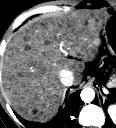
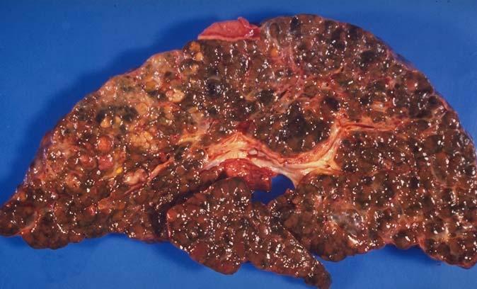
7 Cirrhosis Fibrosis Nodular regeneration Disturbed blood flow CT MR

8 Pseudocirrhosis Clinical In some patients receiving chemotherapy, a morphologic pattern develops similar to that associated with cirrhosis. Most common with breast cancer, but occurs with other cancers and lymphoma May occur with hepatotoxic drugs without liver metastases may be asymptomatic or may cause portal hypertension from venous compression Pathology Findings are suggestive of nodular regenerative hyperplasia which is characterized by the formation of regenerative hepatic nodules with compression and atrophy of parenchyma but without hepatic fibrosis. There may or may not be foci of residual tumor. It is thought to be a chemotherapeutic response due to shrinkage of tumor with subsequent scarring and nodular regeneration of uninvolved areas Imaging A lobular hepatic contour, segmental volume loss, and enlargement of the caudate lobe. Findings evolve over 1-3 months AJR 1994; 163:


9 Cavernous Hemangioma Clinical Common; 7-20% of adults; female 5:1 MRI Very bright on T2WIs Enhancing nodules that become more numerous and confluent over time intensity Follows blood pool on all phases May not fill in
 diagnosis Clinical Associated with chronic wasting diseases,steroid")

10 Peliosis Hepatis Pathology Rare benign disorder causing sinusoidal dilatation & presence of multiple blood filled lacunar spaces Usually a path (not imaging) diagnosis Clinical Associated with chronic wasting diseases,steroid medications, sprue, diabetes, vasculitis, hematological disorders Bacillary peliosis hepatis caused by Bartonella species in HIV-positive patients Complications: Liver failure/cholestasis/portal hypertension/liver rupture leading to shock
11 Peliosis Hepatis CT Findings NECT Multiple hepatic areas of low attenuation CT findings differ with size of lesions, presence or absence of thrombus within cavity & presence of hemorrhage CECT Larger cavities communicating with sinusoids have same attenuation as blood vessels Thrombosed cavities will have same appearance as nonenhancing nodules Arterial phase: Early globular vessel-like enhancement Multiple small accumulations of contrast, hyperdense in center or periphery of lesion Portal phase: Centrifugal or centripetal enhancement without mass effect on hepatic vessels Delayed phase: Late diffuse homogenous hyperattenuation characteristic of phlebectatic type
12 Peliosis Hepatis MR Findings T1WI Hypointense Signal due to presence of subacute blood suggestive of hemorrhagic necrosis T2WI Hyperintense Multiple foci of signal due to presence of subacute blood Post-C Lesions usually show contrast-enhancement Cystic cavity with enhancing rim representing hematoma Strong contrast-enhancement with "branching" appearance caused by vascular component
 Never bleed, no Ca++ Rarely contain lipid Scar hypo on T1 and hyper on T2 Homogenously intensely enhances on HAP Homogenously washes")
13 Focal Nodular Hyperplasia (FNH) Clinical Common; 2-5% of adults; female 4:1 Mostly young women with incidental mass No malignant potential MRI Looks almost like normal liver on non-c scans 13% multiple May be lobulated or have pseudocapsule (no true capsule) Never bleed, no Ca++ Rarely contain lipid Scar hypo on T1 and hyper on T2 Homogenously intensely enhances on HAP Homogenously washes out rapidly Delayed enhancement of scar Only approx 20% have classic features Persistant hyperintensity on delayed scans with Gd-BOPTA
 Not associated with hepatitis B virus, cirrhosis or metabolic abnormalities Usually normal serum markers Better prognosis")
 Compared with FNH, FLC is bigger and more heterogeneous, frequently with calcified (68%) central/eccentric scar & features of malignancy (vessel/biliary obstruction, nodal & lung")
14 Fibrolamellar Carcinoma (FLC) Pathology Diffuse fibrous stroma comprising fibrolamellar bands of collagen and fibrocytes arranged in a lamellar pattern and in delicate bands between nests of tumor cells Clinical Uncommon Primarily young adult (mean age 28) Not associated with hepatitis B virus, cirrhosis or metabolic abnormalities Usually normal serum markers Better prognosis than classic HCC; 5 year survival is 60% Imaging Findings Heterogeneously-enhancing, large, lobulated mass with hypointense central scar and radial septa on T2WI Size: Vary from 5-20 cm (mean 13 cm) Compared with FNH, FLC is bigger and more heterogeneous, frequently with calcified (68%) central/eccentric scar & features of malignancy (vessel/biliary obstruction, nodal & lung metastases)
15 Hepatic Masses FNH FLC Prevalence common rare Blood Products never rare Ca++ rare common Fat unusual never Central Scar common common Late Enhanced Scar common some Homogen Art Phase common never Homogen PV Phase common never

 May calcify")
16 Ciliated Hepatic Foregut Cyst Pathology Derives from embryologic foregut Solitary, unilocular cyst made up of ciliated pseudostratified columnar epithelaium, a subepithelial layer of connective tissue layer, a smooth muscle layer, and an outer fibrous capsule Clinical Rare. Typically appears in 50 yo patients with male predominance Can show malignant transformation (extremely rare) Since it is the only cililated cyst that occurs in the liver, FNA is diagnostic Imaging Usually located in subcapsular location on the anterior aspect of the liver at the insertion of the falciform ligament (medial seg left lobe) May calcify Pathology Oncology Research 2002;8(4): BMC Cancer 2006, 6:244 Radiology 1990;175:
Essentials of Clinical MR, 2 nd edition. 65. Benign Hepatic Masses
 65. Benign Hepatic Masses Pulse sequences acquired for abdominal MRI typically consist of fast acquisition schemes such as single-shot turbo spin echo (i.e. HASTE) and gradient echo schemes such as FLASH
Malignant Focal Liver Lesions
 Malignant Focal Liver Lesions Other Than HCC Pablo R. Ros, MD, MPH, PhD Departments of Radiology and Pathology University Hospitals Cleveland Medical Center Case Western Reserve University Pablo.Ros@UHhospitals.org
CTA/MRA of Pediatric Hepatic Masses Radiology-Pathology Correlation
 Acta Radiológica Portuguesa, Vol.XVIII, nº70, pág. 41-50, Abr.-Jun., 2006 CTA/MRA of Pediatric Hepatic Masses Radiology-Pathology Correlation Marilyn J. Siegel Mallinckrodt Institute of Radiology, Washington
Acknowledgements. Update of Focal Liver Lesions Goals. Focal Liver Lesions. Imaging Choices For Liver Lesions. Focal Liver Lesions
 Acknowledgements Update of Focal Liver Lesions 2012 Giles Boland Massachusetts General Hospital Harvard Medical School No disclosures Dushyant Sahani Mukesh Harisinghani Goals Focal liver lesions Imaging
Liver Tumors. Prof. Dr. Ahmed El - Samongy
 Liver Tumors Prof. Dr. Ahmed El - Samongy Objective 1. Identify the most important features of common benign liver tumors 2. Know the risk factors, diagnosis, and management of hepatocellular carcinoma
HEPATO-BILIARY IMAGING
 HEPATO-BILIARY IMAGING BY MAMDOUH MAHFOUZ MD PROF.OF RADIOLOGY CAIRO UNIVERSITY mamdouh.m5@gmail.com www.ssregypt.com CT ABDOMEN Indications Patient preparation Patient position Scanogram Fasting 4-6 hours
Use of Ultrasound in NAFLD
 Institute for Liver and Digestive Health Use of Ultrasound in NAFLD Dr. Davide Roccarina Specialist in General Medicine Specialist Doctor in Clinical Ultrasound and non-invasive liver assessment Hepatology
MRI OF FOCAL LESIONS IN
 Introduction MRI OF FOCAL LESIONS IN THE NON-CIRRHOTIC LIVER Ivan Pedrosa M.D. Ph.D. Associate Professor of Radiology and Advanced Imaging Research Center University of Texas Southwestern. Dallas, TX Incidental
Financial Disclosure
 Benign Liver Masses Adil Abdalla, MBBS Creighton University-CHI Health August 25, 2018 Financial Disclosure Nothing to disclose Financial Disclosure 1 Objectives To assess patients with benign liver tumors
Recently role of non-invasive diagnostics methods
 CERTAIN ASPECTS OF NLS-DIAGNOSTICS OF LIVER FOCAL PATHOLOGY A.Y. Shvack, V.I. Nesterov, N.L. Ogluzdina This article contains information about NLS-graphy application in diagnostics of liver focal affections:
Normal Sonographic Anatomy
 hapter 2:The Liver DUNSTAN ABRAHAM Normal Sonographic Anatomy Homogeneous, echogenic texture (Figure 2-1) Measures approximately 15 cm in length and 10 12.5 cm anterior to posterior; measurement taken
Hepatocellular carcinoma Cholangiocarcinoma. Jewels of hepatobiliary cancer imaging : what to look for? Imaging characteristics of HCC.
 Outline : Imaging Jewels Jewels of hepatobiliary cancer imaging : what to look for? Hepatocellular carcinoma Cholangiocarcinoma Surachate Siripongsakun, M.D. Chulabhorn Cancer Center Imaging characteristics
Radiological Reasoning: Incidentally Discovered Liver Mass
 AJR Integrative Imaging LIFELONG LEARNING FOR RADIOLOGY This Radiological Reasoning article is available for SAM credit and CME credits when completed with the additional educational material provided
Imaging of liver and pancreas
 Imaging of liver and pancreas.. Disease of the liver Focal liver disease Diffusion liver disease Focal liver disease Benign Cyst Abscess Hemangioma FNH Hepatic adenoma HCC Malignant Fibrolamellar carcinoma
Chief Complain. Liver lesion found in routine health check 41 days ago
 Chief Complain Liver lesion found in routine health check 41 days ago Present Illness On 2005-7-26 at 台北署立醫院 he underwent a health check for the first time. Abdominal US showed suspicious of a 6*5 cm hepatoma,
Ankur A. Gupta 1, Danny C. Kim, Glenn A. Krinsky, Vivian S. Lee
 Pictorial Essay T and MRI of irrhosis and its Mimics nkur. Gupta 1, Danny. Kim, Glenn. Krinsky, Vivian S. Lee irrhosis is among the leading causes of death in the western world. irrhosis and its associated
CT & MRI of Benign Liver Neoplasms Srinivasa R Prasad
 CT & MRI of Benign Liver Neoplasms Srinivasa R Prasad No financial disclosures Acknowledgements Many thanks to Drs. Heiken, Narra & Menias (MIR) Dr. Sahani (MGH) for sharing images Benign Liver Tumors:
Evaluation of Liver Mass Lesions. American College of Gastroenterology 2013 Regional Postgraduate Course
 Evaluation of Liver Mass Lesions American College of Gastroenterology 2013 Regional Postgraduate Course Lewis R. Roberts, MB ChB, PhD Division of Gastroenterology and Hepatology Mayo Clinic College of
We are IntechOpen, the world s leading publisher of Open Access books Built by scientists, for scientists. International authors and editors
 We are IntechOpen, the world s leading publisher of Open Access books Built by scientists, for scientists 3,350 108,000 1.7 M Open access books available International authors and editors Downloads Our
Radiology of hepatobiliary diseases
 GI cycle - Lecture 14 436 Teams Radiology of hepatobiliary diseases Objectives 1. To Interpret plan x-ray radiograph of abdomen with common pathologies. 2. To know the common pathologies presentation.
The Focal Hepatic Lesion: Radiologic Assessment
 The Focal Hepatic Lesion: Radiologic Assessment Kevin Kuo, Harvard Medical School Year III Our Patient: PS 67 y/o female w/ long history of alcohol use Drinking since age 18, up to one bottle of wine/day
Liver Ultrasound - Beyond the Basics. Pamela Parker Lead Sonographer
 Liver Ultrasound - Beyond the Basics Pamela Parker Lead Sonographer Aims Review what we know about the liver Reasons for imaging Focal lesions Diffuse disease Can we do more? The Liver The Liver The Liver
Essentials of Clinical MR, 2 nd edition. 73. Urinary Bladder and Male Pelvis
 73. Urinary Bladder and Male Pelvis Urinary bladder carcinoma is best locally staged with MRI. It is important however to note that a thickened wall (> 5 mm) is a non-specific finding seen in an underfilled
Liver nodules mimicking metastatic disease
 Liver nodules mimicking metastatic disease Poster No.: C-1703 Congress: ECR 2011 Type: Educational Exhibit Authors: F. Vandenbroucke, B. Ilsen, B. Op de Beeck, J. de Mey ; 1 1 2 2 3 2 3 Brussels/BE, Brussel/BE,
Jesse Civan, M.D. Medical Director, Jefferson Liver Tumor Center
 Liver Tumors Jesse Civan, M.D. Medical Director, Jefferson Liver Tumor Center Differential Diagnosis Malignant Metastatic from non-hepatic primary Hepatocellular carcinoma Cholangiocarcinoma Biliary cystcarcinoma
Enhancements in Hepatobiliary Imaging:
 Enhancements in Hepatobiliary Imaging: S. Channual 1, MD; A. Pahwa 2, MD; S. Raman 1, MD. 1 UCLA Medical Center, Department of Radiologic Sciences 2 Olive-View UCLA Medical Center, Department of Radiology
Pediatric Retroperitoneal Masses Radiologic-Pathologic Correlation
 Acta Radiológica Portuguesa, Vol.XVIII, nº 70, pág. 61-70, Abr.-Jun., 2006 Pediatric Retroperitoneal Masses Radiologic-Pathologic Correlation Marilyn J. Siegel Mallinckrodt Institute of Radiology, Washington
CT and MRI of Hepatic Contour Abnormalities
 Hepatobiliary Imaging Pictorial Essay CT and MRI of Jafi A. Lipson 1, Aliya Qayyum 1, David E. Avrin 2, Antonio Westphalen 1, Benjamin M. Yeh 1, Fergus V. Coakley 1 Fig. 1. 38-year-old woman imaged for
Approach to Liver Lesions. Anjana A. Pillai, MD Associate Professor of Medicine Director, Liver Tumor Clinic The University of Chicago Medical Center
 Approach to Liver Lesions Anjana A. Pillai, MD Associate Professor of Medicine Director, Liver Tumor Clinic The University of Chicago Medical Center Objectives Identify common clinical features and imaging
Liver Cancer (Hepatocellular Carcinoma or HCC) Overview
 Overview") Liver Cancer (Hepatocellular Carcinoma or HCC) Overview Recent advances in liver cancer care seek to address the rising incidence of liver cancer, which has steadily increased over the past three decades.
Liver Cancer (Hepatocellular Carcinoma or HCC) Overview Recent advances in liver cancer care seek to address the rising incidence of liver cancer, which has steadily increased over the past three decades.
ACG Clinical Guideline: Diagnosis and Management of Focal Liver Lesions
 ACG Clinical Guideline: Diagnosis and Management of Focal Liver Lesions Jorge A. Marrero, MD, 1 Joseph Ahn, MD, FACG, 2 K. Rajender Reddy, MD, FACG 3 1 University of Texas at Southwestern, Dallas, Texas,
Innovations in HCC Imaging: MDCT/MRI
 Innovations in HCC Imaging: MDCT/MRI Anthony E. Cheng, M.D. Cardinal MRI Center Cardinal Santos Medical Center, Wilson Street, San Juan Innovations in HCC Imaging: Goals/Objectives MDCT/MRI Learn the diagnostic
Alice Fung, MD Oregon Health and Science University
 Alice Fung, MD Oregon Health and Science University Disclosure Comments The speaker Alice Fung, MD Has relevant financial relationships to disclose. Received honorarium from (Guerbet). This individual
Role of imaging in RCC. Ultrasonography. Solid lesion. Cystic RCC. Solid RCC 31/08/60. From Diagnosis to Treatment: the Radiologist Perspective
 Role of imaging in RCC From Diagnosis to Treatment: the Radiologist Perspective Diagnosis Staging Follow up Imaging modalities Limitations and pitfalls Duangkamon Prapruttam, MD Department of Therapeutic
Evangelos Chartampilas Bioclinic Hospital Thessaloniki, Greece
 Evangelos Chartampilas Bioclinic Hospital Thessaloniki, Greece Hepatospecificcontrast agents Gadobenate dimeglumine (Multihance) Gadoxeticacid (Primovist) 3-5% liver uptake 50% liver uptake Hepatobiliary
MRI for HCC surveillance and reporting: LI-RADS. Donald G. Mitchell, M.D. Thomas Jefferson University Philadelphia, PA
 MRI for HCC surveillance and reporting: LI-RADS Donald G. Mitchell, M.D. Thomas Jefferson University Philadelphia, PA Cirrhotic Nodules Regenerative Nodule Atypical Nodule Hyperplastic Nodule Dysplastic
LIVER IMAGING TIPS IN VARIOUS MODALITIES. M.Vlychou, MD, PhD Assoc. Professor of Radiology University of Thessaly
 LIVER IMAGING TIPS IN VARIOUS MODALITIES M.Vlychou, MD, PhD Assoc. Professor of Radiology University of Thessaly Hepatocellular carcinoma is a common malignancy for which prevention, screening, diagnosis,
PATHOLOGY OF LIVER TUMORS
 PATHOLOGY OF LIVER TUMORS Pathobasic, 31.05.2016 WHO Classification Approach to a Liver Mass Lesion in a patient with chronic liver disease? Lesion in a patient without chronic liver disease? Malignant
Hepatic Imaging: What Every Practitioner Should Know
 Hepatic Imaging: What Every Practitioner Should Know Shuchi K. Rodgers, MD Section Chief, Abdominal Imaging Director of Ultrasound Department of Radiology Einstein Medical Center rodgerss@einstein.edu
Behandeling van colorectale levermetastasen. Rol van beeldvorming van de lever bij colorectaal carcinoom
 Behandeling van colorectale levermetastasen Rol van beeldvorming van de lever bij colorectaal carcinoom B. Op de Beeck Universitair Ziekenhuis Antwerpen bart.op.de.beeck@uza.be 10.12.2016 AZ Turnhout campus
Liver Cancer And Tumours
 Liver Cancer And Tumours What causes liver cancer? Many factors may play a role in the development of cancer. Because the liver filters blood from all parts of the body, cancer cells from elsewhere can
Imaging of common diseases of hepatobiliary and GI system
 Imaging of common diseases of hepatobiliary and GI system Natthaporn Tanpowpong, M.D. Diagnostic radiology Faculty of Medicine, Chulalongkorn University Normal plain radiograph A = Common bile duct
HEPATOCYTE SPECIFIC CONTRAST MEDIA: WHERE DO WE STAND?
 HEPATOCYTE SPECIFIC CONTRAST MEDIA: WHERE DO WE STAND? Andrew T. Trout, MD @AndrewTroutMD Disclosures No relevant disclosures Outline Review of hepatocyte specific contrast media Review of hepatocellular
Objectives. HCC Incidence and Mortality. Disclosure Statement HCC. Imaging of Hepatocellular Carcinoma. Treatment of Hepatocellular Carcinoma
 Imaging of Hepatocellular Carcinoma and the use of LI RADS Treatment of Hepatocellular Carcinoma Aaron D. Anderson, D.O. AOCR April 2015 Objectives Show how the use of LI RADS can simplify the diagnosis
Rare primary liver tumors - MRI pictorial review
 Rare primary liver tumors - MRI pictorial review Poster No.: C-2293 Congress: ECR 2017 Type: Educational Exhibit Authors: R. Lameiras, J. Cruz, J. Felício Costa, F. D. Figueiredo, A. 1 1 1 1 2 1 1 1 2
HCC e CEUS. Prof. A. Giorgio. Direttore IX UOC di Malattie Infettive ad Indirizzo Ecointerventistico
 HCC e CEUS Prof. A. Giorgio Direttore IX UOC di Malattie Infettive ad Indirizzo Ecointerventistico The natural history of compensated cirrhosis due to hepatitis C virus: a 17 year cohort study of 214 patients
Diffuse Liver Disease:
 Diffuse Liver Disease: MRI Evaluation Fat Iron Cirrhosis Donald G. Mitchell, M.D. Thomas Jefferson University Philadelphia, PA Fatty Liver Disease Common, increasing» Diabetes» Diet, high fructose corn
RING ENCHANCING LESION BY M.S. HEMHNATH
 RING ENCHANCING LESION BY M.S. HEMHNATH A 21 YRS FEMALE CAME WITH H/O HEADACHE AND SEIZURE FOR THE PAST ONE MONTH. NO OTHER FOCAL NEUROLOGICAL DEFICIT. DIFFERENTIAL DIAGNOSIS For this case are Neurocysticerosis
Pediatric Hepatobiliary, Pancreatic & Splenic US
 Pediatric Hepatobiliary, Pancreatic & Splenic US Susan J. Back, MD Department of Radiology, The Children s Hospital of Philadelphia No Disclosures Objectives Normal Abnormal: cases and US advances Objectives
Anatomy Jessica Ferguson Ashley Dobos May 31, 2006 LIVER
 Anatomy Jessica Ferguson Ashley Dobos May 31, 2006 LIVER 1) Other Names: Reidel s Lobe normal anatomic variant; projection of the right lobe that can extend as far as the iliac crest (Tempkin, p.54, Anatomy).
Paradoxical uptake of Gd-EOB-DTPA of focal hepatic nodule in the hepatobiliary phase
 Paradoxical uptake of Gd-EOB-DTPA of focal hepatic nodule in the hepatobiliary phase Poster No.: C-1869 Congress: ECR 2011 Type: Educational Exhibit Authors: S. M. Ha, C. Lee, K. A. Kim, J. Lee, Y.-S.
Hematologic Malignancies of the Liver : Spectrum of Disease. Zhou Jian
 Hematologic Malignancies of the Liver : Spectrum of Disease Zhou Jian 2015-7-8 Hematologic malignancies include a wide spectrum of lymphoproliferative and myeloproliferative disorders with nodal and extranodal
With the widespread use of hepatic imaging, liver masses
 2B: Liver Assessment of the Liver Mass: What Do You Need to Know? With the widespread use of hepatic imaging, liver masses are detected either unexpectedly or in the course of screening for liver cancer
Liver Failure. The most severe clinical consequence of liver disease is liver failure:
 Liver diseases I The major primary diseases of the liver are: - Viral hepatitis, - Nonalcoholic fatty liver disease (NAFLD), - Alcoholic liver disease, - Hepatocellular carcinoma (HCC) Hepatic damage also
Workup of a Solid Liver Lesion
 Workup of a Solid Liver Lesion Joseph B. Cofer MD FACS Chief Quality Officer Erlanger Health System Affiliate Professor of Surgery UTHSC-Chattanooga I have no financial or other relationships with any
Liver imaging takes a step forward with Ingenia
 Publication for the Philips MRI Community ISSUE 49 2013 / 2 Liver imaging takes a step forward with Ingenia Lyon South Hospital strives to move from several studies first CT, then MR or PET to using just
Pitfalls in the diagnosis of well-differentiated hepatocellular lesions
 2013 Colorado Society of Pathology Pitfalls in the diagnosis of well-differentiated hepatocellular lesions Sanjay Kakar, MD University of California, San Francisco Outline Hepatocellular adenoma: new WHO
Interesting case. Vikas Kundra, M.D., Ph.D. October Vikas Kundra, M.D., Ph.D.
 Interesting case October 2012 Disclosure Information Vikas Kundra, M.D, Ph.D. I have no financial relationships to disclose. I WILL NOT include discussion of investigational or off-label use of a product
Renal masses - the role of diagnostic imaging
 Renal masses - the role of diagnostic imaging Poster No.: C-2471 Congress: ECR 2015 Type: Educational Exhibit Authors: V. Rai#; Bjelovar/HR Keywords: Cysts, Cancer, Structured reporting, Ultrasound, MR,
US LI-RADS v2017 CORE
 US LI-RADS v2017 CORE Screening or surveillance US in patient at high risk for HCC US category US-1 US-2 US-3 Negative Subthreshold Positive Category Concept Definition US-1 Negative US-2 Subthreshold
Large Nonmalignant Hepatic Mass and Role of Pediatric Interventional Radiology
 Originally Posted: December 18, 2014 Large Nonmalignant Hepatic Mass and Role of Pediatric Interventional Radiology Resident(s): Kushal R Parikh, MD Attending(s): Jonathan R Dillman, MD and Ranjith Vellody,
Imaging in breast cancer. Mammography and Ultrasound Donya Farrokh.MD Radiologist Mashhad University of Medical Since
 Imaging in breast cancer Mammography and Ultrasound Donya Farrokh.MD Radiologist Mashhad University of Medical Since A mammogram report is a key component of the breast cancer diagnostic process. A mammogram
Diagnostic Challenges and Pitfalls in MR Imaging with Hepatocyte-specific
 Note: This copy is for your personal non-commercial use only. To order presentation-ready copies for distribution to your colleagues or clients, contact us at www.rsna.org/rsnarights. ABDOMINAL AND GASTROINTESTINAL
ADRENAL LESIONS 10/09/2012. Adrenal + lesion. Introduction. Common causes. Anatomy. Financial disclosure. Dr. Boraiah Sreeharsha. Nothing to declare
 ADRENAL LESIONS Financial disclosure Nothing to declare Dr. Boraiah Sreeharsha MBBS;FRCR;FRCPSC Introduction Adrenal + lesion Adrenal lesions are common 9% of the population Increase in the detection rate
IT 의료융합 1 차임상세미나 복부질환초음파 이재영
 IT 의료융합 1 차임상세미나 2013-4-3 복부질환초음파 이재영 나는오늘누구를위하여 종을울리나? 전통적의료 의사 공학설계자 의사 최첨단진단장비들 USG, CT, MRI 환자 환자 현대의료 사용자중심의사고 US in the Abdomen Detection DDx Look Behavior Response by external stimuli Guiding Tool
State of the Art Imaging for Hepatic Malignancy: My Assignment
 State of the Art Imaging for Hepatic Malignancy: My Assignment CT vs MR vs MRCP Which one to choose for HCC vs Cholangiocarcinoma What special protocols to use for liver tumors Role of PET and Duplex US
RICCARDO LENCIONI,CLOTILDE DELLA PINA, LAURA CROCETTI,DANIA CIONI. Chapter 1
 RICCARDO LENCIONI,CLOTILDE DELLA PINA, LAURA CROCETTI,DANIA CIONI Chapter 1 Impact of European Federation of Societies for Ultrasound in Medicine and Biology (EFSUMB) Guidelines on the Use of Contrast
Afternoon Session Cases
 Afternoon Session Cases Case 1 19 year old woman Presented with abdominal pain to community hospital Mild incr WBC a14, 000, Hg normal, lipase 100 (normal to 75) US 5.2 x 3.7 x 4 cm mass in porta hepatis
Cerebro-vascular stroke
 Cerebro-vascular stroke CT Terminology Hypodense lesion = lesion of lower density than the normal brain tissue Hyperdense lesion = lesion of higher density than normal brain tissue Isodense lesion = lesion
ADRENAL MR: PEARLS AND PITFALLS
 ADRENAL MR: PEARLS AND PITFALLS Frank Miller, M.D. Lee F. Rogers MD Professor of Medical Education Chief, Body Imaging Section and Fellowship Medical Director, MR Imaging Professor of Radiology Northwestern
CT 101 :Pancreas and Spleen
 CT 101 :Pancreas and Spleen Shikha Khullar,, MD, MPH Division of Radiology University of South Alabama The Pancreas Normal Pancreas 3 Phase Pancreatic CT Non contrast Arterial phase : 30-35 35 second
Neuro-endocrine and pancreatic non-adenocarcinomas. Marc Engelbrecht, AMC, Amsterdam
 Neuro-endocrine and pancreatic non-adenocarcinomas Marc Engelbrecht, AMC, Amsterdam Pancreatic Tumors q Epithelial Exocrine q Mesenchymal Ductal Adenocarcinoma (85-95%) Metastasis Lymfoma Acinar Cell Carcinoma
FOR YOUR EYES ONLY: A Guide to Accurate Detection of Diffuse Infiltrators in the Liver Eric C. Ehman, MD 1
 FOR YOUR EYES ONLY: A Guide to Accurate Detection of Diffuse Infiltrators in the Liver Eric C. Ehman, MD 1 INFILTRATOR Brian T. Welch, MD 1 Naoki Takahashi, MD 1 Christine O. Menias, MD 2 Ajit H. Goenka,
IMAGING OF LIVER, BILIARY TREE, PANCREAS
 IMAGING OF LIVER, BILIARY TREE, PANCREAS Department of Radiology West China Hospital, Sichuan University Yao Jin Learning Points The methodology for imaging the LBP (liver, biliary tree, and pancreas )
Extraosseous myeloma: imaging features
 Extraosseous myeloma: imaging features C. Santos Montón, R. Corrales, J. M. Bastida Bermejo, M. Villanueva Delgado, R. E. Correa Soto, J. M. Alonso Sánchez; Salamanca/ES Learning objectives -To review
Contrast Enhanced Ultrasound of Parenchymal Masses in Children
 Contrast Enhanced Ultrasound of Parenchymal Masses in Children Sue C Kaste, DO On behalf of Beth McCarville, MD St. Jude Children s Research Hospital Memphis, TN Overview Share St. Jude experience with
Pathological Classification of Hepatocellular Carcinoma
 3 rd APASL Single Topic Conference: HCC in 3D Pathological Classification of Hepatocellular Carcinoma Glenda Lyn Y. Pua, M.D. HCC Primary liver cancer is the 2 nd most common cancer in Asia HCC is the
Chief Complaint. Retroperitoneal cystic mass incidentally found at health examination center.
 Personal Information Age: 34 y/o Sex: female Past history: major systemic medical history(-) surgical history(-), family history(-) Denied food or drug allergy Chief Complaint Retroperitoneal cystic mass
Question 1 History. Likely Diagnosis Differential. Further Investigation or Management. Requires Paediatric Surgical referral for laparotomy
 Question 1 Male newborn spilling green tinged vomit day 1 of life Imaging Abdominal X-Rays performed on 03/05/2012 Upper and lower gastrointestinal contrast studies performed on 03/05/2012 Abdominal X-Rays
ESUR 2018, Sept. 13 th.-16 th., 2018 Barcelona, Spain
 ESUR 2018, Sept. 13 th.-16 th., 2018 Barcelona, Spain OUR APPROACH Incidental adrenal nodule/mass Isaac R Francis, M.B;B.S University of Michigan, Ann Arbor, Michigan Disclosures None (in memory) M Korobkin,
Intrahepatic Sarcomatoid Cholangiocarcinoma with Portal Vein Thrombosis: A Case Report 1
 Intrahepatic Sarcomatoid Cholangiocarcinoma with Portal Vein Thrombosis: A Case Report 1 Jae-Hoon Lim, M.D., Jin Woong Kim, M.D., Suk Hee Heo, M.D., Yong Yeon Jeong, M.D., Heoung Keun Kang, M.D. A 53-year-old
ADRENAL INCIDENTALOMA. Jamii St. Julien
 ADRENAL INCIDENTALOMA Jamii St. Julien Outline Definition Differential Evaluation Treatment Follow up Questions Case Definition The phenomenon of detecting an otherwise unsuspected adrenal mass on radiologic
Cystic lesions of the liver
 Cystic lesions of the liver Poster No.: C-0408 Congress: ECR 2014 Type: Educational Exhibit Authors: E. Rosado, J. Pereira, S. El Bouchaibi, M. A. A. Bali ; 1 1 2 2 3 3 4 4 Amadora/PT, Lisboa/PT, Bruxelles/BE,
Imaging of Neuroendocrine Metastases
 Imaging of Neuroendocrine Metastases Aoife Kilcoyne, Shaunagh McDermott, Colin McCarthy,Manuel Patino, Dushyant Sahani, Michael Blake Abdominal Imaging Division Massachusetts General Hospital Disclosure
Breast pathology. 2nd Department of Pathology Semmelweis University
 Breast pathology 2nd Department of Pathology Semmelweis University Breast pathology - Summary - Benign lesions - Acute mastitis - Plasma cell mastitis / duct ectasia - Fat necrosis - Fibrocystic change/
RINGS N THINGS: Imaging Patterns in Differential Diagnosis. Anne G. Osborn, M.D.
 RINGS N THINGS: Imaging Patterns in Differential Diagnosis Anne G. Osborn, M.D. ExpDDxs: Intra-axial (Parenchymal) Lesions Ring-enhancing lesions, solitary 1 Ring-enhancing lesion crossing corpus callosum
Liver MRI in 30 minutes
 X Liver MRI in 30 minutes SCBT/MR Annual Meeting Salt Lake City September 18, 2016 Scott B. Reeder, MD, PhD Department of Radiology University of Wisconsin Madison, WI Disclosures University of Wisconsin-Madison
ABDOMINAL DIFFUSION WEIGHTED MR
 ABDOMINAL DIFFUSION WEIGHTED MR Frank Miller, M.D. FACR Professor of Radiology Chief, Body Imaging Section Medical Director, MR Imaging Northwestern University Feinberg School of Medicine fmiller@northwestern.edu
How to Approach a Medical Liver Biopsy. 9 th Bryan Warren School of Pathology Pancreatic and Liver Pathology. Sarajevo, 6 th -7 th November 2015
 1 A Brief Introduction to the Liver Sessions 9 th Bryan Warren School of Pathology Pancreatic and Liver Pathology Sarajevo, 6 th -7 th November 2015 Stefan Hübscher, Institute of Immunology & Immunotherapy,
X-Ray Corner. Imaging Approach to Cystic Liver Lesions. Pantongrag-Brown L. Solitary cystic liver lesions. Hepatic simple cyst (Figure 1)
") THAI J 136 Imaging Approach to Cystic Liver Lesions GASTROENTEROL 2013 X-Ray Corner Imaging Approach to Cystic Liver Lesions Pantongrag-Brown L Cystic liver lesions are common findings in daily practice
THAI J 136 Imaging Approach to Cystic Liver Lesions GASTROENTEROL 2013 X-Ray Corner Imaging Approach to Cystic Liver Lesions Pantongrag-Brown L Cystic liver lesions are common findings in daily practice
Imaging characterization of renal clear cell carcinoma
 Imaging characterization of renal clear cell carcinoma Poster No.: C-0327 Congress: ECR 2011 Type: Educational Exhibit Authors: S. Ballester 1, A. Gaser 2, M. Dotta 1, M. F. CAPPA 1, F. Hammar 1 ; 1 2
Simplifying liver assessment in internal medicine
 Ultrasound Customer story Simplifying liver assessment in internal medicine Philips Affiniti ultrasound for elastography and contrast-enhanced ultrasound (CEUS) Where Sonography Institute, Uster, Switzerland
Centrally-located Sclerosing Hepatocellular Carcinoma: A Case Report with Imaging Findings before and after Transcatheter Arterial Chemoembolization
 Chin J Radiol 2001; 26: 147-152 147 CASE REPORT Centrally-located Sclerosing Hepatocellular Carcinoma: A Case Report with Imaging Findings before and after Transcatheter Arterial Chemoembolization TUN-MEI
Common and unusual CT and MRI manifestations of pancreatic adenocarcinoma: a pictorial review
 Review Article Common and unusual CT and MRI manifestations of pancreatic adenocarcinoma: a pictorial review Min-Jie Yang, Su Li, Yong-Guang Liu, Na Jiao, Jing-Shan Gong Department of Radiology, Shenzhen
Chest Radiology Interpretation: Findings of Tuberculosis
 Chest Radiology Interpretation: Findings of Tuberculosis Get out your laptops, smart phones or other devices pollev.com/chestradiology Case #1 1 Plombage Pneumonia Cancer 2 Reading the TB CXR Be systematic!
Hepatobiliary Contrast Agents for Liver MRI
 Hepatobiliary Contrast Agents for Liver MRI Scott B. Reeder, MD, PhD International Society for Magnetic Resonance in Medicine Sociedad Mexicana de Radiologia e Imagen (SMRI) Mexico City June 4, 2014 Department
Hilar cholangiocarcinoma. Frank Wessels, Maarten van Leeuwen, UMCU utrecht
 Hilar cholangiocarcinoma Frank Wessels, Maarten van Leeuwen, UMCU utrecht Content Anatomy Biliary strictures (Hilar) Cholangiocarcinoom Staging Biliary tract 1 st order Ductus hepatica dextra Ductus hepaticus
objectives Pitfalls and Pearls in PET/CT imaging Kevin Robinson, DO Assistant Professor Department of Radiology Michigan State University
 objectives Pitfalls and Pearls in PET/CT imaging Kevin Robinson, DO Assistant Professor Department of Radiology Michigan State University To determine the regions of physiologic activity To understand