Classification of Alzheimer s disease subjects from MRI using the principle of consensus segmentation
|
|
|
- Allan Evans
- 6 years ago
- Views:
Transcription


1 Classification of Alzheimer s disease subjects from MRI using the principle of consensus segmentation Aymen Khlif and Max Mignotte 1 st September, Maynooth University, Ireland
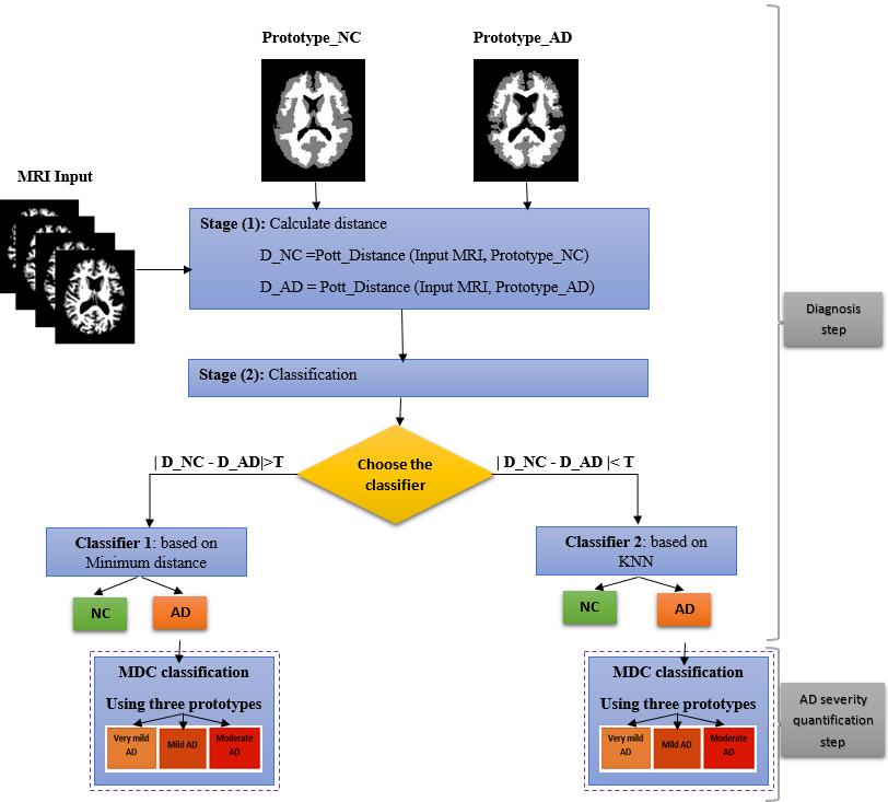
2 Plan Introduction Contributions : Proposed classification model Dataset description MRI data preprocessing Prototypes NC and AD Hybrid classification Experiments and results Conclusion and perspectives 2
")


3 Introduction : Alzheimer's disease (AD) Neurodegenerative and progressive disease Deterioration of cognitive functions Memory, language and behavior disorders Progressive loss of autonomy Disorientation in time and space 3


4 Introduction : Statistics of Alzheimer s Disease 1 In 2050: 115 million in the world, a new case every 7 seconds 13.5 million in the USA ( new cases per year) Global economic impact: $ 600 billion in 2010 Urgent issue: Early diagnosis of AD Implementation of a therapeutic Slow down the neurodegenerative process 1 Association Alzheimer s disease International 4


5 Introduction : Magnetic Resonance Imaging (MRI) Brain analysis: structural, functional and non-invasive Contributing to the early diagnosis of AD Structural alterations Metabolic alterations Detecting changes Macro-structural Micro-structural IRM Anatomic/Structural Diffusion Tensor Imaging 5
 Automatic / semi-automatic: suffers from errors voxel analysis / Voxel To detect significant differences in Grey Matter (GM) between two groups of")

6 AD diagnosis methods Volumetric analysis / Area of interest Study the variation of the volume of a region Manual: time-consuming, depends on the observer (clinician) Automatic / semi-automatic: suffers from errors voxel analysis / Voxel To detect significant differences in Grey Matter (GM) between two groups of subjects by voxel-to-voxel tests Do not require a priori assumptions about the location, the size or number of ROIs to be analyzed, since they provide voxel wise measures determined in the entire brain Help to detect structural changes in MRIs Do not depend on the clinician abilities Methods for group analysis: Individual diagnosis? 6
7 Problematic Individual diagnosis = visual assessment of a new case Learn about similar cases? Detection and characterization of pathological targets? To which class of known subjects can it be associated? Lists of similar images? 7

. Visualisation et mise en cluster des données de segmentation.")
8 Solution Use recent advances made in segmentation and multimedia Indexing 2 and classification for Content Based Visual Information Retrieval (CBVIR). More precisely, use the concept of consensus segmentation to build two segmentation prototypes (Prototype Normal Control and Prototype Alzheimer's Disease) Tools Indexing by visual content of images Principle of consensus segmentation based atlas Using Domain Knowledge" in: Image acquisition MRI Diagnosis of AD 2 A.Khlif and M.Mignotte (2017). Visualisation et mise en cluster des données de segmentation. Outils et applications multimédias, 76 (1),
9 principle of consensus segmentation A consensus segmentation is conceptually the compromise (in terms of level of details, contour accuracy, number of regions, etc.) exhibited by each segmentation map (or spatial clustering) belonging to a set of segmentations In our case, the principle of consensus segmentation allows us to build two reliable segmentation-based prototypes, one corresponding to healthy individuals and the second one corresponding to unhealthy subjects (with AD) The segmentation into three kinds of regions has the merit to efficiently reduce the information content of a brain image and to suppress noise and artifacts which are not relevant for the AD detection These two consensus segmentation-based prototypes allow us to suppress undesired components in the brain image (to be classified) such as the anatomical variability existing between individuals which are not relevant for the detection and quantification of AD 9
10 Data description OASIS (Open Access Series of Imaging Studies: ) Worldwide project Sharing data for research in the treatment and diagnosis of Alzheimer's disease We will consider a subset of the complete cross-sectional OASIS dataset, with 49 controls and 49 AD patients 10


")

11 MRI data preprocessing Affine registration to template Brain masking (BET) Grey/White/CSF segmentation Talairach atlas brain 11

12 Construction of prototypes 12


13 Proposed classification model Pott distances is the normalized number of labels differences in percentage 13

14 Comparison with morphometric methods (NC vs. AD) 14
15 Conclusion Our approach is automatic and does not require the intervention of the clinician during the disease diagnosis It is extensible to other diseases that can be diagnosed by brain MRI such as Schizophrenia and brain tumors Perspectives The method could be extended by combining axial, coronal, and sagittal MRI data for improving the classification accuracy Generalize the approach for the 3D case and compare it with 2D Classification in four classes (NC, Very mild AD, mild AD, moderate AD) Generalize the approach for other criteria for the consensus segmentation (e.g., VOI, GCE, PRI, FCR ) 15
16 Perspectives 16

17 17
Computer based delineation and follow-up multisite abdominal tumors in longitudinal CT studies
 Research plan submitted for approval as a PhD thesis Submitted by: Refael Vivanti Supervisor: Professor Leo Joskowicz School of Engineering and Computer Science, The Hebrew University of Jerusalem Computer
Review of Longitudinal MRI Analysis for Brain Tumors. Elsa Angelini 17 Nov. 2006
 Review of Longitudinal MRI Analysis for Brain Tumors Elsa Angelini 17 Nov. 2006 MRI Difference maps «Longitudinal study of brain morphometrics using quantitative MRI and difference analysis», Liu,Lemieux,
Contributions to Brain MRI Processing and Analysis
 Contributions to Brain MRI Processing and Analysis Dissertation presented to the Department of Computer Science and Artificial Intelligence By María Teresa García Sebastián PhD Advisor: Prof. Manuel Graña
Detection of Mild Cognitive Impairment using Image Differences and Clinical Features
 Detection of Mild Cognitive Impairment using Image Differences and Clinical Features L I N L I S C H O O L O F C O M P U T I N G C L E M S O N U N I V E R S I T Y Copyright notice Many of the images in
Cover Page. The handle holds various files of this Leiden University dissertation
 Cover Page The handle http://hdl.handle.net/1887/26921 holds various files of this Leiden University dissertation Author: Doan, Nhat Trung Title: Quantitative analysis of human brain MR images at ultrahigh
Automated Volumetric Cardiac Ultrasound Analysis
 Whitepaper Automated Volumetric Cardiac Ultrasound Analysis ACUSON SC2000 Volume Imaging Ultrasound System Bogdan Georgescu, Ph.D. Siemens Corporate Research Princeton, New Jersey USA Answers for life.
Experimental Assessment of Infarct Lesion Growth in Mice using Time-Resolved T2* MR Image Sequences
 Experimental Assessment of Infarct Lesion Growth in Mice using Time-Resolved T2* MR Image Sequences Nils Daniel Forkert 1, Dennis Säring 1, Andrea Eisenbeis 2, Frank Leypoldt 3, Jens Fiehler 2, Heinz Handels
POC Brain Tumor Segmentation. vlife Use Case
 Brain Tumor Segmentation vlife Use Case 1 Automatic Brain Tumor Segmentation using CNN Background Brain tumor segmentation seeks to separate healthy tissue from tumorous regions such as the advancing tumor,
Classification and Statistical Analysis of Auditory FMRI Data Using Linear Discriminative Analysis and Quadratic Discriminative Analysis
 International Journal of Innovative Research in Computer Science & Technology (IJIRCST) ISSN: 2347-5552, Volume-2, Issue-6, November-2014 Classification and Statistical Analysis of Auditory FMRI Data Using
NIH Public Access Author Manuscript Hum Brain Mapp. Author manuscript; available in PMC 2014 October 01.
 NIH Public Access Author Manuscript Published in final edited form as: Hum Brain Mapp. 2014 October ; 35(10): 5052 5070. doi:10.1002/hbm.22531. Multi-Atlas Based Representations for Alzheimer s Disease
Automated Whole Brain Segmentation Using FreeSurfer
 Automated Whole Brain Segmentation Using FreeSurfer https://surfer.nmr.mgh.harvard.edu/ FreeSurfer (FS) is a free software package developed at the Martinos Center for Biomedical Imaging used for three
Supporting Information. Demonstration of effort-discounting in dlpfc
 Supporting Information Demonstration of effort-discounting in dlpfc In the fmri study on effort discounting by Botvinick, Huffstettler, and McGuire [1], described in detail in the original publication,
Tumor cut segmentation for Blemish Cells Detection in Human Brain Based on Cellular Automata
 Tumor cut segmentation for Blemish Cells Detection in Human Brain Based on Cellular Automata D.Mohanapriya 1 Department of Electronics and Communication Engineering, EBET Group of Institutions, Kangayam,
Segmentation of Meningiomas and Low Grade Gliomas in MRI
 Segmentation of Meningiomas and Low Grade Gliomas in MRI M. R. Kaus - S. K. Warfield - A. Nabavi - E. Chatzidakis - P. M. Black - F. A. Jolesz - R. Kikinis Surgical Planning Laboratory, Department of Radiology,
Automated Brain Tumor Segmentation Using Region Growing Algorithm by Extracting Feature
 Automated Brain Tumor Segmentation Using Region Growing Algorithm by Extracting Feature Shraddha P. Dhumal 1, Ashwini S Gaikwad 2 1 Shraddha P. Dhumal 2 Ashwini S. Gaikwad ABSTRACT In this paper, we propose
Structural And Functional Integration: Why all imaging requires you to be a structural imager. David H. Salat
 Structural And Functional Integration: Why all imaging requires you to be a structural imager David H. Salat salat@nmr.mgh.harvard.edu Salat:StructFunct:HST.583:2015 Structural Information is Critical
Effective Diagnosis of Alzheimer s Disease by means of Association Rules
 Effective Diagnosis of Alzheimer s Disease by means of Association Rules Rosa Chaves (rosach@ugr.es) J. Ramírez, J.M. Górriz, M. López, D. Salas-Gonzalez, I. Illán, F. Segovia, P. Padilla Dpt. Theory of
Methods on Skull Stripping of MRI Head Scan Images a Review
 DOI 10.1007/s10278-015-9847-8 Methods on Skull Stripping of MRI Head Scan Images a Review P. Kalavathi 1 & V. B. Surya Prasath 2 # Society for Imaging Informatics in Medicine 2015 Abstract The high resolution
EARLY STAGE DIAGNOSIS OF LUNG CANCER USING CT-SCAN IMAGES BASED ON CELLULAR LEARNING AUTOMATE
 EARLY STAGE DIAGNOSIS OF LUNG CANCER USING CT-SCAN IMAGES BASED ON CELLULAR LEARNING AUTOMATE SAKTHI NEELA.P.K Department of M.E (Medical electronics) Sengunthar College of engineering Namakkal, Tamilnadu,
Four Tissue Segmentation in ADNI II
 Four Tissue Segmentation in ADNI II Charles DeCarli, MD, Pauline Maillard, PhD, Evan Fletcher, PhD Department of Neurology and Center for Neuroscience, University of California at Davis Summary Table of
Stereotactic Diffusion Tensor Tractography For Gamma Knife Stereotactic Radiosurgery
 Disclosures The authors of this study declare that they have no commercial or other interests in the presentation of this study. This study does not contain any use of offlabel devices or treatments. Stereotactic
Differentiating Tumor and Edema in Brain Magnetic Resonance Images Using a Convolutional Neural Network
 Original Article Differentiating Tumor and Edema in Brain Magnetic Resonance Images Using a Convolutional Neural Network Aida Allahverdi 1, Siavash Akbarzadeh 1, Alireza Khorrami Moghaddam 2, Armin Allahverdy
Automated detection of abnormal changes in cortical thickness: A tool to help diagnosis in neocortical focal epilepsy
 Automated detection of abnormal changes in cortical thickness: A tool to help diagnosis in neocortical focal epilepsy 1. Introduction Epilepsy is a common neurological disorder, which affects about 1 %
DEFORMABLE ATLASES FOR THE SEGMENTATION OF INTERNAL BRAIN NUCLEI IN MAGNETIC RESONANCE IMAGING
 DEFORMABLE ATLASES FOR THE SEGMENTATION OF INTERNAL BRAIN NUCLEI IN MAGNETIC RESONANCE IMAGING Marius George LINGURARU, Miguel Ángel GONZÁLEZ BALLESTER, Nicholas AYACHE EPIDAURE Research Group, INRIA,
Functional MRI Mapping Cognition
 Outline Functional MRI Mapping Cognition Michael A. Yassa, B.A. Division of Psychiatric Neuro-imaging Psychiatry and Behavioral Sciences Johns Hopkins School of Medicine Why fmri? fmri - How it works Research
Cancer Cells Detection using OTSU Threshold Algorithm
 Cancer Cells Detection using OTSU Threshold Algorithm Nalluri Sunny 1 Velagapudi Ramakrishna Siddhartha Engineering College Mithinti Srikanth 2 Velagapudi Ramakrishna Siddhartha Engineering College Kodali
Early Diagnosis of Alzheimer s Disease and MCI via Imaging and Pattern Analysis Methods. Christos Davatzikos, Ph.D.
 Early Diagnosis of Alzheimer s Disease and MCI via Imaging and Pattern Analysis Methods Christos Davatzikos, Ph.D. Director, Section of Biomedical Image Analysis Professor of Radiology http://www.rad.upenn.edu/sbia
Visualization strategies for major white matter tracts identified by diffusion tensor imaging for intraoperative use
 International Congress Series 1281 (2005) 793 797 www.ics-elsevier.com Visualization strategies for major white matter tracts identified by diffusion tensor imaging for intraoperative use Ch. Nimsky a,b,
DTI fiber tracking at 3T MR using b-1000 value in the depiction of periprostatic nerve before and after nervesparing prostatectomy
 DTI fiber tracking at 3T MR using b-1000 value in the depiction of periprostatic nerve before and after nervesparing prostatectomy Poster No.: C-2328 Congress: ECR 2012 Type: Scientific Paper Authors:
Dosimetric Analysis Report
 RT-safe 48, Artotinis str 116 33, Athens Greece RT-safe +30 2107563691 info@rt-safe.com Dosimetric Analysis Report SAMPLE, for demonstration purposes only Date of report: ------ Irradiation system: ------
A Comparative Study on Brain Tumor Analysis Using Image Mining Techniques
 Available Online at www.ijcsmc.com International Journal of Computer Science and Mobile Computing A Monthly Journal of Computer Science and Information Technology ISSN 2320 088X IMPACT FACTOR: 5.258 IJCSMC,
NIH Public Access Author Manuscript Med Image Comput Comput Assist Interv. Author manuscript; available in PMC 2013 December 06.
 NIH Public Access Author Manuscript Med Image Comput Comput Assist Interv. Author manuscript; available in PMC 2013 December 06. Published in final edited form as: Med Image Comput Comput Assist Interv.
Brain Tumor Segmentation: A Review Dharna*, Priyanshu Tripathi** *M.tech Scholar, HCE, Sonipat ** Assistant Professor, HCE, Sonipat
 International Journal of scientific research and management (IJSRM) Volume 4 Issue 09 Pages 4467-4471 2016 Website: www.ijsrm.in ISSN (e): 2321-3418 Brain Tumor Segmentation: A Review Dharna*, Priyanshu
arxiv: v1 [cs.cv] 17 Aug 2017
![arxiv: v1 [cs.cv] 17 Aug 2017](/thumbs/78/78278805.jpg "arxiv: v1 [cs.cv] 17 Aug 2017") Deep Learning for Medical Image Analysis Mina Rezaei, Haojin Yang, Christoph Meinel Hasso Plattner Institute, Prof.Dr.Helmert-Strae 2-3, 14482 Potsdam, Germany {mina.rezaei,haojin.yang,christoph.meinel}@hpi.de
Deep Learning for Medical Image Analysis Mina Rezaei, Haojin Yang, Christoph Meinel Hasso Plattner Institute, Prof.Dr.Helmert-Strae 2-3, 14482 Potsdam, Germany {mina.rezaei,haojin.yang,christoph.meinel}@hpi.de
Quantitative Neuroimaging- Gray and white matter Alteration in Multiple Sclerosis. Lior Or-Bach Instructors: Prof. Anat Achiron Dr.
 Quantitative Neuroimaging- Gray and white matter Alteration in Multiple Sclerosis Lior Or-Bach Instructors: Prof. Anat Achiron Dr. Shmulik Miron INTRODUCTION Multiple Sclerosis general background Gray
QIBA/NIBIB Final Progress Report
 QIBA/NIBIB Final Progress Report Amyloid Profile Continued Support with Brain Phantom Development Larry Pierce, David Haynor, John Sunderland, Paul Kinahan August 29, 2015 Title: Amyloid Profile Continued
Mammography is a most effective imaging modality in early breast cancer detection. The radiographs are searched for signs of abnormality by expert
 Abstract Methodologies for early detection of breast cancer still remain an open problem in the Research community. Breast cancer continues to be a significant problem in the contemporary world. Nearly
Data-driven Structured Noise Removal (FIX)
") Hamburg, June 8, 2014 Educational Course The Art and Pitfalls of fmri Preprocessing Data-driven Structured Noise Removal (FIX) Ludovica Griffanti! FMRIB Centre, University of Oxford, Oxford, United Kingdom
Hamburg, June 8, 2014 Educational Course The Art and Pitfalls of fmri Preprocessing Data-driven Structured Noise Removal (FIX) Ludovica Griffanti! FMRIB Centre, University of Oxford, Oxford, United Kingdom
Brain tissue and white matter lesion volume analysis in diabetes mellitus type 2
 Brain tissue and white matter lesion volume analysis in diabetes mellitus type 2 C. Jongen J. van der Grond L.J. Kappelle G.J. Biessels M.A. Viergever J.P.W. Pluim On behalf of the Utrecht Diabetic Encephalopathy
Fully-automated volumetric MRI with normative ranges: Translation to clinical practice
 Behavioural Neurology 21 (2009) 21 28 21 DOI 10.3233/BEN-2009-0226 IOS Press Fully-automated volumetric MRI with normative ranges: Translation to clinical practice J.B. Brewer Department of Radiology and
Early Diagnosis of Autism Disease by Multi-channel CNNs
 Early Diagnosis of Autism Disease by Multi-channel CNNs Guannan Li 1,2, Mingxia Liu 2, Quansen Sun 1(&), Dinggang Shen 2(&), and Li Wang 2(&) 1 School of Computer Science and Engineering, Nanjing University
Precision of pre-sirt predictive dosimetry
 International Course on THERANOSTICS AND MOLECULAR RADIOTHERAPY Precision of pre-sirt predictive dosimetry Hugo Levillain Department of Nuclear Medicine Medical Physics Jules Bordet Institute, Université
Segmentation of White Matter Lesions from Volumetric MR Images
 Segmentation of White Matter Lesions from Volumetric MR Images S. A. Hojjatoleslami, F. Kruggel, and D. Y. von Cramon Max-Planck-Institute of Cognitive Neuroscience Stephanstraße 1A, D-04103 Leipzig, Germany
Twelve right-handed subjects between the ages of 22 and 30 were recruited from the
 Supplementary Methods Materials & Methods Subjects Twelve right-handed subjects between the ages of 22 and 30 were recruited from the Dartmouth community. All subjects were native speakers of English,
Automatic Ascending Aorta Detection in CTA Datasets
 Automatic Ascending Aorta Detection in CTA Datasets Stefan C. Saur 1, Caroline Kühnel 2, Tobias Boskamp 2, Gábor Székely 1, Philippe Cattin 1,3 1 Computer Vision Laboratory, ETH Zurich, 8092 Zurich, Switzerland
Investigating the impact of midlife obesity on Alzheimer s disease (AD) pathology in a mouse model of AD
 pathology in a mouse model of AD") Brain@McGill Prize for Neuroscience Undergraduate Research Colleen Rollins Supervisor: Dr. Mallar Chakravarty Revised: August 8, 2017 Investigating the impact of midlife obesity on Alzheimer s disease
Brain@McGill Prize for Neuroscience Undergraduate Research Colleen Rollins Supervisor: Dr. Mallar Chakravarty Revised: August 8, 2017 Investigating the impact of midlife obesity on Alzheimer s disease
5/28/2015. The need for MRI in radiotherapy. Multiparametric MRI reflects a more complete picture of the tumor biology
 Ke Sheng, Ph.D., DABR Professor of Radiation Oncology University of California, Los Angeles The need for MRI in radiotherapy T1 FSE CT Tumor and normal tissues in brain, breast, head and neck, liver, prostate,
Fast cine-magnetic resonance imaging point tracking for prostate cancer radiation therapy planning
 Journal of Physics: Conference Series OPEN ACCESS Fast cine-magnetic resonance imaging point tracking for prostate cancer radiation therapy planning Recent citations - Motion prediction in MRI-guided radiotherapy
LEFT VENTRICLE SEGMENTATION AND MEASUREMENT Using Analyze
 LEFT VENTRICLE SEGMENTATION AND MEASUREMENT Using Analyze 2 Table of Contents 1. Introduction page 3 2. Segmentation page 4 3. Measurement Instructions page 11 4. Calculation Instructions page 14 5. References
Effects Of Attention And Perceptual Uncertainty On Cerebellar Activity During Visual Motion Perception
 Effects Of Attention And Perceptual Uncertainty On Cerebellar Activity During Visual Motion Perception Oliver Baumann & Jason Mattingley Queensland Brain Institute The University of Queensland The Queensland
Biomedical Imaging: Course syllabus
 Biomedical Imaging: Course syllabus Dr. Felipe Orihuela Espina Term: Spring 2015 Table of Contents Description... 1 Objectives... 1 Skills and Abilities... 2 Notes... 2 Prerequisites... 2 Evaluation and
University of Groningen. The traumatized brain Chalavi, Sima
 University of Groningen The traumatized brain Chalavi, Sima IMPORTANT NOTE: You are advised to consult the publisher's version (publisher's PDF) if you wish to cite from it. Please check the document version
End-To-End Alzheimer s Disease Diagnosis and Biomarker Identification
 End-To-End Alzheimer s Disease Diagnosis and Biomarker Identification Soheil Esmaeilzadeh 1, Dimitrios Ioannis Belivanis 1, Kilian M. Pohl 2, and Ehsan Adeli 1 1 Stanford University 2 SRI International
arxiv: v1 [cs.cv] 12 Jun 2018
![arxiv: v1 [cs.cv] 12 Jun 2018](/thumbs/90/102210908.jpg "arxiv: v1 [cs.cv] 12 Jun 2018") Multiview Two-Task Recursive Attention Model for Left Atrium and Atrial Scars Segmentation Jun Chen* 1, Guang Yang* 2, Zhifan Gao 3, Hao Ni 4, Elsa Angelini 5, Raad Mohiaddin 2, Tom Wong 2,Yanping Zhang
Multiview Two-Task Recursive Attention Model for Left Atrium and Atrial Scars Segmentation Jun Chen* 1, Guang Yang* 2, Zhifan Gao 3, Hao Ni 4, Elsa Angelini 5, Raad Mohiaddin 2, Tom Wong 2,Yanping Zhang
Powered by. Dedicated MRI
 Powered by Dedicated MRI Provides the latest software and hardware upgrade configuration powered by exp technology: boosting productivity, increasing image quality, and adding new acquisition techniques.
COMPUTER AIDED DIAGNOSTIC SYSTEM FOR BRAIN TUMOR DETECTION USING K-MEANS CLUSTERING
 COMPUTER AIDED DIAGNOSTIC SYSTEM FOR BRAIN TUMOR DETECTION USING K-MEANS CLUSTERING Urmila Ravindra Patil Tatyasaheb Kore Institute of Engineering and Technology, Warananagar Prof. R. T. Patil Tatyasaheb
APPLICATION OF PHOTOGRAMMETRY TO BRAIN ANATOMY
 http://medifitbiologicals.com/central-nervous-system-cns/ 25/06/2017 PSBB17 ISPRS International Workshop APPLICATION OF PHOTOGRAMMETRY TO BRAIN ANATOMY E. Nocerino, F. Menna, F. Remondino, S. Sarubbo,
BRAIN STATE CHANGE DETECTION VIA FIBER-CENTERED FUNCTIONAL CONNECTIVITY ANALYSIS
 BRAIN STATE CHANGE DETECTION VIA FIBER-CENTERED FUNCTIONAL CONNECTIVITY ANALYSIS Chulwoo Lim 1, Xiang Li 1, Kaiming Li 1, 2, Lei Guo 2, Tianming Liu 1 1 Department of Computer Science and Bioimaging Research
Brain Tumor Detection and Segmentation in MR images Using GLCM and. AdaBoost Classifier
 2015 IJSRSET Volume 1 Issue 3 Print ISSN : 2395-1990 Online ISSN : 2394-4099 Themed Section: Engineering and Technology Brain Tumor Detection and Segmentation in MR images Using GLCM and ABSTRACT AdaBoost
Image Fusion, Contouring, and Margins in SRS
 Image Fusion, Contouring, and Margins in SRS Sarah Geneser, Ph.D. Department of Radiation Oncology University of California, San Francisco Overview Review SRS uncertainties due to: image registration contouring
Diffusion Tensor Imaging in Psychiatry
 2003 KHBM DTI in Psychiatry Diffusion Tensor Imaging in Psychiatry KHBM 2003. 11. 21. 서울대학교 의과대학 정신과학교실 권준수 Neuropsychiatric conditions DTI has been studied in Alzheimer s disease Schizophrenia Alcoholism
Advances in MRI for Radiation Therapy
 Advances in MRI for Radiation Therapy Jing Cai, PhD, DABR Associate Professor Department of Radiation Oncology Duke University Medical Center, Durham NC Advances in MRI Structural Imaging Fast Imaging
Improved Intelligent Classification Technique Based On Support Vector Machines
 Improved Intelligent Classification Technique Based On Support Vector Machines V.Vani Asst.Professor,Department of Computer Science,JJ College of Arts and Science,Pudukkottai. Abstract:An abnormal growth
Dentate imaging: methods and applications
 Dentate imaging: methods and applications Dagmar Timmann Department of Neurology University of Duisburg-Essen, Germany Cerebellar output via deep nuclei Folie 2 Brodal 2010 Cerebellar nuclei Fastigial
Heterogeneous Data Mining for Brain Disorder Identification. Bokai Cao 04/07/2015
 Heterogeneous Data Mining for Brain Disorder Identification Bokai Cao 04/07/2015 Outline Introduction Tensor Imaging Analysis Brain Network Analysis Davidson et al. Network discovery via constrained tensor
Speech recognition in noisy environments: A survey
 T-61.182 Robustness in Language and Speech Processing Speech recognition in noisy environments: A survey Yifan Gong presented by Tapani Raiko Feb 20, 2003 About the Paper Article published in Speech Communication
WHAT DOES THE BRAIN TELL US ABOUT TRUST AND DISTRUST? EVIDENCE FROM A FUNCTIONAL NEUROIMAGING STUDY 1
 SPECIAL ISSUE WHAT DOES THE BRAIN TE US ABOUT AND DIS? EVIDENCE FROM A FUNCTIONAL NEUROIMAGING STUDY 1 By: Angelika Dimoka Fox School of Business Temple University 1801 Liacouras Walk Philadelphia, PA
NIH Public Access Author Manuscript Proc SPIE. Author manuscript; available in PMC 2014 February 07.
 NIH Public Access Author Manuscript Published in final edited form as: Proc SPIE. 2007 March 5; 6512: 651236. doi:10.1117/12.708950. Semi-Automatic Parcellation of the Corpus Striatum Ramsey Al-Hakim a,
Activated Fibers: Fiber-centered Activation Detection in Task-based FMRI
 Activated Fibers: Fiber-centered Activation Detection in Task-based FMRI Jinglei Lv 1, Lei Guo 1, Kaiming Li 1,2, Xintao Hu 1, Dajiang Zhu 2, Junwei Han 1, Tianming Liu 2 1 School of Automation, Northwestern
Photon Attenuation Correction in Misregistered Cardiac PET/CT
 Photon Attenuation Correction in Misregistered Cardiac PET/CT A. Martinez-Möller 1,2, N. Navab 2, M. Schwaiger 1, S. G. Nekolla 1 1 Nuklearmedizinische Klinik der TU München 2 Computer Assisted Medical
Spatial Normalisation, Atlases, & Functional Variability
 Spatial Normalisation, Atlases, & Functional Variability Jörn Diedrichsen Institute of Cognitive Neuroscience, University College London Overview Cerebellar normalisation Anatomical reference High-resolution
UCLA UCLA UCLA 7/10/2015. The need for MRI in radiotherapy. Multiparametric MRI reflects a more complete picture of the tumor biology
 Ke Sheng, Ph.D., DABR Professor of Radiation Oncology University of California, Los Angeles The need for MRI in radiotherapy T1 FSE CT Tumor and normal tissues in brain, breast, head and neck, liver, prostate,
Discriminative Analysis for Image-Based Population Comparisons
 Discriminative Analysis for Image-Based Population Comparisons Polina Golland 1,BruceFischl 2, Mona Spiridon 3, Nancy Kanwisher 3, Randy L. Buckner 4, Martha E. Shenton 5, Ron Kikinis 6, and W. Eric L.
2D-Sigmoid Enhancement Prior to Segment MRI Glioma Tumour
 2D-Sigmoid Enhancement Prior to Segment MRI Glioma Tumour Pre Image-Processing Setyawan Widyarto, Siti Rafidah Binti Kassim 2,2 Department of Computing, Faculty of Communication, Visual Art and Computing,
The latest developments - Automated Breast Volume Scanning. Dr. med. M. Golatta
 The latest developments - Automated Breast Volume Scanning Dr. med. M. Golatta Automated Breast Volume US: Why? o Mammography is limited in dense breasts: high false negative rate o Many of these tumors
Detection of Brain Tumor Using FFBP Neural Networks
 Detection of Brain Tumor Using FFBP Neural Networks Ms.S. Suruthi 1, Mrs.G.Jayanthi 2, Mrs.Dr.G.Gandhimathi 3 1 B.E, M.E Communication systems, Dept. of ECE, Parisutham Institute of Technology & Science,
Comparative Study of K-means, Gaussian Mixture Model, Fuzzy C-means algorithms for Brain Tumor Segmentation
 Comparative Study of K-means, Gaussian Mixture Model, Fuzzy C-means algorithms for Brain Tumor Segmentation U. Baid 1, S. Talbar 2 and S. Talbar 1 1 Department of E&TC Engineering, Shri Guru Gobind Singhji
Proceedings of the UGC Sponsored National Conference on Advanced Networking and Applications, 27 th March 2015
 Brain Tumor Detection and Identification Using K-Means Clustering Technique Malathi R Department of Computer Science, SAAS College, Ramanathapuram, Email: malapraba@gmail.com Dr. Nadirabanu Kamal A R Department
anatomical brain atlas registration tumor template segmented prototypes classification & morphology images pre-processed images
 Adaptive Template Moderated Brain Tumor Segmentation in MRI M. Kaus, S.K. Wareld, F.A. Jolesz and R. Kikinis Surgical Planning Laboratory Department of Radiology, Brigham & Women's Hospital Harvard Medical
A Dorsolateral Prefrontal Cortex Semi-Automatic Segmenter
 A Dorsolateral Prefrontal Cortex Semi-Automatic Segmenter Ramsey Al-Hakim a, James Fallon b, Delphine Nain c, John Melonakos d, Allen Tannenbaum d a Department of Biomedical Engineering, Georgia Institute
MR Advance Techniques. Vascular Imaging. Class II
 MR Advance Techniques Vascular Imaging Class II 1 Vascular Imaging There are several methods that can be used to evaluate the cardiovascular systems with the use of MRI. MRI will aloud to evaluate morphology
Prostate cancer caused necrosis segmentation and registration system for perfusion analysis via ethrive imaging
 Prostate cancer caused necrosis segmentation and registration system for perfusion analysis via ethrive imaging Jacky Ko Ka Long 1,2,3, Wang Defeng 1,2,3, Simon Yu Chun Ho 1 1 Department of Imaging and
Brain Tumour Detection of MR Image Using Naïve Beyer classifier and Support Vector Machine
 International Journal of Scientific Research in Computer Science, Engineering and Information Technology 2018 IJSRCSEIT Volume 3 Issue 3 ISSN : 2456-3307 Brain Tumour Detection of MR Image Using Naïve
Quantitative magnetic resonance imaging of osteoarthritis
 For reprint orders, please contact: reprints@futuremedicine.com PERSPECTIVE Quantitative magnetic resonance imaging of osteoarthritis Felix Eckstein Institute of Anatomy & Musculoskeletal Research, Paracelsus
Assessing Brain Volumes Using MorphoBox Prototype
 MAGNETOM Flash (68) 2/207 33 Assessing Brain Volumes Using MorphoBox Prototype Alexis Roche,2,3 ; Bénédicte Maréchal,2,3 ; Tobias Kober,2,3 ; Gunnar Krueger 4 ; Patric Hagmann ; Philippe Maeder ; Reto
Segmentation of Tumor Region from Brain Mri Images Using Fuzzy C-Means Clustering And Seeded Region Growing
 IOSR Journal of Computer Engineering (IOSR-JCE) e-issn: 2278-0661,p-ISSN: 2278-8727, Volume 18, Issue 5, Ver. I (Sept - Oct. 2016), PP 20-24 www.iosrjournals.org Segmentation of Tumor Region from Brain
Framework for 3D TransRectal Ultrasound (TRUS) Image-Based Tracking - Example of Use Evaluation of 2D TRUS Prostate Biopsies Mapping
 Image-Based Tracking - Example of Use Evaluation of 2D TRUS Prostate Biopsies Mapping") Author manuscript, published in "Johns Hopkins University "Prostate Day", Baltimore : United States (2008)" Framework for 3D TransRectal Ultrasound (TRUS) Image-Based Tracking - Example of Use Evaluation
Author manuscript, published in "Johns Hopkins University "Prostate Day", Baltimore : United States (2008)" Framework for 3D TransRectal Ultrasound (TRUS) Image-Based Tracking - Example of Use Evaluation
Horizon Scanning Technology Summary. Magnetic resonance angiography (MRA) imaging for the detection of coronary artery disease
 imaging for the detection of coronary artery disease") Horizon Scanning Technology Summary National Horizon Scanning Centre Magnetic resonance angiography (MRA) imaging for the detection of coronary artery disease April 2007 This technology summary is based
Horizon Scanning Technology Summary National Horizon Scanning Centre Magnetic resonance angiography (MRA) imaging for the detection of coronary artery disease April 2007 This technology summary is based
Neuroimaging vs. other methods
 BASIC LOGIC OF NEUROIMAGING fmri (functional magnetic resonance imaging) Bottom line on how it works: Adapts MRI to register the magnetic properties of oxygenated and deoxygenated hemoglobin, allowing
Validation of basal ganglia segmentation on a 3T MRI template
 Validation of basal ganglia segmentation on a 3T MRI template Claire Haegelen, Nicolas Guizard, Pierrick Coupé, Florent Lalys, Pierre Jannin, Xavier Morandi, D. Louis Collins To cite this version: Claire
HIPPOCAMPAL VOLUME ASSESSMENT. Using Analyze
 HIPPOCAMPAL VOLUME ASSESSMENT Using Analyze 2 Table Of Contents 1. Introduction page 3 2. Preprocessing Steps page 6 I. Manual AC-PC Alignment of Brain Data page 7 II. Upsampling and Cropping for Improved
Patterns of Brain Tumor Recurrence Predicted From DTI Tractography
 Patterns of Brain Tumor Recurrence Predicted From DTI Tractography Anitha Priya Krishnan 1, Isaac Asher 2, Dave Fuller 2, Delphine Davis 3, Paul Okunieff 2, Walter O Dell 1,2 Department of Biomedical Engineering
Resting-State functional Connectivity MRI (fcmri) NeuroImaging
 NeuroImaging") Resting-State functional Connectivity MRI (fcmri) NeuroImaging Randy L. Buckner et. at., The Brain s Default Network: Anatomy, Function, and Relevance to Disease, Ann. N. Y. Acad. Sci. 1124: 1-38 (2008)
Resting-State functional Connectivity MRI (fcmri) NeuroImaging Randy L. Buckner et. at., The Brain s Default Network: Anatomy, Function, and Relevance to Disease, Ann. N. Y. Acad. Sci. 1124: 1-38 (2008)
Synthesized Magnetic Resonance Imaging
 Synthesized Magnetic Resonance Imaging and SyMRI Theory and Application A White Paper Marcel Warntjes, PhD. Table of contents SyMRI technical overview Synthesized MR image acquisition and reconstruction
Repeatability of 2D FISP MR Fingerprinting in the Brain at 1.5T and 3.0T
 Repeatability of 2D FISP MR Fingerprinting in the Brain at 1.5T and 3.0T Guido Buonincontri 1,2, Laura Biagi 1,3, Alessandra Retico 2, Michela Tosetti 1,3, Paolo Cecchi 4, Mirco Cosottini 1,4,5, Pedro
A new Method on Brain MRI Image Preprocessing for Tumor Detection
 2015 IJSRSET Volume 1 Issue 1 Print ISSN : 2395-1990 Online ISSN : 2394-4099 Themed Section: Engineering and Technology A new Method on Brain MRI Preprocessing for Tumor Detection ABSTRACT D. Arun Kumar
Multi-atlas-based segmentation of the parotid glands of MR images in patients following head-and-neck cancer radiotherapy
 Multi-atlas-based segmentation of the parotid glands of MR images in patients following head-and-neck cancer radiotherapy Guanghui Cheng, Jilin University Xiaofeng Yang, Emory University Ning Wu, Jilin
Automatic Classification of Calcification in the Coronary Vessel Tree
 Automatic Classification of Calcification in the Coronary Vessel Tree R. Shahzad 1,2,3, L. J. van Vliet 2, W. J. Niessen 2,3, and T. van Walsum 3 1 Division of Image Processing, Department of Radiology,
GLCM Based Feature Extraction of Neurodegenerative Disease for Regional Brain Patterns
 GLCM Based Feature Extraction of Neurodegenerative Disease for Regional Brain Patterns Akhila Reghu, Reby John Department of Computer Science & Engineering, College of Engineering, Chengannur, Kerala,
Principles of nuclear metabolic imaging. Prof. Dr. Alex Maes AZ Groeninge Kortrijk and KULeuven Belgium
 Principles of nuclear metabolic imaging Prof. Dr. Alex Maes AZ Groeninge Kortrijk and KULeuven Belgium I. Molecular imaging probes A. Introduction - Chemical disturbances will precede anatomical abnormalities