Evaluation of IL2 and HLA on the Homeostasis and Function of Human CD4 and CD8 T Cells
|
|
|
- Louisa Gaines
- 5 years ago
- Views:
Transcription

1 University of Massachusetts Medical School GSBS Dissertations and Theses Graduate School of Biomedical Sciences Evaluation of IL2 and HLA on the Homeostasis and Function of Human CD4 and CD8 T Cells Philip A. Durost University of Massachusetts Medical School Follow this and additional works at: Part of the Immunity Commons Recommended Citation Durost, PA. Evaluation of IL2 and HLA on the Homeostasis and Function of Human CD4 and CD8 T Cells. (2017). University of Massachusetts Medical School. GSBS Dissertations and Theses. Paper 936. DOI: /M2C This material is brought to you by escholarship@umms. It has been accepted for inclusion in GSBS Dissertations and Theses by an authorized administrator of escholarship@umms. For more information, please contact Lisa.Palmer@umassmed.edu.
2 EVALUATION OF IL2 AND HLA ON THE HOMEOSTASIS AND FUNCTION OF HUMAN CD4 AND CD8 T CELLS A Dissertation Presented By PHILIP A DUROST Submitted to the Faculty of the University of Massachusetts Graduate School of Biomedical Sciences, Worcester in partial fulfillment of the requirements for the degree of DOCTOR OF PHILOSOPHY SEPTEMBER 15, 2017 IMMUNOLOGY AND MICROBIOLOGY PROGRAM
3 EVALUATION OF IL2 AND HLA ON THE HOMEOSTASIS AND FUNCTION OF HUMAN CD4 AND CD8 T CELLS A Dissertation Presented By PHILIP A DUROST This work was undertaken in the Graduate School of Biomedical Sciences Immunology and Microbiology Program Under the mentorship of Michael Brehm, Thesis Advisor The signatures of the Dissertation Defense Committee signify completion and approval as to style and content of the Dissertation Dale Greiner, Member of Committee Anne Rittenhouse, Member of Committee John Harris, Member of Committee Evan Jellison, External Member of Committee The signature of the Chair of the Committee signifies that the written dissertation meets the requirements of the Dissertation Committee Ann Rothstein, Chair of Committee The signature of the Dean of the Graduate School of Biomedical Sciences signifies that the student has met all graduation requirements of the School. Anthony Carruthers, Ph.D., Dean of the Graduate School of Biomedical Sciences
4 Abstract Homeostasis of human T cells is regulated by many factors that control proliferation, differentiation of effector cells and generation of memory. Our current knowledge of the mechanisms controlling human T cell homeostasis in vivo is based on experiments in small animal models. However many differences exist between immune systems of mice and humans, including cell composition, function, and gene expression. Humanized mouse models have shown great value in the study of human immunobiology. I have used novel humanized mouse models to examine the role of human MHC (HLA) and human IL2 in CD8 T cell and CD4 regulatory T cell (Treg) homeostasis. To study human CD8 T cells I engrafted CD8 T cells from healthy donor PBMC into NOD-scid IL2rg null (NSG) mice that lacked expression of murine MHC and that expressed HLA-A2. My data demonstrate that CD8 T cell survival and effector function required the presence of HLA- A2, helper function from human CD4 T cells and exogenous human IL2. To study human Treg homeostasis I used NSG mice engrafted with human fetal thymus and hematopoietic stem cells (BLT model). NSG-BLT mice support the growth of human thymic tissue and enable the efficient development of HLA-restricted Treg and conventional T cells. Using an AAV vector to express human IL2, I demonstrated that functional human Treg but not conventional T cells increased in number in NSG-BLT mice and that this coincided with increases in activated human NK cells. Overall my research has revealed that HLA and human IL2 have an essential role in human T cell survival and function in vivo.
5 Table of contents Part 1: Innate immunity, T cell types and their respective roles role in the immune response Innate immunity Adaptive immunity Part 2: The development, homeostatic proliferation and the function of T cells Development of T cells in the Thymus and Central Tolerance Treg Development in the Thymus and the Periphery Peripheral Homeostasis and Survival of Tconv and Treg The migratory path of naïve T cells and the initiation of the T cell response to foreign antigen. Memory T cell development Part 3: Graft versus host disease GVHD is a Complication of Tissue Transplantation The Phases in the Development of acute GVHD Chronic and mixed forms of GVHD Part 4: Gene therapy A Complicated History With Mixed Results Viral Vectors as Adaptive Gene-Delivery Tools Part 5: The development and application of the humanized mouse model Mutations and Differences in the Lineages of Mice Used in Humanized Mouse Models Models of the Human Immune system Introduction Materials and methods Results CD8 T cells engraft in NSG mice Engrafted CD8 T cells mediate a GVHD-like response and migrate into the host tissues CD8 T cell engraftment and function depends upon the expression of MHC-I Introduction: Cellular and cytokine factors that regulate T cell survival and function Materials and methods
6 Results Purified human CD8 T cells are dependent upon other cell types to successfully engraft Titration of CD8p cells into NSG mice reveals that there is a threshold for engraftment Mice expressing transgenic human IL-7 fail to engraft with purified CD8 T cells Exogenous human IL-2 can support the engraftment of purified CD8 T cells Background on IL-2 and Treg function Materials and methods Results AAV8-huIL-2 increases the proportion of Treg in BLT mice Treg isolated from AAV8-huIL-2 treated animals are responsive to stimuli and functional AAV8-huIL2 treatment does not alter the proportion of conventional CD4 or CD8 T cells in NSG-BLT mice. AAV8-huIL-2 treatment increases the number of NK cells recovered from the spleen Delineating the role of HLA and cytokines in the survival and function of CD8 T cells Implications of this research and general conclusions The role of HLA/MHC-I in the survival and function of CD8 T cells Treg function and dependence on IL-2 Limitations of the current model CD8 T cell function and homeostatic proliferation Treg dependence upon IL-2 for function and proliferation Future Directions Elucidating more of the nuanced functions of CD8 T cell survival and function using the humanized mouse model
7 List of Tables
8 List of Figures
9 List of copyrighted material produced by the author
10 Abbreviations APC (Antigen presenting cell) AIRE (Autoimmune regulator protein) ANA (Anti-nuclear Antibodies) B 2 m (Beta-2-microglobulin) BCR (B Cell Receptor) BLT (Bone Marrow Liver Thymus) CD8e (An Enriched Population of CD8 T cells Generated by Depleting CD4 T cells from Human PBMC) CD8p (Purified CD8 T cells Directly Isolated by Labeling PBMC With Anti-CD8 Antibodies Conjugated to Magnetic Beads) ctec (cortical thymic epithelial cell) CMV (Cytomegalovirus) CSR (Class Switch Recombination) DAMP (Damage-associated molecular pattern) DC (Dendritic Cell) EAE (Experimental Autoimmune Encephalitis) pdc (Plasmacytoid Dendritic Cell) FasL (Fas Ligand; CD95L) FasR (Fas Receptor; CD95) GzB (Granzyme B) GVHD (Graft versus host disease)
11 HLA (Human Leukocyte Antigen) HSC (Hematopoietic Stem Cells) IH (Intra-Hepatic) ILC (Innate Lymphoid Cells) IV (Intra-Venous) MHC (Major Histocompatibility Complex) MIP (Mouse Insulin Promoter) MPEC (Memory Progenitor Effector Cell) MHC-I (Class-I Major Histocompatibility Complex) MHC-II (Class-II Major Histocompatibility Complex) MΦ (Macrophage) mtec (medullary thymic epithelial cell) NOD (Non-Obese Diabetic) NK Cell (Natural Killer Cell) NSG (NOD Scid IL2rg null ) PAMP (Pathogen-associated molecular pattern) PBMC (peripheral blood mononuclear cells) PBL (Peripheral Blood Leukocytes) Prf (Perforin) Prkdc scid (protein kinase, DNA activated, catalytic polypeptide; severe combined immunodeficiency) PRR (Pattern Recognition Receptor)
12 RAG-1/RAG-2 (Recombination Activating genes) RIG-I (Retinoic acid Inducible Gene I) SCID (Severe Combined Immunodeficiency) SHM (Somatic Hypermutation) SLEC (Short Lived Effector Cell) SRC (SCID reconstituting cells) Tcm (Central Memory T cell) TCR (T cell Receptor) Tem (Effector Memory T cell) Th (T helper) Trm (Tissue Resident Memory Cell)
13 Chapter I Introduction The immune system exists in a well-balanced state that is capable of remaining quiescent when not perturbed while also being poised to respond to foreign intrinsic danger signals that indicate the possible presence of an infection. Among the myriad of cells that make up the mammalian immune system, T cells stand out as an important part of both responding to infection and an opposite but just as important role in regulating the intensity of the response. Because of this duality, T cells are a popular target for therapeutic intervention to modulate immune responses. The ultimate goal of my thesis research is to develop treatments that can either act alone or in conjunction with existing therapies to treat autoimmune conditions. Our knowledge regarding the cytokines and cell interactions that human T cells require for both normal homeostasis or when responding to foreign antigen in vivo is primarily based on experiments in rodent models. These experiments have discovered that a large portion of T cell function is conserved, but have also identified numerous differences between the human and mouse immune response. The many differences that exist between immune systems of mice and humans include cell composition, function, and gene expression (1). Because of these differences, there has been an effort to develop a small animal model to study human immunology. After decades of optimization, several strains of immunodeficient mice that are permissive to engraftment with human cells have been developed and utilized as the platform upon which the existing models have been built. In my experiments presented here, I have used several of these humanized mouse models to examine the role of human
14 MHC and of different cytokines, specifically human IL-2 and IL-7, in CD8 T cell and CD4 regulatory T cell (Treg) homeostasis. To study human CD8 T cells, I used the NSG-PBL-SCID model and fractionated the whole PBMC fraction into distinct populations. Specifically, the NSG-PBL-SCID model used in most of these experiments was created using CD8 T cells that were isolated from PBMCs obtained from healthy donors. We injected the cells into NOD-scid IL2rg null (NSG) mice that either expressed murine MHC-I, were deficient in MHC-I expression, or into mice that expressed transgenic forms of human MHC-I (HLA-A02*01). My data demonstrate that CD8 T cell effector functions are dependent upon the presence of either murine MHC-I expression or transgenic expression of human HLA-A02. Survival of these CD8 cells was also dependent upon the presence of a secondary cell population or the exogenous delivery of human IL-2. To study human Treg homeostasis I used the NSG-BLT mouse (Bone marrow Liver Thymus) model. BLT mice are created by transplanting a piece of human fetal thymus and liver under the kidney capsule. These mice are also injected with human CD34+ cells that are isolated from the fetal thymus. The human CD34+ cells expand and begin to produce myeloid and lymphoid cells. The T cell progenitor cells migrate to the human thymus organoid where they are educated on HLA. The BLT model was chosen for two specific reasons. First, the NSG-BLT mice support the growth of human thymic tissue and enable the efficient development of HLA-restricted T cells, and these mice develop
15 stable populations of both Treg and conventional T cells. Using an AAV vector to express human IL2, I also demonstrated that functional human Treg, but not conventional T cells, increased in number in NSG-BLT mice and that this coincided with increases in activated human NK cells. Overall, my research has revealed that HLA and human IL2 have an essential role in human T cell survival and function in vivo. Part 1: Innate immunity, T cell types and their respective roles role in the immune response Innate immunity Across many species, the immune system is made up of a number of cells that are both highly specialized and diverse in their mechanism of action. At the highest level, these immune cells can be broadly divided into two different domains that have distinct and complimentary roles; the innate and the adaptive immune systems (2). Both of these domains are essential to mounting a complete immune response, but the responses of these two domains are distinct in how they identify damage or infection, the nature of the response and the development of memory following activation. The primary distinction between these two domains lies in how foreign molecules or molecules that are indicative of non-apoptotic cell death or damage are sensed. Innate cells express receptors that are invariant and known as pattern recognition receptors (PRRs). These PRRs recognize several different motifs that are indicative of cellular damage or infection with a
16 pathogen. The class of molecule that the PRRs can detect are referred to as either pathogen-associated molecular patterns (PAMPs), if they are characteristically linked with infection or, damage-associated molecular patterns (DAMPs) if they are typically found after injury or non-apoptotic cell death (3). PAMPs are molecules that are either only found during infection or they are molecules that are found in a non-physiological location. Examples include lipopolysaccharide, peptidoglycan, flagella, ureic acid crystals, cytosolic DNA, or double-stranded RNA. DAMPs include molecules found after cells die due to injury or after they undergo necrosis. Typical DAMPs include cytosolic DNA, or the chromatin-associated protein HMGB1. Both stimulate a strong immune response when sensed in the cell in a non-physiologic location. There is some overlap between these groups as both damage or infection may result in the presence of cytosolic DNA, for instance. Also, TLR4 recognizes LPS as well as heat shock proteins, making it capable of recognizing both PAMPs and DAMPs. A list of common PRRs, their primary location, and primary ligand can be found in Table 1.
17 Table 1: PRRs, their location and their ligands
18 The innate immune system consists primarily of cells that belong to the myeloid lineage. However, some of the cells of the innate immune system are of lymphoid origin. For example, NK cells and ILCs (innate lymphoid cells) are activated by invariant receptors that sense danger or they respond to signals that indicate stress in other cells and develop from lymphoid progenitor cells (14). Other innate lymphoid cells such as B-1 cells (15) and γδt cells (16) express receptors with limited variability, but lack the ability to generate a memory response following activation. While some of these innate cells are specialized in direct killing, sequestration of pathogens, or secretion of histamine, others are more specialized in antigen presentation (17). Not only do they process proteins and present antigens in the context of the MHC-I and MHC-II molecules, they also provide co-stimulation, cytokines and chemokines that recruit the cells of the adaptive immune response to the relevant areas. Antigen presentation is a crucial step in both activating and regulating the second adaptive domain of the immune system. Of these innate cells, DCs are particularly important in maintaining tolerance as well as activating the immune system under inflammatory circumstances (18). Immature DCs migrate from lymphoid tissues through peripheral tissues where they take up and present antigen (19, 20). Most of these antigens that the DC samples from the environment come from the normal apoptotic-turnover of cells and is important for maintaining overall peripheral tolerance (19, 21). Antigen presentation by non-inflammatory DC is crucial in the development of tolerance through several different mechanisms (22). T cells that bind to these apoptotic antigens can
19 become anergic (23), can be induced to undergo apoptotic cell death (24), or can cause the T cell to develop into a Treg (25). Under inflammatory conditions, mature DCs are also vital for the normal development of the immune response (26). As these DC migrate from the periphery to secondary lymphoid tissue, they undergo a specific maturation process that results in a number of changes that enhance the DCs ability to activate T cells. DCs constitutively express moderate levels of the immunoproteasome, an enhanced form of the normal cellular proteasome that processes proteins for antigen presentation (27). As they mature, DC expression of the immunoproteasome increases beyond normal levels (28, 29). They begin to express chemokine receptors that facilitate migration to the secondary lymphoid organs and they express more co-stimulatory molecules (30). While during steady-state conditions, the circulating DCs are immunosuppressive, during inflammation they mature into a state that activates the adaptive immune system. This is largely due to changes that occur during DC maturation following the exposure to microbial PAMPs or endogenous DAMPs (31). These signals trigger an increase in the expression of MHC and increase the expression of co-stimulatory molecules such as CD80 and CD86 (32), chemokines (33, 34), and receptors that promote migration into the secondary lymphoid organs (35, 36). Within the T cell zone of lymph node germinal centers, the DC present these antigens to naïve T cells and stimulate the development of effector functions and proliferation (37). Rapid activation in response to conserved molecular patterns allows the innate immune system to quickly respond to potential threats through direct killing, cytokine production and antigen presentation.
20 Table 2: Co-Stimulatory molecules, their ligands, and the effect on T cell activation
21 Table 2: Co-Stimulatory molecules, their ligands, and the effect on T cell activation T cells can express a number of receptors that bind to ligands that may exist at the immunological synapse, depending on the current conditions within the immune system. When the receptor binds to the associated ligand(s), there can be an increase in cell activation or proliferation that can function as signal 2 during T cell activation. Alternatively, the ligand-receptor pairing can result in a diminution of the immune response.
22 Adaptive immunity The adaptive immune system is largely dependent upon antigen presentation, much of which occurs on specialized APC within the innate immune system. The cells that make up the adaptive immune system are divided into T cells and B cells. Both of these cell lineages, like the cells of the innate immune system, are capable of recognizing foreign antigen. However, T cells and B cells differ in that they do not respond to foreign antigen directly through invariant receptors but by specialized receptors. The common distinguishing feature of these B and T cell receptors is that they develop through a process of genetic recombination and mutation that expands the number of potential epitopes that can be recognized beyond what can be encoded in the genome (51). B cells recognize foreign antigen, in a soluble form or expressed on a cell surface with a membrane bound BCR and also function as an APC by presenting antigen to T cells (52). BCR engagement with foreign antigen and appropriate co-stimulation activates the B cells and results in the production and secretion of antibody (53). Following antigen exposure, B cells migrate to germinal centers where they undergo maturation and a process called somatic hypermutation (SHM) that results in the B cells becoming antibody-producing plasma cells (54). In addition to SHM, these cells undergo a second process called class switch recombination (55, 56). Antibodies exist as a number of different isotypes that differ in the constant region. During CSR, the variable region of the antibody heavy chain is unchanged but the constant region of the heavy chain is alternatively spliced resulting in the production of antibodies with a different constant region (57).
23 Both plasma cells and the memory B cells that produce antibodies are long lived. This long-lived nature makes antibody production an essential component of long-term immunity. Because of this, the vast majority of vaccine design and development has focused on developing a B cell response to confer long term immunity (58, 59). B cells and the isotype switching that occurs is ultimately dependent upon T cell help from a specialized type of CD4 T cell called a T follicular helper cell (Tfh) (60). T cells express a membrane-bound receptor that is responsible for the T cells ability to recognize and respond to foreign antigen. This TCR is a heterodimer made up of an alpha and a beta chain that associate in the plasma membrane with either the co-receptor CD4 or CD8. The expression of CD4 or CD8 divides T cells into two distinct categories that are named for the co-receptor that they express. Unlike the BCR, the TCR does not recognize foreign antigens directly. Instead, the TCR binds to peptides that are presented by a complex of proteins called the MHC. CD4 T cells, also known as helper T cells (Th), recognize antigen presented by MHC-II and secrete cytokines which are responsible for many regulatory effects. CD8 T cells are primarily cytotoxic cells and kill cells that express antigen on MHC-I molecules (61). For both of these cell types, the TCR recognize short peptide antigens that have been processed from protein and presented by specialized MHC molecules found on the surface of APCs (MHC-I and II) and most somatic cells (MHC I) (62). In general, peptides that arise from proteins that are produced within the cell are presented via MHC-I (63). Peptides that arise from exogenous proteins that are taken up by phagocytosis and processed by both the phagolysosome and the
24 proteasome and the peptide fragments that are produced are then presented within the MHC-II complex (64). Although the majority of phagocytosed antigens are processed and presented by MHC-II, these antigens can also be presented by MHC-I in a process called cross-presentation (65). MHC-I consists of two proteins, an integral membrane protein that binds to and presents the peptide to the T cell, and the B2M protein (66). Proteins processed in the proteasome complex and subsequently presented in a binding groove formed by the α1 and α2 subunits and are presented at the cell surface for recognition (67). Both CD4 and CD8 T cells depend on TCR-MHC interactions to mediate their effects, but TCR binding to MHC is insufficient to cause T cell activation. T cells depend on three signals for activation (68). The first signal is the antigen-specific interaction between the TCR on the surface of the T cell and a MHC molecule on the surface of an APC (69). The second signal is provided by co-stimulatory molecules that are expressed in conjunction with the MHC-I binding to its receptor on the surface of the T cell. The classic example of co-stimulation is CD28 expressed by T cells binding to CD80/CD86 on an APC (70, 71) but numerous other molecules exist that are capable of acting as a second signal for activation and are listed as ligands in Table 2. Finally, certain cytokines can provide the third signal necessary for T cell activation. Frequently type I interferon (IFNα and IFNβ) and IL-12 act as a third signal (72, 73).
25 Along with their different MHC preference, these two main subsets of T cells have different functions within the immune response. In mice, CD8 T cells are primarily cytotoxic cells and are the T cell type responsible for cellular immunity. These cells function by binding to peptide presented by MHC-I with their TCR. MHC-I is expressed on most cells and it generally presents antigen found within the cell (63). Consequently, CD8 T cells will recognize cells that present intracellular foreign antigens on their surface. During an infection, intracellular pathogen-derived proteins will be processed and presented on the surface of the cell. After recognizing these foreign proteins, a CD8 T cell will undergo a clonal expansion that produces both effector and memory precursor cells (74). The effector cells in particular will then mediate the cytotoxic functions that are characteristic of this cell type. After recognizing infected cells, CD8 T cells will release cytotoxic granules that contain the proteins such as perforin (Prf) and granzyme B (GzB) that ultimately lead to the apoptotic death of the target cell (75). Additionally, CD8 T cells may kill target cells through the expression of FasL, which binds to Fas expressed on target cells, and also triggers apoptotic cell death (76-78). CD8 T cells also produce a number of cytokines that are important for the immune response such as IFNγ. CD8 T cells that are activated in the presence of IL-12 tend to produce IFNγ (79). Certain subsets of CD8 T cells may also play a regulatory role. For example, Qa-1-restricted CD8 T cells have been demonstrated to be crucial for regulating CD4 T cells. Qa-1 or HLA-E is engaged by NKG2A/CD94 respectively and leads to an inhibition of activity on the conjugate CD4 T cell (80).
26 CD4 T cells are often also called helper T cells. This name reflects the role that they play upon activation which is primarily that of a regulator. CD4 T cells bind to antigen that is presented by specialized APCs that express MHC-II and help to regulate the immune response (64). Naïve helper T cells have the ability to react to the wider context of the immune response (typically the cytokines that are present at the time of activation) in order to mature into several different subsets of cells. When mature, CD4 T cells can mediate either positive or negative effects in a number of other cell types. Th cells are classically divided into Th1 and Th2 subtypes (81). In this model, a naïve CD4 T cell encounters a cognate antigen expressed by MHC-II molecule and specialization to Th1 or Th2 is guided through the presence of different cytokines. These distinct subsets are defined by specific cytokines expressed by the Th cells and by the overall impact the cytokine profile has on the immune response. Naïve CD4 T cells that encounter a cognate antigen presented by MHC-II will mature into Th1 cells in the presence of IFN- γ (82) and IL-12 (83) and into a Th2 cell in the presence of IL-4 (84) and IL-2 (85). Th1 cells primarily mediate functions that are related to cellular immunity, specifically producing cytokines that enhance the CD8 T cell response. Th2 cells are classically involved in humoral immunity and are crucial in facilitating B cell maturation and antibody class switching (86). Although the Th1/Th2 paradigm is a useful description, recent findings have necessitated an expansion of these categories to include newly described subsets that do not fit within these categories. Th9, Th17, Th22, and Treg cells all fall outside of the classical paradigm in that they are induced by different cytokines (Figure 1B) and have functions that differ from either Th1 or Th2 cells (Figure 1D) (87). Th9 cells
27 produce IL-9 and IL-21 and have been identified as a possible cell type that is involved in several different autoimmune conditions (88). IL-9 was initially classified as a Th2 cytokine until evidence suggested that there was a subset of presumed Th2 cells that produced IL-9 preferentially and were induced to do so in the presence of TGFb (88, 89). Although only recently discovered, this subset has been classified as a primarily proinflammatory cell type and linked to anti-tumor effects (90) as well as being increased in allergic patients (91) or in a mouse model of airway inflammation (92). Th17 cells are identified by the expression of the transcription factor RORγt (Figure 1C) and produce the cytokines IL-17, IL-22, and IL-25 (Figure 1D) (93). Initially, these cells were identified in a number of autoimmune conditions as well as in the inflammatory response to opportunistic infections by normally commensal bacteria and fungi. However, these cells have proven difficult to study due to a high degree of plasticity. For instance, in autoimmune models such as EAE, Th17 cells were shown to undergo changes during the inflammatory response that led to the expression of cytokines that were considered non- Th17 (94). Th22 cells bear a number of similarities to Th17 cells. Like Th17 cells, Th22 are highly associated with inflammation in autoimmune diseases (95, 96). Likewise, Th22 and IL-22 are both linked with preventing overgrowth, inflammation and infection from commensal bacteria (97). CD4 Treg are distinct from other Th subtypes in that their primary purpose is to dampen the immune response under inflammatory conditions and to maintain tolerance under normal circumstances (98). Although other Th subtypes can be immunosuppressive in
28 certain contexts, the primary purpose of Treg is to limit the immune response to foreign antigen during infection and to prevent autoimmune reactions to self-peptides (99). Furthermore, Treg have specific requirements for survival. Unlike conventional cells that primarily depend upon IL-7 for survival, Treg primarily depend upon IL-2 for both survival and function through the maintenance of the master Treg transcription factor FoxP3 (100, 101). Signaling through IL-2 promotes the expression of stat5 and maintains the expression of Foxp3 (102, 103). This role for IL-2 in promoting survival of Treg provides a feedback loop where activated T cells produce more IL-2 (104, 105). Increased IL-2 promotes Treg function and proliferation which, in turn, limits the immune response.
29 Figure 1: The cytokine milieu present during activation determines terminal effector fate of naïve CD4 cells IFN-γ, IL-12 Th1 Tbet IL4, IL-2 Th2 GATA3 TGFβ, IL-4 Th9 PU.1 TGFβ, IL-6 Th17 RORγt TNFα, IL-6 Th22 AHR IL-6, IL-21 TfH BCL-6 TGFβ, IL-2 Treg FoxP3
30 Figure 1: The cytokine milieu present during activation determines terminal effector fate of naïve CD4 cells Naïve CD4 T cells circulate through the tissues of the body and the secondary lymphoid organs. When these naïve cells (A) encounter antigen under inflammatory conditions they mature into a specialized cell type. This directed development occurs based on the cytokine milieu that they are exposed to during activation (B). These CD4 cells mature when binding to conjugate antigens expressed by APCs. During inflammatory conditions, these cells undergo changes into specific subtypes and can be identified by both the expression of certain transcription factors (C) and, to an extent, the cytokines that they can produce (D) following maturation.
31 Part 2: The development, homeostatic proliferation and the function of T cells Development of T cells in the Thymus and Central Tolerance T cells develop in a well-regulated pattern that is optimized for producing cells that meet two conditions. First, these T cells must have an inherent affinity for the peptide-mhc complex. This MHC restriction ensures that the T cells will capable of recognizing antigen within the context of MHC-1 and II. Second, these T cells must be tolerant to endogenous self-antigens to prevent autoimmune activation. This thymic regulation is referred to as central tolerance and is divided into positive selection and negative selection respectively (106). The result of this development and selection is the expression of a TCR where both the α and β chains have been rearranged through a process of recombination within the TCR locus. The selection process begins with common lymphoid progenitor cells migrating into the cortex of the thymus. At this stage, these cells do not express either CD4 or CD8 (double negative) (Figure 2A). Development in these pre-t cells begins with the expression of an invariant α-chain and the rearrangement and mutation of the β-chain locus. Not all cells will produce a functional β-chain capable of pairing with the invariant α-chain at this stage. Those that do produce a functional β-chain (Figure 2B) proliferate and progress to the next stage in development whereas those that do not will be eliminated (Figure 2C). Signaling through this pre-tcr stops recombination at the β-loci, induces proliferation and promotes the expression of both the CD4 and CD8 co-receptors. At this next stage, recombination occurs at the α-locus in the pre-t cell. Once again, survival and proliferation of the cell depends upon the successful rearrangement of the α-locus. First, the rearranged α chain
32 must be capable of pairing with the rearranged β-chain (Figure 2D). Inability to pair with the β-chain results in the elimination of the T cell (Figure 2E).
33 Figure 2: The thymic development of CD4 and CD8 T cells
34 Figure 2: The thymic development of CD4 and CD8 T cells DN thymocytes migrate into the thymus where they begin to express an invariant α chain of the TCR (A.). The β-locus of the TCR undergoes recombination. Successful rearrangements of the β-locus can pair with the invariant α chain and progress to the DP stage (B.). Unsuccessful recombination results in deletion of the pre-t cell (C.). Signaling through the pre-tcr causes rearrangement of the β-locus to stop and the expression of CD4 and CD8. These DP T cells proliferate and begin recombination at the α-locus. (D. and E.). Loci that successfully undergo recombination must be able to pair with the β-chain. Successful pairing results in survival (D.) whereas an inability to pair results in elimination (E.)
35 This α/β TCR must now be capable of pairing with and recognizing either MHC-I or II in the process referred to as positive selection. The thymocytes expressing both CD4 and CD8 are exposed to self-antigens presented by MHC-I and MHC-II molecules on ctec (Figure 3A and 3B). T cells must express a TCR that has some inherent affinity for the MHC-peptide complex in order to function properly. Cells that have little or no affinity die through the induction of apoptosis (Figure 3A and 3B). Those DP cells that have a high affinity for MHC-I complexes downregulate CD4 (Figure 3C) whereas those that have a high affinity for MHC-II downregulate CD8 (Figure 3D) (107). At this stage, the thymocytes now express either CD4 or CD8 and express a TCR that has an inherent affinity for the MHC-peptide complex. These cells are educated on endogenous peptides and because of this, there is the distinct possibility for these T cells to cause an autoimmune reaction if they migrate into the periphery. To prevent this, the T cells must undergo another stage of development, negative selection, where the cells with too high an affinity are eliminated. In the medulla, these cells encounter medullary thymic epithelial cells (mtecs) that express a specialized transcription factor, AIRE, that controls expression of tissue-specific antigens (108). AIRE-expressing mtecs produce proteins found throughout the body that are usually only expressed by specialized cells. CD8 and CD4 T cells that bind MHC-I or II with a high affinity die by apoptotic cell death or are induced to become Treg. Those with an intermediate affinity survive and emigrate into the periphery to become naïve CD8 (Figure 3E) or CD4 (Figure 3F) cells. Treg development is different from conventional T cell development in a number of ways that begins when CD4 T cells undergo negative selection.

36 Figure 3: T cells are educated on endogenous peptides expressed on both mtec and ctec.
37 Figure 3: T cells are educated on endogenous peptides expressed on both mtec and ctec. Following rearrangement of the α and β loci, the TCR must recognize either MHC-I (A.) or II (B.) MHC. During positive selection, T cells that express a TCR that lacks affinity for MHC-peptide complexes expressed on cortical thymic epithelial cells (ctecs) die due to neglect (B. and D.). During Negative selection, cells that have too high of an affinity for MHC-peptide complexes are deleted through apoptosis. T cells with an affinity for MHC-I will develop into CD8 (F.) whereas those that recognize MHC-II will develop into CD4 (E.).
38 Treg Development in the Thymus and the Periphery CD4 Treg can develop in either the thymus or in the periphery and are respectively referred to as natural or induced Treg (109). Along with the central tolerance that occurs during development in the thymus, Treg play a vital role in peripheral tolerance and are essential to prevent aberrant activation. Although central tolerance is vital to the maintenance of tolerance, an estimated 25-40% of T cells that emigrate from the thymus are cells that have escaped clonal deletion (110). Mutations in FoxP3 were first identified in scurfy mice that develop a fatal lymphoproliferative disease (111). In humans, mutations in FoxP3 that impact Treg development and function also results in a fatal lymphoproliferative condition known as IPEX (112). The initial development of Treg in the thymus follows the same pattern of selection that conventional T cells undergo. The T cells with no or little affinity for the peptide MHC complex die from neglect. The difference emerges during positive selection when T cells with high avidity to MHC-self complexes are typically deleted (113). Some of these high-avidity cells are instead preserved, induced to produce the transcription factor FoxP3, and become regulatory T cells (114). These cells emigrate from the thymus and are essential to maintaining a tolerogenic state and preventing aberrant activation. Once mature T cells emigrate from the thymus, they are nominally tolerant of endogenous antigens. However, potentially self-reactive T cells regularly escape deletion and migrate into the periphery (115).
39 Peripheral Homeostasis and Survival of Tconv and Treg After T cells complete the developmental process in the thymus, naïve CD4 and CD8 T cells emigrate from the thymus and into the periphery where survival is then dependent upon two different factors. First, the naïve T cell must encounter and bind with peptide- MHC complexes to survive (116, 117). While migrating through the periphery under normal conditions, these naïve cells come into contact with APCs and stromal cells (MHC-I) that express self-peptide-mhc complexes. If the ability to signal through the TCR is impaired experimentally by deleting downstream signaling molecules there is a noticeable shortening of the lifespan in these cells compared to T cells that are capable of signaling through the TCR (118). Both CD4 (119) and CD8 (116) T cells require a constant low level of stimulation through the TCR. The second signal required for survival is provided by cytokines. Primarily, these naïve cells depend upon IL-7 for survival but both IL-15 and IL-2 also contribute to survival and proliferation (120). IL-7 was shown to be vital for the survival of naïve cells. A total lack of IL-7 results in a loss of naïve cells whereas overexpression of IL-7 causes higher than physiological levels (121, 122). IL-7 binds to the IL-7R complex composed of CD127 and IL2Rγc and signals through stat5 (123). Under steady-state conditions, IL-2 is required only for Treg survival. Finally, IL-15 is particularly important for the maintenance of normal homeostasis of memory CD8 T cell levels (120, 124). IL-15 is different from most cytokines in that it is not secreted and instead it is membrane bound and it binds to the IL-15R at an immunological synapse (125).
40 The migratory path of naïve T cells and the initiation of the T cell response to foreign antigen. T cells that have emigrated from the thymus survive because of constant MHC stimulation and due to a select few necessary cytokines that are normally present. The naïve phenotype of these cells is characterized by the presence of a number of markers such as CD45RA, CD62L and CCR7 (126, 127). The latter of these two facilitate homing and migration into secondary lymphoid organs. CD62L (also called L selectin) facilitates the entry of T cells into the secondary lymphoid organs by binding CD34 or GlyCAM-1 and slowing the flow rate of these cells through the blood vessels (128). Likewise, CCR7 is expressed on naïve cells and it binds to chemokines that are preferentially produced in secondary lymphoid organs (129). This receptor also facilitates the overall movement of the naïve T cells towards lymph nodes. Lymph nodes are collection points that drain tissues and concentrate antigen through the migration of tissue resident macrophages and dendritic cells. Naïve T cells are then exposed to these antigens and, in the case of infection, T cells that can recognize the foreign antigen activate and begin to multiply (129). Memory T cell development Following exposure to antigen in the periphery, T cells begin to undergo profound changes that begin with the upregulation of certain proteins and an increase in proliferation called clonal expansion. This expansion gives rise to a population of T cells
41 that all have the capability of recognizing the same foreign antigen. Due to differences in cell division, co-stimulation and cytokine stimulation, these cells give rise to a nonhomogenous population of T cells that, although they share a common TCR, are specialized for different roles in the T cell response. Historically, this division gave rise to two populations that are largely differentiated by their migratory patterns and the effector functions the T effector memory (TEM) and the T central memory (TCM) cells (130). TEMs migrate from lymphoid organs and into tissues where they mediate the effector functions seen during the acute immune response whereas the TCMs recirculate throughout the lymphatic system. More recently, however, this paradigm has changed to include a number of other possible fates and specialties including tissue-resident memory cells (TRMs), short lived effector cells (SLECs) and memory precursor effector cells (MPECs) (131). The effector memory cells proliferate quickly, upregulate effector proteins used to combat the infection, and migrate into the periphery to areas of infection (132). At the end of the immune response, the SLEC population undergoes contraction through a process of apoptosis mediated by Bim and FAS-FASL that eliminates the vast majority of the cells ( ). Central memory cells are more resistant to death during this phase and persist long-term after the immune response has resolved and returned to a point of homeostasis ( ). Part 3: Graft versus host disease
42 GVHD is a Complication of Tissue Transplantation Tissue transplantation is often the definitive treatment for a number of diseases but it is a treatment that is not without risks. One such complication is graft versus host disease (GVHD). Three factors determine the risk for GVHD to develop in a transplant recipient. First, the tissue or organ from a donor individual will introduce some number of passenger lymphocytes into the recipient of the graft. The amount of donor cells transferred varies greatly depending on what is transplanted. In healthy individuals with complete immune systems, these passenger lymphocytes are quickly eliminated and pose no risk to the recipient individual. The second condition necessary for GVHD, is that the recipient of the transplanted tissue must be incapable of rejecting the transplanted lymphocytes. As transplant patients typically undergo a regimen of immunosuppression along with transplantation, it is frequently the case that graft recipients cannot reject the transplanted cells (139). Finally, the transplanted lymphocytes must recognize the recipient s tissues as foreign, become activated and mediate an immune response against the host tissues (140). Collectively, these three conditions result in a condition known as graft versus host disease (GVHD) (141). Donor cells from the same species mediate an allogeneic graft versus host response but cell transplants across species can mediate a similar disease state referred to as xenogeneic graft versus host disease. Although possible with most transplantations, GVHD occurs most frequently in patients that have undergone HSC transplantation from either a related or an unrelated donor (142).
43 The TCR has an inherent affinity for the MHC that is finely tuned during T cell development in the thymus. Cells possessing TCRs that have no affinity for MHC are eliminated and those with excessive affinity are also eliminated via positive and negative selection respectively. In an allogeneic or xenogeneic environment, the TCR maintains a strong innate affinity for the MHC that is expressed by the host. However, the donor T cells that are active in GVHD have not undergone central tolerance with regards to the host MHC and, as a result, highly reactive TCR (TCR that bind with high avidity) have not been eliminated. Similar to this, antigens that are presented by the passenger APC can also cause a different reaction. Polymorphisms that exist between individuals (allogeneic- GVHD) or species (xenogeneic-gvhd) can also elicit an immune response. This is not directly related to the structure of the MHC and is instead related to differences in the protein structure and sequence that is presented by MHC-I and II (143). The Phases in the Development of acute GVHD GVHD occurs in 3 distinct phases: the afferent, the efferent and the effector phase (144). The cytokines and cells involved and the resulting pathology are different and are used to distinguish each phase. The afferent phase occurs first and is often described as a priming step where the Th cell types in particular become activated. Patients who are undergoing bone marrow grafts are treated with myeloablative chemotherapy or irradiation to eliminate the endogenous hematopoietic stem cells (HSC) (145). These cause widespread tissue damage that results in inflammation through the presence of DAMPs and PAMPs leading to the activation of the innate immune cells, and the
44 secretion of type I interferon. This collectively leads to the maturation of APCs and these cells become primed to present antigen to incoming T cells while the overall cytokine milieu is one that is conducive to T cell proliferation and activation. The efferent phase occurs after the T cells have recognized the foreign antigen and begin to traffic into the host tissues and proliferate. This phase positions the T cells in the tissues that will be damaged in the final stage of GVHD. The final stage in GVHD, called the effector phase, involves T cell activation causing damage to the host cells. Here, the T cells that were primed/activated during the afferent phase begin to cause direct damage to the host tissues. Classically, the skin, liver and intestinal tract are most affected by this damage but the kidneys as well as the lungs may also be damaged during this phase (146). Chronic and mixed forms of GVHD The time of manifestation after transplantation determine whether the symptoms were indicative of acute (agvhd) or chronic (cgvhd). Commonly, agvhd is defined as occurring within the first 100 days after transplantation and cgvhd after the 100-day mark. This classical division is not an absolute and there are common examples of GVHD that bear characteristics of the agvhd that occur years after transplant and concurrent with a cgvhd type disease (147). There is considerable overlap in the organ systems that are impacted but cgvhd typically occurs in a wider range of tissues unlike in agvhd which typically develops in a predictable pattern, cgvhd can manifest in a number of different ways and with a less predictable course to the overall disease. Also different is that many cgvhd cases seem to mimic symptoms or pathological findings that are typically seen in autoimmune diseases. Frequently, patients develop
45 autoantibodies such as anti-nuclear antibodies typical of lupus (148) or skin lesions that are identical to scleroderma (149). Both conditions are unfortunately common particularly within HSC recipients. Overall mortality is high among HSC recipients, but agvhd is responsible for 40% of that overall mortality (142). Chronic GVHD occurs in 44% of all HSC transplantation and is the leading cause of mortality (147). Part 4: Gene therapy A Complicated History With Mixed Results Since the concept of genetic inheritance was developed, scientists have endeavored to find ways to alter this inheritance to achieve specific goals. Historically, this took place through such practices as animal husbandry and the intentional breeding of plants to encourage desirable traits. Specifically, there is evidence that dogs (150), horses (151), sheep (152), and cattle ( ) have undergone intentional genetic manipulation through breeding. Once the molecule responsible for inheritance was identified this idea matured into inducing changes in DNA directly. At first, such attempts were crude random exposure to mutagens or radiation to hopefully cause some beneficial mutation ( ). Bacterial cultures were exposed to chemical mutagens (alkylating agents such as ethyl methanesulfonate) and for a time, plants were grown in so called atomic gardens or directly exposed to γ radiation. Although crude, some of the most popular plant varieties that exist today, most notably Rio red grapefruit and most varieties of peppermint were developed through this shotgun approach (156). Clearly though, this
46 approach was less than ideal for two primary reasons. First, this random approach was labor intensive and of overall questionable value due to the random nature. Second, such approaches would be untenable as a possible treatment or for medical use in people. A significant breakthrough for the application of genetic modification was the development of the gene gun as a method to physically deliver DNA into the surface areas of plants (159). So called metal microprojectiles were coated with nucleic acids and propelled into plant cells through the cell wall by modifying a pneumatic airgun. Dubbed a biolistic (biological-ballistic), this approach lead to transient expression of genes in several different mono and dicotyledonous plants. Later, this approach was attempted with mammals in an attempt to adapt the technology for human use with the research focusing primarily on the application of genetic vaccines (160). The next logical step was to find a way to adapt this technology to treat genetic disorders. Many such genetic diseases result from genetic mutations that either lead to a loss of function in the resulting protein or an alteration that causes impaired or unsuccessful transcription/translation (161). In either case, definitive treatment would be to deliver a replacement DNA segment for the defective protein. These defects can be difficult to treat with conventional therapies for a number of reasons. For instance, replacing a missing membrane or mitochondrial protein is difficult to deliver due to location of the affected protein, as is the case in cystic fibrosis (162). Even if the defective protein can be delivered effectively, such treatments are frequently costly. An example of this is enzyme replacement therapy for lysosomal storage disorders, which require frequent infusions
47 that are expensive and have limited shelf-life, such as Fabry (163) and Gaucher (164) diseases. In both cases, the ideal treatment would be to either repair the defective gene or to deliver a replacement gene that expresses a physiological amount of the defective gene product. This approach presents a number of challenges. After identifying the defective gene, the first obstacle is to find a way to deliver genetic material to the desired cell type or organ and to ensure that the delivered genes are expressed. To overcome this first obstacle, different approaches have been developed. First, the genes can be delivered by direct injection; cells may be transformed in vitro before they are injected, or by pneumatic delivery (gene gun) (165). These methods are limited and typically confined to targeting those cells from a limited subset of tissues that are either easily accessible or can be removed and transduced ex vivo. Alternative approaches have involved using promoters that allow expression only in dendritic cells (DC). Delivery of genetic material to the skin results only in expression in DC as opposed to all cells in the vicinity of the gene gun delivery area ( ). This approach is somewhat limited in what tissues can be targeted. Subsequent approaches are more diverse in the tissues that can be targeted for transformation. An alternative and more-targeted approach is using viruses to deliver the genes either directly or in vitro before transferring into a recipient host. Such approaches have been successful in treating human diseases (adenosine deaminase deficiency induced severe combined immunodeficiency) (169). Viruses are potentially immunogenic and previous
48 gene therapy interventions, specifically one treating ornithine transcarbamylase deficiency, have resulted in fatalities (170). Despite this risk, different viruses have different tropisms and this specificity allows for the targeting of certain cell-types based on which cells are permissive to infection with a given virus ( ). Because of this, viruses can be chosen to deliver genes based on the cell type that is permissive to infection. Furthermore, viruses possess the necessary proteins to facilitate integration of the replacement gene into the host genome (in the case of retroviruses) or to persist longterm in an extrachromosomal state (AAV). Viral Vectors as Adaptive Gene-Delivery Tools Many viral vectors have been used to deliver genes. The cells that can be modified for any given virus are limited to those who are permissive to infection as viruses depend on different receptors for cell entry. Research has focused on herpesvirus, lentivirus, adenovirus, and adeno-associated viruses (174). Lentiviruses have a strong preference for infecting dividing cells, specifically preferring to infect leukocytes in many cases both in vivo and in vitro. Genes delivered through this vector can integrate into the genome where the delivered gene is maintained despite the frequent division (175). Many of the human trials that have been done in gene therapy have used lentivirus as a vector. Likewise, adeno-associated viruses have also been used to deliver genetic material to a wider variety of target cells for a number of reasons. Primarily, these viruses are replication defective and are not known to cause any sort of productive infection or disease (176). Interestingly, although not known to cause any human diseases, a large
49 portion of the human population has antibodies specific for one or more serotype of AAV (177, 178). As mentioned above, the genes that are delivered using AAV are expressed long-term. Studies in primates (179) and mice (180) have both shown that transferred genes are still expressed from 6 months to 1.5 years after injection. Integration into the genome is rare and the AAV genes typically exist as multiple copies arranged into extrachromosomal concatemers (181). While these viruses lack the specificity of other viruses, they are capable of delivering genetic material to a broad range of cells. Expression of the delivered gene can be tailored as well. The gene may be expressed in a broad range of cells (using the CMV promoter, for instance) or targeted to one particular cell type (such as beta cells with the mouse insulin promoter). Several AAV vectors, primarily AAV1, AAV2, and AAV8, are currently being tested in a number of human phase I, II, and III clinical trials. Many of these are for diseases where a single defective protein is responsible for the symptoms of the disease. Specifically, muscular dystrophy, cystic fibrosis, Canavan s disease, and alpha-1-antitrypsin deficiency (174). Part 5: The development and application of the humanized mouse model Mutations and Differences in the Lineages of Mice Used in Humanized Mouse Models History has shown that although mice and non-human primate models are a reasonable approximation of overall mammalian biology, they fall short as a specific model for predicting human biology. Both have fallen short with disastrous consequences as was the case with CD154 blockade. Blocking CD154 is able to prevent the rejection of skin
50 and cardiac allografts in mice (182) as well as pancreatic islet allografts (183). CD154 blockade was also shown to be effective in non-human primates in extending the survival of renal (184) and islet (185) allografts. Both of these animal models identified CD154 blockade as a promising treatment to prevent the rejection of allogeneic tissues. However, the results that were seen in human clinical trials differed greatly from what these models predicted and resulted in thromboembolic complications (186). In particular, profound differences exist between what has been discovered in the murine system as compared to human immunology. Ethical concerns limit what can be studied in humans in vivo, and ex vivo studies using human cells are limited in the degree to which they simulate the outcomes in vivo. Other models, specifically non-human primate models, have traditionally been used as a more accurate system to study both basic biology and to predict the outcomes of therapeutics developed for human use. However non-human primate models are expensive, require extensive resources, and also have ethical concerns. In an effort to avoid these ethical concerns and to also create a better representation of the human immune system, several groups have explored immunodeficient mouse models as recipients for human tissues (1, 187). The primary requirement in developing a humanized mouse model was creating or identifying strains of mice that are deficient in some aspect of the immune response. Such a deficiency was first observed in mice bearing a mutation that caused hairlessness, later called nude mice. These mice showed impaired development of T cells due to the lack of development of the thymus that is caused by a defect in the FOXN1 gene (188). All subsequent models
51 have been built using strains bearing different spontaneous or induced mutations that impair the function of the immune system. Developing a mouse that was truly permissive to engraftment with human cells was accomplished over several years through identifying mice with mutations in specific genes that would impair immune function and development. First, mutations were identified that impaired the development of the adaptive immune system. Early on in this search, mutations that impair development of B and T cells were identified. Initially a mutation was found that inactivated the Prkdc scid gene, a gene essential for DNA damage repair that is required for generation of antigen-specific receptors, and severely impaired the development of B and T cells ( ). Subsequently, mutations in Rag-1 (192) and Rag-2 (193), expressing proteins involved in recombination of TCRs and BCRs, were found to have a similar phenotype. NSG mice bearing the scid mutation or mutations within the Rag-1 or Rag-2 genes, support engraftment with human immune systems but overall engraftment levels are low and have limited functionality (194). Targeted mutations were also made in the in the IL2rγ, also called the common γ chain, that effectively eliminated high affinity signaling for cytokines such as IL2, IL4, IL7, IL9, IL15 and IL21 and when combined with scid or Rag mutations, enable efficient engraftment of functional human immune systems ( ). The strain background of the immunodeficient recipients will also directly impact the efficiency of human immune system development. For example; NOD mice, which develop an autoimmune form of diabetes, have a number of genetic advantages that make them more permissive to
52 engraftment with human cells and tissues compared to other mouse strains. Such differences include a polymorphism in Sirpα that makes macrophage phagocytosis of human cells less likely (198), impaired DC maturation, reduced cytokine production by macrophages and reduced NK cell number and function (199). Immunodeficient NOD mice have proved to be more permissive to engraftment of human immune systems when compared to alternative strains (1). Models of the Human Immune system Over the years, the many strains of mice that are immunocompromised have been used as a host for human cells to study different aspects of human biology. These mice with impaired immune responses are all permissive to engraftment with human cells to some degree. Of particular interest to the remainder to this thesis is the NOD, Scid, commongamma chain knockout (NSG) strain of mouse described above. Regardless of which strain of mouse is used, the models can be broadly divided into three different categories. These different models recapitulate all or a specified portion of the human immune system (187, 200, 201). Categorically, these models are defined by which types of human cells are injected into the mice and the human cell populations that persist long term (Figure 4). The Hu-PBL-SCID model is an immunodeficient mouse that is injected with human PBMC. This model primarily engrafts with T cells (189, 202). This model is simple to establish and mice engraft with both effector and memory T cells but not with myeloid cells or B cells. Since the engrafted cells are mature T cells from a human, this model allows for the direct study of T cells from donors that have T cell-related
53 disorders. Unfortunately, the PBL-SCID model suffers in that it is difficult to use for long-term experiments due to the gradual development of a xenogeneic GVHD. The human SCID repopulating cell (SRC) mice are generated as newborns through the intravenous or intrahepatic injection of CD34+ human cells or the IV injection of adult mice. The CD34+ HSC may come from newborn umbilical cord blood, bone marrow aspirates, G-CSF mobilized peripheral blood and fetal liver (203). As the mice age, multiple lineages of both myeloid and lymphoid cells develop within the mice and produce a naïve immune system that contains innate immune cells, B cells, and T cells. Although this Hu-SRC SCID model recapitulates most facets of the immune system, the T cells are educated on mouse MHC and engraftment levels tend to be low. Finally, BLT mice are generated by first surgically implanting human fetal liver and thymus under the renal capsule and subsequently injecting human CD34+ cells isolated from the same fetal liver used in the surgery (204). These mice, like the SRC-SCID, develop a full naïve immune system but the T cells that develop in this model are educated on a human thymus as opposed to mouse thymus. The BLT mouse generates HLA restricted T cells in addition to more robust T cell engraftment overall and enables development of conventional and regulatory T cells.
54 Figure 4: An illustration of the differences between the three main types of humanized mouse models
55 Figure 4: An illustration of the differences between the three main types of humanized mouse models Approaches to engraft human immune systems into immunodeficient mice (A.) with genetic defects in the host immune system are either directly engrafted with human cells or undergo a conditioning regimen. One or more of potential cell types are then transplanted or injected into these mice. This results in the reconstitution of one or more aspects of the human immune system (B.). The engraftment of cells depends on which donor cells are used and the resulting humanized mouse is classified as one of three different models (C.) that develop distinct cell populations (D.).
56 Hu-SRC-SCID and BLT mice can also be used to generate both B and T cell responses to infection. BLT mice infected with EBV (204), HIV (205, 206), and dengue virus (207, 208) successfully developed virus-specific immune responses. Although the humanized mouse model is a robust platform with a number of possible applications, it has a number of limitations that need to be addressed. Cytokines that are produced within the mouse are not always cross-reactive. Human IL-2, for instance, binds to both the human and mouse high-affinity IL-2R complex but mouse IL-2 does not bind as well to the human IL-2R trimer (209). B cells develop in both the Hu-SRC-SCID and the BLT mice but these mice do not develop robust B cell responses to antigen and they fail to undergo affinity maturation or isotype switching (210). Mice that have mutations in the IL2rγ gene also tend to have poorly defined secondary lymphoid organs due to a lack of IL-7 signaling (211).
57 Chapter II Role of MHC in the engraftment and function of human CD8 T cells in NSG Hu-PBL-SCID mice Introduction The NSG Hu-PBL-SCID model is optimized to study mature human T cells within the context of a small animal model. To generate Hu-PBL-SCID mice adult human PBMC are injected into NSG mice and allowed to expand and fill this mostly empty niche. Transiently, innate cells and B cells may be detected, but only CD4 and CD8 T cells persist long term. As a result, the NSG mouse will gradually develop a diverse repertoire of CD4 and CD8 T cells that reflect what is found in a human donor, allowing experiments involving transplantation survival, infection, or models of GVHD. The engrafting T cells will mediate a xenogeneic GVHD as the mature human T cells recognize mouse xeno-antigens. Previous published work has indicated that CD4 T cells are independently capable of engrafting and mediating a similar xenogeneic GVHD but it is less clear from the literature whether or not CD8 T cells will behave similarly (212). Homeostasis and survival of both naïve and memory CD8 T cells in the periphery is regulated by several mechanisms, including cytokine signaling and engagement with MHC molecules. Several cytokines have been demonstrated to be important factors in CD8 T cell survival, with IL-2, IL-7, and IL-15 having essential roles. In addition to these cytokines, signaling via CD8 TCR interactions with the MHC-I molecules are critical for survival. Normal CD8 T cell function depends on the TCR expressed by the
58 CD8 T cell recognizing foreign antigen that is presented by MHC-I. In addition to being necessary for CD8 T cells to function, transient low-level interactions between the TCR expressed by the CD8 T cell and MHC-I molecules expressed on other cells is critical for cell survival. The Hu-PBL-SCID system will introduce mature T cells into an environment where they will be continuously exposed to murine MHC-I, enabling the expansion of xeno-reactive human T cells. Previous research has shown that human CD8 T cells can recognize xenogeneic-antigen expressed by either human or mouse cells (213). The engagement of TCR expressed by xeno-reactive CD8 T cells with murine MHC will provide a strong activation signal and stimulate a robust immune response to the xeno-antigens. The strong interactions between TCR and xenogeneic-mhc that dominate in the Hu- PBL-SCID model will likely lead to the activation of CD8 T cells. These mice will also lack CD4 Treg cells, which are also an important part of how the immune system prevents aberrant activation. Both of these conditions will contribute to the CD8 T cells in this model mediating disease in the form of a xenogeneic-gvhd response. Observations made in our lab previously indicate that CD4 and CD8 T cells can engraft into NSG mice that lack the expression of MHC-I. Since the effector functions of the CD8 T cells are tied so closely to the expression of MHC-I, I used two different MHC-I deficient NSG strains to answer the question of whether or not CD8 T cells can persist in an environment that lacks the expression of MHC-I. I have hypothesized that the
59 engraftment and effector functions will be diminished in MHC-I-deficient mice when they are engrafted with human CD8 T cells. Materials and methods Mice NOD.Cg-Prkdc scid Il2rg tm1wjl /SzJ (NOD-scid IL2rγ null, NSG), NSG-B2M null, NSG- (K d D b ) null (NSG-KD null ), mice were obtained from colonies maintained by Dr. Leonard Shultz at The Jackson Laboratory (Bar Harbor, ME). B6 mice expressing HHD-A2 were crossed with NSG-B2M null and NSG-KD null to generate a mouse MHC-I deficient mouse that expresses HLA A02*01. All animals were housed in a specific pathogen free facility in microisolator cages, given autoclaved food and maintained on acidified autoclaved water or sulfamethoxazole-trimethoprim medicated water (Goldline Laboratories, Ft. Lauderdale, FL) provided on alternate weeks. All animal procedures were done in accordance with the guidelines of the Animal Care and Use Committee of the University of Massachusetts Medical School and The Jackson Laboratory and conformed to the recommendations in the Guide for the Care and Use of Laboratory Animals (Institute of Laboratory Animal Resources, National Research Council, National Academy of Sciences, Eighth Edition 2011).
60 Whole blood was collected from healthy donors into heparin treated 50ml conical tubes. Volunteers donated blood under informed consent in accordance with the Declaration of Helsinki and approval from the Institutional Review Board of the University of Massachusetts Medical School. Following collection, the blood was processed the same day by diluting the samples 1:1 with RPMI and overlaying the diluted blood on Ficoll density gradient separation media. Following separation, samples were suspended in RPMI and either injected directly or separated further using MACS LD or LS columns (Miltenyi Biotech). Cell separation The PBMCs were either diluted directly for injection or were labeled with magnetic beads conjugated to CD4 or CD8 antibodies obtained (Miltenyi biotech, Auburn, CA, USA) and used to label a portion of the PBMC cells. CD4 depleted cells, also referred to as CD8e cells, were directly labeled with anti-cd4 microbeads and passed over a Miltenyi LD column. The fraction that passed through was collected and retained for either injection (CD8e groups) or further processed. Purified CD8 T cells (CD8p) were obtained by either labeling the CD8 T cells directly or by labeling the CD4 depleted CD8e cells with CD8 microbeads and passing them over an LS column. The LS column was then flushed with MACS buffer and the bound cells washed and resuspended in RPMI media for injection into mice. In some experiments, the CD4+ fraction remaining after depleting CD4 cells were relabeled with CD4 microbeads and passed over a LS
61 column to yield a purified CD4 Fraction and an CD4-, CD8- fraction, referred to as the flow through cells (FT). Antibodies and flow cytometry Human immune cell populations were monitored in mice using monoclonal antibodies (mabs) specific for the following human antigens; CD45 (clone HI30), CD3 (clone UCHT1), CD4 (clone RPA-T4), CD8 (clone RPA-T8), CD20 (clone 2H7), CD45RA (clone HI100), CD62L (clone DREG-56), HLA-DR (clone G46-6), purchased from ebioscience, BD Bioscience (San Jose, CA) or BioLegend (San Diego, CA). Mouse cells were identified and excluded from analysis by staining with a mab specific for murine CD45 (clone 30-F11, BD Biosciences). Single-cell suspensions of bone marrow and spleen were prepared from engrafted mice, and whole blood was collected in heparin. Single cell suspensions of 1x10 6 cells or 100 μl of whole blood were washed with FACS buffer (PBS supplemented with 2% fetal bovine serum (FBS) and 0.02% sodium azide) and then pre-incubated with rat anti-mouse FcR11b mab (clone 2.4G2, BD Biosciences) to block binding to mouse Fc receptors. Specific mabs were then added to the samples and incubated for 30 min at 4 C. Stained samples were washed and fixed with 2% paraformaldehyde for cell suspensions or treated with BD FACS lysing solution for whole blood. At least 50,000 events were acquired on LSRII or FACSCalibur instruments (BD Biosciences). Data analysis was performed with FlowJo (Tree Star, Inc., Ashland, OR) software.
62 Histological analyses For histological examination, samples of liver, lung, and small intestine were recovered from NSG, NSG-B2M null, NSG-KD null, NSG-B2M null /A2, NSG-B2M null /HHD-A2, NSG- KD null /HHD-A2 mice, immersed overnight in 10% neutral buffered formalin and embedded in paraffin. Sections (5 µm) were cut and stained with haematoxylin and eosin. Immunohistochemical staining was performed with mabs specific for human CD45 (clone 2B11+PD7/26; Dako, Glostrup, Denmark) using a DakoCytomation EnVisionDual Link system implemented on a Dako Autostainer Universal Staining System (Dako). GVHD Model Results CD8 T cells engraft in NSG mice NSG mice that are injected with human PBMC engraft with both CD4 and CD8 T cells (214). These engrafted T cell populations are activated by mouse xeno-antigens, expand in number and ultimately mediate a xenogeneic GVHD. However, the mechanisms that
63 enable the survival of human CD8 T cells in NSG mice and the role for CD8 T cells in the development of GVHD are not well understood. To elucidate the factors regulating T cell engraftment in the Hu-PBL-SCID model, I designed an experimental strategy to specifically study CD8 T cells survival in NSG mice. The first question was to determine if human CD8 T cells are able to engraft in the absence of other cell populations present in PBMC. To do this we first isolated CD8 T cells from the whole fraction of the PBL by either positively or negatively selecting the CD8 T cells (Figure 5). The un-manipulated PBMC fraction (Figure 5A), the CD4-depleted fraction (CD8e, Figure 5B), and the CD8-purified fraction (CD8p), (Figure 5C) were then injected into NSG mice who were then monitored for changes in bodyweight and bled bi-weekly to assess engraftment. The rationale for using both the CD8e andcd8p fractions was to determine if engraftment of human CD8 T cell in NSG mice requires the innate immune cells and/or B cells present in the PBMC preparation or if CD8 T cells are sufficient to engraft as a purified population. The purity level of each population of cells was confirmed by flow cytometry.
64 Figure 5: Purification of different cell fractions from human PBMC
65 Figure 5: Purification of different cell fractions from human PBMC Human PBMC were obtained from normal donors and was separated using the MACS system. After the whole blood was processed using a Ficoll separation gradient, PBMC were retained for injection (A) and a proportion of these cells were also labeled with magnetic beads conjugated to an anti-cd4 antibody and passed through a depletion column. The cells that passed through the column were separated into two groups. The first comprised the CD8e group (B) and the remainder was labeled with magnetic beads conjugated to an anti-cd8 antibody and isolated using a positive selection column (c).
66 NSG mice were injected IV with whole PBMCs (1x10 7 cells), CD8e (5 to 6x10 6 cells), or CD8p (2x10 6 cells). These specific numbers of cells were used to enable the injection of similar numbers of CD8 T cells. To assess engraftment of CD8 T cells blood was collected at day 14 post-injection, stained with antibodies specific for human T cells, including human CD45, CD3, CD4 and CD8 and analyzed the cells by flow cytometry. The distinct human populations within the HuPBL-SCID mice were designated using the gating strategy shown in the representative flow plots of blood samples from NSG mice injected with PBMC (Figure 6). This flow cytometry profile allows human cells (CD45+) to be distinguished from the endogenous mouse cells (mcd45+) and identifies discrete populations of CD3+ CD4+ and CD3+ CD8+ T cells. As shown in Figure 26C, the majority of human cells that engraft in NSG mice injected with PBMC at day 14 are human T cells, with both CD4 and CD8 T cells detectable (Figure 26D). Both the PBMC injected NSG mice and the CD8e injected NSG mice engrafted with CD45+ cells by day 14 (Figure 6E). Similar engraftment was not, however, seen in the CD8p-injected group. Only low levels of human CD45+ cells were detected in NSG mice injected with CD8p fractions. The CD45+ cells detected in all mice at day 14 were predominantly CD3 cells (average = 97% CD3+). Overall these data suggest that purified CD8 T cells are unable to efficiently engraft in NSG mice and require the presence of one or more non-t cell populations to support engraftment.

67 Figure 6: Blood from mice engrafted with CD8e cells has a detectable human population at day K 10 5 SSC-A SSC-A 200K 150K 100K 50K <APC-A>: hucd45 hucd K 100K 150K 200K 250K FSC-A FSC-A <PerCP-Cy5-5/Blue-A>: mly mcd45 CD8 <APC-A> 10 3 CD <Alexa Fluor 700-A> CD3 CD4 30 Day 14 P<0.001 Human CD45+ Cells (%) P< PBMC CD8e CD8p
68 Figure 6: Blood from mice engrafted with CD8e cells has a detectable human population at day 14 NSG mice were engrafted with human PBMC, CD8e or CD8p cells and bled at day 14 to determine if human cells had engrafted. At day 14 blood was drawn from these animals and stained as shown in a-d. First, we gated on lymphocytes (A), and within this population we used hucd45 and mcd45 to determine the human and mouse hematopoietic cells respectively (B). Within the hucd45 population, we determined what cells were T cells by CD3 expression (C). Within the CD3 population we separated the CD4 and the CD8 T cells (D). At day 14, there was a distinguishable population seen in both the PBMC and the CD8e groups but not the CD8p group (E). These cells were overwhelmingly CD3+ cells.
69 Engrafted CD8 T cells mediate a GVHD-like response and migrate into the host tissues The data above demonstrated that human CD8 T cells persist in NSG mice injected with CD8e cells, which allows the further evaluation human CD8 T cells survival and function in NSG mice The ability of the engrafting CD8 T cells to mediate xenogeneic GVHD that is seen the Hu-PBL-SCID model was tested. NSG mice were injected IV with human PBMC, CD8e and CD8p populations as described above. The bodyweight and overall condition of the mice were monitored regularly. NSG mice engrafted with either PBMC (Figure 7A) or CD8e cells (Figure 7B) gradually lost weight and developed symptoms of xenogeneic GVHD. The NSG mice engrafted with CD8p cells, however, maintained their weight throughout the course of the experiment (Figure 7C). Survival of PBMC engrafted and CD8e-engrafted NSG mice was significantly reduced compared to CD8pengrafted mice (Figure 7D). Human cell chimerism levels were monitored in the peripheral blood of engrafted NSG mice bi-weekly by FACS analysis. Levels of engraftment at day 14 correlated with the overall mortality. In addition, the activation status of the CD8 T cells was evaluated and compared to the initial population of CD8 T cells prior to injection. To assess the activation status of the T cells the expression level of CD45RA, an isoform of CD45 that decreases after activation, was evaluated by flow cytometry. Expression of CD45RA on CD8 T cells was decreased after 14 days in both the PBMC and the CD8e groups when compared with the expression on the donor CD8 T cells (Figure 7E). This shows that while the CD8 T cells can engraft in NSG mice, CD4 T cell help may be facilitating CD8 T cell activation and engraftment in the PBMC mice.
70 The CD8p mice were excluded from this analysis as the number of human cells in these animals was too low to allow for an accurate comparison (ie fewer than 100 events within the CD8 gate). These data show that the CD8 T cells can engraft in NSG mice, they develop an activated phenotype, the engrafted cells are functional and mediate a xenogeneic GVHD.
71 Figure 7: NSG Mice engrafted with PBMC or CD8e cells have a gradual weight loss that corresponds with overall mortality and engraftment with hucd NSG 110 NSG CD8e % of Starting Weight % of Starting Weight Day NSG CD8p Day Survival % of Starting Weight Percent survival 50 P= NSG PBMC NSG CD8e NSG CD8p Day CD45RA Expression at Day Day %CD45RA+ CD8 T cells Input NSG PBMC NSG CD8e
72 Figure 7: NSG Mice engrafted with PBMC or CD8e cells have a gradual weight loss that corresponds with overall mortality and engraftment with hucd45 The NSG mice that were engrafted with either PBMC, CD8e, or CD8p cells were monitored for signs of weight loss. The PBMC (A) and the CD8e (B) engrafted NSG mice gradually lost weight. Similar weight loss was not seen in the CD8p engrafted animals (C). This weight loss in the NSG mice correlates with overall mortality (D). When we compared the expression of CD45RA on these cells at the time of input to the expression at day 14 we saw a dramatic decrease in the PBMC and a more moderate decrease in the CD8e group (E).
73 The results above show that human CD8 T cells will engraft in NSG mice and mediate a xeno-gvhd. In NSG mice injected with PBMC, CD4 and CD8 T cells infiltrate a number of peripheral organs including the mouse liver. To determine if CD8 T cells migrate to peripheral tissues in the absence or CD4 T cell help, livers from PBMC engrafted and CD8e engrafted NSG mice were recovered when the mice were sacrificed (body weight<80% of starting weight or signs of distress) and the tissues were stained with H&E to visualize infiltrating leukocytes within these tissues. The liver from an unengrafted NSG mouse shows a lack of infiltrates and an overall uniform appearance (Figure 8A). The 3 adjacent panels show livers from CD8e-engrafted animals that were collected at the time of sacrifice. In all cases, the livers from engrafted NSG mice show numerous infiltrates into the liver concentrated near blood vessels (Figure 8B-D) and overall alterations to the structure of the organ. This type of infiltration is typical in the immunopathology of patients suffering from GVHD with liver involvement (215). In the following panels, liver sections from NSG mice that were engrafted with PBMC (Figure 8 E and H) or CD8e (Figure 8 F and G) were stained with anti-human CD45 to visualize infiltrating human cells. These findings indicate that human CD8 T cells are able to infiltrate peripheral tissues in NSG mice in the absence of CD4 T cells. Collectively, these data together indicate that the engrafted human T cells persist in the NSG mice. The CD8 T cells expand within the mice, develop an activated phenotype, migrate into peripheral tissues, and mediate a xenogeneic GVHD. Furthermore, the CD8 T cells are capable of mediating this disease on their own without CD4 T cell help.


74 Figure 8: Liver tissues isolated from CD8e engrafted animals show extensive mononuclear infiltrates near vessels Unengrafted NSG A B CD8e Engrafted NSG CD8e Engrafted NSG CD8e Engrafted NSG C D
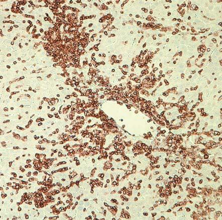
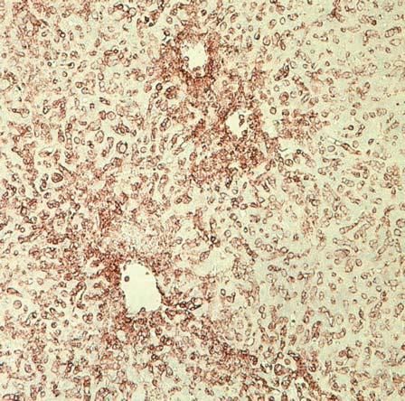
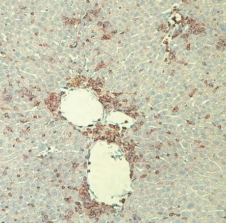

75
76 Figure 2.4: Liver tissues isolated from CD8e engrafted animals show extensive mononuclear infiltrates near vessels Liver sections from unengrafted NSG mice (A) or CD8e engrafted mice were sectioned and stained with H&E to visualize infiltrating cells (B-D). CD8e-engrafted NSG mice had extensive infiltration into the tissues surrounding the blood vessels and profound changes in the overall color of the tissues. In the following panels Liver sections from NSG mice engrafted with PBMC (E and H) or CD8e (F and G) are stained with anti-human CD45 to visualize infiltrating human cells.
77 CD8 T cell engraftment and function depends upon the expression of MHC-I MHC-I has a critical role in maintaining CD8 T cell homeostasis and function (116). To examine the contribution of murine MHC to CD8 T cell survival and function in NSG mice, I used two different types of MHC-I knockout NSG mice that were developed by Dr. Leonard Shultz, at the Jackson Laboratory. The first NSG strain lacks expression of the MHC-I locus (NSG-KD null ), and the second stain lacks the B 2 M molecule necessary for surface expression of MHC-I. Previous studies have shown that NSG-B 2 M null mice have a delay in the development of xenogeneic-gvhd after injection of human PBMC (214). To compare the kinetics of human cell engraftment and of xenogeneic-gvhd between the NSG-B 2 M null mice and the NSG KD null mice, unfractionated PBMC were injected IV into NSG, NSG-KD null and NSG-B 2 M null mice. Injected NSG mice were monitored for weight loss, mortality, engraftment, and T cell activation by bleeding these animals bi-weekly. As before, all of the animals showed a gradual and progressive weight loss (Figure 9A-C) that ultimately lead to the development of GVHD and the euthanasia of the mice. The GVHD symptoms were delayed in the NSG-KD null mice and NSG- B 2 M null (Figure 9B and C). The delay in weight loss correlated with a delay in the time that the animals survived within the parameters of the experiment (Figure 9D). As before, all mice that were engrafted with PBMC were engrafted with CD8 T cells at day 14 and day 28 with a small but not statistically significant trend towards lower engraftment in the NSG-KD null mice (Figure 9E and F).
78 Figure 9: Human PBMC isolated from donors engraft in mice deficient in MHC-I 110 NSG 120 KD null % of Starting Weight % Starting Weight Day B 2 m null Day % Starting Weight Percent survival P=0.085 NSG n=15 KDnull n=14 B2M n= Day Day 14 Engraftment Day Day 28 Engraftment %hucd %hucd NSG NSG KD null NSG B 2 m 0 NSG NSG KD null NSG B 2 m
79 Figure 9: Human PBMC isolated from donors engraft in mice deficient in MHC-I PBMC were injected into either NSG, NSG-KD null, or NSG-B 2 M null mice. All of the groups experienced a gradual weight loss with both of the MHC-I deficient strains having a trend towards slower weight loss when compared to the NSG mice (A-C). The weight loss correlated with the development of GVHD with the NSG-PBMC mice developing symptoms noticeably sooner than the NSG-KD null -PBMC or the NSG-B 2 M null -PBMC mice (D). Engraftment was similar among the three groups, but there was a noticeable trend towards higher engraftment in the NSG group when compared with the MHC-I deficient groups at day 14 and day 28 (E and F).
80 The data shown above with MHC-I deficient NSG mice indicate that following injection with human PBMC, these mice engrafted and develop a xenogeneic-gvhd, although at slower kinetics than NSG mice. I hypothesized that enriched CD8 T cells (CD8e) would engraft into NSG mice lacking expression of MHC-I but would not mediate a xenogeneic-gvhd. Normal NSG mice, NSG-KD null or NSG-B 2 M null mice were injected with 4 to 6x10 6 enriched CD8 cells. Weights were monitored and the mice were bled biweekly. As before, the CD8e cells caused a gradual and progressive weight loss in the NSG mice (Figure 10A). Conversely, NSG-KD null (Figure 10B) and NSG-B 2 M null mice (Figure 10C) did not develop similar weight loss out to 60 days. As expected, this resistance to weight loss correlated with an increase in survival. Whereas the majority of the NSG mice were sacrificed by day 60, no mice in the NSG-KD null or the NSG-B 2 M null mice (Figure 10D) had developed symptoms of GVHD. Interestingly, although the engraftment was reduced, particularly at earlier time points, the CD8 T cells did indeed engraft in the MHC-1-deficient mice and the engraftment increased over the course of the experiment. At day 14, both the NSG-KD null and the NSG-B 2 M null had a significantly lower proportion of hucd45+ T cells at day 14 (Figure 10E). At day 28 the percentage of hucd45 cells was increased in both the NSG-KD null and the NSG-B 2 M null but they both remained significantly lower than the NSG mice (Figure 10F). As before, the engrafted cells developed an activated phenotype, expressing less of the CD45RA isoform. This difference was most noticeable at day 14 (Figure 10G), but persisted out to day 28 (Figure 10H). Overall these data indicate that NSG mice deficient in expression
81 of MHC-I will still support low level engraftment of human CD8 T cells, but these cells will not mediate a severe GVHD.
82 Figure 10: CD8 T cells engraft into MHC-I deficient mice and persist long term 120 NSG Mice + CD8 T cells 120 KD null mice + CD8 T cells % Starting Weight % Starting Weight Day B 2 M null Mice + CD8 T cells Day 110 % Starting Weight Day 50 Day 14 Engraftment 50 Day 28 Engraftment P< P< P<0.001 %hucd P<0.001 %hucd NSG NSG KD null NSG B 2 m 0 NSG NSG KD null NSG B 2 m
83 Figure 10: CD8 T cells engraft into MHC-I deficient mice and persist long term. Day 14 Activation Day 28 Activation %CD45RA+ CD8 T cells %CD45RA+ CD8 T cells NSG NSG KD null NSG B 2 m 0 NSG NSG KD null NSG B 2 m
84 Figure 10: CD8 T cells engraft into MHC-I deficient mice and persist long-term. NSG, NSG-KD null, or NSG-B 2 M null mice were injected with either PBMC, or CD8e cells. We monitored the mice regularly for weight loss. The NSG mice given CD8e cells lost weight (A). The NSG-KD null (B) and the NSG-B 2 M null (C) mice both maintained their bodyweight throughout the course of the experiment. The NSG mice engrafted with CD8e cells gradually lost weight and developed symptoms consistent with GVHD whereas both the NSG-KD null and the NSG-B 2 M null mice persisted until the end of the experiment with weight loss less than 20% of the starting weight (D). The NSG mice engrafted with CD8e cells engrafted with the highest level of human cells, followed by the NSG-KD null mice treated with and the NSG-B 2 M null mice at both day 14 (E) and day 28 (F). Finally, CD8e-injected MHC-I deficient mice had a population of human CD8 cells that developed an activated phenotype at a slower rate than the other groups as defined by a reduction in the expression of CD45RA from day 14 (G) to day 28 (H).
85 NSG MHC-I deficient mice permitted CD8 T engraftment but the mice maintained their bodyweight long-term and the mice did not develop signs of GVHD. I hypothesized that the expression of human MHC-I would restore the ability of the CD8 T cells to mediate GVHD. To test this hypothesis, a NSG-KD null mouse strain expressing human HLA-A02 in the form of the HHD hybrid protein was used. The NSG KD null /HHD mouse was developed by Dr. Shultz. The HHD molecule is a hybrid protein consisting of HLA A0201 expressing a chimeric form of the human MHC-I molecule. Specifically, it is made up of the human β 2 -microglobulin fused to the HLA A0201 α-1 and α-2 domains and a murine α-3 domain from H-2D b (216). These mice were injected with 4 to 6x10 6 CD8e cells from an HLA-A2-negative donor, weighed regularly, and bled bi-weekly to monitor engraftment over the course of the experiment. As expected, NSG-KD null mice engrafted with CD8 T cells maintained their bodyweight throughout the experiment (Figure 11A). The NSG-KD null /HHD mice injected with CD8e cells were again susceptible to the progressive weight loss seen in NSG mice (Figure 11B). Following injection with CD8e cells, the NSG-KD null /HHD mice also experienced weight loss at rates that were similar to that seen in NSG mice (Figure 11C). As predicted, the engraftment levels in the mice expressing transgenic human HLA-A02 was improved when compared with MHC-I deficient animals and was closer to what is typically seen in NSG mice (Figure 11D). In previous experiments, the MHC-I deficient mice developed an activated phenotype slower than the NSG mice, I also decided to evaluate the development of an activated phenotype in the NSG-KD null /HHD mice when compared to
86 the NSG or the NSG-KD null mice. Blood that was collected at day 21 was stained with antibodies to MHC-II (HLA DR) or CD45RO. Expression of CD45RO and HLA DR is elevated on antigen-experienced and acutely activated cells, respectively (217, 218). CD8 T cells recovered from the NSG-KD null /HHD mice that were injected with CD8e cells had significantly higher levels of HLA DR when compared with the NSG-KD null mice. The expression level was similar to that seen in NSG mice that were injected with PBMC (Figure 11E). Likewise, CD8 T cells from the NSG-KD null /HHD mice injected with CD8e cells expressed significantly more CD45RO when compared with the NSG-KD null mice. The expression level of CD45RO was similar to that seen in NSG mice that were injected with PBMC (Figure 11F). Based on this restoration of susceptibility to GVHD, we concluded that the engrafted CD8 T cells were capable of recognizing and interacting with the HHD protein and mediating GVHD. I next tested if the severity of GVHD in NSG NSG-KD null /HHD mice was altered when CD8 cells from HLA-A02+ donors were injected when compared with cells from a HLA- A02- donor. I hypothesized that CD8 T cells from an HLA-A2+ donor should be tolerant to A2-epxressing mouse cells if the primary mechanism of recognition is allo-recognition of the A2 molecule. Thus the GVHD severity following injection of CD8 T cells from a HLA-A2+ donor is expected to be less severe than the disease mediated by CD8 T cells from a A2- individual. To test this PBMC from HLA A02 donors and from non-hla A02 donors were treated as before to generate an enriched population of CD8 cells that were used to inject into NSG, NSG-KD null, or NSG-KD null /HHD mice. Mice were
87 injected with 1x10 7 PBMC or 4 to 6x10 6 CD8e cells, weighed regularly, and bled biweekly to monitor engraftment over the course of the experiment. Interestingly, we did not detect an appreciable difference between the HLA-A02 positive donors and the non- HLA A02 donors. In both cases there was progressive weight loss and eventual removal from the experiment due to weight loss (Figure 11G). Of particular note, the mice that were injected with CD8 T cells that were isolated from an HLA A02+ donor did not differ significantly from the mice who were injected with CD8 T cells from an HLA A02- donor. This indicates both that the CD8 T cells are recognizing the transgenic HLA and that the severity of the reaction to the xenogeneic peptides is as severe as the reaction to the foreign MHC-I seen in normal NSG mice.
88 Figure 11: Expression of transgenic MHC-I restores mouse susceptibility to GVHD. Day 14 Engraftment 50 %hucd KD null /HHD+PBMC KD null /HHD+CD8 KD null +CD8
89 Figure 11: Expression of transgenic MHC-I restores mouse susceptibility to GVHD. P=0.001 KD null /HHD: A2- KD null /HHD: A2+ KD null A2- KD null A2+
90 Figure 11: Expression of transgenic MHC-I restores mouse susceptibility to GVHD. NSG-KD null, NSG-KD null /A2, or NSG mice were injected with CD8e cells from HLA- A02-negative donors. As seen previously, there were marginal changes in the weight of NSG-KD null (A) mice whereas the expression of HHD (B) restored susceptibility to progressive weight loss. The NSG mice expressing HHD had a much higher rate of weight loss (C) and that increase in weight loss correlated with an increase in the ability of CD8 T cells to engraft in these mice (D). The activation level between the NSG-KD null and the NSG-KD null -HHD mice was significantly different. These mice expressed significantly more CD45RO (E) and more HLA DR (F). When we repeated this experiment and compared either A2+ or A2- CD8e cells from donors, we saw no significant difference in weight loss, between the NSG-KD null and the NSG-KD null -HHD mice (G).
91 Discussion The in vivo survival of mouse CD8 T cells is dependent upon regular low-level interactions with the MHC-I molecules expressed on cells (116). In order to determine if human CD8 T cells have a similar requirement for survival, proliferation and function, I have used the Hu-PBL-SCID model with NSG mice. I used an enriched cell population that contains CD8 T cells and engrafted different NSG mouse strains that either express murine MHC-I, express no MHC, or express a transgenic human HLA A02*01 complex. In the three different instances, I saw profound differences in the engraftment and function of the CD8 T cells. Most notably, NSG-KD null and the NSG-B 2 M null mice had a significantly lower proportion of hucd45+ T cells that stayed relatively low throughout the course of the experiments, a range of days. During this time period, there was minimal weight loss and the mice appeared healthy. CD8 T cells were capable of persisting, but only at a relatively low level. CD8 T cells engrafting in the NSG-KD null and the NSG-B 2 M null mice developed an activated phenotype, but with significantly slower kinetics as compared to NSG mice. Although the human CD8 T cells were able to survive in the NSG-KD null and the NSG-B 2 M null mice, these mice did not develop a xeno- GVHD. I hypothesize that the NSG-KD null and the NSG-B 2 M null mice will be useful to study the functionality of human T cells in the absence of the confounding effects of xeno-gvhd. Finally, to generate a model of allogeneic GVHD, we crossed the NSG-KD null mice with a strain that expresses a transgenic HLA A02*01 complex that is a hybrid of human and
92 murine proteins. This HHD complex was expressed by the murine cells, restored the susceptibility of the mice to GVHD and effectively made a different model of GVHD where allogeneic peptides are presented and recognized within the context of the human MHC. This model of allogeneic GVHD offers an opportunity to study human GVHD in a small animal model where the reactivity of naïve and memory CD8 T cells to foreign antigen can be studied and methodologies to reduce or eliminate the reactivity can be explored. This is an improvement to the existing NSG model of GVHD because it eliminates the inherent reactivity between the human TCR expressed by CD8 T cells and the murine MHC-I.
93 Chapter III Role human cytokines on the engraftment, survival, and function of human CD8 T cells Introduction: Cellular and cytokine factors that regulate T cell survival and function In Chapter 2, I observed that CD8 T cells engraft into the NSG Hu-PBL-SCID model when injected as a part of the whole PBMC or when injected as an enriched population lacking CD4 T cells (CD8e cells), but not when injected as a purified population (CD8p). Only when CD8 T cells are injected as a purified population (CD8p) do we see that CD8 T cells generally fail to engraft. The engraftment data with enriched CD8 T cells (CD8e) indicated that non-cd4 cells support CD8 T cell engraftment. The engraftment level of CD8 T cells was significantly higher for the NSG-PBMC group as compared to NSG mice injected with CD8e populations suggesting that CD4 T cells will support CD8 T cell engraftment in NSG mice. The engraftment of purified human CD8 T cells in NSG mice may require help in the form of direct cell contact or the production of cytokines. As mentioned above, there are a handful of cytokines that are known to be primary factors for T cell survival. In particular, IL-2, IL-7 and, to a lesser extent, IL-15 are all important for T cell survival. CD4 T cells are also known to be important for CD8 T cell activation and survival, due in part to the production of IL-2. Given this information, we decided to test if CD4 T cells
94 could support CD8 T cell engraftment by co-injecting purified CD4 T cells. Since we have already observed that CD8e cells will engraft and we know that non-t cells do not persist in this model (212, 214), we can presume that the CD8 T cells need only transient help from other cells in order to engraft. As we also see that rarely CD8p cells will engraft for an unknown reason, I also decided to titrate the dose of CD8p cells to determine if there is a threshold number of CD8p cells necessary to engraft. Materials and methods Mice NOD.Cg-Prkdc scid Il2rg tm1wjl /SzJ (NOD-scid IL2rγ null, NSG), and NSG-Tg(huIL7) mice were obtained from colonies maintained by Leonard Schultz at The Jackson Laboratory (Bar Harbor, ME). All animals were housed in a specific pathogen free facility in microisolator cages, given autoclaved food and maintained on acidified autoclaved water or sulfamethoxazole-trimethoprim medicated water (Goldline Laboratories, Ft. Lauderdale, FL) provided on alternate weeks. All animal procedures were done in accordance with the guidelines of the Animal Care and Use Committee of the University of Massachusetts Medical School and The Jackson Laboratory and conformed to the recommendations in the Guide for the Care and Use of Laboratory Animals (Institute of Laboratory Animal Resources, National Research Council, National Academy of Sciences, Eighth Edition 2011).
95 NSG-huIL7 Tg Mice: Administration of recombinant IL7 to HSC-engrafted NSG mice increases human T cell responses (219). Using BAC technology, Dr. Leonard Shultz developed NSG-huIL7 Tg mice that make physiological levels of human IL7 (1 to 2 pg/ml serum). Blood preparation Whole blood was collected from healthy donors into heparin treated 50ml conical tubes. Volunteers donated blood under informed consent in accordance with the Declaration of Helsinki and approval from the Institutional Review Board of the University of Massachusetts Medical School. Following collection, the blood was processed the same day by diluting the samples 1:1 with RPMI and overlaying the diluted blood on Ficoll density gradient separation media. Following separation, samples were suspended in RPMI and either injected directly or separated further using MACS LD or LS columns (Miltenyi Biotech). Cell separation The PBMCs were either diluted directly for injection or were labeled with magnetic beads conjugated to CD4 or CD8 antibodies obtained (Miltenyi biotech, Auburn, CA, USA) and used to label a portion of the PBMC cells. CD4 depleted cells, also referred to as CD8e cells, were directly labeled with anti-cd4 microbeads and passed over a Miltenyi LD column. The fraction that passed through was collected and retained for either injection (CD8e groups) or further processed. Purified CD8 T cells (CD8p) were
96 obtained by either labeling the CD8 T cells directly or by labeling the CD4 depleted CD8e cells with CD8 microbeads and passing them over an LS column. The LS column was then flushed with MACS buffer and the bound cells washed and resuspended in RPMI media for injection into Mice. In some experiments, the CD4+ fraction remaining after depleting CD4 cells were relabeled with CD4 microbeads and passed over a LS column to yield a purified CD4 Fraction and an CD4-, CD8- fraction, referred to as the flow through cells(ft). Antibodies and flow cytometry Human immune cell populations were monitored in mice using monoclonal antibodies (mabs) specific for the following human antigens; CD45 (clone HI30), CD3 (clone UCHT1), CD4 (clone RPA-T4), CD8 (clone RPA-T8), CD20 (clone 2H7), CD45RA (clone HI100), CD62L (clone DREG-56), HLA-DR (clone G46-6), purchased from ebioscience, BD Bioscience (San Jose, CA) or BioLegend (San Diego, CA). Mouse cells were identified and excluded from analysis by staining with a mab specific for murine CD45 (clone 30-F11, BD Biosciences). Single-cell suspensions of bone marrow and spleen were prepared from engrafted mice, and whole blood was collected in heparin. Single cell suspensions of 1x10 6 cells or 100 μl of whole blood were washed with FACS buffer (PBS supplemented with 2% fetal bovine serum (FBS) and 0.02% sodium azide) and then pre-incubated with rat anti-mouse FcR11b mab (clone 2.4G2, BD Biosciences) to block binding to mouse Fc receptors. Specific mabs were then added to the samples and incubated for 30 min at 4 C. Stained samples were washed and fixed with 2%
97 paraformaldehyde for cell suspensions or treated with BD FACS lysing solution for whole blood. At least 50,000 events were acquired on LSRII or FACSCalibur instruments (BD Biosciences). Data analysis was performed with FlowJo (Tree Star, Inc., Ashland, OR) software. IL2 The IL-2 reagent was obtained from Hoffman La Roche and consisted of an IL-2 Fc fusion protein. Results Purified human CD8 T cells are dependent upon other cell types to successfully engraft As described in Chapter 2, NSG mice that are injected with purified CD8 T cells (CD8p) fail to engraft while those that are injected with CD4 depleted cells (CD8e) engraft with CD8 T cells although with slower kinetics when compared to whole human PBMC. Given this information, we hypothesized that CD8 T cell engraftment could be supported by either the non-t cell fraction, or through help from CD4 T cells that are present in whole PBMC-treated mice. Based on this evidence, I devised an experimental scheme that isolated human cells into 5 different groups, PBMC, CD8e, CD8p, CD4p, and flow through (The remaining CD4- and CD8- cells) (Figure 12).
98 Figure 12: Isolation plan for elucidating the role of other cell types in CD8 T cell engraftment PBMC A PBMC Deplete CD4 CD8e B CD8e Purify CD8 C CD8p D CD4p
99 Figure 12: Isolation plan for elucidating the role of other cell types in CD8 T cell engraftment. Beginning with whole PBMC from a healthy donor, some cells were reserved for the PBMC group (A) and the rest were labeled with CD4-specific magnetic beads and processed with a depletion column. The column was flushed to liberate the bound CD4 cells (CD4+) and some of the resulting CD8e cells were retained for injection (B). The remaining CD8e cells were labeled with CD8-specific microbeads and processed through a purification column. The purified CD8p cells were retained for injection (C). The bound CD4+ fraction from the first separation step was retained for injection (D).
100 I decided to test whether or not CD4 T cells could provide help to CD8 T cells and assist them in engrafting. To conduct this experiment, I isolated CD4p, CD8p and CD8e cells from healthy donors. NSG mice were injected with the CD4p cells (4x10 6 cells) IV at the same time as the CD8p cells (2x10 6 cells). A separate group of NSG mice were injected with CD8p cells alone (2x10 6 cells) or CD8e cells (5x10 6 cells). After allowing the cells to engraft, mice were bled at 2 weeks and the level of engraftment with human cells was analyzed. (Figure 13A) We next decided to test if CD4 T cells that have been lethally irradiated could also support engraftment. Lethally irradiated cells will not proliferate but they do retain a number of effector functions such as cytokine secretion and limited protein synthesis. They may also engage in direct cell interactions with the CD8 T cells. To test this, the experiment outlined in (Figure 13A) was repeated with the addition of lethally irradiating the CD4 T cells before injecting the cells (Figure 13B).
101 Figure 13: Irradiated and non-irradiated CD4 T cells are capable of supporting the engraftment of CD8p T cells into NSG mice Non-Irradiated Irradiated Human CD45+ T Cells (%) P<0.001 P<0.001 Human CD45+ T Cells (%) P<0.001 P< CD8e CD8p CD8+CD4 0 CD8e CD8p CD8+CD4
102 Figure 13: Irradiated and non-irradiated CD4 T cells are capable of supporting the engraftment of CD8p T cells into NSG mice NSG mice were irradiated as previously described and PBMC from healthy donors were separated into CD4p, CD8e, and CD8p groups. The CD4p cells were injected at a dosage of 4x10 6 cells per mouse simultaneously with 2x10 6 CD8p cells. Simultaneously, CD8e cells 6x10 6 or CD8p 2x10 6 CD8p cells were injected to control for engraftment differences. At 2 weeks, the mice were bled to analyze the engraftment levels in the blood (A). To determine if transient CD4 help is sufficient to support CD8 T cell engraftment, we repeated the experiment as described in part (A) but the CD4p cells were lethally irradiated with 2000 rad before injection into the NSG mice (B).
103 Titration of CD8p cells into NSG mice reveals that there is a threshold for engraftment The experiments shown above revealed that injection of 2x10 6 CD8p cells do not engraft without some support from other cell types. The 2x10 6 CD8 T cell number is similar to the amount of CD8s that are injected with 1x10 7 PBMC, the standard dose in my experiments. I next asked the question if there was a threshold for engraftment with this cell population. Specifically, I wanted to know if injecting a large amount of CD8 T cells would enable engraftment. If CD8 survival is dependent upon a cytokine that is produced by CD8 T cells or if there is a rare population of CD8 T cells that can preferentially survive, then injecting an increased number of CD8p cells may allow cells to engraft. I hypothesized that there would be a certain point at which the number of injected cells would become large enough to ensure survival and long-term engraftment of a CD8 T cell population that exists long term. To test this, we obtained PBMC from a healthy donor and purified the CD8 T cells as described above. Mice were then injected with PBMC (10x10 6 cells/mouse) or CD8p cells at a dose ranging from 1x10 6 to 8x10 6 cells/mouse in an effort to determine if there is a threshold amount that, when injected, will result in engraftment. The PBMC group and the highest dose of CD8p cells had an increased level of mice removed from the experiment due to their appearance or weight loss (Figure 14A). PBMC injected mice engrafted at a high level that was evident by the amount of hucd45+ cells seen in the blood at day 21 and day 35 (Figure 14B). Mice that were injected with 1x10 6-2x10 6 cells had negligible engraftment over the course of 49 days (Figure 14C-D). Those mice injected with 4x10 6 cells had a small increase in engraftment of CD8 T cells at week 5 and 7 (Figure 14E). Finally, the mice injected with
104 8x10 6 cells were significantly more engrafted (P<0.01) than all other CD8p groups and the rate of removal of these mice from the experiment due to GVHD was not significantly different from the PBMC groups.
105 Figure 14: Injecting a large amount of CD8 T cells can overcome the inability of CD8p cells to engraft. A 100 B PBMC Engraftment 80 Percent survival 50 PBMC (n=5) 1x10 6 CD8p (n=6) 2x10 6 CD8p (n=10) 4x10 6 CD8p (n=9) 8x10 6 CD8p (n=6) Engraftment (% of hucd45+ cells) Gone C Days Survival 1x10 6 CD8-pure D 0 Week 2 Week 4 2x10 6 CD8-pure Week Engraftment (% of hucd45+ cells) Engraftment (% of hucd45+ cells) E 0 Week 2 Week 4 4x10 6 CD8-pure Week 6 F 0 Week 2 Week 4 8x10 6 CD8-pure Week Engraftment (% of hucd45+ cells) Engraftment (% of hucd45+ cells) Gone 0 Week 2 Week 4 Week 6 0 Week 2 Week 4 Week 6
106 Figure 14: Injecting a large amount of CD8 T cells can overcome the inability of CD8p cells to engraft. NSG mice were engrafted with either PBMC or CD8p cells ranging from 1x10 6 to 8x10 6. The bodyweights were measured regularly and the mice monitored for signs of GVHD. The survival of the mice is shown in (A) with both the PBMC and the 8x10 6 populations showing a marked increase in mortality relative to the other groups.
107 Mice expressing transgenic human IL-7 fail to engraft with purified CD8 T cells As mentioned above, IL-7 is important in the survival of human CD4 and CD8 T cells. Given this information, I first tried to support CD8 T cell engraftment by using a system that had supplemental IL-7. We obtained NSG mice that express human IL-7 and used these mice as recipients for human CD8 T cells. NSG and NSG-TgIL-7 mice were injected IV with either PBMC (10x10 6 cells) or CD8p (2x10 6 cells). We monitored the weight of the animals and noticed the typical decline seen in NSG mice that were engrafted with PBMC (Figure 15A) and that decline was the same as what was seen in NSG-IL-7 mice (Figure 15B). No such weight loss was seen in either the NSG (Figure 15A) or the NSG-IL-7 mice (Figure 15B) that were given CD8p cells. Similarly, there was no difference in the survival of NSG (Figure 15C) or NSG-IL-7 (Figure 15D) mice that were injected with CD8 T cells. Blood taken from the mice at week 2 indicated that there was a detectible population of human cells in both the PBMC NSG and the PBMC NSG-IL-7 groups and there was no significant difference between the two groups (Figure 15E). Conversely, there was little or no detectible human cells seen in either the of the NSG or NSG-IL-7 groups injected with CD8p at week 2 (Figure 15E). The mice were bled again at week 4 and there were still no detectable human cells in either of the CD8p groups (Figure 15F).
108 Figure 15: Transgenic Expression of IL-7 Fails to Support the Engraftment of Human CD8 T Cells 120 NSG 120 NSG-IL % of Starting Weight NSG+PBMC n=8 NSG+CD8 n=3 % of Starting Weight NSG IL7+PBMC n=3 NSG IL7+CD8 n= Day NSG Survival Day NSGIL-7 Survival Percent survival 50 NSG+PBMC n=5 NSG+CD8 n=3 Percent survival 50 NSG-IL-7+PBMC n=3 NSG-IL-7+CD8 n= Engraftment Wk2 Engraftment Wk %CD45 Cells %CD45 Cells NSG+PBMC NSG+CD8 NSG-IL-7+PBMC NSG-IL-7+CD8 0 NSG+PBMC NSG+CD8 NSG-IL-7+PBMC NSG-IL-7+CD8
109 Figure 15: Transgenic IL-7 fails to support the engraftment of human CD8 T cells. NSG or NSG-IL-7 mice were injected with either PBMC or CD8p cells. Bodyweight was regularly monitored for changes. Weights consistently declined in both PBMC groups (NSG (A) and the NSG-IL-7 (B)) but did not decline in either of the strains injected with CD8p cells. (NSG (A) and the NSG-IL-7 (B)). Blood taken from the mice stained with antibodies against CD45 showed a detectible population in the PBMC mice, but not the CD8p mice at both week 2 (C) and week 4 (D).
110 Exogenous human IL-2 can support the engraftment of purified CD8 T cells Given the ability of a transient population of non-cd8 T cells to support CD8 engraftment and the result that showed that IL-7 was not sufficient for CD8 engraftment, we hypothesized that IL-2 might be capable of supporting CD8 T cell engraftment. To test this we injected NSG mice with either CD8e (5 to 6x10 6 cells) or CD8p (2x10 6 cells) cells isolated as seen above. At the time we injected the human cells, half of the CD8p mice were also given a bolus of either PBS, or PBS containing IL-2. Mice were injected SC bi-weekly thereafter with either PBS or PBS containing IL-2. Mice were monitored regularly for changes in bodyweight (data not shown). Blood was taken at day 14 and was stained with antibodies against human CD45, CD3, CD4 and CD8 and analyzed. The NSG-CD8e and the NSG-CD8p-IL2 mice had an identifiable population of human cells whereas the NSG-CD8p mice had no or negligible human cells (Figure 16A) At day 28, we repeated the analysis of the blood and found expansion in all groups but most notably in the CD8e and the CD8p+IL-2 groups. (Figure 16B). I sacrificed the mice on day 28 and compared the number of human cells found in the spleen (Figure 16C) and evaluated the CD8 T cells for activation by assessing the prevalence of CD45RA-expressing cells (Figure 16D)
111 Figure 16: Exogenous human IL-2 can support CD8 T cell engraftment Human CD45+ Cells (%) Human CD45+ Cells (%) CD8-Pure CD8-Pure+IgIL-2 CD8e 0 20 CD8-Pure CD8-Pure+IgIL-2 CD8e Splenocytes Recovered (x10 6 ) %CD45RA+ CD8 T cells CD8-Pure CD8-Pure+IgIL-2 CD8e 0 CD8-Pure CD8-Pure+IgIL-2 CD8e
112 Figure 16: Exogenous human IL-2 can support CD8 T cell engraftment NSG mice were injected with PBMC, CD8e or CD8p. The NSG-CD8p mice were further separated into a PBS and an IL-2 group and were injected bi-weekly with either IL-2 or PBS. At 2 and 4 weeks, blood was collected and stained and the proportion of human CD45 at week 2 (A) and week 4 (B) showed a significant increase in the NSG-CD8p-IL-2 relative to the NSG-CD8p control group. Mice were euthanized at week, the number of splenocytes present were counted (C) and the splenocytes were stained for markers to identify CD45RA expression on CD8 T cells (D).
113 Discussion I began these experiments with the goal of determining the role that cell interactions specifically CD4 help and cytokine production, has on human CD8 T cell survival and function. In the experiments described above, we tested whether CD4 T cell presence, higher cell doses of CD8 T cells and cytokines can promote engraftment of human CD8 T cells in NSG mice. Understanding the factors that regulate and can change CD8 T cell homeostasis is important in order to understand how they will behave during infection or other immune responses. Understanding of how CD8 T cells will respond to the presence or absence of certain stimuli would also be important to help understand why CD8 T cells can lose effectiveness during the course of an infection. Additionally, having a small animal model where CD8 T cells can be exposed to strong stimuli might also provide a platform to test cytokines or drugs that may reverse CD8 exhaustion during chronic activation. Purified CD8 T cells are unable to engraft and survive in NSG mice without the early presence of additional cell components, including CD4 T cells. I hypothesized that the CD4 T cells, like the non-t cell fraction from the CD8e cells, would be able to support CD8 engraftment. In the case of CD4 T cells, it is possible that this help could be in the form of direct interaction or through the production of cytokines. Given the results seen in our IL-2 experiments, it is possible that the CD4 production of IL-2 may be aiding CD8 T cell survival at the critical early stages of engraftment. Interestingly, injections of a higher number of CD8 T cells enabled engraftment of the CD8p cells. This suggests
114 that human CD8 T cells could be producing factors that are able to support survival in NSG mice but only at a low level. Interestingly, IL-7 did not aid in CD8 T cell survival whereas IL-2 did. IL-7 is critical for the survival of naïve CD4 and CD8 T cells whereas IL-2 is normally only necessary for the survival of Treg. However, IL-2 can also be important for CD8 proliferation especially in the absence of CD4 help (220).
115 Chapter IV The regulatory effect of huil-2 on Treg homeostasis and function in NSG BLT mice Background on IL-2 and Treg function The use of recombinant adeno associated virus (raav) for in vivo gene delivery is a promising approach for the treatment of numerous genetic disorders and as a basic research tool (221). AAV is a non-pathogenic, non-envelope parvovirus that infects both dividing and non-dividing cells in vivo and supports stable expression of transgenes by infected cells (222). An additional advantage for the use of AAV as a gene therapy vector is the high level of diversity in AAV-serotypes that enables the targeting of a broad range of cell types in vivo (223). AAV-based vectors have been used in mice for the efficient delivery and long-term expression of transgenes with positive therapeutic results (224). Moreover AAV vectors have shown promise and safety in clinical trials for a number of diseases including hemophilia, rheumatoid arthritis, cystic fibrosis, lipoprotein lipase deficiency, Leber s congenital amaurosis, choroideremia, Canavan s disease, muscular dystrophy, alpha-1-antitrypsan deficiency, Parkinson s disease, macular degeneration and heart disease (174). However, the efficient preclinical testing of novel AAV vectors expressing transgenes that will act specifically on human cells and tissues is hampered by the paucity of appropriate models that recapitulate human biology (225). Previous studies using humanized mice have demonstrated the success of AAV vectors for the delivery of anti-cancer therapies (226, 227), to express PDX1 in human liver cells (228), to target transgene expression to human CD4 T cells (226), and to augment the development and
116 function of human immune systems following CD34+ HSC engraftment ( ). The success of these previous experimental strategies with AAV vectors in humanized mice support the use of humanized mouse models to test AAV vectors for gene delivery, targeting specific human immune cell populations. FOXP3+ CD4+ Treg are an essential component of T cell tolerance and physiological immune homeostasis ( ), and IL2 is critical to maintain normal Treg homeostasis (236). Low dose IL2 therapies have been tested in both mice and in clinical trials as a strategy to increase functional Treg levels and limit autoimmunity and suppress transplant rejection ( ). Low dose IL2 therapies preferentially act through the high affinity IL2 receptor that is expressed by Treg (241). Recent studies in NOD mice have demonstrated that AAV delivery of IL2 has a protective effect on the development of type-1 diabetes (T1D) (237, 238, 242, 243). Injection of NOD mice with an AAV8 vector expressing mouse IL2 under the control of the mouse insulin promoter enabled long term expression of IL2 by pancreatic beta cells, increased the number of functional Treg and delayed progression of T1D (237, 238). Similar effects on Treg levels and delay in diabetes progression were also observed in NOD mice injected with recombinant IL2 (244). Delivery of low dose human IL2 in recent clinical trials has been accomplished by repeated injections of recombinant IL2, Proleukin (Aldesleukin) (240). Clinical trials have shown that injection of patients with recombinant human IL2 at low doses increases human Treg levels and reduces symptoms in uncontrolled chronic GVHD (245), HCVinduced vasculitis (246), alopecia areata (247) and SLE (248, 249). However, a clinical
117 trial of a rapamycin/il2 combination therapy in T1D patients resulted in beta cell dysfunction (250). T1D patients treated with combination rapamycin and low dose IL2 showed a significant increase in levels of human Treg but this was accompanied by a decrease in C-peptide levels and increases in numbers of NK cells and eosinophils (250). Recent studies treating T1D patients with low dose IL2 only have shown efficacy for increasing Treg number, but these studies also demonstrated expansion of human NK cells and eosinophils in a dose-specific manner (239, ). These findings highlight the need for models to test the efficacy of strategies targeting human Treg and the development of novel approaches to deliver low dose IL2. Here we use a humanized mouse model that enables efficient development of functional human T cells, including CD4+/CD25+/FOXP3+/CD125 dim Treg, to study the effectiveness of an AAV vector expressing human IL2. These studies use NOD-scid IL2rγ null (NSG) mice engrafted with human fetal thymus and liver tissues, abbreviated as the BLT (bone marrow/liver/thymus) model. The BLT model supports robust development of HLA-restricted conventional and regulatory T cell subsets (254). Our results show that use of an AAV8 vector expressing human IL2 under control of the mouse insulin promoter increases the levels of Treg in the blood and spleen of NSG-BLT mice. Moreover, the IL2-expanded human Treg cell population maintains the ability to suppress immune responses both in vivo and in vitro. Coinciding with the Treg expansion, a significant increase in the number and activation status of human NK cells is observed, recapitulating the clinical observations. These findings validate that humanized
118 mouse models are effective for the in vivo study of AAV based therapies that specifically target human immune cell subsets. Materials and methods Mice NOD.Cg-Prkdc scid Il2rg tm1wjl /SzJ (NOD-scid IL2rγ null, NSG) mice were obtained from colonies maintained by Dr. Leonard Schultz at The Jackson Laboratory (Bar Harbor, ME). All animals were housed in a specific pathogen free facility in microisolator cages, given autoclaved food and maintained on acidified autoclaved water or sulfamethoxazole-trimethoprim medicated water (Goldline Laboratories, Ft. Lauderdale, FL) provided on alternate weeks. All animal procedures were done in accordance with the guidelines of the Animal Care and Use Committee of the University of Massachusetts Medical School and The Jackson Laboratory and conformed to the recommendations in the Guide for the Care and Use of Laboratory Animals (Institute of Laboratory Animal Resources, National Research Council, National Academy of Sciences, Eighth Edition 2011). Fetal tissue transplantation Human fetal thymus and fetal liver (gestational age between 16 and 20 week) specimens were obtained from Advanced Bioscience Resources (Alameda, CA). Upon receipt, tissues were washed with RPMI supplemented with penicillin G (100U/ml), streptomycin (100mg/ml), fungizone (0.25 μg/ml), and gentamycin (5μg/ml). The thymus tissue and a
119 portion of the liver were sectioned into 1mm 3 fragments for transplantation, as previously described (255). The remaining fetal liver was processed to recover human CD34+ hematopoietic stem cells (HSC) as described below. Recipient NSG mice were irradiated with 200 cgy and implanted with fetal thymus and fetal liver fragments together in the renal subcapsular space, as described previously (256). Enrichment of CD34+ HSC from fetal liver tissue To recover human CD34+ HSC, fetal liver was processed as previously described (255). Briefly, fetal liver was minced and digested at 37 C for 20 min with a collagenasedispase buffer (Liver Digest Medium, Gibco). Recovered cells were washed with RPMI supplemented with 10% fetal bovine serum and filtered through a metal sieve. Red blood cells were removed by ficoll-hypaque density centrifugation. The fetal liver cells were then depleted of CD3+ cells using a magnetic bead negative-selection separation technique (Miltenyi Biotec Inc., Auburn, CA), and the percentage of CD34+ cells was determined by flow cytometry (CD34 specific antibody, clone 581, BD Biosciences). At a minimum of four hours after irradiation, recipient NSG mice were injected IV with CD3-depleted fetal liver cells to achieve 1x10 5 CD34+ HSC per mouse. ds AAV8 vectors The dsaav vectors were engineered and packaged as previously described (257). Briefly, full-length cdna encoding human IL2 or EGFP were subcloned into a dsaav plasmid (258) containing the murine preproinsulin II promoter (mip). dsaav vector
120 packaging was carried out as previously described (238, 259) or produced by the Viral Vector Core at the University of Massachusetts Medical School Horae Gene Therapy Center (Worcester, MA). Recipient mice were injected IP with 2.5x10 11 particles of the purified AAV8-huIL-2 or AAV8-EGFP. ELISA Blood was collected from mice into 1,000U of heparin sodium injection, USP (Pfizer Injectables, New York, NY) and thoroughly mixed to prevent coagulation. Blood was then centrifuged for 30 minutes at 1000xg and the plasma layer collected. Plasma samples were then diluted 1:3 in 1% BSA PBS and frozen until levels of IL2 were measured. IL2 levels were quantified using a human IL2-specific ELISA kit (BioLegend) following the manufacturers protocol. Antibodies and flow cytometry Human immune cell populations were monitored in NSG-BLT mice using monoclonal antibodies (mabs) specific for the following human antigens; CD45 (clone HI30), CD3 (clone UCHT1), CD4 (clone RPA-T4), CD8 (clone RPA-T8), CD16 (clone 3G8), CD20 (clone 2H7), CD25 (clones 2A3 and MA-251), CD45RA (clone HI100), CD56 (clone NCAM16.2), CD62L (clone DREG-56), CD94 (clone HP-3D9), CD127 (clone A019D5), Ki67 (clone B56), FoxP3 (clone 236A/E7), HLA-DR (clone G46-6), NKp46 (clone 9e2/Nkp46), granzyme B (clone GB11) purchased from ebioscience, BD Bioscience (San Jose, CA) or BioLegend (San Diego, CA). Mouse cells were identified and excluded
121 from analysis by staining with a mab specific for murine CD45 (clone 30-F11, BD Biosciences). Single-cell suspensions of bone marrow and spleen were prepared from engrafted mice, and whole blood was collected in heparin. Single cell suspensions of 1x10 6 cells or 100 μl of whole blood were washed with FACS buffer (PBS supplemented with 2% fetal bovine serum (FBS) and 0.02% sodium azide) and then preincubated with rat anti-mouse FcR11b mab (clone 2.4G2, BD Biosciences) to block binding to mouse Fc receptors. Specific mabs were then added to the samples and incubated for 30 min at 4 C. Stained samples were washed and fixed with 2% paraformaldehyde for cell suspensions or treated with BD FACS lysing solution for whole blood. At least 50,000 events were acquired on LSRII or FACSCalibur instruments (BD Biosciences). Data analysis was performed with FlowJo (Tree Star, Inc., Ashland, OR) software. pstat5 Staining pstat5 was stained using the Phosflow staining kit obtained from BD Bioscience (San Jose, CA) and following the manufacture s protocol. Briefly, splenocytes were suspended at 5x10 6 cells/ml in RPMI supplemented with 10% FBS, and 1x10 6 cells were then added to 5mL FACS tubes (BD Biosciences). Splenocytes were then cultured at 37 C/5% CO 2 for 2 hours and either treated with IL2 (50 IU) for 20 minutes at 37 C or left untreated. Following the incubation splenocytes were fixed with Cytofix buffer, permeabilized with Perm Buffer III, and washed twice in Staining Buffer. Cells were stained at room temperature with an antibody specific for pstat5 (clone 47/Stat5 (py694), BD
122 Biosciences) for 60 minutes and then washed with Staining Buffer. Splenocytes were then stained with mabs specific for murine CD45 and human CD4 and CD25. Samples were analyzed immediately after staining. In Vitro Treg Suppression Assay Human Treg were tested for the ability to suppress in vitro proliferation of human CD4 T cells. CD4 Treg were enriched from AAV8-hu-IL2-treated and PBS-treated NSG-BLT mice, and effector CD4 T cells (CD25-) were enriched from a cohort of NSG-BLT mice that were generated with tissues not matched to the AAV treated NSG-BLT mice. CD4+ T cells were enriched from the spleens of NSG-BLT mice by magnetic bead negative selection using a human CD4 T cell isolation kit (Miltenyi Biotech). Enriched CD4+ cells were then stained with mabs to CD25 and CD127, and Treg (CD25+/CD127dim) and effector T cells (CD25-) were purified by cell sorting using a BD-FACS ARIA (BD Biosciences). Effector T cells were labeled with CFSE (Sigma Aldrich) and incubated for 5 days with anti-cd3/anti-cd28-coated beads (Miltenyi Biotech) in the presence or absence of human Treg at graded ratios. After 5 days, cells were stained with fluorochrome-conjugated mabs and Live/dead viability dye (ThermoFisher) and analyzed via FACS for proliferation (CSFE dye dilution). The percentage of proliferation inhibition was calculated as previously described by determining the proportion of dividing effector T cells from each co-culture as compared to the proportion of dividing effectors in the absence of Treg when stimulated with anti-cd3/anti-cd28-coated beads (260).
123 In Vivo Killing Assay NSG-BLT mice and NSG mice not engrafted with human cells were injected IV with PBMC (20x10 6 cells) that were allogeneic to the transplanted tissues in NSG-BLT mice. HLA allelic differences between the injected allogeneic PBMC and the BLT-generated immune system were used to monitor levels of the injected PBMC. Specifically, PBMC from HLA-A2+ or HLA-A3+ donors were injected into BLT mice that were A2- or A3-, respectively. Three days after PBMC injection, splenocytes were recovered from the injected mice and levels of allogeneic PBMC were determined by staining with mabs specific for either HLA-A2 (Clone BB7.2 BD Bioscience) or HLA-A3 (Clone GAP.A3 BD Bioscience). For deletion of CD4 T cells, mice were injected IP with 200mg of OKT- 4 (anti-cd4) mab (BioXcel, Lebanon, NH) on days -8, -7, and -6 before injection with allogeneic PBMC. Depletion of CD4 T cells was confirmed by flow cytometry using the RPA-T4 clone. Statistical Analyses To compare individual pair-wise groupings, we used one-way ANOVA with Bonferroni post-tests and Kruskal-Wallis test with Dunns post-test for parametric and non-parametric data, respectively. Results of proliferation assays were analyzed using a T test. For both analyses, significant differences were assumed for p values <0.05. Statistical analyses were performed using GraphPad Prism software (version 4.0c, GraphPad, San Diego, CA).
124 Results AAV8-huIL-2 increases the proportion of Treg in BLT mice NSG-BLT mice provide an ideal humanized model to study the effects of human-specific therapies on immune system homeostasis and T cell biology. The NSG-BLT model supports the growth of human thymus tissue and enables the selection of HLA-restricted T cells on autologous human thymic epithelium cells. Representative flow cytometry data showing human T cell populations detected in the peripheral blood of NSG-BLT mice at 12 weeks post implant are shown in (Figure 17A and B). Human CD8+ and CD4+ conventional T cells and Treg develop efficiently and populate the periphery of NSG- BLT mice. Human CD4+ Treg represented an average of 3.6±0.7% of total circulating human hematolymphoid cells (human CD45+). The peripheral development of T cell subsets suggests that NSG-BLT mice are potential candidates for pre-clinical evaluation of immune therapies targeting human T cells. Previous studies have shown that delivery of IL2 by AAV is an effective approach to expand functional Tregs in NOD mice and prevent the development of T1D (238). To test the effect of AAV8-delivered huil2 on human Treg and conventional T cell homeostasis, we used the NSG-BLT model. NSG-BLT mice were injected IP with 2.5x10 11 particles of either a human IL2-expressing vector (AAV8-huIL2) or a control vector (AAV8-EGFP). Human IL2 was detected in the plasma of NSG-BLT mice
125 injected with AAV8-huIL2 but not in AAV8-EGFP treated mice beginning at 2 weeks post injection of AAV (Figure 17C). Human cell chimerism (CD45+) and T cell (CD3+/CD4+ and CD3+/CD8+) levels were monitored in the peripheral blood every 2 weeks over a 10-week time frame. No significant differences were observed in the percentages of CD45+ human cells (Figure 17D) and of CD3+ T cells (Figure 17E) (both CD4+ and CD8+) in the peripheral blood of NSG-BLT mice injected with AAV8- huil2 as compared to control NSG-BLT mice (Figure 17F and 17G). These findings suggest that treatment with this AAV vector at this dose establishes constitutive low level expression of human IL2, which does not significantly alter human immune system homeostasis in NSG-BLT mice.
 100 80 60 40 20 0 CD3 CD8 CD4 CD4+ Treg C. 150 AAV8-EGFP D.")
 100 80 60 40 20 0 Pre Bleed 2 Weeks 4 Weeks 6 Weeks 8 Weeks 10 Weeks Human CD4+ T Cells (%) 100 80 60 40 20 0 Pre Bleed 2 Weeks 4 Weeks 6 Weeks 8 Weeks 10 Weeks Human")
126 250K 200K 150K 100K 50K 0 Figure 17: Levels of human CD4 and CD8 T cells are maintained in NSG-BLT mice treated with AAV8-huIL2. Figure 1 A. B. SSC-A CD K 100K 150K 200K 250K FSC-A 86.9 CD Human CD45 CD25 80 FSC-A 9.11 CD127 Human CD45 CD4 CD FoxP Human Immune Cell Subsets (% of hucd45+ Cells) CD3 CD8 CD4 CD4+ Treg C. 150 AAV8-EGFP D. AAV8-huIL2 Human IL2 (pg/ml of serum) Pre Bleed 2 Weeks 4 Weeks 6 Weeks 8 Weeks 10 Weeks Human CD45+ Cells (%) Pre Bleed 2 Weeks 4 Weeks 6 Weeks 8 Weeks 10 Weeks E. F. G. Human CD3 + Cells (% of CD45 + ) Pre Bleed 2 Weeks 4 Weeks 6 Weeks 8 Weeks 10 Weeks Human CD4+ T Cells (%) Pre Bleed 2 Weeks 4 Weeks 6 Weeks 8 Weeks 10 Weeks Human CD8+ T Cells (%) Pre Bleed 2 Weeks 4 Weeks 6 Weeks 8 Weeks 10 Weeks
127 Figure 17: Levels of human CD4 and CD8 T cells are maintained in NSG-BLT mice treated with AAV8-huIL2. NSG-BLT mice were generated as described in the Materials and Methods. Human immune system development was examined in the blood of NSG-BLT mice 12 weeks after implantation of tissues and HSC (A and B). The gating strategy is shown in (A) with identification of CD3+ T cells by gating on human CD45+ cells and then gating on CD4+ and CD8+ T cells. Within the CD4+ population, Treg were identified as CD127 low /CD25+/ FoxP3+. Representative human cell engraftment levels are shown in (B). The data shown in A and B are representative of 4 experiments. (C) NSG- BLT mice were injected IP with either AAV8-EGFP or AAV8-huIL2 (2.5x10 11 particles) and human IL2 levels were monitored in the serum by ELISA. Human immune system engraftment, including overall engraftment of human CD45+ cells, was monitored in the blood of NSG-BLT mice injected with either AAV8-EGFP or AAV8-huIL2 (D) CD3+ T cells (E), CD4 T cells (F) and CD8 T cells (G). These results are representative of 4 experiments.
128 Table 3: Summary of Treg increase seen in other AAV8-huIL-2 experiments
129 We next assessed the effect of AAV-huIL2 on human Treg in NSG-BLT mice (Figure 18). NSG-BLT mice were injected with AAV8-EGFP or AAV-huIL2, and the levels of human Treg (human CD3+/CD4+/CD127-/CD25+/FoxP3+) were monitored in the peripheral blood every two weeks. Over the course of 10 weeks, the proportion of CD4 Treg increased significantly in NSG-BLT mice injected with AAV8-huIL2 (Figure 18). Representative staining of Tregs from the blood of AAV8-EGFP (Figure 18A) and AAV8-huIL2 (Figure 18B) treated NSG-BLT mice shows the changes in the percentage of human Treg over time. A significant increase in human CD127-/CD25+ CD4 Tregs was detectable at week 4 in NSG-BLT mice after injection with AAV-huIL2, and this increase was maintained through week 10 (Figure 18C), as compared to AAV8-EGFP injected NSG-BLT mice. The majority of CD127-/CD25+ Treg in both treatment groups of NSG-BLT mice were FoxP3+ at all time points, as show by the percentage of CD4 T cells that were CD25+/FoxP3+ (Figure 18D) or CD127-/CD25+/FoxP3+ (Figure 18E).
 40 20 0 ** *** *** *** Pre Bleed Week 2 Week 4 Week 6 Week 8 Week 10 D.")
130 Figure 18: Treatment with AAV8-huIL2 increases the percent of human Treg in the blood of NSG-BLT mice. Figure 2 A. AAV8-EGFP Pre-Bleed Week 2 Week 4 Week 6 Week 8 Week B. AAV8-huIL C. AAV8-EGFP 60 AAV8-huIL-2 Human CD25+/CD127- Treg (% of CD4 T cells) ** *** *** *** Pre Bleed Week 2 Week 4 Week 6 Week 8 Week 10 D. Human CD25+/FoxP3+ Treg (% of CD4 T cells) Pre Bleed Week 2 ** Week 4 *** *** *** Week 6 Week 8 Week 10 E. Human CD25+/CD127-/FoxP3+ Treg (% of CD4 T cells) Pre Bleed Week 2 *** *** *** ** Week 4 Week 6 Week 8 Week 10
131 Figure 18: Treatment with AAV8-huIL2 increases the percent of human Treg in the blood of NSG-BLT mice. NSG-BLT mice were generated as described in the Materials and Methods. NSG- BLT mice were injected IP with either AAV8-EGFP(n=5) or AAV8-huIL2 (n=9) (2.5x10 11 particles) and bled every 2 weeks to monitor human Treg levels. Representative staining (CD127 and CD25) for human Treg over time in NSG BLT mice injected with AAV8-EGFP (A) and AAV8-huIL2 (B) are shown. Percent of CD4 Treg subsets over time are shown: CD127 low /CD25+ (C), CD25+/FoxP3+ (D), and CD25+/CD127 low /FoxP3+ (E). These results are representative of 4 experiments. **p<0.01; ***p<0.001.
132 We devised a gating strategy to identify human Treg in the splenocytes from NSG-BLT mice treated with AAV8-EGFP Figures 19A) and AAV8-huIL2 (Figure 19B). Significant increases in both the percentages and numbers of human Treg in the spleen at the 10-week time point were observed (Figure 19C and 19D). To determine if expression of IL2 enhanced human Treg development, we analyzed the CD4+ Treg cell subset within the human thymus graft of NSG-BLT mice injected with AAV8-huIL2 (Figure 19E). The implanted thymus grafts were excised from mice 10 weeks after treatment with either AAV8-EGFP or AAV8-huIL-2 and processed to collect thymocytes for analysis by flow cytometry. No significant increase in human Treg levels was detected within the thymic tissue of NSG-BLT mice treated with AAV8-huIL2. Together these data suggest that delivery of human IL-2 by AAV vectors increased the frequency of peripheral human Tregs in NSG-BLT mice but did not increase development of Treg within the thymus.

 80 60 40 20 0 *** AAV8-EGFP AAV8-huIL2 Human CD25+/CD127-/FoxP3+ (Total Number, x10 6 ) 80 60")
133 Figure 19: Treatment with AAV8-huIL2 increases the percent and number of human CD4 Treg in the spleen of NSG-BLT mice but does not change levels in the thymus. Figure 3 A. AAV8-EGFP 80.3 Human CD FSC-A B. AAV8-huIL Human CD FSC-A C. D. E. Human CD25+/CD127-/FoxP3+ (% of CD4 T cells) *** AAV8-EGFP AAV8-huIL2 Human CD25+/CD127-/FoxP3+ (Total Number, x10 6 ) AAV8-EGFP *** 8 NS AAV8-huIL2 Human CD25+/CD127-/FoxP3+ (% of CD4 T cells) AAV8-EGFP AAV8-huIL2
134 Figure 19: Treatment with AAV8-huIL2 increases the percent and number of human CD4 Treg in the spleen of NSG-BLT mice but does not change levels in the thymus. NSG-BLT mice were generated as described in the Materials and Methods. NSG- BLT mice were injected IP with either AAV8-EGFP(n=5) or AAV8-huIL2 (n=9) (2.5x10 11 particles). At 10 weeks post-treatment Treg levels were determined in spleens and thymus tissues. The gating strategy used for analyzing splenocytes is shown for NSG-BLT mice treated with AAV8-EGFP (A) and AAV8-huIL2 (B). The proportion (C) and numbers (D) of CD127 low /CD25+/FoxP3+ Tregs are shown. The human thymic tissue was recovered from NSG-BLT mice injected with either AAV8-huIL2 or AAV8-EGFP, and the proportion of CD4 Treg was determined (E). These results are representative of 4 experiments. ***p<0.001.
135 Expression of human IL2 in NSG-BLT mice stimulated a significant increase in the levels of peripheral human Treg. To assess their phenotype, we first examined Treg expression of CD45RA and CD62L in AAV8-treated NSG-BLT mice. CD45RA+/CD62L+ human Treg are capable of robust expansion and efficiently suppress immune responses (261). The levels of CD45RA+/CD62L+ human Treg were significantly higher in blood (Figure 20A) and spleen (Figure 20B) of AAV8-huIL2- treated NSG-BLT mice as compared with Treg from AAV8-EGFP-treated NSG-BLT mice. AAV8-huIL2-treated NSG-BLT mice also had a higher proportion of Ki67+ human Treg in the spleen when compared to the AAV8-EGFP-treated NSG-BLT mice (Figure 20C). A higher proportion of Ki67+ human Treg was also detected in the blood of AAV8-huIL2-treated mice but this difference did not reach statistical significance (data not shown). Expression of CD25 (IL2 receptor alpha chain) was increased on human Treg in the blood of NSG-BLT mice 2 weeks after treatment with AAV8-huIL2, and increased CD25 expression was maintained through the 10 week experiment (Figure 20D). To test responsiveness of human Treg to IL2 stimulation we assessed levels of phosphorylated STAT5 (pstat5) after exposure to IL2 in vitro. Splenocytes from NSG- BLT mice injected 10 weeks previously with either AAV8-huIL2 or AAV8-EGFP were stimulated in vitro with recombinant huil2 for 20 minutes and evaluated for pstat5 expression by flow cytometry. CD4 Treg were identified as CD3+/CD4+/CD25+ cells (Figure 20E). Within this population we quantified the mean fluorescent intensity (MFI)
136 of pstat5 staining. Tregs from NSG-BLT mice injected with AAV8-huIL2 or AAV8- EGFP showed a similar increased level STAT5 phosphorylation in response to IL-2 stimulation when compared to one another (Figure 20F). These results demonstrated that human Tregs exposed to chronic IL-2 stimulation maintain responsiveness to IL-2.
 100 *** *** 80 * 60 40 20 0 AAV8-EGFP AAV8-huIL-2 C. Ki67+ CD4 Treg (%) 15 10 5 0 AAV8-EGFP AAV8-huIL-2 D.")
137 Figure 20: AAV8-huIL2 treatment alters the phenotype and homeostasis of human Treg in NSG-BLT. Figure 4 A. CD45RA+/CD62L+ CD4 Treg (% in Blood) AAV8-EGFP AAV8-huIL-2 B. CD45RA+/CD62L+ CD4 Treg (% in Spleen) 100 *** *** 80 * AAV8-EGFP AAV8-huIL-2 C. Ki67+ CD4 Treg (%) AAV8-EGFP AAV8-huIL-2 D. CD25 Expression (MFI of Treg) AAV8-EGFP AAV8-huIL-2 *** *** *** *** ** Weeks Post Injection E. CD4+/CD25+ Treg pstat5 Expression CD CD25 EVENTS Unstimulated 50 IU huil-2 pstat5 F. **** **** AAV8-EGFP AAV8-huIL2
138 Figure 20: AAV8-huIL2 treatment alters the phenotype and homeostasis of human Treg in NSG-BLT. NSG-BLT mice were generated as described in the Materials and Methods and were injected IP with (2.5x10 11 particles) of either AAV8-huIL2 (n=9) or control AAV8- EGFP(n=5) Expression of phenotypic and proliferation markers by the recovered human Treg was determined at 10 weeks post-treatment. The frequency of human Treg co-expressing CD45RA and CD62L was determined in blood (A) and spleen (B). To determine the proportion of human Treg undergoing cell division, expression of Ki67 by splenic Treg was evaluated (C). Expression of CD25 by Tregs that were recovered from the blood of NSG-BLT mice was evaluated every 2 weeks after AAV treatment (D). The data shown in A-D are representative of 4 experiments. STAT5 phosphorylation (pstat) was evaluated in Treg recovered from the spleen of NSG-BLT mice 10 weeks after AAV treatment (E and F). Splenocytes were stimulated with 50 IU of recombinant human IL-2 for 20 minutes or unstimulated (medium only) and then pstat5 levels were evaluated by flow cytometry. The gating strategy (E) and the pstat5 levels detected in CD3+/CD4+/CD25+ cells (F) are shown. The data shown in E and F are representative of 2 experiments. **p<0.01; ***p<0.001; ****p<
139 Treg isolated from AAV8-huIL-2 treated animals are responsive to stimuli and functional Human Treg from AAV8-huIL2 treated NSG-BLT mice maintain functionality. The results presented above show that expression of human IL2 by an AAV vector increases the levels of Treg in NSG-BLT mice but does not indicate whether the Treg population maintains overall functionality in vitro and in vivo. For in vitro suppression assays, human Treg (CD4+/CD127 low /CD25+) were sorted from the spleens of AAV8- EGFP or AAV8-IL2-injected NSG-BLT mice 5 weeks after treatment. Splenic effector human T cells (CD4 + /CD25 - ) were enriched from a separate cohort of otherwise untreated NSG-BLT mice (allogeneic donor). The two recovered populations were then used in a CFSE-based proliferation suppression assay (260). CFSE-labeled, effector CD4+ T cells were stimulated with CD3 and CD28 in the presence or absence of graded numbers of purified human Treg. After 5 days of in vitro culture, proliferation of the CD4+ conventional T cells was assessed by dilution of CFSE. Human Tregs recovered from NSG-BLT mice injected with AAV8-huIL2 or AAV8-EGFP were able to suppress CD4 proliferation and this effect decreased with lower Treg:T effector ratios (Figure 21A). No statistical differences in suppression were observed between Treg recovered from AAV8-huIL2 or AAV8-EGFP treated mice. These results indicate that human Treg responding to IL-2 in NSG-BLT mice maintain in vitro functionality. To assess their in vivo function, PBMC from a donor that was HLA-mismatched with the BLT tissue donor were injected into non-engrafted NSG mice or into NSG-BLT mice
140 treated with either AAV8-EGFP or AAV8-huIL2. Specifically, allogeneic donors that were mismatched at HLA-A2 or A3 were identified, as this enabled the direct monitoring of A2+ or A3+ allogeneic PBMC in the NSG-BLT mice by flow cytometric analysis. Three days after injection of PBMC, the levels of HLA-mismatched PBMC recovered from the spleens of recipient mice were determined. NSG-BLT mice treated with AAV8- EGFP had significantly lower levels of mismatched PBMC as compared to the nonengrafted NSG mice (NSG Control), indicating that the BLT immune system efficiently rejected the injected allogeneic human cells (Figure 21B). In contrast NSG-BLT mice treated with AAV8-huIL2 showed a significant increase in detectable HLA-mismatched PBMC as compared to the AAV8-EGFP group (Figure 21B). These results indicate that treatment with AAV8-huIL2 suppressed the ability of BLT mice to reject allogeneic human PBMC. To determine whether this suppressive effect was due to CD4+ T cells, we injected HLAmismatched PBMC into non-engrafted NSG mice (NSG Control) or AAV8-treated NSG- BLT mice that were depleted of CD4+ cells 5 days prior to injection of allogeneic PBMC. Injection of AAV8-EGFP-treated NSG mice with a CD4-depleting antibody prior to injection of PBMC did not reduce the number of injected PBMC detected in the spleen of these mice (Figure 21C). Consistent with the results shown in Figure 21B, NSG-BLT mice treated with AAV8-huIL2 had a significant increase in the detectable levels of HLA-mismatched PBMC as compared to the AAV8-EGFP treated group. However, depletion of CD4+ cells from NSG-BLT mice treated with AAV8-huIL2 resulted in a
141 significant reduction in detectable HLA-mismatched PBMC as compared to the AAV8- huil2 group that was not CD4-depleted (Figure 21C). The results from the CD4 depletion experiments indicate that the suppression of allo-specific immune responses in NSG-BLT mice treated with AAV8-huIL2 requires a CD4+ T cell population. Together these results demonstrate that AAV delivery of human IL2 stimulates an increase in functional human CD4+ (Treg) in NSG-BLT mice.
142 Figure 21: Human Tregs from AAV8-huIL2 treated NSG-BLT mice function in vitro and in vivo. Figure 5 C. B. A. Recovery of Donor PBMC (Total Number, x10 6 ) AAV8-huIL2 Treg AAV8-EGFP Treg NSG Control **** **** NSG-BLT+ AAV8-EGFP ** NSG-BLT+ AAV8-huIL2 Recovery of Donor PBMC (Total Number, x10 6 ) NS * NS * NSG Control NSG-BLT
143 Figure 21: Human Tregs from AAV8-huIL2 treated NSG-BLT mice function in vitro and in vivo. The ability of human Treg in NSG-BLT mice treated with AAV8-huIL2 to suppress human effector T cell responses was elevated with in vitro (A) and in vivo (B and C) assays. NSG-BLT mice were generated as described in the Materials and Methods, and were injected IP with 2.5x10 11 particles of AAV8-EGFP or AAV8-huIL2. (A) Splenic human Treg (CD4+/CD127 low /CD25+) were sorted from AAV8-EGFP or AAV8-IL-2 treated NSG-BLT mice 5 weeks after treatment. Splenic effector human T cells (CD4 + /CD25 - ) were enriched from a separate cohort of NSG-BLT mice (allogeneic donor). The effector CD4 T cells were labeled with CFSE, cultured with the purified human Treg at the indicated ratios and stimulated with CD3/CD28 coated beads for 4 days. The percent inhibition was calculated as described in the Material and Methods. The data shown are an average of 3 independent experiments. (B) Human PBMC (15x10 6 ) were injected into either non-engrafted NSG mice and NSG-BLT mice treated 4 weeks earlier with either AAV8-EGFP or AAV8-huIL2 (2.5x10 11 particles). The PBMC were from a donor that was HLAmismatched (allogeneic) to the engrafted BLT immune system. Specifically, HLA- A2 and A3 mismatched donors were used. Three days after injection of allogeneic PBMC, splenocytes were recovered and the levels of injected PBMC were determined in the peripheral blood by flow cytometry staining with antibodies to HLA-A2 or HLA-A3 to identify A2+/A3+ donor PBMC. The total number of recovered donor PBMC was then determined. (C) To determine if CD4 T (Treg)
144 suppressed the killing of allogeneic PBMC in NSG-BLT mice treated with AAV8 huil-2, allogeneic donor PBMC were injected into non-engrafted NSG mice and NSG mice treated 4 weeks earlier with either AAV8-EGFP or AAV8-huIL2 as described in B. In addition, mice in the indicated groups were treated with an isotype control antibody or an antibody against human CD4 to deplete CD4 T cells (including Treg) from the BLT immune system 5 days before injection of donor PBMC. Three days after injection of allogeneic PBMC, splenocytes were recovered and the levels of injected PBMC were determined in the peripheral blood by staining with antibodies to HLA-A2 or A3. The total numbers of recovered donor PBMC were then determined. The data shown for B and C are representative of 2 independent experiments. *p<0.05; **p<0.01; ****p<0.0001
145 AAV8-huIL2 treatment does not alter the proportion of conventional CD4 or CD8 T cells in NSG-BLT mice. Conventional human effector and memory CD4 and CD8 T cells constitutively express the IL2 receptor beta (CD122) and common gamma γ (CD132) chains that enable intermediate IL2 binding and signaling. Moreover acutely activated conventional T cells will also upregulate expression of CD25 (IL2 receptor alpha chain), creating a high affinity IL2 receptor (262). To determine if treatment with AAV8-huIL2 stimulates activation of human T cells in NSG-BLT mice, we first evaluated the phenotype and activation status of conventional CD4 and CD8 T cells. At 10 weeks post treatment with AAV8 vectors, CD4 and CD8 T cells from the peripheral blood of NSG-BLT mice were evaluated for expression of HLA-DR as a marker of acute activation and for the presence of naïve cells (CD45RA+/CD62L+), effector/memory cells (CD45RA-/CD62L-) and central memory human CD45RA-/CD62L+ T cells. Consistent with the increased function of human Treg, the percent of conventional CD4 and CD8 T cells expressing HLA-DR in NSG-BLT mice treated with AAV8-huIL2 was significantly lower as compared to control NSG-BLT mice (Figure 22A and 22E). However, no significant differences were observed in the percentages of naïve, effector/memory and central memory CD4 (Figure 22B-D) and CD8 (Figure 22F-H) T cells. These data indicate that AAV8-huIL2 treatment slightly reduces the levels of acutely activated CD4 and CD8 T cells but does not significantly alter the subset distribution of the conventional T cell populations.
146 Figure 22: Conventional human T cells from AAV8 EGFP-treated and AAV8-huIL2 treated NSG-BLT mice have a similar phenotype. Figure 6 A. B. C D. 20 HLA-DR+ CD4 Tconv (%) * Naive CD4 Tcon (% CD45RA + /CD62L + ) E /E Memory CD4 Tconv (% CD45RA - /CD62L - ) Central Memory CD4 Tconv (% CD45RA - /CD62L + ) AAV8-EGFP AAV8-huIL-2 0 AAV8-EGFP AAV8-huIL-2 0 AAV8-EGFP AAV8-huIL-2 0 AAV8-EGFP AAV8-huIL-2 E. F. G. H ** HLA-DR+ CD8 Tconv (%) AAV8-EGFP AAV8-huIL-2 Naive CD8 Tcon (% CD45RA + /CD62L + ) AAV8-EGFP AAV8-huIL-2 E /E Memory CD8 Tconv (% CD45RA - /CD62L - ) AAV8-EGFP AAV8-huIL-2 Central Memory CD8 Tconv (% CD45RA - /CD62L + ) AAV8-EGFP AAV8-huIL-2
147 Figure 22: Conventional human T cells from AAV8 EGFP-treated and AAV8-huIL2 treated NSG-BLT mice have a similar phenotype. NSG-BLT mice were generated as described in the Materials and Methods and were injected IP with either 2.5x10 11 particles of AAV8-EGFP(n=5) or AAV8-huIL2 (n=9). At 10 weeks post-treatment, conventional human CD4 (A-D) and CD8 (E-H) T cells from the blood were evaluated for the expression of HLA-DR (A and E), and for CD45RA and CD62L to determine the proportion of naïve (CD45RA+/CD62L+) (B and F), effector/effector memory (CD45RA-/CD62L-) (C and G), and central memory (CD45RA-/CD62L+) (D and H). These results are representative of 4 experiments. *p<0.05; **p<0.01.
148 AAV8-huIL-2 treatment increases the number of NK cells recovered from the spleen Human NK cells defined as NKp46+/CD94 High/Low constitutively express the low affinity IL-2 receptor complex and will proliferate in response to treatment with IL-2 (238). We next evaluated NK numbers and activation in AAV8-EGFP and AAV8-huIL2 treated NSG-BLT mice. Representative flow panels of NK cells in the spleen at 10 weeks are shown in Figure 23A. NSG-BLT mice treated with AAV8-huIL2 had significantly higher percentages and numbers of human NK cells in the spleen as compared to AAV8- EGFP-treated NSG-BLT mice (Figures 23B-C). Treatment with AAV8-huIL2 also resulted in a significant increase in the number of NK cells that expressed granzyme B, which identifies NK cells that have cytotoxic activity (Figure 23D). Overall these data demonstrate the treatment with AAV8-huIL2 results in a significant increase in NK cell numbers and their activation status.


149 Figure 23: AAV8 huil-2 treatment increases the number and activates human NK cells in NSG-BLT mice. Figure 7 A. AAV8-EGFP C. ** D B. AAV8-huIL ** Human NK Cells (Total Number, x10 6 ) GranzymeB+ Human NK Cells (%)
Adaptive immune responses: T cell-mediated immunity
 MICR2209 Adaptive immune responses: T cell-mediated immunity Dr Allison Imrie allison.imrie@uwa.edu.au 1 Synopsis: In this lecture we will discuss the T-cell mediated immune response, how it is activated,
General Overview of Immunology. Kimberly S. Schluns, Ph.D. Associate Professor Department of Immunology UT MD Anderson Cancer Center
 General Overview of Immunology Kimberly S. Schluns, Ph.D. Associate Professor Department of Immunology UT MD Anderson Cancer Center Objectives Describe differences between innate and adaptive immune responses
TCR, MHC and coreceptors
 Cooperation In Immune Responses Antigen processing how peptides get into MHC Antigen processing involves the intracellular proteolytic generation of MHC binding proteins Protein antigens may be processed
5/1/13. The proportion of thymus that produces T cells decreases with age. The cellular organization of the thymus
 T cell precursors migrate from the bone marrow via the blood to the thymus to mature 1 2 The cellular organization of the thymus The proportion of thymus that produces T cells decreases with age 3 4 1
The development of T cells in the thymus
 T cells rearrange their receptors in the thymus whereas B cells do so in the bone marrow. The development of T cells in the thymus The lobular/cellular organization of the thymus Immature cells are called
T cell maturation. T-cell Maturation. What allows T cell maturation?
 T-cell Maturation What allows T cell maturation? Direct contact with thymic epithelial cells Influence of thymic hormones Growth factors (cytokines, CSF) T cell maturation T cell progenitor DN DP SP 2ry
T Cell Development. Xuefang Cao, MD, PhD. November 3, 2015
 T Cell Development Xuefang Cao, MD, PhD November 3, 2015 Thymocytes in the cortex of the thymus Early thymocytes development Positive and negative selection Lineage commitment Exit from the thymus and
Tolerance, autoimmunity and the pathogenesis of immunemediated inflammatory diseases. Abul K. Abbas UCSF
 Tolerance, autoimmunity and the pathogenesis of immunemediated inflammatory diseases Abul K. Abbas UCSF Balancing lymphocyte activation and control Activation Effector T cells Tolerance Regulatory T cells
1. Overview of Adaptive Immunity
 Chapter 17A: Adaptive Immunity Part I 1. Overview of Adaptive Immunity 2. T and B Cell Production 3. Antigens & Antigen Presentation 4. Helper T cells 1. Overview of Adaptive Immunity The Nature of Adaptive
Effector mechanisms of cell-mediated immunity: Properties of effector, memory and regulatory T cells
 ICI Basic Immunology course Effector mechanisms of cell-mediated immunity: Properties of effector, memory and regulatory T cells Abul K. Abbas, MD UCSF Stages in the development of T cell responses: induction
The Adaptive Immune Response. T-cells
 The Adaptive Immune Response T-cells T Lymphocytes T lymphocytes develop from precursors in the thymus. Mature T cells are found in the blood, where they constitute 60% to 70% of lymphocytes, and in T-cell
CELL BIOLOGY - CLUTCH CH THE IMMUNE SYSTEM.
 !! www.clutchprep.com CONCEPT: OVERVIEW OF HOST DEFENSES The human body contains three lines of against infectious agents (pathogens) 1. Mechanical and chemical boundaries (part of the innate immune system)
Development of B and T lymphocytes
 Development of B and T lymphocytes What will we discuss today? B-cell development T-cell development B- cell development overview Stem cell In periphery Pro-B cell Pre-B cell Immature B cell Mature B cell
SEVENTH EDITION CHAPTER
 Judy Owen Jenni Punt Sharon Stranford Kuby Immunology SEVENTH EDITION CHAPTER 16 Tolerance, Autoimmunity, and Transplantation Copyright 2013 by W. H. Freeman and Company Immune tolerance: history * Some
Structure and Function of Antigen Recognition Molecules
 MICR2209 Structure and Function of Antigen Recognition Molecules Dr Allison Imrie allison.imrie@uwa.edu.au 1 Synopsis: In this lecture we will examine the major receptors used by cells of the innate and
T cell development October 28, Dan Stetson
 T cell development October 28, 2016 Dan Stetson stetson@uw.edu 441 Lecture #13 Slide 1 of 29 Three lectures on T cells (Chapters 8, 9) Part 1 (Today): T cell development in the thymus Chapter 8, pages
Immunology Lecture 4. Clinical Relevance of the Immune System
 Immunology Lecture 4 The Well Patient: How innate and adaptive immune responses maintain health - 13, pg 169-181, 191-195. Immune Deficiency - 15 Autoimmunity - 16 Transplantation - 17, pg 260-270 Tumor
ACTIVATION OF T LYMPHOCYTES AND CELL MEDIATED IMMUNITY
 ACTIVATION OF T LYMPHOCYTES AND CELL MEDIATED IMMUNITY The recognition of specific antigen by naïve T cell induces its own activation and effector phases. T helper cells recognize peptide antigens through
Defensive mechanisms include :
 Acquired Immunity Defensive mechanisms include : 1) Innate immunity (Natural or Non specific) 2) Acquired immunity (Adaptive or Specific) Cell-mediated immunity Humoral immunity Two mechanisms 1) Humoral
T-cell activation T cells migrate to secondary lymphoid tissues where they interact with antigen, antigen-presenting cells, and other lymphocytes:
 Interactions between innate immunity & adaptive immunity What happens to T cells after they leave the thymus? Naïve T cells exit the thymus and enter the bloodstream. If they remain in the bloodstream,
COURSE: Medical Microbiology, MBIM 650/720 - Fall TOPIC: Antigen Processing, MHC Restriction, & Role of Thymus Lecture 12
 COURSE: Medical Microbiology, MBIM 650/720 - Fall 2008 TOPIC: Antigen Processing, MHC Restriction, & Role of Thymus Lecture 12 FACULTY: Dr. Mayer Office: Bldg. #1, Rm B32 Phone: 733-3281 Email: MAYER@MED.SC.EDU
Immune Regulation and Tolerance
 Immune Regulation and Tolerance Immunoregulation: A balance between activation and suppression of effector cells to achieve an efficient immune response without damaging the host. Activation (immunity)
Antigen Presentation and T Lymphocyte Activation. Abul K. Abbas UCSF. FOCiS
 1 Antigen Presentation and T Lymphocyte Activation Abul K. Abbas UCSF FOCiS 2 Lecture outline Dendritic cells and antigen presentation The role of the MHC T cell activation Costimulation, the B7:CD28 family
Chapter 1. Chapter 1 Concepts. MCMP422 Immunology and Biologics Immunology is important personally and professionally!
 MCMP422 Immunology and Biologics Immunology is important personally and professionally! Learn the language - use the glossary and index RNR - Reading, Note taking, Reviewing All materials in Chapters 1-3
Central tolerance. Mechanisms of Immune Tolerance. Regulation of the T cell response
 Immunoregulation: A balance between activation and suppression that achieves an efficient immune response without damaging the host. Mechanisms of Immune Tolerance ACTIVATION (immunity) SUPPRESSION (tolerance)
Mechanisms of Immune Tolerance
 Immunoregulation: A balance between activation and suppression that achieves an efficient immune response without damaging the host. ACTIVATION (immunity) SUPPRESSION (tolerance) Autoimmunity Immunodeficiency
Follicular Lymphoma. ced3 APOPTOSIS. *In the nematode Caenorhabditis elegans 131 of the organism's 1031 cells die during development.
 Harvard-MIT Division of Health Sciences and Technology HST.176: Cellular and Molecular Immunology Course Director: Dr. Shiv Pillai Follicular Lymphoma 1. Characterized by t(14:18) translocation 2. Ig heavy
Tolerance 2. Regulatory T cells; why tolerance fails. Abul K. Abbas UCSF. FOCiS
 1 Tolerance 2. Regulatory T cells; why tolerance fails Abul K. Abbas UCSF FOCiS 2 Lecture outline Regulatory T cells: functions and clinical relevance Pathogenesis of autoimmunity: why selftolerance fails
LESSON 2: THE ADAPTIVE IMMUNITY
 Introduction to immunology. LESSON 2: THE ADAPTIVE IMMUNITY Today we will get to know: The adaptive immunity T- and B-cells Antigens and their recognition How T-cells work 1 The adaptive immunity Unlike
Autoimmunity. Autoimmunity arises because of defects in central or peripheral tolerance of lymphocytes to selfantigens
 Autoimmunity Autoimmunity arises because of defects in central or peripheral tolerance of lymphocytes to selfantigens Autoimmune disease can be caused to primary defects in B cells, T cells and possibly
Introduction to Immunology Part 2 September 30, Dan Stetson
 Introduction to Immunology Part 2 September 30, 2016 Dan Stetson stetson@uw.edu 441 Lecture #2 Slide 1 of 26 CLASS ANNOUNCEMENT PLEASE NO TREE NUTS IN CLASS!!! (Peanuts, walnuts, almonds, cashews, etc)
HLA and antigen presentation. Department of Immunology Charles University, 2nd Medical School University Hospital Motol
 HLA and antigen presentation Department of Immunology Charles University, 2nd Medical School University Hospital Motol MHC in adaptive immunity Characteristics Specificity Innate For structures shared
T Cell Receptor & T Cell Development
 T Cell Receptor & T Cell Development Questions for the next 2 lectures: How do you generate a diverse T cell population with functional TCR rearrangements? How do you generate a T cell population that
Immunology - Lecture 2 Adaptive Immune System 1
 Immunology - Lecture 2 Adaptive Immune System 1 Book chapters: Molecules of the Adaptive Immunity 6 Adaptive Cells and Organs 7 Generation of Immune Diversity Lymphocyte Antigen Receptors - 8 CD markers
Scott Abrams, Ph.D. Professor of Oncology, x4375 Kuby Immunology SEVENTH EDITION
 Scott Abrams, Ph.D. Professor of Oncology, x4375 scott.abrams@roswellpark.org Kuby Immunology SEVENTH EDITION CHAPTER 13 Effector Responses: Cell- and Antibody-Mediated Immunity Copyright 2013 by W. H.
The Adaptive Immune Response: T lymphocytes and Their Functional Types *
 OpenStax-CNX module: m46560 1 The Adaptive Immune Response: T lymphocytes and Their Functional Types * OpenStax This work is produced by OpenStax-CNX and licensed under the Creative Commons Attribution
Introduction. Introduction. Lymphocyte development (maturation)
") Introduction Abbas Chapter 8: Lymphocyte Development and the Rearrangement and Expression of Antigen Receptor Genes Christina Ciaccio, MD Children s Mercy Hospital January 5, 2009 Lymphocyte development
Introduction Abbas Chapter 8: Lymphocyte Development and the Rearrangement and Expression of Antigen Receptor Genes Christina Ciaccio, MD Children s Mercy Hospital January 5, 2009 Lymphocyte development
Examples of questions for Cellular Immunology/Cellular Biology and Immunology
 Examples of questions for Cellular Immunology/Cellular Biology and Immunology Each student gets a set of 6 questions, so that each set contains different types of questions and that the set of questions
Immunological Tolerance
 Immunological Tolerance Introduction Definition: Unresponsiveness to an antigen that is induced by exposure to that antigen Tolerogen = tolerogenic antigen = antigen that induces tolerance Important for
Effector T Cells and
 1 Effector T Cells and Cytokines Andrew Lichtman, MD PhD Brigham and Women's Hospital Harvard Medical School 2 Lecture outline Cytokines Subsets of CD4+ T cells: definitions, functions, development New
Adaptive Immunity: Humoral Immune Responses
 MICR2209 Adaptive Immunity: Humoral Immune Responses Dr Allison Imrie 1 Synopsis: In this lecture we will review the different mechanisms which constitute the humoral immune response, and examine the antibody
CHAPTER 9 BIOLOGY OF THE T LYMPHOCYTE
 CHAPTER 9 BIOLOGY OF THE T LYMPHOCYTE Coico, R., Sunshine, G., (2009) Immunology : a short course, 6 th Ed., Wiley-Blackwell 1 CHAPTER 9 : Biology of The T Lymphocytes 1. 2. 3. 4. 5. 6. 7. Introduction
Objectives. Abbas Chapter 11: Immunological Tolerance. Question 1. Question 2. Question 3. Definitions
 Objectives Abbas Chapter 11: Immunological Tolerance Christina Ciaccio, MD Children s Mercy Hospitals and Clinics February 1, 2010 To introduce the concept of immunologic tolerance To understand what factors
all of the above the ability to impart long term memory adaptive immunity all of the above bone marrow none of the above
 1. (3 points) Immediately after a pathogen enters the body, it faces the cells and soluble proteins of the innate immune system. Which of the following are characteristics of innate immunity? a. inflammation
Animal Models to Understand Immunity
 Animal Models to Understand Immunity Hussein El Saghire hesaghir@sckcen.be Innate Adaptive immunity Immunity MAPK and NF-kB TLR pathways receptors Fast Slow Non-specific Specific NOD-like receptors T-cell
The Immune System: Innate and Adaptive Body Defenses Outline PART 1: INNATE DEFENSES 21.1 Surface barriers act as the first line of defense to keep
 The Immune System: Innate and Adaptive Body Defenses Outline PART 1: INNATE DEFENSES 21.1 Surface barriers act as the first line of defense to keep invaders out of the body (pp. 772 773; Fig. 21.1; Table
Adaptive Immune System
 Short Course on Immunology Adaptive Immune System Bhargavi Duvvuri Ph.D IIIrd Year (Immunology) bhargavi@yorku.ca Supervisor Dr.Gillian E Wu Professor, School of Kinesiology and Health Sciences York University,
Immunology for the Rheumatologist
 Immunology for the Rheumatologist Rheumatologists frequently deal with the immune system gone awry, rarely studying normal immunology. This program is an overview and discussion of the function of the
chapter 17: specific/adaptable defenses of the host: the immune response
 chapter 17: specific/adaptable defenses of the host: the immune response defense against infection & illness body defenses innate/ non-specific adaptable/ specific epithelium, fever, inflammation, complement,
7/6/2009. The study of the immune system and of diseases that occur as a result of inappropriate or inadequate actions of the immune system.
 Diseases of Immunity 2009 CL Davis General Pathology Paul W. Snyder, DVM, PhD Purdue University Acknowledgements Pathologic Basis of Veterinary Disease, 4 th Ed Veterinary Immunology, An Introduction 8
Adaptive (acquired) immunity. Professor Peter Delves University College London
 immunity. Professor Peter Delves University College London") Adaptive (acquired) immunity Professor Peter Delves University College London p.delves@ucl.ac.uk Haematopoiesis Haematopoiesis Lymphocytes = adaptive response Recognition of pathogens by adaptive cells,
Adaptive (acquired) immunity Professor Peter Delves University College London p.delves@ucl.ac.uk Haematopoiesis Haematopoiesis Lymphocytes = adaptive response Recognition of pathogens by adaptive cells,
The Major Histocompatibility Complex (MHC)
") The Major Histocompatibility Complex (MHC) An introduction to adaptive immune system before we discuss MHC B cells The main cells of adaptive immune system are: -B cells -T cells B cells: Recognize antigens
The Major Histocompatibility Complex (MHC) An introduction to adaptive immune system before we discuss MHC B cells The main cells of adaptive immune system are: -B cells -T cells B cells: Recognize antigens
HLA and antigen presentation. Department of Immunology Charles University, 2nd Medical School University Hospital Motol
 HLA and antigen presentation Department of Immunology Charles University, 2nd Medical School University Hospital Motol MHC in adaptive immunity Characteristics Specificity Innate For structures shared
Overview of the Lymphoid System
 Overview of the Lymphoid System The Lymphoid System Protects us against disease Lymphoid system cells respond to Environmental pathogens Toxins Abnormal body cells, such as cancers Overview of the Lymphoid
Allergy and Immunology Review Corner: Chapter 1 of Immunology IV: Clinical Applications in Health and Disease, by Joseph A. Bellanti.
 Allergy and Immunology Review Corner: Chapter 1 of Immunology IV: Clinical Applications in Health and Disease, by Joseph A. Bellanti. Chapter 1: Overview of Immunology Prepared by David Scott, MD, Scripps
Immune System AP SBI4UP
 Immune System AP SBI4UP TYPES OF IMMUNITY INNATE IMMUNITY ACQUIRED IMMUNITY EXTERNAL DEFENCES INTERNAL DEFENCES HUMORAL RESPONSE Skin Phagocytic Cells CELL- MEDIATED RESPONSE Mucus layer Antimicrobial
Test Bank for Basic Immunology Functions and Disorders of the Immune System 4th Edition by Abbas
 Test Bank for Basic Immunology Functions and Disorders of the Immune System 4th Edition by Abbas Chapter 04: Antigen Recognition in the Adaptive Immune System Test Bank MULTIPLE CHOICE 1. Most T lymphocytes
Self Tolerance and MHC Restriction. Dr. Issa Abu-Dayyeh
 Self Tolerance and MHC Restriction Dr. Issa Abu-Dayyeh How do we teach B and T cells not to recognise self antigens as dangerous? How do we restrict T cells to recognise only Ags presented on a self MHC
Question 1. Kupffer cells, microglial cells and osteoclasts are all examples of what type of immune system cell?
 Abbas Chapter 2: Sarah Spriet February 8, 2015 Question 1. Kupffer cells, microglial cells and osteoclasts are all examples of what type of immune system cell? a. Dendritic cells b. Macrophages c. Monocytes
Immunodermatology. Danielle Tartar, MD, PhD Assistant Clinical Professor Co-Director of Inpatient Dermatology University of California - Davis
 Immunodermatology Danielle Tartar, MD, PhD Assistant Clinical Professor Co-Director of Inpatient Dermatology University of California - Davis Outline Adaptive Immunity: T and B cell development, activation,
SINGLE CHOICE. 5. The gamma invariant chain binds to this molecule during its intracytoplasmic transport. A TCR B BCR C MHC II D MHC I E FcγR
 A Name: Group: SINGLE CHOICE 1. Which is the most important ligand of TLR5? A endospore B flagellin C polysaccharide capsule D DNA E pilus 2. The antibody-binding site is formed primarily by... A the constant
The Adaptive Immune Response. B-cells
 The Adaptive Immune Response B-cells The innate immune system provides immediate protection. The adaptive response takes time to develop and is antigen specific. Activation of B and T lymphocytes Naive
Major Histocompatibility Complex (MHC) and T Cell Receptors
 and T Cell Receptors") Major Histocompatibility Complex (MHC) and T Cell Receptors Historical Background Genes in the MHC were first identified as being important genes in rejection of transplanted tissues Genes within the MHC
Major Histocompatibility Complex (MHC) and T Cell Receptors Historical Background Genes in the MHC were first identified as being important genes in rejection of transplanted tissues Genes within the MHC
The Adaptive Immune Responses
 The Adaptive Immune Responses The two arms of the immune responses are; 1) the cell mediated, and 2) the humoral responses. In this chapter we will discuss the two responses in detail and we will start
Principles of Adaptive Immunity
 Principles of Adaptive Immunity Chapter 3 Parham Hans de Haard 17 th of May 2010 Agenda Recognition molecules of adaptive immune system Features adaptive immune system Immunoglobulins and T-cell receptors
Scott Abrams, Ph.D. Professor of Oncology, x4375 Kuby Immunology SEVENTH EDITION
 Scott Abrams, Ph.D. Professor of Oncology, x4375 scott.abrams@roswellpark.org Kuby Immunology SEVENTH EDITION CHAPTER 11 T-Cell Activation, Differentiation, and Memory Copyright 2013 by W. H. Freeman and
General Biology. A summary of innate and acquired immunity. 11. The Immune System. Repetition. The Lymphatic System. Course No: BNG2003 Credits: 3.
 A summary of innate and acquired immunity General iology INNATE IMMUNITY Rapid responses to a broad range of microbes Course No: NG00 Credits:.00 External defenses Invading microbes (pathogens). The Immune
Immune system. Aims. Immune system. Lymphatic organs. Inflammation. Natural immune system. Adaptive immune system
 Aims Immune system Lymphatic organs Inflammation Natural immune system Adaptive immune system Major histocompatibility complex (MHC) Disorders of the immune system 1 2 Immune system Lymphoid organs Immune
The Immune System. These are classified as the Innate and Adaptive Immune Responses. Innate Immunity
 The Immune System Biological mechanisms that defend an organism must be 1. triggered by a stimulus upon injury or pathogen attack 2. able to counteract the injury or invasion 3. able to recognise foreign
Chapter 13 Lymphatic and Immune Systems
 The Chapter 13 Lymphatic and Immune Systems 1 The Lymphatic Vessels Lymphoid Organs Three functions contribute to homeostasis 1. Return excess tissue fluid to the bloodstream 2. Help defend the body against
Determinants of Immunogenicity and Tolerance. Abul K. Abbas, MD Department of Pathology University of California San Francisco
 Determinants of Immunogenicity and Tolerance Abul K. Abbas, MD Department of Pathology University of California San Francisco EIP Symposium Feb 2016 Why do some people respond to therapeutic proteins?
PBS Class #2 Introduction to the Immune System part II Suggested reading: Abbas, pgs , 27-30
 PBS 803 - Class #2 Introduction to the Immune System part II Suggested reading: Abbas, pgs. 15-25, 27-30 Learning Objectives Compare and contrast the maturation of B and T lymphocytes Compare and contrast
Medical Virology Immunology. Dr. Sameer Naji, MB, BCh, PhD (UK) Head of Basic Medical Sciences Dept. Faculty of Medicine The Hashemite University
 Head of Basic Medical Sciences Dept. Faculty of Medicine The Hashemite University") Medical Virology Immunology Dr. Sameer Naji, MB, BCh, PhD (UK) Head of Basic Medical Sciences Dept. Faculty of Medicine The Hashemite University Human blood cells Phases of immune responses Microbe Naïve
Medical Virology Immunology Dr. Sameer Naji, MB, BCh, PhD (UK) Head of Basic Medical Sciences Dept. Faculty of Medicine The Hashemite University Human blood cells Phases of immune responses Microbe Naïve
T Cell Development II: Positive and Negative Selection
 T Cell Development II: Positive and Negative Selection 8 88 The two phases of thymic development: - production of T cell receptors for antigen, by rearrangement of the TCR genes CD4 - selection of T cells
Lecture outline. Immunological tolerance and immune regulation. Central and peripheral tolerance. Inhibitory receptors of T cells. Regulatory T cells
 1 Immunological tolerance and immune regulation Abul K. Abbas UCSF 2 Lecture outline Central and peripheral tolerance Inhibitory receptors of T cells Regulatory T cells 1 The immunological equilibrium:
Significance of the MHC
 CHAPTER 8 Major Histocompatibility Complex (MHC) What is is MHC? HLA H-2 Minor histocompatibility antigens Peter Gorer & George Sneell (1940) Significance of the MHC role in immune response role in organ
ACTIVATION AND EFFECTOR FUNCTIONS OF CELL-MEDIATED IMMUNITY AND NK CELLS. Choompone Sakonwasun, MD (Hons), FRCPT
, FRCPT") ACTIVATION AND EFFECTOR FUNCTIONS OF CELL-MEDIATED IMMUNITY AND NK CELLS Choompone Sakonwasun, MD (Hons), FRCPT Types of Adaptive Immunity Types of T Cell-mediated Immune Reactions CTLs = cytotoxic T lymphocytes
ACTIVATION AND EFFECTOR FUNCTIONS OF CELL-MEDIATED IMMUNITY AND NK CELLS Choompone Sakonwasun, MD (Hons), FRCPT Types of Adaptive Immunity Types of T Cell-mediated Immune Reactions CTLs = cytotoxic T lymphocytes
Attribution: University of Michigan Medical School, Department of Microbiology and Immunology
 Attribution: University of Michigan Medical School, Department of Microbiology and Immunology License: Unless otherwise noted, this material is made available under the terms of the Creative Commons Attribution
Third line of Defense
 Chapter 15 Specific Immunity and Immunization Topics -3 rd of Defense - B cells - T cells - Specific Immunities Third line of Defense Specific immunity is a complex interaction of immune cells (leukocytes)
Andrea s SI Session PCB Practice Test Test 3
 Practice Test Test 3 READ BEFORE STARTING PRACTICE TEST: Remember to please use this practice test as a tool to measure your knowledge, and DO NOT use it as your only tool to study for the test, since
Chapter 10 (pages ): Differentiation and Functions of CD4+ Effector T Cells Prepared by Kristen Dazy, MD, Scripps Clinic Medical Group
: Differentiation and Functions of CD4+ Effector T Cells Prepared by Kristen Dazy, MD, Scripps Clinic Medical Group") FIT Board Review Corner September 2015 Welcome to the FIT Board Review Corner, prepared by Andrew Nickels, MD, and Sarah Spriet, DO, senior and junior representatives of ACAAI's Fellows-In-Training (FITs)
FIT Board Review Corner September 2015 Welcome to the FIT Board Review Corner, prepared by Andrew Nickels, MD, and Sarah Spriet, DO, senior and junior representatives of ACAAI's Fellows-In-Training (FITs)
Chapter 22: The Lymphatic System and Immunity
 Bio40C schedule Lecture Immune system Lab Quiz 2 this week; bring a scantron! Study guide on my website (see lab assignments) Extra credit Critical thinking questions at end of chapters 5 pts/chapter Due
Adaptive Immunity. PowerPoint Lecture Presentations prepared by Mindy Miller-Kittrell, North Carolina State University C H A P T E R
 CSLO7. Describe functions of host defenses and the immune system in combating infectious diseases and explain how immunizations protect against specific diseases. PowerPoint Lecture Presentations prepared
2014 Pearson Education, Inc. Exposure to pathogens naturally activates the immune system. Takes days to be effective Pearson Education, Inc.
 The innate immune interact with the adaptive immune system 1. Damage to skin causes bleeding = bradykinin activated, resulting in inflammation 2. Dendritic phagocytose pathogens Adaptive immunity 4. Dendritic
This is a free sample of content from Immune Tolerance. Click here for more information or to buy the book.
 A ACPAs. See Antibodies to citrullinated peptide antigens Activation-induced cell death (AICD), 25 AICD. See Activation-induced cell death AIRE, 3, 18 19, 24, 88, 103 104 AKT, 35 Alefacept, 136 ALPS. See
WHY IS THIS IMPORTANT?
 CHAPTER 16 THE ADAPTIVE IMMUNE RESPONSE WHY IS THIS IMPORTANT? The adaptive immune system protects us from many infections The adaptive immune system has memory so we are not infected by the same pathogen
Micro 204. Cytotoxic T Lymphocytes (CTL) Lewis Lanier
 Lewis Lanier") Micro 204 Cytotoxic T Lymphocytes (CTL) Lewis Lanier Lewis.Lanier@ucsf.edu Lymphocyte-mediated Cytotoxicity CD8 + αβ-tcr + T cells CD4 + αβ-tcr + T cells γδ-tcr + T cells Natural Killer cells CD8 + αβ-tcr
Micro 204 Cytotoxic T Lymphocytes (CTL) Lewis Lanier Lewis.Lanier@ucsf.edu Lymphocyte-mediated Cytotoxicity CD8 + αβ-tcr + T cells CD4 + αβ-tcr + T cells γδ-tcr + T cells Natural Killer cells CD8 + αβ-tcr
Adaptive Immunity. PowerPoint Lecture Presentations prepared by Mindy Miller-Kittrell, North Carolina State University C H A P T E R
 PowerPoint Lecture Presentations prepared by Mindy Miller-Kittrell, North Carolina State University C H A P T E R 16 Adaptive Immunity The Body s Third Line of Defense Adaptive Immunity Adaptive immunity
Putting it Together. Stephen Canfield Secondary Lymphoid System. Tonsil Anterior Cervical LN s
 Putting it Together Stephen Canfield smc12@columbia.edu Secondary Lymphoid System Tonsil Anterior Cervical LN s Axillary LN s Mediastinal/Retroperitoneal LN s Thoracic Duct Appendix Spleen Inguinal LN
FOCiS. Lecture outline. The immunological equilibrium: balancing lymphocyte activation and control. Immunological tolerance and immune regulation -- 1
 1 Immunological tolerance and immune regulation -- 1 Abul K. Abbas UCSF FOCiS 2 Lecture outline Principles of immune regulation Self-tolerance; mechanisms of central and peripheral tolerance Inhibitory
Dr. Yi-chi M. Kong August 8, 2001 Benjamini. Ch. 19, Pgs Page 1 of 10 TRANSPLANTATION
 Benjamini. Ch. 19, Pgs 379-399 Page 1 of 10 TRANSPLANTATION I. KINDS OF GRAFTS II. RELATIONSHIPS BETWEEN DONOR AND RECIPIENT Benjamini. Ch. 19, Pgs 379-399 Page 2 of 10 II.GRAFT REJECTION IS IMMUNOLOGIC
Immunology Basics Relevant to Cancer Immunotherapy: T Cell Activation, Costimulation, and Effector T Cells
 Immunology Basics Relevant to Cancer Immunotherapy: T Cell Activation, Costimulation, and Effector T Cells Andrew H. Lichtman, M.D. Ph.D. Department of Pathology Brigham and Women s Hospital and Harvard
Adaptive Immunity: Specific Defenses of the Host
 17 Adaptive Immunity: Specific Defenses of the Host SLOs Differentiate between innate and adaptive immunity, and humoral and cellular immunity. Define antigen, epitope, and hapten. Explain the function
Innate immunity. Abul K. Abbas University of California San Francisco. FOCiS
 1 Innate immunity Abul K. Abbas University of California San Francisco FOCiS 2 Lecture outline Components of innate immunity Recognition of microbes and dead cells Toll Like Receptors NOD Like Receptors/Inflammasome
Immunology MIMM-314 MID-TERM II EXAMINATION. 1 hour between 8:30 a.m. - 10:00 a.m. McIntyre Medical Rm 504 (Martin Amphitheatre)
") GROUP (Version) 1 Annotated version April 8, 2011, RGEP DEPARTMENT OF MICROBIOLOGY AND IMMUNOLOGY Immunology MIMM-314 MID-TERM II EXAMINATION Course Coordinator: Dr. Roger Palfree Date: Thursday, March
GROUP (Version) 1 Annotated version April 8, 2011, RGEP DEPARTMENT OF MICROBIOLOGY AND IMMUNOLOGY Immunology MIMM-314 MID-TERM II EXAMINATION Course Coordinator: Dr. Roger Palfree Date: Thursday, March
Immunology Lesson plan for international medical students
 Immunology Lesson plan for international medical students Kermanshah University of medical sciences (KUMS) international affairs School of medicine Department of Immunology Lecturers: Dr. Ali Gorgin Karaji
Immune response. This overview figure summarizes simply how our body responds to foreign molecules that enter to it.
 Immune response This overview figure summarizes simply how our body responds to foreign molecules that enter to it. It s highly recommended to watch Dr Najeeb s lecture that s titled T Helper cells and
Hematopoiesis. Hematopoiesis. Hematopoiesis
 Chapter. Cells and Organs of the Immune System Hematopoiesis Hematopoiesis- formation and development of WBC and RBC bone marrow. Hematopoietic stem cell- give rise to any blood cells (constant number,
Primer on Tumor Immunology. International Society for Biological Therapy of Cancer. C. H. June, M.D. November 10, 2005
 Primer on Tumor Immunology International Society for Biological Therapy of Cancer C. H. June, M.D. November 10, 2005 Outline: Primer on Tumor Immunology T Cell Receptors T Cell Biology Tumor immunology