Helicosporous hyphomycetes from China
|
|
|
- Denis Clarke
- 5 years ago
- Views:
Transcription
1 Fungal Diversity Helicosporous hyphomycetes from China Guozhu Zhao 1, Xingzhong Liu 1 and Wenping Wu 2 1 Key Laboratory of Systematic Mycology & Lichenology, Institute of Microbiology, Chinese Academy of Sciences, Beijing , PR China 2 Novozymes China, Shangdi Zone, Haidian District, Beijing , PR China Zhao, G.Z., Liu, X.Z, and Wu, W.P. (2007). Helicosporous hyphomycetes from China. Fungal Diversity 26: Morphological studies of anamorphic taxa with helicospores (helicosporous fungi) were carried out based on observation of specimens collected in China and comparisons with descriptions in the literature. After examination of more than 300 freshly collected specimens and 100 herbarium specimens, we conclude that 71 species in 14 genera are presently known in mainland China, including 9 new species and 2 new combinations. The new species are Helicomyces denticulatus G.Z. Zhao, Xing Z. Liu & W.P. Wu; Helicosporium dentophorum G.Z. Zhao, Xing Z. Liu & W.P. Wu; Helicosporium sympodiophorum G.Z. Zhao, Xing Z. Liu & W.P. Wu; Helicoma hainanense G.Z. Zhao, Xing Z. Liu & W.P. Wu; Helicoma hyalonema G.Z. Zhao, Xing Z. Liu & W.P. Wu; Helicoma latifilum G.Z. Zhao, Xing Z. Liu & W.P. Wu; Helicoma scarabaeiforme G.Z. Zhao; Xenosporium latisporum G.Z. Zhao, Xing Z. Liu & W.P. Wu; Xenosporium ovatum G.Z. Zhao, Xing Z. Liu & W.P. Wu. The new combinations are Helicoma fumosum (P. Karst.) G.Z. Zhao, Xing Z. Liu & W.P. Wu; Helicofilia irregularis (P.M. Kirk) G.Z. Zhao, Xing Z. Liu & W.P. Wu. Another three new combinations, Helicoma casuarinae (Matsush.) G.Z. Zhao, Xing Z. Liu & W.P. Wu, Helicoma hyalospora (Rao & D. Rao ) G.Z. Zhao, X.Z. Liu & W.P. Wu and Slimacomyces isiola (R.T. Moore) G. Z. Zhao, and a new name, Helicoma palmarum G.Z. Zhao, Xing Z. Liu & W.P. Wu are introduced based on the literature. All species are described, illustrated and discussed. Diagnostic characteristics and keys for most helicosporous genera are provided. Drepanospora is considered to be synonym of Helicosporium and Troposporella and Helicosporina are dealt with as the synonyms of Helicoma. A third group of Xenosporium without secondary conidia was found and four new species are described in this group. Several specimens were unavailable for study and are listed as doubtful records. Key words: anamorph, ascomycota, freshwater fungi, helicospore, hyphomycetes, taxonomy. Introduction Helicosporous hyphomycetes include Helicomyces, Helicosporium, Helicoma, Helicoön, Spirosphaera and allied genera that produce various forms of coiled two- or three-dimensional hollow conidia. They are a morphologically and ecologically fascinating group of microfungi. They are mostly saprobes on plant litter, rotten wood, decaying twigs in moist places or around water. They 313
2 are anamorphic Ascomycota, and their teleomorphs are known in at least five Orders of Ascomycetes: Dothideales, Helotiales, Orbiliales, Pleosporales, Sphaeriales. Generic history and classification Since the publication of Vol. IV of the Sylloge Fungorum (Saccardo 1886), the tribe Helicosporae became a recognized taxonomic entity. Historical reviews and some important papers dealing with this group of fungi include Morgan, 1892; Linder, 1929; Beverwijk, 1953; Moore, 1953, 1954, 1955, 1957, 1962; Tubaki, 1964; Pirozynski, 1966; Ichinoe and Kume, 1970; Matsushima, 1971, 1975, 1980, 1983; Rao and Rao, 1964a, b and Mercado Sierra, Quite a number of genera and species of this group of fungi were published before Taxonomic studies lead to the division in the following main groups. Helicomyces Link (1809), Helicosporium Nees (1817) and Helicoma Corda (1837) are the three earliest erected helicosporous hyphomycete genera. According to the original generic circumscriptions, distinctions between Helicoma, Helicosporium and Helicomyces were often vague due to similarities in coiling of their conidia. Morgan (1892) proposed the term hygroscopic to describe conidial nature of certain species in helicosporous hyphomycetes. Linder (1929) and Moore (1955) applied this feature to separate Helicoma from Helicomyces, Helicosporium and other allied genera. Except for the hygroscopic conidial characteristic, Pirozynski (1972) suggested that the taxonomy of those three genera might be improved by putting more weight on the characters of conidial attachment position, secondary conidia, conidiogenous cells and presence of sclerotes pedicelees than on colour, size and configuration of conidiophores and conidial filaments. Following this viewpoint, more than two hundred species names have been assigned into these three genera in the past 100 years (Linder, 1929; Hughes, 1953, 1978; Moore 1953, 1954, 1955, 1957; Pirozynski, 1972; Lane and Shearer, 1984). Goos (1985b, 1986, 1989) reviewed these three genera and distinguished them as follows: the conidia of Helicoma are non-hygroscopic and the conidial filaments are relatively thick in proportion to their length; whereas conidia of Helicomyces and Helicosporium are hygroscopic and conidial filaments are thin in proportion to their length; conidiophores are well-developed in Helicosporium and much reduced or lacking in Helicomyces. Drepanospora was established by Berkley and Curtis (1875) based on Drepanospora pannosa Berk. & M.A. Curtis. Hughes (1953, 1978) and Pirozynski (1972) suggested placing species that produce erect multiseptate 314
3 Fungal Diversity conidiophores and dark conidia with conidial filaments exceeding 5 µm in width and frequently bearing secondary conidia in Drepanospora. Goos (1989) accepted this suggestion and recognized 2 species in this genus, i.e. D. pannosa and D. viridis [= Helicoma viridis (Corda) S. Hughes (1958)]. However, Moore (1957) noted the close similarity between Helicosporium and Drepanospora. He treated Drepanospora as a synonym of Helicosporium. After detail examination of the specimens collected in China, we agree with the treatment of Moore (1957) and Hughes (1958), and recognize that Helicosporium pannosum (Berk. & M.A. Curtis) R.T. Moore and Helicoma viridis (Corda) S. Hughes (1958) are the correct names. Accordingly, Drepanospora was dealt with as a synonym of Helicosporium. Current generic concepts of Helicosporium are mainly based on Goos, (1989), extended to comprise Drepanospora in present study. Cirrenalia Meyers & R.T. Moore (1960) and Zalerion R.T. Moore & Meyers (1962) were originally described as marine helicosporous hyphomycetes with the main character of excentric coiled conidia. The concept of these genera has been extended to include terrestrial species with centric coiling conidia (Kohlmeyer, 1966; Goos, 1985; Raghu-Kumar et al., 1988; Matsushima, 1996; Kohlmeyer et al., 1997; Somrithipol et al., 2002). The detailed differentiation of these two genera suggested by Moore and Meyers (1962), Kohlmeyer and Kohlmeyer (1979) and Goos (1985a, c) was that the coiled conidial filaments in Cirrenalia enlarge in diameter from the base to the apex and appear conspicuously larger and darker terminal cells while those in Zalerion are uniformity in diameter but irregularly tangle coiled. Xenosporium Penz. & Sacc. (1901) was monotypic until Xenosporium produces muriform and curved conidia, and Saccardo classified the genus into the phaeodictyous Dematiaceae (Sylloge Fungroum). Acrodictys Ellis (1961, 1971) produces erect, macronematous conidiophores and muriform conidia, closely similar to those of Xenosporium. A few Xenosporium species, especially those with ellipsoidal to ovate, non-curved conidia can not be easily distinguished from those of Acrodictys. Early scholars such as Pirozynski (1966) have discussed whether it was suitable to place these fungi into Xenosporium. Pirozynski (1966) considered morphological similarity between Xenosporella Höhnel (1923) and Xenosporium, and referred Xenosporella as a synonym of Xenosporium and transferred the four species from Xenosporella to Xenosporium. Eventually, Xenosporium was characterized by percurrently proliferating conidiogenous cells (ontogeny), muriform slightly coiled or curved conidia and with or without globose secondary conidia in the coiled centre of parent conidia. Based on conidial morphology, Goos (1990) divided the species of Xenosporium into two groups, i.e. dorsiventrally curved conidia, 315
4 and ovate or ellipsoid conidia. Although generic concept emphasized presence of secondary conidia, current study broadens generic circumscription to compose some species without secondary conidia. Helicodendron Peyronel (1918) and Helicoön Morgan (1892) were described as a major group of the aero-aquatic helicosporous fungi including those fungi that grow under water but produce spores above the water (Beverwijk, 1951). Before the creation of the genus Helicoön, members of the group were classified under Helicomyces and Helicosporium. Linder (1929) placed those species with the filaments coiled in three dimensions to form an ovoid barrel-shaped hollow spore body in Helicoön. Similar genus Helicodendron was erected to include species with conidia; chains produced by proliferation of tangled masses of conidia compared with those conidia always produced singly in Helicoön. The aero-aquatic hyphomycetes, Spirosphaera Beverw. (1953), Candelabrum Beverw. (1951), Clathroconium Samson & H.C. Evans (1982), Clathrosphaerina Beverw. (1951), Clathrosphaera Zalewski (1888), Clathrosporium Nawawi & Kuthub. (1987), Pseudoclathrosphaerina Voglmayr (Voglmayr and Krisai-Greilhuber, 1997), Strumella Sacc. (1880) (Non Strumella Fr., Syst. Orb. Veg. 1: ) and Sympodioclathra Voglmayr (Voglmayr and Krisai-Greilhuber, 1997) grow on plant material such as leaves and rotten wood periodically submerged in water. They usually formed large globose or irregular shaped conidial propagules, which envelop air to become buoyant and easily float on the surface of water for dispersion. The conidia are pigmented, multicellular, comprising coiled, branched filaments that disarticulate into arthroconidia when mature. Strumella Sacc. and Clathrosphaera, are two questionable genera. Strumella Sacc. (lectotypified by Strumella olivatra (Sacc.) Sacc.), a later illegitimate homonym of Strumella Fr., was found to be synonymous with Clathrosporium (Hennebert, 1998). Consequently, Strumella olivatra was redisposed in Clathrosporium. Clathrosphaera and Clathrosphaerina were considered to be confused with each other. Based on observations of pure cultures of clathroid spores and the original decription, Van Beverwijk (1951) believed that Clathrosphaera and the only species Cl. spirifera described by Zalewski (1888) are a mixture of four different fungi. The name Cl. spirifera is no longer valid. The name Clathrosphaerina zalewskii was proposed to replace Clathrosphaera spirifera, and accordingly Clathrosphaera was dealt with as synonymous genus of Clathrosphaerina. In order to separate them easily, a key to Spirosphaera-related genera is provided as follows. 316
5 Fungal Diversity Key to the aero-aquatic genera related to Spirosphaera 1. Conidia clathrate globose, composed of multiple or much-branched filaments, rough-walled, yellow-brown at maturity... Clathroconium 1. Conidia smooth-walled, hyaline Conidia at maturity hollow sphere Conidia at maturity solid sphere Conidia clathrate, fusion of the tips of filaments is common... Clathrosphaerina 3. Conidia nonclathrate globose, fusion of the tips of filaments is not common Pseudoclathrosphaerina 4. Conidial filament coiling occurs Conidial filament coiling does not occur Initial conidial filament usually forming one lateral branched, septate coil, growing towards the center, coiling and interwining, then branching again in the same way, branching of the filaments is never opposite, but single... Spirosphaera 5. Pigmented mycelium and conidia producing two opposite daughter filaments, not dichotomous and fusion of the tips of hyphae is not common... Clathrosporium 6. Large fuscous conidia consisting of many overlying sheaths of imperfectly clathrate scalelike plates produced by irregular anastomosly dichotomous branching of the conidial filament... Sympodioclathra 6. Conidium consists of repeatedly dichotomously or trichotomously branched cell with ornamentation in combination with distinctly verrucose end lobes, and more or less stellate structure, peculiar chandelier-like shape of the conidia...candelabrum Overview of genera Goos and his colleagues systematically studied helicosporous fungi and published a series of papers on Cirrenalia, Helicomyces, Helicosporium, Helicoma, Helicoön, Helicodendron, Xenosporium, Zalerion and others (Goos, 1975, 1980, 1985a, b, c, 1986a, b, 1989, 1992; Goos and Pirozynski, 1975; Goos et al., 1985, 1986). A total of 43 genera and about 165 species were included in the papers (Goos, 1987). A number of genera and species have also been described and illustrated in the past 10 years (Matsushima, 1995, 1996; Abdullah et al., 1998a, b; Voglmayr, 1997a, b, c, 1998, 2004; Voglmayr and Fisher, 1997; Voglmayr and Krisai-Greilhuber, 1997; Voglmayr and Delgado- Rodriguez, 2001, 2003). In this paper, a list of genera and numbers of species in each genus is summarized (Table 1). Twenty-one genera are monotypic and seven, including Cirrenalia, Helicoma, Helicodendron, Helicomyces, 317
6 1. List of genera and species number of helicosporous hyphomycetes. Genera No. of species Arnoldiella Castañeda, 1984 (non Arnoldiella Miller, A name used previously 1 for an alga). BrachyHelicoön Arnaud, 1952 (nomen nudum, fide Hawksworth et al., 1983). 1 Circinoconis Boedijn, Cirrenalia Meyers & R.T. Moore, Clathroconium Sampson & Evans, Clathrosphaerina van Beverwijk, Curculiospora Arnaud, Delortia Pat. & Gaillard, Dichotomophthoropsis Ellis, Diplorhynchus Arnaud, 1952 (nomen nudum). 1 Drepanoconis J. Schrot. & Henn., Drepanospora Berk. & M.A. Curtis, 1875 (See Helicosporium Nees). - Everhartia Sacc. & Ellis, Goosiella Morgan-Jones, Helicocephalum Thaxt., Helicoceras Linder, 1931 (nomen confusum). 3 Helicocoryne Corda, (See Helicoma Corda.) - Helicodendron Peyronel, Helicofilia Matsushima, Helicoma Corda, Helicomina L.S. Olive, Name not currently in use (See Pseudocecospora 6 Speg., 1910; Deighton, 1976). Helicominites Barlinge & Paradkar, 1982 (fossil fungi). 1 Helicominopsis Deighton, Helicomyces Link, Helicoön Morgan, Helicoönites Kalgutkar & Sigler, 1995 (fossil fungi). 1 Helicorhoidion S. Hughes, Helicosingula van Wyk, Marasas, Baard & Knox-Davies, Helicosporiates Kalgutkar & Sigler, 1995 (fossil fungi). 1 Helicosporina G. Arnaud ex Rambelli, 1960 (See Helicoma Corda). - Helicosporium Nees, Helicostilbe Von Höhnel, 1902 (See Trochophora Moore) - Helicoubisia Lunghini & Rambelli, Hiospira Moore, Hobsonia Berk. ex Massee, 1891 (Lichenicolous fungi). 1 Hobsoniopsis D. Hawksw., in Sikaroodi, Lawrey, Hawksworth & DePriest, (Lichenicolous fungi). Hyalohelicomina Yokoyama, Hyalotrochophora Finley & Morris, Illosporiopsis D. Hawksw., in Sikaroodi, Lawrey, Hawksworth & DePriest, (Lichenicolous fungi). Inesiosporium Castañeda & W. Gams,
7 Fungal Diversity Table 1 continued. List of genera and species number of helicosporous hyphomycetes. Genera No. of species Moorella P. Rag. Rao & D. Rao, Ophiodendron Arnaud, Name not currently in use (nomen dubium, Kirk et - al., 2001). Paleoslimacomyces Kalgutkar & Sigler, 1995 (fossil fungi). 1 Rogergoosiella A. Hern. & J. Mena, Slimacomyces Minter, Spiralum Mulder, Spirosphaera Beverw., Trochophora R. T. Moore, Troposporella P. Karst., 1892 (See Helicoma Corda). - Troposporium Harkn., Troposporopsis Whitton, McKenzie & Hyde, Vanbeverwijkia Agnihothrudu, Xenosporium Penzig & Saccardo, Zalerion Moore & Meyers, Helicosporium, Helicoön and Xenosporium, have more than ten species. The current concepts of most helicosporous hyphomycete genera stem from Linder (1929) and were emended by Goos ( ). In this study, we summarize and amend the following genera as follows: Helicomyces, conidiophores micronematous or absent, laterally borne on creeping hyphae, conidia coil in two-dimensional plane, hygroscopic; Helicosporium, conidiophores macronematous, conidia coil in two-dimensional plane, hygroscopic. Helicoma, conidiophores macronematous or micronematous, conidia coil in two-dimensional planes, non-hygroscopic. Cirrenalia, conidiophores absent, conidial filaments increasing in diameter from base to apex and the terminal cell distinctly larger; Zalerion, conidiophores absent, conidial filament uniform in diameter but centrically irregularly tangled coils. Xenosporium, conidiophores proliferation lacking or percurrent, conidia curved muriform with or without globose secondary conidia. Helicodendron, conidiophores absent or macronematous, conidia coiling in three-dimensional plane with the hollow barrel and proliferation by conidial chains. Helicoön, conidiophores absent, conidia coiling in three-dimensional plane with the hollow barrel and proliferation by single conidium. 319
8 Spirosphaera, conidiophores bear in clusters to form the conidial propagules, conidial filaments dichotomously branch and forming a globose conidial body by coil anastomosis. Candelabrum, conidiophores absent, conidia consisting of multi-branched lateral cells covered with blunt spines. Species concepts and morphology Morphological characteristics are still the important criteria for classification of helicosporous hyphomycetes. These characteristics include habitat such as saprobic substrate in moist place or waterside, conidia such as coiled in two or three dimensional planes, hygroscopic or non-hygroscopic, color or colorless conidial filaments, denticulate conidiogenous cells and the type of the conidiogenesis. The conidial colour as a criterion was introduced by Saccardo (1886) to classify genera and species of helicosporic hyphomycetes. However, Linder (1929) rejected this characteristic and accepted Morgan s (1892) concept of conidial hygroscopic and non-hygroscopic. Currently taxonomic criterions for distinguishing species are based on a combination of characteristics and not a single parameter. The criteria were listed as follows: (1) habitat features such as marine, freshwater or terrestrial; saprobic or parasitic; (2) conidium colour; (3) direction of coiling of the conidium, whether clockwise or counter-clockwise; (4) thickness of the conidial filament and septate frequency; (5) looseness or tightness of coiling; (6) number of coils and approximate ultimate size of the conidium; (7) cultural characteristics; (8) secondary characters such as synanamorph, secondary conidia, chlamydospore, appendages; (9) anamorph-teleomorph connection. Colony Although most helicosporous hyphomycetes are saprobic, a few members of Dichotomophthoropsis, Helicomina, Helicohoidion and Trochophora are known to be plant pathogens. Generally parasitic fungi are easily recognized with the assistance of the host plants. Saprobic helicosporic fungi such as most members in Helicomyces and Helicosporium usually forms an inconspicuous, effuse, flaky, white or brown loose cottony layer on the substrate; some of them such as members in Helicoma, Helicoön and Xenosporium form indistinctly, tiny punctiform, compacted colonies. Aero-aquatic helicosporous microfungi such as species in Spirosphaera, Helicodendron and Clathrosphaerina commonly colonize on submerged plant litters (leaves and decaying twigs) in water. They are usually difficult to observe when the substrate is immersed in 320
9 Fungal Diversity water without any sporulating structure. The crystal white colonies mainly composed of conidial propagules are visible when the substrates with these fungi float on water or are exposed to atmosphere. Theoretically, most helicosporous hyphmycetes including most members in Helicomyces, Helicosporium, Helicoma, Helicodendron and Helicoön are culturable. Various media and cultural methods have been used to culture these taxa for description and identification. Matsushima (1971, 1975, 1983, 1985, 1993) described most of his fungi on modified CMA (corn meal agar supplemented with yeast extract), V8A (V8 juice agar), PDA and b/c (a piece of steam-sterilized banana leaf put on a CMA plate). Goos et al. (1985, 1986) described Helicoön and Helicodendron species on MEA (Oxoid malt extract agar). Some helicosporous fungi especially those with dark coloured conidia such as members in Helicoön and Xenosporium are difficult to obtain in living culture. We obtained pure cultures by single spore isolation from 25% of the total collected specimens. Most cultures grew very slowly on PDA plates, produced effuse or punctiform, grey, yellow, green colonies with or without sporulation. Some species such as Helicodendron triglitizense grew relatively rapidly and produced initial pale and gradually reddish-brown Fusarium-like colony, reached 4-5 cm diameter after 1-2 weeks incubation and without any spores produced. Further study is needed to evaluate identification value of cultures. Conidiophore Characteristics of conidiophores such as aggregation, colour, size, shape, septation, smooth or verruculose, thin- or thick-wall are of taxonomic significance. The conidiophores of Trochophora aggregate closely into typical synnemata. Some members of Helicoma, Helicosporium, Xenosporium, and Helicoön produce separated, erect, macronematous conidiophores. Conidiophores of Helicomyces, Helicodendron, Cirrenalia, and Zalerion are absent, simple short or micronematous. And another group of fungi such as some members in Helicoma, Everhartia and Delortia produce short conidiophores with closely aggregation to form punctiform sporodochia. Conidiogenesis Conidiogenesis including ontogeny, delimitation and secession of conidia and proliferation and regeneration of conidiogenous cells is important to classify this group of fungi (Hughes, 1979; Kirk et al., 2001). Sympodial conidiophores by laterally extending new conidiogenous cell from detached 321
10 point of initial conidium and bearing new spores were found in part species of Helicoma and Helicoön. The denticulate conidiogenous cells appear in most species of Helicoma, Helicosporium and Helicomyces. Schizolytic conidial secession, occurring by separation across the outer wall at the base of the conidium, produces in some members of Helicoma, Helicomyces and Xenosporium. In Cirrenalia and Zalerion, conidial filaments sometimes are inconspicuously differentiated from conidiophores (or conidiogenous cells) and the detached conidial filaments commonly include the apical parts of conidiophores (conidiogenous cells). Most helicosporous hyphomycetes produce conidia by holoblastic, i.e. all the layers of the wall of conidiogenous cell are involved in the synthesis of the conidium wall. Besides coiled conidia, some species in Helicodendron and Helicosporium produce another kind of conidia (synanamophic conidia), which are phialidic conidiogenesis formed by enteroblastically. Conidia Morphological characteristics of conidia including shape, colour, size, wall (thick or thin, with or without constrict), septation and ornamentation are the main basis for species identification. Except of those common characteristics, conidia of helicosporous hyphomycetes have some special criteria such as hygroscopic conidial filament, which is the main characteristic of Helicosporium and Helicomyces. Coiling direction of conidium (whether clockwise or counter-clockwise) was applied in identification of Helicoön and Helicodendron species. Number of coils of the conidium is also an important criterion for most genera of helicosporous hyphomycetes. Globose secondary conidia were proposed as one of the characteristics for the classification of Xenosporium (Zhao et al., 2005). Synanamorphic conidia are produced in some Helicodendron and Helicoön species. Chlamydospores are the stable and unique characteristic for some species such as Helicoma chlamydosporium Shearer (1987) and Helicoma depressispora Matsush. (1993). Helicosporous fungi in China There was little known on helicosporous fungi in China until Matsushima (1980, 1983, 1987) and Chang (1999) reported some species from Taiwan. Some helicosporous fungi and related Ingoldian fungi (staurosporous hyphomycetes, a type of aquatic hyphomycetes producing stauro-shaped conidia) were recently studied in Hong Kong University and listed in the 322
11 Fungal Diversity checklist of Hong Kong fungi (Lu et al., 2000). We list the helicosporous species in Table 2. Table 2. Helicosporous fungi reported in China before this study. Taxa Localities References Helicoma ambiens Hong Kong Lu et al., 2000 Helicoma depressispora Hong Kong, Taiwan Hyde, 1998; Chang, 1999 Helicoma olivaceum Taiwan Matsushima, 1980 Helicoma sympodiophora Taiwan, Hong Kong Matsushima, 1993; Lu et al., 2000 Helicoma taiwanensis Taiwan Matsushima, 1983 Helicomyces casuarinae Taiwan Matsushima, 1983 Helicomyces colligatus Hong Kong Lu et al., 2000 Helicomyces lilliputeus Hong Kong Lu et al., 2000 Helicomyces macrofilamentosus Taiwan Matsushima, 1983 Helicomyces roseus Hong Kong Lu et al., 2000 Helicomyces torquatus Hong Kong Lu et al., 2000 Helicosporium gigasporum Hong Kong Tsui et al., 2001 Helicosporium griseum Hong Kong Lu et al., 2000; Tsui et al., 2001 Helicosporium guianense Hong Kong Lu et al., 2000 Helicosporium hongkongense Hong Kong Lu et al., 2000; Tsui et al., 2001 Helicosporium lumbricoides Hong Kong Lu et al., 2000 Helicosporium pallidum Hong Kong Lu et al., 2000 Helicosporium panacheum Taiwan Chang, 2001 Helicosporium sp. Hong Kong Lu et al., 2000 Helicoön doliiformis Taiwan Chang, 2001 Spirosphaera floriformis Hong Kong Lu et al., 2000 Helicoubisia coronata Hong Kong Lu et al., 2000 Xenosporium berkeleyi Taiwan Matsushima, 1987; Chang, 2001 Xenosporium indicum Taiwan Matsushima, 1987 Cirrenalia basiminuta Hong Kong Lu et al., 2000 Cirrenalia macrocephala Hong Kong Lu et al., 2000 Cirrenalia palmicola Taiwan Matsushima, 1980 Cirrenalia pseudomacrocephala Hong Kong Lu et al., 2000 Zalerion maritimum Hong Kong Lu et al., 2000 Zalerion varium Hong Kong Lu et al., 2000 Taxonomic perspective Except for traditional morphological characters, current taxonomy of anamorphic fungi has focused on teleomorph-anamorph connection, i.e. the 323
12 whole fungi concept and application of molecular tools. However, little research on these aspects has been carried for helicosporous hyphomycetes. Teleomorph-anamorph connection The helicosporous fungi are a phylogenically diverse group. Goos (1987) stated that in the light of current trends in hyphomycete taxonomy, with emphasis on conidium and developmental ontogeny, anamorph-teleomorph connections, and ultrastructural features. Twenty-seven species have known teleomorphs (Table 3) in at least five orders of Ascomycetes. All of the anamorph-teleomorph connections were established by teleomorph culturing, and there are no any reports on the teleomorphs from anamorphic culturing. Although we tried to induce teleomorphs from anamorphic cultures with treatments of near ultraviolet light, damaging the colonies by cutting, and freezing the cultures, we failed to produce teleomorphs. There is no doubt that the whole fungi (connection of teleomorph and anamorph) is being and will continuously be crucial for fungal taxonomy of anamorphic fungi in the future. Molecular phylogeny Taxonomy of helicosporous fungi has relied on morphological characters. However, those characters overlap between genera and species. With development of molecular techniques during the past two decades, DNA sequence analysis based on PCR technique has been extensively used in taxonomy and systematics of organisms (refs). Several gene markers such as subunit of rdna, mtdna, protein-encoding genes such as RPB2 (RNA polymerase II subunit gene), EF1-α (elongation factor 1-α gene), Bt (β-tubulin gene), calmodulin gene, actin synthase gene, and chitin synthase gene have been applied in taxonomy of fungi (White et al., 1990; Van Wert and Yoder, 1992; Untereiner et al., 1995; Pryor and Bigelow, 2003; Staats et al., 2005; Vijaykrishna et al., 2006). However, little information on the gene sequences of helicosporous fungi is available (Table 4). Bills et al. (1999) reclassified a pneumocandin-producing anamorph fungus in Glarea lozoyensis from Zalerion arboricola based on ITS sequence analysis. Sikaroodi et al. (2001) evaluated the phylogenetic position of some lichenicolous fungi of Hobsonia, Illosporium, and Marchandiomyces by analyzing ITS and SSU rdna sequences and erected Illosporiopsis and Hobsoniopsis from Hobsonia christiansenii and Hobsonia santessonii, respectively. Molecular phylogenetic analysis of nlsu and ITS rdna sequences revealed that Spirosphaera and Clathrosporium are polyphyletic and Spirosphaera cupreorufescens is 324
13 Fungal Diversity Table 3. Known teleomorphs of helicosporous hyphomycetes. Anamorph Teleomorph References Cirrenalia adarca Juncigena adarca Kohlmeyer et al.,1997 Clathrosphaerina zalewskii Hyaloscypha zalewskii Descals and Webster, 1976 Helicodendron giganteum Mollisia gigantean Fisher and Webster, 1983 Helicodendron paradoxum Hymenoscyphus paradoxus Fisher and Webster, 1983 Helicodendron pinicolum Herpotrichiella ponicola Muller et al., 1987 Helicodendron tubulosum Lambertella tubulosa Abdullah and Webster, 1981 Helicoma muelleri Tubeufia pezizula Barr, 1980 Helicoma sp Thaxteriella roraimensis Samuels and Muller, 1978 Helicomyces roseus Tubeufia cylindrothecia Barr, 1980 Helicomyces sp Tubeufia palmarum Samuels et al., 1978 Helicomyces sp Tubeufia paludosa Samuels et al., 1978 Helicomyces sp Acanthostigma minutum Reblova and Barr, 2000 Helicoön sessile Orbilia luteorubella Pfister, 1997 Helicoön farinosum Ascotaiwania hughesii Crane et al., 1998 Helicosporium aureum Acanthostigma scopulum Réblová and Barr, 2000 Helicosporium phragmitis Tubeufia paludosa Sivanesan, 1984; Kodsueb et al., 2004 Helicosporium pannosum (= Drepanospra pannosa) Tubeufia helicoma Hughes, 1978; Sivanesan, 1984 Helicosporium vegetum (= Tubeufia cerea Barr, 1980 Helicosporium virescens) Hiospira hendrickxii Brooksia tropicalis Kendrick, 1979 Moorella speciosa Thaxteriellopsis lignicola Sivanesan et al., 1976 Xenosporium indicum Chaetosphaerulina yasudae Subramanian and Sekar, 1980; Crane et al., Xenosporium larvale Chaetosphaeria parvicapsa Berlese, 1890; Linder, 1929 Xenosporium thaxteri Acanthostigmella thaxteri Linder, 1929; Kendrick, 1979 Zalerion guadalupensis Microthyrium Ramaley, 1999 guadalupensis Zalerion maritima Lulworthia uniseptata Nakagiri, 1984 Zalerion cf varium Corollospora sp. Sakayaroj et al., 2004 Zalerion-like species Haligena-like species Sakayaroj et al., 2004 Zalerion sp. Hadrospora fallax Tanaka and Harada, 2003 Zalerion sp. Trematosphaeria clarkii Fisher and Webster,
14 Table 4. Known gene sequences of helicosporous fungi. Species Isolate No. Gene for sequence Base (bp) GenBank References accession Helicomyces roseus BCC S rdna partial sequence 845 AY Kodsueb et al., (direct submission) Helicoön sessile Unknown 5.8S/ITS rdna 525 HSU72605 Pfister, 1997 Helicosporium sp. p15 HN1 gene complete cds. 851 AB Wakatsuki et al., 2001 Hobsonia christiansenii = ATCC: S ribosomal RNA gene, partial sequence. 803 AF Sikaroodi et al., 2001 (Illosporiopsis christiansenii) Hobsonia mirabilis ATCC: S ribosomal RNA gene, partial sequence AF Sikaroodi et al., 2001 Hobsonia santessonii (= Unknown 18S ribosomal RNA gene, partial sequence AF Sikaroodi et al., 2001 Hobsoniopsis santessonii) Lambertella tubulosa [anamorph: Helicodendron tubulosum (Riess) Linder] AE2 28S rdna partial sequence 832 AY Voglmayr, 2004 Spirosphaera cupreorufescens A1 18S+5.8S/ITS rdna 908 AY Voglmayr, 2004 Spirosphaera cupreorufescens A20 28S rdna partial sequence 853 AY Voglmayr, 2004 Spirosphaera cupreorufescens A89 18S+5.8S/ITS rdna 983 AY Voglmayr, 2004 Spirosphaera cupreorufescens A20 18S+5.8S/ITS rdna 977 AY Voglmayr, 2004 Spirosphaera floriformis A80 28S rdna partial sequence 857 AY Voglmayr, 2004 Tubeufia amazonensis ATCC S rdna partial sequence 850 AY Kodsueb et al., 2004 (direct submission) Tubeufia helicoma JCM S rdna partial sequence 840 AY Kodsueb et al., 2004 (direct submission) Tubeufia helicoma (= Unknown 18S rdna partial sequence 1627 AF Inderbitzin et al., 2001 Thaxteriella helicoma) Tubeufia helicomyces IMI S rdna-3 end 357 L35296 Untereiner et al., 1995 Zalerion arboricola MF S/ITS rdna 498 AF Bills et al., 1999 Zalerion arboricola MF S/ITS rdna 498 AF Bills et al., 1999 Zalerion arboricola Unknown pyrroline carboxylate reductase (P5CR) 1073 ZAU33266 Kelly and Register, 1996 mrna, complete cds. Zalerion maritimum ATCC S/ITS rdna 556 AF Bills et al., 1999 Zalerion sp. olrim S/ITS rdna 557 AY Menkis et al., 2004 Zalerion sp. T2N16c 5.8S/ITS rdna 497 AY Ganley et al., 2004 Zalerion varium GFI S/ITS rdna 558 AJ Sabev et al., 2003 (direct submission) Zalerion varium ATCC S/ITS rdna 524 AF Bills et al.,
15 Fungal Diversity phylogenetically placed within the bitunicate Dothideomycetes (Voglmayr, 2004). A recent study using sequence data to reconstruct systematics of Helicoma, Helicomyces and Helicosporium showed that none of the anamorphic genera were monophyletic and none of the four sections within the genus Helicoma were monophyletic (Tsui et al., 2006). In this paper, attention is paid to morphological systematics of this group of anamorphic fungi. Materials and methods Specimen collection During investigations of helicosporous hyphomycetes in China more than 400 specimens were collected from 28 localities distributed in 16 Provinces: Heilongjiang: Xiaoxinganling (Liangshui), Jingpohu; Jilin: Dunhua, Changbai mountain; Liaoning: Qian mountain; Hebei: Wuling mountain; Beijing: Song mountain; Shandong: Tai mountain, Yellow sea (Rushan and Haiyang coast); Hubei: Shennongjia; Zhejiang: Tianmu mountain; Guangdong: Lufu mountain, Dinghu mountain; Hainan: Jianfengling, Diaoluo mountain, Wuzhi mountain, Danzhou tropical botanical garden; Guangxi: Shiwandashan, Nanning; Yunnan: Gaoligong mountain, Kunming; Tibet: Linzhi, Bomi, Brahmaputra; Xinjiang: Hanasi; Qinghai: Xining; Ningxia: Liupan mountain (Map. 1). Map. 1. Sampling localities for helicosporous hyphomycetes in mainland China ( : sampling place) 327
16 During field trips, submerged wood, decaying leaves, twigs and other parts of plants collected from stagnant, standing bodies of water were separately placed into plastic or paper bags, taken to laboratory, and stored in a refrigerator at 4ºC before microscopic study, or incubated in moist containers (plastic bags or boxes) at room temperature to induce sporulation. Humidity was maintained by adding moistened paper towels. The incubated samples were examined periodically for helicosporous fungi. Microscopic observation Samples were examined periodically for the presence of sporulating structures under a stereo-microscope. A piece of mycelia with sporulating structures of fungi on natural substrates or agar cultures were picked up and transferred to a slide. Water mounts were used for all observations, measurements, line-drawings and photographs under a Nikon 80i microscope with DIC. Semi-permanent slides were prepared by using lactophenol-cotton blue as mount and sealed with clear fingernail polish or neutral balsam. Isolation methods The specimens were cleaned by washing with tap water to remove soil and debris, placed in a chamber moistened by wet tissue paper or filter paper and incubated under light at room temperature. After a few days, conidia and conidiophores can be transferred to agar plates containing antibiotics using a fine needle or an eyelash stuck on a toothpick. Cultural characteristics For morphological descriptions, a 3 mm diameter disc from the margin of an actively growing colony of fungi was placed in the center of a Petri dish (10 cm diameter) containing PDA (potato dextrose agar containing 200 g peeled potato, 20 g dextrose and 20 g agar in 1 liter water) or OMA (Oxoid malt extract agar, usually 0.1% or 2%) and cultured under natural daylight supplemented with continuous near-uv illumination at room temperature for most genera, or at 10-15ºC for aero-aquatic fungi such as Helicoön, Helicodendron and Spirosphaera. The plates were sealed with Parafilm or Sellotape to prevent contamination and avoid water evaporation. To stimulate sporulation, a cm 2 agar block with fungal infestation was placed on a slide and incubated in a 50 ml centrifuge tube. An alternative 328
17 Fungal Diversity method to induce sporulation was to inoculate fungi onto 3 cm diameter sterilized leaf disks of Fagus sylvatica or banana in petri dishes. Measurements All measurements were conducted by SPOTCam software with an oil immersion lens under an Olympus or Nikon microscope with interference phase contrast. The studied specimens are deposited mainly in HMAS (Herbarium of Mycology, Academia Sinica), some in W.P. Wu s mycological herbarium in Novozymes China (WU s Herbarium), and a few in HSAUP (Herbarium of Shandong Agricultural University, Plant Pathology). The semi-permanent slides and living cultures are deposited in HMAS and CGMCC (China General Microbial Culture Collection). Terminology Helicospore: in the Dictionary of the Fungi 9 th (Kirk et al., 2001), the term helicospore was defined as a non-septate or septate spore, with a single (usually elongated) axis curved through at least 180º but may describe one or more complete rotations, in two or three dimensions. Helicosporae: referring to the fungi, which can produce coiled conidia. Goos (1987) described eight coiled types of conidia produced by these fungi as Hemi-circinate, Circinate, Planate, Cochleate, Tortuous, Doliiform, Clathrate and Furcate. Hygroscopic: conidia are expanded and uncoiled or loosely coiled in water (Linder, 1929; Moore, 1955; Goos, 1985b, 1986, 1989). Genera with this character of conidia include Helicomyces and Helicosporium. Non-hygroscopic: Conidia are coiled but not expanded in water. Representative genera include Helicoma. Two-dimensional coiling: conidia coiled two (or more) times in one plane forming a disk-like conidium (Planate). Genera characterized by this type of conidia include: Helicomyces, Helicosporium and Helicoma. Three-dimensional coiling: conidia coiled two or more times in irregular manner, or in ascending coils, forming a more or less barrel-shaped or spring-like conidium. Genera showing this type of conidium include Helicoön and Helicodendron. Centric and excentric coiling: In the centric coiled conidium, the conidiogenous cell and the base of the conidium are both at the perimeter of the helix, while the apex of the conidium is at the center. This is the form of coiling found in most Helicosporae such as Helicomyces, Helicosporium, Helicoma. In 329
18 the excentric coiled conidium, the base of the conidium and the conidiogenous cell is found at the center of the helix, while the apex of the conidium is at the periphery. Cirrenalia and Zalerion provide examples of this type of development (Sutton, 1973; Goos, 1987). Clockwise and counter-clockwise coiling: in describing the coiled direction of conidial filament are defined as follows: an observer situated outside the spiral, and on its axis, describes the structure as clockwise if the nearer end of the coil points in the direction of rotation of the hands of a clock; conversely, when the nearer end of the coil points in a direction contrary to the rotation of the hands of a clock, the spiral is said to be counter-clockwise (Glen- Bott, 1955; Goos et al., 1985). The two terms are often used in species delimitation in Helicoön and Helicodendron. Eccentric: Referring to the conidium attached extra from direction of the main axis of conidiogenous cells or conidiophores. Genera characterized by this type of conidium include Helicomyces. 330
19 Fungal Diversity Results Key to the known Chinese helicosporous fungal genera 1. Conidiophores synnematous; conidia strongly curved or helicoid, transversely septate with a dark, thick band at each septum...trochophora 1. Conidiophores not synnematous Conidiophores with brown branches formed in verticals at intervals along the stipe; conidiogenous cells polyblastic, integrated and terminal on stipe and branches or rarely discrete... Moorella 2. Conidiophores otherwise; conidiogenous cells monoblastic or polyblastic Conidia muriform, usually coiled less than 1 times... Xenosporium 3. Conidia with transverse septa only Conidia coiled in three-dimensional plane to form doliiform spore body 4. Conidia of other shapes Conidia repeatedly coiled in several planes to loosely circinate, usually with a long extended basal filament...inesiosporium 5. Conidia without a extended basal filament 6. Conidia proliferating, forming chains or tangled masses...helicodendron 6. Conidia not proliferating, or if so, forming only secondary conidia... Helicoön 7. Conidia coiled in two-dimensional plane, plate-like Conidia irregularly coiled or of other shapes Conidial filaments non-hygroscopic... Helicoma 8. Conidial filaments hygroscopic Conidia borne on differentiated conidiophores... Helicosporium 9. Conidia borne on denticles on repent mycelium, or on short lateral branches...helicomyces 10. Conidia composed of globose propagules; aero-aquatic fungi Conidial filaments simple or sparingly branched Conidia at maturity globose balls, formed from numerous, branched, septate coils Spirosphaera 11. Conidia not coiled, consisting of repeatedly dichotomously or trichotomously branched cells with ornamentation in combination with distinctly verrucose end lobes...candelabrum 12. Conidia formed from irregularly branched, septate coils...helicofilia 12. Conidia excentrically coiled
20 13. Conidial filaments increasing in diameter from base to apex... Cirrenalia 13. Conidial filaments uniform in diameter... Zalerion Helicomyces Link, Ges. Naturf. Freunde Berlin, Mag. 3: 21, 1809 [Linder, Ann. Missouri Bot. Gard. 16: 270, 1929; Goos, Mycologia 77: , 1985b.] Anamorphic Tubeufiaceae, Pleosporales, Dothideomycetidae, Ascomycota. Type species: Helicomyces roseus Link, Colonies effuse to arachnoid or tuberculate, white to pinkish, or becoming brownish in age. Mycelium immersed or superficial, composed of branched, septate, hyaline to dilute fuscous hyphae. Conidiophores lacking or formed as short, lateral branches of the repent mycelium. Conidiogenous cells mono- or polyblastic, producing conidia from the apex, or synchronously and /or successively from short denticles. Conidia hyaline, dry, hygroscopic, frequently uncoiling in water. Conidial filament coiled 1-8 times, usually in one plane to form a disk-like body, but sometimes in three planes and resembling a loosely coiled spring; basal cell attached eccentrically; conidial secession schizolytic. Notes: The currently circumscription of the genus Helicomyces is mainly based on Goos (1985b). However, we emphasize the undifferentiated conidiophores and hygroscopic conidia in contrast to Helicosporium with fine differentiated conidiophores and Helicoma with non-hygroscopic conidia. In fact, the characteristics overlap among these three genera, and it is difficult to strictly delimite them. Teleomorphs have been identified for four species of Helicomyces (Table 3). Although Samuels et al. (1978) reported that Tubeufia palmarum (Torrend) Samuels, Rossman & E. Müll. (1978) produced a Helicomyces-like anamorph, they did not give it a name. This anamorph produced typical non-hygroscopic conidia, a distinguishing characteristic of Helicoma. We propose an anamorphic name for this fungus, Helicoma palmarum G.Z. Zhao, Xing. Z. Liu & W.P. Wu, nom. nov. Six Helicomyces species have been previously reported in Hong Kong and Taiwan (Table 2). Our study has yielded ten species including one new species and nine new records for mainland China. Checklist of Helicomyces species Helicomyces ambiguus (Morgan) Linder, Annals Missouri Bot. Gard. 16: 273 (1929) Helicomyces bellus Morgan, J. Cincinnati Soc. Nat. Hist. 15: 42 (1892) Helicomyces colligatus R.T. Moore, Mycologia 46: 89 (1954) 332
21 Fungal Diversity Helicomyces denticulatus G.Z. Zhao, Xing.Z. Liu & W.P. Wu, this book. Helicomyces hyderabadensis P. Rag. Rao & D. Rao [as 'hyderabadense'], Mycopath. Mycol. Appl. 24: 28 (1964) Helicomyces lilliputeus R.T. Moore, Mycologia 49: 583 (1957) Helicomyces louisianensis Goos, Mycologia 77: 612 (1985) Helicomyces macrofilamentosus Matsush., Matsu. Mycol. Mem. 3: 11 (1983) Helicomyces roseus Link, Magazin Ges. Naturf. Freunde, Berlin 3: 21 (1809) Helicomyces scandens Morgan, J. Cincinnati Soc. Nat. Hist. 15: 42 (1892) Helicomyces tenuis Speg., Anales Mus. Nac. Hist. Buenos Aires 20: 423 (1910); emended, R.D. Goos, Mycologia 77: 614 (1985) Helicomyces torquatus L.C. Lane & Shearer, Mycotaxon 19: 291 (1984) Reassigned Helicomyces species Helicomyces casuarinae Matsush., Matsush. Mycol. Mem. 4: 9, See Helicoma casuarinae (Matsush.) G.Z. Zhao (page 434). Helicomyces anamorph of Tubeufia paludosa (Crouan & Crouan) Samuels, Rossman & E. Müll., Sydowia 31: , See Helicosporium phragmitis Höhn. Ann. Mycol. 3: 338 (1905) (Sivanesan, 1984; Goos, 1989). Notes: Samuel et al. (1978) connected Tubeufia paludosa with an Helicomyces-like anamorph. Sivanesan (1984) referred Helicosporium phragmitis to the teleomophic Tubeufia paludosa. Helicomyces anamorph of Tubeufia palmarum (Torrend) Samuels, Rossman & E. Müll., Sydowia 31: , See Helicoma palmarum G.Z. Zhao, Xing. Z. Liu & W.P. Wu. Key to the species of Helicomyces from China 1. Conidia coiled more than 4 times...h. louisianensis 1. Conidia coiled 1-4 times Setae present... H. scandens 2. Setae lacking Conidial filaments less than 3 µm in diam Conidial filaments more than 3 µm in diam
22 4. Conidiophores differentiated, stout Conidiophores slightly differentiated; conidia borne on short conidiophores......h. hyderabadensis 5. Conidia borne on short denticulate conidiophores, coiled 3½-3¾ times... H. denticulatus 5. Conidia not borne on short denticulate conidiophores, coiled 1½-3 times...h. lilliputeus 6. Conidia containing oil droplets...h. torquatus 6. Conidia not containing oil droplets Conidial filament 6-12 µm thick, usually irregularly or loosely coiled......h. macrofilamentosus 7. Conidial filament less than 6 µm thick Conidia not attached eccentrically on apex of conidiophores, constricted multiseptate H. ambiguus 8. Conidia attached eccentrically on apex of conidiophores Conidia coiled 1½-3 times... H. colligatus 9. Conidia coiled 2¼-3 times...h. roseus Table 5. Dimensions of Helicomyces species. Species Conidiophores size (µm) Diameter of conidia (µm) Number of coils in conidia Width of conidial filament (µm) H. ambiguus* H. bellus Teeth on repent ½-3½ mycelium H. colligatus* ½ H. denticulatus* ½-3¾ H. hyderbadensis* ½-3½ H. lilliputeus* Up to ½ H. louisianensis* H macrofilamentosus* H. roseus* ¼ H. scandens* H. tenuis H. torquatus* ½ * Species recorded in present study. All measurements are from Goos (1985b) or the original description. 334
23 Fungal Diversity Helicomyces ambiguus (Morgan) Linder, Annals Missouri Bot. Gard. 16: 273, (Fig. 1) Helicoma ambiguum Morgan, Cinci. Soc. Nat. Hist. J. 15: 49, Colonies on natural substrate effuse, brown to dark brown, punctiform, often inconspicuous, composed of dry and compressed conidia on surface of woody substrate. Mycelium partly superficial, partly immersed in the substrate, composed of branched, septate, hyaline and smooth-walled hyphae. Conidiophores hyaline, micronematous, solitary, simple, cylindrical, erect, straight or slightly flexuous, smooth, up to 25 µm long, 3-5 µm wide. Conidiogenous cells mono- or polyblastic, integrated, terminal, cylindrical, smooth, and hyaline. Conidia acrogenous, solitary, dry, first hyaline to subhyaline, gradually becoming brown to dark brown when mature, easily broken into segments, loosely coiled 2-4 times, µm in coiled diam. Conidial filaments multiseptate, distinctly constricted at septa, hygroscopic, µm thick. Substrate: Decaying wood including Platanus and Cryptomeria. Distribution: China, Japan, USA (Ohio). Description and illustration: Linder, 1929; Tubaki, 1958; Goos, 1985b. Specimen examined: HMAS (= ZGZIIII ): on dead branches of unidentified tree, Hainan Province, Wuzhi mountain, 14 Dec. 2003, Guozhu Zhao. Notes: Assignment of this collection into Helicomyces ambiguus was based on its 2-4 times coiled conidia with prominent septa. However, this is doubtful, due to the undifferenated conidiophores, distinctly constricted conidial filaments, and coloured conidia in the Chinese specimen. Further studies on both morphology and pure culture are needed to confirm its taxonomic position. Helicomyces colligatus R.T. Moore, Mycologia 46: 89, (Fig. 2) Colonies effuse, white, ceraceous, coarsely flocculose, forming cottonlike mycelium layer. Mycelium mostly superficial, partly immersed, composed of branched, septate, hyaline, smooth hyphae. Conidiophores macronematous, mononematous, short, erect, robust, unbranched, unseptate or 1-2 septate, up to 40 µm long, µm wide, arising as lateral branches from creeping hyphae. Conidiogenous cells mono- or polyblastic, terminating the conidiophores or borne directly on the mycelium or sympodial, denticulate. Conidia acrogenous, dry, holoblastic, terminal, attached eccentrically, single or in clusters at tips of conidiogenous cells, seceding schizolytically from conidiogenous cells, hyaline, hygroscopic, µm in diam of coils when coiled tightly. Conidial filament coiled 2½-3½ times, usually loosely coiled in the water, µm 335


24 A B C D Fig. 1. Helicosporium ambiguus (HMAS 90279): A-D. Micronematous conidiophores and conidia with a distinctly constricted and multiseptate conidial filament. Bars = 10 µm. thick at the widest point, tapering towards the ends, the basal end µm wide, usually inconspicuously constricted at the septa. Substrate: Rotten wood and grass culms. Distribution: China, India, USA (Iowa and Missouri). Description and illustrations: Moore, 1954; Rao and Rao, 1964; Goos, 1985b. Specimens examined: HMAS (= ZGZII ): on decaying branches of bamboo, Yunnan Province, Gaoligong mountain, Labang, 20 Oct. 2003, Guozhu Zhao. HMAS (= ZGZII ): on decaying branches of unidentified tree, Yunnan Province, Gaoligong mountain, Tongbiguan, 18 Oct. 2003, Guozhu Zhao. HMAS (= ZGZII ): on decaying branches of unidentified tree, Hainan Province, Diaoluo mountain, 12 Dec. 2003, 336


25 Fungal Diversity A B C D E G F H Fig. 2 Helicomyces colligatus. A. C. Conidia. B. Conidia from lateral hyphae (HMAS 90415). D-F. Mono- or polyblastic conidiophores (HMAS 90315). G-H. Conidia attached eccentrically on apex of conidiophores (HMAS 90333). Bars = 10 µm. 337
26 Guozhu Zhao. HMAS (= ZGZII ): on decaying branches of unidentified tree, Hainan Province, Diaoluo mountain, 12 Dec. 2003, Guozhu Zhao. HMAS (= ZGZII ): on rotten wood, Hainan Province, Diaoluo mountain, 12 Dec. 2003, Guozhu Zhao. HMAS (= ZGZII ): on dead branches of unidentified plant, Hainan Province, Wuzhi mountain, 14 Dec. 2003, Guozhu Zhao. HMAS (= ZGZII ): on dead branches of bamboo, Hainan Province, Wuzhi mountain, 14 Dec. 2003, Guozhu Zhao. HMAS (= ZGZII ): on dead bark of palm tree, Hainan Province, tropical botanical garden of Danzhou, 19 Dec. 2003, Guozhu Zhao. HMAS (= ZGZII ): on decaying branches of unidentified tree, Hainan Province, Wuzhi mountain, 14 Dec. 2003, Guozhu Zhao. Notes: This species closely resembles H. ambiguus, H. roseus and H. torquatus in having short conidiophores formed laterally on creeping hyphae and eccentrically attached conidia on conidiogenous cells. H. ambiguus can be distinguished from H. colligatus by its hyaline conidiophores and conidia with conspicuous constricting at septa; H. colligatus differs from H. roseus by smaller conidia and narrower filament; and from H. torquatus by smaller conidia and slight tightly coiled conidial filaments (Linder, 1929; Moore, 1954; Goos, 1985b). Helicomyces denticulatus G.Z. Zhao, Xing Z. Liu & W.P. Wu, sp. nov. MycoBank: (Fig. 3) Etymology: L. denticulatus, referring to the denticulate conidiogenous cells. Coloniae in substrato naturali effusae, laxe pilosae usque laxe velutinae, albae, primo discretae, deinde confluentes. Mycelium partim in ligno substrato immersum vel superficiale, ex hyphis ramosis, septatis, hyalinis, laevibus, 2-3 µm latis compositum. Conidiophora macronematosa, mononematosa, erecta, recta vel leviter flexuosa, simplicia, laevia, 0-2-septata, usque ad 5-13 µm longa, µm lata. Cellulae conidiogenae polyblasticae, in conidiophoris incorporatae, terminales, laeves, sympodialiter prolificae, denticulatae; dentes µm. Conidia holoblastica, solitaria, sicca, helicoidea, complanata, µm diam, multiseptata, subhyalina ad pallide lutea, facile scissilia et secedentia. Filamenta conidica µm crassa, in 3½-3¾ spiris convolute. Holotypus: In ramulis emortuis plantae ignotae, Monte Diaoluo, Hainan Provincia sinica, leg. Guozhu Zhao, 13 XII, 2003, HMAS (= ZGZII ). Colonies on natural substrate effuse, white, loose hairy to cottony, sporodochia-like, discrete, dilute, composed of loose hyphae, but becoming confluent and covered by compacted masses of conidia. Mycelium mostly immersed, composed of branched, septate, hyaline, smooth hyphae, 2-3 µm thick. Conidiophores formed laterally from the repent mycelium, short, macronematous, mononematous, erect, septate, straight or slightly flexuous, simple, smooth, 0-2 septate, 5-13 µm long, µm wide. Conidiogenous cells terminal, polyblastic, proliferating sympodially, pale brown, denticulate; denticles µm. Conidia holoblastic, helical, solitary, dry, µm diam, smooth and thick-walled, hygroscopic, septate, slightly constricted at septa, basal cells slightly swollen, subhyaline to pale yellow, conidial secession schizolytic; conidial filaments µm diam, coiled 3½-3¾ times. 338


.")
27 Fungal Diversity A Fig. 3. Helicomyces denticulatus. A-B. conidiophores, conidiogenous cells and conidia from holotype (HMAS 90347). Bars = 10 µm. B 339
28 Holotype: HMAS (= ZGZII ): on decaying branches of unidentified tree, Hainan Province, Diaoluo mountain, 13 Dec. 2003, Guozhu Zhao. Other specimens examined: HMAS (= ZGZII xiao) and HMAS (= ZGZII ): on decaying branches of unidentified plant, Hainan Province, Wuzhi mountain, 14 Dec. 2003, Guozhu Zhao. Notes: This species shares some common morphology with both Helicoma and Helicosporium, such as the short, denticulate conidiophores of Helicoma, and hygroscopic conidia of Helicosporium. The hygroscopic conidia in H. denticulatus distinguish it from the species of Helicoma; while the poorly differentiated conidiophores make it different from species of Helicosporium. A combination of short and stout conidiophores formed laterally on creeping hyphae and denticulate conidiogenous cells differentiate H. denticulatus from any known species in the genus Helicomyces. Helicomyces hyderabadensis P. Rag. Rao & D. Rao [as 'hyderabadense'], Mycopath. Mycol. Appl. 24: 28, (Fig. 4) Colonies on natural substrates effuse, white, powdery or coarsely flocculose. Mycelium partly superficial, and partly immersed, composed of branched, septate, hyaline to subhyaline, smooth, creeping hyphae, 1-2 µm thick. Conidiophores arising laterally from the creeping mycelium, erect or slightly bent, branched, septate, subhyaline to pale yellow, cylindrical, up to 35 µm long, µm broad. Conidiogenous cells mono- or polyblastic, cylindrical, clavate, or denticulate. Conidia holoblastic, acrogenous, attached eccentrically, subhyline to hyaline, µm in diam. Conidial filaments 3-5 µm thick, inconspicuously constricted at the septa, coiled 2-3½ times. Substrate: On dead branches of unidentified plant, and dead leaves of Borassus flabellifer L. Distribution: China, India. Description and illustration: Rao and Rao, 1964; Goos, 1985b. Specimens examined: HMAS (= ZGZII xiao ): on dead branches of unidentified tree, Hainan Province, Wuzhi mountain, 14 Dec. 2003, Guozhu Zhao. HMAS (= ZGZII ) and HMAS (= ZGZII ): on decaying branches of unidentified trees, Hebei Province, Wuling mountain, eighteen ponds, 17 May 2004, Guozhu Zhao. HMAS (= ZGZII xi ): on dead branches of unidentified tree, Hainan Province, Wuzhi mountain, 14 Dec. 2003, Guozhu Zhao. HMAS (= ZGZII ): on decaying bean-pod of leguminous tree, Hainan Province, Tropical Botanical Garden of Danzhou, 19 Dec. 2003, Guozhu Zhao. HMAS (= ZGZII ): on inner surface of dead bark of unidentified tree, Hainan Province, tropical botanical garden of Danzhou, 19 Dec. 2003, Guozhu Zhao. HMAS (= ZGZII ): on cutting rotten wood, Hainan Province, Tropical Botanical Garden of Danzhou, 19 Dec. 2003, Guozhu Zhao. 340


29 Fungal Diversity A B C D E Fig. 4. Helicomyces hyderabadensis. A- F. Creeping hyphae, conidiophores, conidiogenous cells and conidia. A. from HMAS 90276; B. from HMAS 98816; C-D., from HMAS 98875; E. from HMAS 90279; F. from HMAS Bars = 10 µm. F 341
 Colonies effuse, white, loose, thin, cottony.")


30 Notes: Prior to this study, H. hyderabadensis was known only from the type locality. It resembles H. lilliputeus in having small, coiled and hygroscopic conidia, but H. lilliputeus has slightly broader conidiophores than those of H. hyderabadensis. Helicomyces lilliputeus R.T. Moore, Mycologia 49: 583, (Fig. 5) Colonies effuse, white, loose, thin, cottony. Mycelium mostly superficial, partly immersed, composed of dilute hyaline to pale yellow, branched, septate hyphae, 2-3 µm thick. Conidiophores solitary or grouped, simple or branched, 4-6 µm wide in broadest place, apering to a hyaline, denticle apex, 2-3 µm wide. Conidiogenous cells mono- or polyblastic, integrated, terminally, or intercalary, cylindrical or reduced to denticles. Conidia holoblastic, acropleurogenous or directly forming on lateral creeping hyphae, hyaline to subhyaline, µm in diam. Conidial filaments indistinctly multiseptate, µm thick, hygroscopic, coiled 2-3¼ times. Colonies on PDA media effuse, yellow brown, growing slowly, no conidia produced. Substrate: On dead wood and bamboo. A B C Fig. 5. Helicomyces lilliputeus. A. Conidia directly formed on creeping hyaline (HMAS 98853). B. Conidia polyblastically formed on apex of erect, macronematous conidiophores (HMAS 98853). C. Conidiophores and conidia (WU2018). Bars = 10 µm. 342
31 Fungal Diversity Distribution: China, India, USA (Hawaii). Description and illustrations: Moore, 1957; Rao and Rao, 1964; Goos, 1985b. Specimens examined: HMAS (= ZGZII ): on dead rotten stems of unidentified grass, Hebei Province, Wuling mountain, Luosongtai, altitude 1400m, 18 May 2004, Guozhu Zhao. WU2018: on dead culm of bamboo, China: Guangdong Province, Lufu mountain, Oct , Wenping Wu. WU1852d: on dead branches of Quercus sp., China: Guangdong Province, Dinghu mountain, Oct , Wenping Wu. WU2006a: on rotten wood, China: Guangdong Province, Dinghu mountain, Oct , Wenping Wu. WU 6001c. Notes: This species is barely distinguishable from H. hyderabadensis (Rao and Rao, 1964; Goos, 1985b). Based on the original description and examination of the Chinese specimens, we distinguished them by broader conidiophores, smaller conidial diam and narrower filament in H. lilliputeus than in H. hyderabadensis. Helicomyces louisianensis Goos, Mycologia 77: 612, (Fig. 6) Colonies white, sporodochia initially discrete, but becoming confluent, compact due to the conidia production. Mycelium mostly immersed, composed of branched, septate, hyaline to subhyaline hyphae, µm thick. Conidiophores short, arising in clusters as lateral branches of the repent hyphae, septate, yellowish-brown, up to 10 µm long, µm wide. Conidiogenous cells terminating the conidiophores, mono- or polyblastic, simple, cylindrical, smooth. Conidia holoblastic, acrogenous, dry, attached eccentrically, hyaline to dark green or brown, µm in coiled diam. Conidial filament µm wide, hygroscopic, tightly coiled 4-6 times. Substrate: On dead wood. Distribution: China, USA (Louisiana). Description and illustrations: Goos, 1985b. Specimen examined: WU2072a: on dead fruit of palm, Guangdong Province, China, Oct , Wenping Wu. Notes: Goos (1985b) recognized the species based on three specimens originally assigned to H. roseus. Multiple coiling (with up to 10 coils) of the conidia of H. louisianensis distinguished it from other known species in the genus (Goos, 1985b). In the original description of H. louisianensis, the conidia are coiled in 2 or 3 dimensions and appear as loosely coiled springs, while in our material no such conidia were found. We consider that Goos description of conidia coiled in 3 dimensions may have occurred in a moist condition. 343

. A, B.")


32 A B C D Fig. 6. Helicomyces louisianensis (WU2072a). A, B. Conidia. C-D. Conidiophores, mono- or polyblastic conidiogenous cells and conidia. Bars = 10 µm. 344
33 Fungal Diversity Helicomyces macrofilamentosus Matsush., Matsu. Mycol. Mem. 3: 11, (Fig. 7) Colonies effuse, yellowish-green, patch-like. Mycelium mostly superficial, partly immersed. Hyphae branched, septate, subhyaline to pale brown. Conidiophores erect, macronematous, subhyaline to pale brown, cylindrical, septate, µm long, µm wide. Conidiogenous cells mono- or polyblastic, integrated, terminal, determinate, cylindrical, with a truncate apex, µm wide, hyaline, smooth; new conidiogenous cell extending from lateral ones when old conidia detached, sympodial. Conidia acrogenous, holoblastic, single, seceding schizolytically from conidiogenous cells, white in mass, hyaline, µm in diam. Conidial filament slender, µm thick in broadest part, tapering towards the rounded ends, up to 65 septate, constricted at the septa, coiled 2½-3½ times when tightly coiled, becoming uncoiled or loosely coiled in the water, basal cell elongated and attached slightly eccentrically, with truncate scar, µm. Substrate: On dead branches and foliage of Rhododendron formosum Wall. Distribution: China including Taiwan. Description and illustration: Matsushima, 1983; Goos, 1985b. Specimens examined: WU2208a: on dead branches of unidentified plant, China: Guang Dong Province, Oct , Wenping Wu. HMAS (= ZGZII ): on rotten wood, Hainan Province, Wuzhi mountain, 15 Dec. 2003, Guozhu Zhao. Notes: Matsushima (1983) reported this species on dead foliage of Rhododendron formosum from Taiwan. It was known from only the type collection. Relatively big conidia formed on the surface of woody habitat are easily observed with a dissecting microscope (40 ). It fits well within the generic concept of Helicomyces in having short conidiophores and hygroscopic, helical conidia. Helicomyces macrofilamentosus is similar to H. roseus in having broad conidial filaments. There was no comparison between those two species when H. macrofilamentosus was proposed by Matsushima (1983) and in the systematic dealing with Helicomyces by Goos (1985b). From the original description and illustration of those two species, we consider that the main difference is larger conidial filament of H. macrofilamentosus than H. roseus. H. macrofilamentosus also resembles H. ambiguus in the conidia attached direction along with conidiophore axis, while conidia are larger in H. macrofilamentosus (55-70 µm) than in H. ambiguus (35-40 µm). 345


34 A B C D E F Fig. 7. Helicomyces macrofilamentosus. A, C. Loosely coiled, hygroscopic, conidia (WU2208a). B. Detached conidial filament with apical part of conidiophore. D-F. Irregularly coiled conidia (HMAS 90376). Bars = 10 µm. 346
35 Fungal Diversity Helicomyces roseus Link, Magazin Ges. Naturf. Freunde, Berlin 3: 21, (Fig. 8) = Helicomyces albus Preuss, Linnaea 25: 725, = Helicomyces elegans Morgan, Cinci. Soc. Nat. Hist. J. 15: 45, = Helicomyces clarus Morgan, Cinci. Soc. Nat. Hist. J. 15: 44, = Helicomyces fuscopes Linder. Ann. Missouri Bot. Gard. 18: 15-16, Colonies effuse, forming a thin, flocculose, white to pinkish stratum. Mycelium partly immersed and partly superficial, composed of septate, branched hyaline to light fuscous hyphae. Stalked sclerotia often present. Conidiophores short, erect, straight, unbranched, septate, yellowish brown at the base, hyaline to pale brown at apex, mostly arising as short lateral branches of the repent mycelium, up to 30 µm long, 3-5 µm wide. Conidiogenous cells mono- or polyblastic, terminal, cylindrical, subhyaline to pale brown. Conidia hyaline, white to pinkish in mass, attached eccentrically, sometimes with hyaline secondary conidia, loosely coiled, up to 70 µm in diam. Conidial filament µm in diam, septa inconspicuous, up to 40, tapering to a rounded apical cell and an enlarged, oblique flattened basal cell, coiled 2-2½ times. Substrate: On dead wood and other decaying plant debris. Commonly found on palm litter in tropical areas. Distribution: Austria, Belgium, Bermuda, Brazil, China, Colombia, Cuba, England, France, Germany, Grenada, Hawaii, India, Japan, Okinawa, Tasmania, USA. Description and illustration: Linder, 1929; Seaver and Waterston, 1940; Rao and Rao, 1964 (as Helicomyces fuscopes); Matsushima, 1971, 1975; Pirozynski, 1972 (as H. fuscopes); Goos, 1980 (as H. fuscopes); Goos, 1985b. Teleomorph: Tubeufia cylindrothecia (Seaver) Höhn. (Barr, 1980). Specimens examined: HMAS (= ZGZII da ): on dead branches of unidentified tree, Hainan Province, Wuzhi mountain, 14 Dec. 2003, Guozhu Zhao. HMAS (= ZGZII ) and HMAS (= ZGZII ): on decaying branches of unidentified plant, Hainan Province, Wuzhi mountain, 14 Dec. 2003, Guozhu Zhao. WU2016: on dead branches of unidentified plant, China: Guangdong Province, Lufu mountain, Oct , Wenping Wu. Notes: As the type species of the genus, H. roseus is commonly encountered and distributed worldwide. The size of the spore in this species is extremely variable. Spores were µm in the description of Linder (1929) and µm in the description of Goos (1985b), which are significantly smaller than those from the Chinese collections. It is difficult to accurately measure the diameter of conidia in this species, because of the variable filaments that are commonly uncoiled or loosely coiled in moist condition. 347


36 A B C D E F G H Fig. 8. Helicomyces roseus. Conidiophores and conidia. A. Conidia from HMAS B, G. Conidia from HMAS C-F, H. Conidia erectly borne on creeping hyphae and yellowish brown conidiophores from HMAS Arrows show inflated basal cells of condial filaments and conidiogenous points. Bars = 10 µm. 348


37 Fungal Diversity Helicomyces scandens Morgan, J. Cincinnati Soc. Nat. Hist. 15: 42, (Fig. 9) = Helicostilbe helicina Höhn., Kon. Akad, Wiss. Wien, Math.-Nat. Kl. Sitzungsber. 111: 1028, Colonies effuse, white to pink, setulose. Mycelium partly immersed, mostly superficial, composed of branched, septate, pale brown to hyaline hyphae, hyphae creeping over the substrate. Some hyphae forming erect or clustered, bristle-like setae, thick-walled, dark brown, tapered towards the apex, 5-8 septate, up to 75 µm long and 2-3 µm wide at the base. Conidiophores micronematous or absent, arising laterally from synemata-like structured setae, subhyaline, cylindrical, unbranched, unseptate. Conidiogenous cells mono-or polyblastic, terminal or lateral, hyaline, denticulate with denticles up to µm long and 0.5 µm wide. Conidia holoblastic, solitary, dry, pleurogenous or acropleurogenous, hyaline to subhyaline, simple, µm diam, tightly coiled 2½-3 times. Conidial filament µm thick, hygroscopic, inconspicuously multiseptate. A B Fig. 9. Helicomyces scandens (HMAS 90451). A. Fully mature sporogenous hyphae clustered with numbers of dark setae. B. Conidia. Bars = 10 µm. Substrate: On decaying wood and bark. Distribution: Widely distributed including Austria, Canada, Chile, China, USA. Description and illustration: Linder, 1929; Sutton, 1973; Goos, 1985b. 349
38 Specimens examined: HMAS (= ZGZII ): on inner bark of tree, Yunnan Province, Gaoligong mountain, Bawan, 15 Oct. 2003, Guozhu Zhao. Notes: The synnematous structure formed by anastomosing mycelium and bristle-like setae of H. scandens are unique in the genus. The present fungus collected from China has relatively smaller conidia ( µm in coiled diameter) and somewhat differs from the one described by Goos (1985), in which conidia are described as µm in diam. Helicomyces torquatus L.C. Lane & Shearer, Mycotaxon 19: 291, (Fig. 10) Colonies effuse, white, cottony. Mycelium mostly superficial, partly immersed, composed of branched, septate, subhyaline to pale brown hyphae. Conidiophores erect, macronematous, short, cylindrical, septate, up to 16 µm long, 3-4 µm wide, subhyaline to pale brown. Conidiogenous cells mono- or polyblastic, cylindrical, hyaline, and smooth. Conidia acrogenous, holoblastic, single, seceding schizolytically from conidiogenous cells, white in mass, µm in diam and coiled 2-2½ times when coiled tightly. Conidial filament slender, µm thick, up to 55 septa, constricted at the septa, usually uncoiled or loosely coiled in the water with the slightly eccentrically attached elongated basal cell, µm; Secondary conidia often formed, global, small, 3-4 µm in diam, attached to the margin of the filaments, pale brown to brown. Distribution: China, Panama. Description and illustration: Lane and Shearer, 1984, Goos, 1985b. Specimens examined: HMAS (= ZGZII ): on rotten wood, Daqing Mountain, Dailing, Heilongjiang Province, 13 Sep. 2004, Guozhu Zhao. Notes: Lane and Shearer (1984) distingushed H. torquatus from all other species of Helicomyces by the large sized conidium with a spathulate basal end cell and guttulate conidial cells filled with large quantities of oil droplets. We doubt that Lane and Shearer s criteria are reasonable to differentiate H. torquatus from other species, because the large sized conidia ( µm in diam) given by Lane and Shearer were mainly measured on loosely coiled or uncoiled conidial state. This is not comparable with other species of Helicomyces, since they are mostly measured with a tightly coiled conidial structure. In addition, the spathulate apical cells of the conidia in H. torquatus also occur in some other species, such as H. colligatus, H. roseus and H. louisianensis. The oil droplets in the conidial filaments might be the only character to distinguish it from other species. 350

. A.")


. B.")
39 Fungal Diversity A Fig. 10. Helicomyces torquatus (HMAS 98748). A. Small globose secondary conidia attached to the margin of loosely coiled conidial filaments (arrows). B. Conidiophores and conidia. Bars = 10 µm. B 351
40 Helicosporium Nees, Das System der Pilze und Schwamme: 68, = Drepanospora Berk. & M.A. Curtis [as 'Drepanispora'], in Berkeley, Grevillea 3: 105, = Helicotrichum Nees & T. Nees, Nova Acta Acad. Caesar. Leop. Carol., 9: 246, Anamorphic Tubeufiaceae, Pleosporales, Dothideomycetidae, Ascomycota Type species: Helicosporium vegetum Nees [= Helicosporium virescens (Pers.) Sivan., anamorph of Tubeufia cerea (Berk. & M.A. Curtis) Höhn. (1919), Sivanesen (1984)]. Colonies on natural substrate effuse, black, dispersed, bright coloured to fuscous, hairy or cottony. Mycelium partly superficial, partly immersed, composed of branched, septate hyphae. Conidiophores macronematous, mononematous, unbranched or loosely branched; branches sometimes anastomosing, straight or flexuous; the upper part of conidiophore is often sterile and setiform. Conidiogenus cells mono- or polyblastic, integrated, intercalary and sometimes terminal, occasionally small and discrete, sympodial or determinate, cylindrical, denticulate; denticles cylindrical, often narrow. Conidia solitary, pleurogenous or acropleurogenous, simple, planate to somewhat cochleate, colourless or brightly coloured in mass. Conidial filament hygroscopic, mostly septate, smooth, and helicoids. Notes: Several early papers concering Helicosporium have been published (Linder, 1929; Hughes, 1953, 1978; Pirozynski, 1972; Sivanesan, 1984; Goos, 1985, 1986, 1989). The genus is characterized by fine differentiated macronematous conidiophores and hygroscopic conidia. Goos (1989) systematically dealt with Helicosporium, excluded many synonyms and recognized 16 species. Recently, three species were added to the genus, i.e. Helicosporium raghuveeri V.G. Rao & Varghese [as 'raghuveerii'] (1988), H. gigasporum K.M. Tsui, Goh, K.D. Hyde and Hodgkiss (2001) which is synonymised H. pannosum in the present study, and H. hongkongense K.M. Tsui, Goh, K.D. Hyde and Hodgkiss (2001). A total of 21 species are now accepted in the genus. Their diagnostic characteristics are summarized in Table 6. The genus Drepanospora Berk. & M.A. Curtis was dealt with as synonym of Helicosporium and two previously accepted species were reassigned to Helicosporium and Helicoma, respectively in this paper. Teleomorphs are known for at least four Helicosporium species (Table 7). Eight Helicosporium species were previously reported from Hong Kong and Taiwan (Table 6). In our study, 13 species of Helicosporium including two new species H. sympodiophorum and H. dentophorum are identified and described. 352
41 Fungal Diversity Checklist of Helicosporium species Helicosporium abuense Chouhan & Panwar, Indian Phytopath. 33: 289 (1980) Helicosporium aureum (Corda) Linder, Ann. Missouri Bot. Gard. 16: 279 (1929) Helicomyces aureus Corda, Icones Fung. 1: 9(1837) Helicosporium decumbens Linder, Ann. Missouri Bot. Gard. 16: 284 (1929) Helicosporium dentophorum G. Z. Zhao, Xing Z. Liu & W.P. Wu, this book. Helicosporium gracile (Morgan) Linder, Ann. Missouri Bot. Gard. 16: 281 (1929) Helicosporium griseum Berk. & M.A. Curtis, Grevillea 3: 51 (1874) [non H. griseum (Bon.) Sacc., 1886] Helicosporium guianense Linder [as guianensis ], Ann. Missouri Bot. Gard. 16: 280 (1929) Helicosporium hiospiroides B.S. Reddy, D. Rao & G.V. Rao, Curr. Sci. 39: 215 (1970) Helicosporium hongkongense K.M. Tsui, Goh, K.D. Hyde & Hodgkiss, Mycologia 93: 392 (2001) Helicosporium indicum Rao & D. Rao, Mycopath. Mycol. Appl. 24: 32 (1964) Helicosporium lumbricopsis Linder, Ann. Missouri Bot. Gard. 16: 284 (1929) Helicosporium murinum Goos, Mycologia 81: 367 (1989) Helicoma griseum Bonorden, Handbuch: 74 (1851) [non Helicosporium griseum Berk. & M.A. Curtis, 1874] Helicosporium nizamabadense Rao & D. Rao, Mycopath. Mycol. Appl. 24: 34 (1964) Helicosporium pallidum Ces. in Rabenhorst, Bot. Zeit. 13: 598 (1855) Helicosporium panacheum R.T. Moore, Mycologia 46: 92 (1954) Helicosporium pannosum (Berk. & M.A. Curtis) R.T. Moore, Mycologia 49: 582 (1957) Helicosporium phragmitis Höhn. Ann. Myc. 3: 338 (1905) Helicosporium raghuveeri V.G. Rao & Varghese [as 'raghuveerii'], Int. J. Myc. Lich. 3: 297 (1988) Helicosporium sympodiophorum G.Z. Zhao, Xing Z. Liu & W.P. Wu, this book Helicosporium talbotii Goos, Mycologia 81: 368 (1989) Helicosporium ramosum Talbot, Bothalia 6: 493 (1956) [non H. ramosum (Berk. & M.A. Curtis) Massee, 1893] Helicosporium virescens (Pers.) Sivan., Bitunicate Ascomycetes and their Anamorphs (Vaduz): 591 (1984) 353
42 Excluded and doubtful species and specimens Helicosporium gigasporum K.M. Tsui, Goh, K.D. Hyde & Hodgkiss, Mycologia 93: 392, See Helicosporium pannosum (Berk. & M.A. Curtis) R.T. Moore, Mycologia 49: 582, Matsushima (1975) cited several specimens MFC-2422, MFC-4221, MFC-1518 and MFC-4207 under the same name Helicosporium pannosum and very detailed illustrations were also provided. Based on Matsushima s illustrations, we conclude that most likely these specimens represent conidial types of different species. Herein they are reassigned as: Specimen MFC-2422: This specimen, with large conidial filaments seems to belong to Helicosporium hongkongense. Specimen MFC-4221: This specimen having typical broad, nonhygroscopic conidial filaments, is reassigned to Helicoma atroseptatum. Specimen MFC-1518: This speciemen was reassigned to Helicoma viridis (Corda) S. Hughes (See the review in Chapter Helicoma) Key to the species of Helicosporium from China 1. Conidiophores erect, macronematous Conidiophores usually undifferentiated Conidiophores usually unbranched Conidophores branched Coiled conidia µm in diam; filaments 1-2 µm thick...h. griseum 3. Coiled conidia µm in diam; filaments µm thick... H. lumbricopsis 4. Conidia produce on big bladder-like teeth... H. aureum 4. Conidia produce on minute teeth or directly on the conidiophores Conidia borne on minute teeth... H. virescens 5. Conidia borne directly on the lateral conidiophores...h. murinum 6. Conidiophores more than 4 µm wide; conidial filaments 2-4 µm thick...h. pannosum 6. Conidiophores little differentiated from hyphae, short Conidiophores and conidia uncoloured...h. pallidum 7. Conidiophores and conidia more or less coloured Conidiophores polyblastic; conidia borne at apex of conidiophores or on lateral branches H. panacheum 8. Conidiophores monoblastic or sympodial, short
43 Fungal Diversity 9. Conidia borne on minute sporogenous teeth formed on repent hyphae-like conidiophores......h. talbotii 9. Conidia borne on distinctly differentiated conidiophores Conidia borne on denticles, conidiogenous cells sympodial...h. sympodiophorum 10. Conidiophores or conidiogenous cells non-sympodial Conidiophores less than 40 µm long; conidia borne on teeth... H. dentophorum 11. Conidiophores more than 40 µm long Conidiophores multi-branched... H. guianense 12. Conidiophores less branched...h. gracile Table 6. Diagnostic characteristics of Helicosporium species. Species Conidiophore size (µm) Conidium diam (µm) Filament width (µm) Number of Colony color coils H. abuense Brown H. aureum* Yellow to olive H. decumbens Brown H. denticles* ¼ White H. gracile* Yellow H. griseum* Pinkish grey H. guianense* Yellow H. hiospiroides Yellow H. hongkongense Pale brown H. indicum Brown H. lumbricopsis* Grey H. murinum* Grey H. nizamabadense Grey to white H. pallidum* Grey H. panacheum* White H. pannosum* ½-5 Brown to olivebrown H. phragmitis Brownish grey H. raghuveeri Brown H. sympodiophorum* ½-4 White H. talbotii * White H. virescens* Yellow, brown or grey *Species recorded in China. All measurements and identification characteristics are from the reference of Goos (1989) and original descriptions. 355
44 Helicosporium aureum (Corda) Linder, Ann. Missouri Bot. Gard. 16: 279, (Fig. 11) Helicomyces aureus Corda, Icones Fung. 1: 9, = Helicosporium pilosum Ellis & Everh., Bull. Torrey Bot. Club 24: 476, = Helicosporium citreoviride Tubaki, Trans. Mycol. Soc. Japan 5: 2, Colonies effuse, yellow to greyish green, loose cotton-like layer separable from the substratum. Mycelium mostly superficial and partly immersed, composed of branched, septate, brown, smooth, creeping hyphae, 3-7 µm diam. Conidiophores macronematous, mononematous, simple, septate, erect, stiff and bristle-like, unbranched, smooth, fuscous to dark brown, upper part of conidiophore often sterile and setiform, up to 570 µm long, 5-6 µm wide at base, tapering to 2 µm at apex. Conidiogenous cells monoblastic or polyblastic, hyaline, arising laterally from the lower portions of the conidiophores, bladderlike protrusion, µm diam, bearing 1-3 tiny sporogenous teeth; denticles cylindrical. Conidia pleurogenous, on tooth-like protrusion, µm diam, hyaline; conidial filament µm thick, hygroscopic, mostly coiled 3-3½ times in one plane, or rarely loosely cochleate in two planes, septa, indistinct. Substrate: On decaying vegetation and coniferous wood. Description and illustration: Linder, 1929; Tubaki, 1964; Barr, 1980; Goos, 1987, 1989; Kodsueb et al., Distribution: Widespread including China, Japan, Panama, USA. Teleomorph: Acanthostigma scopulum (Cooke & Peck) Peck (Réblová and Barr, 2000; Kodsueb et al., 2004 [=Tubeufia scopula (Cooke & Peck) Barr (Barr, 1980)]. Specimens examined: HMAS (= ZGZII ): on decorticated dead branch of unidentified plant, Yunnan Province, Gaoligong mountain, 15 Oct. 2003, Guozhu Zhao. HMAS (= ZGZII ): on the cut of stump, Lulang, Bomi, Tibet, altitude 3600m, 19 July 2004, Guozhu Zhao. HMAS (= ZGZII ): on rotten wood, Linzhi, altitude 3100m, Tibet, 13 July 2004, Guozhu Zhao. Notes: Helicosporium citreoviride, proposed by Tubaki (1964), has similar characteristics to H. aureum. Tubaki (1964) distinguished H. citreoviride from H. aureum by the straight, tough, unbranched conidiophore and the wider conidia of H. citreoviride. However, Goos (1989) treated H. citreoviride as a synonym of H. aureum after checking the type specimens. We agree with Goos (1989) since the conidiophore with or without branches is not a major characteristic for separation of the two species and there is a measurement bias often occurs between researchers and groups, so a little of differences in the width of conidial filaments of the two species is not a significant reason to separate them. 356

. A.")


45 Fungal Diversity C A B D E Fig. 11. Helicosporium aureum (HMAS 90438). A. Conidiophore and conidia. B, D-E. The tooth-like arrangement of conidiogenous cells on conidiophores, Conidiogenous cell bladderlike with denticles (arrows). C. Conidia. Bars = 10 µm. 357
46 In Linder s (1929) description, H. guianense was similar to H. aureum, except for slightly larger size of conidia. We doubt if it is reasonable to separate the two species by the size of conidia. The type specimens need to be examined. Helicosporium dentophorum G.Z. Zhao, Xing Z. Liu & W.P. Wu, sp. nov. Mycobank (Fig. 12) Etymology: Latin dentophorum, referring to the denticulate conidiophores. Coloniae effusae, velutinae, albidae vel pallide brunneae. Mycelium partim superficiale et partim in substrato immersum, ex hyphis ramosis, septatis, hyalinis vel pallide brunneis compositum, 4-5 µm latum. Conidiophora ex hyphis lateraliter oriunda, macronematosa, mononematosa, solitaria, simplicia, erecta, recta vel leviter flexuosa, pallide brunnea vel brunnea, ad apicem subhyalina, laevia, usque ad 35 µm longa, µm crassa. Cellulae conidiogenae monoblasticae vel polyblasticae, in conidiophoris incorporatae, terminales, manifeste dentatae; dentes cylindricae, µm. Conidia ex apice conidiophori oriunda, primum hyalina, matura dilute brunnea, holoblatica, acrogena, sicca, laevia, septata. filo 2-2¼-plo convoluto, partim crassissima µm crassa, ut in torto 30-45µm diam. Holotypus: In lingo mortuo arboris latifoliae. Colonies effuse, velvety, white to pale brown. Mycelium partly superficial and partly immersed, composed of branched, septate, hyaline to pale brown hyphae, 4-5 µm wide. Conidiophores arising laterally from hyphae, macronematous, mononematous, erect, straight or slinghtly flexuous, simple, pale brown to brown, becoming toward subhyaline apex, smooth, branched, septate, up to 35 µm long, µm wide in broadest part, tapering to a sympodial, truncate apical conidiogenous cells. Conidiogenous cells monoblastic or polyblastic, integrated, terminal, denticulate; denticles cylindrical, µm, truncate at apical point. Conidia formed singly at the apex of conidiophores, first hyaline, becoming pale brown at maturity, smooth, loosely coiled 2-2¼ times, µm in diam. Conidial filament µm thick, hygroscopic, septa. Substrate: On dead branches. Distribution: China. Holotype: WU2013b: on dead branches of unidentified plant, Lufu mountain, Guangdong Province, China, 15 Oct. 1998, Wenping Wu. Notes: Helicosporium dentophorum produces short but distinct conidiophores arising laterally from the creeping hyphae. It shows some characteristics of Helicomyces, however the macronematous conidiophores and the denticulate conidiogenous cells seem better to assign this species to Helicosporium. Matsushima (1975) described two unnamed Helicosporium species (MFC-1847 and MFC-4345) having similar conidiogenous characters to the new species, but the conidia of the new species are larger (14-20 µm in diam in MFC-1847; µm in diam in MFC-4345). 358
 Linder Ann. Missouri Bot. Gard. 16: 281, 1929. (Fig. 13) = Helicomyces gracilis Morgan, J. Cincinnati Soc. Nat. Hist. 15: 40, 1892.")

47 Fungal Diversity Fig. 12. Helicosporium dentophorum from holotype WU2013 b. A. Conidia. B. Conidiophores and conidiogenous cells with denticles (arrows). Bars = 10 µm. A B Helicosporium gracile (Morgan) Linder Ann. Missouri Bot. Gard. 16: 281, (Fig. 13) = Helicomyces gracilis Morgan, J. Cincinnati Soc. Nat. Hist. 15: 40, Colonies on natural substrate effuse, white or yellow, cottony. Mycelium mostly superficial and partly immersed, composed of branched, septate, smooth-walled, hyaline to subhyaline hyphae, µm diam. Conidiophores macronematous, mononematous, simple, septate, erect, straight, brown to dark brown at base, tapering to pale brown apex, up to 150 µm long, µm wide. Conidiogenous cells monoblastic or polyblastic, intercalary or terminal, determinate, cylindrical or minute denticulate; denticles, µm. Conidia acro- or pleurogenous, solitary or in clusters, subhyaline to pale brown, (34) µm diam, usually loosely coiled 3-4 times; conidial filament µm thick, hygroscopic, distinctly septate. 359
.")


48 A B Fig. 13. Helicosporium gracile (WU2042b). A. Conidiophore and conidia. B-C. Erect conidiophores arising from repent sterile mycelium. Bars = 10 µm. Substrate: Decaying wood and dung. Description and illustration: Morgan, 1892; Linder, 1929; Goos, Distribution: Africa, China, USA (Massachusetts, Ohio, Iowa). Specimens examined: WU2042b: on dead branches of unidentified plant, Guangdong Province, Lufu mountain, China, Oct , Wenping Wu. Notes: Linder (1929) discussed that H. gracile resembled H. aureum and H. vegetum (= H. virescens) but differentiated them by the creeping habit and the pellucid dilute fuscous or dilute olivaceous colour of the mycelium of H. gracile. However, these characters might vary among specimens. We do not think creeping habit is a distinguishing characteristic because they are generally immersed on natural substrate and it is usually difficult to observe in this group of fungi. Based on both specimens and literature study, we found that H. aureum can be separated from H. gracile by its bladder-like conidiogenous C 360
49 Fungal Diversity cells, stiff and long bristle-like conidiophores. Helicosporium vegetum differs from H. gracile by its stiff and long bristle-like conidiophores. Helicosporium griseum Berk. & M.A. Curtis, Grevillea 3: 51, 1874 [non H. griseum (Bon.) Sacc. 1886]. (Fig. 14) = Helicosporium lumbricoides Sacc., Michelia 1: 86, Helicosporium lumbricoides Sacc. emend. Matruchot, Rech. Devel. Quel. Mucid. 5-37, = Helicosporium cinereum Peck, N. Y. State Bot. Rep. 33: 28, Helicomyces cinereus (Peck) Morgan, Cincin. Soc. Nat. Hist. J. 15: 41, = Helicosporium leptosporum Sacc., Syll. Fung. 4: 559, Colonies on natural substrate effuse, cottony, white or yellow. Mycelium partly superficial or immersed, composed of branched, septate, smooth-walled, subhyaline to dark brown hyphae, µm diam., superficial mycelium readily separable from the substratum. Conidiophores arising from a dark repent mycelium, more or less erect, dark brown, septate, irregularly branched, often forming a loop and network by anastomosing, µm thick. Conidiogenous cells holoblastic, monoblastic, intergrated, intercalary or terminal, denticulate; denticles on the lower parts of conidiophores or directly arising on lateral of creeping fertile hyphae, µm. Conidia pleurogenous, borne singly on minute hyaline sporogenous teeth, hyaline, tightly coiled 2½- 3¼ times, diameter of coiled spores µm; conidial filament 1-2 µm thick, hygroscopic, indistinctly septate. Substrate: Decaying wood and other plant litter. Distribution: Belgium, Canada, China including Taiwan, Cuba, Germany, Japan, New Guinea, Panama, South Africa, USA. Description and illustration: Linder, 1929; Talbot, 1956; Ichinoe and Kume, 1970; Matsushima, 1971, 1980; Holubová-Jechová, 1980; Mercado Sierra, 1982; Goos, Specimens examined: HMAS (= ZGZII ): on rotten branches of unidentified tree, Hubei Province, Shennongjia, Hongping, 19 Sep. 2003, Guozhu Zhao; HMAS (= ZGZII ): on rotten branches of unidentified tree, Hubei Province, Shennongjia, Hongping, 14 Sep. 2003, Guozhu Zhao; HMAS (= ZGZII ): on rotten branches of unidentified tree, Hubei Province, Shennongjia, Honghuaping, 16 Sep. 2003, Guozhu Zhao; HMAS (= ZGZII ): on rotten branches of unidentified tree, Hubei Province, Shennongjia, Honghuaping, 16 Sep. 2003, Guozhu Zhao; HMAS (= ZGZII ): on rotten branches of unidentified tree, Hubei Province, Shennongjia, Shenhouxi, 15 Sep. 2003, Guozhu Zhao. HMAS (= ZGZII ): on decaying leaves of palm tree, Hainan Province, Diaoluo mountain, 12 Dec. 2003, Guozhu Zhao. HMAS (= ZGZII ): on dead branches of unidentified tree, Hainan Province, Diaoluo mountain, 12 Dec. 2003, Guozhu Zhao. HMAS (= ZGZII ): on decaying branches of unidentified plant, Hainan Province, Wuzhi mountain, 14 Dec. 2003, Guozhu Zhao. HMAS (= ZGZII ): on decaying branches of unidentified tree, Hebei Province, Wuling mountain, waterfall of dragon ponds, 17 May 2004, Guozhu Zhao. HMAS (= ZGZII ): on dead twigs, Erdaobaihe, 361
50 Changbai mountain, Jilin Province, 18 Sep. 2004, Guozhu Zhao. HMAS (= ZGZII ): on dead branches of an undeterminated tree, Erdaobaihe, Changbai mountain, Jilin province, 18 Sep. 2004, Guozhu Zhao. HMAS (= ZGZII ): on decorticated dead branches of an unidentified tree, Daqing Mountain, Dailing, Heilongjiang Province, 13 Sep. 2004, Guozhu Zhao. HMAS (= ZGZII ): on decorticated dead branches of an unidentified tree, Daqing mountain, Dailing, Heilongjiang Province, 13 Sep. 2004, Guozhu Zhao. HMAS (= ZGZII ): on rotten branch, Lushuihe, Changbai mountain, Jilin Province, 19 Sep. 2004, Guozhu Zhao. HMAS (= ZGZII ): on rotten wood, Daqing mountain, Dailing, Heilongjiang Province, 13 Sep. 2004, Guozhu Zhao. HMAS (= ZGZII ): on rotten wood, Qian mountain, Liaoning Province, 22 Sep. 2004, Guozhu Zhao. WU1851: on rotten branches of unidentified plant, Guangdong Province, Dinghu Mountain, Oct , Wenping Wu. WU2006b: on rotten wood, Guangdong Province, Dinghu Mountain, Oct , Wenping Wu. WU2036: on dead wood, Guangdong Province, Lufu mountain, Oct , Wenping Wu. WU 2042 a: on dead branches of unidentified plant, Guangdong Province, Lufu mountain, Oct , Wenping Wu. WU2033 b: on dead branches of unidentified plant, Guangdong Province, Lufu mountain, Oct , Wenping Wu. WU 7028, WU 7078, WU 7080c, WU Notes: H. griseum is one of the most commonly found species in the genus. It has been reported worldwide, including temperature areas to tropical rain forests (Linder, 1929; Talbot, 1956; Ichinoe and Kume, 1970; Matsushima, 1971, 1980; Mercado Sierra, 1982; Goos, 1989). Helicosporium lumbricopsis is closely related to H. griseum and sometimes it is difficult to distinguish them. Linder (1929) and Moore (1955) pointed out that the differences between the two species were environmental rather than morphological. Moore (1955) and Ichinoe and Kume (1970) questioned whether H. lumbricopsis was distinct from H. lumbricoides, but the upright, dark conidiophores in H. lumbricopsis seem adequate to identify it. Goos (1989) followed above points and accepted H. lumbricopsis as a separate species. We found that H. griseum in general has smaller conidia compared to H. lumbricopsis. The Chinese collections agree well with the diagnosis given by Linder. The formation of repent mycelium and lack of distinct conidiophores in several Helicosporium species such as H. griseum and H. lumbricopsis is very similar to what is found in some Helicomyces species such as H. bellus Morgan (Linder, 1929) and H. tenuis Speg. (Linder, 1929). Published studies have seldomly focused on identifying these similar species and their generic position. This is mainly due to the difficulty in examining early related specimens (Goos, 1985b, 1989). 362

, bar = 1 cm. B.")

. E.")


51 Fungal Diversity A B C D E F Fig. 14. Helicosporium griseum. A. Colonies on PDA culture (HMAS 90301), bar = 1 cm. B. Conidia from WU7028. C. Conidia from HMAS D. Fusion hyphae forming loop structure (HMAS 90355). E. Conidia from HMAS F. Conidia arising from sporogenous denticles which borne on the creeping fertile hyphae (HMAS 90352). B-F. Bars = 10 µm. 363
52 Helicosporium guianense Linder (as guianensis ), Ann. Missouri Bot. Gard. 16: 280, (Fig. 15) Colonies effuse, white, composed of a patch of cotton-like layer. Mycelium mostly superficial composed of branched, septate, smooth, thinwalled hyphae, µm wide. Conidiophores arising from creeping hyphae, well developed, at first erect, simple, then branched laterally, the branches of the conidiophores formed just below the septa of the main axis, golden yellow below, pale towards apex, thick-walled, septate, smooth, up to 400 µm long, µm wide. Conidiogenus cells monoblastic or polyblastic, cylindrical, sometimes inflate bladdery, producing one or more hyaline, minute sporogenous teeth. Conidia holoblastic, borne acro-pleurogenously on the main axis of conidiophore or its branches, hyaline, white in mass, obscurely septate, µm diameterwhen tightly coiled, larger when loose or uncoiled; conidial filament µm thick, hygroscopic, coiled 3-3½ times in one plane or forming a loose spiral. Substrate: Decaying wood, and leaf of Cocos nucifera. Distribution: British Guiana, China, Cuba, India, New Guinea, Panama. Description and illustration: Linder, 1929; Matsushima, 1971; Goos, Specimens examined: HMAS (= ZGZII ): on rotten wood, Hainan Province, Wuzhi mountain, 15 Dec. 2003, Guozhu Zhao. Notes: This species is similar to Helicosporium hiospiroides and H. gracile in many characteristics. They can be distinguished by H. hiospiroides producing conidia that are loosely coiled in two planes, while conidia of H. guianense usually coil in one plane. H. gracile forms erect conidiophores from creeping hyphae, while H. guianense produces stiff and bristle-like conidiophores with bladder-like projection, which usually develop into branched, short conidiophores. Helicosporium lumbricopsis Linder, Ann. Mo. Bot. Gdn 16: 284, (Fig. 16) Colonies on natural substrate effuse, raised, yellowish brown, white tinged with pink when sporulating, reddish-brown or yellowish-green, clustered, cottony. Mycelium mostly superficial and partly immersed, composed of branched, septate, subhyaline, smooth, thin-walled, repent hyphae, µm diam. Conidiophores erect or repent, straight or flexuous, yellowish brown, thick-walled, branched, septate, usually forming a network or loop structure by anastomosis conidiophores and fertile hyphae, µm wide. Conidiogenous cells monoblastic or sometimes polyblastic, integrated, terminal or intercalary, determinate, denticulate; denticles tiny, cylindrical, µm. Conidia holoblastic, acropleurogenous or pleurogenous, solitary, dry, 364


53 Fungal Diversity Fig. 15. Helicosporium guianense. A-E. Conidiophores and conidia from HMAS 90292; Bars =10 µm. A B C D E 365


54 A B C D E F Fig. 16. Helicosporium lumbricopsis. A-F. Conidiophores and conidia (HMAS 90314). Bars = 10 µm. simple, helicoids, colourless to pale brown, µm diam. Conidial filament smooth, indistinctly multiseptate, hygroscopic, µm thick, tightly coiled 3-4 times in one plane. 366
55 Fungal Diversity Substrate: Decaying wood. Distribution: China, Trinidad, USA (New York, Hawaii). Description and illustration: Linder, 1929; Goos, Specimens examined: HMAS (= ZGZII ): on decaying branches of unidentified tree, Yunnan Province, Gaoligong mountain, Tongbiguan, 18 Oct. 2003, Guozhu Zhao. HMAS (= ZGZII ): on decaying branches of unidentified tree, Hubei Province, Shennongjia, Honghuaping, 16 Sept. 2003, Guozhu Zhao. HMAS (= ZGZII ): on decaying branches of unidentified tree, Hainan Province, Jianfengling mountain, 17 Dec. 2003, Guozhu Zhao. WU1850e: on rotten wood of unidentified plant, Guangdong Province, Dinghu mountain, Oct , Wenping Wu. HMAS (= ZGZII ): on rotten wood, Yongjiugou, Linzhi, altitude 3100m, Tibet, 14 July 2004, Guozhu Zhao. Notes: H. lumbricopsis resembles H. griseum in conidial features and the colony on natural substrate. Both form white or yellowish-green colony on the substrate and produce conidia on repent mycelium, which usually forms loop structures by anastomosing. H. griseum differs from H. lumbricopsis by its thinner conidiophores, smaller conidia of µm diameter and thinner conidial filament of 1-2 µm thick. Helicosporium murinum Goos, Mycologia 81: 367, (Fig. 17) = Helicosporium griseum (Bonord.) Sacc., Syll. Fung. 4: 559, 1886 [non Helicosporium griseum Berk. & M.A. Curtis, 1874]. Helicoma griseum Bonord., Handbuch: 74, Colonies on natural substrate effuse, dilute, greyish brown, composed of loose cotton-like mycelium layer. Mycelium mostly superficial and partly immersed, composed of branched, septate, pale brown, smooth hyphae, 2-3 µm thick. Conidiophores macronematous, mononematous, simple, erect, straight, stiff and bristle-like, unbranched, septate, smooth, brown below, lighter above, µm. Conidiogenous cells mono- or polyblastic, integrated, intercalary, cylindrical. Conidia arising holoblastically, pleurogenous, solitary, dry, helical, µm diam, hygroscopic, indistinctly septa, smooth and thin-walled. Conidial filament 1-2 µm thick, soft, loosely coiled 2¼-2½ times. Substrate: On leaf, wood and grass. Distribution: Austria, Canada, China, Cuba, USA. Description and illustration: Linder, 1929; Goos, Specimens examined: HMAS (= ZGZII ): on decaying branches of unidentified tree, Hebei Province, Wuling mountain, waterfall of dragon ponds, 17 May 2004, Guozhu Zhao. HMAS (= ZGZII ): on inner surface of dead bark of unidentified tree, Hainan Province, tropical botanical garden of Danzhou, 19 Dec. 2003, Guozhu Zhao. HMAS (= ZGZII ): on decaying branches of unidentified tree, Hainan Province, Diaoluo mountain, 13 Dec. 2003, Guozhu Zhao. WU2065b: on dead branches of unidentified plant, Guangdong Province, 9 Oct. 1998, Wenping Wu. 367


 Colonies on natural substrate, effuse, white, composed of patch of dilute thin cottony layer.")
56 A B Fig. 17. Helicosporium murinum. A-B. Conidiophores and conidia from HMAS Bars = 10 µm. Notes: H. murinum resembles H. aureum in having the erect and setaelike conidiophore. However, in H. murinum the conidia are directly borne on the conidiogenous cells which usually integrate with the main axis of conidiophores; while the conidia of H. aureum are borne on the bladder-like conidiogenous projections. It was also observed that the conidia of H. murinum are easily detached from the conidiophore and it is difficult to find the attched conidia on the conidiophores. Helicosporium pallidum Ces. in Rabenhorst, Bot. Zeit. 13: 598, (Fig. 18) Colonies on natural substrate, effuse, white, composed of patch of dilute thin cottony layer. Mycelium mostly superficial and partly immersed, composed of branched, septate, hyphae. Conidiophores macronematous, mononematous, unbranched or loosely branched, branches sometimes anastomosing, often 368
57 Fungal Diversity forked close to the base, upper part frequently sterile, colourless to pale brown, sepatate, straight or flexuous, pale brown or subhyaline, up to 250 µm long, 3-4 µm thick at the base, tapering to 2-3 µm at apex. Conidiogenous cells monoblastic, integrated, terminal, sympodial, cylindrical, denticulate; denticles usually tapered to a point. Conidia solitary, dry, pleurogenous, simple, coiled 2½-3½ times in one plane, colourless, µm diam; conidial filament 1-2 µm thick, indistinctly multiseptate. Substrate: Decaying wood. Distribution: China, Europe, USA (Massachusetts). Description and illustration: Linder, 1929; Goos, Specimens examined: HMAS (= ZGZII ): on rotton wood, Hebei Province, Wuling mountain, waterfall of dragon pond, 17 May 2004, Guozhu Zhao. HMAS (= ZGZII ): on decaying branches of unidentified tree, Hebei Province, Wuling mountain, waterfall of dragon ponds, 17 May 2004, Guozhu Zhao. HMAS (= ZGZII ): on decaying branches of bamboo, Hebei Province, Wuling mountain, Eighteenth ponds, 17 May 2004, Guozhu Zhao. HMAS (= ZGZII ) and HMAS (= ZGZII ): on dead wood of Walnut tree, Milin, Tibet, 22 July 2004, Guozhu Zhao. HMAS (= ZGZII ): on rotten branches of bamboos, Yongjiugou, Linzhi, altitude 3100m, Tibet, 14 July 2004, Guozhu Zhao. HMAS (= ZGZII ): on rotten branches of an unidentified tree, Yongjiugou, Linzhi, altitude 3100m, Tibet, 14 July 2004, Guozhu Zhao. HMAS (= ZGZII ): on dead twigs, Basong lake, Linzhi, altitude 3400m, Tibet, 25 July 2004, Guozhu Zhao. HMAS (= ZGZII ): on inner surface of dead culm of bamboo, Linzhi, altitude 3100m, Tibet, 13 July 2004, Guozhu Zhao. HMAS (= ZGZII ): on rotten wood, Qian mountain, Liaoning Province, 22 Sep. 2004, Guozhu Zhao. HMAS (= ZGZII ): on rotten branches, Qian mountain, Liaoning Province, 22 Sep. 2004, Guozhu Zhao. HMAS (= ZGZII ) and HMAS (= ZGZII ): on dead branches of an unidentified plant, Qian mountain, Liaoning Province, 22 Sep. 2004, Guozhu Zhao. WU1857a: on rotten wood of unidentified plant, Guangdong Province, Dinghu mountain, Oct , Wenping Wu. WU 7236, WU 7243, WU 7248a, WU 7309, WU Notes: The hyaline to pale-colored conidiophores and conidia are the characteristics of Helicosporium pallidum. This represents another common species in China. Helicosporium panacheum R.T. Moore, Mycologia 46: 92, (Fig. 19) Colonies on natural substrate, effuse, white, finely arachnoid, punctuated by prominent conidial clusters. Mycelium mostly superficial and partly immersed, composed of branched, septate hyphae, µm wide. Conidiophores macronematous, mononematous, branched or unbranched, straight or flexuous, pale brown gradually becoming dark brown at base part when mature, up to 140 µm long, 4-5 µm wide. Conidiogenus cells mono-or more commonly polyblastic, integrated, terminating the conidiophore or branch, determinate or with sympodial proliferations, cylindrical and bearing tooth-like projections; denticles cylindrical, µm. Conidia hyaline in 369


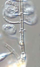


58 A B C D E F Fig. 18. Helicosporium pallidum. A-F. Conidiophores and conidia. A, D, F. from HMAS B. from HMAS C. from HMAS E. from HMAS Bars = 10 µm. 370


59 Fungal Diversity A B C Fig. 19. Helicosporium panacheum. A-C. Conidiophores and conidia from HMAS Bars = 10 µm. mass, (19) µm diam, tightly coiled 3½-4½ times. Conidial filament hygroscopic, indistinctly multisepta, (1) µm thick. 371
60 Substrate: Decaying stems of Zea mays; decaying leaves; fruit of Casuarina equisetifolia; old woody flower stalk. Distribution: Canada, China, Japan, USA (Iowa, Hawaii). Description and illustration: Moore, 1954; Tubaki, 1958; Goos, 1980; Goos, Specimens examined: HMAS (= ZGZII ): on rotten wood of palm tree, Guangdong Province, Zhuhai, 7 Dec. 2003, Guozhu Zhao. Notes: H. panacheum is characterized by subhyaline to very pale brown conidiophores, polyblastic conidiogenous cells with 3-5 denticles, and 3-5 times coiled conidia. These are similar to those found in Helicosporium gracile (Morgan) Linder (1929), but the conidia of H. gracile are coiled only 3-3¼ times. Helicosporium pannosum (Berk. & M.A. Curtis) R.T. Moore, Mycologia 49: 582, (Fig. 20) Drepanospora pannosa Berk. & M.A. Curtis, [as 'Drepanispora pannosa'], Grevillea 3: 105, = Helicosporium elinorae Linder, Ann. Missouri Bot. Gard. 16: 289, = Helicosporium nematosporum Linder, Ann. Missouri Bot. Gard. 16: , = Helicosporium serpentinum Linder, Ann. Missouri Bot. Gard. 16: 288, = Helicosporium linderi R.T. Moore, Mycologia 46: 90, = Helicosporium gigasporum K.M. Tsui, Goh, K.D. Hyde & Hodgkiss, Mycologia 93: 392, Colonies on woody substrate, effuse, white, yellowish brown, cottony. Mycelium partly superficial, partly immersed, composed of branched, septate, smooth-walled, subhyaline hyphae, µm thick. Conidiophores almost simple or branched from basal parts, erect, macronematous, arising laterally on the creeping superficial mycelium, single or often in group, fuscous, paler and tapering towards the apex, µm long, 4-6 µm thick at base, septate, apical region often sterile and setiform. Conidiogenous cells monoblastic, laterally formed from the lower parts of the conidiophores, hyaline to subhyaline, simple, tooth-like protuberance; denticles, cylindrical, µm. Conidia pleurogenous, when tightly coiled, 2-5 coils, µm in diam; filaments slender, subhyaline, thin-walled, smooth, hygroscopic, indistinctly septate, pale at the septa. Conidial filament 2-4 µm in the broadest part, tapering towards both ends, no secondary conidia observed, sclerote-like chlamydospore sometimes present. Substrate: Decayed wood. Distribution: Worldwide including Australia, Brazil, British Guiana, Chile, China, Cuba, Great Britain, New Zealand, USA including Hawaii. 372
61 Fungal Diversity Description and illustration: Linder, 1929; Matsushima, 1971, 1975; Pirozynski, 1972; Hughes, 1978; Barr, 1980; Romero, 1983; Sivanesan, 1984; Goos, 1989 (as Drepanospora pannosa ). Teleomorph: Tubeufia helicoma (Phill. & Plower.) Pirozynski, (= Tubeufia rugosa Booth, fide Pirozynski, 1972). Specimens examined: HMAS (= ZGZII ): on rotten wood, Hainan Province, Wuzhi mountain, 14 Dec. 2003, Guozhu Zhao. HMAS (= ZGZII ): on rotten wood, Yunnan Province, Gaoligong mountain, Dahaoping, 16 Oct. 2003, Guozhu Zhao. HMAS (= ZGZII ): on rotten wood, Hainan Province, Wuzhi mountain, 14 Dec. 2003, Guozhu Zhao. HMAS (= ZGZII ): on decaying branches of unidentified plant, Hainan Province, Wuzhi mountain, 14 Dec. 2003, Guozhu Zhao. HMAS (= ZGZII ): on dead wood, Yunnan Province, Gaoligong mountain, Bawan, 15 Oct. 2003, Guozhu Zhao. WU1795b: on dead wood in water, Jilin Province, Changbai, Sept , Wenping Wu. WU1981a: on rotten wood, Guangdong Province, Lufu mountain, Oct , Wenping Wu. WU1984: on dead branches of unidentified plant, Guangdong Province, Lufu mountain, Oct , Wenping Wu. WU7030. Notes: H. pannosum differs from all other Helicosporium species in having subhyaline to pale yellow conidia consisting of a wide conidial filament. Based on these characteristics, Goos (1989) assigned this species into the genus Drepanospora, which is an early erected genus but was ignored currently. After examining several specimens of this species, we conclude that Drepanospora should be accommodated in Helicosporium. Such a conclusion had been made by Moore (1957). Helicosporium gigasporum and H. hongkongense are similar to H. pannosum in having erect, macronematous conidiophores and cylindrical toothlike conidiogenous cells (Tsui et al., 2001). Tsui et al. (2001) distinguished Drepanospora pannosa from H. hongkongense by the echinulate conidia bearing lateral secondary conidia in the former species, but these characteristics seem to be unstable from our studies and also reports in the literature (Matsushima, 1975). Except for this, we also observed the relatively smaller and hygroscopic conidia in H. hongkongense in the original description. After carefully comparision of H. pannosum and H. gigasporum, we conclude that they are conspecific and thus H. gigasporum is reduced to a synonym of H. pannosum. Our attempt to grow this fungus on agar was not successful. Great variation in the conidial morphology, such as number of coiling and size were observed among different specimens. Furthermore, the very flexible and hygroscopic conidial filament is usually uncoiled in water. All of this makes if difficult to clearly distinguishing H. pannosum from other species. 373


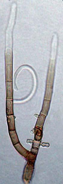
62 A B C D F E G Fig. 20. Helicosporium pannosum (HMAS 90282). A-B. Conidia. C-G. Conidiophores with cylindrical sporogenous denticles (arrows) and conidia. Bars = 10 µm. 374
63 Fungal Diversity Helicosporium sympodiophorum G.Z. Zhao, Xing Z. Liu & W.P. Wu, sp. nov. Mycobank (Fig. 21) Etymology: L. sympodiophorum, referring to the sympodial conidiophores. Coloniae effusae, velutinae, pallide. Mycelium plerumque superficiale sed in substrato partim immersum, ex hyphis ramosis, septatis, subhyalinis vel pallide brunneis, laevibus, µm latis compositum. Conidiophora macronematosa, mononematosa, solitaria, simplicia, erecta, recta vel leviter flexuosa, laevia, usque ad 250 µm longa, superne sympodialiter prolifera, manifeste dentata, inferne fusca sursum pallescentia; dentes conici apice µm lati. Conidia holoblastica, acrogena, solitaria, sicca, ad basim truncata, septata, laevia, pallide brunnea, ex filo 3½-4-plo convoluto, partim crassissima µm in diam., ut in torto µm diam. Holotypus: In ligno mortuo arboris latifoliae. Colonies effuse, velvety, white. Mycelium mostly superficial and partly immersed, composed of branched, septate, subhyaline to pale brown hyphae, µm wide, smooth. Conidiophores macronematous, mononematous, solitary, simple, erect, straight or slightly flexuous, smooth, rarely branched, septate, up to 250 µm long, µm in broadest part, tapering to apex. Conidiogenous cells sympodial, mono- or polyblastic, cylindrical or conical, truncate at the apex, µm wide. Conidia holoblastic, acrogenous, solitary, dry, truncate at base, smooth, pale brown, tightly coiled 3½-4 times, µm in diam. Conidial filament µm thick, slightly hygroscopic, septa. Substrate: on dead wood of broad-leaf tree. Distribution: China. Holotype: WU1858a: on rotten wood of unidentified plant, Dinghu mountain, Guangdong Province, China, Oct , Wenping Wu. Notes: The conidiophores with sympodial proliferation and truncate conidiogenous cells are similar to those in Helicomyces macrofilamentosus and H. roseus. However, the erect and macronematous conidiophores of Helicosporium sympodiophorum fit well into Helicosporium. Matsushima (1975) described Helicosporium sp. (no.276, MFC-1812) producing erect, maronematous conidiophores with sympodial proliferation, which is similar to H. sympodiophorum but differs in conidial morphology. The conidia of H. sympodiophorum are 3½-4 times coiled with a narrower ( µm wide) conidial filament whereas MFC-1812 has 2-2½ times coiled conidia and 5-7 µm wide conidial filament. The conidial septa of H. sympodiophorum are difficult to observe without staining. 375
. A.")


64 A B C Fig. 21. Helicosporium sympodiophorum (WU1858a). A. Conidiophores, sympodial conidiogenous cells tapering to abruptly rounded and truncate conidia-bearing tips (arrows) and conidia, slide mounted in lactophenol-cotton blue. B-C. Conidiophores arising laterally from the creeping hyphae. Bars = 10 µm. 376
65 Fungal Diversity Helicosporium talbotii Goos, Mycologia 81: 368, (Fig. 22) Helicosporium ramosum Talbot, Bothalia 6: 493, 1956 [non H. ramosum (Berk. & M.A. Curtis) Massee, 1893]. Colonies effuse, white, yellowish brown, cottony. Mycelium mostly superficial and partly immersed composed of branched, septate, smooth-walled, subhyaline hyphae. Conidiophores erect, straight, simple or branched, macronematous, laterally on the creeping superficial mycelia, fuscous, up to 120 µm long, 4-5 µm wide at base, septate; apical region often sterile. Conidiogenous cells monoblastic, with a lateral, tooth-like protuberance; the protuberance hyaline to subhyaline, simple, cylindrical, µm wide. Conidia pleurogenous, arising directly from the mycelium or else borne on robust conidiophores or conidiogenous cell. Conidial filament slender, subhyaline, thin-walled, smooth, 2½-3½ coils when tightly coiled, µm in diameter, hygroscopic, indistinctly multiseptate, µm in the broadest part, tapering toward both ends. Substrate: Dead wood and leaves. Distribution: China, Japan, South Africa. Description and illustration: Talbot, 1956; Ichinoe and Kume, 1970; Goos, Specimens examined: HMAS (= ZGZII ): on decorticated dead branches of an unidentified tree, Jilin Province,Lushuihe, Changbai mountain, 19 Sep. 2004, Guozhu Zhao. HMAS (= ZGZII chi): on dead branches of unidentified tree, Hainan Province, Wuzhi mountain, 14 Dec. 2003, Guozhu Zhao. Helicosporium virescens (Pers.) Sivan., Bitunicate Ascomycetes and their Anamorphs (Vaduz): 591, (Fig. 23) Dematium virescens Pers., Romers Neues Mag. Bot. 1: 121, =Helicosporium vegetum Nees, Syst. Pilze Schwamme: 68, Helicosporium state of Tubeufia cerea (Berk. & M.A. Curtis.) Booth, in Ellis, Dematiaceous Hyphomycetes: 193, Helicotrichum vegetum (Nees) Wallr., Fl. Crypt. German. 2: 161, Helicomyces vegetum (Nees) Corda, Icon. Fung. 1: 9, Helicomyces vegetus (Nees) Pound & Clem., Bull. Minn. Geol. Nat. Hist. Surv. 9: 659, = Helicotrichum pulvinatum Nees & T. Nees, Nova Acta Acad. Caes. Leop 9: 246, = Helicosporium olivaceum Peck, Rep. N. Y. State Mus. 27: 102, = Helicosporium neesii R.T. Moore, Mycologia 49: 582, Colonies on natural substrate effuse, yellow, cottony. Mycelium mostly superficial and partly immersed, composed of branched, septate, hyaline, smooth hyphae. Conidiophores usually unbranched, subulate, up to 400 µm long; lower part brown to dark brown, µm wide, fertile; upper part pale, tapering to a point, sterile. Conidiogenous cells monoblastic or polyblastic, integrated, intercalary, determinate, cylindrical, denticulate; denticles hyaline, 377


 Booth, 1964).")
66 conical, µm long, µm wide at the broadest part. Conidia pleurogenous, holoblastic, helical, tightly coiled 3-4 times in one plane, hyaline to yellowish green in mass, µm diam. Conidial filament µm thick, indistinctly septate. A B C Fig. 22. Helicosporium talbotii (HMAS 98782). A. Conidium. B-C. Conidiophores, conidiogenous cells and conidia. Bars = 10 µm. Substrate: Decaying wood. Distribution: Worldwide including Canada, China, Netherlands, USA. Description and illustration: Linder, 1929; Tubaki, 1964; Sivanesan, 1984; Goos, Teleomorph: Tubeufia cerea (Berk. & M.A. Curtis) Höhn., (= Tubeufia cerea (Berk. & M.A. Curtis) Booth, 1964). Specimens examined: HMAS (= ZGZII ): on decaying branches of bamboos, Yongjiugou, Linzhi, altitude 3100m, Tibet, 14 July 2004, Guozhu Zhao. 378
. Bars = 10 µm.")

 referred to the connection between Tubeufia cerea (Tubeufiaceae, Pleosporales, Dothideomycetidae,")
67 Fungal Diversity Fig. 23. Helicosporium virescens. A-C. Conidiophores with denticulate conidiogenous cells and conidia (HMAS 98900). Bars = 10 µm. A B C Notes: Helicosporium murinum is similar to H. virescens in having erect, unbranched conidiophores and tooth-like conidiogenous cells, but differs from H. virescens by longer conidiophores with distinctly cylindrical conidiogenous cells and regularly 3-4 times coiled conidia. Sivanesan (1984) referred to the connection between Tubeufia cerea (Tubeufiaceae, Pleosporales, Dothideomycetidae, Ascomycetes, Ascomycota) and Helicosporium virescens. Hughes (1958) and Booth (1964) demonstrated this connection by producing the anamorph H. vegetum from a culture of T. cerea derived from an ascospore. 379
68 Reassigned Drepanospora species Drepanospora Berk. & M.A. Curtis [as 'Drepanispora'], in Berkeley, Grevillea 3: 105, (See page 352). Type species: Drepanospora pannosa Berk. & M.A. Curtis, [as 'Drepanispora pannosa'], Grevillea 3: 105, Drepanospora pannosa Berk. & M.A. Curtis, Grevillea 3: 105, 1875, in Goos, Mycologia 81: 370, See Helicosporium pannosum (Berk. & M.A. Curtis) R.T. Moore, Mycologia 49: 582, Drepanospora viridis (Corda) Goos [as ' viride'], Mycologia 81: 371, See Helicoma viridis (Corda) S. Hughes, Can. J. Bot. 36: 772, (See page 424) Linder (as Helicoma mulleri), Ann. Missouri Bot. Gard. 16: 307, Helicoma Corda, Icon. fung. 1: 15, = Troposporella P. Karst., Hedwigia 31: 299, = Helicosporina G. Arnaud ex Rambelli, Mycopath. Mycol. Appl. 13: 110, Anamorphic Tubeufia, Tubeufiaceae, Pleosporales, Dothideomycetidae, Ascomycetes, Ascomycota, Fungi. Type species: Helicoma muelleri Corda, Colonies effuse, often hairy or velvety, variable in colour but frequently yellowish, greyish brown, brown or olivaceous. Mycelium immersed or superficial. Conidiophores macronematous, mononematous, or lacking, simple or branched; sometimes apex setiform, pale to olivaceous or dark-brown, smooth. Conidiogenous cells mono- or polyblastic, integrated, terminal and/or intercalary, determinate or sympodial, cylindrical, denticulate. Conidia dry, solitary, acropleurogenous or pleurogenous, simple, helicoid or circinate, hyaline to brown or olivaceous brown, smooth, multiseptate. Conidial filament rather thick in proportion to length, non-hygroscopic (Ellis, 1971; Goos, 1986, 1987). Notes: The early concept of Helicoma was often confused with Helicomyces and Helicosporium for their similar helical conidia. Linder (1929) treated 26 species of Helicoma. He characterized Helicoma as having thickwalled, non-hygroscopic, coiled conidia and applied these features to distinguish Helicomyces and Helicosporium and other allied genera with hygroscopic conidia. Goos (1986) dealt with Helicoma and accepted 31 species, excluded 12 species described by Linder (1929) or subsequent workers. He divided the 380
69 Fungal Diversity genus into four Sections according to their ontogeny, i.e. 10 species in Section Helicoma, 1 in Atroseptatum, 8 in Violaceum, and 12 in Monilipes. Since then, six more species have been described in Helicoma (Holubová-Jechová, 1987; Shearer, 1987; Carris, 1989; Matsushima, 1993; Castañeda Ruíz et al., 1998) and assigned into two Sections. Their diagnostic characteristics and measurements are summarized (Tables 7-10). Considering its conidial morphology, ontogeny and culture characteristics, Helicoma irregulare Kirk (1984) was transferred into Helicofilia Matsushima (1983) in this monograph. Teleomorphs are known for two Helicoma species (Table 3). Hirsutella petchabunensis Hywel-Jones, Goos & E.B.G. Jones (1998) has a Helicoma-like synanamorph. Helicoma chlamydosporium Shearer (1987) and Helicoma depressispora Matsushima (Matsushima, 1993; Hyde, 1998; Chang, 1999) commonly produce dictyochlamydospores on natural substrate, and the helical conidia appear on culture. The coiled parent conidium of H. atroseptatum Linder occasionally yield small, globose secondary conidia attached to the margin of parent conidia (Goos, 1986). Based on an analysis of type species of Troposporella and Helicosporina, these two genera are considered to have similar ontogeny and conidial morphology to Helicoma. In our study they are dealt with as synonyms of Helicoma. Only 4 Helicoma species have been reported from Hong Kong and Taiwan (Table 2). Eighteen Helicoma species including 2 new species and 16 new records to China were found during our study. One new combination Helicofilia irregularis (Kirk) G.Z. Zhao is proposed. Checklist of Helicoma species Helicoma acrophalerium R.T. Moore, Mycologia 49: 584 (1957) Helicoma ambiens Morgan, J. Cincin. Soc. Nat. Hist. 15: 46 (1892) Helicoma anastomosans Linder, Ann. Mo. Bot. Gard. 18: (1931) Helicoma asperothecum Linder, Ann. Mo. Bot. Gard. 16: 311 (1929) Helicoma atroseptatum Linder, Ann. Mo. Bot. Gard. 16: 307 (1929) Helicoma casuarinae (Matsush.) G.Z. Zhao, Xing Z. Liu & W.P. Wu, this paper. Helicomyces casuarinae Matsush., Matsu. Mycol. Mem. 4: 9 (1985) Helicoma chlamydosporum Shearer, Mycologia 79: 468 (1987) Helicoma conicodentatum Linder, Ann. Mo. Bot. Gard. 16: 311 (1929) Helicoma dennisii M.B. Ellis, Mycol. Pap. 87: 23 (1963) Helicoma depressispora Matsush., Matsu. Mycol. Mem. 7: 52 (1993) Helicoma divaricata Hol.-Jech., Ceská Mykol. 41: 33 (1987) 381
70 Helicoma fumosum (P. Karst.) G.Z. Zhao, this book Troposporella fumosa P. Karst., Hedwigia 31: 299 (1892) Helicoma hainanense G.Z. Zhao, this book Helicoma hyalonema G.Z. Zhao, Xing Z. Liu & W.P. Wu, this book Helicoma hyalospora (P.Rag. Rao & D. Rao) G. Z. Zhao, Xing Z.Liu & W.P. Wu, this book Troposporella hyalospora P.Rag. Rao & D. Rao, Mycopathologia 22: 52 (1964) Helicoma inflatum Linder, Mycologia 25: 344 (1933) Helicoma latifilum G.Z. Zhao, Xing Z. Liu & W.P. Wu, this book Helicoma limpidum Morgan, J. Cincin. Soc. Nat. Hist. 15: 47 (1802) Helicoma machaerinae Goos, Mycologia 72: 597 (1980) Helicoma microscopicum (Ellis) Linder, Ann. Mo. Bot. Gard. 16: 299 (1929) Helicosporium microscopicum Ellis, Bull. Torr. Bot. Club 9: 98 (1882) Helicoma minutissimum Linder, Ann. Mo. Bot. Gard. 16: 298 (1929) Helicoma monilipes Ellis & L.N. Johnson, Proc. Acad. Nat. Sci. Phila.: 374 (1894) Helicoma morganii Linder[as 'morgani'], Ann. Mo. Bot. Gard. 16: 304 (1929) Helicoma muelleri Corda, Icon. fung. (Prague) 1: 15 (1837) Helicoma narsapurensis P.Rag. Rao & D. Rao [as 'narsapurense'], Mycopathologia 22: 49 (1964) Helicoma olivaceum (P. Karst.) Linder, Ann. Mo. Bot. Gard. 16: 302 (1929) Helicopsis olivacea P. Karst. [as 'olivaceus'], Rev. Mycol. 11: 96 (1889) Helicoma palmigenum (Penz. & Sacc.) Linder, Ann. Mo. Bot. Gard. 16: 306 (1929) Helicosporium intermedium var. palmigenum Penz. & Sacc., Icones Fungorum Javanicum: 105 (1904) Helicoma palmarum G.Z. Zhao, this book Helicomyces anamorph of Tubeufia palmarum (Torrend) Samuels, Rossman & E. Müll., Sydowia 31: 189 (1978). Helicoma perelegans Thaxt. ex Linder, Ann. Mo. Bot. Gard. 16: 303 (1929) Helicoma phaeosporum Fresenius, Beitrage zur Mykol. 3: 99 (1863) Helicoma polysporum Morgan, Jour. Cincin. Soc. Nat. Hist. 15: 46 (1892) Helicoma pulchra R.F. Castañeda & Guarro, Mycol. Res. 102: 58 (1998) Helicoma recurvum Linder, Ann. Mo. Bot. Gard. 16: 312 (1929) Helicoma repens Morgan, Jour. Cincin. Soc. Nat. Hist. 15: 47 (1892) Helicoma resinae (Lindau) J.L. Crane & Schokn., Mycologia 67: 669 (1975) Torula resinae J. Lindau, in Rabenhorst s Kryptogamen-Fl. Deutsch. Oesterr. Schweiz. 1: 578 (1906) Helicoma roseolum Thaxt., Ann. Mo. Bot. Gard. 16: 297 (1929) Helicoma scarabaeiforme G.Z. Zhao, Xing Z.Lui, & W.P. Wu, this paper) Helicoma stigmateum (Reiss) Linder, Ann. Mo. Bot. Gard. 16: 298 (1929) Lituaria stigmatea Riess, Bot. Zeit. 11: 136 (1853) 382
71 Species Fungal Diversity Helicoma sympodiophorum Matsush., Matsu. Mycol. Mem. 7: 52 (1993) Helicoma taenia R.T. Moore, Mycologia 47: 91 (1955) Helicoma taiwanensis Matsush., Mats. Mycol. Mem. 3: 11 (1983) Helicoma tenuifilum Linder, Ann. Mo. Bot. Gard. 18: 14 (1931) Helicoma vaccinii Carris, Mycotaxon 36: 29 (1989) Helicoma violaceum G. Winter ex Linder, Ann. Mo. Bot. Gard. 16: 303 (1929) Helicoma viridis (Corda) S. Hughes, Can. J. Bot. 36: 772 (1958) Table 7. Diagnostic characteristics of the species of Sect. Helicoma. Conidiophore size (µm) Diameter of conidia (µm) Number of coils in conidia Width of conidial filament (µm) Number of septa in filament H. ambiens* ½-1¾ H. asperothecum ½-1¾ H. casuarinae ¼ H. chlamydosporum ¼-1¾ H. conicodentatum ¼ H. dennisii* ¼-1½ H. divaricata ¼-1½ H. hainanense* ¼-1¾ inconspicuous H. inflatum ¼-1¾ H. muelleri ½-1¾ H. nasapurensis ¼ H. palmarum ¼ H. recurvum ¾ H. scarabaeiforme* ¾ H. taenia ½-1¾ H. taiwanensis ½ H. vaccinii* ½-1¾ *Species recorded in China. All measurements and identification characteristics are from the reference of Goos (1986) and other original descriptions. Key to the Sections of Helicoma (Goos, 1986) 1. Conidia borne acrogenously on small denticles, becoming pleurogenous as a result of continued growth of the conidiophore and the lateral displacement of conidia; basal cell of the conidium tapering to a more or less flattened, slightly thickened, attachment scar Sect. Helicoma 1. Conidia not borne on denticles, or if so, borne pleurogenously from the first
72 2. Conidia borne pleurogenously on lateral peg-like outgrowths; basal cell of the conidium truncate but without prominent scar; conidia attached centrally; secondary conidia often present... Sect. Atroseptatum 2. Conidia not borne pleurogenously Conidia attached across the full diameter of the basal cell, leaving a truncate attachment scar on the conidiogenous cell when shed; conidiophores generally short (less than 75 µm)......sect. Violaceum 3. Conidia borne in diverse manner, but not on denticles, lateral outgrowths, or distinctly truncated conidiogenous cells, or if so, conidiophores macronematous...sect. Monilipes Table 8. Diagnostic characteristics of the species of Sect. Atroseptatum. Species Conidiophore Diameter of Number of Width of Number of size (µm) conidia (µm) coils in conidia conidial filament septa in (µm) filament H. atroseptatum ¼ H. viridis* ¼ *Species recorded in China. All measurements and identification characteristics are from the reference of Goos (1986) and other original descriptions. Species Table 9. Diagnostic characteristics of the species of Sect. Violaceum. Conidiophore size (µm) Diameter of conidia (µm) Number of coils in conidia Width of conidial filament (µm) Number of septa in filament H. anastomosans* ½-1¾ H. depressispora* micronematous ¼-1¾ H. hyalonema* ½ H. hyalospora ½ H. limpidum H. microscopicum* ¾ H. morganii* H. perelegans* ½ H. pulchra* ½ H. repens* short branches ½-2¾ H. sympodiophorum H. tenuifilum ¾-3¼ H. violaceum *Species recorded in China. All measurements and identification characteristics are from the reference of Goos (1986) and other original descriptions. 384
73 Table 10. Diagnostic characteristics of the species of Sect. Monilipes. Fungal Diversity Species Conidiophores Diameter of Number of Width of Number of size (µm) conidia (µm) coils in conidia conidial filament (µm) septa in filament H. acrophalerium Up to 28 H. fumosum* ½ H. latifilum* ½ H. machaerinae lacking ¾ H. minutissimum ¼ H. monilipes* ¼ H. olivaceum* ¼-1¾ H. palmigenum ¾ H. phaeosporium H. polysporium short branches ¾ H. resinae ½ 4- multiple H. roseolum ¼-1½ H. stigmateum denticles 7-10 ¾-1½ *Species recorded in China. All measurements and identification characteristics are from the reference of Goos (1986) and other original description. Key to the species of Helicoma from China 1. Conidiophores and conidia with characteristics of Sect. Atroseptatum... H. viridis 1. Conidiophores and conidia with characteristics of other Sections Conidiophores and conidia with characteristics of Sect. Monilipes Conidiophores and conidia with characteristics of other Sections Conidial filaments more than 6 µm thick... H. latifilum 3. Conidial filaments less than 6 µm thick Conidiogenous cells hyaline, swollen obovoid...h. fumosum 4. Conidiogenous cells pale brown, elongated cylindrical or lacking Coiled conidia µm in diam; filaments µm thick... H. monilipes 5. Coiled conidia µm in diam; filaments µm thick... H. olivaceum 6. Conidiophores and conidia with characteristics of Sect. Helicoma Conidiophores and conidia with characteristics of Sect. Violaceum Conidia coiled less than 1 times, µm in diam...h. scarabaeiforme 7. Conidia coiled more than 1 times Conidia uncoloured, mostly 1-septate, coiled 1¼-1¾ times... H. hainanense 8. Conidia coloured, more than 1-septate
74 9. Conidiophores usually swollen at the apex; conical projections sometimes appear on the fringe of conidial filaments... H. vaccinii 9. Conidiophores not swollen at the apex Conidia with round basal cells...h. ambiens 10. Conidia with tapered basal cells, truncate at the point of attachment... H. dennisii 11. Chlamydospores usually formed, dematiaceous, muriform...h. depressispora 11. Chlamydospores not produced Conidia coloured, with distinct multi-septation Conidia pale colour or indistinct septation Sporodochia formed on natural substrate; coiled conidia 9-12 µm in diam H. microscopicum 13. Sporodochia usually not formed; coiled conidia more than 12 µm in diam Conidia borne on shorted, cylindrical denticles...h. perelegans 14. Conidia borne on enlongated, sympodial conidiophores... H. pulchra 15. Conidiophores distinctly differentiated, macronematous, erect or flexuous Conidiophores indistinctly differentiated Conidiogenous cells conical, truncate; base of conidium conical, truncate H. anastomosans 16. Conidiogenous cells cylindrical, truncate; base of conidium round... H. morganii 17. Conidia borne on lateral of repent hyphae; filaments µm thick... H. repens 17. Conidia borne on short conidiophores; filaments µm thick... H. hyalonema Helicoma ambiens Morgan, J. Cincin. Soc. Nat. Hist. 15: 46, (Fig. 24) Helicosporium ambiens (Morgan) Sacc., Syll. Fung. 11: 639, = Helicoma interveniens Talbot. Bothalia 6: 492, Colonies on natural substrate, effuse, yellowish brown. Mycelium mostly superficial and partly immersed, composed of branched, septate, subhyaline to olive brown, smooth-walled hyphae. Conidiophores simple or rarely branched, erect, straight or slightly curved, single or in clusters, dark brown at base, becoming paler towards the apex, up to 170 µm long, µm in broadest part, tapering towards the apex, thick-walled, smooth. Conidiogenous cells denticulate, hyaline, cylindrical, µm. Conidia acropleurogenous, µm diam, borne singly on short and inconspicuous sporulating denticles. Conidial filament hyaline to dilute fuscous, µm thick, tightly coiled 1¼- 1¾ (1½) times, indistinctly 7-10 septate, both ends abruptly blunted and rounded, base with a truncate scar. Substrate: On decaying wood and rotting fibers of oil palm. 386
75 Fungal Diversity Distribution: Canada (Ontario), China, Cuba, South Africa, Tanzania, USA (Hawaii, Iowa, Ohio). Description and illustration: Morgan, 1892; Linder, 1929; Moore, 1953; Talbot, 1956 (as H. interveniens); Pirozynski, 1972; Goos, 1980; Goos, Specimen examined: HMAS (= ZGZII ): on rotten wood, Qian mountain, Liaoning Province, 22 Sep. 2004, Guozhu Zhao. Notes: The main difference between this species and the other related species in the genus is the morphology of the basal cell of the conidial filament. In H. ambiens it is abruptly blunted and rounded with a characteristic U-shape; while in H. muelleri Corda, the basal cell is more or less V-shaped. Furthermore, H. muelleri has the confirmed teleomorph Tubeufia pezizula (Berk. & M.A. Curtis.) Barr (Barr, 1980; Goos, 1986). Helicoma ambiens also resembles H. asperothecum Linder (1929), H. chlamydosporum Shearer (1987) and H. dennisii M.B. Ellis (1963) in conidial morphology. H. asperothecum differs from H. ambiens by the roughened walls of the conidiophores. H. chlamydosporum is distinguished from H. ambiens by producing intercalary chlamydospores, and short conidiophores. H. dennisiihas eccentrically attached conidia with V-shaped basal cells. Matsushima (1987) considered that H. divaricata Hol.-Jech. was close to the H. ambiens, but differred in its branched conidiophores and smaller size of conidia with a distinct basal conidial scar. Although morphological characters such as shape of the basal cell of the conidia and the branching pattern of the conidiophores has been used to separate the closely related species H. ambiens, H. dennisii and H. divaricata, we doubt if these features alone can sufficiently be used to separate these species. As mentioned by Linder (1929) that The real status of this species can only be determinted by comparative cultural studies or else by connecting it with the perfect stage. We suggest that further study of these characterictics on natural substrates and in pure cultures should be conducted to evaluate their importance in species identification. Helicoma anastomosans Linder, Ann. Mo. Bot. Gard. 18: 13-14, (Fig. 25) Colonies effuse, white cottony. Mycelium partly superficial and partly immersed partly, composed of branched, septate, subhyaline, smooth-walled, µm thick hyphae. Conidiophores macronematous, ascending, septate, flexuous, yellow to golden brown, branched irregularly, arising singly or in clusters from the hyaline repent mycelium, up to 115 µm long, µm in broadest part. Conidiogenous cells monoblastic or rarely polyblastic, pale brown, cylindrical or conical, denticulate, tapering to a truncate end; denticles first subhyaline, conical, 3-4 µm long, µm wide at the base, 1-2 µm wide 387


76 Fig. 24. Helicoma ambiens (HMAS 98785). A. Conidiophores and conidia. B. Conidiophores. Bars = 10 µm. A B 388

.")


77 Fungal Diversity A B C Fig. 25. Helicoma anastomosans. A-B. Conidiophores and conidia (HMAS 90367). C. Conidiophores and conidia (HMAS 90334). Bars =10 µm. at the apex, becoming pale brown to gold brown, developing to conidiophores. Conidia holoblastic, acrogenous or pleurogenous, dry, hyaline to subhyaline, µm diam. Conidial filament 4-5 µm thick, tightly coiled 2-2½ times, 389
78 indistinctly multiseptate, apex rounded, basal cell tapering gradually towards a truncated end. Substrate: On dead wood and decaying palm. Distribution: British Guiana, China. Description and illustration: Linder, 1931; Goos, Specimen examined: HMAS (=ZGZII ): on decaying branches of unidentified tree, Hainan Province, Diaoluo mountain, 12 Dec. 2003, Guozhu Zhao. HMAS (= ZGZII ): on rotten wood, Hainan Province, Wuzhi mountain, 14 Dec. 2003, Guozhu Zhao. Notes: H. anastomosans is similar to Helicoma morganii Linder (1929), H. repens Morgan (1892) and H. tenuifilum Linder (1931) in the conidial shape. H. morganii produces taller but more slender and loosely branched conidiophores with smaller teeth-like sporogenous cells, and larger conidia (21-27 µm in diam); H. repens has less developed conidiophores, shorter and slender conidiogenous cells, and hyaline and multiguttulate conidia; H. tenuifilum has truncated and cylindrical conidiogenous cells. Helicoma dennisii M.B. Ellis, Mycol. Pap. 87: 23, (Figs 26-28) Colonies on natural substrate, effuse, reddish brown, velvety. Mycelium partly superficial and partly immersed, composed of branched, septate, hyaline, smooth hyphae. Conidiophores simple or rarely branched, erect, occasionally anastomosing, septate, smooth-walled, fuscous at the base, light fuscous toward the apical cells, µm wide at the basal part, and tapering to µm wide at the apical part, up to 190 µm long. Conidiogenous cells mono- or polyblastic, integrated, terminal or intercalary, determinate, cylindrical, denticulate, smooth; sporogenous denticles hyaline, inconspicuous, cylindrical, µm. Conidia acropleurogenous, borne singly, tightly coiled 1¼-1¾ (1½) times,10-15 (13.5) µm in diam; conidial filament hyaline to dilute fuscous, (4.5) µm thick, hyaline indistinct 6-9 (8) septa, with rounded apical cells and V-shaped basal cells, with a distinctly darkered conidial scar. Colonies growing slowly on PDA, up to 0.5 cm diam. after 17 d, compact, pulvinate, greyish brown, become powdery with production of large amount of conidia. Aerial myceium first hyaline, becoming pale yellow, µm wide. Conidiophore erect, macronematous, arising on the repent hyphae. Conidia borne on the denticle which is slightly smaller (10-12 µm in diam) than those on natural substrate. Detached conidia often produce a global bulb on the basal cells of filaments. It was considered to be cytoplasm overflow of the conidial body. Substrate: Decayed wood. Distribution: China, Japan, NewGuinea, Taiwan, USA (Alabama, Hawaii), Venezuela. 390
79 Fungal Diversity Fig. 26. Helicoma dennisii: Conidiophores, conidiogenous cells and conidia (HMAS 90319). Bars = 10 µm. Description and illustration: Ellis, 1963, 1976; Matsushima, 1971, 1975; Morgans-Jones, 1974; Goos, 1980,


. B-C.")


80 B A C D E Fig. 27. Helicoma dennisii. A-E. Conidiophores, conidiogenous cells and conidia. A, D-E. from natural substrate (HMAS 90319). B-C. from culture on PDA, arrows shows cytoplasm overflow from detached conidia that forms a globose bubble attached to the basal filament cell. Bars = 10 µm. 392


81 Fungal Diversity A B C D E Fig. 28. Helicoma dennisii. A-E. Conidiophores, denticulate conidiogenous cells and conidia from natural substrate (HMAS 90441). Bars = 10 µm. 393
82 Specimen examined: HMAS (= ZGZII ): on cutting section of rotten wood of unidentified tree, Yunnan Province, Gaoligong mountain, Tongbiguan, 18 Oct. 2003, Guozhu Zhao. HMAS (= ZGZII ): on decaying palm tree, Hainan Province, tropical botanical gardon of Danzhou, 19 Dec. 2003, Guozhu Zhao. HMAS (= ZGZII ): on rotten wood, Yunnan Province, Gaoligong mountain, Nankang, 22 Oct. 2003, Guozhu Zhao. Notes: Helicoma dennisii is closely related to H. mulleri Corda (1837) and was considered as a synonym of the latter by Goos (1980) and Matsushima (1987). The main differences were said to be in the lighter colour of the conidia and conidiophores and the slightly larger conidia of H. dennisii. The conidia of H. dennisii are up to 23.5 µm in diam and the conidial filaments 8-9 µm thick; the conidia of H. mulleri up to 20 µm in diam and the conidial filaments 5-7 µm thick. H. narsapurensis P.R. Rao & D. Rao (1964) and H. recurvum (Petch) Linder (1929) are also similar to H. dennisii. H. narsapurensis differs from H. dennisii in the distance between septa of conidiophore, the conidial measurements (20-27 µm diam) and number of septa (3-8) of the conidia. H. recurvum differs from H. dennisii in having conidiophores with scattered superficial masses of purplish-black crystalloid deposits and the thicker conidial filaments which measure 6-8 µm as against 4-5 µm of H. dennisii. Helicoma ambiens Morgan, H. inflatum Linder (1933), H. chlamydosporum Shearer (1987) and H. divaricata Hol.-Jech. (1987) also possess similar conidia. However, they can be separated by the following features: H. ambiens has conidia with round basal cells, H. inflatum has bladder-like inflated conidiophores, H. chlamydosporum has speciel chlamydospore, and H. divaricata produces dichitomously branched condiophores. All three Chinese collections are well accommodated into H. dennisii, but slight differences in conidial size were observed between the different collections. For example, in specimen HMAS 90441, the conidia are much smaller (10-13 µm in diam) than those in the original description (19-22 µm in diam) (Ellis, 1963). We considered that the differences in the size of conidia may be caused by the different habitats. No conidiophores were observed in specimen HMAS 90337, but the distinguished conidia of H. dennisii are easily recognized. Helicoma depressispora Matsush., Matsu. Mycol. Mem. 7: 52, (Fig. 29) Colonies on natural substrate effuse, superficial, sparse, diffuse, consisting of large, black, shining, dictyochlamydospores. Mycelium partly superficial and partly immersed, composed of branched, septate, hyaline, smooth hyphae. Chlamydospores developed terminally or intercalarly from subhyaline hyphae of 6-10 µm thick through cell division and enlargement, dictyosporous, ellipsoidal, opaque at the septa, light brown at first, becoming 394
83 Fungal Diversity black, smooth, µm; tapering towards both ends and ends extending to propagules with subtending hyphae, pale brown and septate apical and basal part. Helical conidia not observed. Substrate: Decaying wood. Description and illustration: Matsushima, 1993; Hyde and Goh, 1998; Chang, Distribution: Australia, China including Taiwan, Ecuador, Peru. Specimens examined: HSAUPII (= ZGZII 0 660): On a dead submerged wood of Populus sp. in a stream, Tai mountain, Shandong Province, China, Sep. 2000, Guozhu Zhao. Notes: The distinctly large, black and shinning dictyochlamydospores observed in the Chinese collection make it easy to be recongnized as H. depressispora. We successfully obtained a pure culture of this fungus on PDA media. Abundant chlamydospores appeared after 20 days, however, no coiled conidia were observed either in pure culture or on natural wood. The coiled conidia of H. depressispora are usually difficult to see, especially in pure culture (Matsushima, 1993). Matsushima (1993) first described this fungus on decaying petioles of palm at Tambopata, Peru and Rio Yuturi, Ecuador. Chang (1999) collected this fungus on twigs in Taiwan. Two species of Helicoma produce dictyochlamydospores, H. chlamydosporum Shearer (1987) and H. depressispora, and they share some similar characters, but can be distinguished by slight differences in the outer wall of the surface cells of the chlamydospores. Those of H. chlamydosporum are conical and protuberant and septa are constricted obviously dividing spore into multicelled dictyospore, while mature chlamydospores in H. depressispora are black, opaque at the septa except for the pale brown and narrower apical and basal cells. Clear differentiation of these two species requires their helical conidial states. Coiled conidia of H. depressispora are complanate oblate circinate, 13-24µm wide and µm high (Matsushima, 1993), while those of H. chlamydosporium are plate-like circle, µm in diam (Shearer, 1987). Similar chlamydosporous structures also appear in other hyphomycetes: Intercalarispora nigra J.L. Crane & Schokn. (Schoknecht and Crane, 1983; Mercado-Sierra et al., 1998) chlamydospores were assigned as conidia and their formation is similar to chlamydospore formation in species of Xylomyces Goos, Brooks & Lamore (Goos et al., 1977; Goh et al., 1997). The chlamydospores in Xylomyces are generally fusiform and have transverse septa only. Pithomyces pulvinatus (Cook & Massee) M.B. Ellis (Ellis, 1971) with dictyochlamydospore-like conidia can easily be confused with H. depressispora, but the coiled conidia of H. depressispora occasionally appearing on natural substrate present a useful character to identify them. 395
84 Fig. 29. Helicoma depressispora: Chlamydospores on natural substrate (HSAUPII ). Bar = 10 µm. Helicoma fumosum (P. Karst.) G.Z. Zhao, Xing Z. Liu & W.P. Wu, comb. nov. (Fig. 30) Troposporella fumosa P. Karst., Hedwigia 31: 299, Colonies on natural substrate producing dense, minute, yellow ochre tufts. Punctiform sporodochia scattered, pulvinate to subspherical, the surface dry, granular. Mycelium partly superficial and partly immersed, composed of branched, septate, hyaline, smooth hyphae. Conidiophores micronematous or lacking, densely aggregated, sparsely branched, deeply constricted at the septa so as to give a monilioid appearance, dilute to light fuscous, the terminal cells when sterile frequently globose and reaching µm in diam. Conidiogenous cells discrete, catenulate, proximally clavate, pyriform to obovoid, distally 396
85 Fungal Diversity spherical, subspherical, mono- or polyblastic, smooth, hyaline to subhyaline, µm. Conidiogenous cells may generate from pre-existed conidiogenous cells and form a chain. Conidia holoblastic, acrogenous or pleurogenous, thick-walled, µm in diam. Conidial filament nonhygroscopic, µm thick, tightly coiled 1¾-2 times, distinctly septate, constricted at the septa. Substrate: on decaying wood. Description and illustration: Linder, 1929; Ichinoe and Kume, 1970; Ellis, Distributions: China, Europe, USA (Hawaii). Specimen examined: HMAS (= ZGZII ): on rotten branches of unidentified tree, Hubei Province, Shennongjia, Hongping, 14 Sep. 2003, Guozhu Zhao. Notes: Karsten (1892) erected Troposporella with T. fumosa as the type species. Based on the type specimen of T. fumosa, Linder (1929) gave a comprehensive description and illustration with the distinct character of bubbleshaped conidiophores / conidiogenous cells. He also mentioned that generic characteristic appeared to be intermediate between species of Helicoma, represented by H. monilipes and H. olivaceum. He also considered that it differed from the two Helicoma species in that the conidiophores were very closely aggregated to form a definite and compact subglobose fruiting body. However, subsequent studies have led to some confusion between T. fumosa and H. monilipes. For example, Ellis (1971a) and Ichinoe and Kume (1970) illustrated T. fumosa with the elongated basal conidial filament which is usually separate from the core of conidium body. It was normally considered to be the character of H. monilipes. Studies on the type specimens for these species are needed to clarify their relationship. According to the present concept of Helicoma (Ellis, 1971a; Goos, 1986), Troposporella fumosa seems to be better accommodated in Helicoma. A few species similar to T. fumosa have already assigned to the genus Helicoma. Furthermore, Troposporella has not been commonly accepted as a distinct genus by most scholars. Based on these considerations and checking of our material, the new combination, H. fumosum, was proposed for the type species of Troposporella. As a result, Troposporella becomes a synonym of Helicoma. Two described species in the genus Troposporella have already been transferred to other genera, T. monospora (W.B. Kendr.) M.B. Ellis (1976) into Slimacomyces monospora (W.B. Kendr.) Minter (1986), and T. rigidospora R.F. Castañeda & W.B. Kendr., (1990) into Troposporopsis rigidospora (R.F. Castañeda & W.B. Kendr.) Whitton, McKenzie & K.D. Hyde (1999). Troposporella hyalospora P.Rag. Rao & D. Rao (1964) is transferred into Helicoma hyalospora (P.Rag. Rao & D. Rao) G.Z. Zhao (see page 434). 397


86 A B C D Fig. 30. Helicoma fumosum. A-D. Conidiophores, conidiogenous cells and conidia on natural substrate (HMAS 90283). Arrows show the swollen conidiogenous cells. Bars = 10 µm. H. fumosum is morphologically similar to H. monilipes, but the sporodochial conidiomata, hyaline conidiophores with catenulate and swollen conidiogenous cells in H. fumosum are somewhat different from the scattered and branched conidiophores, and the pale brown, slender conidiogenous cells in H. monilipes. The Chinese collections with obviously obovoid conidiogenous cells well agree with H. fumosum as described by Linder (1929). 398
87 Fungal Diversity Helicoma hainanense G.Z. Zhao, Xing Z. Liu & W.P. Wu, sp. nov. Mycobank (Figs 31, 32D-E) Etymology: Named after sampling place, Hainan, a province of China. Coloniae in substrato naturali effusae, sparsae, minutae, albae. Mycelium pro maxima parte superficiale vel partim immersum, ex hyphis anastomosis, ramosis, laevibus, subhyalinis ad pallide brunneis, septatis, µm crassis compositum. Conidiophora recta vel flexuosa, deinceps sympodice prolificantia, brunnea ad atro-brunnea, crassitunicata, septata, denticulate, µm longa, µm crassa, ad basin µm crassa, ad apicem pallidiora et tenuitunicata, attenuata et truncata. Cellulae conidiogenae polyblasticae vel monoblasticae, terminales, sympodiales, cylindricae, denticulata; Dentes conidiiferi cylindrici, truncati, µm. Conidia acropleurogena, hyalina vel subhyalina usque pallide ochracea, levia, µm in diam., filis um crassis, 1-3 (plerumque 1)-septatis, non hygroscopicis, in spiras 1¼-1¾ convolutis, cellulis extremis rotundatis, cellulis basilaribus leviter conico-rotundatis apiculatisque, cicatrices planis µm crassis. Fig. 31. Helicoma hainanense: Conidiophores, conidiogenous denticles and conidia from holotype (HMAS 90366). Bars = 10 µm. 399


88 A B C D Fig. 32. Helicoma species. A-C. Helicoma scarabaeiforme: Conidiophores, conidiogenous denticles and conidia from holotype (HMAS 90464). D-E. Helicoma hainanense: Conidiophores, conidiogenous denticles and conidia from holotype (HMAS 90366). Bars = 10 µm. 400
89 Fungal Diversity Colonies on natural substrate effuse, scattered, small, white cottony, Mycelium partly superficial and partly immersed, composed of anastomosing hyphae repent, branched, smooth, septate, subhyaline to pale brown, µm thick. Conidiophores at first erect, simple, bent-ascending, successively sympodially proliferating, later loosely branched, and tending to become fasciculate by anastomosing, brown to dark brown, thick-walled, septate, continuously attenuated and ending in a truncate conidiogenous denticle, µm long, µm wide at base, µm wide at the paler to subhyaline apex. Conidiogenous cells polyblastic or monoblastic, terminal, sympodial, cylindrical, denticulate; denticles cylindrical, truncate, µm. Conidia acropleurogenous, hyaline or subhyaline to pale yellow-brown, smooth, µm in diam; conidial filament µm wide, with 1-3 (mostly 1) hyaline septa near the basal end, non-hygroscopic, tightly coiled 1¼-1¾ times, bluntly rounded at distal end, tapering to a rounded basal cell, 0.5-1µm wide. Holotype: HMAS (= ZGZII ) on decaying twigs of broad-leaved tree, in Mount Wuzhi, Hainan Province China, December 14, 2003, Coll. G. Z. Zhao. Notes: Helicoma hainanense is well placed in Section Helicoma, because of its conidial ontogeny and coiled shape. It is similar to H. stigmateum (Reiss) Linder (1929) in having 1¼-1¾ times coiled and hyaline to subhyaline conidia, but differs by its macronematous bent-ascending conidiophores and larger conidia (12-16 µm). H. stigmateum was assigned to Section Monilipes for having micronematous conidiophores borne laterally on the hyaline, repent hyphae and conidia which are 7-10 µm in diam. The new species is also close to H. asperothecum Linder (1929), a species from Section Helicoma, but differs by its smaller conidia, thinner, multiseptate (usually1-3) conidial filaments. H. asperothecum produces larger conidia (15-25 µm in diam) with filaments of µm wide and 8-10 septate. Helicoma hyalonema G.Z. Zhao, Xing Z. Liu & W.P. Wu, sp. nov. (Fig. 33) MycoBank Etymology: L. hyalonema, referring to the hyaline conidial filaments. Coloniae in substrato naturali effusae, albae. Mycelium partim in ligno substrato immersum vel superficiale, ex hyphis ramosis, septatis, hyalinis, laevibus compositum. Conidiophora macronematosa, mononematosa, erecta, recta vel leviter flexuosa, simplicia, laevia, septata, usque ad 35 µm longa, 2-3 µm lata. Cellulae conidiogenae polyblasticae, in conidiophoris incorporatae, terminales, laeves, sympodialiter prolificae, denticulatae; dentes µm. Conidia holoblastica, solitaria, sicca, helicoidea, complanata, µm diam, multiseptata, septa (18), subhyalina ad pallide lutea, cellulae facile secedentes. Filamenta conidica µm crassa, in 2-2½ spiris convoluta. Holotypus: In ramulis emortuis indeterminate, Monte Wuzhi, Hainan Provincia sinica, leg. Guozhu Zhao, 14 XII, 2003, HMAS (= ZGZII ). 401


90 Fig. 33. Helicoma hyalonema. A- B. Conidiophores, conidiogenous cells and conidia from holotype HMAS Bars = 10 µm. A B Colonies on natural substrate effuse, dispersed, composed of loose white conidia. Mycelium partly superficial and partly immersed, composed of branched, septate, hyaline, smooth hyphae. Conidiophores macronematous, mononematous, erect, straight or slightly flexual, first hyaline, becoming golden brown towards hyaline apex, unbranched or occasionally branched, thinwalled, up to 35 µm long, 2-3 µm wide in broadest part. Conidiogenous cells polyblastic, terminals, proliferate sympodially, denticulate, pale brown; denticles cylindrical, µm. Conidia holoblastic, helical, hyaline to subhyaline, µm in diam; conidial filament µm thick, non- 402
91 Fungal Diversity hygroscopic, tightly coiled 2-2½ times, indistinctly (18) septate. Secondary conidia sometimes present. Substrate: On dead wood. Distribution: China. Holotype: HMAS (= ZGZII ): on decaying branches of unidentified plant, Hainan Province, Wuzhi mountain, 14 Dec. 2003, Guozhu Zhao. Notes: The micronematous and thin-walled conidiophores in Helicoma are somewhat between Helicosporium-Helicomyces-Helicoma, while the nonhygroscopic conidia ensure it best to be accommodated into Helicoma. It resembles H. anastomosans, H. limpidum, H. morganii, H. polysporum, H. repens and H. tenuifilum in conidial morphology. However, in H. anastomosans the conidiophores are simple and the conidia are 2½-2¾ times coiled; in H. limpidum the conidia are borne laterally at the apex of short conidiophores and the conidiogenous cells are monoblastic; in H. morganii the conidiophores are longer, broader and brown; in H. polysporum the conidia are coiled less than 2 times (1½-1¾ times); in H. repens the conidiophores are macronematous and formed laterally from the creeping hyphae; and in H. tenuifilum the conidia are coiled 2¾-3½ times. Helicoma latifilum G.Z. Zhao, Xing Z. Liu & W.P. Wu, sp. nov. (Fig. 34) MycoBank Etymology: L. latifilum, referring to the broad conidial filaments. Coloniae in substrato naturali nigrae, granulosae, inconspicuae, constantes e sporodochiis arcte aggregates, sub lente sporodochia singula, sparsa, brunnea, visibilia. Mycelium sparsum, pro maxima parte submersum. Conidiophorum micronematovel semimacronemato, simplex vel irregulariter ramosum, septatum, raro exigue constrictum, laeve, 35 µm altum, 3-6 µm latum. Cellulae conidiogenae discretae, catenulatae, proximaliter clavatae, pyriformes ad obovoideas, distales, sphaericae, mono- vel polyblasticae, laeves, hyalinae ad subhyalinas, µm. Catena nova ex cellula conidiogena originali multo genifa. Conidia acrogena vel pleurogena, singulatim producta, helicoidea, 1½ -circinata, complanata, µm diam, 6-8 µm crassa, septata, rotundata ad apicem, cellulis basilaribus rotundato-fastigatis apiculatisque, primo hyalina, tarde brunnea vel atrobrunnea, laevia, guttulata, sicca. Holotypus: in ligno putrido rami emortui arborum incognitarum in Mounte Liupanshan, Ningxia Provincia, Sina, 24 VII 2001, leg. Wenping Wu, WU1120g. Colonies on natural substrate black, inconspicuous, the surface granular, consisting of groups of sporodochia. Conidiomata sporodochia, individually scattered, dry, hard, punctiform, superficial, brown. Mycelium scanty, mostly immersed. Conidiophores micronematous or semi-macronematous, simple or irregularly branched, septate, occasionally slightly constricted at septa, smooth, up to 35 µm long, 3-6 µm wide. Conidiogenous cells mono- or polyblastic, discrete, catenate, clavate, cylindrical, pyriform to obovoid, distally spherical or subspherical, smooth, hyaline to subhyaline, µm. Conidiogenous 403
92 cells proliferation from pre-existing conidiogenous cells. Conidia holoblastic, acrogenous or pleurogenous, produced singly; helicoid, 1½ times, complanate, µm diam. Conidial filament 6-8 µm wide at the widest part, at first colourless, then brown to dark brown at maturity, smooth-walled, guttulate, dry, septa, rounded at the apex, tapering towards a rounded base with a truncated scar. Substrate: On dead wood. Distribution: China. Holotype: WU1120g: on dead branch of unidentified plant, China: Ningxia Province, Liupan Monutain, Liangdianxia, 24 Aug. 1997, Wenping Wu. Notes: The swollen, enlonged, subhyaline conidiogenous cells and the broader conidial filaments of H. latifilum are distinct characters separating it from other Helicoma species. H. latifilum mostly resembles H. monilipes, but differs from the latter which has smaller conidia ( µm in diameter), conidial filaments that are thinner (3.5-5 µm in broadest part) coiled 2¼ times and of equal width (Linder, 1929). H. fumosum is also similar, but has smaller conidia (12-16 µm diam) and narrower conidial filaments (4-4.5 µm thick).. Helicoma microscopicum (Ellis) Linder, Ann. Mo. Bot. Gard. 16: 299, (Fig. 35) Helicosporium microscopicum Ellis, Bull. Torr. Bot. Club 9: 98, = Helicomyces microscopicum (Ellis) Pound & Clement, Minn. Bot. Stud. 9: 659, Colonies on natural substrate, punctiform, compact, black, rigid, minute tuft, forming sporodochia, less than 1 mm. Mycelium mostly immersed and partly superficial, composed of branched, septate, hyaline, smooth hyphae, (1) µm wide. Condiophores micronematous or lacking. Conidiogenous cells monoblastic, acrogenous, or incorporated with creeping hyphae, integrated, intercalary or terminal, thin-walled, smooth, subhyaline, cylindrical. Conidia holoblastic, acro- or pleurogenous, dilute fuscous to brown, 9-12 µm in diam; conidial filament 2-3 µm thick, non-hygroscopic, tightly coiled 1¼-1¾ times, thick-walled, brown to dark brown, conspicuously constricted 6-9 septa, bluntly rounded at the apical cells, tapering towards the enlonged basal cells. Substrate: Decaying catkin of Alnus serrulata. Distribution: China, USA (New Jersey). Description and illustration: Linder, 1929; Goos, Specimens examined: WU5512: on dead branches of palm, Hainan Province, 20 Dec. 2000, Wenping Wu & Yan Huang. 404


93 Fungal Diversity Fig. 34. Helicoma latifilum. A-B. Conidiophores, conidiogenous cells and conidia from holotype WU1120g. Bars = 10 µm. A B 405


94 Fig. 35. Helicoma microscopicum. A. Compacted conidiophores and fasiculate conidia forming the sporodochia on natural substrate from WU5512. B. Hyphae, Conidiophores, conidiogenous cells and conidia. Bars = 10 µm. A B Notes: Goos (1986) mentioned that conidiophores and conidia were not observed after his examination of the slides of type specimen. In conidial morphology, H. microscopicum is closely related to two species, H. monilipes and H. olivaceum, but differs from them in having smaller conidia. H. microscopicum was refered to Section Violaceum by Goos (1986), 406
95 Fungal Diversity while the two latter species were in Monilipes group. The Chinese collection fits well with the original description given by Linder (1929). Helicoma monilipes Ellis & L.N. Johnson, Proc. Acad. Nat. Sci. Phila.: 374, (Fig. 36) = Helicosporina veronae Rambelli, Mycopathologia 13:111, Colonies on natural substrate forming punctiform sporodochia, scattered, stout, pulvinate to subspherical with appearance of granular surface. Mycelium partly superficial and partly immersed, composed of hyaline to pale brown, branched, septate, slender hyphae, 1-1.5µm wide. Conidiophores short, multiseptate, branched, closely aggregated, light fuscous, up to 50 µm long, 3-4 µm wide. Conidiogenous cells monoblastic, integrated, intercalary or terminal, subhyaline to hyaline, cylindrical, µm. Conidia holoblastic, acrogenous, golden brown, helical, µm in diam; conidial filament µm thick, non-hygroscopic, tightly coiled 1½ times, distinctly 9-15 septate, constricted at septa. Substrate: On dead wood and bark of Acer, Populus, Quercus. Distribution: Great Britain, Japan, USA (Michigan, Maine, New York). Description and illustration: Linder, 1929; Moore, 1953; Rambelli, 1960 (as Helicosporina veronae); Ichinoe and Kume, 1970; Goos, 1986; Matsushima, Specimens examined: HMAS (= ZGZII ): on decaying branches of unidentified tree, Hubei Province, Shennongjia, Hongping, 14 Sep. 2003, Guozhu Zhao. Notes: The development model of the conidia in Helicoma monilipes and H. olivaceum is very similar to that of species in Helicosporina. Ichinoe and Kume (1970) questioned if these two pecies should be transferred to Helicosporina. However, according to the revised generic concept of Helicoma, the genus Helicosporina should be accommodated in Helicoma (Ellis, 1971; Goos, 1986). Accordingly, Helicosporina veronae Rambelli (1960), with the same morphology as Helicoma monilipes, should probably be regarded as a synonym of the latter species. Helicoma monilipes morphologically resembles H. olivaceum. Conidia of H. monilipes are µm in diam, composed of µm wide filaments, coiled 2 times, (7-)10-15 septa; while conidia of H. olivaceum are µm in diam, composed of µm wide filaments, coiled 1½-1¾ times, 3-12 black septa. H. monilipes is also similar to Helicoma fumosum [= Troposporella fumosa], H. microscopicum and H. phaeosporum Fresenius (Goos, 1986). H. fumosum differs from H. monilipes by the hyaline, chain-like conidiogenous cells. H. microscopicum can be separated from H. monilipes by its smaller ( µm in diam), subhyaline and yellowish conidia. Helicoma 407


96 A B C D E F Fig. 36. Helicoma monilipes. A-F. Conidiophores and conidia on natural substrate (HMAS 90308). Bars = 10 µm. 408
97 Fungal Diversity phaeosporum can be differentiated from H. monilipes by its erect and macronematous conidiophores. Helicoma morganii Linder [as 'morgani'], Ann. Mo. Bot. Gard. 16: 304, (Fig. 37) = Helicoma berkeleyi Morgan. J. Cincin. Soc. Not. Hist. 15: 48, Colonies on natural substrate effuse, yellowish brown, cottony layer. Mycelium partly superficial and partly immersed, sterile hyphae creeping, translucent, colourless, 1-2 µm wide, smooth, septate, branched. Conidiophores erect or ascending, at first simple, then loosely and irregularly branching, brownish, septate, smooth, up to 145 µm long, 5-7 µm wide in broadest part. Conidiogenous cells mono- or polyblastic, intergrated, intercalary or terminal, denticulate; denticles µm. Conidia holoblastic, helicoid, hyaline, (21) µm in diam; conidial filament 3-4 µm thick, non-hygroscopic, tightly coiled 2½ times, indistinctly septa. Substrate: On decaying wood. Distribution: China, USA (Ohio, Iowa). Description and illustration: Linder, 1929; Moore, 1953; Fungi Canadensis No. 293, 1985; Goos, Specimens examined: HMAS (= ZGZII ): on rotten bark, Yunnan Province, Gaoligong mountain, Nankang, 22 Oct. 2003, Guozhu Zhao. Notes: The species resembles H. violaceum, but forms taller, slender, and loosely branched conidiophores, smaller sporogenous teeth, and smaller conidia that are often obliquely attached to the sporogenous teeth (Linder, 1929). Linder (1929) in his original description of the species mentioned that the conidia were hyaline and inconspicuously septate, while in his illustration he showed the distinct thick and double septa. The Chinese collection fits well with Linder s description of the species except for the inconspicuous septa of the conidia. Helicoma olivaceum (P. Karst.) Linder, Ann. Mo. Bot. Gard. 16: 302, (Fig. 38) Helicopsis olivacea P. Karst. [as 'olivaceus'], Rev. Mycol. 11: 96, = Helicopsis punctata Peck, N.Y. State Mus. Bull. 167: 26, Colonies on natural substrate punctiform, compact, black, rigid, minute tuft, less than 1 mm in diam, mainly composed of compressed conidia. Mycelium partly superficial and partly immersed, composed of branched, septate, hyaline, smooth hyphae. Conidiomata sporodochial, punctiform. Condiophores micronematous or absent. Conidiogenous cells monoblastic, integrated, intercalary or terminal, hyaline to subhyaline, cylindrical. Conidia 409


98 Fig. 37. Helicoma morganii. A-D. Conidiophores and conidia (HMAS 90453). Bars = 10 µm. A B C D 410
. A.")


99 Fungal Diversity A B C D E F G H Fig. 38. Helicoma olivaceum (HMAS 90397). A. Sporodochial conidiomata. B-E. Conidia. F- H. Conidiophores and conidia from pure culture on PDA. Bars = 10 µm. acrogenous, becoming pleurogenous by the elongation of the conidiophores, dilute fuscous to yellowish-green fuscous, µm in diam. Conidial filament µm thick, non-hygroscopic, bluntly rounded at both ends, thick-walled and black, septate, distinctly constricted at septa, tightly coiled 1¾-2 times. Colonies on PDA media growing slowly, yellowish brown, rounded. Mycelium mostly superficial. Conidiophores borne on the repent hyphae, macronematous, straight. Conidial filament usually hypogenesis and coiled not less than 1 times.substrate: Dead wood and bark. Distribution: China, Europe, Japan, North America. 411
100 Description and illustration: Linder, 1929; Matsushima, 1980; Goos, Specimens examined: HMAS (= ZGZII ): on decaying wood of unidentified tree, Hubei Province, Shennongjia, Caiqi, 17 Sep. 2003, Guozhu Zhao. Notes: H. olivaceum resembles H. monilipes in conidiogenesis, but differs by its smaller conidia. Helicoma perelegans Thaxter ex Linder, Ann. Mo. Bot. Gard. 16:303, (Figs 39-45) = Helicosporina globulifera G. Arnaud ex Rambelli, Bull. Trimest. Soc. Mycol. Fr. 69: 292, Colonies on natural substrate effuse, disperse, black, mainly composed of yellowish brown conidial clusters. Mycelium partly superficial and partly immersed, composed of branched, septate, hyaline, smooth hyphae. Conidiophores macronematous, mononematous, septate, brown to dark brown, branched subdichotomously or irregularly, arising singly or in clusters from the fuscous, repent mycelium, 3-4 µm in broadest part, 50 µm long. Conidiogenous cells monoblastic or polyblastic, sympodial, pale brown, cylindrical, tapering to a truncate end, 2-3 µm wide. Conidia acrogenous or pleurogenous, dry, µm in diam, smooth, first hyaline, becoming dilute fuscous to reddish brown with age, except for subhyaline terminal cells, septa dark brown, 12-17; conidial filament µm thick, tightly coiled 2-2¾ times, apical cell rounded, basal cell tapering gradually towards a truncated base. Colonies on PDA media growing slowly under 25 o C, reaching cm diam after 25 days, grey-brown, rounded, first sterile, abundantly sporulating with prolonged incubation time. Mophologically, conidiophores and conidia are same as those from natural substrate. Substrate: On decayed wood and bark. Distribution: China, Japan, New Zealand, USA (Massachusetts). Description and illustration: Linder, 1929; Tubaki, 1964; Hughes, 1978; Goos, Specimens examined: HMAS (= ZGZII ): on rotten wood, Yunnan Province, Gaoligong mountain, Nankang, 22 Oct. 2003, Guozhu Zhao. HMAS (= ZGZII ): on decaying branches of unidentified tree, Hubei Province, Shennongjia, Honghuaping, 16 Sep. 2003, Guozhu Zhao. HMAS (= ZGZII ): on decaying branches of unidentified plant, Hainan Province, Wuzhi mountain, 14 Dec. 2003, Guozhu Zhao. HMAS (= ZGZII ) and PDA culture: on rotten wood, Yunnan Province, Gaoligong mountain, Bawan, 15 Oct. 2003, Guozhu Zhao. HMAS (= ZGZII ): on rotten wood, Yunnan Province, Gaoligong mountain, Nankang, 22 Oct. 2003, Guozhu Zhao. HMAS (= ZGZII ): on dead branches of unidentified plant, Yunnan Province, Gaoligong mountain, Tongbiguan, 18 Oct. 2003, Guozhu Zhao. 412
101 Fungal Diversity Fig. 39. Helicoma perelegans: Conidiophores and conidia from HMAS Bars = 10 µm. Notes: Helicoma perelegans closely resembles H. acrophalerium and H. pulchra. H. acrophalerium differs from H. perelegans by its distinct sinuous conidial septation and dimension and the prominent terminal cell of the conidophore. H. pulchra has dichotomously or irregularly branched conidiophores, which can reach up to 150 µm long and in velvety clusters (Castañeda and Guarro, 1998). Helicosporina globulifera, invalidly published without a Latin description by Arnaud (1953) and validated by Rambelli (1954). Morphological comparison shows that this species is identical to H. perelegans. Ichinoe and Kume (1970) described a fungus under the name of H. intermedium (= H. 413
102 viridis), but based on the description and illustration it should be placed into H. perelegans. Some morphological variations such as number of times coiled, colour and size of conidia, and also conidiophore shape were noted among several Chinese collections of H. perelegans; we believe that those differences should be the variations within species. A B C D Fig. 40. Helicoma perelegans. A-D. Conidiophores and conidia from HMAS Bars = 10 µm. Helicoma pulchra R.F. Castañeda & Guarro, Mycol. Res. 102: 58, (Fig. 46) Colonies effuse, mainly composed of dispersed, black conidia. Mycelium partly superficial and partly immersed; hyphae 1-2 µm wide, hyaline to pale brown, branched, septate, smooth. Conidiophores macronematous, branched dichotomously or irregularly, septate, brown to dark reddish-brown, arising 414
103 Fungal Diversity singly or in clusters from the fuscous repent mycelium, pale brown, up to 300 µm long, µm in broadest part, sympodial proliferation. Conidiogenous cells monoblastic or rarely polyblastic, pale brown, cylindrical, tapering to a truncate end, 3-4 µm wide at the apex. Conidia acrogenous or pleurogenous, dry, first hyaline, becoming dilute fuscous to dark reddish brown with age, except for subhyaline terminal cells, (37) µm in diam; conidial filament µm thick, septa dark brown, 17-27, smooth, tightly coiled 2¼-2¾ times, tapering gradually towards the abruptly rounded or truncate basal end and the tapering, rounded distal end. A B C D E Fig. 41. Helicoma perelegans. A-E. Conidiophores and conidia on natural substrate from HMAS Bars = 10 µm. 415

. A.")


104 A B C D Fig. 42. Helicoma perelegans (HMAS 90365). A. Colonies on PDA plate. Bar = 1 cm. B-D. Developing conidiophores and conidia in pure culture on PDA. E-F. Conidiophores, conidiogenous cells and conidia from natural substrate. B-F. Bars = 10 µm. 416 E F


105 Fungal Diversity B C A E F D H G I Fig. 43. Helicoma perelegans (HMAS 90452). A. Colonies on PDA plate, bar = 1 cm. B-D. Developing conidiophores and conidia in pure culture on PDA. E-F. Fasiculate conidia on natural substrate. G-I.Conidiophores, conidiogenous cells and conidia from natural substrate. Bars = 10 µm. 417

 in erecting H. pulchra mentioned that H.")
106 Fig. 44. Helicoma perelegans (HMAS 90441). A-B. Conidiophores and conidia, conidial filament with hyaline terminal cells. Bars = 10 µm. A B Substrate: On decaying bark, wood including Populus alba. Distribution: Canada, China, Cuba, Japan. Description and illustration: Castañeda and Guarro, Specimens examined: HMAS (= ZGZII ): on decaying branches of unidentified tree, Hubei Province, Shennongjia, Hongping, 14 Sep. 2003, Guozhu Zhao. Notes: This species is similar to H. perelegans. Castañeda and Guarro (1998) in erecting H. pulchra mentioned that H. perelegans differed from their new species by its typical dichotomously or irregularly branched and shorter 418

.")

107 Fungal Diversity conidiophores (which are 150 µm long, while those of H. pulchra are up to 600 µm long) and reddish-brown conidia with subhyaline end cells. After observing many specimens related to H. pulchra and H. perelegans in China, we found that the morphological characters used for distinguishing them were rather variable and over-lapped among different collections. Thus, it was difficult to differentiate them based on the characters such as branched conidiophores and subhyaline apical conidial cell used by Castañeda and Guarro (1998). Further study on the type specimens is needed to elucidate their relationship. The Chinese collection has very similar conidia to H. pulchra, but its conidial filaments are with apical and basal cells frequently remaining subcolourless, which is a typical feature of H. perelegans. Thus, the determination of the Chinese collection as H. pulchra remains questionable. A B C Fig. 45. Helicoma perelegans (HMAS 90443). A. Conidia clustered on surface of substrate. B. Conidiophores and conidia. Bars = 10 µm. 419

. A.")


108 A B C D E Fig. 46. Helicoma pulchra (HMAS 90307). A. Development of conidiophores and conidia on natural substrate. B-E. Conidiophores and conidia; Arrows shows branched conidiophores; Bars = 10 µm. 420


109 Fungal Diversity Helicoma repens Morgan, J. Cincin. Soc. Nat. Hist. 15: 47, (Fig. 47) Helicosporium repens (Morgan) Sacc., Syll. Fung. 11: 639, Colonies on natural substrate minute, white, hardly seen even under the dissecting microscope. Mycelium partly superficial and partly immersed, composed of branched, septate, hyaline, smooth hyphae. Conidiophores erect, arising from ascending or aerial hyphae, sometimes bent, short and stout, 1-3- septate, hyaline, thin-walled, bearing conidia acrogenously, µm or even longer, µm in broadest part, tapering to a truncate apical cell 2-3 µm wide. Conidiogenous cells monoblastic or rarely polyblastic, integrated, terminal, hyaline and cylindrical, µm wide at the apex. Conidia acrogenous, helical, coiled 2-2½ times in one plane or slightly cochleate in three-dimensional plane, hyaline to subhyaline, µm in diam; conidial filament µm thick in broadest part, multiseptate, not or scarcely constricted at the septa, from the middle of the first coil gradually tapering towards the rounded slender distal end and also truncated basal end, ends µm broad; dictyochlamydospores sometimes appear, yellow to dark brown. Substrate: Inner bark of Acer saccharinum. Distribution: China, USA (Ohio). A B C Fig. 47. Helicoma repens. A-C. Conidiophores and conidia on natural substrate (HMAS 90327). Bars = 10 µm. 421
110 Description and illustration: Morgan, 1892; Linder, 1929; Moore, 1953; Goos, Description and illustration: Morgan, 1892; Linder, 1929; Moore, 1953; Goos, Specimen examined: HMAS (= ZGZII ): on dead bark of palm tree, Hainan Province, Tropical Botanical Garden of Danzhou, 19 Dec. 2003, Guozhu Zhao. Notes: The closest species to H. repens in the genus are H. limpidum Morgan(Linder, 1929)and H. polysporum Morgan (1892), all of them have hyaline and short conidiophores borne on repent hyphae, and hyaline coiled conidia. It is difficult to distinguish H. limpidum from H. repens only based on the simple descriptions and illustration made by Linder in Some additional characters might be useful for the identification of the two species, for example, the conidial filaments of H. limpidum are indistinctly septate, which is different from those hyaline and multiguttulate conidia in H. repens. H. polysporum can be separated from H. repens in that the conidial filaments of the former are broad towards the distal end, and tapering to the very slender (1-1.3 µm thick) basal end (Linder, 1929). Helicoma repens also resembles Helicomyces ambiguus (Morgan) Linder (Linder, 1929; Tubaki, 1958; Goos, 1985b) in their hyaline conidiophores formed on the creeping hyphae, and in their hyaline conidia. However, the conidia in H. ambiguus are larger (25-40 µm diam) and hygroscopic. Helicoma scarabaeiforme G.Z. Zhao, Xing Z. Liu & W.P. Wu, sp. nov. MycoBank (Fig. 48) Etymology: L. scarabaeiforme, referring to the shape of conidium. Coloniae in substrato naturali effusae, tenues, pallide pulveraceae, saepe inconspicuae. Mycelium partim superficiale et partim immersum, ex hyphis subhyalinis vel pallide brunneis, septatis, laevibus. Conidiophora macronemata, mononemata, brevissima, plerumque simplicia vel raro ramosa, flexuosa, brunnea vel atro-brunnea, cellulis apicalibus pallide brunneis, 4-7- septata, usque ad 40 µm longa, µm lata. Cellulae conidiogenae polyblasticae vel monoblasticae, terminales, sympodiales, cylindricae, denticulatae; dentes conici, truncati µm. Conidia holoblastica acropleurogena, solitaria, sicca, laevia, hyalina vel subhyalina, µm diam, circinata, scarabaeiforma, ambitus ¾-1-plo convoluta, haud hygroscopica, 2-3 (3)-septata; conidiorum filamento 2-3 µm crasso. Colonies on natural substrate effuse, thin, white powdery, inconspicuous. Mycelium partly immersed and partly superficial; hyphae septate, subhyaline to pale brown, smooth. Conidiophores macronematous, mononematous, short, erect, brown to dark brown, narrow, smooth, 4-7-septate, simple or occasionally branched, flexuous, paler towards the apex, up to 40 µm long, µm wide. Conidiogenous cells polyblastic or monoblastic, terminal becoming intercalary, sympodial, cylindrical, denticulate. Denticles conical and truncate, µm. Conidia holoblastic, acropleurogenous, solitary, dry, smooth, circinate 422
111 Fungal Diversity curved, scarabaeiform, hyaline to subhyaline, coiled ¾-1 times in a single plane, non-hygroscopic, µm in diam. Conidial filament 2-3 µm thick, 2-3 (usually 3) septa. Holotype: HMAS (= ZGZII ) on decaying branches of broad-leaf tree, Jianfengling, Hainan Province, China, December 17, 2003, Coll. G. Z. Zhao. Fig. 48. Helicoma scarabaeiforme. Conidiophores, conidiogenous denticles and conidia from holotype (HMAS 90464). Bar = 10 µm. Notes: The denticulate conidiophores and non-hygroscopic conidia usually coiled in one plane are the typical characteristics of Helicoma. It is difficult to observe the conidia and their septa of H. scarabaeiforme due to the small size and pale colour of conidia under normal microscopy. Differential interference contrast (DIC) microscopy is recommended to observe this fungus. H. scarabaeiforme belongs in Section Helicoma, a group composed of 14 species with the conidia borne acrogenously on distinct denticles. H. scarabaeiforme differs from other members of the group by producing smaller conidia (6-7.5 µm in diam) with less than 1 times coiled (¾-1), while the other members in this section produce conidia more than 7 µm in diam and with more than 1 times coiled. In addition the scarabaeiform conidia of H. scarabaeiforme are clearly distinct from any other members in the Section. 423
112 Helicoma vaccinii Carris, Mycotaxon 36: 29, (Fig. 49) Colonies on natural substrate effuse, velvety, dark brown. Mycelium immersed and superficial, composed of branched, septate, brown, smooth hyphae, µm wide. Conidiophores macronematous, mononematous, erect, solitary or 3-4 forming a group, simple or short-branched, slightly flexuous and nodulose, septate, dark brown at base, pale toward apex, µm in broadest part, up to 130 µm long. Conidiogenous cell mono- or polyblastic, integrated, terminal, cylindrical, denticulate; denticles µm, truncate at distal end. Conidia acropleurogenous, holoblastic, hyaline or subhyaline, nonhygroscopic, µm in diam. Conidial filament µm thick, tightly coiled 2-2½ times, 9-13 septa, the distal cell rounded, the basal cell attenuated. Conical protuberances sometimes appear excentrically on the outside of the conidia coils. Substrate: On decaying Palmae-petioles and stem scars of Vaccinium elliotii. Distribution: China, Peru, USA (Georgia). Description and illustration: Carris, 1989; Matsushima, Specimens examined: WU4575b: on dead branches of woody plants, Jilin Province, Dunhua, 14 Aug. 2000, Wenping Wu & Yang Huang. Notes: The conidia of H. vaccinii have a long basal cell, which tapers towards a narrow base and usually detached from the compacted coiled conidia body. This character makes it very easy to distinguish from other species. H. vaccinii was originally found to colonize a vertical scar on a stem section of V. elliotii in USA, however there is no evidence of pathogenesis. Matsushima (1993) reported this species on decaying petioles of palm from Peru. The Chinese collection showed some differences in conidial morphology from the original description and illustration by Carris (1989). It is closer to the fungus recorded by Matsushima (1993). The conical projections that appear on the verge of outside of coiled filaments probably function as producing secondary conidia, but secondary conidia were not seen in the materials. Pure culture was unsuccessful. Helicoma viridis (Corda) S. Hughes, Can. J. Bot. 36: 772, 1958 (Figs 50-53) Helicocoryne viridis Corda [as 'viride'], Icon. fung. (Prague) 6: 9, = Helicosporium viride (Corda) Sacc., Syll. fung. 4: 558, = Drepanospora viridis (Corda) Goos [as ' viride'], Mycologia 81: 371, = Helicosporium brunneolum Berk. & Curt., Grevillea 3: 51, Helicomyces brunneolus (Berk. & Curt.) Pound & Clements, Bull. Minn. Geol. Nat. Hist. Survey 9: 658, = Helicoma proliferens Linder, Ann. Missouri Bot. Gard. 16: 309, = Helicosporium intermedium Penzig & Sacc., Icon. Fung. Javan., p. 105,


113 Fungal Diversity A B C Fig. 49. Helicoma vaccinii (WU4575b). A. Conidia. B-C. Conidiophores and conidia, arrows show conical protuberances that appear excentrically on the outside of the coils; Bars = 10 µm. 425
114 Helicoma intermedium (Penzig & Sacc.) Linder, Ann. Missouri Bot. Gard. 16: 306, Linder (as Helicoma mulleri), Ann. Missouri Bot. Gard. 16: 307, Matsushima (specimens no.4221 and no.1518, as Helicosporium pannosum) Colonies on dead wood, yellowish-brown, effuse, loose, attenuate. Mycelium partly superficial and partly immersed, composed of branched, septate, subhyaline, smooth-walled hyphae, 1-2 µm wide. Conidiophores simple or rarely branched, erect, macronematous, borne laterally on the creeping superficial mycelium, single, fuscous, paler and tapering towards the apex, µm long, 5-8 µm thick at base, septate, laterally bearing stout, hyaline to subhyaline, cylindrical sporulating teeth, apical region often fertile. Conidiogenous cells mono- or polyblastic, consisting of cells of conidiophore with lateral, tooth-like projections; denticle cylindrical, µm, bearing 1-3 conidia. Conidia holoblastic, acro- or pleurogenous, pale yellowish green, thin-walled, smooth, µm in diam. Conidial filament 5-6 µm thick in the broadest part, tightly coiled 1½-1¾ times, non-hygroscopic, 8-15 pale septa, tapering towards both ends; no secondary conidia observed; sclerotial chlamydospores sometimes present. The fungus grows slowly on PDA, reaching 1-2 cm diam. in 30 days at 25 o C. Colonies effuse, yellow-brown, cottony, rounded, first no conidia produced but abundant after 20 days inoculation. Conidiophores erect, macronematous, brown, up to 130 µm long, 5-7 µm in broadest part. Conidia µm in diam; conidial filaments 5-8 µm wide, coiled 1½ times, pale brown; each cell of mature conidia can germinating producing conidiogenous teeth; teeth cylindrical, µm. Substrate: Decaying wood. Distribution: Worldwide. Description and illustration: Linder, 1929 (as Helicoma proliferens); Pirozynski, 1972 (as Helicosporium viride); Goos, 1975 (as Helicoma proliferens); Goos, 1986 (as Helicosporium viride and H. intermedium); Goos, 1989 (as Drepanospora viride). Specimens examined: HMAS (= ZGZII ): on split of dead branches of unidentified tree, Hubei Province, Shennongjia, Caiqi, 19 Sep. 2003, Guozhu Zhao. HMAS (= ZGZII ): in inner plane of decaying bark of unidentified tree, Hebei Province, Wuling mountain, waterfall of dragon pond, 17 May 2004, Guozhu Zhao. HMAS (= ZGZII ): on split of decaying branches of unidentified tree, Hebei Province, Wuling mountain, waterfall of dragon ponds, 17 May 2004, Guozhu Zhao. HMAS (= ZGZII ): on decaying branches of unidentified tree, Hebei Province, Wuling mountain, waterfall of dragon ponds, 17 May 2004, Guozhu Zhao. HMAS (= ZGZII ): on decorticated dead branches of an unidentified tree, Lushuihe, Changbai mountain, Jilin province, 19 Sep. 2004, Guozhu Zhao. HMAS (= ZGZII ): on rotten wood, Qian mountain, Liaoning Province, 22 Sep. 2004, Guozhu Zhao. 426
115 Fungal Diversity Notes: Due to the widespread distribution of this species, there are many synonyms and combinations by different authors even by the one author. Hughes (1958) first assigned the species to Helicoma due to its nonhygroscopic conidia with lesser coiled conidia and subsequently followed by Ichinoe and Kume (1970). However, Goos (1989) assigned this fungus to Drepanospora based on its fuscous to dark conidia with filaments exceeding 5 µm in width. Here we follow Hughes and consider the unbranched, erect, macronematous conidiophores and non-hygroscopic conidial filaments are closer to some Helicoma species such as Helicoma atroseptatum. Thus, H. viridis remains in the genus Helicoma. Helicoma viridis is close to Helicosporium pannosum, both of which produce macronematous, erect conidiophores and stout, cylindrical conidiogenous teeth, but H. pannosum differs by producing hygroscopic conidia. Matsushima (1975) identified and illustrated several of his specimens as Helicosporium pannosum, but some of them, such as no.1518, might well belong to H. viridis. H. viridis also resembles H. atroseptatum Linder (1929), but differs by having larger conidia (30-35 µm in diam) with distinctly constricted and dark-coloured septa and thicker conidial filament (8-11µm thick) in H. atroseptatum. As pointed out by Pirozynski (1972), Helicoma muelleri and H. curtisii were confused by Linder (1929), who erroneously adopted the name H. mulleri for what is now known as H. viridis, and described H. muelleri under the name H. curtisii Berk. Here we follow Pirozynski in treating H. curtisii as a synonym of H. mulleri, and use the name Helicoma viridis for the fungus Linder (1929) described as H. muelleri. Ichinoe and Kume (1970) described a fungus (NHLH 40012) under the name Helicoma intermedium (= H. viridis), but most likely this was a misidentification and the fungus should probably be placed into H. perelegans. The small globose secondary conidia were usually described to appear at the outside margin of the conidial filaments in H. viridis (Goos, 1986, 1989). However, they were not seen in our material. The Chinese collections produce conidia on lateral and apical tooth-like projections of conidiophore. In most literature the conidiophores of this species are described as sterile at the apex, which is different from the Chinese collection (Linder, 1929; Goos, 1975; Goos, 1986, 1989, Ichinoe and Kume, 1970; Matsushima, 1975 specimens no. 1518). However, this single difference is not enough to consider the Chinese collections as a different species. 427


116 A C D B E F G H I J K Fig. 50. Helicoma viridis. A-K. Conidiophores and conidia from HMAS Arrows show conidiogenous cells with denticles. G. Branched polyblastic sporogenous denticle indicated by arrow. Bars = 10 µm. 428
117 Fungal Diversity A B C Fig. 51. Helicoma viridis (HMAS 98775). A-C. Conidiophores, tooth-like conidiogenous cells (arrow) and conidia photographed under the DIC (Differential Interference Contrast, oil object lens, 100 ) microscope. Bars = 10 µm. 429


. A-E.")


118 A B D C E Fig. 52. Helicoma viridis (HMAS 98817). A-E. Conidiophores, conidiogenous cells and conidia. Bars = 10 µm. 430


119 Fungal Diversity B A D E C F G Fig. 53. Helicoma viridis (HMAS 98858). A-B. Conidiophores and conidia on natural substrate. C-G. Conidiophores, conidiogenous cell and conidia on PDA culture under the DIC microscope; F. Germinating conidia bear cylindrical, tooth-like conidiogenous cells. Bars = 10 µm. 431
120 Helicofilia irregularis (P.M. Kirk) G.Z. Zhao, Xing Z.Liu & W.P. Wu, comb. nov. (Fig. 54) Helicoma irregulare P.M. Kirk, in Kirk & Spooner, Kew Bull. 38: 588, MycoBank Colonies on natural substrate effuse, black, scattered, punctiform, hard, composed of aggregated punctiform sporodochia. Mycelium partly superficial and partly immersed. Conidiophores micronematous or absent. Conidia solitary, complex, branched; conidial filaments anastomosed and formed irregularly coiled conidial body, µm in diam, dark brown to black, septate, µm thick. Colony on PDA grows slowly, reaching 1 cm after 20 days incubation at 25 C, effuse, gray, velvety, at first no conidia produced, but few conidia appear after 10 days. Hyphae hyaline to pale brown, smooth, septate, µm wide. Conidiophores micronematous or absent. In early stage of development, the conidia usually produce two arms of filaments on one common basal conidial cell, then these developing arms usually further grow and branch laterally in the same way and the same process can repeat several times to form conidial complex. These branched arms grow towards different directions, coiled and anastomosed each other, and finally develop into irregularly helical conidia. Conidial filaments µm thick, first brown, becoming into dark brown to black when mature. Specimens examined: HMAS (= ZGZII ) on rotten branches of unidentified tree, Shennongjia National Natural Reservation, Guanmen mountain, Hubei Province, China. Sep.16, 2003, Coll. G.Z. Zhao. Notes: This fungus originally collected from dead leaves of Pseudowintera colorata (Raoul) Dandy in Gigha, was described as Helicoma irregulare P.M. Kirk (1984). Differing from other members of Helicoma on conidial morphology, it s placement in the genus Helicoma was doubtful by Kirk. Nevertherless, the Chinese collection fits well to the description of the species given by Kirk. Matsushima (1983) erected a helicosporous hyphomycete genus Helicofilia for a single species on dead leaves of Cocos nucifera L. The genus is characterized by miconematous or absent conidiophores, and spiral or convolute fuscous, non-hygroscopic, septate, one- to more lateral branched secondary conidial filaments which similar to the primary conidial filaments. Comparison of a combination of both morphological and cultural characters concluded that the Chinese collections clearly belong to Helicofilia on the irregularly branched helicoid conidia and its ontogeny. Therefore, Helicoma irregulare was transferred into the genus Helicofilia and the new combination is proposed accordingly. Helicofilia irregularis is distinguished from H. madrasensis by the size of conidium and conidial filament. H. irregularis has larger conidia (


121 Fungal Diversity A B D C G E F H I Fig. 54. Helicofilia irregularis. A-F. Developing conidia on PDA culture. Arrows show the branched nodes. G-I. Irregularly coiled mature conidia on natural substrate from HMAS Bars = 10 µm. 433
122 µm) and narrower filaments of 3-4 µm thick, while the conidia of H. madrasensis are µm diam and the conidial filaments are µm thick. New combinations and name of Helicoma The following new combinations and name are proposed based on the literatures. Helicoma casuarinae (Matsush.) G.Z. Zhao, Xing Z. Liu & W.P. Wu, comb. nov. = Helicomyces casuarinae Matsush., Matsu. Mycol. Mem. 4: 9, Mycobank Notes: Matsushima (1985) described this fungus with distinguishable non-hygroscopic coiled conidia. It is here transferred into Helicoma. The erected, denticulate, conidiophores of the fungus fit well in Section Helicoma by Goos (1986). No type material has been seen for this fungus by us. Helicoma hyalospora (P.Rag. Rao & D. Rao ) G.Z. Zhao, Xing Z. Liu & W.P. Wu, comb. nov. = Troposporella hyalospora P.R. Rao & D. Rao, Mycopathologia 22: 52, Mycobank Notes: Rao and De Rao (1964) provided the species a detailed description with a good illustration, which shows well characteristics of Helicoma according current generic concept (Goos, 1986). Helicoma palmarum G.Z. Zhao, Xing Z. Liu & W.P. Wu, nom. nov. = Helicomyces anamorph of Tubeufia palmarum (Torrend) Samuels, Rossman & E. Müll., Sydowia 31: 190, Mycobank Notes: Samuels et al. (1978) mentioned that the anamorph of T. palmarum is peculiar on conidial morphology. The conidia fromed from the fine denticles on the conidiogenous cells are unicellular, hooked rather than coiled. This conidial state cannot be easily accommodated in any of the known genera of helicosporous hyphomycetes. He placed the anamorph of Tubeufia palmarum in Helicomyces, but did not assign the epithet of anamorph species. Matsushima (1985, 1989, 1993) reported the conidia of this fungus with 0-3 indistinct septa. Although no connection with teleomorph was established by Matsushima, the anamorph of Tubeufia palmarum described by Samuels et al is identical with Matsushima s fungus. A distinguishable characteristic of the anamorphic fungus is non-hygroscopic coiled conidia, which is fully in compliance with the current Helicoma concepts erected by Goos in
123 Fungal Diversity Excluded species of Helicoma Helicoma isiola R.T. Moore, Mycologia 49: 584, Slimacomyces isiola (R.T. Moore) G.Z. Zhao, comb. nov. Mycobank Notes: This species is not a typical Helicoma species and the represented specimens were only deposited in QM herbarium (Moore, 1957). Goos (1986) referred that H. isiola morphologically is quite similar to Helicoma monospora Kendrick. Ellis (1976) transferred the latter species to Troposporella, as T. monospora (W.B. Kendr.) M.B. Ellis. Minter (1986) erected Slimacomyces with S. monospora (=Troposporella monospora) as types species. Morphologically Helicoma isiola was considered better fit with the genus Slimacomyces. Here we proposed a new combination. Slimacomyces isiola also possess characteristics of Cirrenalia Meyers & Moores, such as poorly developed conidiophores and curved or coiled conidia. It mostly resembles Cirrenalia acericola Mel nik (Mel nik, 1988) in having hyaline, micronematous conidiophores and curved conidia with hyaline end cells and the dark brown in middle cells. However in S. isiola, the conidia are 6-7-septate and with one apical and 3-4 basal hyaline cells, while in C. acericola the conidia are mostly 4-5-septate and with one apical and one basal hyaline cell. Helicoma irregulare P.M. Kirk, in Kirk & Spooner, Kew Bull. 38: 588, See Helicofilia irregularis (P.M. Kirk), G.Z. Zhao, Xing Z. Liu & W.P. Wu (page 432). Helicoma cannabis Ponnappa, Indian J. Mycol. Pla. Path. 7: 140, See Helicomina cannabis Ponnappa, Indian J. Mycol. Plant Path. 7: 140, Notes: An error generic name record as Helicoma cannabis found in indexfungorum database. Reassigned Troposporella and Helicosporina species According to the current generic circumscription, Troposporella and Helicosporina have been dealt with as synonyms of Helicoma based on their type species morphology. Accordingly, the members in the two genera are also reassigned based on literatures and our collections. 435
124 Troposporella fumosa P. Karst., Hedwigia 31: 299, Helicoma fumosum (P. Karst.) G.Z. Zhao, Xing Z. Liu & W.P. Wu. (type species of Troposporella, see description and discussion of Helicoma fumosum on page 396). Troposporella hyalospora P.Rag. Rao & D. Rao, Mycopathologia 22: 52, Helicoma hyalospora (P.Rag. Rao & D. Rao) G.Z. Zhao, Xing Z. Liu & W.P. Wu (see comments of the species on page 434). Notes: Short, macronematous conidiophores and non-hygroscopic, 1-1½ times coiled conidia made the species better accomodate to Helicoma. Here, a new combination is proposed. Troposporella monospora (W.B. Kendr.) M.B. Ellis, More Dematiaceous Hyphomycetes: 47, Helicoma monospora W.B. Kendr., Trans. Br. Mycol. Soc. 41: 446, See Slimacomyces monospora (W.B. Kendr.) Minter, Bull. Brit. Mycol. Soc. 20: 23, Troposporella rigidospora R.F. Castañeda & W.B. Kendr., Univ.Waterloo Bio. Ser. 32: 43, See Troposporopsis rigidospora (R.F. Castañeda & W.B. Kendr.) Whitton, McKenzie & K. D. Hyde, Fungal Divers. 3: 176, Helicosporina G. Arnaud ex Rambelli, Mycopath. Mycol. Appl. 13: 110, Type species: Helicosporina globulifera G. Arnaud ex Rambelli, The genus Helicosporina was erected by Arnaud (1953) based on H. globulifera Arnaud without a Latin diagnosis. Rambelli (1960) reported Helicosporina veronae and provided a Latin diagnosis to validate the genus. Barron (1968) discussed the taxonomic status of the genus. Hawksworth et al. (1983) cited this genus as Helicosporina Arnaud a nomen incertae sedis (Goos, 1987). In studying two species published under the genus of Helicosporina, we found Helicosporina globulifera seems conspecific with Helicoma perelegans and Helicosporina veronae seems conspecific with Helicoma monilipes. They were assigned to Helicoma, respectively. As a result, Helicosporina was dealt with as a synonym of Helicoma. Helicosporina globulifera G. Arnaud ex Rambelli, Bull. Trimest. Soc. Mycol. Fr. 69: 292, Helicoma perelegans Thaxt. ex Linder, Ann. Mo. Bot. Gard. 16: 303, (See review and description of Helicoma perelegans on page 412). Helicosporina veronae Rambelli, Mycopathologia 13: 111,
125 Fungal Diversity Helicoma monilipes Ellis & L.N. Johnson. Proc. Acad. Nat. Sci. Phila.: 374, (See review and description of Helicoma monilipes on page 407). Cirrenalia Meyers & R.T. Moore, Am. J. Bot. 47: 346, Incertae sedis, Ascomycota Type species: Cirrenalia macrocephala (Kohlm.) Meyers & R.T. Moore, Colonies pulvinate and punctiforme or effuse, reddish-brown to black. Mycelium superficial or immersed. Conidiophores micronematous or semimacronematous, sometimes with inflated cells, or lacking. Conidiogenous cells monoblastic, integrated, terminal, determinate, sometimes inflated. Conidia helicoids or irregularly coiled, acrogenous, solitary, dry, septate, sometimes constricted at the septa, brown, often with some cells darker than others, smooth. Notes: Cirrenalia was established by Meyers and Moore (1960) based on the marine fungus C. macrocephala (= Helicoma macrocephala Kohlm., 1958) as type species. Cirrenalia is characterized as helicosporous, dematiaceous, with conidia strongly constricted at the septa, cells distinctly non congruent, increasing in diam. irregularly from the base to the apex, terminal cell generally conspicuously larger and darker than the subterminal cells. Cirrenalia was considered a genus of marine fungi initially, which was emended by Kohlmeyer (1966) who extended the range of habitats. Since the first terrestrial species Cirrenalia donnae B. Sutton (Sutton, 1973) was added to the genus, many taxa have been described from marine and terrestrial environments. Goos (1985) systematically dealt with the genus and discussed ten accepted species with a key. Raghu-Kumar et al. (1988) described the additional species Cirrenalia basiminuta Raghuk. & Zainal from marine habitats and amended the diagnostic characteristics of the species of Cirrenalia summerized by Goos (1985). Subsequently years Cirrenalia acericola Mel nik (Mel nik, 1988), C. Caffra Matsush. (Matsushima, 1996) and Cirrenalia adarca Kohlm. (Kohlmeyer et al., 1997) were added to the genus. Cirrenalia nigrospora Somrithipol, Chatmala & E.B.G. Jones was described from fruits and seeds in a forest in Thailand (Somrithipol et al., 2002). Up to now, there are 15 species, including 7 marine lignicolous species and 8 terrestrial species. A synopsis of 16 species of Cirrenalia (including a new species) is provided (Table 11). Kohlmeyer et al. (1997) established the connection between C. adarca and its teleomorph Juncigena adarca Kohlm., Volkm.-Kohlm. & O.E. Erikss. by culturing ascospores to produce the anamorphic stage on seawater agar. This is the first time that a teleomorph has been connected to a member of genus Cirrenalia. 437
126 Our study has yielded six Cirrenalia species, one of which collected from dead wood in Yunnan is different from the other species in having fuscous to black conidia with a long, multi-septate basal conidial filament usually separated from conidial body. It is described as new to science (Zhao and Liu, 2005). Five other species of Cirrenalia new to China are noted. Checklist of Cirrenalia species Cirrenalia acericola Mel nik, Mikologiya i Fitopatologiya 22: 496 (1988) Cirrenalia adarca Kohlm., Volkm.-Kohlm. & O.E. Erikss., Botanica Marina 40: 292 (1997) Cirrenalia basiminuta Raghuk. & Zainal, in Raghu-Kumar, Zainal & Jones, Mycotaxon 31: 163 (1988) Cirrenalia caffra Matsush. [as 'caffera'], Matsu. Mycol. Mem. 9: 6 (1996) Cirrenalia donnae B. Sutton, Mycol. Pap. 132: 33 (1973) Cirrenalia fusca I. Schmidt, Mycotaxon 24: 419 (1985) Cirrenalia indica G.V. Rao & A.P. Reddy, Indian J. Mycol. Res. 16: 368 (1978) Cirrenalia japonica Sugiy., Trans. Mycol. Soc. Japan 22: 47 (1981) Cirrenalia lignicola P.M. Kirk, Trans. Br. Mycol. Soc. 77: 283 (1981) Cirrenalia longipes G.Z. Zhao & Xing Z. Liu, Fungal Divers. 18: 204 (2005) Cirrenalia macrocephala (Kohlm.) Meyers & R.T. Moore, Am. J. Bot. 47: 347 (1960) Helicoma macrocephala Kohlm., Ber. Bayer. Bot. Ges. 71: 99 (1958) Cirrenalia nigrospora Somrithipol, Chatmala & E.B.G. Jones, Nova Hedwigia 75: 477 (2002) Cirrenalia palmicola Matsush., Matsu. Mycol. Mem. 1: 17 (1980) Cirrenalia pseudomacrocephala Kohlm., Mycologia 60: 266 (1968) Cirrenalia pygmea Kohlm., Ber. Bayer. Bot. Ges. 79: 35 (1966) Cirrenalia tropicalis Kohlm., Mycologia 60: 267 (1968) Key to the species of Cirrenalia from China 1. Fungus collected from marine habitats... C. macrocephala 1. Fungi collected from terrestrial habitats in this paper Species isolated from soil Species on lignicolous substrate
127 Fungal Diversity 3. Conidial filaments with equal diam from base to apex...c. palmicola 3. Conidial filaments irregularly contorted, distinctly increasing filament diam from base to apex...c. fusca 4. Basal conidial filaments separating from the body...c. longipes 4. Conidia reddish-brown, globose; basal cells not separate from the body Coiled conidia µm in diam; conidial filament µm thick... C. donnae 5. Coiled conidia µm in diam; conidial filament terminal cell µm high, 6-12 µm diam...c. nigrospora Cirrenalia donnae B. Sutton, Mycol. Pap. 132: 33, (Figs 55, 56) On natural substrate sporodochia formed, scattered, punctiform, dark brown, compress tightly, shiny, up to mm. Mycelium mostly immersed and partly superficial. Conidiophores micronematous or semi-macronematous, brown. Conidia acrogenous, solitary, dry, helicoid, µm diam., dark brown to black, mostly coiled times, 4-11 but mostly 5 septate, constricted septa thick and very dark in mature conidia, smooth; conidial filament µm thick at the broadest part; immature conidia brown, cells increasing in diam. from base to apex, basal cell conical truncate. Substrate: On bark of Abies balsamea; on bark of Sorbus. Description and illustration: Sutton, 1973; Goos, 1985a. Distribution: Canada, China, England. Specimens examined: HMAS (= ZGZII ): on decaying branches of unidentified tree, Gaoligong mountain, Tongbiguan, Yunnan Province, China, 20 Oct. 2003, Coll. G.Z. Zhao. Notes: The Chinese collection has distinctly larger conidia than those in the original description ( µm diam.) of Cirrenalia donnae (Sutton, 1973). However, there are no obvious differences between these two collections in other morphological characteristics. Only the difference in conidial size itself does not support that the Chinese specimen represents a distinguishable species. Cirrenalia fusca I. Schmidt, Mycotaxon 24: 419, (Fig. 57) Cirrenalia fusca I. Schmidt. Feddes Repertorium 80: 110, (invalid publication). Mycelium scanty, mostly immersed, composed of branched, septate, yellow or brown hyphae, µm wide. Conidiophores micronematous, simple, unbranched, straight or flexuous, pale-brown to brown, smooth, elongate, up to 100 µm long and 2-4 µm in width. Conidiogenous cells monoblastic, integrated, terminal and determinate. Conidia holoblastic, acrogenous, solitary, brown to dark brown, dry, more or less helicoid or 439
128 Table 11. Synopsis of Cirrenalia species. Species/ habitat Conidiophore Conidia morphology size (µm) Colour Coiling Size (µm) Septa Diam. of filament (µm) Marine species C. adarca Pale to dark brown Irregular C. basiminuta Light brown ¼ C. fusca 33 2 Brown to dark brown 1½ C. macrocephala Bright reddish brown ¼ C. pseudomacrocephala Fuscous to grey brown 1¼ C. pygmea Black ½ C. tropicalis Light reddish brown 1-1½ Terrestrial species C. acericola Dark brown ½ C. caffra Pale brown to brown ½-1½ C. donnae Reddish brown C. indica Pale to dark brown ¼-½ C. japonica Reddish brown ½ C. lignicola 2 Olivaceous brown 1½ Up to C. longipes 4-6 Dark brown to black 1½-2½ Up to C. nigrospora Black C. palmicola Dark brown *Species recorded in China. All measurements and identification characters are from reference of Goos (1985a) or original description. 440


129 Fungal Diversity Fig. 55. Cirrenalia donnae (HMAS 88753). Conidia on natural substrate. Bar = 25 µm. Fig. 56. Cirrenalia donnae (HMAS 88753). Conidia on natural substrate. Bar = 20 µm. C A B D 441
130 irregularly contorted, coiled ½-1½ times, smooth-walled, 2-5 septate, increasing in diameter and pigmentation from base to apex; spirals µm in size, terminal cell µm in diam. Substrate: Dead rhizomes of Phragnites communis and unidentified reeds, soil. Description and illustration: Schmidt, 1969; Schmidt, 1985; Goos, 1985a. Distribution: Baltic Sea, China, France, Germany; Mediterranean. Specimen examined: Isolated from soil, XJJ-193-2, Coll. Junjie Xu. Culture, slides and strains were deposited in HSAUP. Fig. 57. Cirrenalia fusca (XJJ-193-2). Conidiophores and conidia on natural substrate. Bar = 10 µm. 442
131 Fungal Diversity Notes: This fungus was isolated by a former colleague of the senior author, who kindly presented the detailed description as well as the subtle illustration. Judging from which, we believe this fungus is consistent with C. fusca. Schmidt (1969) first described this species collected from dead rhizomes of Phragnites communis in the Baltic Sea. However, Schmidt (1985) considered that the taxon was not validly published according to ICBN (International Code of Botanical Nomenclature, 1983), because the designation of the type was omitted. Goos (1985a) reviewed Cirrenlia and in error cited the publication date of C. fusca as Although previous records of this species were mainly from marine habitats, the Chinese collection represents a terrestrial record. Cirrenalia fusca shows some morphological similarities with C. tropicalis in the conidial morphology, but differs in the number of the conidial septa (2-4 septa in C. fusca; 6-12 septa in C. tropicalis). Cirrenalia longipes G.Z. Zhao & X.Z. Liu, Fungal Diversity 18: 204, (Figs 58, 59) Colonies on natural substrate black, granular, inconspicuous, consisting of groups of sporodochia. Under the stereomicroscope, individually scattered, superficial, brown sporodochia are visible. Mycelium scanty, mostly immersed. Conidiophores short, simple, unbranched, erect, straight or flexuous, pale-brown to brown, smooth, 4-6 µm wide. Conidiogenous cells monoblastic, integrated, terminal, determinate. Conidia acrogenous, solitary, dry, helicoid, smooth-walled. Immature conidia reddish brown, mostly coiled times; width of filament increasing from base to apex, up to 26 µm at the broadest part, septa 4-7, apex obtuse; basal parts long tapering, usually kept separated from the conidium body, 121 µm long and µm wide at the widest part. Mature conidia gradually becoming fuscous to black, septa usually opaque, up to 35, spiraled diam µm, width of filament usually slightly increasing from base to apex, unlike those in immature conidia, µm wide, coiled 1½-2½ times, basal cells brown, markedly constricted at the septa, conidia usually with the long basal conidial filament which is separate from the body. Holotype: HMAS (= ZGZII ): on dead wood, Gaoligong mountain, Tongbiguan, Yunnan Province, China, 20 Oct. 2003, Coll. G.Z. Zhao. Distribution: China. Description and illustration: Zhao and Liu, Notes: The long basal conidial filament of C. longipes presents a characteristic, which distinctly differs from all marine and terrestrial species in the genus. Of all the accepted species, C. longipes resembles C. nigrospora and C. donnae. However, in C. nigrospora the conidia are tightly and regularly coiled with larger intermediate cells, while in C. donnae the conidia have a shorter basal filament (Zhao and Liu, 2005). 443
132 A B Fig. 58. Cirrenalia longipes (HMAS 88754, holotype,). Conidiophores and conidia on natural substrate. A. Immature conidia. B. Mature conidi. Bar = 25 µm. 444

133 Fungal Diversity Fig. 59. Cirrenalia longipes (holotype, HMAS 88754). Conidiophores and conidia on natural substrate. A-D. Developing conidia. E-H. Mature conidia. Bar = 20 µm. 445
134 Cirrenalia macrocephala (Kohlm.) Meyers & R.T. Moore, Am. J. Bot. 47: 347, (Figs 60, 61) Helicoma macrocephala Kohlm., Ber. Dtsch. Bot. Ges. 71: 99, Fig. 60. Cirrenalia macrocephala (HYM022). Conidiophores and conidia on natural substrate. Bar = 25 µm. Colonies on natural substrate effuse, disperse and black. Mycelium mostly immersed and partly superficial; hyphae 3-5 µm wide, septate, hyaline to pale brown. Conidiophores micronematous, pale, terminal, integrated, monoblastic and determinate, µm. Conidiogenous cells monoblastic, integrated, terminal, determinate. Conidia solitary, helicoids, semi-contorted, mostly coiled times, µm in diam., 3-4 (5) septate, constricted at the septa, bright reddish brown; cells increase in size and pigments from base to apex: apical cell conspicuously swollen, subglobose, darker than the others, µm; basal cell cylindrical and tapering, µm long and µm wide at the widest part. Substrate: Dead wood exposed to sea water. Description and illustration: Meyers and Moore, 1960; Kohlmeyer, 1979; Goos, 1985a; Zhao and Liu, Distribution: Worldwide. Specimen examined: On wood exposed to seawater, occurs in Haiyang coastal water, Shandong Province, China, May 20, 2003, Coll. Jinjing. HYM022 (permanent-slide) was preserved in HMAS. Notes: The closest species C. pseudomacrocephalacan be distinguished from this species by the colour and size of the conidia and also the size of the conidiophores (fuscous to grey brown, the terminal cells µm in diam; conidiophore µm). 446


135 Fungal Diversity A B C Fig. 61. Cirrenalia macrocephala (HYM022). Conidiophores and conidia on natural substrate. Bars: A = 10 µm, B-D = 20 µm. Cirrenalia nigrospora Somrithipol, Chatmala & E.B.G. Jones, Nova Hedwigia 75: 479, (Figs 62, 63) Colonies on natural substrate dispersed, black, granular, composed of punctiform sporodochia. Mycelium partly superficial and partly immersed, composed of septate, brown, 3-4 µm wide hyphae. Conidiophores micronematous or semi-macronematous, µm wide, simple, acrogenous or arising laterally on hyphae, light brown. Conidiogenous cells monoblastic, integrated, terminal, determinate. Conidia acrogenous, D 447
136 solitary, dry, helicoids, mostly 1-3 times contorted, 8-12 septa, not constricted at the septa, black; cells increasing in diam. from base to apex, distinctly dissimilar; spiral µm diam.; terminal cell µm high, 6-12 µm diam., cylindrical; central cells obtusely conical or doliiform. Fig. 62. Cirrenalia nigrospora (HMAS 86270). Conidiophores and conidia on natural substrate. Bar = 25 µm. Pure culture grows slowly on PDA, reaching cm diam. in 20 days at 25 C. Colony effuse, grey, brown or dark brown, hairy or velvety, round, sporulating well. Conidia brown to dark brown, similar to those on natural substrate. Substrate: Fruit of Delonix regia; fallen pod, seed including Hevea brasiliensis, Borassus machadonis, Choerospondias axillaris. Description and illustration: Somrithipol et al., 2002; Zhao and Liu, Distribution: China, Thailand. Specimen examined: HMAS (= ZGZII ): on dead fallen branches of indetermined tree, Dinghu mountain, Zhaoqing, Guangdong Province, China. Dec. 9, 2003, Coll. G.Z. Zhao. 448

 Mycelium mostly immersed and partly superficial composed of brown, septate hyphae 3-5 µm in diam. Conidiophores micronematous, pale brown, up to 40 µm long, 1-2 µm wide.")
137 Fungal Diversity A B C Fig. 63. Cirrenalia nigrospora (HMAS 86270). Conidiophores and conidia on natural substrate. Bar = 20 µm. Notes: This species, which occurs on decomposing seeds and pods is widely distributed throughout Thailand from the northeastern region to the southern part and occurs in both terrestrial and freshwater (Somrithipol et al., 2002). Cirrenalia palmicola Matsush., Matsu. Mycol. Mem. 1: 17, (Fig. 64) Mycelium mostly immersed and partly superficial composed of brown, septate hyphae 3-5 µm in diam. Conidiophores micronematous, pale brown, up to 40 µm long, 1-2 µm wide. Conidiogenous cells monoblastic, integrated, terminal, determinate. Conidia formed acrogenously, solitary, elongate, cylindrical, semi-contorted, helicoids, µm in diameter, coiled ½-1 times; conidial filaments µm long, µm thick, 5-25 septate, constricted at the septa. Substrate: On dead wood and stem of Cocos nucifera. D 449
138 Description and illustration: Matsushima, 1980; Goos, 1985a; Mercado-Sierra et al., Distribution: China including Taiwan, Cuba, Mexico. Specimen examined: Isolated form soil, XJJ-192-2, Coll. Junjie Xu. Culture, slides and strains were deposited in HSAUP. Notes: We are obliged to Dr. J.J. Xu, who kindly presented the detailed description as well as subtle illustration of the fungus. Judging from which, we believe it is consistent with C. palmicola. Fig. 64. Cirrenalia palmicola (XJJ-192-2). Conidiophores and conidia on natural substrate. Bar = 10 µm. Zalerion R.T. Moore & Meyers, Can. J. Microbiol. 8: 408, Incertae sedis, Ascomycota Type species: Zalerion maritima (Linder) Anastasiou, 1963 (syn. Zalerion nepura R.T. Moore & Meyers). Colonies effuse, mostly dark greyisholive or olivaceous brown, velvety. Mycelium superficial and immersed. Conidiophores micronematous or semimacronematous, loosely branched or unbranched, straight or flexuous, colorless or brown, smooth or verruculose. Conidiogenous cells monoblastic, integrated, almost always terminal, determinate, cylindrical, doliiform or clavate. Conidia solitary, helicoids; conidial filament sometimes branched, multiseptate, constricted at the septa, often in several planes, excentric coiled (Sutton, 1973) and sometimes forming a knot or ball of cells. 450
139 Fungal Diversity Notes: The genus Zalerion Moore and Meyers (1962) was originally described as a marine fungal genus including four species: Zalerion nepura (the type species) and three other species Z. eistla, Z. raptor and Z. xylestrix. Conidia constituted of irregular excentric spirals in three dimensions characterize the genus. Anastasiou (1963) considered these four species to be merely physiological races of Helicoma maritimum Linder (1944), and as a result, they were dealt with as synonyms of H. maritimum; and the genus Zalerion is reconstituted to include Z. maritima (Linder) Anastasiou and Z.varia Anastasiou.. The first terrestrial species, Zalerion arboricola Buczacki, was added to the genus in With the enlarging of the ecological habitat, more taxa have been described from both marine and terrestrial environment. Goos (1985c) reviewed the genus Zalerion and provided a key and descriptions of the four accepted species. Since then, four taxa have been added to the genus. Among them, Zalerion longispiralis R.F. Castañeda (1985) has been transferred into a recently erected genus as Inesiosporium longispirale (Castañeda) Castañeda (1997). The autonym of Zalerion varia var. varia was created to replace initial name Zalerion varia because of the erection of Zalerion varia var. terrestris Cabello, Aramb. & Liggieri (1993), both of them were considered unacceptable (see review in following content). In total, 6 species, including 2 marine species and 4 terrestrial species, are currently accepted in Zalerion. Their identification characteristics are provided in Table 12. At least four Zalerion species have been associated with their teleomorph (Table 3). Tanaka and Harada (2003) found that the Zalerion state appeared in the old cultures of the ascomycete Hadrospora fallax grown for several months on PDA. From the description and illustration this anamorphic state most likely belongs to Zalerion arboricola. The members of Zalerion are very similar to those found in other two marine fungal genera, Cirrenalia and Cumulospora Schmidt (1985). They share a common habitat and also the conidial appearance of irregularly coiled, multiseptate, often tightly coiled and muriform conidia (Kohlmeyer et al., 1997; Chatmala et al., 2004). They can be separated from each by some special morphological characters, such as the conidial filaments of Cirrenalia are increasing in diameter and pigmentation from base to apex; the conidial filaments of Zalerion are distinct spiral; while those of Cumulospora seem to be more muriform in conidial appearance. 451
140 Table 12. Diagnostic characteristics of Zalerion species. Species Conidiophores Size of Width of Number Substrate Teleomorph (µm) conidia (µm) conidial filament (µm) of septa in filament (µm) Marine species Z. maritima On wood and other cellulosic substrates submerged in sea water. Lulworthia uniseptata Nakagiri, 1985 Z. varium On Tamarix aphylla wood submerged in the Unknown Salton Sea, intertidal and drifting wood, coconut, submerged test panels, pilings submerged leaves and seedlings of Rhizophora mangle. Terrestrial species Z. arboricola micronematous or lacking Isolated from cankers of Larix deciduas caused Unknown by Trichoscyphella willkommii and from stem wounds of Picea abies. Z. guadalupensis micronematous or lacking to many On dead leaves of Nolina micrantha. Microthyrium guadalupensis Ramaley, 1999 Z. sachidanandii to many On dead culms of Cocos nucifera L. Unknown Z. thermophylli micronematous to many Isolated from beech test blocks placed at the inlet point of the cooling tower and collecting lagoon. Unknown 452
141 Fungal Diversity Checklist of Zalerion species Zalerion arboricola Buczacki, Trans. Br. mycol. Soc. 59: 159 (1972) Zalerion guadalupensis A.W. Ramaley, Mycotaxon 70:13 (1999) Zalerion maritima (Linder) Anastasiou, Can. J. Bot. 41: 1136 (1963) Helicoma maritimum Linder, in Linder, Farlowia 1: 405 (1944) Zalerion sachidanandii S.R. Reddy, S.S. Reddy & S.M. Reddy, Indian Phytopath. 33: 73 (1980) Zalerion thermophylli Udaiyan, Journal of Economic and Taxonomic Botany 15: 644 (1991) Zalerion varia Cabello, Aramb. & Liggieri, Can. J. Bot. 41: 1136 (1963) Zalerion varia var. varia, autonym = Zalerion varia var. terrestris Cabello, Aramb. & Liggieri, Darwiniana 32: 262 (1993) Zalerion maritima (Linder) Anastasiou, Can. J. Bot. 41: 1136, (Fig. 65) Helicoma maritima Linder, Farlowia, 1: 405, = Helicoma salinum Linder, Farlowia, 1: 406, = Zalerion nepura Moore & Meyers, Can. J. Microbiol. 8: 413, = Zalerion eistla Moore & Meyers, Can. J. Microbiol. 8: 413, = Zalerion xylestrix Moore & Meyers, Can. J. Microbiol. 8:414, = Zalerion raptor Moore & Meyers, Can. J. Microbiol. 8: 415, Colonies effuse, black. Mycelium partly superficial, partly immersed. Hyphae pale to mid brown, smooth, µm thick. Conidiophores µm long, µm in diameter, micronematous or semimacronematous, simple or rarely branched, light to dark fuscous. Conidiogenous cells acrogenous or pleurogenous, monoblastic, integrated, terminal, determinate. Conidia acrogenous or sessile, solitary, holoblastic; conidial filament nonhygroscopic, variable in length and septation, 4-25 septate, slightly or strongly constricted at the septa, mid to dark brown, smooth, 4-7 µm thick, excentric irregularly coiled 2-4 times in three dimensions, to form a conidium µm in diam. Substrate: On dead wood immersed in sea-water. Description and illustration: Anastasiou, 1963; Ellis, 1976; Kohlmeyer and Kohlmeyer, 1979; Nakagiri, 1984; Goos, 1985c. Distribution: Worldwide. Teleomorph: Lulworthia uniseptata Nakagiri, Trans. Mycol. Soc. Japan 25: , Specimen examined: Slides and cultures isolated from dead wood submerged in the sea-water at coast of Rushan, Shandong Procince, China. RSM006, Aug. 10, 2003, coll. Jing Jin. Notes: This is a common marine hyphomycete. Nakagiri (1984) for the first time connected it to its teleomorph Lulworthia uniseptata by producing the conidial state from single ascospore culture. However, Bills et 453


142 A B C F D E G H Fig. 65. Zalerion maritima. A. Colony on PDA media on the reverse view. B. Colony on PDA media on the top view. C. Conidia in culture. D-G. Developing conidia on PDA culture. H-I. Mature conidia on natural substrate. Bars: A, B = 1 cm; C = 50 µm; D-I = 10 µm. I 454
143 Fungal Diversity al. (1999) doubted that this anamorph is Z. maritima, but rather an undescribed Zalerion sp. Reassigned taxa of Zalerion Zalerion varia var. terrestris Cabello, Aramb. & Liggieri, Darwiniana 32: 262 (1993) Zalerion varia var. varia autonym See Zalerion varia Cabello, Aramb. & Liggieri, Can. J. Bot. 41: 1136 (1963) Notes: Cabello et al. (1993) erected a new variety Z. varia var. terrestris for their isolates from terrestrial habitats in contrast to the original marine habitats of Z. varia. In view of the fact that terrestrial habitats are now known for several species of Zalerion, it seems superfluous to continue recognition of a variety based on this characteristic. Accordingly, we treat the name as a synonym of Z. varia. Xenosporium Penz. & Sacc., Malpighia 15: 248, 1901 = Xenosporella Höhn., Zentbl. Bakt. ParasitKde, Abt. II 60: 17 (1923) Anamorphic Tubeufiaceae, Pleosporales, Dothideomycetidae, Ascomycetes, Ascomycota, Fungi Type species: Xenosporium mirabile Penz. & Sacc Colonies effuse, thin to dense, velvety, grey, brown to black, saprobic on dead vegetation. Mycelium superficial or partly superficial, partly immersed in the substrate, composed of brown, septate, branched hyphae bearing conidiophores. Conidiophores short, erect or repent, simple or branched, straight to geniculate, hyaline to dark brown. Conidia produced singly from broadly flattened tips of usually percurrently proliferating conidiophores, muriform, in some species strongly compressed laterally, ovoid, curved or coiled, pale to dark brown, usually bearing globose to subglobose, 1-to many-celled secondary conidia. Notes: The genus Xenosporium is characterized by erect, macronematous conidiophores, dorsiventrally curved and muriform conidia. Xenosporium are typically saprobe, mostly associated with various dead woody plant materials (Linder, 1929; Deighton and Pirozynski, 1966; Ellis, 1971, 1976; Matsushima, 1975, 1987; Panwar et al., 1973; Rao and Rao, 1973; Rao and Varghese, 1977; Hughes, 1978; Vittal, 1981; Holubová- Jechová, 1988; Karandikar and Patwardhan 1992; Bussaban et al., 2003; Zhao et al., 2006). Based on the conidial morphology, Goos (1990) systematically reviewed the genus and divided 12 accepted species into two groups i.e. one group with dorsiventrally curved conidia, and the other group with ovate or ellipsoid conidia. Four Chinese species X. latisporum, X. ovatum, X. helicominum, and X. tibetanum were compiled in this genus because their characteristics fit Xenosporium well despite the absence of secondary 455
144 conidia. As the presence of secondary conidia was considered a key character of Xenosporium, we suggested establishing a third group of Xenosporium, which lack secondary conidia. Accordingly, the generic delimitation of Xenosporium is enlarged to include species possessing erect macronematous conidiophores, monoblastic, percurrent conidiogenous cells and muriform, curved or ovate conidia, with or without secondary conidia. Several Xenosporium species have been connected to their teleomorphs (Table 13). However, most of these teleomorph-anamorph connections were made by observing their growing together on the natural substrate and not confirmed by pure cultural study; therefore they must be considered as questionable (Pirozynski, 1966; Goos 1990). Xenosporium indicum has been recognized as the anamorph of Chaetosphaerulina yasudae Hino through the isolation of single ascospores (Subramanian and Sekar, 1980), which remains the only confirmed connection of an ascomycetous teleomorph with a Xenosporium anamorph. Ten species of Xenosporium, including 2 new species and 8 new records for mainland China, were collected from dead and decaying parts of various plants in wet habitats in China. Checklist of Xenosporium species Xenosporium africanum Piroz., in Deighton & Pirozynski, Mycol. Pap. 105: 33 (1966) Xenosporium amomi Bussaban, in Bussaban et al., Fungal Divers. 14: 62 (2003) Xenosporium berkeleyi (M.A. Curtis) Piroz., in Deighton & Pirozynski, Mycol. Pap. 105: 27 (1966). Helicoma berkeleyi M.A. Curtis, Amer. J. Sci., II 6: 352 (1848) Xenosporium boivinii S. Hughes, N.Z. Jl Bot. 16: 354 (1978) Xenosporium cubense Hol.-Jech., Ceská Mykol. 42: 28 (1988) Xenosporium helicominum G.Z. Zhao, Nova Hedwigia, 82:128 (2006) Xenosporium indicum Panwar, Purohit & Geholt, Curr. Sci. 42: 689 (1973) Xenosporium intermedium Vittal, Trans. Br. mycol. Soc. 76: 513 (1981) Xenosporium larvale (Morgan) Piroz., in Deighton & Pirozynski, Mycol. Pap. 105: 29 (1966). Helicoma larvale Morgan, Saccardo's Syll. fung. XI: 638 (1892) Xenosporium latisporum G.Z. Zhao, Xing Z. Liu & W.P. Wu, this paper Xenosporium mirabile Penz. & Sacc., Malpighia 15: 248 (1901) Xenosporium ovatum G. Z. Zhao, Xing Z. Liu & W.P. Wu, this paper Xenosporium pirozynskii K.G. Karand. & Patw., Mycotaxon 43: 22 (1992) Xenosporium pleurococcum (Höhn.) Piroz., in Deighton & Pirozynski, Mycol. Pap. 105: 27 (1966). Xenosporella pleurococca Höhn., Zentbl. Bakt. ParasitKde, Abt. II 60: 17 (1923) Xenosporium shoranoorense V. Rao & P.Rama Rao, Curr. Sci. 42: 616 (1973) 456
145 Fungal Diversity Xenosporium subramanianii V.G. Rao & Varghese [as 'subramanii'], J. Univ. Poona, Sci. Techn. 50: 235 (1977) Xenosporium thaxteri (Linder) Piroz., in Deighton & Pirozynski, Mycol. Pap. 105: 30 (1966). Xenosporella thaxteri Linder, Ann. Mo. bot. Gdn 16: 320 (1929) Xenosporium tibetanum G.Z. Zhao, Nova Hedwigia 82:130 (2006) Key to the species of Xenosporium from China 1. Conidia not producing globose secondary conidia Conidia producing globose secondary conidia Conidia curved or coiled Conidia ovate, not coiled...x. ovatum 3. Conidia slightly dorsiventrally curved less than ½ times...x. tibetanum 3. Conidia curved more than ½ times Conidia brown to dark brown, µm... X. latisporum 4. Conidia pale brown, µm diam, filaments thick...x. helicominum 5. Conidia ovate or ellipsoid Conidia muriform, curved or coiled Conidia ovate, producing secondary conidia on apex of parent conidia...x. africanum 6. Conidia ellipsoid Secondary conidia produced unilaterally on each conidium, at the base, and on the body of the conidium below the apical extension...x. boivinii 7. Secondary conidia poorly defined, 1-3 when present, borne unilaterally on hemispherical conidial cells that bulge out... X. intermedium 8. Conidia semi-helicoid, curved dorsiventrally and flattened from side to side, dark brown, smooth, muriform, µm long, µm broad, 9-25 µm thick... X. mirabile 8. Conidial filament composed of two rows of distinctly thick-walled cells, coiled ¾-1 times Conidia with a thin-walled hyaline apical papilla, coiled ¾-1 times, µm in diam; conidial filament 9-15 µm thick...x. pleurococcum 9. Conidia without a thin-walled hyaline apical papilla, coiled ¾-1 times, µm in diam; filament µm thick... X. berkeleyi 457
146 Table 13. Synopsis of the species of Xenosporium (*Species recorded in China). Species Conidiophore size Conidia morphology Secondary conidia (µm) Size (µm) Shape Colour Size (µm) Number A. Conidia dorsiventrally curved: X. amomi Curved Hyaline to dark brown X. berkeleyi* Curved Dark yellow brown X. cubense Curved Brown or dark brown X. indicum Curved Pale brown X. larvale Curved Pale yellowish-brown X. mirabile* Curved Dark brown to black X. pirozynskii Curved Brown X. pleurococcum Curved Dark brown X. thaxteri Curved Brown B. Conidia ovate or ellipsoid: X. africanum* Ellipsoidal Very dark brown X. boivinii* Ellipsoidal Brown to black and opaque X. intermedium* Ellipsoidal Dark brown X. shoranoorense Ovate Pale to dark blackishbrown X. subramanianii Ellipsoidal Pale to dark brown At least 1 C. Conidia lacking secondary conidia: 15. X. helicominum* (diam) Curved and coiled ½ -1 Pale brown None Lacking times 16. X. latisporum* Curved Brown to dark brown None Usually lacking 17. X. ovatum* Ovate Dark brown None Lacking 18. X. tibetanum* Curved Pale yellow None Lacking 458
147 Xenosporium africanum Piroz., Mycol. Pap. 105: 33, (Figs 66, 67) Colonies on natural substrate black, thinly effused; seen under a dissecting microscope (40 ) as a loose network of straight, dark brown hyphae bearing sparse, scattered, ovoid, shiny black conidia. Mycelium mostly immersed, partly superficial, composed of dark brown, septate and branched hyphae. Conidiophores arising laterally from the superficial repent hyphae, erect, straight or curved, 1-4-septate, µm long, µm wide, at first thin-walled and pale yellow-brown, becoming thick-walled and olive-brown; the apical portion remains thin-walled, sub-hyaline and may bear 1 or 2 indistinct annulations. Conidia straightly or obliquely borne on the tips of conidiophores, single, broad ellipsoid to ovoid spherical to obclavate, µm, muriform, dark brown except for the pale colored basal cell, µm; the conidium usually surrounded by a 1-cell deep cortical layer composed of flattened, thin-walled, subhyaline cells. Secondary conidia 1-6, usually about 3-5 on each conidium, clustered near the apex, more or less globose, multicellular, muriform, dark brown, µm diam. Substrate: On dead and fallen culms of bamboo, dead culm of unidentified plant, dead branches of unidentified tree, rotten wood. Distribution: China, India, Tanzania. Description and illustration: Pirozynski, 1966; Ellis, 1976; Goos, Specimens examined: HMAS (= ZGZII ): on dead wood, China, Yunnan Province: Labang, Gaoligong Mountain, 20 Oct 2003, G. Z. Zhao. HMAS (= ZGZII ): Hainan Province: Wuzhi Mountain, 14 Dec 2003, G. Z. Zhao; HMAS (= ZGZII ): Wuzhi mountain, 14 Dec 2003, G. Z. Zhao; HMAS (= ZGZII ): Wuzhi mountain, 15 Dec 2003, G. Z. Zhao,. Notes: Xenosporium shoranoorense Rao & Rao (1973) resembles X. africanum but has much smaller conidia. There are many overlapping characteristics between X. shoranoorense and X. africanum. We examined many specimens of the two species and concluded that no matter the size and shape of the conidia or its manner, secondary conidia production and their position have a continued variation between these two species. It is difficult to make a clear distinction for the two species. We once considered X. shoranoorense as a doubtful species and better combined into X. africanum, but such a conclusion needs to be based on an examination of both type specimens. In terms of conidial size, the Chinese collections are similar to X. shoranoorense. However, the production pattern of the secondary conidia and the shape of the conidia are more similar to those of X. africanum. For the present, these collections are assigned to X. africanum. Xenosporium berkeleyi (M.A. Curtis) Piroz., Mycol. Pap., 105: 27, (Figs 68, 69) Helicoma berkeleyi M.A. Curtis, Am. J. Sci., II, 6:352, Helicosporium berkeleyi (M.A. Curtis) Sacc., Syll. Fung., 4:560, Xenosporiella berkeleyi (M.A. Curtis) Linder, Ann. Mo. Bot. Gdn, 16: 318, = Helicoma binale Berk. & M.A. Curtis ex Berk. & Broome, J. linn. Soc., Bot., 14: 100,
148 Helicosporium binale Berk. & M.A. Curtis, Sacc., Syll. Fung., 4: 560, = Helicosporium diplosporum Ellis & Everh., Proc. Acad. Nat. Sci. Philad., 1891: 93, Helicomyces diplosporus (Ellis & Everh.) Pound & Clements, Minn. Bot. Stud., 9: 658, = Helicoma bambusae Henn., Hedwigia, 41: 310, Fig. 66. Xenosporium africanum: Conidiophores and conidia on natural substrate (HMAS 90415). Bar = 25 µm. Colonies on natural substrate black, punctiform, composed of conidiophores and conidia. Mycelium partly superficial, partly immersed in the substratum, composed of branched, septate, dark brown hyphae. Conidiophores arising laterally, singly or in groups of 1-4 from repent hyphae of the superficial mycelium, macronematous, mononematous, erect or repent, straight to geniculate, simple, dark reddish-brown throughout, distinctly 1-7-septate, µm. Conidiogenous cells monoblastic, integrated, terminal, determinate. Conidia formed singly on tips of conidiophores, at first pale brown, then dark yellow-brown, coiled, µm in diam, µm wide; conidial filament µm thick, composed of two rows of distinctly thickwalled cells, base truncate, coiled ¾-1 times, bearing one globose, muriform secondary conidium, dark brown, µm in diam. Colony growing slowly, reaching cm diam in 14 days at 25 C on PDA: effuse, grey, brown or dark blackish brown, hairy or velvety, rounded; 460


149 no conidia present in the beginning. Conidia produced after 14 days incubation, brown to dark brown, and rough in appearance; secondary conidia usually lacking, other conidial morphology is similar to those on natural substrate. Substrate: On dead wood, decaying culms and stems of bullrush. Fig. 67. Xenosporium africanum (from HMAS 90415). Conidiophores and conidia on natural substrate. Bars = 10 µm. Distribution: Bermuda, Brazil, China, Cuba, New Zealand, Peru, South Africa, Taiwan, Trinidad, Uganda, USA (Florida, Hawaii, Louisiana, Tennessee, Virginia). 461
150 Description and illustration: Linder, 1929 (as Xenosporiella berkeleyi ); Pirozynski, 1966; Ellis, 1971; Hughes, 1978; Matsushima, 1980, 1987, 1993; Goos, 1980, 1987, Fig. 68. Xenosporium berkeleyi: Conidiophores and conidia on natural substrate (HMAS 90446). Bar = 25 µm. Specimens examined: HMAS (= ZGZII ): China.Yunnan Province: Tongbiguan Nature Reserve, 20 Oct 2003, G. Z. Zhao. Notes: It was found that the Chinese collection has rough-walled conidia from both natural substrate and pure culture. This is different from the smoothwalled conidia from the early descriptions of X. berkeleyi (Pirozynski, 1966; Ellis, 1971). However, the other morphological characters in the Chinese collection are in compliance with those of X. berkeleyi, thus this collection is assigned to the species. Xenosporium boivinii S. Hughes, N.Z. J. Bot. 16: 354, (Figs 70, 71) Colonies on natural substrate black, punctiform, dispersed, composed of single conidium. Mycelium mostly immersed composed of branched, septate, hyaline to very pale brown hyphae. Conidiophores erect, simple, straight or flexuous, hyaline to subhyaline, more or less cylindrical, µm wide, up to 25 µm long. Conidia borne obliquely, developing solitarily at the apex of conidiophores; ellipsoidal to broadly ellipsoidal, fusiform, and terminating in 462
151 an obliquely bent hyaline to subhyaline extension up to 25 µm long. Basal and apical extension slightly curved in one orientation, commonly with about transverse septa, muriform, brown to dark brown to almost black and opaque, Conidia µm, with age the apical extension collapses. Up to 7 secondary conidia produced unilaterally on each conidium, at the base, and on the body of the conidium below the apical extension, sessile or shortly stalked, spherical, brown, muriform, µm in diameter. Substrate: On decaying vegetation and rotten wood. Fig. 69. Xenosporium berkeleyi (HMAS 90446). Conidiophores and conidia on natural substrate. Bars = 10 µm. Distribution: China, New Zealand. Description and illustration: Hughes, 1978; Goos, Specimens examined: HMAS (= ZGZII ): China. Yunnan Province: Gaoligong Mountain, 15 Oct 2003, G. Z. Zhao. 463
152 Fig. 70. Xenosporium boivinii: Conidiophores and conidia on natural substrate (HMAS 90448). Bar = 25 µm. Xenosporium helicominum G.Z. Zhao, Nova Hedwigia 82: 128, (Fig. 72, 83A-C) Colonies on natural substrate effuse, consisting of groups of sporodochia. Under stereomicroscope, individually scattered, superficial, brown sporodochia are visible with scattered developing conidia. Mycelium scanty, mostly immersed, composed of pale to dark brown, branched and septate hyphae. Conidiophores crowded, arising laterally from hyphae, erect or flexuous, simple, pale brown to brown, smooth, up to 21.5 µm long, 2-3 µm wide. Conidia formed singly at the apex of conidiophores, first hyaline, becoming pale brown at maturity, composed of 2 rows of cells with pale, thin walls, muriform, curved, coiled ½-1 times, µm wide, spiralled diameter µm; non-hygroscopic, apical cells cylindrical, pale brown, basal cells truncate, hyaline, slightly constricted. Conidial base tapered towards the 464


153 Fig. 71. Xenosporium boivinii (HMAS 90448). Conidiophores and conidia on natural substrate. Arrows show the secondary conidia produced unilaterally on each conidium, at the base, and on the body of the conidium below the apical extension, sessile or shortly stalked. Bars = 10 µm. 465
154 conidiophore, with a slight constriction between conidium and conidiophore. Secondary conidia absent. Distribution: China. Description and illustration: Zhao et al., Specimen examined: WU4551a: On dead bark of Betula sp., Jilin province, Dunhua, China, 14 Aug. 2000, Wenping Wu & Yang Huang. Fig. 72. Xenosporium helicominum (holotype, WU4551a). Conidiophores and conidia on natural substrate from holotype. Bar = 25 µm. Notes: Xenosporium helicominum resembles X. indicum (Panwar, Purohit & Geholt, 1973) and X. larvale (Deighton and Pirozynski, 1966), but lacks secondary conidia. Furthermore, the conidia in X. indicum are µm in curved diameter, 8-12 µm wide, curved to subreniform, pointed at the apex and truncated at the base, with the end cells hyaline; the conidia in X. larvale are µm in curved diameter, 6-10 µm wide, obtuse at the apex and very pale in colour. The conidia of X. helicominum show some similarities to those of the genus Helicoma (Goos, 1986) in having relatively narrow conidial filaments, ½-1-times coiled conidia, and lacking secondary conidia. However, the 1-2 cell rows of the conidial filaments, and the pale-coloured, muriform conidia, with coils usually curving less than 360 in X. helicominum most closely resemble structures typical of the genus Xenosporium. 466
155 Xenosporium intermedium Vittal, Trans. Br. Mycol. Soc. 76: 513, (Figs 73, 74) Fig. 73. Xenosporium intermedium: Conidiophores and conidia on natural substrate (HMAS 90462). Bar = 25 µm. Colonies on natural substrate thinly effused, consisting of scattered, single, black conidia. Mycelium partly superficial, partly immersed. Conidiophores arising laterally from repent hyphae, simple, erect or curved, unbranched, subhyaline to pale yellow-brown, 3-5 septate, µm long, 4-5 µm wide. Conidia borne singly on tips of conidiophores, at first subhyaline, transversely septate and 2 main longitudinal septate with cells arranged in regular files, becoming 2-4 mainly longitudinal septa, up to 17 transverse septa at maturity, muriform and dark brown, ellipsoid or slightly curved, µm, with apical cell hyaline to subhyaline extension which is up to 15 µm long. Secondary conidia poorly defined, 1-3 when present, borne unilaterally on hemispherical conidial cells that bulge out, 4-6 µm diam. Substrate: On dead stems of Elettariae cardamomi and rotten wood. Distribution: China, India. 467
156 Description and illustration: Vittal, 1981; Goos, Specimens examined: HMAS (= ZGZII ): China. Hainan Province: Wuzhi mountain, 15 Dec 2003, G. Z. Zhao. Fig. 74. Xenosporium intermedium (HMAS 90462). Conidiophores and conidia on natural substrate. Bars = 25 µm. Xenosporium latisporum G.Z. Zhao, Xing Z. Liu & W.P. Wu, sp. nov. MycoBank: (Figs 75, 76) Etymology: L. latisporum, referring to the broad lower conidial filament. Coloniae in substrato naturali nigrae, granulosae, conspicuae. Sporodochia punctiformia, sparsa vel aggregata et saepe dispersa, atra. Mycelium plerumque in substrato immersum, interdum superficiale, hyphis subhyalinis vel pallide brunneis, septatis, laevibus. Conidiophora macronematosa, mononematosa, singula vel 2-3 aggregata, non-ramosa, atrobrunnea, levia, septata, usque ad 20 µm alta, µm lata; Cellulae conidiogenae integratae, monoblasticae, terminales. Conidia singula ex apice conidiophori oriunda, curvata vel subreniformia, sicca, laevia, brunnea vel atrobrunnea, muriformia µm; filum ad basim µm crassum, apicem versus decrescens, µm crassum, in spiras ½ - ¾ 468
157 convolutum, cellulis basilaribus rotundatis, cellulis extremis leviter conico-rotundatis apiculatisque; conidia secunda sphaerica, septata, µm diam. Holotypus: in ramo emortuo deiecto arboris ignotae, in Monte Shennongjia, Caiqi, Hubei Provincia sinica, 17 IX 2003, leg. G. Z. Zhao, HMAS (=ZGZII ). Fig. 75. Xenosporium latisporum: Conidiophores and conidia on natural substrate (HMAS 90394). Bar = 25 µm. Colonies on natural substrate black, granular, conspicuous. Sporodochia punctiform, black, scattered or in groups. Mycelium mostly immersed, occasionally growing superficially on the substratum with hyphae subhyaline to pale brown, septate, smooth. Conidiophores macronematous, mononematous, single, rarely in groups of 2-3, unbranched, dark brown, smooth, septate, up to 20 µm high, µm wide. Conidiogeneous cells integrated, monoblastic, terminal. Conidia formed singly at the apex of the conidiophores, curved to subreniform, dry, smooth, brown to dark brown, muriform, µm, width of body usually distinctly decreasing from base to apex, lower part conspicuously swollen, multicelled muriform, globose, µm, at the widest part, base blunt round; upper part tapering, incurved hook-like, µm wide, mostly coiled ½ - ¾ times; formation of secondary conidia rare or 1-4, spherical, septate, µm in diameter, borne from innermost part of conidia body. 469
: China.")


158 A B C D Fig. 76. Xenosporium latisporum (from HMAS 90394). Conidiophores and conidia on natural substrate. Bars = 25 µm. Substrate: On dead fallen branches of undetermined tree. Holotype: HMAS (= ZGZII ): China. Hubei Province: Caiqi, Shennongjia, 17 Sep 2003, G. Z. Zhao. Notes: X. latisporum is distinguished from other species of the genus by its multiseptate, dorsiventrally curved conidia with broader lower part. Secondary conidia, one of the main characteristics of Xenosporium, are usually lacking in X. latisporum. We hesitated to assign this fungus into Xenosporium, but it was included due to its multiseptate and dorsiventrally curved conidial body, which are the typical features of Xenosporium. 470
159 X. latisporum is similar to X. mirabile. However the latter species has the semi-helicoid, broad lunar-formed, curved dorsiventrally and flattened conidia, which usually bear along the inner curved side 1 or more secondary conidia. Xenosporium mirabile Penz. & Sacc., Malpighia 15: 248, (Figs 77, 78) Fig. 77. Xenosporium mirabile: Conidiophores and conidia on natural substrate (HMAS 90373). Bar = 25 µm. Colonies on natural substrate grey to black, punctiform or effuse. Mycelium partly superficial, partly immersed. Conidiophores erect, macronematous, mononematous, fasciculate, simple, pale brown to brown, smooth, septate, percurrent, up to 20 µm long, 5-7 µm thick. Conidiogenous cells monoblastic, integrated, terminal, determinate or percurrent, cylindrical. Conidia black and shiny by reflected light, solitary, dry, acrogenous, simple, semi-helicoid, curved dorsiventrally and flattened from side to side, dark brown, smooth, muriform, µm long, µm broad, 9-25 µm thick, bearing along the inner curved side 1 or more spherical or sub-spherical, 471


160 Fig. 78. Xenosporium mirabile (HMAS 90373). Conidiophores and conidia on natural substrate. Bars =10 µm. 472
161 smooth, brown, muriform secondary conidia, µm diam.; basal cell sometimes protuberant, brown, truncate at the base, µm wide. Substrate: On dead and decaying petioles of undetermined palm. Distribution: China, Java. Description and illustration: Ellis, 1963, 1971; Pirozynski, 1966; Goos, Specimens examined: HMAS (= ZGZII ): China. Hainan Province: Five Finger Mountain, 15 Dec 2003, G. Z. Zhao. Notes: The rather large, reniform, semicircular to flabelliform conidia in X. mirabile are visible on the substratum with a dissecting microscope. The scattered, large, black and shiny conidia of X. mirabile resemble small pieces of coal. This appearance is similar to the conidia of Mycoenterolobium platysporum Goos (Goos, 1970; Zhao and Zhang, 2002), both of them producing unusual strongly flattened dictyosporous conidia. However, M. platysporum differs from X. mirabile in lacking well-defined conidiophores and also in lacking secondary conidia. X. mirabile is also similar to Acrodictys deightonii Ellis (Ellis, 1971) but differs from the latter by producing large flattened, fan-shaped, curved dorsiventrally conidia and secondary conidia. In A. deightonii the conidia are variable in shape from subpyriform to irregular. Xenosporium ovatum G. Z. Zhao, Xing Z. Liu & W.P. Wu, sp. nov. MycoBank (Figs 79, 80) Etymology: L. ovatum, referring to the shape of the conidia. Coloniae in substrato naturali tenuiter effusae, nigrae, granulosae, sub lente conidia singula sparsa brunnea visibilia. Mycelium sparsum, pro maxima parte submersum ex hyphis atro-brunneis, septatis, parce ramosis, µm latis compositum. Conidiophora ex hyphis repentibus superficialibus, lateraliter oriunda, brevia, simplicia, non ramosa, erecta, recta vel flexuosa, pallide brunnea vel brunnea 1-4-septata, µm longa, µm lata. Conidia singula ex apice conidiophori oriunda, late ellipsoidea vel ovoidea, µm, atrobrunnea, muriformia. Conidia secunda 1-3, apice vel prope apicem conidii fasciculata, vel lateralia, globosa, multicellularia, atro-brunnea, µm, nonnunquam 18 µm diam. Holotypus: in culmis emortuis bambusae, in Monte Wuzhi, Hainan Provincia sinica, 15 XII 2003, leg. G. Z. Zhao, HMAS (= ZGZII ). Colonies on natural substrate black, thinly effused, granular, inconspicuous; under the stereomicroscope, individually scattered, superficial, ovoid, brown conidia are visible. Mycelium scanty, mostly immersed, composed of dark brown, septate, sparingly branched hyphae µm wide. Conidiophores arising laterally from superficial, repent hyphae, short, simple, unbranched, erect, straight or flexuous, pale brown to brown, µm long, µm wide, with 1-4 septa near the base and 0-4 annellations towards the apex. Conidia borne singly and usually oblique with long axis of the conidium on broad tips of conidiophores, broadly ellipsoid to ovoid, non coiled, µm, muriform and very dark brown except for the apical cell which remains pale coloured. Sometimes a 1-celled deep cortical layer, composed of flattened, thin-walled, sub-hyaline cells, surrounds the conidium. Secondary conidia usually lacking or rarely 1-3 are clustered at or near the apex of the 473
.")


162 A B C D E Fig. 79. Xenosporium ovatum (HMAS 90379). Conidiophores and conidia on natural substrate. A. Conidium detached from conidiophore with a scar on basal inflated cell (arrow). E. Percurrent conidiogenus cells with successive short, cylindrical proliferations (arrow). Bars = 25 µm. 474
163 conidium, more or less globose, multicellular, dark brown, (sometimes up to 18) µm diam. Substrate: On dead culms of bamboo. Holotype: HMAS (= ZGZII ): China. Hainan Province: Wuzhi mountain, 15 Dec 2003, G. Z. Zhao. Fig. 80. Xenosporium ovatum. Conidiophores and conidia on natural substrate (HMAS 90379). Bar = 25 µm. Notes: The muriform conidia in X. ovatum are similar to most Acrodictys M.B. Ellis (Ellis, 1971a; Baker et al., 2002a, b; Baker and Morgan-Jones, 2003) species. However, the short conidiophores and the large, ovate conidia in this species differ from the usually long conidiophores and oblong, pyriform conidia of Acrodictys. X. ovatum is characterized by its ovate conidia, which are very similar to X. shoranoorense and X. africanum, but secondary conidia are usually lacking, the conidia in X. shoranoorense are also much smaller ( µm). 475

 Xenosporella pleurococca Höhn., Zentbl.")


164 Xenosporium pleurococcum (Höhn.) Piroz., in Deighton & Pirozynski, Mycol. Pap. 105: 27, (Fig. 81) Xenosporella pleurococca Höhn., Zentbl. Bakt. ParasitKde, Abt. II 60: 17, A B C D E Fig. 81. Xenosporium pleurococcum (WU8226). A, B, D. Conidia clustered on surface of natural substrate. C. Coiled conidia with global secondary conidia. E. Hyphae, conidiophores, swollen conidiogenous cells (arrow) and conidia. Bars =10 µm. 476
165 Colonies on natural substrate black, punctiform, composed of conidiophores and conidia. Mycelium partly superficial, partly immersed in the substratum, composed of branched, septate, dark brown hyphae. Conidiophores arising laterally, singly or in groups of 1-4 from repent hyphae of the superficial mycelium, macronematous, mononematous, erect or repent, straight to geniculate, simple 1- or 2-times branched, thin-walled, subhyaline above, pale brown towards the base, µm, continuous or with 1-3 obscure septa near the base and 0-4 annellations towards the apex. Conidiogenous cells monoblastic, integrated, terminal, determinate, sometimes swollen. Conidia formed singly on tips of conidiophores, at first pale brown, then dark yellowbrown, except for a thin-walled hyaline apical papilla and the basal cell, base truncate, coiled, µm in diam, rough; conidial body 9-15 µm thick, composed of two rows of distinctly thick-walled cells, coiled ¾-1 times, bearing one globose, non-septate or obscurely septate secondary conidium, darker than conidia, 7-11 µm in diam. Substrate: On dead wood, decaying branches. Distribution: Austria, China. Description and illustration: Linder, 1929 (as Xenosporiella pleurococca ); Pirozynski, 1966; Ellis, 1976; Goos, Specimens examined: WU8226. On rotten wood, Hubei Province, Shengnongjia, Sept , Wenping Wu. Notes: X. pleurococcum morphologically resembles X. berkeleyi, X. larvale, and X. indicum, but differs from them in conidial size and other characters. The conidia of X. berkeleyi are µm and without hyaline apical cell; the conidia of X. larvale are µm, hyaline; the conidia of X. indicum are µm and apical cell is pointed. Xenosporium tibetanum G.Z. Zhao, Nova Hedwigia 82: 130, (Figs 82, 83D-F) Colonies on natural substrate effuse, yellow-brown, conidia fasciculate and forming sporodochia. Mycelium superficial, composed of branched, septate, repent, brown, smooth hyphae. Conidiophores arising laterally from hyphae, erect or flexuous, simple, hyaline to pale brown, smooth, up to 27 µm long, µm wide. Conidia µm, formed singly at the apex of the conidiophore, at first hyaline, erect, gradually becoming pale brown, composed of 3-4 rows of pale brown, thin- and smooth-walled cells, muriform, curved, at each end with a hyaline and distinctly cylindrical cell of µm. Secondary conidia absent. Mature conidia curved with both ends bent towards the substrate, thus appearing arched. Distribution: China. Description and illustration: Zhao et al., Specimen examined: HMAS (= ZGZII ): on decaying culm of bamboos, Yongjiugou, Linzhi, altitude 3100 m, Tibet, 14 July 2004, Guozhu Zhao (holotype). 477
166 a b Fig. 82. Xenosporium tibetanum from holotype HMAS Conidiophores and conidia on natural substrate. A. Developing conidia on conidiophores. B. Mature and arched conidia. Bar = 25 µm. Notes: The conidia of the species on natural substrate are curved, transparent, yellow, and nematode-like, conidiophores fasciculate in sporodochia or dispersed, resembling nematode eggs as seen under the dissecting microscope. In conidial morphology, the new species is similar to X. amomi, described from dead pseudostems of Amomum siamense in Thailand (Bussaban et al., 2003). The conidia in both species have an elongated subhyaline and cylindrical cell at each end, giving them a arched appearance. The conidia of X. amomi are narrower ( µm), composed of 2-3 rows of conidial cells and with 1-3 secondary conidia; while the conidia of X. tibetanum are broader ( µm), composed of 3-4 rows of conidial cells and without secondary conidia. X. tibetanum was collected from an altitude of 3100 m, while most species of Xenosporium originate from lower altitudes. 478


. F.")
167 B A C D E F Fig. 83. Xenosporium species. A-C. Xenosporium helicominum from holotype WU4551a. Conidiophores and conidia on natural substrate. D-F. Xenosporium tibetanum from holotype HMAS D. Developing conidium on conidiophore. E. Conidia on natural substrate fasciculate, resembling nematode eggs (dissecting microscope). F. Mature and arched conidia. Bars: A-D, F = 10 µm; E = 100 µm. 479
168 Helicodendron Peyronel and Helicoön Morgan, aero-aquatic helicosporous hyphomycete genera The aero-aquatic fungi as an ecological fungal group include those fungi that grow under water but produce spores above the water-level (Beverwijk, 1951). Two helicosporous genera, Helicoön and Helicodendron, belong to this group and live at lower optimum temperature usually 10-20ºC at the bottom of stagnant to slow-flowing aquatic systems such as stagnant pools and ditches. They are saprotrophs mainly on fallen leaf litter and decaying twigs introduced into the water body from the surroundings and are able to survive at low levels of oxygen and high H 2 S-concentration (Field and Webster, 1983, 1985; Voglmayr, 1997, Thormann and Rice, 2007). Helicoön and Helicodendron are characterized by cylindrical to doliiform conidia produced by the conidial filament being tightly coiled in three dimensions. In the hollow centre of the conidium air becomes enclosed which results in good buoyancy of the conidia for their dispersal in water. Members of the two genera closely resemble each other by producing cylindrical to barrel-shaped conidia, but differ in species of Helicodendron proliferations occur from previously formed conidia which results in the formation of tangled masses of conidia (Goos et al., 1985, 1986), while in Helicoön the conidia are always produced singly and proliferation does not occur. Methods for the collection, isolation and culture of the aero-aquatic species have been described in Chapter 2 of this book. Two terms to describe the coiled direction of conidial filament, clockwise and counter-clockwise are defined on page 322. Helicodendron Peyronel, Nuovo Giornale Botanico Italiano, N.S. 25: 460, Anamorphic Hymenoscyphus, Helotiaceae, Helotiales, Leotiomycetidae, Ascomycota Type species: Helicodendron paradoxum Peyronel, Colonies effuse, velvety or loosely cottony, at first white, shining, later greenish to olive, grey or brown. Mycelium partly immersed, partly superficial, composed of branched and septate hyphae. Conidiophores micronematous or semi-macronematous, mononematous, branched, flexuous, colourless to brown or olivaceous, smooth. Conidiogenous cells mono- or polyblastic, integrated, terminal and intercalary, determinate, cylindrical. Conida catenate, dry, acropleurogenous, simple, coiled in 3 planes to form a cylindrical, ovoid or ellipsoidal spore body or with only a few loose coils, forming a discoid body, forming chains by continuous blastic proliferation from previously formed conidia; filaments colourless to green, olivaceous or brown, smooth, septate (Ellis, 1971; Goos et al., 1985). Notes: The genus Helicodendron is the largest genus within the aeroaquatic fungi. The generic concept was extended by Linder (1929) to include 480
169 all species with spores coiled to form a helix and in which the conidia proliferate to form irregular chains or clusters. Goos et al. (1985) reviewed the genus and acceped 18 species. Helicodendron pinicola, being the anamorph of Tyrannosorus pinicola (Petrini & P.J. Fisher) Untereiner & Malloch (Müller et al., 1987; Untereiner, Straus & Malloch, 1995) has never been validly published. In a review of Helicodendron (Goos et al., y1985), it is cited as Helicodendron pinicola E. Müller et al. ined., a fact which is corrected and validated in the publication of Voglmayr and Fisher (1997). Another nine species (including re-validate Helicodendron pinicola) were added to the genus since Their diagnostic characteristics are provided in Table 14. The phialidic conidiogenous cells and microconidia (spermatia?) have been observed in several species of Helicodendron, such as H. conglomeratum (Glen-Bott, 1955), H. giganteum (Field, 1979), H. paradoxum (Fisher, unpublished), H. triglitziense (Goos and Bandoni, 1977) and H. tubulosum (Barron, 1961; Abdullah, 1980). Teleomorphs have been connected to four Helicodendron species (Table 3). Three Helicodendron species have been reported in Hong Kong and Taiwan (Table 2). Our study has yielded three new records for mainland China. Checklist of Helicodendron species Helicodendron amazonense Matsush., Matsu. Mycol. Mem. 3: 10 (1983) Helicodendron articulatum Glen-Bott, Trans. Br. Mycol. Soc. 38: 23 (1955) Helicodendron conglomeratum Glen-Bott, Trans. Br. Mycol. Soc. 38: 21 (1955) Helicodendron coniferarum Voglmayr, Plant Systematics and Evolution 205: 186 (1997) Helicodendron cumbriense Abdullah, Trans. Br. Mycol. Soc. 81: 638 (1983) Helicodendron fractum P.J. Fisher, in Abdullah, Fisher & Webster, Trans. Br. Mycol. Soc. 72: 324 (1979) Helicodendron fuscum (Berk. & M.A. Curtis) Linder, Ann. Mo. bot. Gdn 16: 332 (1929) Helicosporium fuscum Berk. & M.A. Curtis, Symbolae Mycologicae 3: 78 (1873) Helicodendron giganteum Glen-Bott, Trans. Br. Mycol. Soc. 34: 276 (1951) Helicodendron hyalinum Linder, Ann. Mo. bot. Gdn 16: 333 (1929) Helicodendron indicum Varghese & V.G. Rao, Norw. Jl Bot. 27: 55 (1980) Helicodendron intestinale Abdullah, in Abdullah, Fischer & Webster, Trans. Br. Mycol. Soc. 72: 328 (1979) Helicodendron japonicum Abdullah, Nova Hedwigia 44: 340 (1987) Helicodendron longisporum Abdullah, Nova Hedwigia 44: 339 (1987) Helicodendron longitubulosum Voglmayr, Plant Systematics and Evolution 205: 189 (1997) Helicodendron luteoalbum Glen-Bott, Trans. Br. Mycol. Soc. 38: 24 (1955) 481
170 Helicodendron microsporum Abdullah, Cano, Descals & Guarro, Mycol. Res. 104: 376 (2000) Helicodendron minutum Goos & Uecker, Mycologia 84: 326 (1992) Helicodendron multicatenulatum Beverw., Trans. Br. Mycol. Soc. 36: 115 (1953) Helicodendron multiseptatum Abdullah, Trans. Br. Mycol. Soc. 81: 639 (1983) Helicodendron paradoxum Peyr. Nuote. Giorn. Bot. Ital. N. S. 25: 460 (1918) Helicodendron pinicola E. Müll., Petrini, P.J. Fisher, Samuels & Rossman ex Voglmayr & P.J. Fisher, in Goos, Abdullah, Fisher & Webster, Mycol. Res. 101: 1124 (1997) Helicodendron praetermissum Voglmayr, Can. J. Bot. 75: 1775 (1997) Helicodendron triglitziense (Jaap) Linder, [as 'triglitziensis'], Ann. Mo. Bot. Gard. 16: 330 (1929) Helicomyces triglitziensis Jaap, Verhandl. Bot. Ver. Prov. Brandenb. 58: 43 (1916) Helicodendron tubulosum (Riess) Linder, Ann. Mo. Bot. Gard. 16: 330 (1929) Helicomyces tubulosus Riess, Beitr. Pilzk. 11: 140 (1853). Helicodendron websteri Voglmayr & P.J. Fisher, Mycol. Res. 101: 1122 (1997) Helicodendron westerdijkae Beverw., Trans. Br. Mycol. Soc. 36: 117 (1953) Key to the species of Helicodendron from China 1. Conidia 1¼-2½ times convolute, the coiled conidia µm in diameter... H. triglitiziense 1. Conidia coiled more than 2 times Conidia (12.5) µm, hyaline to pale brown; the filament µm thick, indistinctly multiseptate, loosely coiled 2-7 times per conidium...h. hyalinum 2. Conidia, µm; the filament 3-5 µm thick, coiled more or less tightly 5-11 times in a clockwise direction to form a three dimensional doliiform helix...h. luteoalbum Helicodendron hyalinum Linder, Ann. Mo. Bot. Gdn 16: 333, (Fig. 84) Aero-aquatic fungus. Colonies on natural substrate glistening crystal white, inconspicuous. Mycelium partly superficial and partly immersed, composed of hyaline, septate, branched hyphae, 1-2 µm wide. Conidiophores micronematous, mononematous, hyaline, septate, varying in length, µm wide, sparingly branched. Conidiogenous cells blastic, integrated, terminal or intercalary. Conidia acropleurogenous, (12.5) µm, hyaline to pale brown, conidia usually clustered together, smooth; conidial filament µm thick, indistinctly multiseptate, loosely coiled 2-7 times per conidium to form a three dimensional helix, cylindrical and doliiform. 482
171 Table 14. Diagnostic characteristics of Helicodendron species. Species Conidium colour Filament diam (µm) Number of coils Conidium size (µm) Direction of coil 1. H. amazonense white ± 2. H. articulatum white H. conglomeratum white H. coniferarum fuscous H. cumbriense fuscous H. fractum white H. fuscum fuscous H. giganteum white H. hyalinum* white ± 10.H. indicum white H. intestinale white H. japonicum light brown H. longisporum light brown H. longitubulosum white H. luteoalbum* white H. microsporum white H. minutum dark brown 2-4 ¾-1½ 7-10 ± 18. H. multicatenulatu fuscous H. multiseptatum fuscous H. paradoxum White to green ± 21.H. pinicola fuscous ± 22.H. praetermissum brown ± 23.H. triglitziense* white ± 24.H. tubulosum 25. H. websteri white white H. westerdijkae fuscous Notes: *Species recorded in China. +, clockwise; ±, coiled in 2 directions; -, counterclockwise. All measurements and identification characteristics are from Goos et al. (1985) or original descriptions. Substrate: On decaying twigs and wood and pine needles. Distribution: China, Great Britain, USA (Mass.). Description and illustration: Linder, 1929; Goos et al., Specimen examined: WU6058: China: Hunan Province, Mangshan, April 15, 2002, Wenping Wu. Notes: H. hyalinum is very similar to H. fractum in having small and hyaline conidia. Except for a slight difference in conidial length and directions of coils, there is little reason to separate them as two species. 483
 Aero-aquatic fungus. Colonies on natural substrate glistening crystal white. Mycelium partly superficial and partly immersed, composed of hyaline, septate, branched hyphae 2-2.5 µm wide.")

172 A B Fig. 84. Helicodendron hyalinum (WU6058). A-B. Coiled, hyaline conidia on substrate. Bars = 10 µm. Helicodendron luteoalbum Glen-Bott, Trans. Br. Mycol. Soc. 38: 24, (Fig. 85) Aero-aquatic fungus. Colonies on natural substrate glistening crystal white. Mycelium partly superficial and partly immersed, composed of hyaline, septate, branched hyphae µm wide. Conidiophores micronematous to semimacronematous, mononematous, hyaline, septate, varying in length, µm diam, sparingly branched. Conidiogenous cells blastic, integrated, terminal or intercalary. Conidia acropleurogenous, hyaline, smooth, µm; secondary conidia produced by lateral proliferation from parent conidium, forming branched chains; the chains are easily broken into single conidia under pressure; conidial filament 3-5 µm thick, indistinctly multiseptate without constrictions, coiled more or less tightly 5-11 times in a clockwise direction to form a three dimensional doliiform helix. Substrate: On decaying leaves and wood including Betula, Quercus, Fagus, Salix and Acer by the side of streams and pond. Distribution: China, Great Britain. Description and illustration: Glen-Bott, 1955; Ellis, 1976; Goos et al., Specimens examined: WU1796: on rotten wood in water, China, Jilin Province, Changbai mountain, Sep , Wenping Wu. Notes: The size of the spores seen in the Chinese collection are somewhat between those of H. conglomeratum ( µm, Glen-Bott, 1955) and H. luteoalbum ( µm, Glen-Bott, 1955). It is, however, closer to the latter in having hyaline conidial filaments with indistinctly constricted septa. 484
 Linder, [as 'triglitziensis'], Ann. Mo. Bot. Gard. 16: 330, 1929. (Fig. 86) Helicomyces triglitziensis Jaap, Verhandl. Bot. Ver. Prov. Brandenb. 58: 43, 1916.")

173 A B Fig. 85. Helicodendron luteoalbum (WU1796). A-B. Hyaline, coiled, doliiform conidia with denticulate secondary conidiogenous points (arrows), mounted in cotton blue. Bars = 20 µm. Helicodendron triglitziense (Jaap) Linder, [as 'triglitziensis'], Ann. Mo. Bot. Gard. 16: 330, (Fig. 86) Helicomyces triglitziensis Jaap, Verhandl. Bot. Ver. Prov. Brandenb. 58: 43, = Helicodesmus albus Linder, Am. J. Bot. 12: pl , Aero-aquatic fungus. Colonies on natural substrate glistening crystal white. Mycelium partly superficial and partly immersed, composed of hyaline, septate, branched, hyphae µm wide. Conidiophores arising singly as short, hyaline, mostly simple or sparingly branched, varying in length, aerial branches bearing conidia acropleurogenously. Conidiogenous cells blastic, integrated, terminal or intercalary. Conidia acropleurogenous, hyaline, 1¼-2½ times convolute, the coiled conidia µm in diameter, indistinctly multiseptate, smooth, slightly constricted at the septa; secondary conidia produced by lateral proliferation from parent conidium, forming branched chains; the chains are easily broken into fragments under pressure; conidial filament µm thick, rounded-tapering teeth-like at the basal end, the distal end bluntly rounded. Colonies on PDA growing rapidly, reaching 2-4 cm diam. in 1 week at 25 o C, rounded, aerial mycelium sparse, no conidia observed. Substrate: On decaying leaves and twigs by the side of streams and pond. 485


174 A B C D Fig. 86. Helicodendron triglitziense (HMAS 98864). A-B. Colonies on natural substrate, arrows showing glistening crystal white colonies; Bars: A = 5 mm, B = 1 mm; C-E. Conidiophores and coiled conidia, single, dispersed or tangled conidia in chains, arrows showing the conidiogenous point from which the new conidia are produced. Bars = 20 µm. E 486
175 Distribution: Canada, China, Germany, Great Britain, Netherlands, USA. Description and illustration: Linder, 1929; Beverwijk, 1953; Glen-Bott, 1955; Ellis, 1976; Goos and Bandoni, 1977; Abdullah and Webster, 1980; Goos et al., 1985, Roldán et al., Specimens examined: HMAS (= ZGZII ): on decaying branches of unidentified tree, Hebei Province, Wuling mountain, waterfall of dragon pond, 17 May 2004, Guozhu Zhao. HMAS (= ZGZII ): on decaying branches of unidentified tree, Hebei Province, Wuling mountain, waterfall of dragon ponds, 17 May 2004, Guozhu Zhao. HMAS (= ZGZII ): on decorticated dead twigs near river, Changbai mountain, Jilin province, 18 Sep. 2004, Guozhu Zhao. HMAS (=ZGZII ): on decorticated dead twigs, Daqing mountain, Dailing, Heilongjiang Province, 13 Sep. 2004, Guozhu Zhao. HMAS (= ZGZII ): on dead branches of an unidentified plant, Liangshui, Heilongjiang Province, 13 Sep. 2004, Guozhu Zhao. Notes: This fungus seems to be the commonest species of Helicodendron; it has been found in sample of leaves and twigs collected from the bottom of stagnant ponds and riverside. H. triglitziense closely resembles H. paradoxum, and there seems little reason to distinguish them, except for the white conidia with more septa in the former species. The Chinese collections are intermediate in conidial morphology between those of H. triglitziense and H. paradoxum, but with the above reason are all assigned to H. triglitziense. Study of type specimens and molecular biology is needed to clarify the relationship between these two species. Helicoön Morgan, J. Cincinnati Soc. Nat. Hist. 15: 49, Incertae sedis, Ascomycetes, Ascomycota, Fungi Type species: Helicoön sessile Morgan, Colonies effuse, yellow, grey, olivaceous or brown, velvety or loosely cottony. Mycelium partly superficial, partly immersed. Conidiophores macronematous or micronematous, mononematous, unbranched or branched, straight or flexuous, hyaline to brown, smooth or verrucose. Conidiogenous cells polyblastic or monoblastic, integrated, terminal and intercalary, sympodial or determinate, denticulate; denticles cylindrical. Conidia solitary, dry, acropleurogenous or pleurogenous, simple, coiled in 3 planes to form an ellipsoidial or sometimes a cylindrical spore body, colourless to brown; conidial filament smooth, multiseptate (Ellis, 1971; Goos, 1986). Notes: Helicoön is a homogeneous genus characterized by the cylindrical or ellipsoidal hollow spore bodies and shows no transitional tendencies towards any other genera with the exception of Helicodendron, from which it is separated by the conidia not being in chains, but produced singly. As mentioned by Linder (1929), it should be emphasized that a very careful examination should be made to determine this point, since in certain species of Helicodendron, the spores fall apart very readily. Goos (1986) reviewed the genus Helicoön and described all the eight accepted species. Since then, Several species have been added to the genus (Matsushima, 1993; Aa and Samson, 1994; Goh and Hyde, 1996; Abdullah et al., 1996; Godeas and Aramb, 1996; Voglmayr, 1997; Abdullah et al., 1998; 487
176 Chang, 2001). In total, 17 species are accepted in Helicoön and their diagnostic characteristics are provided in Table 15. A fossil fungus, Helicoönites goosii Kalgutkar and Sigler (1995) was found to be associated with Helicoön. Two Helicoön species have been connected to their teleomorphs (Table 3). One Helicoön species has been reported in Taiwan (Table 2). Our study reports two Helicoön species. Checklist of Helicoön species Helicoön auratum (Ellis) Morgan, Cincinnati Soc. Nat. Hist. 15: 50 (1892) Helicosporium auratum Ellis, Bull. Torrey Bot. Cl. 6: 106 (1876) Helicoön chlamydosporum Abdullah & J. Webster, Trans. Br. Mycol. Soc. 75: 512 (1980) Helicoön dendroides Voglmayr, Mycol. Res. 101: 339 (1997) Helicoön doliiformis Chang, Bot. Bull. Acad. Sin. 42: (2001) Helicoön ellipticum (Peck) Morgan, Cincinnati Soc. Nat. Hist. 15: 50 (1892) Helicosporium ellipticum Peck, Rept N Y St Botanist 27: 103 (1877) Helicoön farinosum Linder, Ann. Mo Bot. Gdn 16: 324 (1929) Helicoön fuscosporum Linder, Ann. Mo Bot. Gdn 16: 326 (1929) Helicoön gigantisporum Goh & K.D. Hyde, Mycol. Res. 100: 1487 (1996) Helicoön macrosporum Aa & Samson, Mycol. Res. 98: 75 (1994) Helicoön maioricense Abdullah, Cano, Descals & Guarro [as 'maioricensis'], Mycologia 90: 916 (1998) Helicoön microsporum Abdullah, Guarro & Figueras, Mycotaxon 60: 481(1996) Helicoön myosuroides Voglmayr, Mycol. Res. 101: 337 (1997) Helicoön peruamazonense Matsush., Matsush. Mycol. Mem. 3: 52 (1993) Helicoön pluriseptatum Beverw., Antonie van Leeuwenhoek 20: 5 (1954) Helicoön richonis (Boud.) Linder, Ann. Mo. Bot. Gdn 16: 323 (1929) Helicosporium richonis Boud. Icon. Mycol. 3: 599 (1910) Helicoön septatissimum Godeas & Aramb., Mycotaxon 60: 481(1996) Helicoön sessile Morgan, Cincinnati Soc. Nat. Hist. 15: 50 (1892). Helicoön farinosum Linder, Ann. Mo Bot. Gdn 16: 324, (Fig. 87) Aero-aquatic fungus. Colonies on natural substrate white, powdery. Mycelium partly superficial and partly immersed, composed of hyaline, septate, branched, hyphae µm wide. Conidiophores arising singly, short hyaline, mostly simple or sparingly branched, varying in length. Conidiogenous cells blastic, integrated, terminal or intercalary, hyaline, cylindrical. Conidia acropleurogenous, hyaline, coiled conidia µm in diameter, smooth; conidial filament µm thick, tightly convoluted 7-10 times to form a cylindrical, doliiform, hollow spore body, distinctly multiseptate. Substrate: On decaying leaves and twigs by the side of streams and pond. 488
177 Table 15. Diagnostic characteristics of the species of Helicoön. Species Conidiophore size Conidium size (µm) (µm) Number Width of of coils in conidial conidia filament Conidium colour H. auratum yellow H. chlamydosporum fuscous H. dendroides fuscous H. doliiformis yellow a fuscous H. ellipticum light brown H. farinosum white H. fuscosporum fuscous H. gigantisporum fuscous H. macrosporum fuscous H. maioricense fuscous H. microsporum hyaline H. myosuroides fuscous H. peruamazonense fuscous H. pluriseptatum H. richonis H. septatissimum H. sessile fuscous brown light brown white Distribution: China, Great British, USA (Massachusetts, Wisconsin). Description and illustration: Linder, 1929; Goos et al., 1986; Fallah et al., Teleomorph: Ascotaiwania hughesii Fallah, J. L. Crane & Shearer, Specimens examined: HMAS (= ZGZII ): on dead stems of an unidentified herbaceous plant, Qian mountain, Liaoning Province, 22 Sep. 2004, Guozhu Zhao. Notes: This fungus is difficult to clearly observe because of its indistinct conidiophores and hyaline conidia. Helicoön farinosum shares some similarities with Helicodendron tubulosum (Riess) Linder (1929), however no secondary conidia are produced to form chains. The Chinese collection has slightly larger conidia with multiseptate filaments than those in the original description by Linder (1929) ( µm in conidial size, conidial filament 5-8 septa per coil). The teleomorph of this species was confirmed by Fallah et al. (1998) as Ascotaiwania hughesii Fallah et al., (1998) by culturing single ascospores to produce Helicoön-like conidia. No teleomorph was found in connection with the Chinese collection. 489

 = Helicosporium richonis Boudier, Icon.")

, individually scattered or in groups,")


178 A B C Fig. 87. Helicoön farinosum (HMAS 98778). A-D. Conidiophores and hyaline, doliiform, hollow conidia; Bars = 20 µm D Helicoön richonis (Boud.) Linder, Ann. Mo. Bot. Gdn. 16: 323, (Fig. 88) = Helicosporium richonis Boudier, Icon. Mycol. 3: 599, = Helicoön elegans Arnaud, Bull. Soc. Mycol. Fr. 69: 294, Colonies on natural substrate showing patch of black layer; under the stereomicroscope (40 ), individually scattered or in groups, arising terminally from short, undifferentiated, superficial, hyaline hyphal branches, shiny black, ellipsoidal conidia are visible. Mycelium scanty, mostly immersed. Conidiophores micronematous, short, branched, mid pale olivaceous brown, µm. Conidiogenous cells polyblastic or monoblastic, integrated, terminal, sympodial or determinate, cylindrical, subhyaline to pale brown. Conidia holoblastic, acropleurogenous, simple, pale brown to dark fuscous, nearly always clockwise tightly coiled 7-9 times in 3 planes to form a ovoid, spherical, ellipsoidal, hollow, doliiform spore body, µm; 490


179 A C E B D Fig. 88. Helicoön richonis ( HMAS 90375). A-B. Clockwise coiled conidial filaments in top view. C-F. Conidia in side view, arrows showing conidial pedicels where detached from conidiophores. Bars =20 µm. F 491
180 conidial filament multiseptate, with 5-8 septa per coil, slightly constricted at septa, 5-9 µm thick. Substrate: On decaying leaves and wood of Quercus, Pinus, Salix, Populus. Distribution: Canada, China, France, Great Britain, Netherlands. Description and illustration: Linder, 1929; Abdullah, 1980; Michaelides and Kendrick, 1982 (as H. elegans); Goos et al., Specimens examined: HMAS (= ZGZII ): on decaying branches of unidentified tree, Hainan Province, Wuzhi mountain, 15 Dec. 2003, Guozhu Zhao. Notes: This species closely resembles H. doliiformis, H. ellipticum, H. gigantisporum and H sessile in having micronematous or hyphal conidiophores and doliiform conidia coiled in a three-dimensional plane. However, in H. gigantisporum the conidia are larger µm; in H. doliiformis the conidial filament of each conidium coils 4-7 times and is broader ( µm thick); in H. ellipticum and of H. sessile the conidia are smaller ( µm and µm, respectively) than those of H. richonis. Goos (1986) mentioned a subfossil specimen with conidia similar to those of H. richonis in shape and colour from an archaeological site in England, the age of the specimens is about 650 years. In the meanwhile, he treated Helicoön elegans (Arnaud 1954; Michaelides and Kendrick, 1982) as a synonym of H. richonis for their similar conidial dimensions. However, according to Arnaud and Michaelides and Kendrick s description and illustration, H. elegans possess distinctly macronematous conidiophores, a characteristic not founf in H. richonis (Linder, 1929). Further study of type specimens is needed to clarify their relationships. Spirosphaera Beverw. and Candelabrum Beverw., aero-aquatic hyphomycete genera Aero-aquatic hyphomycetes Spirosphaera and several allied genera produce a globose conidium with coiled conidial filaments. These hyphomycetes grow on plant material such as dead leaves and rotten wood periodically submerged under water. They usually form large globose or irregularly shaped conidial propagules. These propagules enclose air and are buoyant, easily suspend in water and float on the surface of water for their dispersion. The conidia are pigmented, multicellular, consisting of coiled and branched hyphae that disarticulate into arthroconidia when mature. Hyaloscypha zalewskii Descals & Webster and Clathrosphaerina zalewskii is the only one with the teleomorph and anamorph connection found in aero-aquatic fungi (Descals and Webster, 1976). There is little known on this group of fungi in China. The single record, Spirosphaera floriformis Beverw., was reported from Hong Kong (Table 2). In this book, four species including two Spirosphaera species and two Candelabrum species are described. 492
181 Spirosphaera Beverw., Trans. Br. Mycol. Soc. 36: 121, Incertae sedis, Ascomycota Type species: Spirosphaera floriformis Beverw. [as 'floriforme'], Colonies effuse, consisting of thin, granulate, powdery propagules; conidia developing as large, compact, scattered to often confluent propagules, white to pale green. Conidiophores semimacronematous or micronematous, mononematous or several parallel growing into clusters, arising as lateral branches on hyphae, hyaline, simple or branched, septate, smooth or slightly roughened. Conidiogenous cells monoblastic, integrated, undifferentiated from the conidiophores. Conidial propagules dry, powdery, developing largely on the natural substrata, yellowish white to pale green, globose to subglobose, varying in size, composed of tangled hyphae and numerous conidia. Conidia clathroid, distinctly irregularly globose, formed by branched, loosely spirally interwoven filaments, hyaline, pale green or yellowish brown; conidial filament smooth, septate, somewhat constricted at the septa, with several spirals on the main coiled axis, each spiral developing as a single lateral branch on one of the basal cells of the parent spiral, attached with a broad base, growing towards the centre, coiling and intertwining, and then branching again in the same way. When crushed, the mature conidial filament breaks into single cells or multicellular curved segments of coils that can readily germinate. Notes: The genus Spirosphaera was erected by van Beverwijk in The type species S. floriformis was found on decaying Betula leaves under the water surface of a pond in the Netherlands. Spirosphaera is characterized by forming conidial propagules consisting of interwoven filaments, each filament produces several lateral branches with the elongation of the original (parent) filament, coiling and intertwining and producing new coiling branches in the same way to form globose to subglobose or loose balls of irregular shapes. All species of Spirosphaera are known from aquatic habitats and colonize a variety of decaying plant materials which have fallen in small stagnant ponds or in small rivers and streams with slow running water in woodlands (Abdullah et al., 1998). Nine species are currently assigned to the Spirosphaera, their diagnostic characteristics are provided in Table 16. However, are considered questionable by Voglmayr (2004). Spirosphaera verruculosa and S. carici-graminis, with conidial filament consistently dichotomously branching at oppositing position (daughter filaments per cell usually two), do not match the current morphological concept of the genus and are better placed in Clathrosporium. Although Hennebert (2004) mentioned that Spirosphaera and Clathrosporium in the strict sense are polyphyletic and molecular phylogenetic analyses of partial nlsu and of the ITS1-5.8S-ITS2 rdna sequences demonstrated that Spirosphaera in its current circumscription is polyphyletic, no taxonomic change was proposed for these deviating species. S. lignicola is another questionable species. Abdullah et al. (1998) described the species with the development of more than one lateral branch on the parent filament, coiling 493
182 and intertwining in different directions. This does not appear to be a typical characteristic supporting S. lignicola as a species in Spirosphaera. Marvanová and Bärlocher (1998) described S. dimorpha having two different morphological types, e.g. Spirosphaera form above water and Lambdasporium form under water. Checklist of Spirosphaera species Spirosphaera beverwijkiana Hennebert, Trans. Br. Mycol. Soc. 51: 16 (1968) Spirosphaera carici-graminis Voglmayr, Can. J. Bot. 75: 1772 (1997) Spirosphaera dimorpha Marvanová & Bärl., Mycotaxon 68: 33 (1998) Spirosphaera floriformis Beverw. [as 'floriforme'], Trans. Br. Mycol. Soc. 36: 121 (1953) Spirosphaera keratinophila Udagawa & Uchiy., Can. J. Bot. 76: 1643 (1998) Spirosphaera lignicola Abdullah, Gené & Guarro, Mycotaxon 66: 268 (1998) Spirosphaera minuta Hennebert, Trans. Br. Mycol. Soc. 51: 19 (1968) Spirosphaera verruculosa Abdullah, Y. Horie & Udagawa, Nova Hedwigia 43: 508 (1986) Spirosphaera cupreorufescens Voglmayr, Stud. Mycol 50: 222 (2004) Key to Spirosphaera species 1. Producing one morphological conidial state Producing two morphologically different conidial states: Spirosphaera state formed above water and Lambdasporium state formed below water... S. dimorpha 2. Conidia composed of rough-walled filaments Conidia composed of smooth-walled filaments Conidia white to pale yellow, keratinophilous; each conidium µm in diameter S. keratinophila 3. Conidia olive green, µm in diam; conidial filament with consistently dichotomous, opposite branching daughter filaments...s. verruculosa 4. Strongly constricted hyaline conidial filaments Conidial filaments not strongly constricted; one daughter filament usually present Conidia globose to subglobose balls, greyish brown to brown, (-250) µm in diam......s. floriformis 5. Conidia paler, loose balls, irregular in shape Conidia white, µm in diam; filaments irregular, µm wide with 1-5 septa per spiral... S. minuta 6. Conidia hyaline at first, becoming pale brown to chocolate brown, µm in diam; filaments 4 µm wide with 4-18 septa per spiral... S. beverwijkiana 7. Parent filaments often give rise to two opposite daughter filaments... S. carici-graminis 7. Parent filaments do not form two opposite daughter filaments
183 8. Branching of the conidial filament dichotomous, straight to slightly bent but not spirally coiling...s. lignicola 8. Branching of the conidial filament truly unilateral, distinctly spirally coiling; conidia coppery red-brown... S. cupreorufescens Spirosphaera carici-graminis Voglmayr, Can. J. Bot. 75: 1772, (Fig. 89) Colonies on natural substrate growing slowly, consisting of thin, vegetative mycelium; conidia developing as large, compact, scattered to often confluent propagules, yellow white, yellowish green to greyish green. Conidiophores semimacronematous or micronematous, mononematous or in clusters, arising as lateral branches on hyphae, hyaline, simple or branched, septate, up to 80 µm long, 2-3 µm in diameter, smooth. Conidiogenous cells monoblastic, integrated, undifferentiated from the conidiophores. Conidial propagules dry, powdery, developing largely, yellowish white to yellowish green, globose to subglobose, varying in size, up to µm in diameter, composed of tangled hyphae and numerous conidia. Conidia acrogenous, subglobose to irregularly globose, µm diam, formed by branched, loosely spirally interwoven filaments; conidial filament hyaline to pale yellow, smooth, septate, constricted at the septa, parent filaments cells 6-11 µm long, 4-7 µm wide, daughter filaments usually produce opposite in the middle of centre mother cell, each spiral developing as a single lateral branch on one of the basal cells of the parent spiral, attached with a broad base, growing towards the centre, coiling and intertwining, and then branching again in the same way. When crushed, the mature conidial filament broken into single cell or multicellular curved segments of coils that can readily germinate. Substrate: On submerged leaves and culms of Calamagrostis epigejos (L.) Roth, Juncus effsus L., and Carex brizoides L. Distribution: Austria, China. Description and illustration: Voglmayr, Specimen examined: HMAS139763(=ZGZII ): on decaying branches of unidentified tree, Zhejiang Province, Tianmushan mountain, Dashuwang, 21 Apr. 2005, Guozhu Zhao. HMAS139764(=ZGZII ): on decaying branches of unidentified tree, Zhejiang Province, Tianmushan mountain, Dashuwang, 21 Apr. 2005, Guozhu Zhao. Notes: Distinctly opposite daughter filaments are a useful character for distinguishing this species from the other species in Spirosphaera. However, Voglmayer (2004) mentioned that such a character did not follow into the current morphological concept of the genus Spirosphaera since Nawawi and Kuthubutheen (1987) established the genus Clathrosporium. In the initial stages of conidium development, Clathrosporium shows distinct affinities with the helicosporous genus Spirosphaera. The branching of the conidial filaments in Spirosphaera is always single which differs from Clathrosporium in which the branching of the conidial filaments is opposite and coiling does not occur. However, no taxonomic change was made by Voglmayer (2004). Here this 495
184 Table 16. Diagnostic characteristics of Spirosphaera species. Species Identification characters Conidiophore size (µm) S. beverwijkiana Conidial filament regularly Micronematous coiled, not to slightly constricted varying in length at the septa. S. Keratinophilous, terrestrial Micronematous, - cupreorufescens Conidia morphology Colour Shape Hyaline Irragular mostly becoming brown subglobose with age Size (µm) Coppery brown Irregularly globose Diam. of filament (µm) Habitat On decaying Quercus leaves in small stream 5-6 On dead wood and submerged leaf of Alnus glutinosa S. caricigraminis Opposite branching conidial filaments Hyaline Globose to irregularly globose On submerged monocotyledonous remains S. dimorpha Producing two morphologically Simple or absent - Ellipsoidal or Stream foam different conidial states subglobose S. floriformis One lateral branch filaments Little differentiated branches Hyaline to light fuscous Spherical to subspherical On decaying leaves in water S. keratinophila Verrucose conidial filaments Yellowish white Globose to On a fallen crow feather in a to pale yellow subglobose copse wood forest S. lignicola Branching of the conidial filament dichotomous, but not spirally coiling - 3-4, short Olive green to dark green Globose ball of threads On a submerged unidentified twig in a stream S. minuta Conidia small; filament thinner Undifferentiated Hyaline Globose On decaying leaves of deciduous trees S. verruculosa Verrucose conidial filaments Pale olive green to dark green Globose ball of interwoven filaments , roughened with minute warts On decaying unidentified submerged leaf in a small pond 496
185 Fungal Diversity A B C D E F G H I Fig. 89. Spirosphaera carici-graminis. A-E. Developing conida, asterisks showing conidial filaments cells bearing two opposite daughter filaments. F-G. Mature conidia. H-I. Multiconidiophores bearing conidial propagules. Bars = 10 µm. species is kept as its original assignment, until type material of S. caricigraminis is re-examined. 497
186 Spirosphaera floriformis Beverw. [as 'floriforme'], Trans. Br. Mycol. Soc. 36: 121, (Fig. 90) Colonies on natural substrate effuse, consisting of thin, granulate, powdery propagules; conidia developing as large, compact, scattered to often confluent propagules, white to pale green. Conidiophores semimacronematous or micronematous, mononematous or growing parallel into clusters, arising as lateral branches on hyphae, hyaline, simple or branched, septate, up to 50 µm long, µm in diameter, smooth or slightly roughened. Conidiogenous cells monoblastic, integrated, undifferentiated from the conidiophores. Conidial propagules dry, powdery, yellowish white to pale green, globose to subglobose, varying in size, up to 150 µm in diameter, composed of tangled hyphae and numerous conidia. Conidia clathroid, distinctly irregularly globose, µm diam, formed by branched, loosely spirally interwoven filaments, hyaline, pale green or yellowish brown; filaments 3-6 µm wide, smooth, septate, somewhat constricted at the septa, with several spirals on the main coiled axis, each spiral developing as a single lateral branch on one of the basal cells of the parent spiral, attached with a broad base, growing towards the centre, coiling and intertwining, and then branching again in the same way. When crushed, the mature conidial filament broken into single cell or multicellular curved segments of coils that can readily germinate. Colony growing slowly on PDA at 25ºC, reaching cm diam in 3 week, grey-green. White aerial mycelium without conidia appear on colony in the earlier stage of culture, then a few white crystal conidia produced as the media becoming dry. Conidia grow in clusters and develop to globose conidial propagules, extremely variable in size and shape. Substrate: On dead wood, decaying Betula leaves and submerged twig of Picea abies. Distribution: Austria, China, England, Netherlands, South Africa. Description and illustration: van Beverwijk, 1953; Hennebert, 1968; Matsushima, Specimen examined: On decaying twigs of unidentified tree: HMAS (= ZGZII ) from Hubei Province, Shennongjia, Jiuhuping, 18 Sep. 2003, Guozhu Zhao; HMAS (= ZGZII ) from Yunnan Province, Gaoligong mountain, Tongbiguan, 18 Oct. 2003, Guozhu Zhao; HMAS (= ZGZII ) from Hebei Province, Wuling mountain, eighteen ponds, 17 May 2004, Guozhu Zhao. HMAS (= ZGZII ): on dead branches, Lake of Basongcuo, Linzhi, altitude 3400m, Tibet, 25 July 2004, Guozhu Zhao. HMAS (= ZGZII ): on rotten wood, Qian mountain, Liaoning Province, 22 Sep. 2004, Guozhu Zhao. Notes: This species appears to be widely distributed and commonly found on leaf litter, decaying twigs and dead wood in ponds, streams, ditches or puddles in moist forests (Hennebert, 1968; Matsushima, 1996; Voglmayer, 2004). 498


187 Fungal Diversity A B C D E F G H I J K L Fig. 90. Spirosphaera floriformis. A. Conidia from natural substrate; bar = 70 µm. B-E, G. Developing conidia on PDA media, arrows showing dichotomous conidial filaments coiling toward core of conidia; bars = 10 µm. F, H. Conidia from natural substrate; bars = 10 µm. I-L. Mature conidia from natural substrate; bars = 15 µm. 499
188 Although S. floriformis is morphologically close to S. cupreorufescens, molecular phylogenetic analyses support their separation. S. floriformis grouped within the unitunicate Leotiomycetidae sensu lato, while S. cupreorufescens clustered into the bitunicate Dothideomycetes (Voglmayer, 2004). Candelabrum Beverw. Antonie van Leeuwenhoek 17: 283, Incertae sedis, Ascomycota Type species: Candelabrum spinulosum Beverw., Aero-aquatic fungi. Colonies effuse, dispersed, white, with indistinctly thin-powdery layer, composed of white, dispersed, conidia. Mycelium mostly immersed and partly superficial, composed of hyaline, septate and branched hyphae. Conidiophores usually simple, micronematous or lacking, hyaline, with a single terminal conidium formed above the surface of the water. Conidiogenous cells acrogenous, monoblastic, integrated, determinate, hyaline. Conidia acrogenous, solitary, holoblastic, consisting of multi-branched or an H- shaped body composed of four central cells and eight lateral cells, hyaline. The lateral cells grow out perpendicular to the right and left of the apices of the central cells. The central cells are smooth, the lateral cells densely covered with blunt spines. Notes: The genus Candelabrum was established by van Beverwijk in 1951 based on the type species C. spinulosum which was found on decaying plant material in water in Holland, Sweden and Switzerland. The original generic concept was defined as the conidium hyaline, pluricellular, four central cells bearing each two lateral cells at their apices, and the direction of growth of the lateral cells is perpendicular to that of the central cells (van Beverwijk, 1951). When more species were added to the genus, this concept for the genus was emended by Tubaki (1975) and Voglmayr (1998). This was necessary to include those species where the central structure of the conidium consists only of a repeatedly branched dichotomous or trichotomous body. In appearance, colonies of Candelabrum on natural substrate composed of white, glittering and translucent powdery layer resembling those of immature Spirosphaera. The two genera can only be distinguished by microscopic characteristics such as conidial filament forming mode. Checklist of species in Candelabrum Candelabrum brocchiatum Tubaki, Trans. Mycol. Soc. Japan 16: 134 (1975) Candelabrum clathrosphaeroides Voglmayr, Mycol. Res. 102: 412 (1998) Candelabrum desmidiaceum Voglmayr, Mycol. Res. 102: 410 (1998) Candelabrum japonense Tubaki, J.Hattori Bot. Lab. 20: 149 (1958) 500
189 Fungal Diversity Candelabrum macrosporum Matsush., Matsu. Mycol. Mem. 9: 5 (1996) Candelabrum microsporum R.F. Castañeda & W.B. Kendr., Univ.Waterloo Biol. Series 35: 16 (1991) Candelabrum spinulosum Beverw., Antonie van Leeuwenhoek 17: 283 (1951) Candelabrum brocchiatum Tubaki, Trans. Mycol. Soc. Japan 16: 134, (Fig. 91) Colonies on natural substrate effuse, diffuse, pale yellow, powdery, composed of patch of conidial propagules. Mycelium immersed, formed by branched, septate, hyaline, thin-walled hyphae. Conidiophores micronematous or lacking, unbranched, straight, smooth, hyaline. Conidiogenous cells terminal, monoblastic, determinate, slightly inflated. Conidial body repeatedly dicho-or (sometimes) trichotomously branched many times in all directions, and each branch divides in a similar manner in all directions subsequently; each short and tubular branch delimited by a septum. By several division of branches to form spherical cell, terminal branch inflated with a short dichotomy, each of which is ornamented with minute tubercles, resulting in regularly globose conidia. Mature conidia solitary, acrogenous, spherical, hyaline to brown-orange, µm in diam. Substrate: On balsa-wood block in water. Distribution: China, Japan. Description and illustration: Tubaki, 1975; Abdullah et al., Specimen examined: HMAS (=ZGZII ): on decaying branches of unidentified tree, Zhejiang Province, Tianmushan mountain, Dashuwang, 21 Apr. 2005, Guozhu Zhao. HMAS (=ZGZII ): on rotten wood, Zhejiang Province, Tianmu mountain, Dashuwang, 21 Apr. 2005, Guozhu Zhao. Notes: Tubaki (1975) mentioned that conidiogenous cells have dichotomous or sometimes trichotomous branches. This is obviously a mistake, confusing conidial characteristics with conidiogenous cells characteristics. According to the original generic concept (van Beverwijk, 1951) and current concept (Matsushima, 1996; Voglmayr, 1998), the branched conidiogenous cells mentioned in Tubaki s description should be treated as the whole conidial body. The branches usually cannot be separated from the apical conidial cells. This species resembles C. microsporum in conidial morphology, but differs in larger conidial size of C. brocchiatum ( µm diam.), while the conidia of C. microsporum are µm diam. The Chinese collections have distinctly larger conidia measuring µm diam, with the conidial body repeatedly dicho- or (sometimes) trichotomously branched in all directions, resulting in regularly globose conidia and conidia becoming distinctly yellow to brown-orange at maturity. These 501


190 A B C D E F Fig. 91. Candelabrum brocchiatum. A. White, globose conidia on natural substrate; bar = 100 µm. B. Immature conidia showing several division of branches; bar = 10 µm. C-F. Mature conidia; bars = 15 µm; A, D, E. From HMAS139775; B, C, F. From HMAS
191 Fungal Diversity features are common to the conidial type of C. microsporum and C. brocchiatum, while the larger conidia from these specimens make them better assigned into C. biocchiatum. Candelabrum spinulosum Beverw., Antonie van Leeuwenhoek 17: 283, (Fig. 92) Colonies on natural substrate effuse, dispersed, white, with indistinctly thin-powdery layer, composed of white, dispersed, conidia. Mycelium mostly immersed and partly superficial, composed of hyaline, septate, branched hyphae. Conidiophores usually simple, micronematous or lacking, hyaline, with a single terminal conidium formed above the surface of the water. Conidiogenous cells acrogenous, monoblastic, integrated, determinate, hyaline. Conidia acrogenous, solitary, holoblastic, consisting of an H-shaped body of four central cells, and eight lateral cells, hyaline, µm diameter in top view. The lateral cells grow out to the right and left of the apices of the central cells in a direction of growth perpendicular to that of the central cells. The central cells are smooth, the lateral cells densely covered with blunt spines. Substrate: On decaying plant material in water. Distribution: China, Netherlands, Sweden, Switzerland. Description and illustration: van Beverwijk, Specimen examined: HMAS (=ZGZII ): on decaying branches of unidentified tree, Zhejiang Province, Tianmu mountain, Dashuwang, 21 Apr. 2005, Guozhu Zhao. Notes: The species was collected from decaying plant material in water. van Beverwijk (1951) described it as an aero-aquatic fungus with the characteristics of mycelium branches in the debris on the leaf, the erect conidiophores growing through the water film on the leaf and develop conidia just above the water surface. The conidial size was 14 (12-15) µm diam in top view and µm high in side view. Our collections with conidia of µm diam in top view fit well the measurement of C. spinulosum. This species is very similar to C. japanense in conidial morphology, but C. japanense produces larger conidia of µm diam. Inesiosporium R.F. Castañeda & W. Gams, Moorella P. Rag. Rao & D. Rao and Trochophora R.T. Moore Inesiosporium R.F. Castañeda & W. Gams, Nova Hedwigia 64: 485, Incertae sedis, Ascomycota Type species: Inesiosporium longispirale (R.F. Castañeda) R.F. Castañeda & W. Gams,
 seen from above; Bars = 10 µm.")


192 A Fig. 92. Candelabrum spinulosum. A. White globose conidia on natural habitat under dissecting microscope; Bar = 200 µm. B-C. Terminal branches the mature conidia with short dichotomies showing minute ornamentation (blunt spines) seen from above; Bars = 10 µm. Colonies on natural substrate effuse, woolly, brown or olivaceous. Mycelium partly superficial and partly immersed. Conidiophores not differentiated, bearing only intercalary conidiogenous cells, which are short subulate, dark brown. Conidiogenous cells intercalary with a lateral, monoblastic, cylindrical to subulate, determinate, dark brown projection. Conidia repeatedly coiled in several planes to loosely circinate, solitary, acrogenous, multiseptate, brown to olivaceous. Notes: This is a small genus recently erected to include two species. It is characterized by determinate, intercalary, monoblastic conidiogenous cells producing brown to olivaceous, multiseptate conidia with a rather long stalk and extended 3-dimensional coils in the distal part. Castañeda (1997) described the differences between this genus and some similar genera such as Ceratophorum, Helicoön, Helicosingula, Helicosporina, Troposporella and Zalerion. In this part, the species Inesiosporium longispirale is first reported from China. Checklist of Inesiosporium species B Inesiosporium longispirale (R.F. Castañeda) R.F. Castañeda & W. Gams, Nova Hedwigia 64: 486 (1997). C 504
193 Fungal Diversity Zalerion longispiralis R.F. Castañeda, Deuteromycotina de Cuba, Hyphomycetes II: 16 (1985) Inesiosporium mauiense (B. Sutton & Hodges) R.F. Castañeda & W. Gams, Nova Hedwigia 64: 490 (1997) Ceratophorum mauiense B. Sutton & Hodges, Nova Hedwigia 35: 793 (1981) Inesiosporium longispirale (R.F. Castañeda) R.F. Castañeda & W. Gams, Nova Hedwigia 64: 486, (Fig. 93) Zalerion longispiralis R.F. Castañeda, Deuteromycotina de Cuba, Hyphomycetes II: 16, Colonies on natural substrate effuse, woolly, brown. Mycelium mostly superficial and partly immersed, composed of branched, septate, smooth, brown to dark brown hyphae, 1-3 µm wide. Conidiophores not differentiated. Conidiogenous cells monoblastic, intercalary, determinate, cylindrical, dark brown, smooth, with a short subulate lateral neck, truncate at end. Conidia acrogenous, solitary, spirally twisted, usually attenuated and truncate at the base, coiled 2-13 times, multiseptate, dry, smooth-walled, basal extended part µm long with coil of µm long and µm wide; conidial filament 5-7 (-9) µm wide, pale olivaceous to pale brown; basal cell dark brown to brown. Substrate: on fallen leaves of Calophyllum antillanum, Clusia rosea and dead branches. Distribution: China, Cuba. Description and illustration: Castañeda, 1985 (as, Zalerion longispiralis ); Castañeda and Gams, Specimen examined: HMAS (=ZGZII ): on cut wood, Hainan Province, Jianfengling, 17 Dec. 2003, Guozhu Zhao. Notes: This species is similar to I. mauiense in having intercalary, T- shaped conidiogenous cells with cylindrical to slightly conical neck and solitary conidia with coiling filaments, but differs by its more tightly coiled filaments than those of I. mauiense. The fungus was previously found on dead fallen leaves (Castañeda, 1985), while the Chinese collection was from dead branches. Moorella P. Rag. Rao & D. Rao, Mycopathologia 22: 51, Anamorphic Ascomycota Type species: Moorella speciosa P.Rag. Rao & D. Rao, Colonies effuse, dark brown to black, velvety. Mycelium partly superficial, partly immersed. Conidiophores macronematous, mononematous, erect, straight or slightly flexuous, septate, dark brown, smooth, with numerous short, brown branches formed in verticils at intervals along the stipe. Conidiogenous cells polyblastic, integrated and terminal on stipe and branches or rarely discrete, subspherical or ellipsoidal, denticulate. Conidia solitary, dry, 505


194 A B C D E Fig. 93. Inesiosporium longispirale. A-E. Coiled conidia with extended basal filaments (HMAS 90469). Bars = 10 µm. 506
195 Fungal Diversity developing several at a time or successively from a pale brown, thin-walled protrusion at the apex of each conidiogenous cell, simple, helicoids, septate, hyaline or subhyaline, smooth. Checklist of Moorella species Moorella monocephala Matsush., Matsush. Mycol. Mem. 7: 58 (1993) Moorella speciosa P.Rag. Rao & D. Rao, Mycopathologia 22: 52 (1964) Moorella speciosa P. Rag. Rao & D. Rao, Mycopath. Mycol. Appl. 22: 52, 1964a. (Fig. 94) Colonies on natural substrate, effuse, dispersed. Mycelium mostly immersed and partly superficial, composed of branched, septate, hyaline to subhyaline, smooth, creeping hyphae. Conidiophores macronematous, mononematous, erect, straight or slightly flexuous, septate, dark brown, smooth, up to 300 µm long, 7-10 µm thick at the base, 4-6 µm at the apex, with numerous short, brown branches formed in verticals at intervals along the stipe. Conidiogenous cells polyblastic, integrated and terminal on stipe and branches or rarely discrete, subspherical or ellipsoidal, denticulate. Conidia borne solitarily on tip of denticle, dry, simple, helicoids, 9-14 µm diameter; conidial filament hyaline, subhyaline to pale brown, smooth, non-hygroscopic, tightly coiled 1¼-1¾ (1½) times, 4-7(mostly 6) septate, µm thick, bluntly rounded at distal end, tapering to a rounded basal cell. Substrate: on dead wood and bark of Aesculus and other trees. Distribution: China, India, Pakistan. Description and illustration: Rao & De Rao, 1964a; Ellis, Specimens examined: HMAS (=ZGZII ): on decaying bark of an unidentified tree. Tongbiguan, Gaoligong Mountain, Yunnan Province, China. 20 Oct. 2003, Guozhu Zhao. Notes: Moorella speciosa is similar to Helicoma dennisii, H. ambiens, H. inflatum, H. chlamydosporum and H. divaricata in the conidial shape. However, it can be separated from these species by its conidiophores with multiple head-like sporogenous nodes. M. speciosa also resembles M. monocephala, but M. monocephala has only one fertile head, while M. speciosa has multiple fertile heads verticils along the conidiophores. Trochophora R.T. Moore, Mycologia 47: 90, Incertae sedis, Ascomycota Type species: Trochophora fasciculata (Berk. & M.A. Curtis) Goos, [= Trochophora simplex (Petch) R.T. Moore]. 507


196 A B C D Fig. 94. Moorella speciosa. A. Coiled conidia (HMAS 90467). B. Conidiogenous cells. C-D. Conidiophores with head-like conidiogenous cells. Bars = 10 µm. Colonies hypophyllous, small, often square or oblong, mid to dark olivaceous brown, with numerous scattered, erect synnemata. Stroma none, often a spreading plate of closely interwoven hyphae at the base of each synnema. Conidiophores macronematous, synnematous; individual thread unbranched or rarely with 1 or 2 short branches, straight or flexuous, narrow, cylindrical and closely adpressed along most of their length but swollen and splaying out at the apex, pale to mid brown or olivaceous brown, smooth. 508
197 Fungal Diversity Conidiogenous cells polyblastic, intergrated, terminal, apparently determinate but possibly sometimes sympodial, clavate. Conidia solitary, dry, arising at a number of points on the curved surface of the swollen conidiogenous cell, simple, strongly curved or helicoids, pale to mid brown, smooth, transversely septate with a dark, thick band at each septum. Notes: Only one species has been included under the genus, but the species has many synonyms. Goos (1986) examined the specimens of Helicoma simplex (Sydow) Linder, H. fasciculatum Berkeley & Curtis and Trochophora simplex (Petch) Moore and concluded that they should be the same species. Trochophora fasciculata (Berk. & M.A. Curtis) Goos [as 'fasciculatum'], Mycologia 78: 759, (Fig. 95) Helicoma fasciculatum Berk. & M.A. Curtis, in U.S. North Pacific Exped., No Helicosporium fasciculatum (Berk. & M.A. Curtis) Sacc., Syll. Fungorum 4: 560, Helicomyces fasciculatus (Berk. & M.A. Curtis) Pound & Clements. Bull. Minn. Geol. Nat. Hist. Survey 9: 658, 1896 =Helicosporium simplex Syd., Herb. Boissier Mem. 4: 7, Helicoma simplex (Syd.) Linder, Ann. Mo. Bot. Gard. 16: 315, Helicostilbe simplex Petch, Roy. Bot. Gard. Peradeniya, Ann. 7: 321, Trochophora simplex (Petch) R.T. Moore, Mycologia 47: 90, Colonies hypophyllous, small, often square or oblong, mid to dark olivaceous brown, with numerous scattered, erect synnemata. Synnemata dark brown or brown, µm high, 8-35 µm thick immediately below the head, µm at the base. Conidiophores macronematous, synnematous; individual threads unbranched or rarely with 1 or 2 short branches, straight or flexuous, narrow, cylindrical and closely adpressed along most of their length but swollen and splaying out at the apex, pale to mid brown or olivaceous brown, smooth, 2-4 µm thick, swollen at the apex to 3-6 µm. Conidiogenous cells polyblastic, intergrated, terminal, apparently determinate but possibly sometimes sympodial, clavate. Conidia solitary, dry, arising at a number of points on the curved surface of the swollen conidiogenous cell, simple, strongly curved or helicoids, pale to mid brown, smooth, non-hygroscopic, mostly transversely 3- septate with a dark, thick band at each septum, 10-20µm long with the enlongated basal filament; conidial filaments 3-6 µm thick, coil 8-12 µm diam. Substrate: On living leaves of Daphniphyllum. Distribution: Sri Lanka, China including Hong Kong and Taiwan, India. Description and illustration: Moore, 1955 (as Trochophora simplex ); Ellis, 1971 (as T. simplex ); Matsushima, 1985 (as T. simplex ); Goos, Specimens examined: WU6105. On living leaves of Rhododendron sp., Hunan Province, Mangshan, April , Wenping Wu. 509


198 A B D C E Fig. 95. Trochophora fasciculata. A. Synnematous conidiophores and coiled conidia; Bar=20 µm. B. head-like apex of synnema; Bar =30µm. C. Separated conidiophores, Bar =20 µm. D Synnema, conidiophores, conidiogenous cells and conidia. E. Coiled conidia. D-E. Bars = 10µm. 510
Additions to helicoid fungi from India
 Current Research in Environmental & Applied Mycology 6 (4): 248 255 (2016) ISSN 2229-2225 www.creamjournal.org Article CREAM Copyright 2016 Doi 10.5943/cream/6/4/2 Online Edition Additions to helicoid
Goosiomyces bambusicola - A new cheirosporous anamorphic species from Western Ghats, India.
 Current Research in Environmental & Applied Mycology 4 (2): 211 216 (2014) ISSN 2229-2225 www.creamjournal.org Article CREAM Copyright 2014 Doi 10.5943/cream/4/2/8 Online Edition Goosiomyces bambusicola
Xenosporium amomi sp. nov. from Zingiberaceae in Thailand
 Fungal Diversity Xenosporium amomi sp. nov. from Zingiberaceae in Thailand Boonsom Bussaban¹ *, Pipob Lumyong², Eric H.C. McKenzie³, Kevin D. Hyde 4 and Saisamorn Lumyong¹ ¹Department of Biology, Faculty
World Journal of Microbiology Vol. 1(2), pp , September, ISSN: XXXX-XXXX
, pp , September, ISSN: XXXX-XXXX") World Journal of Microbiology Vol. 1(2), pp. 013-016, September, 2014. www.premierpublishers.org, ISSN: XXXX-XXXX WJM Research Article Study of Fungal Genus Gyrothrix Corda from the forest flora of Indian
World Journal of Microbiology Vol. 1(2), pp. 013-016, September, 2014. www.premierpublishers.org, ISSN: XXXX-XXXX WJM Research Article Study of Fungal Genus Gyrothrix Corda from the forest flora of Indian
A review of Cirrenalia (hyphomycetes) and a new species
 and a new species") A review of Cirrenalia (hyphomycetes) and a new species Fungal Diversity G.Z. Zhao and X.Z. Liu * Key Laboratory of Systematic Mycology & Lichenology, Institute of Microbiology, Chinese Academy of Sciences,
A review of Cirrenalia (hyphomycetes) and a new species Fungal Diversity G.Z. Zhao and X.Z. Liu * Key Laboratory of Systematic Mycology & Lichenology, Institute of Microbiology, Chinese Academy of Sciences,
Dokmaia monthadangii gen. et sp. nov., a synnematous anamorphic fungus on Manglietia garrettii
 Dokmaia monthadangii gen. et sp. nov., a synnematous anamorphic fungus on Manglietia garrettii I. Promputtha1, K. D. Hyde2, P. Lumyong3, E. H. C. McKenzie4 & S. Lumyong1 1 Department of Biology, Faculty
REINWARDTIA Published by Herbarium Bogoriense LBN, Bogor Vol. 10, Part 2, pp (1985) THE ANAMORPH OF SARAWAKUS SUCCISUS RIFAI
 THE ANAMORPH OF SARAWAKUS SUCCISUS RIFAI") REINWARDTIA Published by Herbarium Bogoriense LBN, Bogor Vol. 10, Part 2, pp. 265 270 (1985) THE ANAMORPH OF SARAWAKUS SUCCISUS RIFAI MIEN A. RIFAI, KARTINI KRAMADIBRATA Herbarium Bogorievnc LBN, Bogor,
REINWARDTIA Published by Herbarium Bogoriense LBN, Bogor Vol. 10, Part 2, pp. 265 270 (1985) THE ANAMORPH OF SARAWAKUS SUCCISUS RIFAI MIEN A. RIFAI, KARTINI KRAMADIBRATA Herbarium Bogorievnc LBN, Bogor,
Dactylella shizishanna sp. nov., from Shizi Mountain, China
 Dactylella shizishanna sp. nov., from Shizi Mountain, China XueFeng Liu 1,2 and KeQin Zhang 1 * 1 Laboratory for Conservation and Utilization of Bio-resource, Yunnan University, Kunming, Yunnan 650091,
Received for publication: November 15, 2014; Accepted: December 21, 2014.
 Original Research Article Annals of Plant Sciences ISSN: 2287-688X OPEN ACCESS www.annalsofplantsciences.com Fresh Water Fungi from Pachmarhi (Madhya Pradesh) Suhas A Chaudhari 1* and Patil VR 2 1 Dr.Annasaheb
BOTANICA HUNGARICA (Antea: Fragmenta Botanica)
") STUDIA XXIII. BOTANICA HUNGARICA (Antea: Fragmenta Botanica) 1992 pp. 63-68 A new species of Triadelphia from Hungary By Á.RÉVAY (Received 30 April, 1990) Abstract: A new dematiaceous hyphomycete species,
STUDIA XXIII. BOTANICA HUNGARICA (Antea: Fragmenta Botanica) 1992 pp. 63-68 A new species of Triadelphia from Hungary By Á.RÉVAY (Received 30 April, 1990) Abstract: A new dematiaceous hyphomycete species,
Journal of Chemical and Pharmaceutical Research, 2017, 9(1): Review Article. Graphium Salixicum: A New Species Explored from Salix Alba
: Review Article. Graphium Salixicum: A New Species Explored from Salix Alba") Available online www.jocpr.com Journal of Chemical and Pharmaceutical Research, 2017, 9(1):69-74 Review Article ISSN : 0975-7384 CODEN(USA) : JCPRC5 Graphium Salixicum: A New Species Explored from Salix
Available online www.jocpr.com Journal of Chemical and Pharmaceutical Research, 2017, 9(1):69-74 Review Article ISSN : 0975-7384 CODEN(USA) : JCPRC5 Graphium Salixicum: A New Species Explored from Salix
New species of Dictyosporium and Digitodesmium from submerged wood in Yunnan, China
 New species of Dictyosporium and Digitodesmium from submerged wood in Yunnan, China Lei Cai1'2, Keqin Zhang2, Eric H. C. McKenzie3 & Kevin D. Hyde1 1 2 Centre for Research in Fungal Diversity, Department
LESSON ASSIGNMENT. Introduction to Medical Mycology. After completing this lesson, you should be able to:
 LESSON ASSIGNMENT LESSON 1 Introduction to Medical Mycology. TEXT ASSIGNMENT Paragraphs 1-1 through 1-7. TASKS OBJECTIVES After completing this lesson, you should be able to: 1-1. Select the statement
Stilbella holubovae, a new synnematous hyphomycete species on driftwood from the Philippines and South Africa
 Stilbella holubovae, a new synnematous hyphomycete species on driftwood from the Philippines and South Africa Keith A. Seifert 1, Susan J. Stanley 2 and Kevin D. Hyde 2 Centre for Land and Biological Resources
Braunomyces dictyosporus gen. sp. nov. from Vietnam ARTICLE. Introduction. Material and Methods
 doi:10.5598/imafungus.2014.05.01.01 IMA Fungus volume 5 no 1: 1 5 Braunomyces dictyosporus gen. sp. nov. from Vietnam Vadim A. Mel nik 1 and Pedro W. Crous 2 1 Laboratory of the Systematics and Geography
A NOTE ON THE CONIDIAL SCAR IN THE XYLARIACEAE
 New PhytoL (1967) 66, 65-66. A NOTE ON THE CONIDIAL SCAR IN THE XYLARIACEAE BY G. N. GREENHALGH The Hartley Botanical Laboratories, University of Liverpool {Received 5 July 1966) The conidial scars produced
A Cladosarum-like spontaneous mutant of Aspergillus aureolatus
 A Cladosarum-like spontaneous mutant of Aspergillus aureolatus By M. Muntanjola-Cvetkovic & J. Bata Institute for Biological Research Botanicki Zavod, Takovska 43, Belgrade, Yugoslavia Summary. A spontaneous
NEW FUNGAL RECORDS ON EUCALYPTUS SPP. FROM DISTRICT FAISALABAD PAKISTAN
 Pak. J. Bot., 42(5): 3317-3321, 2010. NEW FUNGAL RECORDS ON EUCALYPTUS SPP. FROM DISTRICT FAISALABAD PAKISTAN SYED QAISER ABBAS, TEHREEMA IFTIKHAR, MUBASHIR NIAZ AND NAILA SADAF Department of Botany, Government
Phialomyces fusiformis sp. nov. from soil in Singapore is identified and described
 Mycologia, 95(5), 2003, pp. 896 901. 2003 by The Mycological Society of America, Lawrence, KS 66044-8897 Phialomyces fusiformis sp. nov. from soil in Singapore is identified and described G. Delgado Rodriguez
Conidial fungi from the semi-arid Caatinga biome of Brazil. Ellisembiopsis gen. nov., new variety of Sporidesmiella
 Conidial fungi from the semi-arid Caatinga biome of Brazil. Ellisembiopsis gen. nov., new variety of Sporidesmiella and some notes on Sporidesmium complex Santa Izabel TS 1*, Cruz ACR 1 and Gusmão LFP
Mango Dieback and Gummosis in Sindh, Pakistan Caused by Lasiodiplodia theobromae
 2004 Plant Management Network. Accepted for publication 16 January 2004. Published 2 March 2004. Mango Dieback and Gummosis in Sindh, Pakistan Caused by Lasiodiplodia theobromae Muhammad Ali Khanzada,
Colonial and Morphological Characteristics of various fungi Species Isolated from soil in Bangalore city
 Bulletin of Environment, Pharmacology and Life Sciences Bull. Env. Pharmacol. Life Sci., Vol 6[1] December 2016: 17-21 2016 Academy for Environment and Life Sciences, India Online ISSN 2277-1808 Journal
First Report of Penicillium adametzioides from Decayed Grapes (Vitis vinifera) in Pakistan
 in Pakistan") International Journal of Current Microbiology and Applied Sciences ISSN: 2319-7706 Volume 5 Number 12 (2016) pp. 316-320 Journal homepage: http://www.ijcmas.com Original Research Article http://dx.doi.org/10.20546/ijcmas.2016.512.034
International Journal of Current Microbiology and Applied Sciences ISSN: 2319-7706 Volume 5 Number 12 (2016) pp. 316-320 Journal homepage: http://www.ijcmas.com Original Research Article http://dx.doi.org/10.20546/ijcmas.2016.512.034
Two new species of Corynespora from Uttar Pradesh, India
 Two new species of Corynespora from Uttar Pradesh, India Kumar S 1,2*, Singh R 2, Gond DK 1 and Saini DC 1 1 Birbal Sahni Institute of Palaeobotany, 53, University Road, Lucknow 226007(U.P.), India. 2
Identification of Botryosphaeriaceae associated with the die-back of ornamental trees in the Western Balkans
 COST action FP1401 A global network of nurseries as early warning system against alien tree pests- Global warning Training school Fungal taxonomy and fungal identification using traditional (i.e. not molecular)
MYCOTAXON. Volume 103, pp January March South Florida microfungi: a new species of Stanjehughesia (hyphomycetes) from Sabal palm
 from Sabal palm") MYCOTAXON Volume 103, pp. 229 234 January March 2008 South Florida microfungi: a new species of Stanjehughesia (hyphomycetes) from Sabal palm Gregorio Delgado gdelgado@emlab.com EMLab P&K, Southeastern
MYCOTAXON Volume 103, pp. 229 234 January March 2008 South Florida microfungi: a new species of Stanjehughesia (hyphomycetes) from Sabal palm Gregorio Delgado gdelgado@emlab.com EMLab P&K, Southeastern
Endophragmiella boewei and Beltrania rhombica
 Flora Mediterranea 3-1993 233 B. Mulas, M. Pasqualetti & A. Rambelli Influence or the substratum on the morphology or Endophragmiella boewei and Beltrania rhombica Abstract B. Mulas, M. Pasqualetti & A.
Four interesting Hyphomycetes from Himachal Pradesh
 Journal on New Biological Reports 3(3): 159 166 (2014) ISSN 2319 1104 (Online) Four interesting Hyphomycetes from Himachal Pradesh I. B. Prasher and R.K. Verma* Mycology & Plant Pathology Laboratory, Department
7-001b: Malt agar method for the detection of Alternaria dauci on Daucus carota (carrot)
") International Rules for Seed Testing Annexe to Chapter 7: Seed Health Testing Methods 7-001b: Malt agar method for the detection of Alternaria dauci on Daucus carota (carrot) Published by: International
International Rules for Seed Testing Annexe to Chapter 7: Seed Health Testing Methods 7-001b: Malt agar method for the detection of Alternaria dauci on Daucus carota (carrot) Published by: International
7-001a: Blotter method for the detection of Alternaria dauci on Daucus carota (carrot)
") International Rules for Seed Testing Annexe to Chapter 7: Seed Health Testing Methods 7-001a: Blotter method for the detection of Alternaria dauci on Daucus carota (carrot) Published by: International
International Rules for Seed Testing Annexe to Chapter 7: Seed Health Testing Methods 7-001a: Blotter method for the detection of Alternaria dauci on Daucus carota (carrot) Published by: International
7-011: Detection of Pyricularia oryzae on Oryza sativa (Rice)
") International Rules for Seed Testing Annexe to Chapter 7: Seed Health Testing Methods Published by: International Seed Testing Association (ISTA), Bassersdorf, Switzerland 2014 DISCLAIMER: whilst ISTA
International Rules for Seed Testing Annexe to Chapter 7: Seed Health Testing Methods Published by: International Seed Testing Association (ISTA), Bassersdorf, Switzerland 2014 DISCLAIMER: whilst ISTA
This is the written version of our Hot Topic video presentation available at: MayoMedicalLaboratories.com/hot-topics
 This is the written version of our Hot Topic video presentation available at: MayoMedicalLaboratories.com/hot-topics Welcome to Mayo Medical Laboratories hot topics. These presentations provide short discussion
Key words wild passion-fruit, Mycosphaerellaceae, tropical fruits, cercosporoid fungi
 A NEW FUNGAL DISEASE CAUSED BY A PSEUDOCERCOSPORA SPECIES ON PASSIFLORA SETACEA IN PLANALTINA-DF, BRAZIL Alexei C. Dianese 1, Ana M. Costa 1 & José C. Dianese 2 ( 1 Embrapa Cerrados, Br-020, Km 18, 73310-970
Wood inhabiting marine fungi from the coast of Shandong, China Ⅲ
 Mycosystema 菌物学报 15 January 2008, 27(1): 66-74 jwxt@im.ac.cn ISSN1672-6472 CN11-5180Q 2008 Institute of Microbiology, CAS, all rights reserved. Wood inhabiting marine fungi from the coast of Shandong,
A novel method for the management of mealybug in Cotton.
 A novel method for the management of mealybug in Cotton. Gulsar Banu J Principal Scientist, Central Institute for Cotton Research, Regional Station, Coimbatore-641 003. Tamil Nadu Introduction Due to large
I) Aero-aquatic Hyphomycetes
 Aero-aquatic Hyphomycetes") CHAPTER-VI MITOSPORIC FUNGI I) Aero-aquatic Hyphomycetes Genus: Helicomyces Link Ges. Naturf. Freunde Berlin, Mag. 3: 21 (1809). The genus Helicomyces was introduced by Link (1809) with H. roseus Link
CHAPTER-VI MITOSPORIC FUNGI I) Aero-aquatic Hyphomycetes Genus: Helicomyces Link Ges. Naturf. Freunde Berlin, Mag. 3: 21 (1809). The genus Helicomyces was introduced by Link (1809) with H. roseus Link
Two new lichenicolous species of Hainesia (asexual Ascomycetes) growing on Cladonia
 growing on Cladonia") Two new lichenicolous species of Hainesia (asexual Ascomycetes) growing on Cladonia Paul Diederich 1 & Pieter van den Boom 2 1 Musée national d histoire naturelle, 25 rue Munster, L 2160 Luxembourg, Luxembourg
Two new lichenicolous species of Hainesia (asexual Ascomycetes) growing on Cladonia Paul Diederich 1 & Pieter van den Boom 2 1 Musée national d histoire naturelle, 25 rue Munster, L 2160 Luxembourg, Luxembourg
Identification of Yeasts. Medical Mycology Training Network 15 November 2018 Dr Tan Ai Ling Department of Microbiology, Singapore General Hospital
 Identification of Yeasts Medical Mycology Training Network 15 November 2018 Dr Tan Ai Ling Department of Microbiology, Singapore General Hospital Definition of Yeasts Eukaryote cells have defined nucleus
STUDIES ON CULTURAL, MORPHOLOGICAL AND PATHOGENIC VARIABILITY AMONG THE ISOLATES OF FUSARIUM OXYSPORUM F. SP. CICERI CAUSING WILT OF CHICKPEA
 Volume-7, Issue-1 Jan-Mar-2017 Coden: IJPAJX-CAS-USA, Copyrights@2015ISSN-2231-4490 Received: 1 st Oct-2016 Revised: 26 th Nov-2016 Accepted: 27 th Nov-2016 DOI: 10.21276/Ijpaes http://dx.doi.org/10.21276/ijpaes
7-012: Detection of Alternaria padwickii on Oryza sativa (Rice)
") International Rules for Seed Testing Annexe to Chapter 7: Seed Health Testing Methods Published by: International Seed Testing Association (ISTA), Bassersdorf, Switzerland 2014 DISCLAIMER: whilst ISTA
International Rules for Seed Testing Annexe to Chapter 7: Seed Health Testing Methods Published by: International Seed Testing Association (ISTA), Bassersdorf, Switzerland 2014 DISCLAIMER: whilst ISTA
MYCOTAXON. Volume 91, pp January March Dactylella coccinella sp. nov., an anamorphic species. 1
 MYCOTAXON Volume 91, pp. 127 132 January March 2005 Dactylella coccinella sp. nov., an anamorphic species YING YANG 1,2 & XING ZHONG LIU 1 liuxz@sun.im.ac.cn 1 Key Laboratory of Systematic Mycology & Lichenology
Evaluation of an alternative slide culture technique for the morphological identification of fungal species
 Research Article Evaluation of an alternative slide culture technique for the morphological identification of fungal species Abstract M H Wijedasa 1, L V C Liyanapathirana 1. Sri Lanka Journal of Infectious
A BIOMETRIC STUDY OF THE CONIDIA OF MACROSPORIUM AND ALTERNARIA.
 A BIOMETRIC STUDY OF THE CONIDIA OF MACROSPORIUM AND ALTERNARIA. By F. W. Wakefield, District Forester, Southern Tasmania. (Witli 1 Text Figure;.) (Read 10th April, 1922.) Alternaria and Macrosporiurn
Variability Among the Isolates of Sarocladium Oryzae Incitant of Rice Sheath Rot
 Available online at www.ijpab.com DOI: http://dx.doi.org/10.18782/2320-7051.5525 ISSN: 2320 7051 Int. J. Pure App. Biosci. 5 (6): 1528-1537 (2017) Research Article Variability Among the Isolates of Sarocladium
Dematiaceous hyphomycetes from soil in tropical primordial rain forest of Jianfengling, Hainan Province of
 Mycosystema 菌物学报 15 January 2008, 27(1): 29-38 jwxt@im.ac.cn ISSN1672-6472 CN11-5180Q 2008 Institute of Microbiology, CAS, all rights reserved. Dematiaceous hyphomycetes from soil in tropical primordial
Introduction. Study of fungi called mycology.
 Fungi Introduction Study of fungi called mycology. Some fungi are beneficial: ex a) Important in production of some foods, ex: cheeses, bread. b) Important in production of some antibiotics, ex: penicillin
Revision of the morphology and biogeography of Thysanorea papuana
 Mycosphere 7 (6): 820 827 (2016) ISSN 2077 7019 www.mycosphere.org Article Mycosphere Copyright 2016 Online Edition Doi 10.5943/mycosphere/7/6/13 Revision of the morphology and biogeography of Thysanorea
Two new intertidallignicolous Swampomyces species from Red Sea mangroves in Egypt
 Fungal Diversity Two new intertidallignicolous Swampomyces species from Red Sea mangroves in Egypt M.A. Abdel-Wahab1, H. El-Sharouneyl and E.B.G. Jones2 IDepat1ment of Botany, South Valley University,
Microfungi on the Pandanaceae: Acrodictys, with two new species
 Microfungi on the Pandanaceae: Acrodictys, with two new species Stephen R. Whitton1,2, Eric H.C. McKenzie3 and Kevin D. Hyde1* 'Centre for Research in, Department of Ecology and Biodiversity, The University
7-002a: Blotter method for the detection of Alternaria radicina on Daucus carota (carrot)
") International Rules for Seed Testing Annexe to Chapter 7: Seed Health Testing Methods 7-002a: Blotter method for the detection of Alternaria radicina on Daucus carota (carrot) Published by: International
International Rules for Seed Testing Annexe to Chapter 7: Seed Health Testing Methods 7-002a: Blotter method for the detection of Alternaria radicina on Daucus carota (carrot) Published by: International
Rank: Type 021N ranks 3rd in number of predominance. (SVI values can exceed 500 mg/l) Rapid growth of this filament can cause
 Rapid growth of this filament can cause") Ok, Let's start with the standard information for the filament Type 021N Identification: Relatively large, non-motile filaments (100->1000 µm). Straight or smoothly curved, sometimes coiled filaments with
Ok, Let's start with the standard information for the filament Type 021N Identification: Relatively large, non-motile filaments (100->1000 µm). Straight or smoothly curved, sometimes coiled filaments with
Pseudocercospora aspidospermatis - a new combination for Bactrodesmiella aspidospermatis. Jose C. Dianese & M. P. S. Cämara
 Pseudocercospora aspidospermatis - a new combination for Bactrodesmiella aspidospermatis Jose C. Dianese & M. P. S. Cämara Dcpartamento de Fitopatologia, Universidade de Brasilia, 70910-900 Brasilia, DF,
7-007: Detection of Alternaria linicola, Botrytis cinerea and Colletotrichum lini on Linum usitatissimum (flax) seed
 seed") International Rules for Seed Testing Annexe to Chapter 7: Seed Health Testing Methods 7-007: Detection of Alternaria linicola, Botrytis cinerea and Colletotrichum lini on Linum usitatissimum (flax) seed
International Rules for Seed Testing Annexe to Chapter 7: Seed Health Testing Methods 7-007: Detection of Alternaria linicola, Botrytis cinerea and Colletotrichum lini on Linum usitatissimum (flax) seed
Colletotrichum gloeosporioides, the causal agent of citrus anthracnose in Guizhou Province
 Colletotrichum gloeosporioides, the causal agent of citrus anthracnose in Guizhou Province Jiang YL, Tan P, Zhou XY, Hou XL and Wang Y* Department of Plant Pathology, Guizhou University, Guiyang, Guizhou,
7-002b: Malt agar method for the detection of Alternaria radicina on Daucus carota (carrot)
") International Rules for Seed Testing Annexe to Chapter 7: Seed Health Testing Methods 7-002b: Malt agar method for the detection of Alternaria radicina on Daucus carota (carrot) Published by: International
International Rules for Seed Testing Annexe to Chapter 7: Seed Health Testing Methods 7-002b: Malt agar method for the detection of Alternaria radicina on Daucus carota (carrot) Published by: International
A new fungicolous species of Titaea and new reports of Bahusaganda indica and Exosporium ampullaceum
 A new fungicolous species of Titaea and new reports of Bahusaganda indica and Exosporium ampullaceum (hyphomycetes) from tropical rainforests in Panama Roland Kirschner 1* and Meike Piepenbring 1 1 Botanisches
First report of Erysiphe baptisiae on Baptisia in UK with emended description
 Schlechtendalia 28 (2014) First report of Erysiphe baptisiae on Baptisia in UK with emended description Roger T. A. COOK, Alick HENRICI & Uwe BRAUN Abstract: Cook, R. T. A., Henrici, A. & Braun, U. 2014:
CHAPTER II THE EFFECT OF TEMPERATURE AND CULTURE MEDIUM ON THE GROWTH AND SPORULATION OF DRECHSLERA CATENARIA
 CHAPTER II THE EFFECT OF TEMPERATURE AND CULTURE MEDIUM ON THE GROWTH AND SPORULATION OF DRECHSLERA CATENARIA 61 \ INTRODUCTION Sporulation and growth of fungi are affected by light, temperature, relative
Cultural and Physiological Variation Between Isolates of Stemphylium botryosum the Causal of Stemphylium Blight Disease of Lentil (Lens culinaris)
") World Journal of Agricultural Sciences 5 (): 94-98, 2009 ISSN 87-3047 IDOSI Publications, 2009 Cultural and Physiological Variation Between Isolates of Stemphylium botryosum the Causal of Stemphylium Blight
World Journal of Agricultural Sciences 5 (): 94-98, 2009 ISSN 87-3047 IDOSI Publications, 2009 Cultural and Physiological Variation Between Isolates of Stemphylium botryosum the Causal of Stemphylium Blight
Blotter method for the detection of Alternaria dauci on Daucus carota
 International Rules for Seed Testing Annexe to Chapter 7: Seed Health Testing Methods 7-001a: Blotter method for the detection of Alternaria dauci on Daucus carota Published by: International Seed Testing
Study of wilt producing Fusarium sp. from tomato (Lycopersicon esculentum Mill)
") ISSN: 2319-7706 Volume 3 Number 8 (2014) pp. 854-858 http://www.ijcmas.com Original Research Article Study of wilt producing Fusarium sp. from tomato (Lycopersicon esculentum Mill) Patel Harikrushana 1,
ISSN: 2319-7706 Volume 3 Number 8 (2014) pp. 854-858 http://www.ijcmas.com Original Research Article Study of wilt producing Fusarium sp. from tomato (Lycopersicon esculentum Mill) Patel Harikrushana 1,
Subcutaneous Mycosis
 Subcutaneous Mycosis Fungal infections 1. Superficial mycosis. 2. Coetaneous mycosis: Dermatophytoses. 3. Subcutaneous mycosis. 4. Systemic mycosis. 5. Opportunistic mycosis. Subcutanus mycoses Fungal
THE OCCURRENCE AND EFFECTS OF CEPHALOTHECIUM ROSEUM ON AVOCADO
 California Avocado Society 1951 Yearbook 36: 179-185 THE OCCURRENCE AND EFFECTS OF CEPHALOTHECIUM ROSEUM ON AVOCADO John Wesley Yale, Jr. and George R. Johnstone John Wesley Yale, Jr. (at the time of writing)
Observations on Erysiphe platani in Germany
 Observations on Erysiphe platani in Germany Kirschner R* Department of Life Sciences, National Central University, Jhongli City, 320 Taoyuan County, Taiwan R.O.C. Kirschner R. 2011 Observations on Erysiphe
Some fossil fungal form-taxa from the Maastrichtian and Palaeogene agest
 Mycal. Res. 99 (5): 513-522 (1995) Prinled in Greal Brilain 513 Some fossil fungal form-taxa from the Maastrichtian and Palaeogene agest R. M. KALGUTKAR 1 '" AND LYNNE SIGLER 2 l Instilule of Sedimenlary
Keywords Chlamydotubeufia Dictyospora Helicosporium lignicolous fungi phylogeny taxonomy
 Mycosphere 8 (7): 917 933 (2017) www.mycosphere.org ISSN 2077 70 Article Doi 10.5943/mycosphere/8/7/8 Copyright Guizhou Academy of Agricultural Sciences Phylogenetic investigations on freshwater fungi
Scanning Electron Microscopy (SEM) of Seed Infected with Seed Borne Fungi
 of Seed Infected with Seed Borne Fungi") International Journal of Current Microbiology and Applied Sciences ISSN: 2319-7706 Volume 7 Number 07 (2018) Journal homepage: http://www.ijcmas.com Original Research Article https://doi.org/10.20546/ijcmas.2018.707.476
International Journal of Current Microbiology and Applied Sciences ISSN: 2319-7706 Volume 7 Number 07 (2018) Journal homepage: http://www.ijcmas.com Original Research Article https://doi.org/10.20546/ijcmas.2018.707.476
Diversity, distribution and taxonomy of Cladosporium associated with Celastraceae
 Plant Pathology & Quarantine 6(1): 48 52 (2016) ISSN 2229-2217 www.ppqjournal.org Article PPQ Copyright 2016 Online Edition Doi 10.5943/ppq/6/1/7 Diversity, distribution and taxonomy of Cladosporium associated
Mycological investigation on the Fungal Genus Gyrothrix Corda in the forest flora of Madhya Pradesh India
 World Research Journal of Agricultural Sciences Vol. 1(3), pp. 017-021, October, 2014. www.premierpublishers.org, ISSN: 3115-2864 WRJAS Research Article Mycological investigation on the Fungal Genus Gyrothrix
Curviclavula, a new genus of anamorphic Helotiales (Leotiomycetes) isolated from air
 isolated from air") Mycol Progress (2015) 14: 3 DOI 10.1007/s11557-015-1025-3 ORIGINAL ARTICLE Curviclavula, a new genus of anamorphic Helotiales (Leotiomycetes) isolated from air Gregorio Delgado & Andrew N. Miller & Fernando
Mycol Progress (2015) 14: 3 DOI 10.1007/s11557-015-1025-3 ORIGINAL ARTICLE Curviclavula, a new genus of anamorphic Helotiales (Leotiomycetes) isolated from air Gregorio Delgado & Andrew N. Miller & Fernando
Investigation on Diversity, Distribution and Periodicity of Fungi in Shanthi Sagar Lake of Davangere District, Karnataka, India
 Sengupta, M. and Dalwani, R. (Editors). 2008 Proceedings of Taal 2007: The 12th World Lake Conference: 2009-2013 Investigation on Diversity, Distribution and Periodicity of Fungi in Shanthi Sagar Lake
Microscopic Observation and Pathogenicity Determination of Common Molds on Postharvest Longan Fruit cv. Daw
 Biology Research in Thailand Microscopic Observation and Pathogenicity Determination of Common Molds on Postharvest Longan Fruit cv. Daw Pitchayaporn SUWANAKOOD 1 *, Vicha SARDSUD 2, Somsiri SANGCHOTE
Forest Pest Management SD14 M CI Report 87-12
 Forest Pest Management SD14 M CI Report 87-12 A-3 27._ 3450 December 1987 MI)RTALITY OF BAREROOT COLORADO BLUE SPRUCE SEEDLINGS MONTANA STATE NURSERY, MISSOULA by R. L. James, Plant Pathologist INTRODUCTION
Morphological adaptation and spore pleomorphism in the form-complex Dichomera-Camarosporium and Fusicoccum-Dothiorella. H. Butin
 Morphological adaptation and spore pleomorphism in the form-complex Dichomera-Camarosporium and Fusicoccum-Dothiorella H. Butin Institut für Pflanzenschutz im Forst, Biologische Bundesanstalt für Land-
CULTURAL, MORPHOLOGICAL AND BIOCHEMICAL VARIATIONS OF ALTERNARIA SOLANI CAUSING DISEASES ON SOLANACEOUS CROPS
 NSave Nature to Survive 9(3): 1295-1300, 2014 (Supplement on Plant Pathology) www.thebioscan.in CULTURAL, MORPHOLOGICAL AND BIOCHEMICAL VARIATIONS OF ALTERNARIA SOLANI CAUSING DISEASES ON SOLANACEOUS CROPS
Morphological Characteristics of Conidiogenesis in Cordyceps militaris
 Mycobiology 33(2): 69-76 (2005) Copyright 2005 by The Korean Society of Mycology Morphological Characteristics of Conidiogenesis in Cordyceps militaris Bhushan Shrestha, Sang-Kuk Han, Kwon-Sang Yoon 1
MYCOTAXON. Volume 104, pp April June Raffaelea lauricola, a new ambrosia beetle symbiont and pathogen on the Lauraceae
 MYCOTAXON Volume 104, pp. 399 404 April June 2008 Raffaelea lauricola, a new ambrosia beetle symbiont and pathogen on the Lauraceae T. C. Harrington 1*, S. W. Fraedrich 2 & D. N. Aghayeva 3 * tcharrin@iastate.edu
Endoconidiogenesis in Endoconidioma populi and Phaeotheca fissurella
 Mycologia, 96(5), 2004, pp. 1136 1142. 2004 by The Mycological Society of America, Lawrence, KS 66044-8897 Endoconidiogenesis in Endoconidioma populi and Phaeotheca fissurella A. Tsuneda 1 Department of
Microfungi on the Pandanaceae: Nakatopsis gen. nov., a new hyphomycete genus from Malaysia
 Microfungi on the Pandanaceae: Nakatopsis gen. nov., a new hyphomycete genus from Malaysia Stephen R. Whitton 1, Eric H.C. McKenziel and Kevin D. Hyde2 ILandcare Research, Private Bag 92170, Auckland,
Ascoyunnania aquatica gen. et sp. nov., a freshwater fungus collected from China and its microcylic conidiation
 Fungal Diversity Ascoyunnania aquatica gen. et sp. nov., a freshwater fungus collected from China and its microcylic conidiation Lei Cai 1, 2*, Keqin Zhang 1 and Kevin D. Hyde 2 1 Laboratory for Conservation
Fungi and their pathogenesis
 Fungi and their pathogenesis -Important -Extra -Notes -In boy s slides -In girl s slides In this link, you will find any correction or notes unmentioned in the team s work. Please check the link below
Taxonomy of Dactylella complex and Vermispora. I. Generic concepts based on morphology and ITS sequences data
 Taxonomy of Dactylella complex and Vermispora. I. Generic concepts based on morphology and ITS sequences data Juan Chen 1,2, Ling-Ling Xu 1,2, Bin Liu 1,3 and Xing-Zhong Liu 1* 1 Key Laboratory of Systematic
Incidence of mycopathogens infecting oat birdcherry aphid, Rhopalosiphum padi L. (Homoptera: Aphididae) infesting wheat plants at Assiut
 infesting wheat plants at Assiut") Incidence of mycopathogens infecting oat birdcherry aphid, Rhopalosiphum padi L. (Homoptera: Aphididae) infesting wheat plants at Assiut Prof/ Mohamed A. A. Abdel-Rahman Contents: Introduction Objectives
Incidence of mycopathogens infecting oat birdcherry aphid, Rhopalosiphum padi L. (Homoptera: Aphididae) infesting wheat plants at Assiut Prof/ Mohamed A. A. Abdel-Rahman Contents: Introduction Objectives
FRESHWATER HYPHOMYCETES FROM MADHYA PRADESH (INDIA) (Corresponding author: Borse K.N.
 (Corresponding author: Borse K.N.") FRESHWATER HYPHOMYCETES FROM MADHYA PRADESH (INDIA) Patil D. K. 1, Patil K. B. 2 Borse K. N. 3* and Borse B. D. 4 1 R. C. Patel College, Shirpur - 425405, M. S., India. 2 Arts, comm. and Science College,
FRESHWATER HYPHOMYCETES FROM MADHYA PRADESH (INDIA) Patil D. K. 1, Patil K. B. 2 Borse K. N. 3* and Borse B. D. 4 1 R. C. Patel College, Shirpur - 425405, M. S., India. 2 Arts, comm. and Science College,
The (Emerging) Reality of Corynespora cassiicola: Insights from a literature review
 Reality of Corynespora cassiicola: Insights from a literature review") http://www.bitkisagligi.net http://www.bitkisagligi.net G. Vallad (2011) The (Emerging) Reality of Corynespora cassiicola: Insights from a literature review Abraham Fulmer M.S. Student afulmer@uga.edu
http://www.bitkisagligi.net http://www.bitkisagligi.net G. Vallad (2011) The (Emerging) Reality of Corynespora cassiicola: Insights from a literature review Abraham Fulmer M.S. Student afulmer@uga.edu
Brazilian Semi-Arid Ascomycetes III: New records of Dothideomycetes and Sordariomycetes
 CNova Hedwigia Vol. 105 (2017) Issue 3 4, 487 494 published online June 19, 2017; published in print November 2017 Article Brazilian Semi-Arid Ascomycetes III: New records of Dothideomycetes and Sordariomycetes
Diplolaeviopsis symmictae (Helotiales, Ascomycota), a new lichenicolous fungus on Lecanora symmicta
, a new lichenicolous fungus on Lecanora symmicta") Diplolaeviopsis symmictae (Helotiales, Ascomycota), a new lichenicolous fungus on Lecanora symmicta Paul Diederich 1 & Brian Coppins 2 1 Musée national d histoire naturelle, 25 rue Munster, L-2160 Luxembourg,
Diplolaeviopsis symmictae (Helotiales, Ascomycota), a new lichenicolous fungus on Lecanora symmicta Paul Diederich 1 & Brian Coppins 2 1 Musée national d histoire naturelle, 25 rue Munster, L-2160 Luxembourg,
In vitro antagonistic activity of Trichoderma species against Fusarium oxysporum f. sp. melongenae
 International Journal of Applied Agricultural Research ISSN 0973-2683 Volume 12, Number 1 (2017) pp. 87-95 Research India Publications http://www.ripublication.com In vitro antagonistic activity of Trichoderma
En tornology. FUNGI PARASITIC ON THE NYMPH OF MOGANNZA HEBES WALKER IN TAIWAN L. S; Leu and Z. N. Wang Tainan Sugar Experiment Station Tainan, Taiwan
 En tornology FUNGI PARASITIC ON THE NYMPH OF MOGANNZA HEBES WALKER IN TAIWAN L. S; Leu and Z. N. Wang Tainan Sugar Experiment Station Tainan, Taiwan ABSTRACT Two species of parasitic fungi, lsaris sinclairii
Lignicolous freshwater Ascomycota from Thailand: Melanochaeta and Sporoschisma anamorphs
 Mycol. Res. 104 (4): 478 485 (April 2000). Printed in the United Kingdom. 478 Lignicolous freshwater Ascomycota from Thailand: Melanochaeta and Sporoschisma anamorphs Somsak SIVICHAI, Nigel L. HYWEL-JONES
EAR AND KERNEL ROTS. When to look for: Mid-August to October (and during storage)
") EAR AND KERNEL ROTS When to look for: Mid-August to October (and during storage) Reduction in yield and grain quality due to infection by fungi of ears and kernels both during the growing season, and while
EAR AND KERNEL ROTS When to look for: Mid-August to October (and during storage) Reduction in yield and grain quality due to infection by fungi of ears and kernels both during the growing season, and while
Pyricularia grisea causes blight of buffel grass (Cenchrus ciliaris) in Queensland, Australia
 in Queensland, Australia") Tropical Grasslands (999) Volume??, 00 00 Pyricularia grisea causes blight of buffel grass (Cenchrus ciliaris) in Queensland, Australia ROSS F. PERROTT AND SUKUMAR CHAKRABORTY CSIRO Tropical Agriculture,
Tropical Grasslands (999) Volume??, 00 00 Pyricularia grisea causes blight of buffel grass (Cenchrus ciliaris) in Queensland, Australia ROSS F. PERROTT AND SUKUMAR CHAKRABORTY CSIRO Tropical Agriculture,
Biological control of Tradescantia fluminensis with pathogens report August 2011
 Biological control of Tradescantia fluminensis with pathogens report August 2011 Robert W. Barreto 1 Davi M. Macedo 1 1 Departamento de Fitopatologia, Universidade Federal de Viçosa, Viçosa, MG, 3657-000,
New and interesting records of South African fungi. XI. Eucalyptus leaf fungi
 300 S.AfrJ.Bot., 1993, 59(3): 300-304 New and interesting records of South African fungi. XI. Eucalyptus leaf fungi P.w. CrouS*1 and E.J. van der Linde 2 1 Department of Plant Pathology, University of
Animal Behavior
 ADVANCED PLACEMENT BIOLOGY Laboratory Animal Behavior 74-663 74-664 TEACHER S MANUAL World-Class Support for Science & Math This protocol has been adapted from the Advanced Placement Biology Laboratory
Early Stages of Infection of Lily Leaves by Botrytis elliptica and B. cinerea
 Plant Pathology Bulletin 12:103-108, 2003 Early Stages of Infection of Lily Leaves by Botrytis elliptica and B. cinerea Ping-Fu Hou 1 and Chao-Ying Chen 1,2 1 Department of Plant Pathology, National Taiwan
Laboratory Exercise 1 Laboratory Examination of a Basal Deuterostome: Sea Urchin Embryos
 S. S. SUMIDA BIOLOGY 340 Comparative Embryology Introduction Laboratory Exercise 1 Laboratory Examination of a Basal Deuterostome: Sea Urchin Embryos Many laboratories that examine sea urchin embryos do
The relationships of Virgariella verrucosa sp.nov. on Aloe leaves from Turkey B.C. Sutton
 The relationships of Virgariella verrucosa sp.nov. on Aloe leaves from Turkey B.C. Sutton International Mycological Institute, Bakeham Lane, Englefield Green, Egham, Surrey, TW20 9TY, U.K. Sutton, B.C.
New Record of Anthracnose Disease of Pleomele (Dracaena reflexa Lam.) from West Bengal, India
 from West Bengal, India") International Journal of Current Microbiology and Applied Sciences ISSN: 2319-7706 Volume 6 Number 12 (2017) pp. 1394-1398 Journal homepage: http://www.ijcmas.com Original Research Article https://doi.org/10.20546/ijcmas.2017.612.156
International Journal of Current Microbiology and Applied Sciences ISSN: 2319-7706 Volume 6 Number 12 (2017) pp. 1394-1398 Journal homepage: http://www.ijcmas.com Original Research Article https://doi.org/10.20546/ijcmas.2017.612.156
Microorganisms as Freight Haulage Systems. Colin Ingham Wageningen University, NL
 Microorganisms as Freight Haulage Systems Colin Ingham Wageningen University, NL Swarming bacteria Surface, collective, flagellar-driven, motility common in soil bacteria. Often involves surfactants/lubricants