New species of Chaetosphaeria, Melanopsammella and Tainosphaeria gen. nov. from the Americas
|
|
|
- Barbara Richards
- 5 years ago
- Views:
Transcription
1 New species of Chaetosphaeria, Melanopsammella and Tainosphaeria gen. nov. from the Americas Fernando A. Fernández * and Sabine M. Huhndorf The Field Museum, Department of Botany, 1400 S. Lake Shore Drive, Chicago, Illinois , USA Fernández, F.A. and Huhndorf, S.M. (2005). New species of Chaetosphaeria, Melanopsammella and Tainosphaeria gen. nov. from the Americas. Fungal Diversity 18: Ten new species of Chaetosphaeria, and one new species of Melanopsammella are described from North temperate and tropical America. The new genus Tainosphaeria is also described and Chaetosphaeria capitata is reported from the Neotropics for the first time. Seven different, distinctive anamorphs are reported and connected to Chaetosphaeria teleomorphs. The morphological diversity in anamorphs of Chaetosphaeria and its phylogenetic significance is discussed. Key words: anamorph, Chaetosphaeriaceae, Lasiosphaeriaceae, Sordariales, Striatosphaeria, systematics, Trichosphaeriaceae, Zignoëlla. Introduction Chaetosphaeria Tul. and Tul. and related genera are common saprobic pyrenomycetous ascomycetes which reproduce on extensively decomposed plant substrates and are worldwide in distribution. Ascomata are very small (ca µm in diam.), superficial, glabrous or setose, and are commonly found on decorticated and highly decayed wood, at or in close proximity to the ground. In the tropics, any highly decomposed, lignin-containing substrate (logs, branches, twigs, wood fragments, palm petioles) supports the fruiting of these fungi. Chaetosphaeria had been placed in the Lasiosphaeriaceae (Barr, 1990), and it is currently in the Chaetosphaeriaceae (Réblová et al., 1999) in the newly introduced Chaetosphaeriales (Huhndorf et al., 2004). Morphological characters in teleomorphs of Chaetosphaeria are relatively few and generally simple, whereas corresponding anamorphs are, relatively speaking, distinctive and morphologically diverse. These circumstances have led to an emphasis on * Corresponding author: F.A. Fernández; ffernandez@fieldmuseum.org 15
2 morphological characters of the anamorphs in order to distinguish species in the genus (Gams and Holubová-Jechová, 1976). This in turn has resulted in species with clearly distinctive anamorphs but hardly distinguishable teleomorphs. However anamorph data for many species are still unavailable. Only 24 out of more than 100 Chaetosphaeria species described have been connected to anamorphs, either by association on the substrate and/or by cultural studies. European and South temperate species of Chaetosphaeria have been studied in detail (Booth, 1957, 1958; Gams and Holubová-Jechová, 1976) and recent papers have treated new species of Chaetosphaeria (Réblová, 1998; Hyde et al., 1999; Réblová and Gams, 1999; Huhndorf et al., 2001; Réblová, 2000). Survey work in the neotropics indicates that species of Chaetosphaeria are common components of the local mycota (Huhndorf, 1997). Additionally, a few species have been described from Southeast Asia (Hyde et al., 1999). Our specimen collecting at various locations in the United States, Central and South America, and the Caribbean has yielded several new taxa with placement in Chaetosphaeria and related genera. Melanopsammella Höhn. was synonymized under Chaetosphaeria (Gams and Holubová-Jechová, 1976) and was later reinstated (Réblová et al., 1999). It is characterized by small superficial ascomata with one-septate ascospores that disarticulate into part-spores and Chloridium Link sensu stricto anamorphs (Réblová et al., 1999). It currently includes three species: M. chloroconia (W. Gams & Hol.-Jech.) Réblová, M.E. Barr & Samuels, M. inaequalis (Grove) Höhn. and M. vermicularioides (Sacc. & Roum.) Réblová, M.E. Barr & Samuels. Chaetosphaeria preussii W. Gams & Hol.-Jech. was originally included in the genus (Réblová et al., 1999) but it was later found to have closer phylogenetic affinities to Chaetosphaeria innumera Berk. & Broome ex Tul. & C. Tul. (Réblová and Winka, 2000). A collection from Puerto Rico, identified as a species of Chaetosphaeria, was found to have phylogenetic affinities outside Chaetosphaeria based on analyses of DNA sequence data (Fernández et al., unpublished). Microscopic observations indicate that this species produces an anamorph in culture with morphological features that can be distinguished from those found in Chaetosphaeria anamorphs. This taxon is therefore described as a new genus in the Chaetosphaeriaceae. All the new taxa here described produce anamorphs that have not been either connected to and/or reported for those two genera. Teleomorph-anamorphs connections on the substrate have been confirmed by culture studies for some of these new species. 16
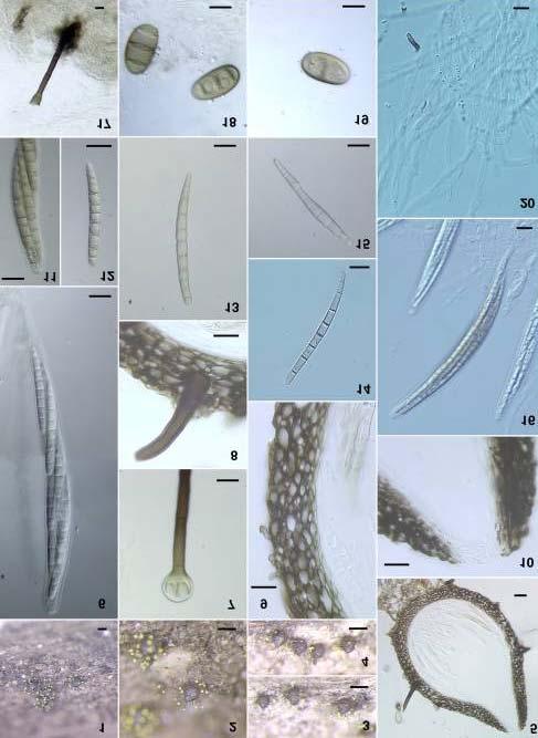
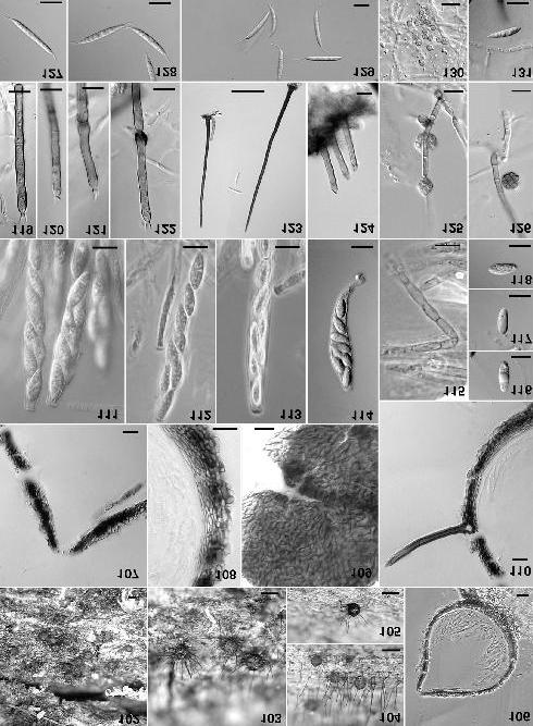
3 Materials and methods Ascomatal contents from specimens were extracted and spread onto water agar plates. After 24 hours, germinating single and/or multiple ascospores were cut out from the medium and transferred to cornmeal agar (CMA) and/or malt-extract agar (MEA) in 6 cm diam. petri plates, and incubated for 30 days at room temp. Data on colony morphology and colony growth rate were recorded for up to three weeks. Colonies were also observed with a dissecting microscope for growth and presence of reproductive structures, particularly anamorphs. Microscopic observations of morphological structures were also made from squash mounts of ascomata. Ascomata were sectioned at 5 µm for light microscopy using the techniques of Huhndorf (1991) and images were captured using bright field (BF), phase contrast (PH) and differential interference microscopy (DIC). Images were captured and photographic plates were produced following the methods of Huhndorf and Fernández (1998). Abbreviations for collectors are SMH = S.M. Huhndorf and FF = F. Fernández. When no collector is listed, the collector s initials are given with the specimen number. All SMH collections are deposited in the Field Museum Mycology Herbarium (F). Latitude and longitude are given in degrees or calculated decimal equivalents. All specimens were collected from decorticated wood unless otherwise noted and dimensions given for the substrates are diameters. Results Chaetosphaeria capitata Sivan. & H.S. Chang, Mycological Research 99: (1995) (Figs. 1-20) Ascomata globose to broadly ovoid, dark brown, µm in diam., µm in height, separate, scattered to gregarious, superficial on the substratum, papillate, with scattered setae, brown, multiseptate, µm, apex capitate, globose to subglobose, 6-14 µm. Yellow pruina covering apices of setae and surface of ascomata. Ascomatal wall opaque in surface view in water, of textura angularis in lactophenol; µm thick in longitudinal section. Ascomatal apex papillate, short. Paraphyses sparse, simple, septate, µm wide. Asci cylindrico-clavate, short-stalked, µm, unitunicate, thin-walled, with a distinctive apical ring, with 8 ascospores irregularly arranged. Ascospores fusiform, with rounded end cells, µm, sub-hyaline to light brown, 7-10 septate. Culture: Seven-day-old colonies of SMH 3239 on CMA 2 mm diam., beige, mycelium dense, floccose, aerial mycelium abundant as tufts, border even, fringed, 17
4 Figs Chaetosphaeria capitata Ascomata on substrate. 5. Longitudinal section through ascoma. 6. Ascus. 7. Apex of ascomal seta. 8. Developing seta attached to ascomal wall. 9. Section through ascomal wall. 10. Section through ascomal neck. 11. Ascus apex Ascospores. 16. Asci. 17. Conidiophore on CMA. 18, 19. Conidia on CMA. 20. Paraphyses. Figs. 1-4 by photomacrography; Figs by DIC. Figs. 1, 2, 5-12, 15, from SMH 3239; Figs. 3, 4, 13, 14, 16, 20 from holotype IMI Bars: 1-4 = 200 µm; 5 = 20 µm; 6-10, = 10 µm; 11 = 5 µm. reverse off-white. Seven-day-old colonies on MEA 4 mm diam., beige, mycelium dense, floccose, aerial mycelium abundant as tufts, border even, fringed, slimy, reverse off-white. Three-week-old colonies on CMA 4 mm diam., white, mycelium dense, appressed, moist, aerial mycelium sparse, border uneven, fringed, reverse white. Conidiophores produced throughout the colony. Three-week-old colonies on MEA 8-12 mm diam., varying from pale gray to black, mycelium dense, cottony-funiculose, moist, aerial mycelium abundant, some isolates developing dark concentric zones, border even, effuse, reverse white/pale brown/black. Conidiophores on CMA, mononematous, macronematous, brown, with few septa, µm at the base, at the apex. Conidiogenous cell a phialide, obclavate, proliferating percurrently, (base) (apex) µm. Conidia on CMA cylindrical to cylindrico-clavate, brown, 3 septate, µm. Anamorph: Exserticlava vasiformis (Matsush.) S. Hughes. Habitat: On decorticated wood Known distribution: Costa Rica, Puerto Rico, Taiwan. Material examined: COSTA RICA, Guanacaste Province, Liberia ACG, Sector Santa María, Estación Biológica, trail to el Bosque Encantado, 750 m, [ , ], 26 June 1997, 5 cm branch, SMH3239. PUERTO RICO, Caribbean National Forest, Luquillo Mts., El Verde Research Area, 16-ha Grid, 350 to 425 m, [ , ], 5 October 1995, on 1 cm branch, SMH TAIWAN. Taichung: Puli, 18 January 1994, on undetermined rotting wood, H.S. Chang (IMI , holotype). Chaetosphaeria chlorotunicata F.A. Fernández & Huhndorf, sp. nov. (Figs ) Etymology: Refers to the outer coating of ascomata. Ascomata ovoidea, tunica viride vel griseo-brunnea, µm diam., µm alta, separata, superficialia, setae sparsis ascomatis brunnea multiseptatae, µm ad base, µm decrescens apicem acutatis. Paries ascomatis cum aspectu superficiaris opacus, sectione longitudinali µm crassus, cellulis pseudoparenchymatis, stratum superficialis 4-10 µm latus. Apicem ascomatis papillatis. Paraphyses simplices, septatae, hyalinae, 2-3 µm latae. Asci cylindrici, µm, octospori. Ascosporae cylindricae vel fusiformes, µm, 7-septatae, interdum 9-septatae, avec cellulas medias brunneas et cellulas terminales hyalinas. 18
5 19
6 20
7 Figs Chaetosphaeria chlorotunicata Ascomata on substrate. 25. Longitudinal section through ascoma. 26. Section through ascomal wall. 27. Section through ascomal neck. 28. Pieces of the ascomal wall showing the outer coating. 29. Paraphyses. 30. Young ascus. 31. Mature ascus Ascospores. 37. Conidiophore on CMA. 38, 39. Conidium attached to a phialide. 40. Conidia and a phialide. 41. Conidium showing basal hilum (arrow). Figs by photomacrography; Figs , by DIC; Fig. 29 by PH. Figs from MO 1038; Figs , 29 from SMH 3483; Figs. 28, 30, 31, 33, 35, 36 from SMH 3136, Fig. 32 from SMH , Figs. 34, from holotype SMH Bars: = 200 µm; 25, 28 = 20 µm; 26, 27, 29-36, = 10 µm; 37 = 50 µm. Ascomata ovoid, with greenish coating turning gray with age, µm in diam., µm in height, separate, superficial, papillate, with sparse, scattered setae, dark brown, multiseptate, µm long, µm at the base, tapering to µm at the apex. Ascomatal wall opaque in surface view, inner wall pseudoparenchymatous, dark brown, µm wide, outer coating 4-10 µm wide. Ascomatal apex papillate. Paraphyses sparse, simple, septate, µm wide. Asci cylindrical, short-stalked, µm, unitunicate. Ascospores cylindrical-fusiform, mostly inequilateral, µm, mostly 7-septate, sometimes 9-septate, dark brown, terminal cells hyaline and short. Culture: One-week-old colonies on MEA 1 mm diam., white, mycelium dense appressed, aerial mycelium abundant, reverse white. No measurable growth on CMA. Two-week-old colonies on MEA 9 mm diam., white with a gray center, mycelium dense appressed, aerial mycelium sparse, border even, reverse white with dark gray center. Two-week-old colonies on CMA 4 mm diam., white, mycelium dense appressed, aerial mycelium sparse, border even, reverse white. Conidiophores mononematous, macronematous, dark brown, on the substrate µm for most of their length, widening to µm at the apex; on CMA µm long, up to 650 µm, µm at the base, broadening to a single apical, funnel-shaped phialide, µm wide. Conidia cylindrical, brown, thick-walled, distoseptate, µm on the substrate, µm on CMA, with a distinctive basal hilum, centric or slightly eccentric. Anamorph: Exserticlava S. Hughes (Hughes, 1978) Habitat: On decorticated wood and palm petioles Known distribution: Costa Rica, Jamaica, Panama, Puerto Rico. Material examined: COSTA RICA, Alajuela Province, Cantón Upala, Bijagua, Alto los Brenes, [10.73, -854], 6 June 1999, I. Lopez IL473 (INB); Guanacaste Province, Cantón Cañas, Hacienda Montezuma, 715 m, [ , ], 7 July 2000, on 2 cm branch, FAF with G.M. Mueller, B. Strack, J.P. Schmit, L. Umaña SMH4258; Area de Conservación Tempisque, Puntarenas Province, Nicoya peninsula, Reserva Absoluta Cabo Blanco, [9.59, -85.1], 13 August 2000, Cabuya station, Sueco trail, M. Oses MO1038 (INB); Puntarenas Province, Area de Conservación Osa, Parque Nacional Corcovado, Sirena Station, Las Ollas trail, 10 m, 21
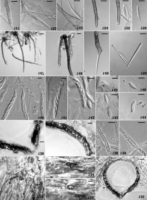
8 [8.4806, ], 16 July 2000, on wood fragment, FAF, with G.M. Mueller, B. Strack, J.P. Schmit, L. Umaña SMH4276. JAMAICA, Trelawny Parish, 1.5 miles beyond the village of Crown Lands, 610 m, [ , ], 10 June 1999, on wood fragment, FAF SMH4062; Winsor Trail, 115 m, [ , ], 12 June 1999, on wood fragment, FAF SMH4076. PANAMA, Barro Colorado Island National Monument, Thomas Barbour trail, 50 to 150 m, [9.1667, ], 18 September 1997, on 3 cm branch, SMH, FAF, SMH3483; on petiole, SMH3487; Snyder-Molino trail, 19 September 1997, on 1 cm branch, SMH, FAF, SMH PUERTO RICO, Caribbean National Forest, El Verde Research Area, 16-ha Grid, Luquillo Mts., 350 to 425 m, [ , ], 25 September 1995, on log, S.M. Huhndorf SMH1565 (F; holotype designated here); 25 September 1995, on 30 cm log, SMH1580; 29 January 1996, on 12 cm log, SMH2094; 20 January 1997, on palm petiole, SMH, FAF, SMH3074; 25 January 1997, on 5 cm branch, SMH, FAF, SMH3136. Chaetosphaeria conirostris F.A. Fernández & Huhndorf, sp. nov. (Figs ) Etymology: Refers to the conical shape of the ostiolar beak Ascomata obpyriformis, atrobrunnea, µm lata, µm alta, solitaria, superficialia, setae sparsis ascomatis brunnea multiseptatae µm, apicem capitatus, hyalino, altus 7-11 µm latus, obtectus substantia hyalina aut ochracea. Setae adsunt super substrata. Paries ascomatis cum aspectu superficiaris opacus in aqua ut textura angularis in lactophenol, sectione longitudinali µm crassus, cellulis pseudoparenchymatis. Collis ascomatis conicus, µm ad base, decrescens ad apicem µm latus, papillatus, albidus, plectenchymatus. Paraphyses sparse, simplices, septatae, hyalinae, (7.1) µm latae. Asci cylindrico-clavati, brevi pedicellati µm, unitunicatae, annuli apicales tenues, octospori, ascosporae dispositio irregulares. Ascosporae cylindricae vel fusiformes, µm, hyalinae, plerumque uniseptatae, aut triseptatae, inequilaterales, rectae vel leviter curvatae. Ascomata obpyriform, dark brown, µm in diam., µm in height, separate, superficial, with sparse setae, brown, multiseptate, µm, and a capitate apex, µm, covered with a hyaline to dark yellow droplet. Capitate setae are also present on the substrate. Ascomatal wall in surface view, opaque in water, textura angularis in lactophenol, µm thick in longitudinal section, composed of pseudoparenchymatic cells. Ascomatal beak, conical, µm at the base, tapering to µm, papilla off-white, plectenchymatous. Paraphyses sparse, unbranched, septate, (7.1) µm wide. Asci cylindrico-clavate, short-stalked, µm, unitunicate, thin-walled, thin apical ring, with 8 ascospores irregularly arranged. Ascospores cylindrico-fusiform, hyaline, mostly oneseptate, sometimes three-septate, µm, inequilateral, straight to slightly curved. Culture: Seven day old colonies on CMA 3 mm in diam., light brown, mycelium mostly immersed, superficial mycelium sparse, floccose, margin effuse, reverse white. Twenty-one day old colonies on CMA 16 mm in diam., brown, zonate and lighter colored at the edges, mycelium mostly immersed, superficial mycelium sparse, subhyaline, margin effuse, 22

9 Figs Chaetosphaeria conirostris. 42, 43. Ascomata on substrate. 44. Longitudinal section through ascoma. 45. Section through ascomal wall. 46. Section through ascomal neck. 47. Ascus apex and ascospore. 48. Ascomal seta. 49, 50. Paraphyses. 51. Asci. 52. Ascus showing apical ring. 53, 54. Ascospores. 55, 60. Phialide and conidium on CMA. 56, 57, 59. Phialides on CMA. 58. Conidium on CMA. Figs. 42, 43 by photomacrography; Figs. 45, 46, 48, 49, by DIC; Figs. 44, 46, 47, 50, 51 by PH. All figures from holotype SMH Bars: 42, 43 = 200 µm; 44-46, 51 = 20 µm; 47-50, = 10 µm. 23
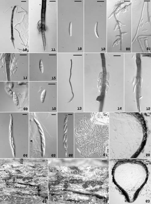
10 Figs Chaetosphaeria lateriphiala 61, 62. Ascomata on substrate. 63. Longitudinal section through ascoma. 64. Young ascus. 65, 66. Asci. 67. Paraphyses. 68. Section through ascomal wall. 69, 70, 72. Ascospores. 71. Ascospore from natural substrate showing microcycle conidiation. 73. Conidiophore on substrate. 74, 75. Lateral phialides on conidiophores from natural substrate. 76, 77. Conidiophore and lateral arrangement of phialides, on CMA. 78, 79. Conidia from natural substrate 80. Degenerate phialides on CMA. 81. Conidia on CMA. Figs. 61, 62 by photomacrography; Figs , by DIC; Fig. 67 by PH. Figs. 61, 62, 64, 65, 67, 70, 72 from SMH 3294; Figs. 63, 66, 68, 69, 76, 77, 80, 81 from SMH ; Figs. 71, 73-75, 78, 79 from SMH Bars: 61, 62 = 200 µm; 63, 67, 76 = 20 µm; 64-66, 68-72, 74-75, = 10 µm; 73 = 50 µm. reverse off-white. Conidiophores absent or short, multiseptate, cylindrical, light brown. Phialides terminal, single, subhyaline to light brown, mostly clavate, µm at the base, at the widest point; collarette absent or, when present, hyaline, obconical, very-thin walled, at the base, flaring to at the apex. Proliferation of conidiogenous cells was not observed in culture. Conidia hyaline, oblong to reniform, one-celled, µm. A few conidia are limoniform, one-celled, µm. Anamorph: It resembles Craspedodydimum Hol.-Jech. (Holubová- Jechová, 1972), observed only in culture. Habitat: On decorticated wood. Known distribution: Costa Rica, Ecuador. Material examined: COSTA RICA, Limón Province, Cantón Limón, Area de Conservación la Amistad Caribe, Reserva Biológica Hitoy Cerere, [9.67, -833], 28 April 2000, tepezcuintle trail, M. Umaña MU1001 (INB); Puntarenas Province, Las Cruces Biological Station, San Vito, Rio Jaba trail, 1050 m, [8.7858, ], 5 May 1996, on 5 cm branch, SMH, FAF, SMH2183 (F; holotype designated here); Area de Conservación la Amistad Pacífico, Cantón Coto Brus, fila Cedro, [8.92, ], 5 June 2001, finca Cafrosa, E. Navarro EN3295 (INB); [94, ], 19 August 2001, Pittier station, E. Navarro EN3716 (INB); Cantón Buenos Aires, Altamira research station, Los Arboles Gigantes trail, [94, -832], 29 June 2002, on wood fragment, FAF1027; trail to Valle del Silencio, [94, ], 2 July 2002, on wood fragment, FAF1061. ECUADOR, Orellana Province, Yasuni National Park, Botanico trail, [-.6713, ], 5 March 2001, on wood fragment, FAF, A.N. Miller, R. Briones, SMH4332; on 30 cm log, SMH4338. Chaetosphaeria lateriphiala F.A. Fernández & Huhndorf, sp. nov.(figs ) Etymology: Refers to the lateral arrangement of phialides on conidiophores. Ascomata globosa vel obpyriformis, atrobrunnea, µm lata, µm alta, solitaria vel aggregata, superficialia vel leviter immersa, papillata. Paries ascomatis cum aspectu superficiaris opacus in aqua et in lactophenol, sectione longitudinali µm crassus, cellulis pseudoparenchymatis, extimus stratum 2-5 µm latus, ad 15 µm latus base collis. Collis ascomatis papillatis. Paraphyses simplices, hyalinae, µm latae. Asci cylindrici, brevi 24
11 25
12 pedicellati, µm, unitunicatae, annuli apicales µm lati, µm alti, octospori, ascosporae dispositio irregulares. Ascosporae fusiformes, µm, hyalinae, plerumque triseptatae, aut uniseptatae, interdum constrictae. Ascomata globose to broadly obpyriform, dark brown, µm wide, µm high, glabrous, scattered to gregarious, superficial to slightly immersed in the substratum. Ascomatal wall opaque in surface view in water, also opaque in lactophenol, µm thick in longitudinal section, composed of pseudoparenchymatic cells, with an opaque outermost layer, 2-5 µm wide, up to 15 µm thick at base of the neck. Ascomatal apex, papillate. Paraphyses simple, hyaline, µm wide. Asci cylindrical, short-stalked, µm, unitunicate, apical ring present, µm wide, µm deep, with 8 ascospores irregularly arranged. Ascospores fusiform to broadly fusiform, with rounded end cells, µm, hyaline, often threeseptate, sometimes one-septate, sometimes constricted. Culture: Isolates from SMH and SMH 3294 showed no observable growth on CMA or MEA after seven days. Three-week-old colonies on CMA 2 mm diam., dark brown, mycelium mostly immersed, aerial mycelium sparse, silky, border uneven, effuse, reverse dark brown. Three-week-old colonies on MEA 6 mm diam., dark gray-brown, aerial mycelium dense, light gray, border uneven, effuse, reverse dark brown. Three-week-old colonies of SMH 3294 on MEA 3 mm diam., dark reddish-dark brown, mycelium woolly, aerial mycelium sparse, border uneven, reverse reddish-dark brown. Conidiophores mononematous, macronematous, dark brown, becoming lighter in color towards the apex, distinctively multiseptate, µm µm at base, tapering to a hyaline apical cell, µm on MEA; on the substrate: µm at base and tapering towards the apex, sometimes branched, branches µm. Phialides ampulliform, light brown, on MEA: µm at the widest point; on the substrate: µm at the widest point, at single, one or more apical conidiogenous openings, µm wide. Phialides are sessile, originating from the uppermost part of the conidiophore cell, typically arranged in whorls. In culture, the number of phialides in whorls diminishes with successive subculturing until only single phialides and extremely reduced conidiophores are produced directly on vegetative hyphae. Conidia one celled, fusiform, hyaline, µm on MEA, µm on the substrate. Anamorph: It resembles Zanclospora S. Hughes & W.B. Kendr. (Hughes and Kendrick, 1965). Habitat: On decorticated wood. Known distribution: USA (Indiana, North Carolina, Wisconsin). Material examined: USA, Indiana, Lawrence Co., Hoosier National Forest, Hickory Ridge trailhead, 26 July 1996, on 10 cm branch, SMH (F; holotype designated here); 26
13 NORTH CAROLINA, Macon Co., Highlands Biological Station, Highlands, 20 July 1997, on wood fragment, FAF, SMH 3294; Jackson Co., Panthertown Valley, [ , ], 22 July 1997, on wood fragment, FAF, SMH 3320; WISCONSIN, Green Co., New Glarus State Park, [42.71, ], 13 September 1995, on 20 cm log, SMH Chaetosphaeria lignomollis F.A. Fernández & Huhndorf, sp. nov. (Figs ) Etymology: Refers to the soft wood on which it occurs. Ascomata ovoidea vel globosa, atrobrunnea, µm lata, µm alta, solitaria, superficialia, setae sparsi vel pauci, brunnea, multiseptatae, µm, decrecens apicem tenuis. Paries ascomatis cum aspectu superficiaris ut textura angularis, sectione longitudinali µm crassus, cellulis pseudoparenchymatis. Apicem ascomatis papillatis. Paraphyses simplices, septatae, hyalinae, 2-3 µm latae. Asci cylindrici, µm, octospori, cum ascosporae distichae. Ascosporae cylindricae vel fusiformes, µm, hyalinae, 7-septatae. Ascomata broadly ovoid to obpyriform, dark brown, µm in diam., µm in height, separate, superficial on the substratum, with a few, scattered setae, brown, multiseptate, slender, µm, tapering to an attenuated apex. Ascomatal wall of textura angularis in surface view, µm thick in longitudinal section, composed of pseudoparenchymatic cells. Ascomatal apex broad, papillate, short. Paraphyses unbranched, septate, hyaline, 2-3 µm wide. Asci cylindro-clavate, shortstalked, µm, unitunicate, thin-walled, with 8 biseriately arranged ascospores, apical ring µm wide, µm deep. Ascospores cylindrical-fusiform, sometimes inequilateral, sometimes one end slightly curved, µm, hyaline to subhyaline, seven-septate, septa sometimes diagonal or at angles. Culture: No visible growth on CMA after seven days. Twenty-one day old colonies on CMA 2.5 mm in diam., light brown, mostly immersed, with knots of dark brown cells, reverse brown. Conidiophores produced singly on CMA, in fascicles of three or more on the substrate, unbranched, dark brown becoming light brown towards the apex, multiseptate, µm on CMA, µm on the substrate. Conidiogenous cell a phialide, on CMA: cylindrical, µm, with either a single apical collarette, obconical, µm wide and µm deep, on the substrate: a polyphialide with a single apical collarette and multiple lateral old conidiogenous loci that appear as refractive pegs. Proliferation mostly sympodial; percurrent proliferation was also observed. Conidia hyaline, cylindrical, broadly rounded at the apex, with a truncate base, 3-6 septa, straight or diagonal, unevenly spaced, µm on CMA, on the substrate. 27
14 Figs Chaetosphaeria lignomollis. 82, 83. Ascomata on substrate. 84. Longitudinal section through ascoma. 85. Paraphyses. 86. Section through ascomal wall. 87. Section through ascomal neck Asci Ascospores. 94. Conidiophore on CMA. 95. Conidiophore showing sympodial proliferation of the conidiogenous cell on CMA. 96. Conidiophore and conidia on CMA. 97. Conidium on CMA. 98, 99. Conidia from the natural substrate. 100, 101. Conidiophore showing polyphialides, from the natural substrate. Figs. 82, 83 by photomacrography; Figs by DIC; Figs. 84, 85 by PH. Figs , 86, 87, 90, 92 from SMH 1829; Figs. 85, 88, 89, 91, 93, from holotype SMH 3015; Figs. 98, 101 from SMH 2888; Figs. 99, 100 from SMH Bars: 82, 83 = 200 µm; 84 = 20 µm; = 10 µm. Anamorph: It resembles Kylindria DiCosmo, S.M. Berch & W.B. Kendr. (DiCosmo et al., 1983). Conidiophores and conidia are present on the substrate in collections SMH1642 and SMH2888. Habitat: On decorticated wood. Known distribution: Costa Rica, Puerto Rico. Material examined: COSTA RICA, Guanacaste Province, Parque Nacional Guanacaste, Santa Cecilia, Sector Pitilla, 700 m, [ , ], 23 June 1997, on 5 cm branch, SMH3209. PUERTO RICO, Caribbean National Forest, El Verde Research Area, 16-ha Grid, Luquillo Mts., 350 to 425 m, [ , ], 16 January 1997, on wood fragment, S.M. Huhndorf, F.A. Fernández, SMH3015 (F; holotype designated here); 29 September 1995, on 1 cm branch, SMH1642; 9 October 1995, on 30 cm log, SMH1829; 14 January 1996, on 25 cm log, SMH1883; 12 January 1997, on 7.5 cm root, SMH, FAF, SMH2888. Chaetosphaeria longiseta F.A. Fernández & Huhndorf, sp. nov.(figs ) Etymology: Refers to the long setae borne on the ascomata. Ascomata globosa vel ovoidea, atrobrunnea, µm lata, µm alta, solitaria, superficialia, papillata; setae sparsi vel pauci, brunnea, multiseptatae, µm ad base, µm decrescens apicem acutatis. Paries ascomatis cum aspectu superficiaris ut textura epidermoidea in aqua et in lactophenol, sectione longitudinali 9-15 µm crassus, cellulis pseudoparenchymatis, tunica extima tenuissime pallida. Apicem ascomatis papillatis, brevis. Paraphyses simplices, septatae, hyalinae, µm latae. Asci unitunicati, cylindricoclavati, brevi pedicellati, µm, octospori. Ascosporae hyalinae, fusiformes vel ellipsoideae, non-septatae vel uniseptatae, µm. Ascomata subglobose to broadly ovoid, dark brown, µm in diam., µm in height, separate, superficial, papillate; setae sparse, scattered, brown, multiseptate, slender, µm at the base, µm for most of its length, tapering to an acute apex. Ascomatal wall of textura epidermoidea in surface view in water, and in lactophenol, 9-15 µm thick in longitudinal section, composed of pseudoparenchymatic cells, with a thin, light-colored outer coating. Ascomatal apex papillate, acute, short. Paraphyses sparse, unbranched, hyaline, septate, µm wide. Asci cylindro-clavate, short-stalked, µm, unitunicate, thin-walled, broad apical cap, with 8 ascospores irregularly arranged. Ascospores hyaline, broadly fusiform to 28
15 29
16 Figs Chaetosphaeria longiseta Ascomata on substrate Longitudinal section through ascoma Section through ascomal neck Section through ascomal wall Surface view of ascomal wall Section through ascomal wall showing seta. 111, 112. Asci Ascus showing apical ring Ascus Paraphysis Ascospores Conidiophores with phialides and percurrent proliferations on CMA Setae associated with conidiophore on the substrate Conidiophores (polyphialides) from the substrate Conidiophore showing percurrent proliferations and clusters of microconidia on CMA Cluster of microconidia on CMA Conidia from natural substrate Microconidia on CMA Aberrant conidium after repeated culturing on CMA. Figs by photomacrography; Figs , 114, by DIC; Figs. 106, 112, 113, 115 by PH. Figs. 102, 103 from SMH 2316; Figs , , 115, , from holotype SMH 3048; Figs. 109, 116, 125, 126, 130 from SMH 3854; Figs. 114, 117, 131 from SMH Bars: = 200 µm; , = 10 µm; 123 = 50 µm. ellipsoid, rounded ends, nonseptate, rarely one-septate, µm. Culture: Colony on CMA 7 mm in diam. after seven days. Twenty-one day old colonies on CMA 20 mm in diam., white, mostly immersed, reverse white or light brown, surface mycelium floccose, margins effuse, conidiophores produced abundantly. Conidiophores single, brown, multiseptate, µm on CMA, µm on the substrate. Conidiogenous cells cylindrical, µm on CMA, with a single, tubular, apical collarette, µm, proliferating percurrently, or phialides with multiple, conidiogenous loci, resulting from sympodial proliferation, often ending in an apical collarette, µm, exclusively found on the substrate. Setae associated with conidiophores, multiseptate, brown, tapering to an acute apex, µm at the base, sometimes two most apical cells dark brown. Conidia of two morphologically distinct types produced from the same phialides on CMA: one celled, ellipsoid, hyaline to light-brown, µm; and one-celled, hyaline, narrowly fusiform, µm, with a single setula at each end, µm. The small conidia are produced in tight, mucilagenous clusters and remain attached after the conidiogenous cell proliferates, appearing as a series of intercalary masses on the conidiophore. On the substrate, conidia hyaline, fusiform, µm, with a single setula at each end, µm. Anamorph: It resembles Dictyochaeta Speg. (Spegazzini, 1923). Habitat: On decorticated wood of twigs and branches. Known distribution: Costa Rica, Ecuador, Puerto Rico, USA (South Carolina). Material examined: COSTA RICA, Puntarenas Province, Area de Conservación la Amistad Pacífico, Cantón Coto Brus, Zona Protectora Tablas, fila Cedro, [8.91, ], 27 June 2002, on wood fragment, F.A. Fernández FAF1016; ECUADOR, Orellana Province, 30
17 31
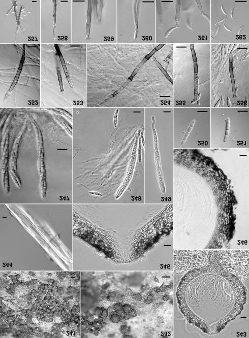
18 Figs Chaetosphaeria luquillensis Ascomata on substrate Longitudinal section through ascoma Section through ascomal neck Section through ascomal wall. 138, 139. Paraphyses Asci Ascospores Conidiophores and setae from natural substrate Polyphialide from natural substrate Phialide and a developing conidium on CMA Conidia from natural substrate Developing phialide on CMA Phialide and developing conidium on CMA Conidium on CMA. 154, 155. Proliferating phialide on CMA Conidium on CMA. Figs by photomacrography; Figs , by DIC; Figs. 138, 139 by PH. All figures from holotype SMH Bars: = 200 µm; 135, 147 = 20 µm; , = 10 µm. Yasuni National Park, Botanico trail, [-.6713, ], 5 March 2001, on 1.5 cm branch, FAF, A.N. Miller, R. Briones, SMH4335; Bariso trail, 7 March 2001, on branch, FAF, A.N. Miller, R. Briones, SMH4380. PUERTO RICO, Caribbean National Forest, El Verde Research Area, 16-ha Grid, Luquillo Mts., 350 to 425 m, [ , ], 18 January 1997, on 1 cm branch, S.M. Huhndorf, F.A. Fernández, SMH3048 (F; holotype designated here); 4 October 1995, on 40 cm log, SMH1725. USA, South Carolina, Oconee Co., Sumter National Forest, Walhalla State Fish Hatchery, left trail through pine area to East Fork Trail, [ , ], 31 July 1998, on 10 cm branch, FAF, SMH3854. Chaetosphaeria luquillensis F.A. Fernández & Huhndorf, sp. nov. (Figs ) Etymology: Refers to Luquillo, name of collection locality in Puerto Rico. Ascomata ovoidea, atrobrunnea, µm lata, µm alta, solitaria, superficialia vel leviter immersa, papillata, setae sparsi vel pauci, atrobrunnea, multiseptatae, apicem tenuissimae. Paries ascomatis cum aspectu superficiaris opacus in aqua et textura epidermoidea in lactophenol, sectione longitudinali µm crassus, cellulis pseudoparenchymatis. Apicem ascomatis papillatis, acutatus, breve. Paraphyses simplices, hyalinae, septatae. Asci unitunicati, cylindrico-clavati, brevi pedicellati, µm, annulo apicali tenuis, octospori. Ascosporae hyalinae, fusiformes, µm, inaequalis, 1-septatae, interdum 2-3-septatae, tunica gelatinosa praeditae. Ascomata broadly ovoid, dark brown, µm in diam., µm in height, separate, superficial to partly immersed, papillate, with sparse, scattered setae, light brown, multiseptate, slender, tapering to an acute apex. Ascomatal wall in surface view, opaque in water, textura epidermoidea in lactophenol, µm thick in longitudinal section, composed of pseudoparenchymatic cells. Ascomatal apex papillate, acute, short. Paraphyses sparse, simple, septate. Asci unitunicate, cylindro-clavate, short-stalked, µm, firm-walled, thin apical cap, with 8 ascospores irregularly arranged. Ascospores hyaline, fusiform, µm, sometimes inequilateral, sometimes ends curved opposite directions, mostly one-septate, sometimes two or three-septate, covered with a gelatinous sheath. Culture: Colony on CMA 8 mm in diam. after seven days. Twenty-one day old colonies 32
19 33
20 on CMA 16 mm in diam., light brown, mostly immersed, reverse light brown, surface mycelial growth dense, margins feathery. Conidiophores single, light brown, cylindrical, mostly multiseptate, µm on CMA, µm on the substrate. Conidiogenous cell a phialide, cylindrical, most often proliferating sympodially to produce multiple lateral conidiogenous loci, sometimes proliferating percurrently, µm on CMA, µm on the substrate. Setae singly on the substrate, multiseptate, light brown, µm, tapering to a rounded apex, µm wide. Conidia obclavate, mostly bent and rounded at apex on CMA, straight and acute at apex on the substrate, hyaline, µm on CMA, µm on the substrate. Anamorph: Dematiaceous phialidic. Habitat: On decorticated wood of dead trunks. Known distribution: Puerto Rico. Material examined: PUERTO RICO, Caribbean National Forest, El Verde Research Area, 16-ha Grid, Luquillo Mts., 350 to 425 m, [ , ], 15 January 1997, on 30 cm log, S.M. Huhndorf, F.A. Fernández SMH2973 (F; holotype designated here). Chaetosphaeria minuta F.A. Fernández & Huhndorf, sp. nov. (Figs ) Etymology: Refers to the small size of ascomata. Ascomata globosa vel subglobosa, atrobrunnea, µm lata, µm alta, solitaria vel aggregata, superficialia, propria papillata. Paries ascomatis cum aspectu superficiaris opacus in aqua et textura angularis in lactophenol, sectione longitudinali µm crassus, cellulis pseudoparenchymatis. Apicem ascomatis papillatis, opacus. Paraphyses simplices, septatae, hyalinae, µm latae, tenuis apicem. Asci cylindrico-clavati, brevi pedicellati, µm, unitunicati, octospori. Ascosporae hyalinae, fusiformes vel ellipsoideae, µm, uniseptatae. Ascomata globose to sub-globose, dark brown, µm in diam., µm in height, solitary to densely gregarious, superficial on a thin subiculum, distinctly papillate. Ascomatal wall in surface view, opaque in water, textura angularis in lactophenol, µm thick, composed of pseudoparenchymatic cells. Ascomatal apex papillate, opaque. Paraphyses unbranched, septate, µm wide, tapering. Asci cylindro-clavate, shortstalked, µm, unitunicate, thin-walled, apical ring not observed, with 8 ascospores irregularly arranged. Ascospores hyaline, fusiform to narrow-ellipsoid, µm, one-septate. Culture: Seven-dayold colonies on CMA 6 mm diam., light to dark gray, mycelium appressed, mostly immersed, aerial mycelium sparse, abundant conidiophores throughout, developing concentric zones, border even, fringed, reverse gray. Seven-day-old colonies on MEA 10 mm diam., off-white, appressed, aerial mycelium sparse, border even, fringed, reverse off-white.three-week-old colonies on CMA 12 mm diam., pale gray-brown, mycelium mostly immersed, aerial mycelium 34

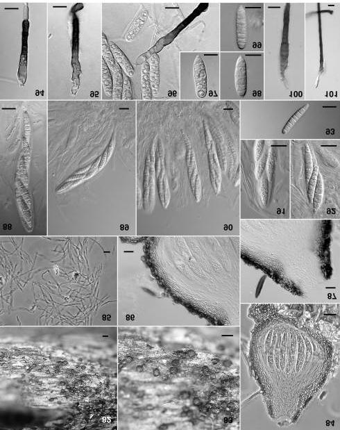
21 Figs Chaetosphaeria minuta. 157, 158. Ascomata on substrate Longitudinal section through ascoma. 160, 161. Asci Ascospores Section through ascomal wall Conidiophore from natural substrate Lateral phialide with conidium, from natural substrate Conidiophores on CMA Conidiophore apex showing terminal phialide with conidium, from CMA Apex of conidiogenous cell showing multiple collarettes, from natural substrate Conidium from natural substrate Conidia on CMA Apex of conidiophore showing terminal phialide, on CMA Bases of conidiophores showing lateral phialides on CMA. Figs. 157, 158 by photomacrography; Figs by DIC. All figures from holotype SMH Bars: 157, 158 = 200 µm; = 10 µm. 35
22 sparse, developing concentric zones, border fringed, reverse light brown agar. Conidiophores and conidia produced abundantly throughout the colony. Threeweek-old colonies on MEA 18 mm diam., white, mycelium mostly immersed, appressed, border uneven, effuse, lobed, reverse white. Conidiophores produced in center, on the old agar block. Conidiophores single, unbranched, multiseptate, brown, on CMA: µm at the base, tapering to an apical phialide, µm, several unilateral phialides produced along the midsection; on the substrate µm at the base, narrowing to µm in width. Conidiogenous cells are phialides, ovoid, brown, produced along the conidiophore midsection, on CMA µm at the widest point, collarettes small, funnel-shaped, µm wide at the apex, µm deep; on the substrate µm wide, with several collarettes in a sympodial arrangement, or sometimes with percurrent proliferations. Conidia narrow fusiform, hyaline, one-celled, µm on CMA, µm on the substrate. Anamorph: It resembles Chaetopsis Grev. Habitat: On decorticated wood of branches on the ground. Known distribution: Panama. Material examined: PANAMA, Barro Colorado Island National Monument, Fausto trail, 50 to 150 m, [9.1667, ], 15 September 1997, on 4 cm branch, S.M. Huhndorf, F.A. Fernández SMH3396 (F; holotype designated here). Chaetosphaeria spinosa F.A. Fernández & Huhndorf, sp. nov. (Figs ) Etymology: Refers to the spiny appearance of ascomata. Ascomata globosa vel ovoidea, atrobrunnea, µm diametro, µm alta, solitaria, superficialia, abundans setae brunnea, µm. Paries ascomatis cum aspectu superficiaris textura epidermoidea in aqua et in lactophenol, sectione longitudinali µm crassus. Paraphyses simplices, septatae, hyalinae, µm latae. Asci cylindrico-clavati, brevi pedicellati, µm, unitunicati, octospori. Ascosporae hyalinae, filiformes, µm, multiseptatae. Ascomata globose to ovoid, dark brown, µm in diam., µm in height, separate, superficial on the substratum, setae abundant, rigid, dark brown, opaque, µm, wider at the base, tapering toward apex. Ascomatal wall of textura epidermoidea in surface view in water, and in lactophenol, µm thick in longitudinal section. Paraphyses unbranched, hyaline, septate, µm wide. Asci cylindrical-clavate, short-stalked, µm, unitunicate, thin-walled, with 8 ascospores, apical ring distinctive, 2-3 µm wide, 1-2 µm high. Ascospores filiform, sometimes slightly bent, µm, hyaline, nonseptate, with numerous guttules. Culture: No measurable growth either on CMA or MEA after seven days. Three-weekold colonies on CMA 6 mm diam., white, mycelium mostly immersed, aerial mycelium sparse, appressed, border even, reverse white Three-week-old 36

23 Figs Chaetosphaeria spinosa. 176, 177. Ascomata on substrate Longitudinal section through ascoma Surface view of the ascomal wall Ascomal seta. 181, 182. Longitudinal sections through ascomal walls Ascus apex showing apical ring. 184, 185. Asci. 186, 187. Ascospores Phialide with a cluster of conidia on CMA Phialide on CMA Conidia on CMA. Figs. 176, 177 by photomacrography; Figs by DIC. All figures from holotype SMH Bars: 176, 177 = 200 µm; = 10 µm. 37
24 colonies on MEA 10 mm diam., white, aerial mycelium densely funiculose in center, appressed elsewhere, border even, reverse white. Conidiophores semimacronematous on CMA. Conidiogenous cell a phialide, ampulliform, hyaline, µm, with a funnel-shaped collarettes, µm deep, µm at opening. Conidia one-celled, ellipsoid to globose, hyaline, µm, in clusters at tips of phialides. Anamorph: Phialidic. Habitat: On decorticated wood (birch). Known distribution: USA (North Carolina, Texas) Material examined: USA, North Carolina, Macon Co., Whiteside Mountain, Highlands, 1000 m, [35192, ], 7 October 1996, on inside surface of peeling birch bark, S.M. Huhndorf, F.A. Fernández, with Q.X. Wu, J.C. Wei, G.M. Mueller, SMH2754 (F; holotype designated here); TEXAS, Hardin Co., Texas Nature Conservancy Roy Larson Sandyland Sanctuary, ca. 10 mi N of Beaumont, 10 June 2000, on 20 cm birch log, A.N. Miller, SMH4232. Chaetosphaeria sylvatica F.A. Fernández & Huhndorf, sp. nov.(figs ) Etymology: Refers to its common occurrence in tropical forests. Ascomata ovoidea vel obpyriformis, atrobrunnea, µm lata, µm alta, solitaria, superficialia, aggregata, papillata, sparsim setosa. Paries ascomatis cum aspectu superficiaris opacus in aqua et in lactophenol, sectione longitudinali µm crassus, cellulis pseudoparenchymatis. Apicem ascomatis papillatis, cum collis, ad 33 µm altis, ad 60 µm basis latis. Paraphyses simplices, septatae, hyalinae, in matrix gelatineus. Asci cylindrici, brevi pedicellati, µm, unitunicati, tenuis paries, tennuis annulus apicalis, octospori. Ascosporae fusiformes, hyalinae, µm, 3-septatae. Ascomata broadly ovoid to obpyriform, dark brown, µm in diam., µm in height, separate, superficial, solitary to gregarious, sometimes sparsely setose. Ascomatal wall in surface view opaque in water and in lactophenol, µm thick in longitudinal section, composed of pseudoparenchymatic cells. Ascomatal apex papillate, beaked, up to 33 µm in length, 60 µm at the base. Paraphyses sparse, simple, septate, hyaline, embedded in a gel matrix. Asci cylindrical, short-stalked, µm, unitunicate, thin-walled, shallow apical cap, with 8 ascospores irregularly arranged. Ascospores fusiform, hyaline, µm, three-septate. Culture: Colony on CMA 7 mm in diam. after seven days. Twenty-one day old colonies on CMA 31 mm in diam., grayish black, mostly immersed, reverse black, surface mycelial growth sparse, grayish, margins effuse. Conidiophores semi-macronematous, black. Conidiogenous cell a phialide, cylindrical to narrowly lageniform, terminal, (base) (venter) (apex) µm, collarettes inconspicuous or absent. Conidia cylindrical to clavate, hyaline, µm on CMA. Anamorph: It resembles Phaeostalagmus W. Gams. Habitat: On decorticated wood. 38

25 Figs Chaetosphaeria sylvatica. 191, 192. Ascomata on substrate Longitudinal section through ascoma Surface view of the ascomal wall in lactophenol Section through ascomal neck Section through ascomal wall. 197, 198. Asci Paraphyses 200, 201. Ascospores Ascus apex showing apical ring Conidiophores and conidia from CMA. Figs. 191, 192 by photomacrography; Figs by DIC; Fig. 193 by PH. Figs. 191, 193, 195, 196 from SMH 1319; Figs. 192, 194, 197, 200, from holotype SMH 2893; Figs. 199, 201, 202 from SMH 1909; Fig. 198 from SMH Bars: 191, 192 = 200 µm; 193 = 20 µm; = 10 µm. 39
26 Known distribution: Jamaica, Puerto Rico. Material examined: JAMAICA, Trelawney Parish, Winsor Trail, 115 m, [ , ], 13 June 1999, on wood fragment, FAF, with T. Armstrong, T. Barroni, S. Cantrell, T. Commock, K.H. Larssen & L. Ryvarden, SMH4081. PUERTO RICO, Caribbean National Forest, El Verde Research Area, 16-ha Grid, Luquillo Mts., 350 to 425 m, [ , ], 12 January 1997, on 30 cm log, S.M. Huhndorf, F.A. Fernández, SMH2893 (F; holotype designated here); 27 April 1995, on wood fragment, SMH1173; 4 May 1995, on 1 cm twig, SMH1319; 16 January 1996, on 4 cm branch, SMH1909; Caribbean National Forest, El Yunque Road, La Coca trail, Luquillo Mts., [18.283, ], 10 June 1998, on wood fragment, FAF, A.N. Miller, SMH3799. Chaetosphaeria tropicalis F.A. Fernández & Huhndorf, sp. nov.(figs ) Etymology: Refers to its common occurrence in the tropics. Ascomata globosa vel subglobosa, atrobrunnea fulgens cinerascens, µm lata, µm alta, superficialia, papillata, dense aggregata, asperum, abundans setae brunnea multiseptatae sinuosa apicem obtusum ab basi ascomatis. Paries ascomatis cum aspectu superficiaris ut textura angularis in aqua et in lactophenol, sectione longitudinali µm crassus, cellulis pseudoparenchymatis. Apicem ascomatis papillatis, breve. Paraphyses simplices, septatae, hyalinae, µm latae. Asci cylindrico-clavati, brevi pedicellati, µm, unitunicati, tennuis annulus apicalis, octospori. Ascosporae fusiformes, hyalinae, µm, 3-septatae, curvatura minima distinguibilis. Ascomata globose to subglobose, dark brown with a gray luster, µm in diam., µm in height, superficial, papillate, with a roughened surface, often in dense clusters, with abundant, brown multiseptate, sinuous, tubular or tapering setae, apically rounded, arising from the base of ascomata, abundant on the substrate and forming a subiculum. Ascomatal wall of textura angularis in surface view in water and in lactophenol, µm thick in longitudinal section, composed of pseudoparenchymatic cells. Ascomatal apex papillate, short. Paraphyses sparse, simple, septate, µm wide. Asci cylindrico-clavate, short-stalked, µm, unitunicate, thinwalled, thin apical refractive ring, with 8 ascospores biseriately arranged. Ascospores fusiform, hyaline, sometimes very pale brown, three-septate, µm, with a slight distinctive curve. Culture: Colony on CMA 6 mm in diam. after seven days. Twenty-one day old colonies on CMA 16 mm in diam., greenish black, mostly immersed, reverse greenish black, surface mycelial light gray, cottony, margins effuse. Conidiophores semimacronematous, dark brown. Phialides on CMA cylindrical to narrowly ampulliform, in terminal or lateral whorls, µm at the base or up to 3.8 µm at midsection, tapering to µm just below the collarette, collarettes small, cylindrical to funnel-shaped, µm deep, µm at opening. Conidia narrow oblong, hyaline, µm on CMA. The cluster arrangement of the phialides disappears with repeated subculturing and is replaced by production of single, lateral phialides. Anamorph: It resembles Phaeostalagmus. 40

27 Figs Chaetosphaeria tropicalis. 207, 208. Ascomata on substrate Longitudinal section through ascoma Surface view of the ascomal wall. 211, 212. Sections through ascomal walls Section through ascomal neck Ascus apex Ascus. 216, 217. Paraphyses Ascomal seta. 219, 220. Ascospores. 221, 223, 224. Conidiophores with phialides on CMA Conidia on CMA. Figs. 207, 208 by photomacrography; Figs , 215, by DIC; Figs. 214, 216, 217 by PH. Figs. 207, 208 from SMH 1312; Figs. 209, from SMH 2833; Figs. 210, , 219, 220 from holotype SMH 1267; Figs from SMH 2250; Fig. 218 from SMH Bars: 207, 208 = 200 µm; 209 = 20 µm; = 10 µm. 41
28 Habitat: On decorticated wood and bark of twigs and branches. Known distribution: Costa Rica, Jamaica, Puerto Rico, Thailand, Venezuela Material examined: COSTA RICA, Puntarenas Province, Parque Internacional La Amistad Pacifico, Los Alturas Biological Station, trail to Cerro Echandi, 1st 500 m, 1580 m, [8.9500, ], 6 May 1996, on 2.5 cm branch, SMH, FAF, SMH2224 (F); on 60 cm log, SMH2242 (F); on wood, SMH2250 (F); on 50 cm log, SMH2258 (F). JAMAICA, Trelawney Parish, Winsor Trail, 115 m, [ , ], 13 June 1999, on wood fragment, FAF, with T. Armstrong, T. Barroni, S. Cantrell, T. Commock, K.H. Larssen & L. Ryvarden, SMH4080; Portland Parish, Ecelesdown, John Crow Mts., [18433, ], 16 June 1999, on wood fragment, FAF, SMH PUERTO RICO, Caribbean National Forest, El Verde Research Area, 16-ha Grid, Luquillo Mts., 350 to 425 m, [ , ], 1 May 1995, on 4 cm branch, S.M. Huhndorf SMH1267 (F; holotype designated here); 25 April 1995, on log, SMH, D.J. Lodge, SMH1148; 27 April 1995, on wood, SMH1182; 28 April 1995, on 25 cm log, SMH1202; 4 May 1995, on 4 cm branch, buried under litter, SMH1312; 10 June 1995, on 15 cm log, SMH1457; 26 September 1995, on 38 cm log, SMH1590; 2 October 1995, on log, SMH1692; 3 October 1995, on 20 cm log, SMH1711; 10 October 1995, on 30 cm log, SMH1859; 16 January 1996, on 25 cm log, SMH1926; 18 January 1996, on 15 cm log, SMH1944; 26 January 1996, on 38 cm branch, SMH2071; 12 January 1997, on 3.75 cm branch, SMH, FAF, SMH2896; 14 January 1997, on wood fragment, SMH, FAF, SMH2932; 14 January 1997, on 30 cm trunk, SMH, FAF, SMH2945; 18 January 1997, on 3 cm branch, SMH, FAF, SMH3040; Caribbean National Forest, El Yunque Road, El Toro trail, Luquillo Mts., 5 June 1998, on wood fragment, FAF, A.N. Miller, SMH3772; near Rio Sabana, NW of junction of Rte 983 & 991, Luquillo Mts., 70 m, [ , ], 17 January 1996, on log, SMH with, D.J. Lodge, D. Pfister, M. Harrington, SMH1940. THAILAND, Khao Suk National Park, 19 November 1996, on branch fragment, SMH2833. VENEZUELA, Edo. Aragua, Parque Nacional Rancho Grande, Estacion Biológica Henry Pittier, Periquito Peak, 1100 m, [ , ], 30 August 1999, on bark, FAF, SMH4154. Melanopsammella gonytrichii F.A. Fernández & Huhndorf, sp. nov. (Figs ) Etymology: Refers to genus name given to anamorph. Ascomata ovoidea vel subglobosa, atrobrunnea, µm lata, µm alta, solitaria, superficialia, papillata, setae sparsi vel pauci, brunnea, multiseptatae, apicem tenuissimae. Paries ascomatis cum aspectu superficiaris ut textura angularis, sectione longitudinali µm crassus, cellulis pseudoparenchymatis. Apicem ascomatis papillatis, opacus. Paraphyses simplices, septatae, hyalinae, µm latae, tenuis apicem µm. Asci cylindrico-clavati, brevi pedicellati, µm, unitunicati, octospori. Ascosporae ellipsoideae, hyalinae, µm, uniseptatae, secedens ad septum. Ascomata broadly ovoid to globose, dark brown, µm in diam., µm in height, separate, papillate, with a few, scattered setae, brown, multiseptate, slender, tapering to an attenuated apex. Ascomatal wall of textura angularis in surface view, µm thick in longitudinal section, composed of pseudoparenchymatic cells. Ascomatal apex papillate, opaque. Paraphyses unbranched, septate, hyaline, µm at the base, tapering to µm at the apex. Asci cylindrical-clavate, short-stalked, µm, unitunicate, 42

29 Figs Melanopsammella gonytrichii Ascoma on substrate Ascoma mounted in water Seta on the ascoma Longitudinal section through ascoma Section through ascomal wall Conidiophore from natural substrate Paraphyses. 232, 233. Asci Ascospores Conidiophore apex showing coiled terminal setae, from natural substrate Conidiophores showing lateral and terminal phialides, from CMA Conidia from natural substrate Lateral phialides, from natural substrate 239. Lateral and terminal phialides, from CMA Conidia from CMA. Fig. 225 by photomacrography; Figs , by DIC; Fig. 231 by PH. All figures from holotype SMH Bars: 225 = 200 µm; 226 = 100 µm; 227, 228, 230, 235, 236 = 20 µm; 229, , = 10 µm. 43
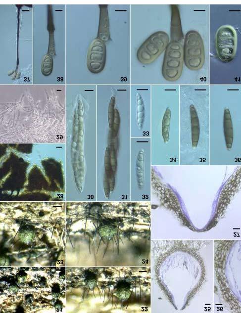
30 thin-walled, with 8 ascospores arranged uniseriately. Ascospores ellipsoid, hyaline, µm, 1-septate, easily disarticulating into part-spores. Culture: One-week-old colonies on CMA 26 mm diam., two-week-old colonies 45 mm diam., white, mycelium mostly superficial, aerial mycelium abundant, floccose, border even, reverse white. Conidiophores and conidia produced sparsely throughout the colony. One-week-old colonies on MEA 25 mm diam., two-week-old colonies 50 mm diam., light and dark grayish green, mycelium superficial, aerial mycelium abundant, floccose, border even, reverse dark green with a white border. Conidiophores and conidia produced abundantly in concentric rings. Conidiophores, single, unbranched, dark brown becoming light brown towards the apex, multiseptate, µm on CMA, with 5-8 whorls of phialides in midsection, a single phialide at the apex; conidiophores on the substrate µm at the base, tapering to a terminal phialide, µm setiform branches subterminal or terminal, tapering to a coiled apex, light-brown becoming hyaline towards the apex, 1 to 4 on a whorl, 1 or 2 whorls per conidiophore. Conidiogenous cell a phialide, cylindrical to lageniform, producing conidia from multiple conidiogenous loci, phialides borne on collar hyphae around the conidiophore, percurrent proliferation observed on the substrate. Conidia ellipsoid, light green, µm on CMA, on the substrate. Anamorph: It resembles Gonytrichum Nees & T. Nees. Habitat: On decorticated wood. Known distribution: Puerto Rico. Material examined: PUERTO RICO, Caribbean National Forest, El Verde Research Area, Luquillo Mts., 350 to 425 m, [ , ], 9 June 1998, on 4 cm branch, comm. J. McKemy, FAF, A.N. Miller, SMH3785 (F; holotype designated here). Tainosphaeria F.A. Fernández & Huhndorf, gen. nov. Etymology: Taino referring to the Taino indians, pre-hispanic inhabitants of Puerto Rico and the Caribbean, + sphaeria meaning sphere. Ascomata subglobosa vel ovoidea, atrobrunnea. Paraphyses simplices, septatae, hyalinae. Asci unitunicati, cylindrici, stipitati, annulo apicali adsum. Ascosporae fusiformes, hyalinae. Typus generis: Tainosphaeria crassiparies F.A. Fernández & Huhndorf Ascomata subglobose to ovoid. Paraphyses simple, septate, hyaline. Asci unitunicate, cylindrical, stipitate, with an apical ring. Ascospores hyaline, septate. Tainosphaeria crassiparies F.A. Fernández & Huhndorf, sp. nov. (Figs ) Etymology: Crassi refers to the relatively thick ascomal wall. Ascomata subglobosa vel ovoidea, atrobrunnea, µm lata, µm alta, cum exterior aspero, solitaria, superficialia, aggregata, papillata. Paries ascomatis cum aspectu 44
31 superficiaris opacus in aqua et textura angularis in lactophenol, sectione longitudinali µm crassus, cellulis pseudoparenchymatis. Apicem ascomatis papillatis. Paraphyses simplices, septatae, hyalinae. Asci cylindrici, brevi pedicellati, µm, unitunicati, tenuis paries, tennuis annulus apicalis, octospori. Ascosporae fusiformes, hyalinae, µm, 3-septatae, 4- et 5-septatae raro. Ascomata subglobose to broadly ovoid, dark brown, µm in diam., µm in height, roughened surface, separate to gregarious, superficial on the substratum, distinctly papillate. Ascomatal wall in surface view, opaque in water, of textura angularis in lactophenol, µm thick in longitudinal section. Ascomatal apex broad, papillate, short. Paraphyses unbranched, septate, hyaline, tapering, µm wide. Asci cylindro-clavate, short-stalked, µm, unitunicate, thin-walled, apical ring very small, with 8 ascospores biseriately arranged. Ascospores narrow-fusiform, with rounded ends, often inequilateral, µm, hyaline, mostly three-septate, rarely 4- or 5-septate. Culture: Colony on CMA 25 mm in diam. after seven days. Twenty-one day old colonies on CMA 45 mm in diam., subhyaline, mycelium mostly superficial, moist, appressed, margin effuse, reverse white, conidiophores produced throughout the colony. Conidiophores single, multiseptate, unbranched, brown becoming light brown towards the apex or viceversa, µm at the base, tapering to µm. Phialides cylindrical, light brown, µm, collarette generally present, funnel-shaped, µm deep, at the base, widening to µm at apex. Conidiogenous cell proliferating repeatedly in a percurrent manner. Conidia in culture ranging from one celled, ellipsoid, hyaline, µm, to one-celled, clavate, hyaline, rounded at the apex, acute at the base, sometimes with a short basal vertical slit, µm, produced from the same phialides. On the substrate, conidiophores are mononematous, macronematous, brown, µm at the base, tapering to a terminal, single phialide, often showing percurrent proliferations. Phialides cylindrical, light brown, at the base, narrowing to µm just below the collarette. Collarettes light brown, funnel shaped, µm at the opening, µm deep. Conidia falcate, inequilateral, hyaline, basal end truncate, apical end rounded, µm, a single setula at both ends, µm long. Anamorph: It resembles Codinaea Maire (Maire, 1937). Habitat: On Hymenia pod and on erumpent stromata of overmature ascomycetes. Known distribution: Puerto Rico. Material examined: PUERTO RICO, Sabana, near Rio Sabana, NW of junction of Rte. 983 & 991, hill above chicken farm, 70 m, N, W, [18.35, ], 17 January 1996, on Hymenaea seed pod, S.M. Huhndorf, with D.J. Lodge, D. Pfister, M. Harrington, SMH1934 (F; holotype designated here). 45
32 46
Two new intertidallignicolous Swampomyces species from Red Sea mangroves in Egypt
 Fungal Diversity Two new intertidallignicolous Swampomyces species from Red Sea mangroves in Egypt M.A. Abdel-Wahab1, H. El-Sharouneyl and E.B.G. Jones2 IDepat1ment of Botany, South Valley University,
Ascoyunnania aquatica gen. et sp. nov., a freshwater fungus collected from China and its microcylic conidiation
 Fungal Diversity Ascoyunnania aquatica gen. et sp. nov., a freshwater fungus collected from China and its microcylic conidiation Lei Cai 1, 2*, Keqin Zhang 1 and Kevin D. Hyde 2 1 Laboratory for Conservation
Stilbella holubovae, a new synnematous hyphomycete species on driftwood from the Philippines and South Africa
 Stilbella holubovae, a new synnematous hyphomycete species on driftwood from the Philippines and South Africa Keith A. Seifert 1, Susan J. Stanley 2 and Kevin D. Hyde 2 Centre for Land and Biological Resources
Synaptospora olandica, a new species from Sweden
 Synaptospora olandica, a new species from Sweden Martina Reblovä Institute of Botany, Dept. Plant Taxonomy and Biosystematics, Academy of Sciences, CZ-252 43 Prühonice, Czech Republic Reblovä, M. (2002):
BOTANICA HUNGARICA (Antea: Fragmenta Botanica)
") STUDIA XXIII. BOTANICA HUNGARICA (Antea: Fragmenta Botanica) 1992 pp. 63-68 A new species of Triadelphia from Hungary By Á.RÉVAY (Received 30 April, 1990) Abstract: A new dematiaceous hyphomycete species,
STUDIA XXIII. BOTANICA HUNGARICA (Antea: Fragmenta Botanica) 1992 pp. 63-68 A new species of Triadelphia from Hungary By Á.RÉVAY (Received 30 April, 1990) Abstract: A new dematiaceous hyphomycete species,
Lignicolous freshwater Ascomycota from Thailand: Melanochaeta and Sporoschisma anamorphs
 Mycol. Res. 104 (4): 478 485 (April 2000). Printed in the United Kingdom. 478 Lignicolous freshwater Ascomycota from Thailand: Melanochaeta and Sporoschisma anamorphs Somsak SIVICHAI, Nigel L. HYWEL-JONES
This work is licensed under a Creative Commons Attribution- NonCommercial-NoDerivatives 4.0 International License.
 Title A new freshwater species of Saccardoella from Hong Kong and South Africa Author(s) Tsui, KM; Hyde, KD; Hodgkiss, IJ; Goh, TK Citation Mycologia, 1998, v. 90 n. 4, p. 701-704 Issued Date 1998 URL
Creative Commons: Attribution 3.0 Hong Kong License
 Title Ascomycetes from freshwater habitats: Ascolacicola aquatica gen. et sp. nov., and a new species of Ascotaiwania from wood submerged in a reservoir in Hong Kong Author(s) Ranghoo, VM; Hyde, KD Citation
MYCOTAXON. Aliquandostipite crystallinus, a new ascomycete species from wood submerged in freshwater habitats
 MYCOTAXON Volume 91, pp. 207 215 January March 2005 Aliquandostipite crystallinus, a new ascomycete species from wood submerged in freshwater habitats HUZEFA A. RAJA, ASTRID FERRER & CAROL A. SHEARER raja@uiuc.edu
Dactylella shizishanna sp. nov., from Shizi Mountain, China
 Dactylella shizishanna sp. nov., from Shizi Mountain, China XueFeng Liu 1,2 and KeQin Zhang 1 * 1 Laboratory for Conservation and Utilization of Bio-resource, Yunnan University, Kunming, Yunnan 650091,
Arnium gigantosporum, a new ascomycete species from fresh water in Florida
 Arnium gigantosporum, a new ascomycete species from fresh water in Florida Huzefa A. Raja *, and Carol A. Shearer Department of Plant Biology, University of Illinois, 265 Morrill Hall, 505 South Goodwin
Dokmaia monthadangii gen. et sp. nov., a synnematous anamorphic fungus on Manglietia garrettii
 Dokmaia monthadangii gen. et sp. nov., a synnematous anamorphic fungus on Manglietia garrettii I. Promputtha1, K. D. Hyde2, P. Lumyong3, E. H. C. McKenzie4 & S. Lumyong1 1 Department of Biology, Faculty
A new species of Podospora from Taiwan
 CHANG Bot. Bull. and Acad. WANG Sin. (2005) A new 46: 169-173 species of Podospora 169 A new species of Podospora from Taiwan Jong-How CHANG 1 and Yei-Zeng WANG 2, * 1 Department of Life Science, National
Two new species of Meliola (Ascomycetes) from Kenya
 from Kenya") Two new species of Meliola (Ascomycetes) from Kenya R.K. Mibey and J.O. Kokwaro Department of Botany, University of Nairobi, P.O Box 30197, Nairobi, Kenya Mibey, R.K. and Kokwaro, J.O. (1999). Two new
Two new species of Meliola (Ascomycetes) from Kenya R.K. Mibey and J.O. Kokwaro Department of Botany, University of Nairobi, P.O Box 30197, Nairobi, Kenya Mibey, R.K. and Kokwaro, J.O. (1999). Two new
Fungi from palms. XIX 1. Caudatispora palmicola gen. et sp. nov. from Ecuador
 Fungi from palms. XIX 1. Caudatispora palmicola gen. et sp. nov. from Ecuador Jane Fröhlich & Kevin D. Hyde Department of Ecology and Biodiversity, University of Hong Kong, Pokfulam Road, Hong Kong Fröhlich,
Goosiomyces bambusicola - A new cheirosporous anamorphic species from Western Ghats, India.
 Current Research in Environmental & Applied Mycology 4 (2): 211 216 (2014) ISSN 2229-2225 www.creamjournal.org Article CREAM Copyright 2014 Doi 10.5943/cream/4/2/8 Online Edition Goosiomyces bambusicola
REINWARDTIA Published by Herbarium Bogoriense LBN, Bogor Vol. 10, Part 2, pp (1985) THE ANAMORPH OF SARAWAKUS SUCCISUS RIFAI
 THE ANAMORPH OF SARAWAKUS SUCCISUS RIFAI") REINWARDTIA Published by Herbarium Bogoriense LBN, Bogor Vol. 10, Part 2, pp. 265 270 (1985) THE ANAMORPH OF SARAWAKUS SUCCISUS RIFAI MIEN A. RIFAI, KARTINI KRAMADIBRATA Herbarium Bogorievnc LBN, Bogor,
REINWARDTIA Published by Herbarium Bogoriense LBN, Bogor Vol. 10, Part 2, pp. 265 270 (1985) THE ANAMORPH OF SARAWAKUS SUCCISUS RIFAI MIEN A. RIFAI, KARTINI KRAMADIBRATA Herbarium Bogorievnc LBN, Bogor,
New species of Dictyosporium and Digitodesmium from submerged wood in Yunnan, China
 New species of Dictyosporium and Digitodesmium from submerged wood in Yunnan, China Lei Cai1'2, Keqin Zhang2, Eric H. C. McKenzie3 & Kevin D. Hyde1 1 2 Centre for Research in Fungal Diversity, Department
Life-History Studies of Brazilian Ascomycetes 1.
 Life-History Studies of Brazilian Ascomycetes 1. Two new genera of the Sphaeriaceae having, respectively, Sporoschisma-like and Codinaea anamorphs Gary J. SAMUELS & Emil MÜLLER DSIR, Plant Diseases Division,
Xylaria kaumanae sp. nov. from the Island of Hawaii (Hawaii, USA)
") Xylaria kaumanae sp. nov. from the Island of Hawaii (Hawaii, USA) Jack D. Rogers 1, D. E. Hemmes 2 & Y.-M. Ju 3 1 Department of Plant Pathology, Washington State University, Pullman, WA 99165-6430, USA
Xylaria kaumanae sp. nov. from the Island of Hawaii (Hawaii, USA) Jack D. Rogers 1, D. E. Hemmes 2 & Y.-M. Ju 3 1 Department of Plant Pathology, Washington State University, Pullman, WA 99165-6430, USA
Key words wild passion-fruit, Mycosphaerellaceae, tropical fruits, cercosporoid fungi
 A NEW FUNGAL DISEASE CAUSED BY A PSEUDOCERCOSPORA SPECIES ON PASSIFLORA SETACEA IN PLANALTINA-DF, BRAZIL Alexei C. Dianese 1, Ana M. Costa 1 & José C. Dianese 2 ( 1 Embrapa Cerrados, Br-020, Km 18, 73310-970
World Journal of Microbiology Vol. 1(2), pp , September, ISSN: XXXX-XXXX
, pp , September, ISSN: XXXX-XXXX") World Journal of Microbiology Vol. 1(2), pp. 013-016, September, 2014. www.premierpublishers.org, ISSN: XXXX-XXXX WJM Research Article Study of Fungal Genus Gyrothrix Corda from the forest flora of Indian
World Journal of Microbiology Vol. 1(2), pp. 013-016, September, 2014. www.premierpublishers.org, ISSN: XXXX-XXXX WJM Research Article Study of Fungal Genus Gyrothrix Corda from the forest flora of Indian
PERSOONIA. Part 2, pp (1962) Studies on discomycetez I. of Ascobolus and Saccobolus. herbarium, Rijksherbarium, Leiden. inquirenda. on loan.
 Studies on discomycetez I. of Ascobolus and Saccobolus. herbarium, Rijksherbarium, Leiden. inquirenda. on loan.") FIG. PERSOONIA Published by the Rijksherbarium, Leiden Volume 2, Part 2, pp. 195-199 (1962) Studies on discomycetezi. s of species of Ascobolus and Saccobolus in Spegazzini s herbarium J. van Brummelen
FIG. PERSOONIA Published by the Rijksherbarium, Leiden Volume 2, Part 2, pp. 195-199 (1962) Studies on discomycetezi. s of species of Ascobolus and Saccobolus in Spegazzini s herbarium J. van Brummelen
Xenosporium amomi sp. nov. from Zingiberaceae in Thailand
 Fungal Diversity Xenosporium amomi sp. nov. from Zingiberaceae in Thailand Boonsom Bussaban¹ *, Pipob Lumyong², Eric H.C. McKenzie³, Kevin D. Hyde 4 and Saisamorn Lumyong¹ ¹Department of Biology, Faculty
MYCOTAXON. Volume 114, pp October-december A new species of Ijuhya, I. antillana, from the French West Indies. 1*
 MYCOTAXON Volume 114, pp. 91 94 October-december 2010 A new species of Ijuhya, I. antillana, from the French West Indies CHRISTIAN LECHAT 1* & REGIS COURTECUISSE 2 1* lechat@ascofrance.fr 64 route de Chizé,
CZECH MYCOLOGY Publication of the Czech Scientific Society for Mycology
 CZECH MYCOLOGY Publication of the Czech Scientific Society for Mycology Volume 50 April 1998 Number 3 A new Chaetosphaeria with a Dictyochaeta anamorph M a r t in a R e b l o v a Institute of Botany, Academy
Occultitheca costaricensis gen. et sp. nov. and Apiocamarops pulvinata sp. nov. from Costa Rica
 Occultitheca costaricensis gen. et sp. nov. and Apiocamarops pulvinata sp. nov. from Costa Rica Jack D. Rogers1 & Y.-M. Ju 2 1 Department of Plant Pathology, Washington State University, Pullman, WA 99164-6430,
Guignardia bispora and G. ellipsoidea spp. nov. and other Guignardia species from palms (Arecaceae)
") Mycosphere Guignardia bispora and G. ellipsoidea spp. nov. and other Guignardia species from palms (Arecaceae) Wulandari NF 1,2, To Anun C. 1*, McKenzie EHC 3 and Hyde KD 4,5 1 Department of Plant Pathology,
Mycosphere Guignardia bispora and G. ellipsoidea spp. nov. and other Guignardia species from palms (Arecaceae) Wulandari NF 1,2, To Anun C. 1*, McKenzie EHC 3 and Hyde KD 4,5 1 Department of Plant Pathology,
Two new species of Corynespora from Uttar Pradesh, India
 Two new species of Corynespora from Uttar Pradesh, India Kumar S 1,2*, Singh R 2, Gond DK 1 and Saini DC 1 1 Birbal Sahni Institute of Palaeobotany, 53, University Road, Lucknow 226007(U.P.), India. 2
Observations on the genus Cresporhaphis (Trichosphaeriaceae), with a key to the known species, and C. ulmi sp. nov.
, with a key to the known species, and C. ulmi sp. nov.") Mycol. Res. 105 (1): 122 126 (January 2001). Printed in the United Kingdom. 122 Observations on the genus Cresporhaphis (Trichosphaeriaceae), with a key to the known species, and C. ulmi sp. nov. Vicent
Mycol. Res. 105 (1): 122 126 (January 2001). Printed in the United Kingdom. 122 Observations on the genus Cresporhaphis (Trichosphaeriaceae), with a key to the known species, and C. ulmi sp. nov. Vicent
This work is licensed under a Creative Commons Attribution- NonCommercial-NoDerivatives 4.0 International License.
 Title Paraniesslia tuberculata gen. et sp. nov., and new records or species of Clypeosphaeria, Leptosphaeria and Astrosphaeriella in Hong Kong freshwater habitats Author(s) Tsui, KM; Hyde, KD; Hodgkiss,
Journal of Chemical and Pharmaceutical Research, 2017, 9(1): Review Article. Graphium Salixicum: A New Species Explored from Salix Alba
: Review Article. Graphium Salixicum: A New Species Explored from Salix Alba") Available online www.jocpr.com Journal of Chemical and Pharmaceutical Research, 2017, 9(1):69-74 Review Article ISSN : 0975-7384 CODEN(USA) : JCPRC5 Graphium Salixicum: A New Species Explored from Salix
Available online www.jocpr.com Journal of Chemical and Pharmaceutical Research, 2017, 9(1):69-74 Review Article ISSN : 0975-7384 CODEN(USA) : JCPRC5 Graphium Salixicum: A New Species Explored from Salix
Cordana versicolor sp. nov. (dematiaceous hyphomycete) causing leaf-spot on Canna denudata (Cannaceae) in Brazil, with observations on Cordana musae
 causing leaf-spot on Canna denudata (Cannaceae) in Brazil, with observations on Cordana musae") Cordana versicolor sp. nov. (dematiaceous hyphomycete) causing leaf-spot on Canna denudata (Cannaceae) in Brazil, with observations on Cordana musae Dartanhã J. Soares 1 ; Katia L. Nechet 2 and Robert
Cordana versicolor sp. nov. (dematiaceous hyphomycete) causing leaf-spot on Canna denudata (Cannaceae) in Brazil, with observations on Cordana musae Dartanhã J. Soares 1 ; Katia L. Nechet 2 and Robert
MYCOTAXON. Volume 106, pp October December 2008
 MYCOTAXON Volume 106, pp. 403 407 October December 2008 Annulatascus apiculatus sp. nov., a new freshwater ascomycete from the semi-arid Caatinga biome of Brazil Flavia Rodrigues Barbosa Luís Fernando
Two new species of Diplococcium from the tropics
 Title Two new species of Diplococcium from the tropics Author(s) Goh, TK; Hyde, KD; Umali, TE Citation Mycologia, 1998, v. 90 n. 3, p. 514-517 Issued Date 1998 URL http://hdl.handle.net/10722/53359 Rights
MYCOTAXON. Volume 103, pp January March South Florida microfungi: a new species of Stanjehughesia (hyphomycetes) from Sabal palm
 from Sabal palm") MYCOTAXON Volume 103, pp. 229 234 January March 2008 South Florida microfungi: a new species of Stanjehughesia (hyphomycetes) from Sabal palm Gregorio Delgado gdelgado@emlab.com EMLab P&K, Southeastern
MYCOTAXON Volume 103, pp. 229 234 January March 2008 South Florida microfungi: a new species of Stanjehughesia (hyphomycetes) from Sabal palm Gregorio Delgado gdelgado@emlab.com EMLab P&K, Southeastern
LESSON ASSIGNMENT. Introduction to Medical Mycology. After completing this lesson, you should be able to:
 LESSON ASSIGNMENT LESSON 1 Introduction to Medical Mycology. TEXT ASSIGNMENT Paragraphs 1-1 through 1-7. TASKS OBJECTIVES After completing this lesson, you should be able to: 1-1. Select the statement
Freshwater ascomycetes: new and noteworthy species from aquatic habitats in Florida
 Mycologia, 100(3), 2008, pp. 467 489. # 2008 by The Mycological Society of America, Lawrence, KS 66044-8897 Freshwater ascomycetes: new and noteworthy species from aquatic habitats in Florida Huzefa A.
CZECH MYCOLOGY Publication of the Czech Scientific Society for Mycology
 CZECH MYCOLOGY Publication of the Czech Scientific Society for Mycology Volume 51 January 1999 Num ber 1 Teleomorph-anamorph connections in Ascomycetes. 1. Cylindrotrichum and Cacumisporium anamorphs of
New species of Cordana and Spadicoides from decaying bamboo culms in China
 New species of Cordana and Spadicoides from decaying bamboo culms in China Lei Cai 1, Eric H. C. McKenzie 2 & Kevin D. Hyde 1 Centre for Research in Fungal Diversity, Department of Ecology & Biodiversity,
Diplolaeviopsis symmictae (Helotiales, Ascomycota), a new lichenicolous fungus on Lecanora symmicta
, a new lichenicolous fungus on Lecanora symmicta") Diplolaeviopsis symmictae (Helotiales, Ascomycota), a new lichenicolous fungus on Lecanora symmicta Paul Diederich 1 & Brian Coppins 2 1 Musée national d histoire naturelle, 25 rue Munster, L-2160 Luxembourg,
Diplolaeviopsis symmictae (Helotiales, Ascomycota), a new lichenicolous fungus on Lecanora symmicta Paul Diederich 1 & Brian Coppins 2 1 Musée national d histoire naturelle, 25 rue Munster, L-2160 Luxembourg,
Dinemasporium (coelomycetes)
") Fungal Diversity Dinemasporium (coelomycetes) Junxin Duan 1,2, Wenping Wu 2* and X.Z. Liu 1 1 Institute of Microbiology, The Chinese Academy of Science, Beijing 100085, PR China 2 Novozymes China, 14 Xinxi
Fungal Diversity Dinemasporium (coelomycetes) Junxin Duan 1,2, Wenping Wu 2* and X.Z. Liu 1 1 Institute of Microbiology, The Chinese Academy of Science, Beijing 100085, PR China 2 Novozymes China, 14 Xinxi
Mycosphaerella eutnusae and its anamorph Pseudocercospora eumusae spp. nov.: causal agent of eumusae. leaf spot disease of banana
 Mycosphaerella eutnusae and its anamorph Pseudocercospora eumusae spp. nov.: causal agent of eumusae leaf spot disease of banana Pedro W. Crous1 & Xavier Mourichon2 1 2 Department of Plant Pathology, University
Rec. zoot. Surv. India, 97 (Part-2) : , 1999
 : , 1999") Rec. zoot. Surv. India, 97 (Part-2) : 167-172, 1999 DESCRIPTIONS OF TWO NEW GALL MIDGES (DIPTERA : CECIDOMYIIDAE).FROM NILGIRI BIOSPHERE RESERVE, KARNATAKA R. M. SHARMA Zoological Survey of India, Western
Rec. zoot. Surv. India, 97 (Part-2) : 167-172, 1999 DESCRIPTIONS OF TWO NEW GALL MIDGES (DIPTERA : CECIDOMYIIDAE).FROM NILGIRI BIOSPHERE RESERVE, KARNATAKA R. M. SHARMA Zoological Survey of India, Western
Crassoascus monocaudatus and Iodosphaeria podocarpi, two new species on Podocarpus parlatorei from Las Yungas, Argentina
 Crassoascus monocaudatus and Iodosphaeria podocarpi, two new species on Podocarpus parlatorei from Las Yungas, Argentina Catania M del V 1 and Romero AI 2 1 Fundación Miguel Lillo, Laboratorio de Micología.
New species of Anthostomella on fynbos, with a key to the genus in South Africa
 Mycol. Res. 107 (3): 360 370 (March 2003). f The British Mycological Society 360 DOI: 10.1017/S0953756203007184 Printed in the United Kingdom. New species of Anthostomella on fynbos, with a key to the
Two new species of Dactylaria (anamorphic fungi) from Australian rainforests and an update of species in Dactylaria sensu lato
 from Australian rainforests and an update of species in Dactylaria sensu lato") Fungal Diversity Two new species of Dactylaria (anamorphic fungi) from Australian rainforests and an update of species in Dactylaria sensu lato Barbara Paulus 1*, Paul Gadek 1 and Kevin D. Hyde 2 1 School
Fungal Diversity Two new species of Dactylaria (anamorphic fungi) from Australian rainforests and an update of species in Dactylaria sensu lato Barbara Paulus 1*, Paul Gadek 1 and Kevin D. Hyde 2 1 School
A new species of Conidiobolus with distended conidiophores.
 Separat-Abdruck aus Sydowia, Annales Mycologici Beiheft I. Festschrift fiir Franz Petrak Verlag Ferdinand Berger, Horn, N.-O., Austria A new species of Conidiobolus with distended conidiophores. By Charles
7-001a: Blotter method for the detection of Alternaria dauci on Daucus carota (carrot)
") International Rules for Seed Testing Annexe to Chapter 7: Seed Health Testing Methods 7-001a: Blotter method for the detection of Alternaria dauci on Daucus carota (carrot) Published by: International
International Rules for Seed Testing Annexe to Chapter 7: Seed Health Testing Methods 7-001a: Blotter method for the detection of Alternaria dauci on Daucus carota (carrot) Published by: International
Two new species of Neocosmospora from Japan, with a key to the currently accepted species
 Two new species of Neocosmospora from Japan, with a key to the currently accepted species S.-I. UDAGAWA National Institute of Hygienic Sciences, Kamiyoga 1-Chome, Setagaya-ku, Tokyo 158, Japan Y. HORIE
ALPAKESIOPSIS GEN. NOV., AND ADDITION TO COELOMYCETES FROM PAKISTAN
 Pak. J. Bot., 35(2): 249-255, 2003. ALPAKESIOPSIS GEN. NOV., AND ADDITION TO COELOMYCETES FROM PAKISTAN S.Q. ABBAS, B.C. SUTTON *, A.GHAFFAR ** AND A. ABBAS Department of Botany, Federal Urdu University
Ascal ultrastructural study in Annulatascus hongkongensis sp. nov., a freshwater ascomycete. Ho, WH; Ranghoo, VM; Hyde, KD; Hodgkiss, IJ
 Title Ascal ultrastructural study in Annulatascus hongkongensis sp. nov., a freshwater ascomycete Author(s) Ho, WH; Ranghoo, VM; Hyde, KD; Hodgkiss, IJ Citation Mycologia, 1999, v. 91 n. 5, p. 885-892
MYCOTAXON. Volume 91, pp January March Dactylella coccinella sp. nov., an anamorphic species. 1
 MYCOTAXON Volume 91, pp. 127 132 January March 2005 Dactylella coccinella sp. nov., an anamorphic species YING YANG 1,2 & XING ZHONG LIU 1 liuxz@sun.im.ac.cn 1 Key Laboratory of Systematic Mycology & Lichenology
Additions to helicoid fungi from India
 Current Research in Environmental & Applied Mycology 6 (4): 248 255 (2016) ISSN 2229-2225 www.creamjournal.org Article CREAM Copyright 2016 Doi 10.5943/cream/6/4/2 Online Edition Additions to helicoid
Brunneodinemasporium jonesii and Tainosphaeria jonesii spp. nov. (Chaetosphaeriaceae, Chaetosphaeriales) from southern China
 from southern China") Mycosphere 7 (9): 1322 1331 (2016) www.mycosphere.org ISSN 2077 7019 Article special issue Doi 10.5943/mycosphere/7/9/6 Copyright Guizhou Academy of Agricultural Sciences Brunneodinemasporium jonesii and
Mycosphere 7 (9): 1322 1331 (2016) www.mycosphere.org ISSN 2077 7019 Article special issue Doi 10.5943/mycosphere/7/9/6 Copyright Guizhou Academy of Agricultural Sciences Brunneodinemasporium jonesii and
Phialomyces fusiformis sp. nov. from soil in Singapore is identified and described
 Mycologia, 95(5), 2003, pp. 896 901. 2003 by The Mycological Society of America, Lawrence, KS 66044-8897 Phialomyces fusiformis sp. nov. from soil in Singapore is identified and described G. Delgado Rodriguez
Fungi on submerged wood and bamboo in the Plover Cove Reservoir, Hong Kong
 Fungi on submerged wood and bamboo in the Plover Cove Reservoir, Hong Kong Teik Khiang Goh* and Kevin D. Hyde Centre for Research in Fungal Diversity, Department of Ecology and Biodiversity, The University
Two new lichenicolous species of Hainesia (asexual Ascomycetes) growing on Cladonia
 growing on Cladonia") Two new lichenicolous species of Hainesia (asexual Ascomycetes) growing on Cladonia Paul Diederich 1 & Pieter van den Boom 2 1 Musée national d histoire naturelle, 25 rue Munster, L 2160 Luxembourg, Luxembourg
Two new lichenicolous species of Hainesia (asexual Ascomycetes) growing on Cladonia Paul Diederich 1 & Pieter van den Boom 2 1 Musée national d histoire naturelle, 25 rue Munster, L 2160 Luxembourg, Luxembourg
New records of conidial fungi for Brazil
 Journal on New Biological Reports JNBR 4(1) 20 25 (2015) ISSN 2319 1104 (Online) Published by www.researchtrend.net New records of conidial fungi for Brazil Marcos Fabio Oliveira Marques 1* and Luis Fernando
Blotter method for the detection of Alternaria dauci on Daucus carota
 International Rules for Seed Testing Annexe to Chapter 7: Seed Health Testing Methods 7-001a: Blotter method for the detection of Alternaria dauci on Daucus carota Published by: International Seed Testing
Microfungi on the Pandanaceae: Acrodictys, with two new species
 Microfungi on the Pandanaceae: Acrodictys, with two new species Stephen R. Whitton1,2, Eric H.C. McKenzie3 and Kevin D. Hyde1* 'Centre for Research in, Department of Ecology and Biodiversity, The University
7-001b: Malt agar method for the detection of Alternaria dauci on Daucus carota (carrot)
") International Rules for Seed Testing Annexe to Chapter 7: Seed Health Testing Methods 7-001b: Malt agar method for the detection of Alternaria dauci on Daucus carota (carrot) Published by: International
International Rules for Seed Testing Annexe to Chapter 7: Seed Health Testing Methods 7-001b: Malt agar method for the detection of Alternaria dauci on Daucus carota (carrot) Published by: International
Identification of Botryosphaeriaceae associated with the die-back of ornamental trees in the Western Balkans
 COST action FP1401 A global network of nurseries as early warning system against alien tree pests- Global warning Training school Fungal taxonomy and fungal identification using traditional (i.e. not molecular)
Received for publication: November 15, 2014; Accepted: December 21, 2014.
 Original Research Article Annals of Plant Sciences ISSN: 2287-688X OPEN ACCESS www.annalsofplantsciences.com Fresh Water Fungi from Pachmarhi (Madhya Pradesh) Suhas A Chaudhari 1* and Patil VR 2 1 Dr.Annasaheb
Anacardiaceae cashew family
 Anacardiaceae cashew family Mostly tropical, the family includes nearly 600 species; four species are native to Nova Scotia. Leaflets are pinnately or palmately compound. Typical are small, regular flowers,
Brazilian Semi-Arid Ascomycetes III: New records of Dothideomycetes and Sordariomycetes
 CNova Hedwigia Vol. 105 (2017) Issue 3 4, 487 494 published online June 19, 2017; published in print November 2017 Article Brazilian Semi-Arid Ascomycetes III: New records of Dothideomycetes and Sordariomycetes
Braunomyces dictyosporus gen. sp. nov. from Vietnam ARTICLE. Introduction. Material and Methods
 doi:10.5598/imafungus.2014.05.01.01 IMA Fungus volume 5 no 1: 1 5 Braunomyces dictyosporus gen. sp. nov. from Vietnam Vadim A. Mel nik 1 and Pedro W. Crous 2 1 Laboratory of the Systematics and Geography
The Genus Pithomyces in South Africa by
 Bothalia, 10, 4: 509-516 The Genus Pithomyces in South Africa by W. F. O. Marasas* and Ingrid H. Schumann* A bstract Descriptions are given of South African isolates of Pithomyces sacchari (Speg.) M. B.
Two new species of Chaetomidium (Sordariales)
") STUDIES IN MYCOLOGY 50: 215 220. 2004. Two new species of Chaetomidium (Sordariales) Alberto M. Stchigel 1*, Josep Guarro 1, Victoria Jato 2 and María J. Aira 3 1 Unitat de Microbiologia, Facultat de Medicina
STUDIES IN MYCOLOGY 50: 215 220. 2004. Two new species of Chaetomidium (Sordariales) Alberto M. Stchigel 1*, Josep Guarro 1, Victoria Jato 2 and María J. Aira 3 1 Unitat de Microbiologia, Facultat de Medicina
Fungi of Gorakhpur. XVII. Sarcinella.
 Fungi of Gorakhpur. XVII. Sarcinella. EAMAL & R. P. SINGH Department of Botany, University of Gorakhpur, Gorakhpur-273001, U. P., India Introduction The Gorakhpur region consitutes a major part of the
Life-History Studies of Brazilian Ascomycetes 4 *)
") Life-History Studies of Brazilian Ascomycetes 4 *) Three species of Herpotrichia and their Pyrenochaeta-like anamorphs Gary J. SAMUELS & Emil MÜLLER DSIR, Plant Diseases Division, Private Bag, Auckland,
Life-History Studies of Brazilian Ascomycetes 4 *) Three species of Herpotrichia and their Pyrenochaeta-like anamorphs Gary J. SAMUELS & Emil MÜLLER DSIR, Plant Diseases Division, Private Bag, Auckland,
A NOTE ON THE CONIDIAL SCAR IN THE XYLARIACEAE
 New PhytoL (1967) 66, 65-66. A NOTE ON THE CONIDIAL SCAR IN THE XYLARIACEAE BY G. N. GREENHALGH The Hartley Botanical Laboratories, University of Liverpool {Received 5 July 1966) The conidial scars produced
7-012: Detection of Alternaria padwickii on Oryza sativa (Rice)
") International Rules for Seed Testing Annexe to Chapter 7: Seed Health Testing Methods Published by: International Seed Testing Association (ISTA), Bassersdorf, Switzerland 2014 DISCLAIMER: whilst ISTA
International Rules for Seed Testing Annexe to Chapter 7: Seed Health Testing Methods Published by: International Seed Testing Association (ISTA), Bassersdorf, Switzerland 2014 DISCLAIMER: whilst ISTA
This is the written version of our Hot Topic video presentation available at: MayoMedicalLaboratories.com/hot-topics
 This is the written version of our Hot Topic video presentation available at: MayoMedicalLaboratories.com/hot-topics Welcome to Mayo Medical Laboratories hot topics. These presentations provide short discussion
Microfungi on the Pandanaceae: Nakatopsis gen. nov., a new hyphomycete genus from Malaysia
 Microfungi on the Pandanaceae: Nakatopsis gen. nov., a new hyphomycete genus from Malaysia Stephen R. Whitton 1, Eric H.C. McKenziel and Kevin D. Hyde2 ILandcare Research, Private Bag 92170, Auckland,
New Xylariaceae taxa from Brazil
 New Xylariaceae taxa from Brazil Jadergudson Pereira 1 *, Jack D. Rogers 2 & Jose Luiz Bezerra 1 1 Departamento de Ciencias Agrarias e Ambientais, Universidade Estadual de Santa Cruz, Rod. Ilheus-Itabuna
Mycological investigation on the Fungal Genus Gyrothrix Corda in the forest flora of Madhya Pradesh India
 World Research Journal of Agricultural Sciences Vol. 1(3), pp. 017-021, October, 2014. www.premierpublishers.org, ISSN: 3115-2864 WRJAS Research Article Mycological investigation on the Fungal Genus Gyrothrix
Subcutaneous Mycosis
 Subcutaneous Mycosis Fungal infections 1. Superficial mycosis. 2. Coetaneous mycosis: Dermatophytoses. 3. Subcutaneous mycosis. 4. Systemic mycosis. 5. Opportunistic mycosis. Subcutanus mycoses Fungal
Observations on Erysiphe platani in Germany
 Observations on Erysiphe platani in Germany Kirschner R* Department of Life Sciences, National Central University, Jhongli City, 320 Taoyuan County, Taiwan R.O.C. Kirschner R. 2011 Observations on Erysiphe
First Report of Penicillium adametzioides from Decayed Grapes (Vitis vinifera) in Pakistan
 in Pakistan") International Journal of Current Microbiology and Applied Sciences ISSN: 2319-7706 Volume 5 Number 12 (2016) pp. 316-320 Journal homepage: http://www.ijcmas.com Original Research Article http://dx.doi.org/10.20546/ijcmas.2016.512.034
International Journal of Current Microbiology and Applied Sciences ISSN: 2319-7706 Volume 5 Number 12 (2016) pp. 316-320 Journal homepage: http://www.ijcmas.com Original Research Article http://dx.doi.org/10.20546/ijcmas.2016.512.034
Conioscyphascus, a new ascomycetous genus for holomorphs with Conioscypha anamorphs
 STUDIES IN MYCOLOGY 50: 95 108. 2004. Conioscyphascus, a new ascomycetous genus for holomorphs with Conioscypha anamorphs Martina Réblová 1* and Keith A. Seifert 2 1 Department of Plant Taxonomy and Biosystematics,
Phylogeny of Discosia and Seimatosporium, and introduction of Adisciso and Immersidiscosia genera nova
 Persoonia 26, 2011: 85 98 www.ingentaconnect.com/content/nhn/pimj RESEARCH ARTICLE doi:10.3767/003158511x576666 Phylogeny of Discosia and Seimatosporium, and introduction of Adisciso and Immersidiscosia
Re-evaluation of Cryptosporiopsis eucalypti and Cryptosporiopsis-like species occurring on Eucalyptus leaves
 Fungal Diversity (2010) 44:89 105 DOI 10.1007/s13225-010-0041-5 Re-evaluation of Cryptosporiopsis eucalypti and Cryptosporiopsis-like species occurring on Eucalyptus leaves R. Cheewangkoon & J. Z. Groenewald
Luciotrichus lasioboloides, a new genus and a new species of the Pezizales
 CZECH MYCOL. 47 (4), 1994 Luciotrichus lasioboloides, a new genus and a new species of the Pezizales RICARDO GAL..\N and AIN RAITVIIR 1 ) Department of Plant Biology, University of Alcalli 288 71 Alcalli
A review of Cirrenalia (hyphomycetes) and a new species
 and a new species") A review of Cirrenalia (hyphomycetes) and a new species Fungal Diversity G.Z. Zhao and X.Z. Liu * Key Laboratory of Systematic Mycology & Lichenology, Institute of Microbiology, Chinese Academy of Sciences,
A review of Cirrenalia (hyphomycetes) and a new species Fungal Diversity G.Z. Zhao and X.Z. Liu * Key Laboratory of Systematic Mycology & Lichenology, Institute of Microbiology, Chinese Academy of Sciences,
Scanning Electron Microscopy (SEM) of Seed Infected with Seed Borne Fungi
 of Seed Infected with Seed Borne Fungi") International Journal of Current Microbiology and Applied Sciences ISSN: 2319-7706 Volume 7 Number 07 (2018) Journal homepage: http://www.ijcmas.com Original Research Article https://doi.org/10.20546/ijcmas.2018.707.476
International Journal of Current Microbiology and Applied Sciences ISSN: 2319-7706 Volume 7 Number 07 (2018) Journal homepage: http://www.ijcmas.com Original Research Article https://doi.org/10.20546/ijcmas.2018.707.476
258 DANIEL A. TEXTORIS Vol. 63
 258 DANIEL A. TEXTORIS Vol. 63 STUDIES OF THE GYPONINAE 1. THE GENUS MARGANANA DELONG* (HOMOPTERA: CICADELLIDAE) DWIGHT M. DELONG AND PAUL H. PREYTAG Department of Zoology and Entomology, The Ohio State
Structure & Life Cycle of Anthoceros
 Structure & Life Cycle of Anthoceros Anthoceros General Characters Gametophytic Plant Body (The Adult gametophyte) Vegetative Structure: External Features It occurs in moist, shaded habitats in sub-tropical
Hypocrea flaviconidia, a new species from Costa Rica with yellow conidia
 STUDIES IN MYCOLOGY 50: 401 407. 2004. Hypocrea flaviconidia, a new species from Costa Rica with yellow conidia Irina S. Druzhinina 1, Priscila Chaverri 2,4, Payam Fallah 2,5, Christian P. Kubicek 1 and
A new species of Coniochaeta with a key to the species known in Argentina
 Mycol. Res. 103 (6): 689 695 (1999) Printed in the United Kingdom 689 A new species of Coniochaeta with a key to the species known in Argentina A. I. ROMERO 1, C. C. CARMARA N 1 * AND L. E. LORENZO Departamento
A new fungicolous species of Titaea and new reports of Bahusaganda indica and Exosporium ampullaceum
 A new fungicolous species of Titaea and new reports of Bahusaganda indica and Exosporium ampullaceum (hyphomycetes) from tropical rainforests in Panama Roland Kirschner 1* and Meike Piepenbring 1 1 Botanisches
Evaluation of an alternative slide culture technique for the morphological identification of fungal species
 Research Article Evaluation of an alternative slide culture technique for the morphological identification of fungal species Abstract M H Wijedasa 1, L V C Liyanapathirana 1. Sri Lanka Journal of Infectious
Research & Reviews: Journal of Microbiology and Biotechnology
 e-issn:2320-3528 Research & Reviews: Journal of Microbiology and Biotechnology Two Novel Eupenicillium Species Isolated from Soil in China Hong-Kai Wang, Yi-Chen Cao, Fu-Cheng Lin* State Key Laboratory
M. A. WAHID, R. J. STEIB, S. J. P. CHILTON REFERENCES. In the absence of the authors the following paper was laid on the table.
 M. A. WAHID, R. J. STEIB, S. J. P. CHILTON REFERENCES 1 ABBOTT, E. V 1938. Red rot of sugarcane. U S D.A. Tech. Bull. 641 96 pp. 2 CHONA, R. L. and C. W. PADWICK. 1942. More light on the red rot epidemic.
Several rare entopathogenic fungi from the Western Sichuan mountains
 Fungal Diversity Several rare entopathogenic fungi from the Western Sichuan mountains Zong Qi Liang 1*, Bo Wang 2 and Ji Chuan Kang 3 1 Laboratory of Fungus Resources, Guizhou University, Huaxi, Guiyang,
Preliminary Survey of Bionectriaceae and Nectriaceae (Hypocreales, Ascomycetes) from Jigongshan, China
 from Jigongshan, China") Preliminary Survey of Bionectriaceae and Nectriaceae (Hypocreales, Ascomycetes) from Jigongshan, China Ye Nong 1, 2 and Wen-Ying Zhuang 1* 1 Key Laboratory of Systematic Mycology and Lichenology, Institute
Preliminary Survey of Bionectriaceae and Nectriaceae (Hypocreales, Ascomycetes) from Jigongshan, China Ye Nong 1, 2 and Wen-Ying Zhuang 1* 1 Key Laboratory of Systematic Mycology and Lichenology, Institute
Endophragmiella boewei and Beltrania rhombica
 Flora Mediterranea 3-1993 233 B. Mulas, M. Pasqualetti & A. Rambelli Influence or the substratum on the morphology or Endophragmiella boewei and Beltrania rhombica Abstract B. Mulas, M. Pasqualetti & A.
7-002b: Malt agar method for the detection of Alternaria radicina on Daucus carota (carrot)
") International Rules for Seed Testing Annexe to Chapter 7: Seed Health Testing Methods 7-002b: Malt agar method for the detection of Alternaria radicina on Daucus carota (carrot) Published by: International
International Rules for Seed Testing Annexe to Chapter 7: Seed Health Testing Methods 7-002b: Malt agar method for the detection of Alternaria radicina on Daucus carota (carrot) Published by: International
SEVEN NEW SPECIES OF THE GENUS APHALARA (HOMOPTERA: CHERMIDAE)
") SEVEN NEW SPECIES OF THE GENUS APHALARA (HOMOPTERA: CHERMIDAE) JOHN S. CALDWELL Ohio State University Aphalara constricta n. sp. Length to tip of forewing 3.-4. mm.; forewing 2.-3. mm. Color: Greenish
SEVEN NEW SPECIES OF THE GENUS APHALARA (HOMOPTERA: CHERMIDAE) JOHN S. CALDWELL Ohio State University Aphalara constricta n. sp. Length to tip of forewing 3.-4. mm.; forewing 2.-3. mm. Color: Greenish
Foliicolous anamorphs of Mycosphaerella from South America
 Foliicolous anamorphs of Mycosphaerella from South America Pedro W. Crous 1, Ruth L. Benehimol 2, Fernando C. Albuquerque^ & Acelino C. Alfenas :i 1 Department of Plant Pathology, University of Stellenbosch,