Review of the 2005 Mycology QAP Results
|
|
|
- Baldric Shields
- 5 years ago
- Views:
Transcription

1 Review of the 2005 Mycology QAP Results Royal North Shore Hospital, Sydney Wednesday 28 th June 2006 Dispatch 2005:2:7A,B,C Maria Carino,, SDS Pathology Dispatch 2005:4:7A,B,C Kerry Weeks, PaLMS Dispatch 2005:6:7A,B,C Okcha Lee & Catherine Wu, ICPMR Dispatch 2005:8:7A,B,C Mirka Rej, Symbion Health
2 RCPA QUALITY ASSURANCE PROGRAM DISPATCH 2: 2005 MYCOLOGY Maria Carino
3 Clinical: Case 1 Culture A was isolated from a CAPD bag from a 30 year old man with peritonitis.
4 Any ideas?
5 Rhodotorula mucilaginosa
6 Rhodotorula mucilaginosa On Sabourauds dextrose agar cultures are: fast growing smooth, glistering or dull sometimes roughened or soft moist to mucoid yeast-like in appearance cream to pink, coral red, orange in colour. Microscopic morphology: spherical to elongate budding yeast-like cells or blastoconidia, x um in size. India ink preparation: small capsules present.
7 Culture on Cornmeal and Tween 80 agar: budding blastoconidia only. No pseudohyphae are formed. Physiological tests: germ tube test is negative hydrolysis of urea is positive growth on cycloheximide medium is negative growth at 37ºC is variable. Fermentation reactions: negative for Glucose; Sucrose; Lactose; Galactose; Maltose;Trehalose.
8 Rhodotorula mucilaginosa is the current name for the species formerly known as Rhodotorula rubra.
9 Final Results % of laboratories got it correct to the genus level 60% of laboratories got it to correct to the species level Morphologically the isolate is very distinctive and a presumptive identification can be made. Identification systems. (ID32C) Keeping up to date with name changes.
10 Case 2: Culture B grew from a scalp scraping taken from a 12 year old girl who recently arrived from Sudan.
11 Any ideas?


12 Trichophyton soudanense
13 Trichophyton soudanense On Sabourauds dextrose agar cultures are: slow growing flat to folded, suede-like surface often there is a broad fringe of submerged growth surface and reverse pigment are characteristically a deep apricot-orange in colour. Microscopy morphology: hyphae often show reflexive or right-angle branching pyriform microconidia may occasionally be present numerous chlamydoconidia are often found in older cultures
14 Clinical significance: T. soudanense is an anthropophilic fungus which is a frequent cause of tinea capitis in Africa. Invaded hairs show an endothrix infection but do not fluoresce under Wood s ultra-violet light. Distribution is mainly in Africa with occasional isolates from Europe, Brazil and U.S.A.
15 Final results % of laboratories got it correct to the genus level 57% of laboratories got it correct to the species level Follow a schematic key. Process of elimination Selective media. If available The absence or the occasional presence of microconidia Reflexing hyphae. Bamboo affect Geographical information
16 Case 3: Culture C was isolated from a subcutaneous abscess on the forearm in a 29 year old HIV positive patient.
17 Any ideas?

18 Cladophialophora carrionii
19 Cladophialophora carrionii On Sabourauds dextrose agar cultures are: slow growing (reaching 3-4cm in dia. after 1 month compact suede-like to downy surface colonies are olivaceous-black in colour and have well defined margins Microscopy morphology: elongate conidiophores producing branched acropetal chains of smoothwalled conidia, x um in size maximum growth temperature 35-37ºC
20 Clinical significance: Cladophialophora carrionii is a recognised agent of chromoblastomycosis and it has been isolated from soils and fence posts made from Eucalyptus sp. Cases of chromoblastomycosis caused by C.carrionii are commonly found in Australia, Venezuela, Madagascar and South America.
21 Final Results % of laboratories got it correct to the genus level 59% of laboratories got it correct to the species level Cladosporium carrionii has been transferred to the Cladophialophora genus Cladophialophora can be distinguished from Cladosporium by the absence or poorly differentiated conidiophores, by unpigmented conidial scars and by being unable to liquify gelatin.
22 Cladosporium species have occasionally been found as opportunists in humans, having a wide clinical spectrum. In contrast, each Cladophialophora species provokes a mycoses which is characteristic for that species. Cladophialophora is related to the black yeast Keeping up to date with name changes Following a schematic key Clinical information
23 Acknowledgements Voula Henry - Biomerieux Dr Pitman - SDS Pathologist Dr fungus website - Images Quality Assurance Program
24 Review of Mycology QAP Results for 2005:4: 7A, B, C By Kerry Weeks Mycology Laboratory PaLMS, NSCCAHS
25 Item 4: 7A Clinical history A 60 y.o. farmer presented with swelling & a dark, blood-tinged discharge from the nose. A biopsy taken from the paranasal sinus grew culture 4: 7A
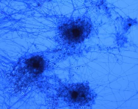
26 4: 7A Conidiobolus coronatus This fungus didn t survive freeze drying by QAP = most labs failed to isolate this fungus Conidiobolus spp. Phylum: Zygomycota, Order: Entomophthorales 27 species Most common sp: C. coronatus, C. incongruus & C. lamprauges
, wide hyphae, coenocytic or irregular septate Simple conidiophores single forcibly discharged terminal conidium")
27 Conidiobolus spp. Rapid growth of waxy colonies (routine agar), wide hyphae, coenocytic or irregular septate Simple conidiophores single forcibly discharged terminal conidium papilla scar remains. Ballistospores- growth on petri dish lid Zygospores intercalary, thick walled, hyaline, w/o beaks Photo from Mycology Online
28 Differentiating between Conidiobolus species Species Colony (PDA 3 days) Forcibly discharged conidia w or w/o secondary conidia Primary conidia size/shape Villose (hair-like) conidia Conidiobolus coronatus > 40 mm Yes 40 um/ prominent papillate base Yes (in older cultures) C. lamprauges < 30 mm No um/ spherical, thin walled, 1-1 many papilla after liberation No C. incongruus < 30 mm Yes um, single tapering basal papilla No


29 Conidiobolus coronatus Multiplicative conidia Papilla RNSH photo (high magnification) Photo from Mycology Online Primary conidia of C. coronatus
, & papillae (site of")
30 C. coronatus Papillae Villose conidia C. coronatus -mature, spherical villose conidia (hair-like protrusions), & papillae (site of former attachment to conidiophore) Photo from Mycology Online
31 Differentiating between Basidiobolus & Conidiobolus Microscopic features Basidiobolus spp. Conidiobolus spp. Ejected sporangioles with papilla Sporangiophores with swollen apices Zygospores Zygospores with conjugation beaks No Yes Yes Yes Yes No No- C. coronatus Yes- C. incongruus C. lamprauges No (Yes 2 beaks in C. lamprauges)


32 Basidiobolus ranarum B. ranarum- beaked zygospores Discharged globose conidia & conidiophores Photos from Mycology Online


33 Rhino-facial infections caused by Conidiobolus Photos from Mycology Online Infections mainly in tropics- eg. Africa, Asia, Central America, India. Infection not evident until swelling/ deformity occurs.
34 Epidemiology Found in soil & decaying debris (found on bananas, rotten wood & a fruit warehouse etc). Ubiquitous- wide range of areas- Alsaksa,, temperate to tropical regions (eg( eg.. PNG soil) Insect pathogen (mosquitoes, termites, aphids) Infects man (80% cases are males) (typically) nasal mucosa & para-nasal sinus, (sometimes) subcutaneous, (rarely) pulmonary & pericardial infections firm, subcutaneous nodules/polyps Immune-competent & immune-suppressed suppressed- emerging opportunist. Occur sporadically but rarely. Route: inhalation Infects animals- horses, sheep, deer, dogs & chimpanzees. Hard to treat- high MIC s?itraconazole, Amp B + terbinafine, flucon, keto, iodides, plastic surgery
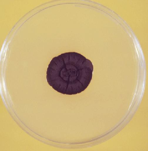
35 Item 4: 7B Clinical history This culture was isolated from sputum collected from a lung transplant patient who presented with a chronic cough
36 4:7B Geotrichum candidum Teleomorph: Galactomyces candidum Description: Colonies, fast growing (routine agar), white, rough texture, hairy, creeping, mostly submerged, dry. Fruity odour. Hyaline hyphae, aerial hyphae fragment forming arthroconidia (cylindrical, barrel shaped or ellipsoidal). No blastospores produced. No capsules. [Trichosporon spp. produce blastospores along pseudohyphae] Differential features of G. candidum: Hyphae 12 um wide with dichotomous branching at colony margin. Assimilation: xylose POS, cellobiose NEG NG at 40C.

37 Colonial morphology G. candidum Sabs agar (28C 4 days)

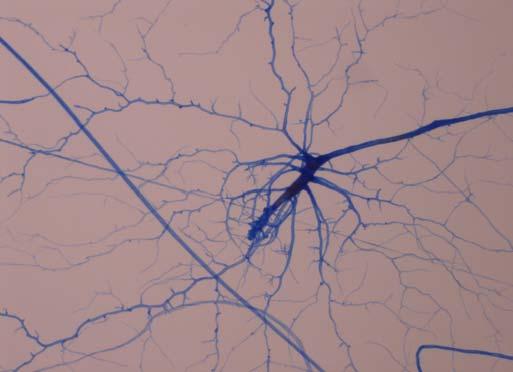
38 Microscopy Photo from Mycology Online Geotrichum candidum- Course true hyphae (no pseudohyphae) that segment into arthoconidia. main & lateral branches with arthroconidia. Dichotomously branching.
39 Similar species to Geotrichum Trichosporon sp- blastoconidia & pseudohyphae (not seen in Geotrichum has true hyphae) Scytalidium sp- dematiacous arthroconidia (hyaline arthos in Geotrichum) Arthrographis & Oidiodendron sp- have condiophores (which Geotrichum lacks) Malbranchea sp- arthroconida release by disjunctor cells ( (Geotrichum produces arthros by fission) Coccidioides sp- arthros alternate with empty cells ( (Geotrichum- consecutive)
40 Biomerieux ID 32c result Profile number: Xylose assimilation POS Cellobiose NEG Result: Geotrichum candidum
41 Differentiating between Geotrichum species G. candidum G. capitaum G. clavatum Cellobiose Neg Neg Pos D-xylose Pos Neg Neg Salicin Neg Neg Pos Vitamin free Pos Neg Neg Arbutin Neg Neg Pos Growth at 37C V Pos Pos [Ref: dehoog, Guarro et al. Atlas of Clinical Fungi p. 227]
42 4:7:B QAP results Geotrichum candidum Geotrichum sp. G. pennicillatum G. captatum G. klebahni Candida kefyr Unable to ID 82 labs (73.2%) 16 (14.3%) 7 (6.3%) 1 (0.9%) 1 (0.9%) 1 (0.9%) 4 (3.6%) 96% labs correct to genus level 73% correct to species level Results obtained from Microbiology QAP
43 Epidemiology G. candidum found worldwide (tundra, temperate, tropical climates) in soil, marine habitats, water, air, sewage, plants,, freq dairy, cereals (eg.frozen( fruit cake, bread), tomatoes, animals skin & in droppings etc. Found in gas oil in Germany, paper pulp factory in France, isolated from irradiated soil. Normal human flora- isolated from faeces & sputum Opportunistic infections in immune compromised ( Geotrichosis ) Endogenous- oral, bronchial, systemic & exogenous- skin, allergic & trauma. Disseminated infection- poor prognosis. Route: Ingestion, inhalation or trauma?also may cause environmental damage- eg. destroy data- storing polycarbonate resin found in CDs! (disc becomes transparent). Also sig cause of citrus fruit rot.
44 Item 4: 7C Clinical history This culture was isolated from a corneal scarping collected from a 12 y.o. with an eye injury
45 4:7:C Candida kefyr Synonym: Candida pseudotropicalis Teleomorph: Kluyveromyces marxianus Colony- white, cream- coloured, smooth texture Microscopy (CMA tween 80) shows elongated blastospores, parallel logs in a stream, curved along pseudohyphae Differential features of C. kefyr: Raffinose POS, 2-keto 2 gluconate (2KG) NEG, ethylamine POS, growth on 0.1% cyclohex G at 37C, G w/o niacin.
 ie. typical yeast morphology.")
46 Colonial morphology Candida kefyr colony white, cream coloured, butyrous (buttery) ie. typical yeast morphology. (Sabs 28C 3 days)

47 Microscopy Pseudohyphae Candida kefyr elongated blastoconidia Elongated blastoconidia Photo from Boekhout et al. CD-ROM Yeasts of the World
 asci with ascospores ascospore Photo from Boekhout et al.")
48 Kluyveromyces marxianis asci (Teleomorph of C. kefyr) asci with ascospores ascospore Photo from Boekhout et al. CD-ROM Yeasts of the World
49 Biomerieux ID 32c result Profile number: Raffinose assimilation POS 2 KG Neg Result: C. kefyr 99.9% T 0.88
50 4:7:C QAP results Candida kefyr Candida sp. Candida (not albicans) Candida tropicalis Geotrichum candidum Unable to ID 102 labs (90.3%) 6 (5.3%) 1 (0.9%) 1 (0.9%) 1 (0.9%) 2 (1.8%) 97% labs correct to genus level 90% correct to species level Results obtained from Microbiology QAP
51 Epidemiology Found worldwide in humans, other mammals & dairy foods Emerging fungal pathogen. Infections- occasionally superficial candidiasis. Reported cases of pulmonary infections, disseminated infection, oesophagitis in immune compromised.
52 A Review of 2005 Mycology QAP (despatchdespatch 6) Okcha Lee Catherine Wu Mycology Unit, CIDMLS, ICPMR Westmead hospital
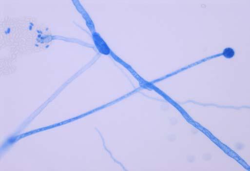
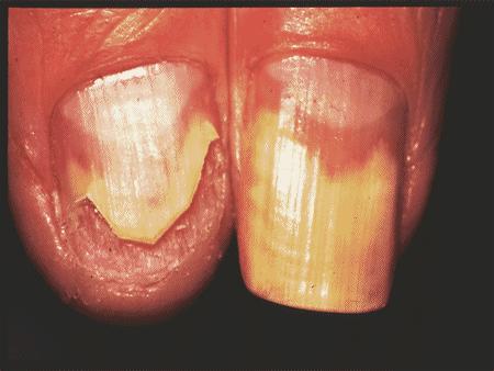
53 Item 2005:6:7A Clinical notes: Culture was isolated from fingernail scrapings taken from a 23 year-old African man with suspected onychomycosis. Direct microscopy showed the presence of septate hyphae.


54 Colony Initially whitish, finally becoming dark gray Spreading, fast


55 Microscopy Hyphae: melanized or hyaline, falling into athroconidia Chlamydospore: conidia single or in chains, dark brown, thick walled, swollen up to 7µm 7 m wide
56 Pathogenicity Identification Scytalidium spp Onychomycosis Dermatophytosis like infection Ecology Soil fungus or plant pathogen in tropical and subtropical areas * 60% correct to genus level
57 Item 2005:6:7B Clinical notes: Culture was isolated from a wound swab taken from a 30 year-old man recovering from severe burns.


58 Colony Bright yellow with dark yellow reverse.


59 Microscopy
60 Microscopy Hyphae: sparsely septated Sporangiophores: Sporangia: <100µm Columella: spherical Sporangiospores: variable in size and shape Chlamydospores: present Rhizoids: present Growth temperature: growth at 37ºC no growth at 42 ºC
61
62 Comparison between Rhizomucor variabilis and Mucor hiemalis colony Sporangio- phores sporangia columella Rhizomucor variabilis Greyish-ochraceous Up to 2mm long, 9-23um 9 wide, branched Subspherical,, up to 100um diameter Spherical, ellipsoidal to cylindrical in various shape, up to 40µm wide Mucor hiemalis Whitish to ochraceous Up to 15mm long, 14um wide, branched Up to 80um diameter Ellipsoidal, spherical, up to um
63 Comparison continued sporangios pores chlamydos pores rhizoids Maximum growth Tm Rhizomucor variabilis Hyaline, smoothwalled,, very variable um abundant abundant 38ºC Mucor hiemalis ellipsoidal, smooth-walled um ºC
64 Molecular ITS1 ITS3 LR1 D1 D2 ITS2 ITS4 LR16 18S rdna (SSU) ITS 1 5.8S rdna ITS 2 28S rdna (LSS) Schematic diagram of ribosomal gene cluster of fungi
65 Comparison of sequencing results between Rhizomucor variabilis and Mucor hiemalis ITS 1 and ITS 2 region Rhizomucor variabilis not in database Mucor hiemalis 92% (452/487 bp) SSU Base pairs ( bp) 99% (487/488 bp) 98% (484/488 bp) Base pairs ( bp) 100% 557/557 bp) 98% (547/557 bp) LSU D1/D2 region ( bp) 99% (638/644 bp) 97% 642/658 bp)
66 Relationship between Rhizomucor variabilis and Mucor hiemalis Rhizomucor variabilis may concern degenerate cultures of Mucor hiemalis Phylogenetically it proved to be very close to Mucor hiemalis Identification Rhizomucor variabilis * 63% answered to Mucor spp and 3.6% to Rhizomucor variabilis
67 Item 2005:6:7C Clinical notes: Culture was isolated from a lung biopsy taken from a 15 year-old girl with acute lymphocytic leukaemia.


68 Colony Usually green, cream-buff or honey-yellow yellow where cleistothecia form Reverse may be olive to purple-brown
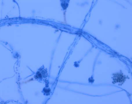
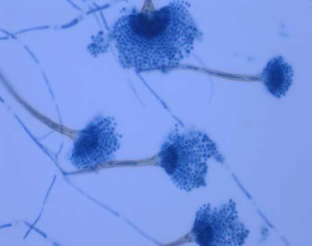

69 Microscopy
70 Microscopy Conidiophores: short (< 250um) Conidial heads: short columnar (< 80um) Conidiogenous cells: biseriate, metulae and phialide Conidia: globose ( um in diam) Teleomorph: Emericella nidulans cleistothecia with reddish ascospores are often surrounded by HülleH cells (up to 25um in diam)
71 Identification Aspergillus nidulans Pathogenicity Opportunistic infection Ecology Soil fungus Prominent colonizer of decomposing plant debris * 94% correct to genus level and 74% to species level
72 RCPA Quality Assurance Programs A Review of the 2005 Mycology QAP Mycology Items 2005:8:7 A,B,C Mirka Rej Microbiology Department Symbion and Laverty Pathology North Ryde
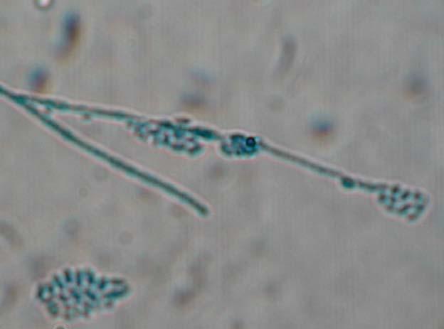
73 Item 2005:8:7A Clinical Notes: Culture was isolated from a knee aspirate taken from a ten year old leukemic boy with osteomyelitis of the right knee.

74 Identification: Macromorphology Young colony is cottony or moist (yeasty) and light grey to black. Mature colony becomes dark grey to black and may develop white mycelial tufts with age. Reverse is grey to black.
. Swelling of the basal portion of the annellides.")
75 Identification: Micromorphology Septate hyphae Unbrached conidiogenous cells (annellides). Swelling of the basal portion of the annellides. Swan necks
76 Conidia are one celled, smooth and ovoid with a slightly narrowed, truncated base. Conidigenous cells locally aggregated into small brushes. Sympotial conidia
77 Clinical importance of Scedosporium prolificans Documented as a cause of: osteomyelitis septic arthritis onychomycosis otomycosis endophthalmitis sinusitis gangrenous skin ulcers pulmonary infections peritonitis systemic infections (may be isolated from blood cultures)
78 Clinical importance of Scedosporium prolificans Scedosporium prolificans in nature occurs in soil and is traumatically introduced into humans by thorns or splinters. In the body it has a predilection for cartilage and joint areas. In immunocompromised patients or transplantation patients fatal dissemination may occur. A fatal case of endocarditis was reported. Isolates are often resistant to antifungal agents.
79 RCPA Quality Assurance Programs Unable to identify Yeast species Candida guillermondii Pseudallescheria boydii Sporothrix schenckii Sporotrichum schenckii Wangiella dermatidis Exophiala jeanselmei Fonsecaea species Phialophora richardsiae Phialophora species Phialophora verrucosa Scedosporium inflatum Scedosporuim species Scedosporium apiospermum Scedosporium prolificans 84% correct to genus level. 76% correct to species level
80 growth on media contraining cycloheximide Differences between S. apiospermum and S. prolificans S. apiospermum S. prolificans + - annellides cylindrical flask shaped, swollen basal portion annellidic rings difficult to see long thin appendages (swan neck) formation of + (P.boydi) - clastothecia synnemata + - cultures white cottony mycelium later turns grey to brown first moist grey to black later dark grey, white tufts in the centre. growth at 45 degrees - +

81 Scedosporium prolificans Bottle shaped annellides Annellides with parrallel sides may also be seen.
82 Item 2005:8:7B Clinical Notes: Culture was isolated from the fingernail scraping taken from a 50 year old woman with chronic paronychia.

83 Identification: Macromorphology White to cream coloured, smooth, glabrous yeast-like colonies.

84 Identification: Micromorphology Budding yeast like cells. cells spherical to broadly ellipsoidal x µm.
85 Identification For the identification of germ tube negative yeasts, morphological (Dalmau plate culture) physiological and biochemical tests are essential. Reliable commercially available yeast identification kits are the API 20C AUX, ATB32C, Vitek systems.

86 Microscopy Morphology on Cornmeal-Tween 80 agar Fairly short, fine pseudohyphae. Clusters of blastoconidia at septa.
87 Identification: Physiological tests: Germ Tube test: Hydrolysis of Urea: Growth on Cycloheximide: Growth at 37C: Negative Negative Positive Positive
88 Fermentation reactions: Identification: Glucose: Lactose: Sucrose: Galactose: Trehalose: Maltose: Variable - -
89 Differential Diagnosis The species can be recognised by the following growth characters: D-glucosamine D-arabinose cellobiose inulin erythritol L-arabinitol D-glucuronate
90 Clinical Importance of Candida guilliermondii is found from normal skin, in sea water, faeces of animals, fig wasps, buttermilk, leather, fish and beer. has been isolated from numerous human infections.

91
92 Clinical Importance Yeasts are considered opportunistic pathogens causing disease in patients: with a breakdown in the body s immune system on prolonged treatment with antibiotics on corticosteroids or cytotoxic drugs with intravascular catheters with diabetes mellitus known to be intravenous drug abusers
93 RCPA Quality Assurance Programs No growth Unable to identify Candida glabrata Candida famata Candida species Candida guilliermondii % correct to genus level. 89% correct to species level.
94 Item 2005:8:7C Clinical Notes: A 40 year old market gardener presented with a tender subcutaneous nodule on the leg. Fluid aspirated from the nodule grew Culture C.

95 Identification: Macromorphology Initially smooth, black, mucoid and yeast like colonies. Revers is black Grows slower or not at all at 37C

96 Identification: Macromorphology Colonies become raised and develop tufts of aerial mycelium with age. Often dome shaped and suede like in texture.

97 Identification: Micromorphology Yeast like cells Torulose hyphae


98 Identification: Micromorphology The conidiogenous cells are slender annellides. Tips of annellides are narrow, elongated and rocket shaped. Conidia gather in clusters at the end, sides of conidiophore and at points along the hyphae. Inflated germinating cells. Budding yeast like cells. Conidia 1-3 x 2-5 µm in size, hyaline, smooth, thin walled and broadly ellipsoidal.
99 Clinical Importance of Exophiala jeanselmei Mainly associated with phaeohyphomycotic cysts. The fungus has also been documented as a cause of black-grained mycetoma. Has been reported from a small number of cases of chromoblastomycosis.
100
101 RCPA Quality Assurance Programs Unable to identify Yeast species Cladosporium carrionii Cladosporium species Sporothrix schenckii Homonema deratioides Hortaea werneckii Phialophora gougerotii Phialophora species Philophora verrucoas Fonsecaes pedrosoi Fonsecaea dermatidis Fonsecaea species Wangiella dermatitidis Exophila castellanii Exophila spinifera Exophila dermatitidis Exophiala species Exophiala jeanselmei 76% correct to genus level 52% correct to species level
102 Differences betweem Exophiala jeanselmei and Wangiella dermatitidis (Exophiala dermatitidis) growth at 42 degrees conidiogenous cell Exophiala Wangiella jeanselmei dermatitidis - + annellide tapering extensions to the tip. phialide. do not show exidence of extension.

103 Wangiella dermititidis Exophiala jeanselmei
104 Books: References Ellis, D., Davis, S., Alexiou,, H., Pfeiffer, T., Manatakis,, Z. (1992). " "Descriptions of Medicat QAP fungi". Mycology Unit, Adelaide Children's Hospital. Hocking, A., Woodgyer,, A. "Clinically Significant Non-Dermatophyte Fungi a Practical Guide to Identification". AIMS National Scientific Meeting 2001, Melbourne Hoog G., Guarro,, J. (1995). " "Atlas of Clinical Fungi" Centraalbureau voor Schimmelcultures,, The Netherlands, and Universitat Rovira i Virgili,, Spain] Larone,, D. (1995). "Medically Important Fungi" 3rd Edition.. American Society for Microbiology, Washington. Murry,, P., Baron, E., Pfaller,, M., Tenover,, F., Yolken,, R. (1995). " "Manual of Clinical Microbiology" 6th Edition.. American Society for Microbiology, Washington. Websites: microbiology.mtsinai.on.ca/mig/defungi/index2.shtml ematiaceous)/exo phiala/jeanselmei.html dii/ onychomycosis.com/images/table%201.htm
Mycology. BioV 400. Subcutaneous Mycoses. Ecological associations. Geographic distribution World-wide
 BioV 400 Mycology Handout 8 Subcutaneous Mycoses Lymphocutaneous sporotrichosis Chromoblastomycosis Phaeohyphomycosis Zygomycosis Mycetoma Lymphocutaneous sporotrichosis Sporothrix schenckii Chronic infection
Identification of Yeasts. Medical Mycology Training Network 15 November 2018 Dr Tan Ai Ling Department of Microbiology, Singapore General Hospital
 Identification of Yeasts Medical Mycology Training Network 15 November 2018 Dr Tan Ai Ling Department of Microbiology, Singapore General Hospital Definition of Yeasts Eukaryote cells have defined nucleus
LESSON ASSIGNMENT. Introduction to Medical Mycology. After completing this lesson, you should be able to:
 LESSON ASSIGNMENT LESSON 1 Introduction to Medical Mycology. TEXT ASSIGNMENT Paragraphs 1-1 through 1-7. TASKS OBJECTIVES After completing this lesson, you should be able to: 1-1. Select the statement
Subcutaneous Mycosis
 Subcutaneous Mycosis Fungal infections 1. Superficial mycosis. 2. Coetaneous mycosis: Dermatophytoses. 3. Subcutaneous mycosis. 4. Systemic mycosis. 5. Opportunistic mycosis. Subcutanus mycoses Fungal
Definition. Phaeohyphomycosis
 Phaeohyphomycosis Synonyms: Cerebral chromomycosis, chromoblastomycosis, chromomycosis, cladosporiosis, phaeomycotic cyst, phaeosporotrichosis, subcutaneous mycotic cyst. Definition Phaeohyphomycosis consists
Introduction. Study of fungi called mycology.
 Fungi Introduction Study of fungi called mycology. Some fungi are beneficial: ex a) Important in production of some foods, ex: cheeses, bread. b) Important in production of some antibiotics, ex: penicillin
Mycology. BioV 400. Clinical classification. Clinical classification. Fungi as Infectious Agents. Thermal dimorphism. Handout 6
 BioV 400 Mycology Handout 6 Fungi as Infectious Agents True or primary fungal pathogens invades and grows in a healthy, noncompromise d host Most striking adaptation to survival and growth in the human
Dermatophytes Dr. Hala Al Daghistani
 Dermatophytes Dr. Hala Al Daghistani Dermatophytoses are superficial infections of the skin and its appendages, commonly known as ringworm, athlete s foot, and jock itch. They are caused by species of
Subcutaneous Fungi 10/13/2009. General Characteristics. Pathogenesis. Epidemiology. Laboratory Diagnosis. Specimens. Growth rate: 1-4 weeks
 General Characteristics Growth rate: 1-4 weeks Subcutaneous Fungi Clinical Laboratory Science Program Carol Larson MSEd, MT(ASCP) Dematiaceous septate hyphae Hyaline septate hyphae Branching GPR Epidemiology
1. Multiple choice (30 2 each); circle the number of the correct choice. b. Trichophyton schoenleinii is traditionally most associated with
; circle the number of the correct choice. b. Trichophyton schoenleinii is traditionally most associated with") NAME SS# EXAM 2 March 26, 2002 BIO 329 Directions: All explanations, definitions, and descriptions should be presented in good English This means complete sentences should be used except when lists or
NAME SS# EXAM 2 March 26, 2002 BIO 329 Directions: All explanations, definitions, and descriptions should be presented in good English This means complete sentences should be used except when lists or
Histopathology Description:
 2013-2-1 CANINE HEART Ahmed M. Abubakar BOVINE PATHOLOGY CONTRIBUTING INSTITUTION : The Royal Veterinary college, Dept. of Pathology and Biology Signalment: 11-month-old male Border Collie dog (Canis familiaris)
LESSON ASSIGNMENT. Upon completion of this lesson, you should be able to
 LESSON ASSIGNMENT LESSON 4 Yeasts of Medical Importance. LESSON ASSIGNMENT Paragraphs 4-1 through 4-10. LESSON OBJECTIVES Upon completion of this lesson, you should be able to 4-1. Select the statement
Absidia corymbifera. Absidia corymbifera showing a typical pyriform-shaped sporangium with a conical-shaped columella and pronounced apophysis.
 Absidia corymbifera Absidia corymbifera showing a typical pyriform-shaped sporangium with a conical-shaped columella and pronounced apophysis. Colonies are fast growing, floccose, white at first becoming
All three dermatophytes contain virulence factors that allow them to invade the skin, hair, and nails. Keratinases. Elastase.
 DERMATOPHYTOSIS (=Tinea = Ringworm) Infection of the skin, hair or nails caused by a group of keratinophilic fungi, called dermatophytes Microsporum Epidermophyton Hair, skin Skin, nail Tih Trichophyton
Ali Alabbadi. Sarah Jaar ... Nader
 24 Ali Alabbadi Sarah Jaar... Nader Intro to Mycology *underlined text was explained in the lecture but is not found in the slides -mycology: the study of the mycoses of man (fungal infections) -less than
Introduction Medical Mycology. Prof. Dr. Asem Shehabi Faculty of Medicine University of Jordan
 Introduction Medical Mycology Prof. Dr. Asem Shehabi Faculty of Medicine University of Jordan General Fungi-1 Medical Mycology deals with fungi cause human diseases directly (mycoses, allergies) or indirectly
Nursing college, Second stage Microbiology Dr.Nada Khazal K. Hendi Medical Microbiology
 1 Nursing college, Second stage Microbiology Medical Microbiology Lecture-1- Fungi (Mycosis) They are a diverse group of saprophytic and parasitic eukaryotic organisms. Human fungal diseases (mycoses)
VPM 201: Veterinary Bacteriology and Mycology 23-24/11/2011 LABORATORY 11: MYCOLOGY
 VPM 201: Veterinary Bacteriology and Mycology 23-24/11/2011 LABORATORY 11: MYCOLOGY I. Overview of Major Groups of Pathogenic Fungi. Although the Kingdom Fungi have been undergoing considerable phylogenetic
Tinea nigra. Dr. Ensieh Zibafar
 Piedra & Tinea nigra Dr. Ensieh Zibafar SUPERFICIAL MYCOSES -Tinea versicolor -Tinea nigra -Piedra -Otomycosis -Erythrasma * -Trichomycosis Axillaris * Piedra Means Stone in Spanish White Piedra Black
Fungal infection in the immunocompromised patient. Dr Kirsty Dodgson
 Fungal infection in the immunocompromised patient Dr Kirsty Dodgson Aims Discuss different types of fungi Overview of types of clinical infections Clinical Manifestations Fungus Includes Moulds Aspergillus
Epidemiology and ecology of fungal diseases
 Epidemiology and ecology of fungal diseases Healthcare Focus on: - individual - diagnosis - treatment Public Health Focus on: - population - prevention The nature of fungi Kingdom Fungi (lat. fungus, -i)
Pathogens with Intermediate Virulence Dermatophytes opportunistic Pathogens
 Pathogens with Intermediate Virulence Dermatophytes opportunistic Pathogens Cryptococcus neoformans Candida albicans Aspergillus species Pneumocystis carinii 1 Dermatophytes Named for derma skin Cause
Opportunistic Mycoses
 CANDIDIASIS SOFYAN LUBIS DEPARTEMEN MIKROBIOLOGI FAK.KEDOKTERAN USU MEDAN 2009 Opportunistic Mycoses Opportunistic mycoses are fungal infections that do not normally cause disease in healthy people, but
The Differentiation of Yeast and Yeast-Like Forms in Human Tissues. Introduction. Histochemical Stains Used to Detect Fungi. Histopathologic Diagnoses
 The Differentiation of Yeast and Yeast-Like Forms in Human Tissues Gary W. Procop, MD Chair, Clinical Pathology Staff, Anatomic Pathology Director, Molecular Microbiology, Mycology, and Parasitology Cleveland
Fungi. CLS 311 Mrs. Ohoud alhumaidan
 Fungi CLS 311 Mrs. Ohoud alhumaidan Outlines Intruduc8on General Characteris8cs of Fungi beneficial & harmful effect of fungi Classifica8on of fungi structure of fungi Reproduc8on of fungi important terms
Fungi. Eucaryotic Rigid cell wall(chitin, glucan) Cell membrane ergosterol Unicellular, multicellular Classic fungus taxonomy:
 Cell membrane ergosterol Unicellular, multicellular Classic fungus taxonomy:") MYCOLOGY Mycology I Fungi Eucaryotic Rigid cell wall(chitin, glucan) Cell membrane ergosterol Unicellular, multicellular Classic fungus taxonomy: Morphology Spore formation FFungi Yeast Mold Yeastlike
MYCOLOGY Mycology I Fungi Eucaryotic Rigid cell wall(chitin, glucan) Cell membrane ergosterol Unicellular, multicellular Classic fungus taxonomy: Morphology Spore formation FFungi Yeast Mold Yeastlike
This is the written version of our Hot Topic video presentation available at: MayoMedicalLaboratories.com/hot-topics
 This is the written version of our Hot Topic video presentation available at: MayoMedicalLaboratories.com/hot-topics Welcome to Mayo Medical Laboratories hot topics. These presentations provide short discussion
About the Editor Gerri S. Hall, Ph.D.
 About the Editor Gerri S. Hall, Ph.D. Dr. Hall s professional career has been focused on clinical microbiology: direct clinical activities of various areas such as bacteriology, mycobacteria, STD testing,
Biology of FUNgi. Lecture 21 The Ugly - localized disease-causing fungi
 Biology of FUNgi Lecture 21 The Ugly - localized disease-causing fungi Flashback - fungitoxins Toxins you should know about The deadly Amanita phalloides Galerina autumnalis Group I: Amatoxin and phyllotoxin
NATIONAL BIORESOURCE DEVELOPMENT BOARD Dept. of Biotechnology Government of India, New Delhi
 NATIONAL BIORESOURCE DEVELOPMENT BOARD Dept. of Biotechnology Government of India, New Delhi For office use: MARINE BIORESOURCES FORMS DATA ENTRY: Form- 1(general ) Ref. No.: (please answer only relevant
OPPORTUNISTIC MYCOSES
 OPPORTUNISTIC MYCOSES Candida yeasts that is, fungi that exist predominantly in a unicellular form. small (4-6 µm), thin-walled, ovoid cells (blastospores) that reproduce by budding. do not require special
number Done by Corrected by Doctor د.حامد الزعبي
 number Fungi#1 Done by نرجس الس ماك Corrected by مهدي الشعراوي Doctor د.حامد الزعبي Introduction to Mycology -Terms: -Medical Mycology: The study of mycosis and their etiological agents -Mycosis: Disease
Mycology Review. Background. Background. Specimen Collection. Calcofluor White. Methods. Yeasts. Moulds. Melissa B. Miller, Ph.D.
 Background Mycology Review Melissa B. Miller, Ph.D. April 4, 2008 Yeasts Unicellular Divide by budding or binary fission Moulds Filamentous hyphae interweave to form mycelium Saprobic phase: airborne,
Medical Mycology. Dr. Hala Al Daghistani
 Medical Mycology Dr. Hala Al Daghistani FAre eukaryotes that grow in two basic forms, a yeasts and molds (or moulds). Growth in the mold form occurs by production of multicellular filamentous colonies.
Journal of Chemical and Pharmaceutical Research, 2017, 9(1): Review Article. Graphium Salixicum: A New Species Explored from Salix Alba
: Review Article. Graphium Salixicum: A New Species Explored from Salix Alba") Available online www.jocpr.com Journal of Chemical and Pharmaceutical Research, 2017, 9(1):69-74 Review Article ISSN : 0975-7384 CODEN(USA) : JCPRC5 Graphium Salixicum: A New Species Explored from Salix
Available online www.jocpr.com Journal of Chemical and Pharmaceutical Research, 2017, 9(1):69-74 Review Article ISSN : 0975-7384 CODEN(USA) : JCPRC5 Graphium Salixicum: A New Species Explored from Salix
Medical Mycology. Dr. Hala Al Daghistani
 Medical Mycology Dr. Hala Al Daghistani Mycotic Infections GENERAL CONCEPTS A. The fungi represent a diverse, heterogeneous group of eukaryotic B. Most of these organisms are plant pathogens and relatively
CUTANEOUS MYCOSES. Introduction
 1 CUTANEOUS MYCOSES Dr. Mohamed El-Sakhawy Epidermis Introduction Outermost layer of the skin Its layers are made of Mostly dead cells. Most of the cells of the epidermis undergo rapid cell division (mitosis).
Types of fungi Diseases that can be caused by filamentous fungi or yeast which can cause: Aspergillusis-mold infection Micro conidia Microspore
 1 Overview: Types of fungi Diseases that can be caused by filamentous fungi or yeast which can cause: Tinea corporis-skin infection Tinea capitis-hair infection Tinea Unguium-Nail infection Aspergillusis-mold
Evaluation of an alternative slide culture technique for the morphological identification of fungal species
 Research Article Evaluation of an alternative slide culture technique for the morphological identification of fungal species Abstract M H Wijedasa 1, L V C Liyanapathirana 1. Sri Lanka Journal of Infectious
Epidemiology and Laboratory Diagnosis of Fungal Diseases
 Medical Mycology (BIOL 4849) Summer 2007 Dr. Cooper Epidemiology of Mycoses Epidemiology and Laboratory Diagnosis of Fungal Diseases Mycosis (pl., mycoses) - an infection caused by a fungus Two broad categories
Mycobacteria and fungal infections of the respiratory tract
 Before you start: You must read the slides! The Dr. did not bother to explain them all Mycobacteria and fungal infections of the respiratory tract - TB is a global problem that the WHO is trying to combat.
Fungal infection. Jantima Jantima Tanboon,MD
 Fungal infection Jantima Jantima Tanboon,MD Yeast Mold Diagnosis -Wood s light -KOH preparation -Periodic acid-schiff (PAS) -Gomorimethinaminesilver (GMS) -India ink, mucicarminestain -Calcofluorwhite
Staphylococci. Gram stain: gram positive cocci arranged in clusters.
 Microbiology lab Respiratory system Third medical year Lab contents: Gram positive bacteria (Staphylococcus and Streptococcus spp), two types of filamentous fungi (Aspergillus and Penicillium spp), and
NATIONAL BIORESOURCE DEVELOPMENT BOARD Dept. of Biotechnology Government of India, New Delhi
 NATIONAL BIORESOURCE DEVELOPMENT BOARD Dept. of Biotechnology Government of India, New Delhi For office use: MARINE BIORESOURCES FORMS DATA ENTRY: Form- 1(general ) Ref. No.: (please answer only relevant
Osteolytic Phaeohyphomycosis in a German Shepherd Dog Caused by Phialemonium obovatum
 JOURNAL OF CLINICAL MICROBIOLOGY, May 1986, p. 987-991 Vol. 23, No. 5 0095-1137/86/050987-05$02.00/0 Copyright C) 1986, American Society for Microbiology Osteolytic Phaeohyphomycosis in a German Shepherd
Impression smear from a nasal mass on a 2 year old cat Presented with: one month duration of epistaxis
 Impression smear from a nasal mass on a 2 year old cat Presented with: one month duration of epistaxis Identify the structures neutrophils macrophages x40 Organisms 8 30 micron in size, with variable capsule
MSES consultants, inc.
 MSES consultants, inc. 609 West Main Street P.O. Drawer 190 Clarksburg, WV 26302-0190 304.624.9700 304.622.0981 304.842.3325 http://www.msesinc.com Office September 13, 2012 Project Number: 12-437 Mr.
Speciation of Candida Isolated from Various Clinical Samples and their Antifungal Susceptibility Profile in a Tertiary Care Hospital
 International Journal of Current Microbiology and Applied Sciences ISSN: 2319-7706 Volume 5 Number 7 (2016) pp. 54-60 Journal homepage: http://www.ijcmas.com Original Research Article http://dx.doi.org/10.20546/ijcmas.2016.507.004
Definition. Paracoccidioidmycosis. History. Paracoccidioides brasiliensis. Epidemiology
 Definition Paracoccidioidmycosis Hillary Howard Carlisle Heinselman Sarah Maher Arjun Vikuntam A systematic endemic disease produced by Paracoccidioides brasiliensis Primarily a pulmonary disease Disseminated
NATIONAL BIORESOURCE DEVELOPMENT BOARD Dept. of Biotechnology Government of India, New Delhi
 NATIONAL BIORESOURCE DEVELOPMENT BOARD Dept. of Biotechnology Government of India, New Delhi For office use: MARINE BIORESOURCES FORMS DATA ENTRY: Form- 1(general ) Ref. No.: (please answer only relevant
Common Fungi. Catherine Diamond MD MPH
 Common Fungi Catherine Diamond MD MPH Birth Month and Day & Last Four Digits of Your Cell Phone # BEFORE: http://tinyurl.com/kvfy3ts AFTER: http://tinyurl.com/lc4dzwr Clinically Common Fungi Yeast Mold
Management of fungal infection
 Management of fungal infection HKDU symposium 17 th May 2015 Speaker: Dr. Thomas Chan MBBS (Hons), MRCP, FHKCP, FHKAM Synopsis Infection caused by fungus mycoses Skin infection by fungus is common in general
First Report of Penicillium adametzioides from Decayed Grapes (Vitis vinifera) in Pakistan
 in Pakistan") International Journal of Current Microbiology and Applied Sciences ISSN: 2319-7706 Volume 5 Number 12 (2016) pp. 316-320 Journal homepage: http://www.ijcmas.com Original Research Article http://dx.doi.org/10.20546/ijcmas.2016.512.034
International Journal of Current Microbiology and Applied Sciences ISSN: 2319-7706 Volume 5 Number 12 (2016) pp. 316-320 Journal homepage: http://www.ijcmas.com Original Research Article http://dx.doi.org/10.20546/ijcmas.2016.512.034
REINWARDTIA Published by Herbarium Bogoriense LBN, Bogor Vol. 10, Part 2, pp (1985) THE ANAMORPH OF SARAWAKUS SUCCISUS RIFAI
 THE ANAMORPH OF SARAWAKUS SUCCISUS RIFAI") REINWARDTIA Published by Herbarium Bogoriense LBN, Bogor Vol. 10, Part 2, pp. 265 270 (1985) THE ANAMORPH OF SARAWAKUS SUCCISUS RIFAI MIEN A. RIFAI, KARTINI KRAMADIBRATA Herbarium Bogorievnc LBN, Bogor,
REINWARDTIA Published by Herbarium Bogoriense LBN, Bogor Vol. 10, Part 2, pp. 265 270 (1985) THE ANAMORPH OF SARAWAKUS SUCCISUS RIFAI MIEN A. RIFAI, KARTINI KRAMADIBRATA Herbarium Bogorievnc LBN, Bogor,
15 CLASS Superficial mycoses. Superficial candidiasis. Histoplasmosis. Blastomycosis. Chromomycoses. Sporotrichosis
 15 CLASS Superficial mycoses. Superficial candidiasis. Histoplasmosis. Blastomycosis. Chromomycoses. Sporotrichosis SUPERFICIAL MYCOSES PITYRIASIS VERSICOLOR Pityriasis versicolor is a chronic mild superficial
A class IIa medical device intended for mild-to-moderate fungal nail infection PRODUCT MONOGRAPH
 A class IIa medical device intended for mild-to-moderate fungal nail infection PRODUCT MONOGRAPH AWB-2052628721 Date of Preparation March 2017 Introduction to Bayer Bayer is a Life Science company with
Impact of migration on health care: When medicine meets politics Some case reports. Dr. Louis Ide, senator
 Impact of migration on health care: When medicine meets politics Some case reports Dr. Louis Ide, senator Case 1 In February to March, 2008, four patients were diagnosed with tinea capitis. Wood s light
L11 Fungal Infection SCBM341: GENERAL PATHOLOGY. Niwat Kangwanrangsan, Ph.D. Department of Pathobiology Faculty of Science, Mahidol University
 SCBM341: GENERAL PATHOLOGY L11 Fungal Infection Niwat Kangwanrangsan, Ph.D. Department of Pathobiology Faculty of Science, Mahidol University Outline Introduction Pathogenesis
Cutaneous phaeohyphomycosis in an immunocompromised host
 Hong Kong J. Dermatol. Venereol. (2014) 22, 85-89 Case Report Cutaneous phaeohyphomycosis in an immunocompromised host YXE Tay, JY Pan, SSJ Lee Phaeohyphomycosis is an infection caused by dematiaceous
Trichophyton Microsporum Epidermophyton. dermatomycosis. Dematiaceous(pigmented fungi ) Dimorphic fungi Yeast and yeast like saprophyte
 Dimorphic fungi Yeast and yeast like saprophyte") Cutaneous candidiasis dermatophytosis Trichophyton Microsporum Epidermophyton dermatomycosis Dematiaceous(pigmented fungi ) Dimorphic fungi Yeast and yeast like saprophyte dermatomycosis Yeast & yeast
Cutaneous candidiasis dermatophytosis Trichophyton Microsporum Epidermophyton dermatomycosis Dematiaceous(pigmented fungi ) Dimorphic fungi Yeast and yeast like saprophyte dermatomycosis Yeast & yeast
2046: Fungal Infection Pre-Infusion Data
 2046: Fungal Infection Pre-Infusion Data Fungal infections are significant opportunistic infections affecting transplant patients. Because these infections are quite serious, it is important to collect
RAFI, M. AND SAJJAD-UR-RAHMAN Department of Veterinary Microbiology, University of Agriculture, Faisalabad 38040, Pakistan
 INTERNATIONAL JOURNAL OF AGRICULTURE & BIOLOGY 1560 8530/00/04 4 553 558 http://www.ijab.org Isolation and Identification of Indigenous Penicillium chrysogenum Series RAFI, M. AND SAJJAD-UR-RAHMAN Department
Dr Hamed Alzoubi. Fungal infections
 Dr Hamed Alzoubi Fungal infections Skin & subcutaneous Mycoses 1-Superficial mycoses such as 2-Cutaneous mycoses such as 3-Subcutaneous mycoses Tinea versicolor or Pityriasis versicolor Ring worm or Tinea
Autopsy findings in 51 year-old man with mantle cell lymphoma
 Autopsy findings in 51 year-old man with mantle cell lymphoma Bobbi S. Pritt, MD, MSc Professor of Laboratory Medicine and Pathology Mayo Clinic Disclosure of Relevant Financial Relationships USCAP requires
7-002b: Malt agar method for the detection of Alternaria radicina on Daucus carota (carrot)
") International Rules for Seed Testing Annexe to Chapter 7: Seed Health Testing Methods 7-002b: Malt agar method for the detection of Alternaria radicina on Daucus carota (carrot) Published by: International
International Rules for Seed Testing Annexe to Chapter 7: Seed Health Testing Methods 7-002b: Malt agar method for the detection of Alternaria radicina on Daucus carota (carrot) Published by: International
number Done by Corrected by Doctor
 Mycology number 2 Done by Corrected by Doctor Hamed Al Zoubi 30 11/12/2017 Fungal infections Dr Hamed Alzoubi Skin & subcutaneous Mycoses 1-Superficial mycoses such as 2-Cutaneous mycoses such as 3-Subcutaneous
Hyaline Molds. Aspergillus clavatus. Aspergillus flavus. Aspergillus fumigatus. Aspergillus glaucus. Aspergillus nidulans. Aspergillus niger Complex
 Hyaline Molds Aspergillus clavatus Aspergillus flavus Aspergillus fumigatus Aspergillus glaucus Aspergillus nidulans Aspergillus niger Complex Aspergillus terreus Complex Aspergillus ustus Aspergillus
In Vitro Studies with R 51,211 (Itraconazole)
") ANTIMICROBIAL AGENTS AND CHEMOTHERAPY, JUIY 1984, p. 5-9 Vol. 26, No. 1 0066-4804/84/070005-05$02.00/0 Copyright 1984, American Society for Microbiology In Vitro Studies with R 51,211 (Itraconazole) ANA
ANTIMICROBIAL AGENTS AND CHEMOTHERAPY, JUIY 1984, p. 5-9 Vol. 26, No. 1 0066-4804/84/070005-05$02.00/0 Copyright 1984, American Society for Microbiology In Vitro Studies with R 51,211 (Itraconazole) ANA
Fungi and their pathogenesis
 Fungi and their pathogenesis -Important -Extra -Notes -In boy s slides -In girl s slides In this link, you will find any correction or notes unmentioned in the team s work. Please check the link below
North American Endemic Fungi
 North American Endemic Fungi Boni Elizabeth Elewski, MD Chair Department of Dermatology University of Alabama at Birmingham James Elder Professor of Graduate Medical Education DISCLOSURE OF FINANCIAL RELATIONSHIPS
7/13/09. Definition. Infections Due to Malassezia. Case Report 1. Case Report 1 (cont.) Case Report 1 (cont.)
 Case Report 1 (cont.)") Definition Infections Due to Malassezia Various species of Malassezia cause both opportunistic, superficial infections and occasionally systemic infections Common superficial infections include: Pityriasis
Definition Infections Due to Malassezia Various species of Malassezia cause both opportunistic, superficial infections and occasionally systemic infections Common superficial infections include: Pityriasis
Pulmonary Infections: Fungus. Part I: Background Information and Dimorphic Fungus. Part II: Opportunistic Yeast and Molds
 Pulmonary Infections: Fungus Part I: Background Information and Dimorphic Fungus Part II: Opportunistic Yeast and Molds Howard J. Sachs, MD www.12daysinmarch.com Classification: Unicellular Budding Yeast,
. 1.5 cm, Fonsecaea pedrosoi. 1.5 cm, Fig. 1.. Bowen,. Phialophora. 240, Fukushiro 1. . Hepatitis C Virus. . Fig. 2.
 Jpn. J. Med. Mycol. Vol. 48, 85 89, 2007 ISSN 0916 4804 Fonsecaea pedrosoi 1 1 2 2 2 1 1 2 3 17, 2006. 12 15, 2006 67 1. 1.5 cm. Fonsecaea pedrosoi., Phialophora., 5 mm,. 3., 1955 1981 Fukushiro 296, 1982
CASE REPORT CHROMOBLASTOMYCOSIS MASQUERADING AS DERMATOPHYTOSIS, WITH THE DESCRIPTION OF A NEW OPPORTUNISTIC SPECIES
 CASE REPORT CHROMOBLASTOMYCOSIS MASQUERADING AS DERMATOPHYTOSIS, WITH THE DESCRIPTION OF A NEW OPPORTUNISTIC SPECIES Kowit Kampirapap 1, Sutthirat Reangchainam 1, Pornpit Ornpaew 2 and Poohglin Tresukosol
Rheem Totah, Office H172M, Ph Office hours MWF 11:30 12:20 or by arrangement
 Rheem Totah, Office H172M, Ph 206-543-9481 rtotah@uw.edu Office hours MWF 11:30 12:20 or by arrangement Date/Time Topic Readings Mon March 26 Antifungal agents Foye s Chapter 40 Wed March 28 Antifungal
Diagnosis of Invasive Septate Mold Infections A Correlation of Microbiological Culture and Histologic or Cytologic Examination
 Microbiology and Infectious Disease / DIAGNOSIS OF SEPTATE MOLD INFECTIONS Diagnosis of Invasive Septate Mold Infections A Correlation of Microbiological Culture and Histologic or Cytologic Examination
Mycology SIG meeting 6/4/11. Bernie Hudson Staff Specialist Microbiology& ID Dept RNSH. Kerry Weeks Senior Scientist, Mycology RNSH
 Mycology SIG meeting 6/4/11 Bernie Hudson Staff Specialist Microbiology& ID Dept RNSH & Kerry Weeks Senior Scientist, Mycology RNSH Tree & Bud What Sputum Grows after a Sri Lankan Holiday Photos by Stella
Received 6 May 2010/Returned for modification 21 June 2010/Accepted 9 July 2010
 JOURNAL OF CLINICAL MICROBIOLOGY, Sept. 2010, p. 3053 3061 Vol. 48, No. 9 0095-1137/10/$12.00 doi:10.1128/jcm.00917-10 Copyright 2010, American Society for Microbiology. All Rights Reserved. Agar Block
International Journal of Advanced Research in Biological Sciences ISSN : Research Article
 International Journal of Advanced Research in Biological Sciences ISSN : 2348-8069 www.ijarbs.com Research Article First Report of Penicillium digitatum (Pers. ex Fr.) Sacc. causing a postharvest green
Epidemiology of Fungal Diseases
 Lecture 2 Epidemiology of Fungal Diseases Disclaimer: This lecture slide presentation is intended solely for educational purposes. Many of the images contained herein are the property of the original owner,
U.S. ARMY MEDICAL DEPARTMENT CENTER AND SCHOOL FORT SAM HOUSTON, TEXAS
 U.S. ARMY MEDICAL DEPARTMENT CENTER AND SCHOOL FORT SAM HOUSTON, TEXAS 78234-6100 MYCOLOGY SUBCOURSE MD0859 EDITION 100 DEVELOPMENT This subcourse is approved for resident and correspondence course instruction.
Am. J. Life. Sci. Res. Vol. 2, Issue 3, , 2014
 2014, World of Researches Publication Am. J. Life. Sci. Res. Vol. 2, Issue 3, 379-383, 2014 American Journal of Life Science Researches www.worldofresearches.com ORIGINAL ARTICLE Received 12 Jan. 2014
Prevalence of Nondermatophytes in Clinically Diagnosed Taeniasis
 ISSN: 2319-7706 Volume 4 Number 7 (2015) pp. 541-549 http://www.ijcmas.com Original Research Article Prevalence of Nondermatophytes in Clinically Diagnosed Taeniasis Sarada Dulla*, Poosapati Ratna kumari
Mycological Profile of Bronchial Wash Specimens in Patients with Lower Respiratory Tract Infections
 International Journal of Current Microbiology and Applied Sciences ISSN: 2319-7706 Volume 6 Number 11 (2017) pp. 176-182 Journal homepage: http://www.ijcmas.com Original Research Article https://doi.org/10.20546/ijcmas.2017.611.022
8/2/10. Sanaz Jalali, Jennifer Demler, Jeremy King. Histoplasmosis is an intracellular mycotic infection of the reticuloendothelial system.
 Histoplasmosis is an intracellular mycotic infection of the reticuloendothelial system. Type of chronic respiratory infection http://www.eregimens.com/regimens/antifungal%20general.htm Sanaz Jalali, Jennifer
L11 Fungal Infection. Introduction. Outline. Fungi. Site of infection
 Outline SCPA501: GENERAL PATHOLOGY L11 Fungal Infection Niwat Kangwanrangsan, Ph.D. Department of Pathobiology Faculty of Science, Mahidol University Introduction Pathogenesis of fungal infection Pathology
7-001b: Malt agar method for the detection of Alternaria dauci on Daucus carota (carrot)
") International Rules for Seed Testing Annexe to Chapter 7: Seed Health Testing Methods 7-001b: Malt agar method for the detection of Alternaria dauci on Daucus carota (carrot) Published by: International
International Rules for Seed Testing Annexe to Chapter 7: Seed Health Testing Methods 7-001b: Malt agar method for the detection of Alternaria dauci on Daucus carota (carrot) Published by: International
Subcutaneous phaeohyphomycosis:
 J Clin Pathol 1985;38:288-292 Subcutaneous phaeohyphomycosis: a histopathological study of nine cases from Malawi PJ O'DONNELL, MSR HUTT From the Department ofhistopathology, St Thomas's Hospital Medical
Fungal biology. Fungal Infections. Fungal cell structure. Pathogenesis
 Fungal Infections Once exotic and rare; now increasingly common Fungi are not virulent But they are good at taking advantage Opportunistic in many senses Fungal biology Eukaryotic (organized nucleus and
Actinomycosis and aspergillosis in the nose of a diabetic: A case report
 Volume 2 Issue 3 2012 ISSN 2250-0359 Actinomycosis and aspergillosis in the nose of a diabetic: A case report 1 Meenu Khurana Cherian 1*, Rajarajeswari 2 1 Department of ENT, Gulf Medical College Hospital
Labquality External Quality Assesment Programmes General Bacteriology 1 1/2010
 Labquality External Quality Assesment Programmes General Bacteriology 1 1/2010 Photos and text: Markku Koskela, M.D., Ph.D. Clinical microbiology specialist Oulu, Finland Sample 1/2010 Pus from an infected
Agar block smear preparation: a novel method of slide preparation for preservation
 JCM Accepts, published online ahead of print on 21 July 2010 J. Clin. Microbiol. doi:10.1128/jcm.00917-10 Copyright 2010, American Society for Microbiology and/or the Listed Authors/Institutions. All Rights
Archives of Infect Diseases & Therapy, 2017
 Research Article Archives of Infectious Diseases & Therapy Antifungal sensitivity profile of Fusarium spp. resulting keratitits LSM Sigera 1, PI Jayasekera 1, ULF Shabry 1 and MAI Malkanthi 1 1 Dept. of
Notice to EMPAT Participants
 Notice to EMPAT Participants 1. Please note the following contacts: Please direct all import permit questions and sample receiving issues to Applied Environmental, Inc. by email at empat.coordinator@appliedhrs.com.
Outline Dermatomycoses Definition: diseases or fungal infections of the skin Transmission of Dermatomycoses Case Report 1 Presentation of Disease
 Outline Dermatomycoses Tinea corporis,tinea capitis,tinea pedis, Tinea cruris, Definition: diseases or fungal infections of the skin Dermatophyte infections are caused by Trichophyton, Microsporum, and
Fungal biology. Pathogenesis. Fungal cell structure. Fungal Infections MID 25 & 26. Eukaryotic (organized nucleus and cell structure) Non-motile
 Non-motile") Fungal Infections Once exotic and rare; now increasingly common Fungi are not virulent But they are good at taking advantage Opportunistic in many senses Fungal biology Eukaryotic (organized nucleus and
Fungal Infections Once exotic and rare; now increasingly common Fungi are not virulent But they are good at taking advantage Opportunistic in many senses Fungal biology Eukaryotic (organized nucleus and
Colonial and Morphological Characteristics of various fungi Species Isolated from soil in Bangalore city
 Bulletin of Environment, Pharmacology and Life Sciences Bull. Env. Pharmacol. Life Sci., Vol 6[1] December 2016: 17-21 2016 Academy for Environment and Life Sciences, India Online ISSN 2277-1808 Journal
Original Article Characterization of candida species isolated from cases of lower respiratory tract infection
 Kathmandu University Medical Journal (2006), Vol. 4, No. 3, Issue 15, 290-294 Original Article Characterization of candida species isolated from cases of lower respiratory tract infection Jha BK 1, Dey
MANAGEMENT OF HOSPITAL-ACQUIRED FUNGAL INFECTIONS
 MANAGEMENT OF HOSPITAL-ACQUIRED FUNGAL INFECTIONS Paul D. Holtom, MD Associate Professor of Medicine and Orthopaedics USC Keck School of Medicine Numbers of Cases of Sepsis in the United States, According