N ew and Interesting Records of South African Fungi, Part V*
|
|
|
- Dora Terry
- 5 years ago
- Views:
Transcription
, at 590 Vermeulen Street, Pretoria. 1.")

1 Bothalia 9, 1: N ew and Interesting Records of South African Fungi, Part V* by W. F. O. Marasasf, G. C. A. van der W esthuizenf, K. T. van W arm elof, and M. C. Papendorfi Fifteen species are described below. Dried down cultures or specimens on natural substrata of all these species have been deposited in the mycological collection of the N ational Herbarium (PRE), at 590 Vermeulen Street, Pretoria. 1. Tritirachium roseum Beyma, in Antonie van Leeuwenhoek 8 : (1942); M acleod, D.M., Can. Journ. Bot. 32: (1954). Figs. 1, 2. Colony on 3 per cent malt agar slow-growing, raised, cottony, Rhodonite pink in colour, reverse Clay colo u r ; mycelium subhyaline, reddish orange in mass under the microscope, septate, ^ in diameter, sparingly branched, usually near septa; conidiophores either arising singly from the mycelium or compound, erect or recumbent, branched, either single or in whorls of 2-4; fertile branches short, forming a angle with the hypha or conidiophore, sterile portion with septum at base, cylindrical to subulate, hyaline, *5 x //, across widest part, tapering to rachis-like zigzag, hyaline fertile region, x 0 *9-1-2/*; conidia hyaline, subglobose to ovoid, X 1*4-2-1/*. Specimen examined: PRE 43023, dried culture on 3 per cent malt agar, isolated from maize silage, Mafeking. Feb This specimen is very similar to T. album described by Limber (Mycologia 32: ), except for the colour. It agrees very well with the brief description of T. roseum given by MacLeod (loc. cit.). This fungus was isolated from decomposing maize silage together with other saprophytic fungi, and is the first record of the occurrence of this genus and species in South Africa. K.T. v. W. 2. Ascobolus immersus Persoon, in Obs. Myc. 1: 35 (1796); Seaver, F. J. The N orth American Cup Fungi (Operculates) 83 (1942). Fig. 3. Apothecia gregarious, partially immersed, subturbinate, up to 1-5 mm in diameter, yellow becoming brown on drying, hymenium convex; asci few, very large, clavate, projecting above the hymenium at maturity, eight-spored, operculate, interspersed with paraphyses, X /* when extended; spores irregularly biseriate, * Parts I-IV appeared in Bothalia 6: ; ibid. 6: ; ibid. 7: and ibid. 9: respectively, t Plant Protection Research Institute, Private Bag 134, Pretoria. XDepartment o f Botany, Potchefstroom University, Potchefstroom.

 dung from the Kruger National Park, Transvaal. The occurrence of this species in South Africa has not been reported before. K.T. v. W. 3. Podospora anserina ( Rabh.")
2 230 ellipsoid, hyaline darkening to deep violet, unicellular, surrounded by a gelatinous sheath, surface sculptured by anastomosing bands, x 25-34//; paraphyses slender, hyaline, sparsely septate, of varying length, // in diameter. Specimen exam ined: PRE 42927, dried apothecia from horse dung. The author has found this species several times on horse dung from several localities, e.g. Pietermaritzburg, Natal and Pretoria, Transvaal, as well as on H ippopotamus (Hippopotamus amphibius L.) dung from the Kruger National Park, Transvaal. The occurrence of this species in South Africa has not been reported before. K.T. v. W. 3. Podospora anserina ( Rabh.) Niessl, in Hedwigia 22: 156 (1883). Sordaria anserina (Ces.) Winter in Abh. Nat. Ges. Halle 13: 99 (1873); Cain, R. F. Univ. Toronto Studies No. 38: 46 (1934). Figs. 4, 5. Perithecia superficial or semi-immersed, dark brown appearing black under reflected light, pyriform, lacking appendages, x //; neck conical, setose, setae long, thin, straight, brown, aggregated, X 3 4//; asci hyaline, cylindricalclavate, on long slender stipes, thin-walled, four-spored, X 25-32// (pars sporif x 25-32//); spores obliquely uni-seriate at maturity, tadpole-shaped when young, appendiculate, hyaline, becoming uniseptate; upper cell swollen, darkening to black at maturity, ellipsoid, X 16-23//; basal cell hyaline, cylindrical, straight or curved, X 8//; gelatinous appendages long and coiled, attached eccentrically at spore apex; paraphyses slender, septate, hyaline, X 3-5//. Specimen examined: PRE 42925, dried perithecia from horse dung. This species was found several times on dung of various herbivorous animals, e.g. horse, sheep, hippopotamus, from various localities, e.g. Pietermaritzburg, Natal, Pretoria, Transvaal and the Kruger National Park, Transvaal. Its occurrence in South Africa has not been recorded before. K.T. v. W. 4. Sporormia minima Auersw., in Hedwigia 7: 66 (1868). Figs. 6, 7, 8, 9. Ascocarps on horse dung numerous, single, very small, totally immersed with only the short papillate ostiole exposed, brown to black, smooth, // in diameter; asci eight-spored, cylindrical-saccate, widest below the middle, briefly stipitate, bitunicate, X 16-18//; pseudoparaphyses few, deliquescing at maturity, spores obliquely arranged in the ascus, cylindrical, three-septate, deeply constricted at the septa, hyaline at first but darkening rapidly to olivaceous, X //, surrounded by a gelatinous sheath about 4// thick. After liberation the spores separate along the middle septa. Specimen examined: PRE 42926, dried ascocarps from horse dung, Onderstepoort Vet. Res. Inst., July This is the first collection of this species in South Africa. K.T. v. W. 5. Tripterospora longicaudata Cain, in Can. Journ. Bot. 34: (1956). Figs. 10, 11, 12. Ascocarps on dung scattered, brown appearing black by reflected light, globose, superficial, inostiolate, without appendages, // in diameter; surface mycelium sparse; peridium brown, pseudoparenchymatous, about 8n thick, composed of


3 231 irregularly shaped interlocking cells; asci eight-spored, clavate, with thin basal stipe, thin-walled, deliquescing at maturity, X 14-18//, spore mass held together by transparent membrane not composed of cells; ascospores irregularly biseriate, tadpoleshaped when young, becoming uniseptate at base of swollen portion, upper cell at first hyaline, darkening through brown to black at maturity, containing a single oil globule, ellipsoid, X 8-9//; basal cell remaining hyaline, cylindrical, straight or slightly curved, X 3//, disintegrating at maturity resulting in unicellular ellipsoid spores with truncate bases. Specimen examined: PRE 42928, dried ascocarps from horse dung, Onderstepoort Vet. Res. Inst., July, This is the first record of the occurrence of this species in South Africa. K.T. v. W. 6. Tripterospora erostrata (Griff.) Cain, in Can. Journ. Bot. 34: (1956). Pleurage erostrata Griff, in Mem. Torrey Bot. Club 11: 71 (1901). Figs. 13, 14. Ascocarps on dung scattered, dark brown appearing black by reflected light, globose, superficial, inostiolate, // in diameter, covered with very long, flexuous, septate, brown hairs up to 2 mm in length and 3-5// in diameter at the base; surface mycelium sparse; peridium brown, about 10// thick, composed of somewhat angular pseudoparenchymatous cells; asci eight-spored, clavate, with thin basal stipe, thinwalled, deliquescing at maturity, X 13-18//, spore mass held together by transparent membrane not composed of cells; ascospores usually biseriate, tadpole-shaped when young, becoming uniseptate at base of swollen portion, upper cell at first hyaline, darkening through brown to black at maturity, ellipsoid with apical germ pore, 9 12 X 6-7*5//; basal cell remaining hyaline, cylindrical, straight, 7-8 x 3//, disintegrating at m aturity resulting in unicellular ellipsoid spores with truncate bases. Specimen examined: PRE 42942, dried ascocarps from horse dung, Onderstepoort Vet. Res. Inst., August, This is the first record of the occurrence of this species in South Africa. The two species described here agree very closely with Cain's (loc. cit.) descriptions of the type specimens. These fungi were found on horse dung kept in a moist chamber and are, apparently, the first collections of these two species of this rare genus outside C anada. K.T. v. W. 7. Auxarthron umbrinum (Boudier) Orr & Plunkett, in Can. Journ. Bot. 33: (1963). Figs. 15, 16. Colonies on potato-carrot agar slow-growing, cottony, white at first, turning buff later and becoming orange-brown in the centre as ascocarps mature, reverse of colony orange-red with pigment insoluble in water and not diffusing into the medium. Hyphae buff, 0* 5-2*0// in diameter, septate, branched. Ascocarps superficial, spherical, reddish-orange to orange-brown, diameter 80* //; peridial hyphae yellow-orange to orange-brown, asperulate, sometimes more or less smooth, thick-walled, 1*5-2 *5// in diameter, septate and usually swollen at the septa forming knuckle-joints measuring 3 *0-4 *5ii in diameter, branching more or less dichotomously and anastomosed and interwoven to form a reticulate peridium; peripheral peridial elements forming inverted Y-shaped arches from which short and elongate appendages arise, free apices of arches often truncate suggesting that appendages were either not formed or broken off; short


4 232 appendages spine-like with apices acute or rounded, aseptate or with 1 or 2 septa, /t long; elongate appendages not abundant, smooth, yellow-orange to orangebrown paling towards a delicate apex, simple, straight, bent or hooked apically, nonseptate or often with 1, 2 or rarely 3 knuckle-joints above the junction of appendage and peridium, more or less 3-0/* in diameter and up to 650/* long; asci hyaline, subglobose to ovoid, thin-walled, evanescent, 8-spored, X /*; ascospores pale yellow-green, echinulate at all stages, globose to subglobose with diameter fi or ovoid and measuring X /*. No asexual stage was observed. Specimen examined: Potchefstroom Pure Culture Collection, No. 99; PRE 43068, dried culture on potato-dextrose agar. Isolated from leaf-litter of Acacia karroo, Potchefstroom, Transvaal, Jan./Febr This isolate agrees very closely with Auxarthron umbrinum as described by Orr and Plunkett (1963). It differs from it mainly in the maximum diameter of the ascocarps, the maximum length of the long appendages and especially in the diameter of the knuckle-joints. The maximum measurements given for these structures are 600/*, 1080/t and 12-0/t respectively as compared with 300//, 650/* and 4*5/* for the Potchefstroom isolate. The poorer medium on which this isolate was grown could account for the difference in measurements but does not seem to explain the difference in ratio between the maximum diameter of appendage and knuckle-joint. Orr and Plunkett s ratio is 1: 4 while mine is only 1: 1-8. According to this the knuckle-joints of the Potchefstroom isolate must be far less prom inent than those o f some other specimens. A culture of this specimen was submitted to Dr. C. F. Orr for observation and diagnosis and he kindly confirmed the indentification. This is the first record of the occurrence of this genus in South Africa. M.C.P 8. Beltrania rhombica O. Penzig, in Nuovo G. bot. ital., 14: 72 (1884). Michelia. 2: 474; F. ital., tab. 1204; Pirozynski, Mycol. Papers, Commonwealth Mycol. Inst. 90: 7 (1963). Beltrania indica C. V. Subram anian, Proc. Indian Acad. Sci., B. 36: 45 (1952). Beltrania multispora H. J. Swart, Leeuwenhoek ned. Tijdschr. 24: 221 (1958). Fig. 17. Colonies growing rapidly on potato-carrot agar, white, spreading, with little aerial mycelium, becoming grey-brown as fertile conidiophores appear, often zonate when young. Mycelium scanty, partly superficial and partly immersed in the m edium ; hyphae hyaline or light greenish-yellow to faintly straw-coloured, becoming brownish later, thin-walled, septate, branched, up to 6 0/* in diameter; setae rarely produced, growing vertically from prostrate hyphae or from the upper part of vertical hyphae bearing conidiophores lower down, simple, erect, straight, thick-walled, darker than conidiophores, septate, smooth, X /*, tapering to an acute apex; conidiophores vertical, arising from prostrate hyphae, often from lobate cells, sometimes from the lower cells of hyphae that become setiform towards the apex, conidiophores flexuous, straw-coloured to yellow-brown, thin-walled, septate, mostly simple, occasionally branched, often proliferating and geniculate, apex hyaline to subhyaline, often inflated, denticulate, bearing varying numbers of conidia and/or one to several separating cells, 25*0-130*0 X 2 *5-4*0/*; separating cells numerous, arising as blown-out ends of conidiophores, subhyaline to faintly coloured, thin-walled, ovoid to obovoid, 1-denticulate at the base, 1-2-denticulate above, secondary separating cells may develop, 7*0-15*0 x 4*0-7*0/*; conidia aoundant, borne directly on conidiophores or on


5 233 separating cells, 1-celled, rhombic or equally to unequally biconic, smooth, olive-brown spore wall two-layered with the outer layer of uniform thickness and the inner layer thinner immediately above the widest part of the conidium where consequently a pale transverse band resembling a broad septum is formed, X /i, basal end 1-denticulate or rounded, apex furnished with a rigid spike-like, hyaline appendage, u long. Specimen examined: Potchefstroom Pure Culture Collection, No. 67; PRE 43066, dried culture on potato-dextrose agar. Isolated from leaf-litter of Acacia karroo, Potchefstroom, Transvaal, Jan./Febr The identity of this specimen was confirmed by the Director of the Centraalbureau voor Schimmelcultures in the Netherlands. This is the first record of the occurrence of this species in South Africa. M.C.P 9. Myrothecium verrucaria (Alb. & Schwein.) Ditmar ex Fries, in Syst. Myc. 3: (1829); Preston, Trans. Brit. Mycol. Soc. 26: 167 (1943). Figs. 18, 19, 20, 25, 27. Colonies develop rapidly on potato-carrot agar, appressed with slightly raised aerial mycelium, cottony to floccose, white. Hyphae smooth, occasionally verrucose, hyaline, septate, sparingly branched, diameter ,//. Sporodochia minute and confluent into larger masses up to 2 mm in diameter, discoid or irregular in outline, sessile and raised above the surface of the medium, composed of loosely arranged hyphae arising from prostrate mycelium or from parenchymatous aggregations of hyphae, terminating in a dense disc-like layer surrounded by small numbers of long, sterile, simple or sparingly branched, verrucose or tuberculate hyphae which occasionally protrude from and extend above the surface of the disc; conidiophores composed of fertile hyphae and phialides, fertile hyphae erect, branched, hyaline or faintly greenish-yellow, 3^-celled with cells up to 40/* long and about 2-5/t wide, branches up to 38/* long, 1-2-celled, arising singly or in pairs or whorls directly from the apical cell of the main axis or immediately below a septum of an intermediate cell, each branch bearing a terminal whorl of phialides, fertile hyphae and phialides closely intertwined and forming a compact palisade-like layer; phialides narrowly clavate or sub-cylindrical, straight or bent near the base, hyaline to faintly yellow-green, arranged in whorls at the apices of the fertile hyphae and their branches, occasionally arising singly or in small whorls below a distal septum of an intermediate cell, X //; conidia elliptical with the base truncate and the apex usually slightly pointed or rounded, showing a germ pore at each end, smooth, one-celled, subhyaline to pale olive-green, X //, usually bearing an extremely delicate membranous, funnelshaped or bicornute appendage at the pointed end, // long and // wide distally; spore-mass globular or conical, diameter 10-0// and more and up to 2-0 mm, viscid, green at first, becoming black later. Specimen examined: Potchefstroom Pure Culture Collection, No. 65: PRE 43064, dried culture on potato-dextrose agar. Isolated from leaf-litter of Acacia karroo, Potchefstroom, Transvaal, Jan./Feb This isolate agrees very closely with Myrothecium verrucaria as described by Preston (loc. cit.) but differs from it mainly in the width of the conidia which was, in this case, never found to be as much as 4-5//. An interesting feature of the conidia which, as far as I know, has never been reported previously, is the extremely delicate, membranous appendage borne on the pointed end of most of the spores. It is generally funnel-shaped or triangular in outline, often apparently with a wide, more or less


6 234 V-shaped notch making it appear bicornute. It is // long and // wide at its broad, distal end. It was not possible to establish with absolute certainty whether the appendage is a flat, two-dimensional or a hollow, three-dimensional structure. It appears to be the latter because it is often seen with a recurved edge. Spores with similar appendages characterize the genera Starkeyomyces and Lomachashaka. Species of both genera differ from the fungus described here in having hyaline or bright-coloured sporodochia and irregularly branched conidiophores in Starkeyomyces and simple conidiophores in Lomachashaka. The difference between the fungus described here and these two genera appears to be greater than between this fungus and Myrothecium. Because the appendages on the spores apparently do not persist in old cultures and because the type specimen of Myrothecium verrucaria was not available for examination and comparision, this fungus is assigned to M. verrucaria with which it agrees very closely in all other respects. The described appendages are not visible with ordinary illumination but become quite distinct when phase contrast equipment is used (Figs. 18, 19). A culture of this isolate was submitted to the Director of the Centraalbureau voor Schimmelcultures in the Netherlands who confirmed the identification. This is the first record of the occurrence of this species in South Africa. M.C.P. 10. Robillarda sessilis Saccardo, in Michelia 2: 8 ; Syll. Fung. 3: 408 (1884). Figs. 21, 22, 24, 28. Colonies on potato-carrot agar restricted, appressed, aerial mycelium sparse, cottony, white. Hyphae hyaline, septate, branched, up to 3-5// in diameter. Pycnidia abundant, scattered, partly immersed in the medium, at first minute, globose, faintly yellow-brown and becoming darker, when mature black in incident and reddish-brown in transmitted light, globose to sub-globose or lacrymoid, ostiolate, often beaked, up to 450/^ in diameter; wall composed of radially arranged rows of yellow brown, thick-walled cells, // in diameter and lined internally with large, hyaline, thinwalled more or less isodiametric or flaskshaped sporogenous cells // or X // respectively with highly vacuolated contents, often forming short 2-3-celled filaments projecting into the pycnidial cavity; spores cylindrical to cylindric-elliptical with ends more or less tapering or rounded, X n, faintly olivaceous, often slightly constricted at the single median septum, apical cell rounded or truncate, bearing a short, clavate, hyaline appendage branching into 2-4 simple or branched, filiform, straight or bent, hyaline, widely divergent setulae X 0*5/*, basal cell rounded or more or less truncate where it was attached to the bearer cell. Specimen examined: Potchefstroom Pure Culture Collection, No. 74; PRE 43065, dried culture on potato-dextrose agar. Isolate from leaf-litter of Acacia karroo, Potchefstroom, Transvaal, Jan./Febr In his paper on Robillarda phragmitis, Cunnell (Trans. Brit. Mycol. Soc. 41: ) refers to the diverse opinions which exist as to whether the appendages of the spores of the various Robillarda species are situated basally or apically and stresses the necessity for detailed studies on the development of the spores in order to reach agreement on this point. From his paper it appears that the appendages of R. phragmitis are borne basally. The development of the spores of R. sessilis differs completely from that of R. phragmitis as recorded by Cunnell (loc. cit.).
.")
.")
7 235 The young spore appears as a rounded or oblong projection from a bearer-cell. It soon assumes a clavate or oval shape and becomes separated from the bearer-cell by a cross-wall (Fig. 28: 1, 2). The young spore now enlarges until it reaches a size of approximately 10-0 x 2-0,«(Fig. 28: 2). At this stage the spore grows out apically and forms an oval-shaped protuberance or extension which is attached to the main body by means of a slender neck (Fig. 28: 3). The appendages are then initiated when 2 4 papillate outgrowths appear apically and/or laterally on the apical protuberance (Fig. 24, 28: 4). These initials elongate and develop into the characteristic filiform setulae of the mature spore (Fig. 22, 28: 5). Because the developing spore is highly vacuolate at all stages it is extremely difficult to observe the exact stage at which the median and apical septum at the junction between the neck and the main body are formed. It probably happens before the spore becomes detached because septate spores are often found still attached to their bearer-cells. The identity of this isolate was confirmed by the Director of the Centraalbureau voor Schimmelcultures in the Netherlands. This is the first record of the occurrence of this species in South Africa. M.C.P. 11. Hyalotia viridis (Torrend) Guba, in Monograph of Monochaetia and Pestalotia 310 (1961). Figs. 23, 26. Colonies develop rapidly on potato-carrot agar, white, floccose to granulose. Hyphae hyaline, profusely branched, septate, // in diameter. Fruiting pustule suggestive of a pycnidium, abundant, scattered, lightly covered with a loose reticulum of dark hyphae often aggregating to form ribbon-like strands, shape variable, subglobose to oblong or subcylindrical with definite or indefinite or irregular outline, single or coalescing, up to 400// in diameter, dark-brown to red-brown in transmitted and black in incident light, distinctly ostiolate when young or completely closed and opening irregularly, carbonaceous shell when crushed straw-coloured to light-brown; spores cylindrical to cylindric-fusiform, erect, sometimes slightly curved, hyaline to light greenish-yellow, all excepting apical cells with finely granular, iridescent contents, 3-4-septate, x //, apical cell empty, narrow-conical, acute, rather long and crested with 2 or usually 3 simple, widely divergent, hyaline, setulae, // wide at the base, /x long, basal cell obconical with a rounded or truncate base supported by a slender, filiform, straight or curved pedicel which is either basal or attached eccentrically or sub-laterally, // long. Specimen examined: Potchefstroom Pure Culture Collection, No. 73; PRE 43067, dried culture on potato-dextrose agar. Isolated from leaf-litter of Acacia karroo, Potchefstroom, Transvaal, Jan./Febr This specimen was kindly examined by Dr. Emil F. Guba and Dr. J. A. von Arx who supplied valuable comments and also verified the identification. Guba (1961) described the fruiting body of Hyalotia as an acervulus but stated that he found the pustule of H. lateripes to be a globose pycnidium with ostiole. From his description of H. viridis it appears that the pustule of this species is considered to be an acervulus. According to my observations the structure is a pycnidium which is distinctly ostiolate, especially when still young, but sometimes it is completely closed and opening irregularly (Fig. 23). In a private communication Dr. Guba also pointed out that he found the conidia o f H. viridis to be 1-4-septate. This is the first record of the occurrence of this genus in South Africa. M.C.P.


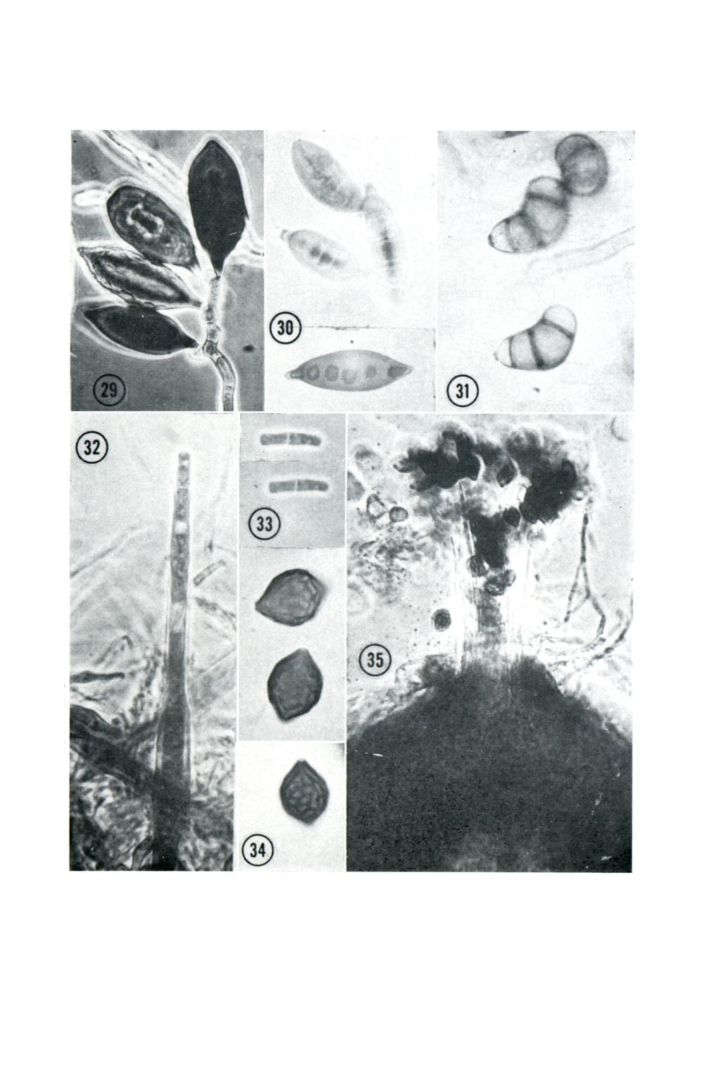
8 Helminthosporium pedicellatum Henry, in Univ. Minn. Agr. Exp. Sta. Bull. 22: 42 (1924). Bipolaris pedicellata (Henry) Shoemaker in Can. Journ. Bot. 37: 884 (1959). Figs. 29, 30. Colonies on potato dextrose agar Olivaceous Black, darkening as sporulation increases, fast-growing, covering the entire plate in five days at 25 C. Conidiophores sparingly branched, 5-6/* in diameter, brownish olive at the apices, lighter towards the base, relatively straight up to the position of the first spore then geniculate and bearing one to four conidia acropleurogenously, leaving prom inent scars upon detachment; conidia broadly fusiform, straight, brownish-olive, widest near the middle and decidedly attenuated towards both ends, apical cell apiculate or broadly rounded in some spores, basal cell narrowed to a short, cylindrical pedicel, epispore strongly thickened with the hilum included within the wall of the basal cell, germinating from both ends but usually from the base, 3-7 septate (mostly 6-septate), X 23-28/*. Specimen examined: PRE 42940, dried culture on potato dextrose agar, isolated from corn (Zea mays) roots by Mr. J. J. du Toit, Plant Protection Research Institute, Pretoria, Dec H. pedicellatum was first isolated by Henry (loc. cit.) from diseased wheat roots in Minnesota, U.S.A. He found this species to be only weakly parasitic. Tveit (Rev. Appl. Mycol. 33: ) isolated this fungus from oat seed from Brazil. Hassan (Plant Dis. Reptr. 40: ) isolated H. pedicellatum from the basal parts of oat plants. In pathogenicity tests he found this fungus to be non-pathogenic to certain varieties of oats, barley and wheat. Shepherd, Hall & Pendry (Phytopathology 52: ) reported H. pedicellatum as the causal organism of a severe root necrosis o f corn in California. They also isolated this fungus from diseased sorghum roots. H. pedicellatum has been found to be in constant association with root rot of corn in the Transvaal province (du Toit, private communication). This is the first record of the occurrence of this fungus in South Africa. W.F.O.M. 13. Curvularia trifolii (Kaufm.) Boed. f.sp. gladioli Parmelee & Luttrell apud Parmelee, in Mycologia 48: (1956). Fig. 31. Culture on potato dextrose agar dark olive grey, woolly, zonate with margin pale or hyaline and darkening to olivaceous black in the centre. Hyphae prostrate and submerged, branching, septate, sub-hyaline at first, darkening to pale-brown or fuscous [i in diameter, and forming intercalary or terminal sclerotia consisting of inflated, globose or subglobose cells, hyaline and thin-walled at first, later with thickened dark-brown walls, forming irregular masses of about x /* mostly embedded in the agar: conidiophores erect, sub-hyaline to fuscous, arising from trailing hyphae and the sclerotia, septate, straight, unbranched and narrow, '5 n, in the lower portioi up to the first conidium, or with one or two branches below the first conidium then geniculate and widening to /* and more frequently septate, bearing one to several conidia acropleurogenously, length very variable /*; conidia unequally ventricose fusitorm, rounded at the apex, tapering towards the prominent, protruding, basal hilum, strongly curved, often sharply bent almost at right angles, usually three-septate occasionally with one or two additional septa, the terminal cells hyaline or sub-hyaline, the middle cells larger fuscous or dark brown thicker-walled, the penultimate cell largest, swollen and usually unequally distended im parting the curvature to the conidia, X /*.
 who emphasized the protruding hilum which distinguishes this species from C. lunata (Wakker) Boed.")

9 237 Specimen examined: PRE 42934, dried culture on potalo-dextrosc agar, isolated from gladiolus corm, Springs District, Tvl., Feb This specimen agrees very well with the descriptions and data given by Parmelee (loc. cit.) who emphasized the protruding hilum which distinguishes this species from C. lunata (Wakker) Boed. Parmelee (Plant Dis. Reptr. 28: ) also mentioned the undifferentiated masses of mycelium made up of globose pigmented cells which he found on the infected host tissues and in culture. On moist filter paper, these bodies, which he regarded as structures which ensure survival of the fungus, produced clumps of conidiophores. The South African collection was found intermingled with a Botrytis sp. on a single corm. Although no lesions similar to those illustrated by Parmelee were present, there is no doubt that this isolate is identical to the fungus described by him. This is the first record of the occurrence of this fungus in South Africa. G. C. A. v.d. W. 14. Chalara terrestris Agnihothrudu & Barna, in Lloydia 25: (1962). Figs. 32, 33. On potato dextrose agar growth is slow, restricted, colony up to 50 mm in diameter after 4 weeks at 26 C. Colony somewhat sodden at first, wrinkled around the inoculum, isabelline, slowly developing fine, compact, downy white mycelium in patches which later turn grey as conidiophores develop; hyphae hyaline, very narrow, branching, septate // in diameter; conidiophores borne singly or in groups of 2-3 on the narrow hyaline hyphae, dark yellowish brown, erect or reclining, long subulate or narrowly obclavate to almost cylindrical, widest in lower or middle part of the sporogenous cell tapering gradually upwards towards the tube or narrowing abruptly above the middle, thickwalled, with 1-20 septa in lower part, tube sub-hyaline to hyaline, slightly flared, thin-walled and with a prominent septum above the sporogenous cell, forming conidia endogenously (80)-90-l20-(223) X //, tube /t in diameter; conidia cylindrical, ends truncate or slightly rounded, hyaline, thin-walled, two-celled, X //. Specimen examined: PRE 42936, isolated on potato dextrose agar from decaying roots of young Eucalyptus saligna, Tzaneen, Transvaal, April The fungus described here, agrees very well with the description by Agnihothrudu and Barna (loc. cit.) of a fungus isolated from the roots of Camellia sinensis suffering from Hittiali disease in upper Assam. Although the South African fungus was isolated from a different host, there is little doubt that this fungus is identical with the species found on the roots of tea bushes in Assam. The conditions under which these isolations were made, however, suggested this species to be a soil saprophyte rather than a root parasite (v.d. Westhuizen, S. A. For. Journ. 54: ). One other species Chalara kriegeriana Bres. had been reported from soil in Switzerland (Gilman, J. C., A Manual of Soil Fungi, p. 331, Iowa, 1957) but this species differs from C. terrestris by having smaller conidia. Saccardo (Syll. Fung. IV pp ) listed C. fusidioides Corda, C. heterospora Sacc. and C. montellica Sacc. on species of Eucalyptus. These three species had been found on dead or dying branches or wood of the host and differ from C. terrestris in the characters of the conidiophores and conidia
 Sacc. in Syll. Fung. 2: 560 (1893); Petch, Trans. Brit. Myc. Soc. 21: 254 (1938); M artin, Mycologia 47: 606-608 (1955). Figs. 34, 35.")

10 238 This is the first record of the occurrence of this genus in South Africa. G. C. A. v.d. W. 15. Melanospora episphaeria Phillips & Plowright, in Grevillea 10: 71 (1881). Sphaeroderma episphaeria (Phill. & Plowr.) Sacc. in Syll. Fung. 2: 560 (1893); Petch, Trans. Brit. Myc. Soc. 21: 254 (1938); M artin, Mycologia 47: (1955). Figs. 34, 35. On malt agar colony thin, fast growing, reaching a diameter of about 70 mm in one week; mycelium thin cottony, sub-hyaline to pale ochraceous b u ff raised at first, later collapsing and often with woolly overgrowth of mycelium in patches; hyphae narrow, branched, hyaline, thin-walled, septate //; perithecia appearing at first as small mucilaginous droplets on the mycelium, darkening gradually to amber, at maturity, scattered, superficial, gelatinous and without subiculum, subglobose, glabrous, // in diameter, the dark spores visible through the wall; ostiole slightly papillate, surmounted by a collar of straight, erect, hyaline setae // long; wall thick, consisting of translucent, pale amber, parenchymatous cells; asci hyaline, clavate, thin-walled 8-spored, deliquescing early, X 12-18//, paraphyses lacking; ascospores dark brown, limoniform, somewhat flattened with epispore covered by reticulate ridges and with minute light-coloured germ pores at the apicula, X 10-15//. Specimen examined: PRE on malt agar, isolated from decaying roots of Eucalyptus saligna, Tzaneen, Transvaal, March The fungus described here, agrees very well with M artin s (loc. cit.) description of Sphaeroderma episphaeria (Phill. & Plowr.) Sacc. which he isolated from surface litter under beeches in Surrey, England. Although M artin s isolate had smaller spores than those mentioned by Petch (loc. cit.) in his description of the species, M artin regarded his isolate as representative of the fungus described by Phillips and Plowright (loc. cit.) and transferred to Sphaeroderma by Saccardo (loc. cit.). M artin further regarded early gelatinization of the asci, maturation of the ascospores in the gelatinous cavity of the ascocarp and their eventual discharge in a gelatinous tendril as alien to the process of spore formation in typical members of the Hypocreales. He suggested that Sphaeroderma should be grouped with other species having the same type of ascus dehiscence. According to Petch (loc. cit.) species of Sphaeroderma Fuckel differ from those of Melanospora Corda by the absence of perithecial necks. Von Arx and Muller (Beit. Krypt. FI. Schweiz II (I): ) regard perithecial necks as variable and their absence or presence subject to influence by environmental conditions. Furthermore, perithecia of the two genera are very similar in their internal structure. The soft gelatinous perithecial wall and asci deliquescing early, leaving the ascospores to mature inside the perithecia, are important characters of Melanospora Corda. For these reasons, Von Arx and Muller (loc. cit.) regarded Sphaeroderma as synonymous with Melanospora Corda. Since the fungus described above agrees with these characters, it is considered to be best placed in the genus Melanospora in which it was described originally. This is the first record of the occurrence of this species in South Africa. G. C. A. v.d. W.


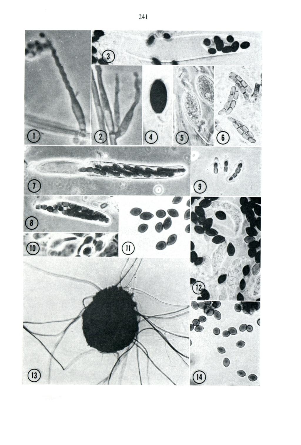
11 239 EXPLANATION OF FIG URES F ig. I. Tritirachium roseum, fertile branch, x F ig. 2. T. roseum, branching, x F ig. 3. Ascobolus immersus, mature ascus, x 130. F ig. 4. Podospora anserina, mature ascospore, x 520. F ig. 5. P. anserina, immature ascospore, x 520. F ig. 6. Sporormia minima, mature ascospores, x 520. F ig. 7. S. minima, extended ascus, x 520. Fig. 8. S. minima, unextended ascus, x 520. Fig. 9. S. minima, ascospores showing mucus sheath, x 520. F ig. 10. Tripterospora longicaudata, ascospores with appendage, x 520. F ig. 11. T. longicaudata, mature ascospores, x 520. F ig. 12. T. longicaudata, asci and ascospores. x 520. F ig. 13. T. erostrata, ascocarp, x 130. F ig. 14. T. erostrata, mature ascospores, x 520. F ig. 15. Auxarthron umbrinum, reticulate peridium showing inverted Y-shaped arches, short ann** dages and elongate appendage, x 250. F ig. 16. A. umbrinum, mature ascocarps, x 100. F ig. 17. Beltrania rhombica, conidiophores, separating cells and conidia, x 330. F ig. 18 & 19. Myrothecium verrucaria, conidia with membranous appendages, x 830. F ig. 20. Myrothecium verrucaria, conidiophores, X 715. F ig. 21. Robillarda sessilis, flask-shaped spre-bearing cells with young conidia, x 715. F ig. 22. R. sessilis, spre-bearing cells with young conidia developing appendages, x 715. F ig. 23. Hyalotia viridis, pycnidia, X 110. F ig. 24. Robillarda sessilis ypung conidia with developing appendages, x 715. F ig. 25. Myrothecium verrucaria, phialides, X 715. F ig. 26. Hyalotia viridis, mature conidia, x 715. F ig. 27. Myrothecium verrucaria, conidiophores, and conidia, X F ig. 28. Robillarda sessilis, five stages in conidial development, x F ig. 29. Helminthosporium pedicellatum, conidiophore and conidia, x 500. F ig. 30. H. pedicellatum conidia, x 500. F ig. 31. Curvularia trifolii f.sp. gladioli, conidia, X 500. F ig. 32. Chalara terrestris, conidiophore with emerging conidium, x F ig. 33. C. terrestris, two-celled conidia, x F ig. 34. Melanospora episphaeria, three ascospores showing form, surface reticulation and germ pore, X F ig. 35. M. episphaeria perithecium showing ostiole, hyaline setae and extruded ascospores, X 250.
12 20y 10l \
13


14 242 4-OH loo/u m m m m
15 243
PERSOONIA. Part 2, pp (1962) Studies on discomycetez I. of Ascobolus and Saccobolus. herbarium, Rijksherbarium, Leiden. inquirenda. on loan.
 Studies on discomycetez I. of Ascobolus and Saccobolus. herbarium, Rijksherbarium, Leiden. inquirenda. on loan.") FIG. PERSOONIA Published by the Rijksherbarium, Leiden Volume 2, Part 2, pp. 195-199 (1962) Studies on discomycetezi. s of species of Ascobolus and Saccobolus in Spegazzini s herbarium J. van Brummelen
FIG. PERSOONIA Published by the Rijksherbarium, Leiden Volume 2, Part 2, pp. 195-199 (1962) Studies on discomycetezi. s of species of Ascobolus and Saccobolus in Spegazzini s herbarium J. van Brummelen
REINWARDTIA Published by Herbarium Bogoriense LBN, Bogor Vol. 10, Part 2, pp (1985) THE ANAMORPH OF SARAWAKUS SUCCISUS RIFAI
 THE ANAMORPH OF SARAWAKUS SUCCISUS RIFAI") REINWARDTIA Published by Herbarium Bogoriense LBN, Bogor Vol. 10, Part 2, pp. 265 270 (1985) THE ANAMORPH OF SARAWAKUS SUCCISUS RIFAI MIEN A. RIFAI, KARTINI KRAMADIBRATA Herbarium Bogorievnc LBN, Bogor,
REINWARDTIA Published by Herbarium Bogoriense LBN, Bogor Vol. 10, Part 2, pp. 265 270 (1985) THE ANAMORPH OF SARAWAKUS SUCCISUS RIFAI MIEN A. RIFAI, KARTINI KRAMADIBRATA Herbarium Bogorievnc LBN, Bogor,
A new species of Podospora from Taiwan
 CHANG Bot. Bull. and Acad. WANG Sin. (2005) A new 46: 169-173 species of Podospora 169 A new species of Podospora from Taiwan Jong-How CHANG 1 and Yei-Zeng WANG 2, * 1 Department of Life Science, National
LESSON ASSIGNMENT. Introduction to Medical Mycology. After completing this lesson, you should be able to:
 LESSON ASSIGNMENT LESSON 1 Introduction to Medical Mycology. TEXT ASSIGNMENT Paragraphs 1-1 through 1-7. TASKS OBJECTIVES After completing this lesson, you should be able to: 1-1. Select the statement
A NOTE ON THE CONIDIAL SCAR IN THE XYLARIACEAE
 New PhytoL (1967) 66, 65-66. A NOTE ON THE CONIDIAL SCAR IN THE XYLARIACEAE BY G. N. GREENHALGH The Hartley Botanical Laboratories, University of Liverpool {Received 5 July 1966) The conidial scars produced
World Journal of Microbiology Vol. 1(2), pp , September, ISSN: XXXX-XXXX
, pp , September, ISSN: XXXX-XXXX") World Journal of Microbiology Vol. 1(2), pp. 013-016, September, 2014. www.premierpublishers.org, ISSN: XXXX-XXXX WJM Research Article Study of Fungal Genus Gyrothrix Corda from the forest flora of Indian
World Journal of Microbiology Vol. 1(2), pp. 013-016, September, 2014. www.premierpublishers.org, ISSN: XXXX-XXXX WJM Research Article Study of Fungal Genus Gyrothrix Corda from the forest flora of Indian
BALLISTICS IN CERTAIN ASCOMYCETES
 BALLISTICS IN CERTAIN ASCOMYCETES BY C. T. INGOLD Birkbeck College, University of London (Received 5 January 1961) (With 3 figures in the te.xt) SUMMARY Horizontal spore discharge has been studied in the
report on PLANT DISEASE FUNGAL LEAF SPOTS OF BLACK WALNUT
 report on PLANT DISEASE RPD No. 600 1987 DEPARTMENT OF CROP SCIENCES UNIVERSITY OF ILLINOIS AT URBANA-CHAMPAIGN FUNGAL LEAF SPOTS OF BLACK WALNUT Several important fungal leaf spot diseases of black walnut
THE UNIVERSITY OF ILLINOIS LIBRARY AGRICULTURAL LIBRARY
 THE UNIVERSITY OF ILLINOIS LIBRARY AGRICULTURAL LIBRARY BULLETIN No. 220 BLACK SPOT OF ONION SETS BY FEANK LINCOLN STEVENS AND ESTHER YOUNG TRUE TIRBANA, ILLINOIS, MAY, 19] 9 CONTENTS OF BULLETIN No.
Journal of Chemical and Pharmaceutical Research, 2017, 9(1): Review Article. Graphium Salixicum: A New Species Explored from Salix Alba
: Review Article. Graphium Salixicum: A New Species Explored from Salix Alba") Available online www.jocpr.com Journal of Chemical and Pharmaceutical Research, 2017, 9(1):69-74 Review Article ISSN : 0975-7384 CODEN(USA) : JCPRC5 Graphium Salixicum: A New Species Explored from Salix
Available online www.jocpr.com Journal of Chemical and Pharmaceutical Research, 2017, 9(1):69-74 Review Article ISSN : 0975-7384 CODEN(USA) : JCPRC5 Graphium Salixicum: A New Species Explored from Salix
An Electron-Microscope Study of Germination of Conidia of Botrytis cinerea
 J. gen. Microbiol. (1963), 33, 43-46 With 2 plates Printed in Great Britain 43 An Electron-Microscope Study of Germination of Conidia of Botrytis cinerea BY LILIAN E. HAWKER AND R. J. HENDY Department
Structure & Life Cycle of Anthoceros
 Structure & Life Cycle of Anthoceros Anthoceros General Characters Gametophytic Plant Body (The Adult gametophyte) Vegetative Structure: External Features It occurs in moist, shaded habitats in sub-tropical
A Cladosarum-like spontaneous mutant of Aspergillus aureolatus
 A Cladosarum-like spontaneous mutant of Aspergillus aureolatus By M. Muntanjola-Cvetkovic & J. Bata Institute for Biological Research Botanicki Zavod, Takovska 43, Belgrade, Yugoslavia Summary. A spontaneous
7-012: Detection of Alternaria padwickii on Oryza sativa (Rice)
") International Rules for Seed Testing Annexe to Chapter 7: Seed Health Testing Methods Published by: International Seed Testing Association (ISTA), Bassersdorf, Switzerland 2014 DISCLAIMER: whilst ISTA
International Rules for Seed Testing Annexe to Chapter 7: Seed Health Testing Methods Published by: International Seed Testing Association (ISTA), Bassersdorf, Switzerland 2014 DISCLAIMER: whilst ISTA
Diplolaeviopsis symmictae (Helotiales, Ascomycota), a new lichenicolous fungus on Lecanora symmicta
, a new lichenicolous fungus on Lecanora symmicta") Diplolaeviopsis symmictae (Helotiales, Ascomycota), a new lichenicolous fungus on Lecanora symmicta Paul Diederich 1 & Brian Coppins 2 1 Musée national d histoire naturelle, 25 rue Munster, L-2160 Luxembourg,
Diplolaeviopsis symmictae (Helotiales, Ascomycota), a new lichenicolous fungus on Lecanora symmicta Paul Diederich 1 & Brian Coppins 2 1 Musée national d histoire naturelle, 25 rue Munster, L-2160 Luxembourg,
Mango Dieback and Gummosis in Sindh, Pakistan Caused by Lasiodiplodia theobromae
 2004 Plant Management Network. Accepted for publication 16 January 2004. Published 2 March 2004. Mango Dieback and Gummosis in Sindh, Pakistan Caused by Lasiodiplodia theobromae Muhammad Ali Khanzada,
5. Diseases of Maize
 5. Diseases of Maize Downy mildew/crazy top Sorghum downy mildew - Peronosclerospora sorghi Phlippine downy mildew - Peronosclerospora philippinensis Crazy top - Sclerophthora macrospora The most characteristic
STUDIES ON CULTURAL, MORPHOLOGICAL AND PATHOGENIC VARIABILITY AMONG THE ISOLATES OF FUSARIUM OXYSPORUM F. SP. CICERI CAUSING WILT OF CHICKPEA
 Volume-7, Issue-1 Jan-Mar-2017 Coden: IJPAJX-CAS-USA, Copyrights@2015ISSN-2231-4490 Received: 1 st Oct-2016 Revised: 26 th Nov-2016 Accepted: 27 th Nov-2016 DOI: 10.21276/Ijpaes http://dx.doi.org/10.21276/ijpaes
NEW FUNGAL RECORDS ON EUCALYPTUS SPP. FROM DISTRICT FAISALABAD PAKISTAN
 Pak. J. Bot., 42(5): 3317-3321, 2010. NEW FUNGAL RECORDS ON EUCALYPTUS SPP. FROM DISTRICT FAISALABAD PAKISTAN SYED QAISER ABBAS, TEHREEMA IFTIKHAR, MUBASHIR NIAZ AND NAILA SADAF Department of Botany, Government
Sporocarp of Marsilea. Dr.Sukanya Baruah Chaliha. Asst. Professor Dept of Botany Class-3 rd Sem(Major) MDKG College,Dibrugarh.
 MDKG College,Dibrugarh.") Sporocarp of Marsilea Dr.Sukanya Baruah Chaliha. Asst. Professor Dept of Botany Class-3 rd Sem(Major) MDKG College,Dibrugarh. External Morphology Soft and green when young but turns dark brown and hard
Sporocarp of Marsilea Dr.Sukanya Baruah Chaliha. Asst. Professor Dept of Botany Class-3 rd Sem(Major) MDKG College,Dibrugarh. External Morphology Soft and green when young but turns dark brown and hard
7-001b: Malt agar method for the detection of Alternaria dauci on Daucus carota (carrot)
") International Rules for Seed Testing Annexe to Chapter 7: Seed Health Testing Methods 7-001b: Malt agar method for the detection of Alternaria dauci on Daucus carota (carrot) Published by: International
International Rules for Seed Testing Annexe to Chapter 7: Seed Health Testing Methods 7-001b: Malt agar method for the detection of Alternaria dauci on Daucus carota (carrot) Published by: International
Two new intertidallignicolous Swampomyces species from Red Sea mangroves in Egypt
 Fungal Diversity Two new intertidallignicolous Swampomyces species from Red Sea mangroves in Egypt M.A. Abdel-Wahab1, H. El-Sharouneyl and E.B.G. Jones2 IDepat1ment of Botany, South Valley University,
ALTHOUGH Piloboliis has been extensively studied both by myco-
 [58] THE SPORANGIOPHORE OF PILOBOLUS BY C. T. INGOLD Department of Botany, University of Reading (With 2 figures in the text) ALTHOUGH Piloboliis has been extensively studied both by myco- ^ logists and
7-001a: Blotter method for the detection of Alternaria dauci on Daucus carota (carrot)
") International Rules for Seed Testing Annexe to Chapter 7: Seed Health Testing Methods 7-001a: Blotter method for the detection of Alternaria dauci on Daucus carota (carrot) Published by: International
International Rules for Seed Testing Annexe to Chapter 7: Seed Health Testing Methods 7-001a: Blotter method for the detection of Alternaria dauci on Daucus carota (carrot) Published by: International
Scanning Electron Microscopy (SEM) of Seed Infected with Seed Borne Fungi
 of Seed Infected with Seed Borne Fungi") International Journal of Current Microbiology and Applied Sciences ISSN: 2319-7706 Volume 7 Number 07 (2018) Journal homepage: http://www.ijcmas.com Original Research Article https://doi.org/10.20546/ijcmas.2018.707.476
International Journal of Current Microbiology and Applied Sciences ISSN: 2319-7706 Volume 7 Number 07 (2018) Journal homepage: http://www.ijcmas.com Original Research Article https://doi.org/10.20546/ijcmas.2018.707.476
Stilbella holubovae, a new synnematous hyphomycete species on driftwood from the Philippines and South Africa
 Stilbella holubovae, a new synnematous hyphomycete species on driftwood from the Philippines and South Africa Keith A. Seifert 1, Susan J. Stanley 2 and Kevin D. Hyde 2 Centre for Land and Biological Resources
Subcutaneous Mycosis
 Subcutaneous Mycosis Fungal infections 1. Superficial mycosis. 2. Coetaneous mycosis: Dermatophytoses. 3. Subcutaneous mycosis. 4. Systemic mycosis. 5. Opportunistic mycosis. Subcutanus mycoses Fungal
2. Diseases of Sorghum
 2. Diseases of Sorghum Downy Mildew - Peronosclerospora sorghi The fungus causes systemic downy mildew of sorghum. It invades the growing points of young plants, either through oospore or conidial infection.
The genus Podospora (Lasiosphaeriaceae, Sordariales) in Brazil
 in Brazil") Mycosphere 6 (2): 201 215(2015) ISSN 2077 7019 www.mycosphere.org Article Mycosphere Copyright 2015 Online Edition Doi 10.5943/mycosphere/6/2/10 The genus Podospora (Lasiosphaeriaceae, Sordariales) in
Mycosphere 6 (2): 201 215(2015) ISSN 2077 7019 www.mycosphere.org Article Mycosphere Copyright 2015 Online Edition Doi 10.5943/mycosphere/6/2/10 The genus Podospora (Lasiosphaeriaceae, Sordariales) in
THE OCCURRENCE AND EFFECTS OF CEPHALOTHECIUM ROSEUM ON AVOCADO
 California Avocado Society 1951 Yearbook 36: 179-185 THE OCCURRENCE AND EFFECTS OF CEPHALOTHECIUM ROSEUM ON AVOCADO John Wesley Yale, Jr. and George R. Johnstone John Wesley Yale, Jr. (at the time of writing)
7-007: Detection of Alternaria linicola, Botrytis cinerea and Colletotrichum lini on Linum usitatissimum (flax) seed
 seed") International Rules for Seed Testing Annexe to Chapter 7: Seed Health Testing Methods 7-007: Detection of Alternaria linicola, Botrytis cinerea and Colletotrichum lini on Linum usitatissimum (flax) seed
International Rules for Seed Testing Annexe to Chapter 7: Seed Health Testing Methods 7-007: Detection of Alternaria linicola, Botrytis cinerea and Colletotrichum lini on Linum usitatissimum (flax) seed
Ascoyunnania aquatica gen. et sp. nov., a freshwater fungus collected from China and its microcylic conidiation
 Fungal Diversity Ascoyunnania aquatica gen. et sp. nov., a freshwater fungus collected from China and its microcylic conidiation Lei Cai 1, 2*, Keqin Zhang 1 and Kevin D. Hyde 2 1 Laboratory for Conservation
North American Pezizales: Greletia and Marcellina
 V Sydowia, Annales Mycologici Ser. II. Vol. 38: 235-240 (1985) Verlag Ferdinand Berger & Söhne Gesellschaft m.b.h., 3580 Horn, Austria North American Pezizales: Greletia and Marcellina Donald H. PFISTER
Blotter method for the detection of Alternaria dauci on Daucus carota
 International Rules for Seed Testing Annexe to Chapter 7: Seed Health Testing Methods 7-001a: Blotter method for the detection of Alternaria dauci on Daucus carota Published by: International Seed Testing
Muscle Tissue. General concepts. Classification of muscle. I. Functional classification is based on the type of neural control.
 Muscle Tissue LEARNING OBJECTIVES 1. Identify the three types of muscle tissue at the light microscopic level. 2. List and compare the structural and functional features of each of the three muscle fiber
Introduction. Study of fungi called mycology.
 Fungi Introduction Study of fungi called mycology. Some fungi are beneficial: ex a) Important in production of some foods, ex: cheeses, bread. b) Important in production of some antibiotics, ex: penicillin
7-002b: Malt agar method for the detection of Alternaria radicina on Daucus carota (carrot)
") International Rules for Seed Testing Annexe to Chapter 7: Seed Health Testing Methods 7-002b: Malt agar method for the detection of Alternaria radicina on Daucus carota (carrot) Published by: International
International Rules for Seed Testing Annexe to Chapter 7: Seed Health Testing Methods 7-002b: Malt agar method for the detection of Alternaria radicina on Daucus carota (carrot) Published by: International
This is the written version of our Hot Topic video presentation available at: MayoMedicalLaboratories.com/hot-topics
 This is the written version of our Hot Topic video presentation available at: MayoMedicalLaboratories.com/hot-topics Welcome to Mayo Medical Laboratories hot topics. These presentations provide short discussion
Structural aspects regarding formation and emission of Diaporthe (Phomopsis) helianthi ascospores
 helianthi ascospores") Structural aspects regarding formation and emission of Diaporthe (Phomopsis) helianthi ascospores Valentina Androsova 1, Irina Balakhnina 1, Thomas Gulya 2 1 All-Russian Research Institute of Biological
Structural aspects regarding formation and emission of Diaporthe (Phomopsis) helianthi ascospores Valentina Androsova 1, Irina Balakhnina 1, Thomas Gulya 2 1 All-Russian Research Institute of Biological
Tree and Shrub Disease
 n t h r a A n t h r a c n o s e A number of different trees are affected by anthracnose disease. This fungal disease can cause severe leaf blighting and deformation, but in many cases the damage to the
Rec. zoot. Surv. India, 97 (Part-2) : , 1999
 : , 1999") Rec. zoot. Surv. India, 97 (Part-2) : 167-172, 1999 DESCRIPTIONS OF TWO NEW GALL MIDGES (DIPTERA : CECIDOMYIIDAE).FROM NILGIRI BIOSPHERE RESERVE, KARNATAKA R. M. SHARMA Zoological Survey of India, Western
Rec. zoot. Surv. India, 97 (Part-2) : 167-172, 1999 DESCRIPTIONS OF TWO NEW GALL MIDGES (DIPTERA : CECIDOMYIIDAE).FROM NILGIRI BIOSPHERE RESERVE, KARNATAKA R. M. SHARMA Zoological Survey of India, Western
(Iteceived for publication December 3, 1915)
") TRANSPLANTABLE SARCOMATA OF THE RAT LIVER ARISING IN THE WALLS OF PARASITIC CYSTS G. L. ROHDENBURG, M.D., AND F. D. BULLOCK, M.D. From Colurnbia University, George Crocker Special Re-search Fund, F. C.
TRANSPLANTABLE SARCOMATA OF THE RAT LIVER ARISING IN THE WALLS OF PARASITIC CYSTS G. L. ROHDENBURG, M.D., AND F. D. BULLOCK, M.D. From Colurnbia University, George Crocker Special Re-search Fund, F. C.
Some Fungi Isolated from Alkaline Soils of Uttar Pradesh, India
 Some Fungi Isolated from Alkaline Soils of Uttar Pradesh, India By M. P. Haware and M. S. Pavgi Faculty of Agriculture, Banaias Hindu University. India Acres of land around Varanasi and other areas of
ACANTHOMULUS. NOTE XIII. On two new species of the genus Acanthodrilus, Perr. from Liberia. Dr. R. Horst. Perr. from New-Caledonia,
 ACANTHOMULUS. 103 NOTE XIII. On two new species of the genus Acanthodrilus, Perr. from Liberia BY Dr. R. Horst Among the Invertebrates collected by Büttikofer and the late Sala during their journey in
Structure & Life Cycle of Funaria
 Structure & Life Cycle of Funaria Funaria General Characters Gametophytic Plant Body Vegetative Structure: External Features It is a common type of water moss which grows on moist, shady, and damp soil,
BOTANICA HUNGARICA (Antea: Fragmenta Botanica)
") STUDIA XXIII. BOTANICA HUNGARICA (Antea: Fragmenta Botanica) 1992 pp. 63-68 A new species of Triadelphia from Hungary By Á.RÉVAY (Received 30 April, 1990) Abstract: A new dematiaceous hyphomycete species,
STUDIA XXIII. BOTANICA HUNGARICA (Antea: Fragmenta Botanica) 1992 pp. 63-68 A new species of Triadelphia from Hungary By Á.RÉVAY (Received 30 April, 1990) Abstract: A new dematiaceous hyphomycete species,
Goosiomyces bambusicola - A new cheirosporous anamorphic species from Western Ghats, India.
 Current Research in Environmental & Applied Mycology 4 (2): 211 216 (2014) ISSN 2229-2225 www.creamjournal.org Article CREAM Copyright 2014 Doi 10.5943/cream/4/2/8 Online Edition Goosiomyces bambusicola
Physiological and Morphogenetic Studies of Fern Gametophyte and Sporophyte by Aseptic Culture VI. Notes on the Alternation of Generations
 Bot. Map. Tokyo 78:187-193 (June 25, 1965) Physiological and Morphogenetic Studies of Fern Gametophyte and Sporophyte by Aseptic Culture VI. Notes on the Alternation of Generations by Yukio KATO * Received
New and interesting records of South African fungi. XI. Eucalyptus leaf fungi
 300 S.AfrJ.Bot., 1993, 59(3): 300-304 New and interesting records of South African fungi. XI. Eucalyptus leaf fungi P.w. CrouS*1 and E.J. van der Linde 2 1 Department of Plant Pathology, University of
ANNOTATIONES ZOOLOGICAE JAPONENSES. Volume 55, No. 2-June Published by the Zoological Society of Japan
 ANNOTATIONES ZOOLOGICAE JAPONENSES Volume 55, No. 2-June 1982 Published by the Zoological Society of Japan AOKI and Mr. H. HARADA, Yokohama National University, in Eastern Kalimantan, Borneo. Included
Key words wild passion-fruit, Mycosphaerellaceae, tropical fruits, cercosporoid fungi
 A NEW FUNGAL DISEASE CAUSED BY A PSEUDOCERCOSPORA SPECIES ON PASSIFLORA SETACEA IN PLANALTINA-DF, BRAZIL Alexei C. Dianese 1, Ana M. Costa 1 & José C. Dianese 2 ( 1 Embrapa Cerrados, Br-020, Km 18, 73310-970
Peony Flower Anatomy I
 Peony Flower Anatomy I Don Hollingsworth, APS Director Maryville, Missouri What Makes a Peony Flower Luxurious? Rich luxury of the flowers explains why peonies are wanted, why loved and why known in history
Lecture 03 - Diseases of Banana (2 Lectures)
") Lecture 03 - Diseases of Banana (2 Lectures) Panama disease :Fusarium oxysporum f. spcubense Economic Importance The first major disease which attacked banana was called Panama disease from the area where
Lecture 03 - Diseases of Banana (2 Lectures) Panama disease :Fusarium oxysporum f. spcubense Economic Importance The first major disease which attacked banana was called Panama disease from the area where
First Report of Penicillium adametzioides from Decayed Grapes (Vitis vinifera) in Pakistan
 in Pakistan") International Journal of Current Microbiology and Applied Sciences ISSN: 2319-7706 Volume 5 Number 12 (2016) pp. 316-320 Journal homepage: http://www.ijcmas.com Original Research Article http://dx.doi.org/10.20546/ijcmas.2016.512.034
International Journal of Current Microbiology and Applied Sciences ISSN: 2319-7706 Volume 5 Number 12 (2016) pp. 316-320 Journal homepage: http://www.ijcmas.com Original Research Article http://dx.doi.org/10.20546/ijcmas.2016.512.034
D1120 Connective Tissue and Muscle Laboratory Module. 1) Connective tissue
 Connective tissue") D1120 Connective Tissue and Muscle Laboratory Module 1) Connective tissue Objectives: 1) identify the components (cells, fibres) present in "ordinary" connective tissue 2) differentiate the three types
D1120 Connective Tissue and Muscle Laboratory Module 1) Connective tissue Objectives: 1) identify the components (cells, fibres) present in "ordinary" connective tissue 2) differentiate the three types
Arnium gigantosporum, a new ascomycete species from fresh water in Florida
 Arnium gigantosporum, a new ascomycete species from fresh water in Florida Huzefa A. Raja *, and Carol A. Shearer Department of Plant Biology, University of Illinois, 265 Morrill Hall, 505 South Goodwin
A revision of the genus Acaníhocordax Günther, 1929 (Dermaptera, Forficulidae)
") ANNALES HISTORICO-NATURALES MUSEI NATIONALIS HUNGARICI Tomus 80. Budapest, 1988 p. 51-56. A revision of the genus Acaníhocordax Günther, 1929 (Dermaptera, Forficulidae) by H. STEINMANN, Budapest H. STEINMANN:
ANNALES HISTORICO-NATURALES MUSEI NATIONALIS HUNGARICI Tomus 80. Budapest, 1988 p. 51-56. A revision of the genus Acaníhocordax Günther, 1929 (Dermaptera, Forficulidae) by H. STEINMANN, Budapest H. STEINMANN:
THE [PUBLISHED MARCH 31ST, 1915.] GEO. K. SUTHERLAND. [WITH FOUR FIGURES IN THE TEXT].
![THE [PUBLISHED MARCH 31ST, 1915.] GEO. K. SUTHERLAND. [WITH FOUR FIGURES IN THE TEXT].](/thumbs/92/110459948.jpg "THE [PUBLISHED MARCH 31ST, 1915.] GEO. K. SUTHERLAND. [WITH FOUR FIGURES IN THE TEXT].") THE HEW PHVTOIiOGIST. VOL. XIV, Nos. 2 & 3. FEB. & MAR., 1915. [PUBLISHED MARCH 31ST, 1915.] NEW MARINE FUNGI ON BY GEO. K. SUTHERLAND. PELVETIA. [WITH FOUR FIGURES IN THE TEXT]. THE small number ot known
THE HEW PHVTOIiOGIST. VOL. XIV, Nos. 2 & 3. FEB. & MAR., 1915. [PUBLISHED MARCH 31ST, 1915.] NEW MARINE FUNGI ON BY GEO. K. SUTHERLAND. PELVETIA. [WITH FOUR FIGURES IN THE TEXT]. THE small number ot known
7-002a: Blotter method for the detection of Alternaria radicina on Daucus carota (carrot)
") International Rules for Seed Testing Annexe to Chapter 7: Seed Health Testing Methods 7-002a: Blotter method for the detection of Alternaria radicina on Daucus carota (carrot) Published by: International
International Rules for Seed Testing Annexe to Chapter 7: Seed Health Testing Methods 7-002a: Blotter method for the detection of Alternaria radicina on Daucus carota (carrot) Published by: International
The Flower, Pollination, and Seeds
 The Flower, Pollination, and Seeds Class 9 th Chapters 6,7,8 1 The Flower A complete or a perfect flower, has all the four Whorls. If, even one whorl is missing, it is an Incomplete Flower. The fourth
ARNICA (WHOLE PLANT) FOR HOMOEOPATHIC PREPARATIONS ARNICA MONTANA FOR HOMOEOPATHIC PREPARATIONS
 FOR HOMOEOPATHIC PREPARATIONS ARNICA MONTANA FOR HOMOEOPATHIC PREPARATIONS") ARNICA (WHOLE PLANT) FOR HOMOEOPATHIC PREPARATIONS ARNICA MONTANA FOR HOMOEOPATHIC PREPARATIONS Arnica montana ad praeparationes homoeopathicas DEFINITION Whole, fresh, blooming plant Arnica montana L.
ARNICA (WHOLE PLANT) FOR HOMOEOPATHIC PREPARATIONS ARNICA MONTANA FOR HOMOEOPATHIC PREPARATIONS Arnica montana ad praeparationes homoeopathicas DEFINITION Whole, fresh, blooming plant Arnica montana L.
7-011: Detection of Pyricularia oryzae on Oryza sativa (Rice)
") International Rules for Seed Testing Annexe to Chapter 7: Seed Health Testing Methods Published by: International Seed Testing Association (ISTA), Bassersdorf, Switzerland 2014 DISCLAIMER: whilst ISTA
International Rules for Seed Testing Annexe to Chapter 7: Seed Health Testing Methods Published by: International Seed Testing Association (ISTA), Bassersdorf, Switzerland 2014 DISCLAIMER: whilst ISTA
Pyrenomycetous fungi on a birch (Betula pendula) substratum in Bulgaria
 substratum in Bulgaria") Violeta I. Fakirova Pyrenomycetous fungi on a birch (Betula pendula) substratum in Bulgaria Abstract Fakirova, V. I.: Pyrenomycetous fungi on a birch (Betula pendula) substratum in Bulgaria. - Bocconea
Violeta I. Fakirova Pyrenomycetous fungi on a birch (Betula pendula) substratum in Bulgaria Abstract Fakirova, V. I.: Pyrenomycetous fungi on a birch (Betula pendula) substratum in Bulgaria. - Bocconea
Forest Pest Management
 PRONG BINDER Forest Pest Management SD Ii4-4 Alq yip. 85 - D3 Report 85-23 PATHOGENIC FUSARIUM ON SPRUCE SEED FROM THE TCWNER NURSERY, NORTH DAKOTA 3450 September 1985 by R. L. James, Plant Pathologist
****************************************************************************************************** INTEGUMENTARY SYSTEM
 BIOLOGY 211: HUMAN ANATOMY & PHYSIOLOGY ****************************************************************************************************** INTEGUMENTARY SYSTEM ******************************************************************************************************
Two new species of Corynespora from Uttar Pradesh, India
 Two new species of Corynespora from Uttar Pradesh, India Kumar S 1,2*, Singh R 2, Gond DK 1 and Saini DC 1 1 Birbal Sahni Institute of Palaeobotany, 53, University Road, Lucknow 226007(U.P.), India. 2
MYCOTAXON ISSN (print) (online) Mycotaxon, Ltd. 2017
 (online) Mycotaxon, Ltd. 2017") MYCOTAXON ISSN (print) 0093-4666 (online) 2154-8889 Mycotaxon, Ltd. 2017 April June 2017 Volume 132, pp. 459 470 https://doi.org/10.5248/132.459 Sporormiella longicolla sp. nov. and new Sporormiella records
MYCOTAXON ISSN (print) 0093-4666 (online) 2154-8889 Mycotaxon, Ltd. 2017 April June 2017 Volume 132, pp. 459 470 https://doi.org/10.5248/132.459 Sporormiella longicolla sp. nov. and new Sporormiella records
Guignardia bispora and G. ellipsoidea spp. nov. and other Guignardia species from palms (Arecaceae)
") Mycosphere Guignardia bispora and G. ellipsoidea spp. nov. and other Guignardia species from palms (Arecaceae) Wulandari NF 1,2, To Anun C. 1*, McKenzie EHC 3 and Hyde KD 4,5 1 Department of Plant Pathology,
Mycosphere Guignardia bispora and G. ellipsoidea spp. nov. and other Guignardia species from palms (Arecaceae) Wulandari NF 1,2, To Anun C. 1*, McKenzie EHC 3 and Hyde KD 4,5 1 Department of Plant Pathology,
DOUBLE TEETH IN THE SPERM WHALE (PHYSETER MACROCEPHALUS L.)
") DOUBLE TEETH IN THE SPERM WHALE (PHYSETER MACROCEPHALUS L.) by H. BOSCHMA Recently a fairly large number of teeth of the sperm whale were acquired for the collections of the Rijksmuseum van Natuurlijke
DOUBLE TEETH IN THE SPERM WHALE (PHYSETER MACROCEPHALUS L.) by H. BOSCHMA Recently a fairly large number of teeth of the sperm whale were acquired for the collections of the Rijksmuseum van Natuurlijke
Tissues. tissue = many cells w/ same structure and function. cell shape aids its function tissue shape aids its function
 Tissues tissue = many cells w/ same structure and function cell shape aids its function tissue shape aids its function Histology = study of tissues 4 types of tissues Epithelial coverings contact openings
EAR AND KERNEL ROTS. When to look for: Mid-August to October (and during storage)
") EAR AND KERNEL ROTS When to look for: Mid-August to October (and during storage) Reduction in yield and grain quality due to infection by fungi of ears and kernels both during the growing season, and while
EAR AND KERNEL ROTS When to look for: Mid-August to October (and during storage) Reduction in yield and grain quality due to infection by fungi of ears and kernels both during the growing season, and while
(From The Rockefeller Institute) Materials and Methods. Observations with the Electron Microscope
 Materials and Methods. Observations with the Electron Microscope") ELECTRON MICROSCOPE STUDY OF THE DEVELOPMENT OF THE PAPILLOMA VIRUS IN THE SKIN OF THE RABBIT* BY ROBERT S. STONE,~ M.D., RICHARD E. SHOPE, M.D., DAN H. MOORE, P,~.D. (From The Rockefeller Institute) PLATES
ELECTRON MICROSCOPE STUDY OF THE DEVELOPMENT OF THE PAPILLOMA VIRUS IN THE SKIN OF THE RABBIT* BY ROBERT S. STONE,~ M.D., RICHARD E. SHOPE, M.D., DAN H. MOORE, P,~.D. (From The Rockefeller Institute) PLATES
Colonial and Morphological Characteristics of various fungi Species Isolated from soil in Bangalore city
 Bulletin of Environment, Pharmacology and Life Sciences Bull. Env. Pharmacol. Life Sci., Vol 6[1] December 2016: 17-21 2016 Academy for Environment and Life Sciences, India Online ISSN 2277-1808 Journal
Hands-on identification of vegetable diseases: Roses
 Hands-on identification of vegetable diseases: Roses Theme: How to diagnose a specific disease from diseases or disorders with similar symptoms. Mathews Paret, Susannah Da Silva, Binoy Babu, Fanny Iriarte,
Delitschia gigaspora var. pescanii: a new variety of coprophilous fungus from Brazil
 Mycosphere 6 (1): 122 126 (2015) ISSN 2077 7019 www.mycosphere.org Article Mycosphere Copyright 2015 Online Edition Doi 10.5943/mycosphere/6/1/12 Delitschia gigaspora var. pescanii: a new variety of coprophilous
En tornology. FUNGI PARASITIC ON THE NYMPH OF MOGANNZA HEBES WALKER IN TAIWAN L. S; Leu and Z. N. Wang Tainan Sugar Experiment Station Tainan, Taiwan
 En tornology FUNGI PARASITIC ON THE NYMPH OF MOGANNZA HEBES WALKER IN TAIWAN L. S; Leu and Z. N. Wang Tainan Sugar Experiment Station Tainan, Taiwan ABSTRACT Two species of parasitic fungi, lsaris sinclairii
DENTIN It a hard vital tissue, surrounds the pulp & underlies the enamel on the crown & the cementum on the roots of the teeth.
 Lec. 7 Dr. Ali H.Murad DENTIN It a hard vital tissue, surrounds the pulp & underlies the enamel on the crown & the cementum on the roots of the teeth. Physical properties: 1-Dentin is pale yellow in color,
CELL AND TISSUE INJURY COURSE-II PATHOLOGY LABORATORY
 CELL AND TISSUE INJURY COURSE-II PATHOLOGY LABORATORY PATHOLOGY of INFECTIOUS DISEASES MICROSCOPY Rengin Ahıskalı Macroscopy samples are shown in the macroscopy presentations of the first two courses.
Blotter method for the detection of Alternaria radicina on Daucus carota
 International Rules for Seed Testing Annexe to Chapter 7: Seed Health Testing Methods 7-002a: Blotter method for the detection of Alternaria radicina on Daucus carota Published by: International Seed Testing
Phialomyces fusiformis sp. nov. from soil in Singapore is identified and described
 Mycologia, 95(5), 2003, pp. 896 901. 2003 by The Mycological Society of America, Lawrence, KS 66044-8897 Phialomyces fusiformis sp. nov. from soil in Singapore is identified and described G. Delgado Rodriguez
Xenosporium amomi sp. nov. from Zingiberaceae in Thailand
 Fungal Diversity Xenosporium amomi sp. nov. from Zingiberaceae in Thailand Boonsom Bussaban¹ *, Pipob Lumyong², Eric H.C. McKenzie³, Kevin D. Hyde 4 and Saisamorn Lumyong¹ ¹Department of Biology, Faculty
258 DANIEL A. TEXTORIS Vol. 63
 258 DANIEL A. TEXTORIS Vol. 63 STUDIES OF THE GYPONINAE 1. THE GENUS MARGANANA DELONG* (HOMOPTERA: CICADELLIDAE) DWIGHT M. DELONG AND PAUL H. PREYTAG Department of Zoology and Entomology, The Ohio State
DROSERA CAPENSIS L.: HISTO-ANATOMY OF THE VEGETATIVE ORGANS. Introduction
 Analele ştiinţifice ale Universităţii Al. I. Cuza Iaşi Tomul LV, fasc. 1, s.ii a. Biologie vegetală, 2009 DROSERA CAPENSIS L.: HISTO-ANATOMY OF THE VEGETATIVE ORGANS IRINA STĂNESCU *, C. TOMA ** Abstract.
SEX BEHAVIOUR AND SEX DETERMINATION IN CREPIDVLA FORNICATA L.
 [34] SEX BEHAVIOUR AND SEX DETERMINATION IN CREPIDVLA FORNICATA L. BY JAN Z. WILCZYNSKI Lebanese State University, Beirut, Lebanon (Received 21 November 1957) INTRODUCTION The problem of sex determination
Studies on cultural, morphological variability in isolates of Fusarium solani (Mart.) Sacc., incitant of dry root-rot of Citrus
 Sacc., incitant of dry root-rot of Citrus") Studies on cultural, morphological variability in isolates of Fusarium solani (Mart.) Sacc., incitant of dry root-rot of Citrus M. Ravi Chandran and M. Reddi Kumar* Department of Plant Pathology, S.V.
Studies on cultural, morphological variability in isolates of Fusarium solani (Mart.) Sacc., incitant of dry root-rot of Citrus M. Ravi Chandran and M. Reddi Kumar* Department of Plant Pathology, S.V.
Rank: Type 021N ranks 3rd in number of predominance. (SVI values can exceed 500 mg/l) Rapid growth of this filament can cause
 Rapid growth of this filament can cause") Ok, Let's start with the standard information for the filament Type 021N Identification: Relatively large, non-motile filaments (100->1000 µm). Straight or smoothly curved, sometimes coiled filaments with
Ok, Let's start with the standard information for the filament Type 021N Identification: Relatively large, non-motile filaments (100->1000 µm). Straight or smoothly curved, sometimes coiled filaments with
Variability Among the Isolates of Sarocladium Oryzae Incitant of Rice Sheath Rot
 Available online at www.ijpab.com DOI: http://dx.doi.org/10.18782/2320-7051.5525 ISSN: 2320 7051 Int. J. Pure App. Biosci. 5 (6): 1528-1537 (2017) Research Article Variability Among the Isolates of Sarocladium
Skeletal muscle. General features :
 Muscular tissues In the first embryonic life the muscular tissues arise from mesoderm, The function of movement in multicellular organisms is usually assumed by specialized cells called muscle fibers which
The Genus Pithomyces in South Africa by
 Bothalia, 10, 4: 509-516 The Genus Pithomyces in South Africa by W. F. O. Marasas* and Ingrid H. Schumann* A bstract Descriptions are given of South African isolates of Pithomyces sacchari (Speg.) M. B.
This work is licensed under a Creative Commons Attribution- NonCommercial-NoDerivatives 4.0 International License.
 Title A new freshwater species of Saccardoella from Hong Kong and South Africa Author(s) Tsui, KM; Hyde, KD; Hodgkiss, IJ; Goh, TK Citation Mycologia, 1998, v. 90 n. 4, p. 701-704 Issued Date 1998 URL
European Union Community Plant Variety Office
 European Union Community Plant Variety Office PROTOCOL FOR DISTINCTNESS, UNIFORMITY AND STABILITY TESTS Gerbera Cass. GERBERA UPOV Species Code: GERBE Adopted on 31 st October 2002 Page 1 I - SUBJECT OF
Dactylella shizishanna sp. nov., from Shizi Mountain, China
 Dactylella shizishanna sp. nov., from Shizi Mountain, China XueFeng Liu 1,2 and KeQin Zhang 1 * 1 Laboratory for Conservation and Utilization of Bio-resource, Yunnan University, Kunming, Yunnan 650091,
First report of Erysiphe baptisiae on Baptisia in UK with emended description
 Schlechtendalia 28 (2014) First report of Erysiphe baptisiae on Baptisia in UK with emended description Roger T. A. COOK, Alick HENRICI & Uwe BRAUN Abstract: Cook, R. T. A., Henrici, A. & Braun, U. 2014:
Tongue In the buccal cavity of the digestive system
 Tongue In the buccal cavity of the digestive system same layers as those of tubular organs Mucosa, submucosa, and muscularis muscularis = the muscularis externa no muscularis mucosa 1 Tongue ling = tongue
Symptomatology, proving pathogenicity and standardization of inoculum density of Fusarium oxysporum causing wilt of fenugreek
 2017; 6(6): 505-509 E-ISSN: 2278-4136 P-ISSN: 2349-8234 JPP 2017; 6(6): 505-509 Received: 12-09-2017 Accepted: 13-10-2017 Rani N Yashoda R Hegde Nargund VB Symptomatology, proving pathogenicity and standardization
New host reports of rust fungi (Pucciniales) from Khyber Pakhtunkhwa, Pakistan
 from Khyber Pakhtunkhwa, Pakistan") Plant Pathology & Quarantine 9(1): 72 76 (2019) ISSN 2229-2217 www.ppqjournal.org Article Doi 10.5943/ppq/9/1/8 New host reports of rust fungi (Pucciniales) from Khyber Pakhtunkhwa, Pakistan Fahad M 1*,
Plant Pathology & Quarantine 9(1): 72 76 (2019) ISSN 2229-2217 www.ppqjournal.org Article Doi 10.5943/ppq/9/1/8 New host reports of rust fungi (Pucciniales) from Khyber Pakhtunkhwa, Pakistan Fahad M 1*,
Planktothuria diaphana, gen. et sp. n.
 PLANKTOTHCJEIA DIAPHANA. 375 Planktothuria diaphana, gen. et sp. n. By J. I>. F. Gilcluist, M.A., D.Sc, Ph.D., Professor of Zoology in the University of Cape Town. "With 4 Text-figures. THE subject of
CHAPTER II THE EFFECT OF TEMPERATURE AND CULTURE MEDIUM ON THE GROWTH AND SPORULATION OF DRECHSLERA CATENARIA
 CHAPTER II THE EFFECT OF TEMPERATURE AND CULTURE MEDIUM ON THE GROWTH AND SPORULATION OF DRECHSLERA CATENARIA 61 \ INTRODUCTION Sporulation and growth of fungi are affected by light, temperature, relative
Plant Terminology. Floral Symmetry
 Plant Terminology Parts of a Flower Pedicel--the stalk of an individual flower Calyx--outermost whorl of a flower Sepal--one member of the calyx Corolla--second whorl of a flower Petal--one member of the