ARD Online First, published on June 7, 2005 as /ard
|
|
|
- Edgar Gilmore
- 5 years ago
- Views:
Transcription
1 ARD Online First, published on June 7, 2005 as /ard TITLE: Inter-observer reliability in musculoskeletal ultrasonography: results from a "Teach-the-Teachers" rheumatologist course AUTHORS: Esperanza Naredo, MD ¹; Ingrid Möller, MD 2 ; Carmen Moragues, MD 3 ; Juan J. de Agustín, MD 4 ; Alexander K. Scheel, MD 5 ; Walter Grassi, MD, PhD 6 ; Eugenio de Miguel, MD 7 ; Marina Backhaus MD, PhD 8 ; Peter Balint, MD, PhD 9 ; George A.W. Bruyn, MD, PhD 10 ; Maria Antonietta D`Agostino, MD 11 ; Emilio Filippucci, MD 12 ; Annamaria Iagnocco, MD 13 ; David Kane, MD, PhD 14 ; Juhani M. Koski, MD, PhD 15 ; Lucia Mayordomo, MD 16 ; Wolfgang A. Schmidt, MD 17 ; Wijnand A.A. Swen, MD, PhD 18 ; Marcin Szkudlarek, MD, PhD 19 ; Lene Terslev, MD, PhD 20 ; Soren Torp-Pedersen, MD 21 ; Jacqueline Uson, MD 22 ; Richard J. Wakefield, MD 23 ; Carola Werner, MSc 24. EULAR Working Group for Musculoskeletal Ultrasound From the Departments: 1 Rheumatology, Hospital Severo Ochoa, Madrid, Spain 2 Head of the Department of Rheumatology, Instituto Poal, Barcelona, Spain 3 Rheumatology, Hospital de Bellvitge, Barcelona, Spain 4 Rheumatology, Hospital Vall d Hebron, Barcelona, Spain 5 Nephrology and Rheumatology, Georg-August-University, Göttingen, Germany 6 Head of the Department of Rheumatology, Universita Politecnica delle Marche, Ancona, Italy 7 Rheumatology, Hospital La Paz, Madrid, Spain 8 Rheumatology and Clinical Immunology, Charité University Hospital, Berlin, Germany 9 3 rd General and Paediatric Rheumatology Department, National Institute of Rheumatology and Physiotherapy, Budapest, Hungary 10 Rheumatology, Medisch Centrum Leeuwarden, Leeuwarden, The Netherlands 11 Rheumatology, Ambroise Paré Hospital, UVSQ University, Boulogne Billancourt, France 12 Rheumatology, Universita Politecnica delle Marche, Ancona, Italy 13 Rheumatology, University La Sapienza, Rome, Italy 14 Rheumatology, University of Newcastle, Newcastle Upon Tyne, United Kingdom 15 Rheumatology, Mikkeli Central Hospital, Mikkeli, Finland 16 Rheumatology, Hospital de Valme, Sevilla, Spain 17 Medical Centre for Rheumatology Berlin-Buch, Berlin, Germany 18 Rheumatology, Medisch Centrum Alkmaar, Alkmaar, The Netherlands 19 Rheumatology, University of Copenhagen, Hvidovre Hospital, Copenhagen, Denmark 20 Rheumatology, The Parker Institute, Frederiksberg Hospital, Frederiksberg, Denmark 21 Radiology, The Parker Institute, Frederiksberg Hospital, Frederiksberg, Denmark 22 Rheumatology, Hospital de Mostoles, Madrid, Spain 23 Academic Department of Musculoskeletal Medicine, Leeds General Infirmary, Leeds, United Kingdom 24 Medical Statistics, Georg-August-University Göttingen, Göttingen, Germany Address reprint request to E. Naredo, M.D. Calle Arturo Soria 259, 4º A, Madrid, Spain. esnaredo@eresmas.com. Telephone number: KEY WORDS: MUSCULOSKELETAL ULTRASONOGRAPHY, INTER-OBSERVER RELIABILITY Running Head: ULTRASONOGRAPHY, INTER-OBSERVER RELIABILITY Ann Rheum Dis: first published as /ard on 7 June Downloaded from on 3 December 2018 by guest. Protected by copyright. Copyright Article author (or their employer) Produced by BMJ Publishing Group Ltd (& EULAR) under licence.
2 ABSTRACT OBJECTIVE: To assess the inter-observer reliability of the main peri- and intra-articular ultrasonographic pathologies and to establish the principal disagreements on scanning technique and diagnostic criteria between a group of experts in musculoskeletal ultrasonography. METHODS: The shoulder, wrist/hand, ankle/foot or knee of 24 patients with rheumatic diseases were evaluated by 23 musculoskeletal ultrasound experts from different European countries randomly assigned to 6 groups. The participants did not reach consensus on scanning method or diagnostic criteria prior to the investigation. They were unaware of the patients clinical and imaging data. The experts from each group performed a blinded ultrasound examination of the 4 anatomical regions. The ultrasound investigation included the presence/absence of joint effusion/synovitis, bony cortex abnormalities, tenosynovitis, tendon lesions, bursitis, and power Doppler signal. Afterwards, they compared the ultrasound findings and re-examined together the patients while discussing their results. RESULTS: Overall agreements were 91% for joint effusion/synovitis and tendon lesions, 87% for cortical abnormalities, 84% for tenosynovitis, 83.5% for bursitis and 83% for power Doppler signal. Kappa values were good for the wrist/hand and knee (0.61 and 0.60) and fair for the shoulder and ankle/foot (0.50 and 0.54). The principal differences in scanning method and diagnostic criteria between experts were related to dynamic examination, definition of tendon lesions and pathological versus physiological fluid within joints, tendon sheaths and bursae. CONCLUSION: In our study, musculoskeletal ultrasound has a moderate to good inter-observer reliability. Further consensus on standardization of scanning technique and diagnostic criteria is necessary to improve musculoskeletal ultrasonography reproducibility. Ann Rheum Dis: first published as /ard on 7 June Downloaded from on 3 December 2018 by guest. Protected by copyright. 2
3 Introduction High-resolution musculoskeletal (MSK) ultrasonography (US) effectively depicts superficial periarticular and intraarticular structures involved in rheumatic diseases (1,2). US has considerable advantages over other imaging modalities including non-invasiveness, speed of performance, relative low cost, ability to scan multiple joints, repeatability and high patient acceptability. In addition, it can be used routinely to perform dynamic examination. Last but not least, rheumatologists can perform in-office US, avoiding the referal to radiologists saving time and money. Recently, US has demonstrated better sensitivity than clinical evaluation and plain radiography for the detection of rheumatoid synovitis (3-6) and joint erosions (6,7), respectively. These encouraging reports have directed US research towards the assessment of early inflammatory and structural changes and monitoring therapeutic response in patients with chronic inflammatory arthritis. However, US has been viewed as one of the most operator-dependent imaging technique. This fact is partly due to the intrinsic real time nature of US image acquisition. The recorded US images largely display the subjective findings observed by the individual performing the examination. In addition, US intra- and inter-observer reliability have been assessed in a minority of papers (4,5,7-13).Therefore, US urgently needs a strictly standardized scanning technique and diagnostic criteria in order to compare the results of US reports, develop multicentre studies and teach uniformly this technique. European League Against Rheumatism (EULAR) Working Group for Musculoskeletal Ultrasound consists of the faculty of the EULAR Sonography Courses. In 2001, the group published the guidelines for MSK US scanning in rheumatology (14), in 2004, reached a consensus on the first preliminary US pathological definitions at OMERACT 7 (CA, USA) and performed the first inter-observer variability study between 14 experts in MSK US during a "Train-the-Trainers" course in Berlin, Germany (13). Afterwards, the group decided to organize a second, more detailed "Teach-the- Teacher" meeting in Sitges (Barcelona, Spain) in October, The "Teach-the- Teacher" course had 23 participants expert in MSK US and two objectives: - Firstly, to assess the inter-observer reliability of US to detect the main rheumatical periarticular and intra-articular pathologic features. - Secondly, to compare and discuss the US scanning technique, image interpretation and diagnostic criteria between the experts by examining patients together and re-evaluating recorded video clips or images in order to address the main differences to be standardized in future EULAR/OMERACT exercises. Patients and Methods Twenty-two rheumatologists and one radiologist expert in MSK US, members of the EULAR Working Group for Musculoskeletal Ultrasound, from 9 European countries ( Denmark 3; France 1; Finland 1; Germany 3; Hungary 1; Italy 3; The Netherlands 2; United Kingdom 2; Spain 7) participated in the "Teach-the-Teacher" course. The meeting started on Friday afternoon and finished on Sunday morning. It took 16 hours divided into 4 consecutive sessions, each of them for US examination of an anatomical region: session 1, shoulder (4 hours); session 2, wrist/hand (4 hours); session 3 ankle/foot (4 hours); session 4, knee (4 hours). The 23 experts were assigned to six groups of 3 (1 group) or 4 (5 groups) members in each part of the study. Then, the members of the 6 groups were rotated for each anatomical region examination. The distribution of the experts was done randomly while avoiding, as far as possible, that participants from the same country were in the same group. Patients Twenty-four patients (8 male, 16 women, mean age 56.9±14.2 years, ranging from 26 to 75 years) were recruited from the outpatient rheumatology clinic of Instituto Poal, Hospital de Bellvitge and Hospital Vall d Hebron (Barcelona, Spain). Six patients were selected for shoulder examination, 6 for wrist/hand examination, 6 for ankle/foot examination and 6 for knee examination. Patients had been diagnosed by clinical evaluation, plain radiography as well as magnetic resonance imaging (MRI) and/or US performed within 1 month before the study by staff members of the hospitals. Patients diagnoses were degenerative shoulder disorder (3 patients) and rheumatoid arthritis (RA) (3 patients) for the shoulder part; RA (6 patients) for the wrist/hand part; RA (3 patients), spondylarthropathy (2 patients) and osteoarthritis (OA) (1 patient) for the ankle/foot part; RA (2 patients) and OA (4 patients) for the knee part. All patients were symptomatic at the time of the study. An US examination was performed in all patients, within 2 days prior to the study in order to confirm the presence of abnormalities in the anatomical region of interest. The clinically dominant region in each patient was selected for US examination: right shoulder in 4 patients, left shoulder in 2, right wrist/hand in 5, left wrist/hand in 1, right ankle/foot in 3, left ankle/foot in 3, right knee in 3 and left knee in 3. US examination US examination was performed with 6 commercially available ultrasound real-time scanners (3 Logiq 5 Pro, General Electric Medical Systems, Kyunngi, Korea, 2 Technos MPX, Esaote, Genoa, Italy and 1 Sonoline Antares,.Siemens, Mountainview, CA, USA) using multi-frequency linear transducer (7-14 MHz) and power Doppler (PD) function. Each group was randomly assigned to an US machine and a patient for assessing shoulder, wrist/hand, ankle/foot and knee. The participants did not reach consensus on scanning method or diagnostic criteria prior to the investigation. They were asked to perform their routine scanning technique and diagnose according to their usual diagnostic criteria. They were blinded to patients diagnosis and previous US and MRI data. The US investigation was quite similar to that used in the "Train-the Trainer" study (Berlin, Germany) (13) and included the presence or absence of the US pathologic findings listed on Table I. 3 Ann Rheum Dis: first published as /ard on 7 June Downloaded from on 3 December 2018 by guest. Protected by copyright.
4 During the first part of each session (one hour), the 3 or 4 members of each group blindly, independently and consecutively examined the patient assigned. Each expert was given a maximum of 15 minutes for scanning the corresponding anatomical region and anonymously filling a standardized report sheet with the US findings. Each examiner was informed of the selected anatomical region (right/left). An application specialist from the US company was near each machine in order to solve technical adjustment problems. The results of the blinded 3 or 4 examinations of each group were used to estimate inter-observer reliability. For the following one hour and a half of each session, the 3 or 4 experts of each group compared their results. Then, they re-examined together their patient while discussing the scanning method and diagnostic criteria used by each of them and recording their different results. During the last part of each session (one hour and a half), each group was given 15 minutes for explaining and discussing with the rest of the experts the main agreements and differences in scanning technique and/or diagnostic criteria found by using recorded video clips or images. Statistical analysis The US findings were grouped for statistical analysis according to the following US diagnoses: joint effusion/synovitis; bony cortex abnormalities including bone erosions and osteophytes; tenosynovitis or paratenonitis; tendon lesions that included tendinosis, enthesopathy, calcification, partial and complete tear; bursitis, including Baker s cyst; power Doppler signal. Overall agreement, defined as the percentage of observed exact agreement was calculated for each US diagnosis in each region and in all regions. Inter-observer reliability was calculated test for each group and anatomical region using the unweighted Kappa. Kappa value could also be calculated for those US diagnoses that were investigated in more than 4 locations in a region. Values of Kappa < 0.40 reflect poor agreement, between 0.40 and 0.75 fair to good agreement, and > 0.75 excellent agreement(15). Results Inter-observer reliability The overall agreements by US diagnosis in each region and in all regions are described in Table II. They were from 83% for PD signal to 91% for joint effusion/synovitis and tendon lesions. The overall agreement for rotator cuff impingement, plantar fasciitis, femoral articular cartilage lesion, and for partial and complete tear of the medial collateral ligament were 87.5%, 96%, 86%, 87.5% and 100% respectively. Table III shows the kappa values by group and anatomical region and the overall kappa by region. Inter-observer agreement was good for the wrist/hand and knee and fair for the shoulder and ankle/foot. The mean Kappa values for the detection of wrist/hand and ankle/foot effusion /synovitis were 0.73 and 0.69, respectively. There was a good agreement for the diagnosis of ankle and knee tendon lesions (kappa 0.71 and 0.72, respectively) while agreement was fair for shoulder tendon lesions (kappa 0.50). The kappa value was excellent for the detection of knee bursitis and Baker s cyst (kappa 0.82), good for wrist/hand and ankle/foot cortical abnormalities (kappa 0.64 and 0.63, respectively) and fair for shoulder cortical abnormalities and ankle tenosynovitis (kappa 0.50 and 0.47, respectively) The principal differences in scanning method and diagnostic criteria between experts were the following: 1. Although all experts performed most of the standard scans recommended by the EULAR guidelines (13), some of them used more multiplanar and dynamic image acquisition that facilitate the detection of subtle abnormalities. However, all experts agreed in scanning various recess in each joint for detecting effusion/synovitis because there are not enough studies comparing their sensitivity. 2. There was no agreement on the definition of rotator cuff tendon lesions such as tendinosis, partial and full-thickness tear. These discrepancies caused different interpretation by the experts of the same US pathologic findings. 3. There was disagreement on the definition of normality/pathology with regard to the minimum fluid within synovial recesses, tendon sheaths and large bursae such as the subacromial-subdeltoid and retrocalcaneal bursa found in both rheumatological patients and many normal subjects. Neither the measure of normal versus pathological fluid nor the location to detect it were standardized between the experts. This resulted in the diagnoses of mild tenosynovitis, bursitis and joint effusion by some experts that were considered normal findings by other experts. Some experts argued that the presence of local clinical symptoms should be decisive for this differential diagnosis. In addition, US findings in the opposite side should be taken into account. Two illustrative US images are shown in Figures 1 and 2. Discussion US has been considered the most operator-dependent imaging technique. The paucity of studies on its validity, reliability and sensitivity to change has largely contributed to this fact and has limited the development of multicentre and longitudinal US studies. European rheumatologists highly experienced in MSK US have been the faculty of the nine training courses on MSK US organized in different European countries under the auspices of the EULAR Standing Committee for Education and Training since They have a teaching and research curriculum in this field. Many of them chair and organize US training for rheumatologists in their countries. Ann Rheum Dis: first published as /ard on 7 June Downloaded from on 3 December 2018 by guest. Protected by copyright. 4
5 For the last four years, the EULAR Working Group for Musculoskeletal Ultrasound has made an effort to standardize US scanning method (14) and diagnostic criteria and to develop reliability studies. The first official Ultrasound Special Interest Group (SIG) met at OMERACT 7 (Asilomar, California) in May The principal activities of the US SIG have been a systematic review of the literature and a consensus on preliminary pathological definitions of synovial hyperthrophy, tenosynovitis, enthesopathy and bone erosion. The first "Train-the Trainers" meeting was held in Berlin, Germany, before the 8 th EULAR Sonography Course organized by Backhaus and Schmidt in June Fourteen teachers from this course participated in this study that had two main objectives: to assess the inter-scanner variabilty between the 14 examiners and to evaluate the agreement of US diagnoses with MRI findings as gold standard in 4 anatomical regions (shoulder, knee, wrist/finger and ankle/toe) of 4 patients respectively, with inflammatory rheumatic diseases (13). Prior to the study by Scheel et al (13), US inter-observer reliability had only been tested between two examiners (4,5,7-12). Swen et al (8 ) reported a good kappa value (0.63) in the detection of rotator cuff (RC) full-thickness tear. Middleton et al (12) found a high agreement (92%) in the diagnosis of RC partial and full-thickness tear. The Kappa values for US detection of wrist synovitis, tenosynovitis and erosions were from 0.73 to 0.89 according to Iagnocco et al (11). In the study by Szkudlarek et al (4) the overall agreements-kappa values for the semiquantitative assessment of effusion, synovitis, PD signal and erosions in small joints of the hand and foot were 79%-0.48, 86%-0.63, 87%-0.55 and 91%-0.68, respectively. However, Filippucci et al (10) reported higher kappa values for the detection of effusion/synovitis and PD signal (0.86 and 0.95, respectively) in the wrist and small joints of the hand and foot. In addition, the Kappa value for US identification of metacarpophalangeal erosions was 0.76 in the study by Wakefield et al (7). Lastly, Hauzeur (9) and Karim (5) reported kappa values of 0.90 and 0.71, for the detection of knee effusion and synovitis, respectively. Although the results of the "Train-the-Trainers" inter-observer study were moderate to good (overall kappa for all examined joints 0.76), we organized the "Teach-the-Teacher" course 4 months later in order to re-evaluate the inter-observer reliability of the main peri- and intra-articular US diagnoses and reveal the principal disagreements between the participants by scanning together patients in real time. Even though we demonstrated a high overall agreement, our kappa values were lower than those reported in individual studies by some rheumatologists of the group (4,5,7,8,10,11). Several reasons can be given to explain these differences. In previous reliability reports the two examiners worked at the same hospital and used the same machine, probably had a commom US background and usually reached consensus on scanning and diagnostic criteria prior to the study. However in the present study as well as in the study by Scheel et al (13), the experts in spite of meeting for a few days several times the last 6 years, work in different hospitals and/or countries and many of them were not familiar with the US equipments. The latter may explain the interobserver variability for PD findings among participants. In addition, the examiners were unaware of the patient s clinical data and did not train together before the investigation. Indeed, the aim of the study was to assess the inter-observer reliability of the spontaneous US evaluation performed by experts, within the usual time spent on it, in daily clinical practice. Nevertheless, our US inter-observer reliability was similar or better then those described in studies on MRI reliability in the detection of rotator cuff disorders (8,16) or joint synovitis, erosions and tenosynovitis (17) as well as on inter-observer variability of the clinical examination of joint inflammation (12,18). Both, clinical evaluation and MRI are widely considered the gold standard in clinical trials. With regard to the second objective of our study, a number of technical issues were identified. As Scheel et al (13) reported, multiplanar and dynamic scans were not performed by all the experts. Dynamic US examination is very useful to detect subtle MSK abnormalities such as small bone erosions, tendon tears and minimum fluid within synovial recesses and tendon sheaths, and probably should be used for all MSK US studies. Then, a more intensive training in standardization of scanning method would improve the sensitivity and reliability of MSK US. Another point of interest is which recesses of each joint should be scanned for detecting synovitis. Since the sensitivity of US detection of synovitis has not yet been compared in the different joint recesses, most experts scan all of them although it makes the US examination longer. Future studies providing evidence of the more sensitive joint recesses for detecting intraarticular inflammation would be very useful to shorten scanning time. In addition, more accurate definitions of tendinosis, tendon partial tear and complete tear, mainly rotator cuff lesions, based on validation studies of the US semiology are needed to improve inter-observer agreement. Finally, it was not easy to reach consensus among experts on the subjective diagnosis of pathological mild joint effusion, tenosynovitis or bursitis versus normality. Physiological fluid in joint recesses, synovial sheath of tendons and large bursae as well as hypoechoic rims in joints that correspond to normal synovial fluid and/or articular cartilage are commonly detected with highresolution US machines in normal subjects (19). Although in our study the experts used the same machine for scanning the same patient, their different US backgrounds could influence the final diagnosis. Objective diagnostic criteria of pathological fluid within joints, tendon sheaths and bursae are necessary to distinguish, independently of the US machine used, normality versus mild pathology. This emphasises the relevance of the study by Schmidt et al (19) who determined standard reference values for MSK US in a large series of healthy adults. Nevertheless, a rheumatological US approach correlating US findings with clinical symptoms is always recommended. Ann Rheum Dis: first published as /ard on 7 June Downloaded from on 3 December 2018 by guest. Protected by copyright. 5
6 Some limitations of our study should be mentioned. Kappas could not be calculated for each US diagnosis in all the regions because the observers in each group changed during the study. This point is an inconvenience from a statistical point of view. However, the main goal of the "Teach-the Teacher" course was to work with as large number of different experts as possible. In conclusion, further meetings of the EULAR/OMERACT MSK US group for training in standardization of scanning method, establishing definition and quantification of US pathologies and assessing reproducibility, sensitivity to change and intermachine variability are highly warranted. These future exercises will contribute to the expanding use of MSK US in clinical and research rheumatology to improve the evaluation of inflammatory activity and therapeutic response in patients with rheumatic diseases. Ann Rheum Dis: first published as /ard on 7 June Downloaded from on 3 December 2018 by guest. Protected by copyright. 6
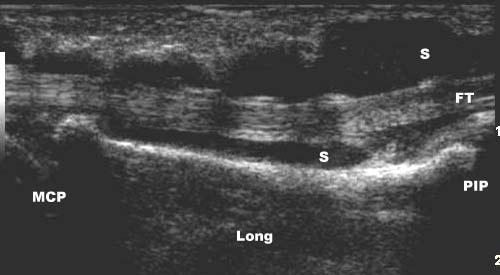
7 The study was supported by Abbott Laboratories S.A.,General Electric Medical Systems España S.A., Esaote España S.A., Siemens S.A., Zambon S.A., Merck Sharp Dohme España S.A and. Vita Científica SL. Acknowledgments: We thank Mr L.A. Ortega, Mr J. Gálvez, Mr F. Chica and Mr C. Matarranz, from General Electric Medical Systems España S.A, Mr A. López and Mr J. Masó from Esaote España S.A., and Mrs I. Hernández from Siemens S.A. for providing the ultrasound equipments and technical support. We would like to thank the staff members of the Department of Rheumatology from Bellvitge and Vall d Hebron Hospital, Barcelona, Spain, for allowing us to examine their patients. FIGURES FIGURE 1. Longitudinal US image of the radiocarpal joint with synovitis and intense power Doppler signal in a patient with RA. Long= longitudinal. FIGURE 2. Second finger flexor tenosynovitis in a patient with RA. Longitudinal US image shows increased hypoechoic synovitis (S) within the sheath surrounding the tendon (FT). MCP= metacarpophalangeal joint; PIP= proximal interphalangeal joint; Long= longitudinal. Ann Rheum Dis: first published as /ard on 7 June Downloaded from on 3 December 2018 by guest. Protected by copyright. 7
8 References 1. Grassi W, Cervini C. Ultrasonography in rheumatology: an evolving technique. Ann Rheum Dis 1998; 57: Wakefield RJ, Gibbon WW, Emey P. The current status of ultrasonography in rheumatology.rheumatology 1999; 38: Kane D, Balint PV, Sturrock RD. Ultrasonography is Superior to Clinical Examination in the Detection and Localization of Knee Joint Effusion in Rheumatoid Arthritis. J Rheumatol 2003; 30: Szkudlarek M, Court-Payen M, Jacobsen S, Klarlund M, Thomsen HS, Ostergaard M. Interobserver Agreement in Ultrasonography of the Finger and Toe Joints in Rheumatoid Arthritis. Arthritis Rheum 2003; 48: Karim Z, Wakefield RJ, Quinn M, Conaghan PG, Brown AK, Veale DJ, O Connor P, Reece R, Emery P. Validation and Reproductibility of Ultrasonography in the Detection of Synovitis in the knee. Arthritis Rheum 2004; 50: Szkudlarek M, Narvestad E, Klarlund M, Court-Payen M, Thomsen HS, Østergaard M. Ultrasonography of the Metatarsophalangeal Joints in Rheumatoid Arthritis. Arthritis Rheum 2004; 50: Wakefield RJ, Gibbon WW, Conaghan PG, O Connor P, McGonagle D, Pease C, et al. The value of sonography in the detection of bone erosions in patients with rheumatoid arthritis. Arthritis Rheum 2001;43: The value of sonography in the detection of bone erosions in patients with rheumatoid arthritis. Arthritis Rheum 2001;43: Swen WAA, Jacobs JWG, Algra PR, Manoliu RA, Rijkmans J, Willems WJ, et al. Sonography and magnetic resonance imaging equivalent for the assessment of full-thickness rotator cuff tears. Arthritis Rheum 1999;42: Middleton WD, Teefey SA, Yamaguchi K. Sonography of the Rotator Cuff: Analysis of Interobserver Variability. AJR 2004: 183: Iagnocco A, Ossandon A, Coari G, Conti F, Priori R, Alessandri C, et al. Wrist joint involvement in systemic lupus erythematosus. An ultrasonographic study. Clin Exp Rheumatol 2004; 22: Filippucci E, Farina A, Carotti M, Salaffi F, Grassi W. Grey scale and pewer Doppler sonographic changes induced by intraarticular steroid injection treatment. Ann Rheum Dis 2004; 63: Hauzeur EP, Mathy L, De Maertelaer V. Comparison between clinical evaluation and Ultrasonography in detecting hydrathrosis of the knee. J Rheumatol 1999; 26: Scheel AK,.Schmidt WA, Hermann KG, Bruyn GA, D Agostino MA, Grassi W, et al. Interobserver reliability of rheumatologist performing musculoskeletal ultrasonography: results from a EULAR "Train the Trainer" course. Ann Rheum Dis Published Online First January 7, doi: /ard Backhaus M, Burmester GR, Gerber T, Grassi W, Machold KP, Swen WA, et al. Guidelines for musculoskeletal ultrasound in rheumatology. Ann Rhem Dis 2001; 60: Landis JR, Koch GG. The measurement of observer agreement for categorical data. Biometrics 1977; 33: Robertson PL, Schweitzer ME, Mitchell DG, Schlesinger F, Epstein RE, Frieman BG, et al. Rotator Cuff Disorders: Interobserver and Intraobserver Variation in Diagnosis with MR Imaging. Radiology 1995; 194: Hoving JL, Buchbinder R, Hall S, Lawler G, Coombs P, McNealy S, Bird P, Connell D. A Comparison of Magnetic Resonance Imaging, Sonography, and Radiography of the Hand in Patients with Early Rheumatoid Arthritis. J Rheumatol 2004; 31: Naredo E, Bonilla G, Gamero F, Uson J, Carmona L, Laffon A. Assessment of Inflammatory Activity in Rheumatoid Arthritis: A Comparative Study of Clinical Evaluation with Gray-Scale and Power Doppler Ultrasonography. Ann Rheum Dis 2005; 64: Schmidt WA, Schmidt H, Schicke B, Gromnica-Ihle E. Standard reference values for musculoskeletal ultrasonography. Ann Rheum Dis 2004; 63: Ann Rheum Dis: first published as /ard on 7 June Downloaded from on 3 December 2018 by guest. Protected by copyright. 8
9 TABLES Table I. Ultrasonographic investigation ANATOMICAL STRUCTURE Long biceps tendon SHOULDER Subscapularis, supraspinatus and infraspinatus tendons Subacromial-subdeltoid and subcoracoid bursae Acromio-clavicular joint Glenohumeral joint (axillary, anterior and posterior recesses) Humeral head (anterior, lateral, posterior and axillary aspects) Supraspinatus tendon WRIST/HAND Radiocarpal joint (dorsal and palmar aspects) Wrist tendons (dorsal, palmar, extensor carpi ulnaris) First carpometacarpal joint Second metacarpophalangeal joint Second proximal interphalangeal joint Second finger flexor tendons ANKLE/FOOT Tibiotalar joint (anterior and posterior aspects) Talonavicular joint Anterior tibialis, flexor, peroneus and posterior tibialis tendons Posterior tibialis tendon Achilles tendon Retrocalcaneal bursa Calcaneus (posterior and plantar aspects) Plantar fascia First metatarsophalangeal joint Fifth metatarsophalangeal joint Suprapatellar recess Bone surfaces KNEE Quadriceps and patellar tendons Prepatellar, superficial and deep infrapatellar, anserin bursae Gastrocnemius-semimembranous bursa Femoral articular cartilage Medial collateral ligament ULTRASONOGRAPHIC FINDINGS tenosynovitis, partial tear, complete tear tendinosis, partial tear, full-thickness tear, calcification bursitis effusion/synovitis, erosions, osteophytes effusion/synovitis erosions impingement effusion/synovitis, power Doppler signal tenosynovitis effusion/synovitis, erosions, osteophytes effusion/synovitis, erosions, osteophytes, power Doppler signal effusion/synovitis, erosions, osteophytes tenosynovitis effusion/synovitis effusion/synovitis, erosions, osteophytes tenosynovitis partial tear, complete tear tendinosis, enthesitis, paratenonitis, partial tear, complete tear bursitis erosions, spur fasciitis effusion/synovitis, erosions, osteophytes, power Doppler signal effusion/synovitis, erosions, osteophytes effusion, synovial hypertrophy erosions, osteophytes tendinosis, enthesitis, partial tear, complete tear bursitis Baker s cyst lesion partial tear, complete tear 9 Ann Rheum Dis: first published as /ard on 7 June Downloaded from on 3 December 2018 by guest. Protected by copyright.
10 Table II Overall agreements by US diagnosis in each region and in all regions: SHOULDER WRIST/HAND ANKLE/FOOT KNEE ALL REGIONS Joint effusion/synovitis 88.5% 95% 89% 91.5% 91% Bony cortex abnormalities 84.5% 88.5% 92% 84.5% 87% Tenosynovitis/paratenonitis 76% 88.5% 88.5% NE 84% Tendon lesions 88% NE 92% 94% 91% Bursitis/cyst 85.5% NE 73.5% 92% 83.5% Power Doppler signal NE 92.5% 73.5% NE 83% NE= diagnosis not evaluated Ann Rheum Dis: first published as /ard on 7 June Downloaded from on 3 December 2018 by guest. Protected by copyright. 10
11 Table III. Kappa values by group and anatomical region and overall (mean) kappa by region:. Group 1 Group 2 Group 3 Group 4 Group 5 Group 6 Mean kappa Shoulder Wrist/hand Ankle/foot Knee Ann Rheum Dis: first published as /ard on 7 June Downloaded from on 3 December 2018 by guest. Protected by copyright. 11
12 " The Corresponding Author has the right to grant on behalf of all authors and does grant on behalf of all authors, an exclusive licence (or non exclusive for government employees) on a worlwide basis to the BMJ Publishing Group Ltd and its licensees, to permit this article (if accepted) to be published in ARD and any other BMJPG products and to exploit all subsidiary rights, as set out in our licence ( Ann Rheum Dis: first published as /ard on 7 June Downloaded from on 3 December 2018 by guest. Protected by copyright. 12

13
14
Interobserver reliability in musculoskeletal ultrasonography: results from a Teach the Teachers rheumatologist course
 14 EXTENDED REPORT Interobserver reliability in musculoskeletal ultrasonography: results from a Teach the Teachers rheumatologist course E Naredo, I Möller, C Moragues, J J de Agustín, A K Scheel, W Grassi,
David Bong (Spain), MD Faculty member of the EULAR MSUS courses since 2011.
, MD Faculty member of the EULAR MSUS courses since 2011.") MUSCULOSKELTAL SONOGRAPHY COURSE Intermediate Level Sept 30 & Oct 1 & 2, 2016 Dubai, United Arab Emirates Organizing Office: Compass Conferences ENDORSED Organizers Ingrid Möller (Scientific Organizer)
MUSCULOSKELTAL SONOGRAPHY COURSE Intermediate Level Sept 30 & Oct 1 & 2, 2016 Dubai, United Arab Emirates Organizing Office: Compass Conferences ENDORSED Organizers Ingrid Möller (Scientific Organizer)
"EULAR endorsed course"
 MUSCULOSKELTAL SONOGRAPHY COURSE InterMediate Level 15 17 September, 2017 Dubai, UAE Organizing Office: Compass Conferences "EULAR endorsed course" Organizers Ingrid Möller (Scientific Organizer) Esperanza
MUSCULOSKELTAL SONOGRAPHY COURSE Basic Level 4 6 November, 2016 Manama, Kingdom of Bahrain Organizing Office: Compass Conferences ENDORSED
 MUSCULOSKELTAL SONOGRAPHY COURSE Basic Level 4 6 November, 2016 Manama, Kingdom of Bahrain Organizing Office: Compass Conferences ENDORSED Organizers Möller (Scientific Organizer) Ayman Fahim (Organizer)
ELENI ANDIPA General Hospital of Athens G. Gennimatas
 ELENI ANDIPA General Hospital of Athens G. Gennimatas Technological advances over the last years have caused a dramatic improvement in ultrasound quality and resolution An established imaging modality
Clinical Study Reliability Exercise for the Polymyalgia Rheumatica Classification Criteria Study: The Oranjewoud Ultrasound Substudy
 Hindawi Publishing Corporation International Journal of Rheumatology Volume 2009, Article ID 738931, 5 pages doi:10.1155/2009/738931 Clinical Study Reliability Exercise for the Polymyalgia Rheumatica Classification
EULAR 24th EULAR Sonography Course Basic, Intermediate and Advanced Musculoskeletal Ultrasound (MSUS) in Rheumatology
 in Rheumatology") EULAR 24th EULAR Sonography Course Basic, Intermediate and Advanced Musculoskeletal Ultrasound (MSUS) in Rheumatology Sunday 11th Wednesday 14th June 2017 Madrid, Spain ORGANISATION AND COMMITTEE Scientific
EULAR 24th EULAR Sonography Course Basic, Intermediate and Advanced Musculoskeletal Ultrasound (MSUS) in Rheumatology Sunday 11th Wednesday 14th June 2017 Madrid, Spain ORGANISATION AND COMMITTEE Scientific
"EULAR endorsed course"
 MUSCULOSKELTAL SONOGRAPHY COURSE Basic Level 20 22 July, 2018 Abu Dhabi, United Arab Emirates Organizing Office: Compass Conferences "EULAR endorsed course" Organizers Möller (Scientific Organizer) Esperanza
Ultrasound in Rheumatology
 Arthritis Research UK Primary Care Centre Winner of a Queen s Anniversary Prize For Higher and Further Education 2009 Ultrasound in Rheumatology Alison Hall Consultant MSK Sonographer/Research Fellow Primary
EULAR 25th Jubilee EULAR Sonography Course Basic, Intermediate and Advanced Musculoskeletal Ultrasound (MSUS) in Rheumatology
 in Rheumatology") EULAR 25th Jubilee EULAR Sonography Course Basic, Intermediate and Advanced Musculoskeletal Ultrasound (MSUS) in Rheumatology Sunday 10th Wednesday 13th June 2018 Amsterdam, Netherlands ORGANISATION AND
EULAR 25th Jubilee EULAR Sonography Course Basic, Intermediate and Advanced Musculoskeletal Ultrasound (MSUS) in Rheumatology Sunday 10th Wednesday 13th June 2018 Amsterdam, Netherlands ORGANISATION AND
EULAR 24th EULAR Sonography Course Basic, Intermediate and Advanced Musculoskeletal Ultrasound (MSUS) in Rheumatology
 in Rheumatology") EULAR 24th EULAR Sonography Course Basic, Intermediate and Advanced Musculoskeletal Ultrasound (MSUS) in Rheumatology Sunday 11th Wednesday 14th June 2017 Madrid, Spain ORGANISATION AND COMMITTEE Scientific
EULAR 24th EULAR Sonography Course Basic, Intermediate and Advanced Musculoskeletal Ultrasound (MSUS) in Rheumatology Sunday 11th Wednesday 14th June 2017 Madrid, Spain ORGANISATION AND COMMITTEE Scientific
26 th Sonography Course MSUS Intermediate
 26 th Sonography Course MSUS Intermediate Madrid, Spain Sunday, 9 th June Wednesday, 12 th June 2019 The 26 th EULAR Sonography Course will be run simultaneously at three levels, basic, intermediate and
8 th Sonography Course Ultrasound Trainers [Teach the Teachers, TTT]
![8 th Sonography Course Ultrasound Trainers [Teach the Teachers, TTT]](/thumbs/96/127818637.jpg "8 th Sonography Course Ultrasound Trainers [Teach the Teachers, TTT]") 8 th Sonography Course Ultrasound Trainers [Teach the Teachers, TTT] Madrid, Spain Saturday, 8 th June Sunday, 9 th June 2019 1 ORGANISATION COMMITTEE Scientific Organisers Esperanza Naredo Ingrid Möller
8 th Sonography Course Ultrasound Trainers [Teach the Teachers, TTT] Madrid, Spain Saturday, 8 th June Sunday, 9 th June 2019 1 ORGANISATION COMMITTEE Scientific Organisers Esperanza Naredo Ingrid Möller
EULAR 25th Jubilee EULAR Sonography Course Basic, Intermediate and Advanced Musculoskeletal Ultrasound (MSUS) in Rheumatology
 in Rheumatology") EULAR 25th Jubilee EULAR Sonography Course Basic, Intermediate and Advanced Musculoskeletal Ultrasound (MSUS) in Rheumatology Sunday 10th Wednesday 13th June 2018 Amsterdam, Netherlands ORGANISATION AND
EULAR 25th Jubilee EULAR Sonography Course Basic, Intermediate and Advanced Musculoskeletal Ultrasound (MSUS) in Rheumatology Sunday 10th Wednesday 13th June 2018 Amsterdam, Netherlands ORGANISATION AND
15 th EULAR SONOGRAPHY COURSE. June 08 th - 11 th, 2008 Paris, France. Basic, Intermediate and Advanced Musculoskeletal Ultrasound in Rheumatology
 15 th EULAR SONOGRAPHY COURSE Basic, Intermediate and Advanced Musculoskeletal Ultrasound in Rheumatology June 08 th - 11 th, 2008 Paris, France Societé française de rhumatologie Université de Versailles
Scoring and Grading B-Mode Synovitis and Doppler findings in pediatric MSKUS. Johannes Roth MD PhD FRCPC RhMSUS
 Scoring and Grading B-Mode Synovitis and Doppler findings in pediatric MSKUS Johannes Roth MD PhD FRCPC RhMSUS Pathology - Definition Synovitis Synovitis on ultrasonography in children B-mode and Doppler
Concise report RHEUMATOLOGY
 RHEUMATOLOGY Rheumatology 2012;51:2034 2038 doi:10.1093/rheumatology/kes124 Advance Access publication 30 July 2012 Concise report Head-to-head comparison of quantitative and semi-quantitative ultrasound
International Musculoskeletal Ultrasound Course MITOS
 Basic and Intermediate Levels th International Musculoskeletal Ultrasound Course MITOS Final Course Program 30 November - 2 December 2017 Wyndham Grand Athens Hotel Athens Greece www.synthesispco.com/mitoscourse2017
Ultrasound in Rheumatology
 Ultrasound in Rheumatology Alison Hall Consultant MSK Sonographer Research Institute for Primary Care & Health Sciences, Keele University Department of Rheumatology, Cannock Hospital, Royal Wolverhampton
Paediatric rheumatology
 Paediatric rheumatology Ultrasonography vs. clinical examination in children with suspected arthritis. Does it make sense to use poliarticular ultrasonographic screening? G. Filippou, L. Cantarini, I.
The use of musculoskeletal ultrasound (MSKUS) as a. Musculoskeletal Ultrasound as a Diagnostic and Prognostic Tool in Rheumatoid Arthritis
 as a. Musculoskeletal Ultrasound as a Diagnostic and Prognostic Tool in Rheumatoid Arthritis") 215 Musculoskeletal Ultrasound as a Diagnostic and Prognostic Tool in Rheumatoid Arthritis Manish Jain, M.D., and Jonathan Samuels, M.D. Abstract The use of musculoskeletal ultrasound (MSKUS) has increased
215 Musculoskeletal Ultrasound as a Diagnostic and Prognostic Tool in Rheumatoid Arthritis Manish Jain, M.D., and Jonathan Samuels, M.D. Abstract The use of musculoskeletal ultrasound (MSKUS) has increased
Bulgarian Association for Musculoskeletal Ultrasound
 MUSCULOSKELETAL ULTRASOUND COURSE INTERMEDIATE LEVEL ORGANIZATION AND COMMITTEE Scientific Organiser Prof. Annamaria Iagnocco Local Organisers Prof Anastas Batalov Dr Rodina Nestorova Dr Plamen Todorov
Ultras ono graphic Evaluation of Rotator Cuff Tendons in Patients with Rheumatoid Arthritis
 Med. J. Cairo Univ., Vol. 83, No. 1, June: 395-399, 215 www.medicaljournalofcairouniversity.net Ultras ono graphic Evaluation of Rotator Cuff Tendons in Patients with Rheumatoid Arthritis HALA I. ELGENDY,
Imaging. Introduction Systemic lupus erythematosus (SLE) is an autoimmune multisystem disorder
 is an autoimmune multisystem disorder") Clinical and Experimental Rheumatology 2009; 27: 00-00 Imaging Ultrasound imaging for the rheumatologist XXIV. Sonographic evaluation of wrist and hand joint and tendon involvement in systemic lupus erythematosus
Clinical and Experimental Rheumatology 2009; 27: 00-00 Imaging Ultrasound imaging for the rheumatologist XXIV. Sonographic evaluation of wrist and hand joint and tendon involvement in systemic lupus erythematosus
September 5 th 7 th, 2018 Innsbruck, Austria
 MUSCULOSKELETAL SONOGRAPHY COURSE FOR RHEUMATOLOGISTS - INTERMEDIATE LEVEL - September 5 th 7 th, 2018 Innsbruck, Austria This course is scientifically endorsed by: GENERAL INFORMATION Course opening:
Scientific Programme
 Scientific Programme (preliminary version) Musculoskeletal Sonography Course for Rheumatologists Warszawa 2010 th st September 28 - October 01, 2010 Warszawa, Poland under scientific patronage of the European
Ultrasound in assessing disease severity and therapeutic response in rheumatoid arthritis
 For reprint orders, please contact: reprints@futuremedicine.com REVIEW Ultrasound in assessing disease severity and therapeutic response in rheumatoid arthritis Helen I Keen, Richard J Wakefield & Philip
Original article RHEUMATOLOGY
 RHEUMATOLOGY Original article Rheumatology 2012;51:184 190 doi:10.1093/rheumatology/ker331 Advance Access publication 24 November 2011 CLINICAL SCIENCE Practice of ultrasound-guided arthrocentesis and
26 th Sonography Course MSUS Advanced
 26 th Sonography Course MSUS Advanced Madrid, Spain Sunday, 9 th June Wednesday, 12 th June 2019 The 26 th EULAR Sonography Course will be run simultaneously at three levels, basic, intermediate and advanced,
The Egyptian Journal of Hospital Medicine (October 2017) Vol. 69 (4), Page
 Vol. 69 (4), Page") The Egyptian Journal of Hospital Medicine (October 2017) Vol. 69 (4), Page 2294-2300 Role of Magnetic Resonance Imaging and Ultrasonography in Diagnosis and Follow Up Rheumatoid Arthritis in Hand and Wrist
The Egyptian Journal of Hospital Medicine (October 2017) Vol. 69 (4), Page 2294-2300 Role of Magnetic Resonance Imaging and Ultrasonography in Diagnosis and Follow Up Rheumatoid Arthritis in Hand and Wrist
Imaging. Ultrasound imaging for the rheumatologist XLIV. Ultrasound of the shoulder in healthy individuals
 Imaging Ultrasound imaging for the rheumatologist XLIV. Ultrasound of the shoulder in healthy individuals A. Iagnocco 1, E. Filippucci 2, G. Sakellariou 3, F. Ceccarelli 1, L. Di Geso 2, L. Carli 4, L.
Reporting Ultrasound Findings and Diagnosis
 Reporting Ultrasound Findings and Diagnosis Rodina Nestorova MD Rheumatology Centre St. Irina, Sofia Bulgarian MSUS Society Basic MSU Course 14-16 Jan 2016 Plovdiv, Bulgaria ULTRASOUND REPORT COLLECTION
The reliability of musculoskeletal ultrasound in the detection of cartilage abnormalities at the metacarpo-phalangeal joints
 Osteoarthritis and Cartilage 20 (2012) 1142e1146 The reliability of musculoskeletal ultrasound in the detection of cartilage abnormalities at the metacarpo-phalangeal joints A. Iagnocco y *, P.G. Conaghan
The EULAR OMERACT rheumatoid arthritis MRI reference image atlas: the wrist joint
 i23 The EULAR OMERACT rheumatoid arthritis MRI reference image atlas: the wrist joint B Ejbjerg, F McQueen, M Lassere, E Haavardsholm, P Conaghan, P O Connor, P Bird, C Peterfy, J Edmonds, M Szkudlarek,
Guidelines for musculoskeletal ultrasound in rheumatology
 Ann Rheum Dis 2001;60:641 649 641 REVIEW Rheumatology and Clinical Immunology, Charité University Hospital, Humboldt University, Berlin, Germany M Backhaus G-R Burmester Rheumatology and Physical Medicine,
Role of Ultrasound and MRI in Detection of Hand and Wrist Joints Erosions in Rheumatoid Arthritis Patients, Comparative Study
 Med. J. Cairo Univ., Vol. 83, No. 1, September: 615-620, 2015 www.medicaljournalofcairouniversity.net Role of Ultrasound and MRI in Detection of Hand and Wrist Joints Erosions in Rheumatoid Arthritis Patients,
Immanuel Krankenhaus Berlin, Medical Centre for Rheumatology Berlin - Buch; 2
 Low-field MRI versus ultrasound: which is more sensitive in detecting inflammation and bone damage in MCP and MTP joints in mild or moderate rheumatoid arthritis? W.A. Schmidt 1, B. Schicke 2, B. Ostendorf
M usculoskeletal ultrasonography is a diagnostic
 988 EXTENDED REPORT Standard reference values for musculoskeletal ultrasonography W A Schmidt, H Schmidt, B Schicke, E Gromnica-Ihle... See end of article for authors affiliations... Correspondence to:
EDUCATIONAL COURSE. MUSCULOSKELETAL ULTRASOUND in RHEUMATOLOGY BASIC COURSE. Course Coordinator Annamaria Iagnocco
 EDUCATIONAL COURSE 2019 MUSCULOSKELETAL ULTRASOUND in RHEUMATOLOGY BASIC COURSE Rome, March 21-23, 2019 Course Coordinator Annamaria Iagnocco COURSE PRESENTATION Musculoskeletal ultrasound in rheumatology
June 26 th 28 th, 2014 Innsbruck, Austria
 MUSCULOSKELETAL SONOGRAPHY COURSE FOR RHEUMATOLOGISTS - INTERMEDIATE LEVEL - June 26 th 28 th, 2014 Innsbruck, Austria This course is scientifically endorsed by: GENERAL INFORMATION Course opening: Thursday,
The development of a preliminary ultrasonographic scoring system for features of hand osteoarthritis
 1 Academic Unit of Musculoskeletal Disease, University of Leeds, Leeds, UK; 2 Rheumatology B, Cochin Hospital, Paris France; 3 Ambroise Pare Hospital, Boulogne-Billancourt, France; 4 Diakonhjemmet Hospital,
MUSCULOSKELETAL SONOGRAPHY COURSE FOR RHEUMATOLOGISTS - BASIC LEVEL - June 27 th 29 th, 2013 Innsbruck, Austria
 MUSCULOSKELETAL SONOGRAPHY COURSE FOR RHEUMATOLOGISTS - BASIC LEVEL - June 27 th 29 th, 2013 Innsbruck, Austria GENERAL INFORMATION Course opening: Thursday, June 27 th, 2013 - h 12.00 Course closing:
UltraSound Course. International Musculoskeletal MITOS. Athens, Greece. Basic and Intermediate Levels - Course Program. 29 Nov - 1 Dec 2018
 Basic and Intermediate Levels - Course Program th International Musculoskeletal UltraSound Course 29 Nov - 1 Dec 2018 Wyndham Grand Athens Hotel Athens, Greece MITOS Musculosceletal Imaging Techniques
A Comparative Study of Ultrasonographic Findings with Clinical and Radiological Findings of Painful Osteoarthritis of the Knee Joint
 Med. J. Cairo Univ., Vol. 84, No. 3, December: 97-, www.medicaljournalofcairouniversity.net A Comparative Study of Ultrasonographic Findings with Clinical and Radiological Findings of Painful Osteoarthritis
September 14 th 16 th, 2017 Innsbruck, Austria
 MUSCULOSKELETAL SONOGRAPHY COURSE FOR RHEUMATOLOGISTS - BASIC LEVEL - September 14 th 16 th, 2017 Innsbruck, Austria This course is scientifically endorsed by: GENERAL INFORMATION Course opening: Thursday,
International Musculoskeletal Ultrasound Course MITOS
 Basic and Intermediate Levels th International Musculoskeletal Ultrasound Course MITOS General information and Course program 30 November - 2 December 2017 Wyndham Grand Athens Hotel Athens Greece www.synthesispco.com/mitoscourse2017
MUSCULOSKELETAL ULTRASOUND COURSE - BASIC LEVEL- Graz, November 2 nd 4 th, 2016
 MUSCULOSKELETAL ULTRASOUND COURSE - BASIC LEVEL- Graz, November 2 nd 4 th, 2016 GENERAL INFORMATION Course opening Wednesday, Novemeber 2 nd, 2016 - h 17.00 Course closing Friday, November 4 th, 2012 -
Imaging. Ultrasound imaging for the rheumatologist
 Imaging Ultrasound imaging for the rheumatologist E. Filippucci 1, A. Iagnocco 2, G. Meenagh 3, L. Riente 4, A. Delle Sedie 4, S. Bombardieri 4, G. Valesini 2, W. Grassi 1 1 Cattedra di Rheumatologia,
Targeted Ultrasound of the Fifth Metatarsophalangeal Joint in an Early Inflammatory Arthritis Cohort
 Arthritis & Rheumatism (Arthritis Care & Research) Vol. 61, No. 7, July 15, 2009, pp 1004 1008 DOI 10.1002/art.24564 2009, American College of Rheumatology CONTRIBUTIONS FROM THE FIELD Targeted Ultrasound
Imaging Ultrasound imaging for the rheumatologist XLVIII. Ultrasound of the shoulders of patients with rheumatoid arthritis
 Imaging Ultrasound imaging for the rheumatologist XLVIII. Ultrasound of the shoulders of patients with rheumatoid arthritis G. Sakellariou 1, A. Iagnocco 2, E. Filippucci 3, F. Ceccarelli 2, L. Di Geso
2 nd MUSCULOSKELETAL SONOGRAPHY COURSE FOR RHEUMATOLOGISTS BASIC LEVEL. Athens, December 17 th - 19 th, 2015
 MITOS 2 nd MUSCULOSKELETAL SONOGRAPHY COURSE FOR RHEUMATOLOGISTS BASIC LEVEL Athens, December 17 th - 19 th, 2015 Musculoskeletal sonography course for rheumatologists, Athens 2015 intends to be a combination
3 rd MUSCULOSKELETAL SONOGRAPHY COURSE FOR RHEUMATOLOGISTS
 3 rd MUSCULOSKELETAL SONOGRAPHY COURSE FOR RHEUMATOLOGISTS UNDER SCIENTIFIC ENDORSEMENT OF THE EUROPEAN LEAGUE AGAINST RHEUMATISM BASIC AND INTERMEDIATE LEVEL Belgrade, March 29th-31st, 2012 G E N E R
Imaging. Ultrasound imaging for the rheumatologist XXXVI. Sonographic assessment of the foot in gout patients
 Imaging Ultrasound imaging for the rheumatologist XXXVI. Sonographic assessment of the foot in gout patients E. Filippucci 1, G. Meenagh 2, A. Delle Sedie 3, G. Sakellariou 4, A. Iagnocco 5, L. Riente
Sonographic appearance of chronic inflammatory rheumatism
 Sonographic appearance of chronic inflammatory rheumatism Poster No.: C-2237 Congress: ECR 2013 Type: Educational Exhibit Authors: H. Elfattach, F. Houari, O. Addou, M. Maaroufi, S. Tizniti ; 1 1 1 1 2
6 th MUSCULOSKELETAL SONOGRAPHY COURSE. Belgrade, 5 th 7 th October, 2017 INTERMEDIATE LEVEL COURSE
 6 th MUSCULOSKELETAL SONOGRAPHY COURSE INTERMEDIATE LEVEL COURSE Belgrade, 5 th 7 th October, 2017 ORGANIZER Rheumatology Association of Serbia www.belgrade-course.org SCIENTIFICALLY ENDORSED BY eular
Table of contents. Foreword. Preface. 1 Introduction Historical Perspective 00
 Table of contents Foreword Preface 1 Introduction 00 1.1 Historical Perspective 00 2 Fundamentals of musculoskeletal ultrasound 00 2.1 Frequency and wavelength 00 2.2 Generating ultrasound waves 00 2.3
Early diagnosis of Rheumatoid
 26 Original Article Diagnostic Accuracy of Ultrasonography in Detection of Destructive Changes in Small Joints of Hands in Patients of Rheumatoid Arthritis: A Comparison with Magnetic Resonance Imaging
General information and Course programme of the. 4 MUSCULOSKELETAL SONOGRAPHY COURSE FOR RHEUMATOLOGISTS - BASIC LEVEL - st
 General information and Course programme of the th 4 MUSCULOSKELETAL SONOGRAPHY COURSE FOR RHEUMATOLOGISTS - BASIC LEVEL - st 1 MUSCULOSKELETAL SONOGRAPHY COURSE FOR RHEUMATOLOGISTS - INTERMEDIATE LEVEL
Pragmatic ultrasound in the diagnosis of soft tissue rheumatic pain. Plamen Todorov
 Pragmatic ultrasound in the diagnosis of soft tissue rheumatic pain Plamen Todorov INTRODUCTION Soft tissue rheumatism: nonsystemic, focal pathological syndromes involving the periarticular structures.
MUSCULOSKELETAL ULTRASOUND in RHEUMATOLOGY
 EDUCATIONAL COURSE 2018 MUSCULOSKELETAL ULTRASOUND in RHEUMATOLOGY BASIC COURSE This course is endorsed by Rome, March 15-17, 2018 Course Coordinator Annamaria Iagnocco COURSE PRESENTATION Musculoskeletal
Sonographic appearance of fluid in peripheral joints and bursae of healthy asymptomatic Chinese population
 Original Article Sonographic appearance of fluid in peripheral joints and e of healthy asymptomatic Chinese population Liyun Wang, Xi Xiang, Yuanjiao Tang, Yujia Yang, Li Qiu Department of Ultrasound,
C. A. Guillén Astete, A. Boteanu, and A. Zea Mendoza. 1. Introduction
 e Scientific World Journal, Article ID 563981, 5 pages http://dx.doi.org/10.1155/2014/563981 Research Article Comparison of revalence of Synovitis by Ultrasound Assessment in Subjects Exposed or Not to
3rd MUSCULOSKELETAL SONOGRAPHY COURSE FOR RHEUMATOLOGISTS - INTERMEDIATE LEVEL -
 3rd MUSCULOSKELETAL SONOGRAPHY COURSE FOR RHEUMATOLOGISTS - INTERMEDIATE LEVEL - Bucharest, Romania October, 13th-15th 2016 Organizer: Romanian Society of Rheumatology Prof. Dr. Ruxandra Ionescu Course
Index. Note: Page numbers of article titles are in boldface type.
 Note: Page numbers of article titles are in boldface type. A ACJ. See Acromioclavicular joint (ACJ) Acromioclavicular joint (ACJ) procedures of, 557 559 Ankle and foot procedures of, 649 671 (See also
Graz, October 3 rd 5 th, 2013
 MUSCULOSKELETAL SONOGRAPHY COURSE IN RHEUMATOLOGY INTERMEDIATE LEVEL Graz, October 3 rd 5 th, 2013 This course has been scientifically endorsed by: GENERAL INFORMATION Course opening: Thursday, October
Musculoskeletal Ultrasound for Rheumatologists
 Hong Kong Bull Rheum Dis 2009;9:1-7 Review Article Musculoskeletal Ultrasound for Rheumatologists Ka-Lai Lee Abstract: Keywords: In the past two decades, musculoskeletal ultrasonography has become an important
ONLINE. J.A. Mendonça 1, M. Yazbek 1, I.M.M. Laurindo 2 and M.B. Bertolo 1
 Brazilian Journal of Medical and Biological Research Online Provisional Version ISSN 0100-879X This Provisional PDF corresponds to the article as it appeared upon acceptance. Fully formatted PDF and full
Clinical Practice Guideline. Ultrasound in Rheumatological Settings. Version
 Clinical Practice Guideline Ultrasound in Rheumatological Settings Version 1.1.2017 November 2017 Table of Contents Abbreviations...3 Introduction...4 Diagnostic Musculoskeletal Ultrasound...6 Definition
Imaging. Ultrasound imaging for the rheumatologist XLI. Sonographic assessment of the hip in OA patients
 Imaging XLI. Sonographic assessment of the hip in OA patients A. Iagnocco 1, E. Filippucci 2, L. Riente 3, G. Meenagh 4, A. Delle Sedie 3, G. Sakellariou 5, F. Ceccarelli 1, C. Montecucco 5, S. Bombardieri
The Role of Ultrasonography in the Assessment of Rheumatic Diseases. Current and Potential Role of Ultrasonography in Rheumatology
 The Role of Ultrasonography in the Assessment of Rheumatic Diseases Karina D. Torralba, MD, MACM, CCD, Assistant Professor of Medicine, Keck School of Medicine, University of Southern California Objective:
Ultrasound in Rheumatoid Arthritis
 Review Med Ultrason 2013, Vol. 15, no. 3, 199-208 DOI: 10.11152/mu.2013.2066.153.cr1fc2 Ultrasound in Rheumatoid Arthritis Chiara Rizzo, Fulvia Ceccarelli, Angelica Gattamelata, Caterina Vavala, Guido
Ultrasound assessment of most frequent shoulder disorders
 Ultrasound assessment of most frequent shoulder disorders Poster No.: C-2026 Congress: ECR 2014 Type: Educational Exhibit Authors: S. P. Ivanoski; Ohrid/MK Keywords: Trauma, Athletic injuries, Arthritides,
MUSCULOSKELETAL ULTRASOUND COURSE - BASIC LEVEL- Graz, November 2 nd 4 th, 2016
 MUSCULOSKELETAL ULTRASOUND COURSE - BASIC LEVEL- Graz, November 2 nd 4 th, 2016 Dear colleague, musculoskeletal ultrasound is increasingly used by rheumatologist and other health professionals for diagnostic
2 MUSCULOSKELETAL SONOGRAPHY COURSE FOR RHEUMATOLOGISTS - INTERMEDIATE LEVEL -
 General information and Course programme of the nd 2 MUSCULOSKELETAL SONOGRAPHY COURSE FOR RHEUMATOLOGISTS - INTERMEDIATE LEVEL - rd th Zagreb, December 3 December 5, 2018. This course has been scientifically
2 MUSCULOSKELETAL SONOGRAPHY COURSE FOR RHEUMATOLOGISTS - BASIC - Zagreb, January 31 - February 2, 2013.
 General information and Course programme of the nd 2 MUSCULOSKELETAL SONOGRAPHY COURSE FOR RHEUMATOLOGISTS - BASIC - st nd Zagreb, January 31 - February 2, 2013. This course has been scientifically endorsed
Intermediate Ultrasound Course. in Belek near Antalya, Turkey. October 4-6, Programme
 Intermediate Ultrasound Course in Belek near Antalya, Turkey October 4-6, 2018 Programme Venue: Susesi Hotel, Belek, Turkey Course fee including course registration, hotel for 3 nights & meals: 950 Number
Role of High Resolution Diagnostic Ultrasound in Musculoskeletal Physiotherapy
 Role of High Resolution Diagnostic Ultrasound in Musculoskeletal Physiotherapy Dr.Amandeep Singh* *Professor, Department of Physiotherapy, Chandigarh University, Gharuan. Email ID: aman_ruby@yahoo.com
Unit of Rheumatology, Ospedale Sacro Cuore, Negrar, Verona, Italy; 2
 Comprehensive evaluation of finger flexor tendon entheseal soft tissue and bone changes by ultrasound can differentiate psoriatic arthritis and rheumatoid arthritis I. Tinazzi 1, D. McGonagle 2, A. Zabotti
3rd MUSCULOSKELETAL SONOGRAPHY COURSE FOR RHEUMATOLOGISTS - BASIC LEVEL Zagreb, May 29th - May 31st, 2014.
 General information and Course programme of the 3rd MUSCULOSKELETAL SONOGRAPHY COURSE FOR RHEUMATOLOGISTS - BASIC LEVEL Zagreb, May 29th - May 31st, 2014. This course has been scientifically endorsed by
Knee, Ankle, and Foot: Normal and Abnormal Features with MRI and Ultrasound Correlation. Disclosures. Outline. Joint Effusion. Suprapatellar recess
 Knee, Ankle, and Foot: Normal and Abnormal Features with MRI and Ultrasound Correlation Jon A. Jacobson, M.D. Professor of Radiology Director, Division of Musculoskeletal Radiology University of Michigan
We are IntechOpen, the world s leading publisher of Open Access books Built by scientists, for scientists. International authors and editors
 We are IntechOpen, the world s leading publisher of Open Access books Built by scientists, for scientists 3,500 108,000 1.7 M Open access books available International authors and editors Downloads Our
Is the Doppler Ultrasound Score More Reliable than the Grey Scale Ultrasound Score in Assessing the Rheumatoid Arthritis Patient s Joints?
 ORIGINAL ARTICLE Is the Doppler Ultrasound Score More Reliable than the Grey Scale Ultrasound Score in Assessing the Rheumatoid Arthritis Patient s Joints? Copotoiu Monica 1, Georgescu Lia 1, Poór G 2,
Schäfer et al. BMC Musculoskeletal Disorders 2013, 14:358
 Schäfer et al. BMC Musculoskeletal Disorders 2013, 14:358 RESEARCH ARTICLE Open Access Evaluation of the novel ultrasound score for large joints in psoriatic arthritis and ankylosing spondylitis: six month
Ultrasound of Mid and Hindfoot Pathology
 Ultrasound of Mid and Hindfoot Pathology Levon N. Nazarian, M.D. Professor of Radiology Thomas Jefferson University Hospital Disclosures None relevant to this presentation Educational Objective Following
Diagnostic utility of musculoskeletal ultrasound in patients with suspected arthritis a probabilistic approach
 Rezaei et al. Arthritis Research & Therapy 2014, 16:448 RESEARCH ARTICLE Open Access Diagnostic utility of musculoskeletal ultrasound in patients with suspected arthritis a probabilistic approach Hamed
S tructural joint damage, a major outcome in
 i3 An introduction to the EULAR OMERACT rheumatoid arthritis MRI reference image atlas M Østergaard, J Edmonds, F McQueen, C Peterfy, M Lassere, B Ejbjerg, P Bird, P Emery, H Genant, P Conaghan... This
Ultrasound in osteoarthritis
 Ultrasound in osteoarthritis A. Iagnocco Reumatologia, Dipartimento di Medicina Interna e Specialità Mediche, Sapienza Università di Roma, Rome, Italy. Annamaria Iagnocco, MD, Adjunct Professor of Rheumatology
Ultrasonographic Evaluation of Painful Shoulder joint in rural population
 Original article: Ultrasonographic Evaluation of Painful Shoulder joint in rural population Dr. Pankaj Garg*, Dr. V.N. Marathe, Dr. S. G. Gandage, Dr.S.G.Kachewar Department of Radiology, Rural Medical
Correspondence should be addressed to Zuhudha Hussain Manik;
 Scientifica Volume 2016, Article ID 5609132, 7 pages http://dx.doi.org/10.1155/2016/5609132 Research Article Ultrasound Assessment of Synovial Thickness of Some of the Metacarpophalangeal Joints of Hand
Musculoskeletal Ultrasound
 EFSUMB Course Book Student Edition Editors: Jan Tuma, Radu Badea, Christoph F. Dietrich Musculoskeletal Ultrasound Giorgio Tamborrini Ultrasound Center, Switzerland Corresponding author: KD Dr. med. Giorgio
Message of the Month for GPs June 2013
 Message of the Month for GPs June 2013 Dr Winn : Consultant Musculoskeletal Radiologist, Manchester Royal Infirmary Imaging of the musculoskeletal system Musculoskeletal pain is a common problem in the
EULAR-PReS PAEDIATRIC MUSCULOSKELETAL COURSE. Monday 12th Wednesday 14th June 2017 Madrid, Spain
 EULAR-PReS PAEDIATRIC MUSCULOSKELETAL COURSE Monday 12th Wednesday 14th June 2017 Madrid, Spain ORGANISATION AND COMMITTEE Scientific organisers Esperanza Naredo Tadej Avcin Jelena Vojinovic Paz Collado
US finding of the shoulder (with live demonstration) 인제의대상계백병원 안재기
 인제의대상계백병원 안재기") US finding of the shoulder (with live demonstration) 인제의대상계백병원 안재기 Shoulder US Biceps tendon & Rotator Cuff Long Head of Biceps Tendon Subscapularis tendon Supraspinatus tendon Infraspinatus tendon Teres
US finding of the shoulder (with live demonstration) 인제의대상계백병원 안재기 Shoulder US Biceps tendon & Rotator Cuff Long Head of Biceps Tendon Subscapularis tendon Supraspinatus tendon Infraspinatus tendon Teres
Role of Magnetic Resonance Imaging in Internal Derangement of Shoulder
 IOSR Journal of Dental and Medical Sciences (IOSR-JDMS) e-issn: 2279-0853, p-issn: 2279-0861.Volume 15, Issue 5 Ver. I (May. 2016), PP 22-26 www.iosrjournals.org Role of Magnetic Resonance Imaging in Internal
Soft Tissue Rheumatism. Elinor Mody, MD Chief, Division of Rheumatology Reliant Medical Group
 Soft Tissue Rheumatism Elinor Mody, MD Chief, Division of Rheumatology Reliant Medical Group Some problems are difficult, but diagnosing and treating most causes of joint pain are not! Common areas of
General information and Course programme of the 4 MUSCULOSKELETAL SONOGRAPHY COURSE FOR RHEUMATOLOGISTS
 General information and Course programme of the 4 MUSCULOSKELETAL 2nd MUSCULOSKELETAL SONOGRAPHY COURSE SONOGRAPHY COURSE FOR RHEUMATOLOGISTS FOR RHEUMATOLOGISTS - BASIC LEVEL - INTERMEDIATE LEVEL th Zagreb,
Introduction to Ultrasound Guided Shoulder Injections. Alison Hall Consultant Sonographer Keele University Cannock Chase Hospital
 Introduction to Ultrasound Guided Shoulder Injections Alison Hall Consultant Sonographer Keele University Cannock Chase Hospital Safe Robust Aim: to provide a service that is Cost effective To enable patients
RHEUMATOLOGISTS WHO SUPPORT THE EFSUMB COMPETENCY ASSESSMENTS FOR RHEUMATOLOGISTS
 RHEUMATOLOGISTS WHO SUPPORT THE EFSUMB COMPETENCY ASSESSMENTS FOR RHEUMATOLOGISTS Lene Terslev,MD, PhD Copenhagen University Hospital at Glostrup, Marcin Szkudlarek, MD, PhD Dept. of Rheumatology University