Scoring and Grading B-Mode Synovitis and Doppler findings in pediatric MSKUS. Johannes Roth MD PhD FRCPC RhMSUS
|
|
|
- Alfred Horatio Green
- 6 years ago
- Views:
Transcription

1 Scoring and Grading B-Mode Synovitis and Doppler findings in pediatric MSKUS Johannes Roth MD PhD FRCPC RhMSUS

2 Pathology - Definition Synovitis Synovitis on ultrasonography in children B-mode and Doppler Depending on the joint, synovitis can be diagnosed on the basis of B-mode abnormalities alone Abnormal B-mode includes synovial effusion or synovial hypertrophy

3 Pathology - Definition Synovitis ctd. Synovial effusion is defined as abnormal, intraarticular, anor hypoechoic fluid that is displaceable Synovial Hypertrophy is defined as abnormal, intraarticular, hypoechoic material that is non-displaceable Abnormal Doppler signals within an area of synovial hypertrophy Physiologic Doppler signals can be present in any area of the joint hypo- and anechoic (non-)displaceable Doppler
4 Scores B-Mode and Doppler Rationale: 1) Serial clinical assessment and comparability 2) Research
5 How to report? 1)Descriptive Effusion in the tibiotalar joint with increased Doppler, measure? 2) Qualitative Mild, moderate, severe 3) Semiquantiative Scoring 4) Pixel count or other (RI)?
6 Challenges Variability in equipment Solution: Ensure optimization of settings, especially frequency and PRF Do not use standardized settings! But: pay great attention to low flow settings and frequency
7 Ultrasound Scoring Systems
8 Semi-quantitative US-Score 4 step semi quantitative grading system (Grad 0 3) separately for: Joint effusion Synovial hypertrophy Bone erosions Power Doppler activity of 5 selected joints unilateral: MCP II, III, PIP II and MTP I and II from dorsal Interobserver Kappa agreement Szkudlarek M et al: Arthritis Rheum. 2001;44:
9 Semi-quantitative Synovitis Score Grade 0: no elevation of joint capsule, no synovitis Grade 1: sligth elevation of joint capsule, mild synovitis, parallel to bone line Grade 2: paralel elevation of joint capsule to skin, moderate synovitis Grade 3: convex elevation of joint capsule severe synovitis Sum scores of synovitis of MCP II V and PIP II V Assess dorsal and volar Scheel AK, Backhaus M et al., Arthritis Rheum 2005: 52;

10 Pathology Scores Adults B-Mode Scheel AK, Backhaus M et al., Arthritis Rheum 2005: 52;

11 Pathology Scores Adults Doppler Scheel AK, Backhaus M et al., Arthritis Rheum 2005: 52;
12 Composite score Grade 0 or normal: no synovial hypertrophy, no Doppler signal Grade 1 or minimal: minimal synovial hypertrophy, with (or without) no more than grade 1 Doppler signal Grade 2 or moderate: moderate synovial hypertrophy with no more than grade 2 Doppler signal minimal synovial hypertrophy and grade 2 Doppler signal Grade 3 or severe: severe synovial hypertrophy with or without Doppler signal minimal/moderate synovial hypertrophy and grade 3 Doppler signal D Agostino MA, et al. J Rheumatol 2009 Naredo E, et al. J Rheumatol 2011
13 How many joints to assess? US Scores joints High correlation coefficients between all these scores for grey scale and Power Doppler Hammer-H et al ACR 2011
14 Which joints to assess? Loeuille D AR 2006 Frequency of Synovitis in RA 62 RA patients PD positive synovitis: Wrist > MCP2 > MCP3 > MTP2 > MTP3 > MTP5 > MCP5 Arctic Group ACR 2016
15 Mandl P et al J Rheum 2013 Table 4. Evaluation of responsiveness of the 7-joint score developed by Backhaus, et al 25 in an additional dataset obtained from Naredo, et al 26. Data for this analysis is courtesy of Marina Backhaus and Esperanza Naredo. Joint Count Gray-scale Synovitis Power Doppler Activity Mean decrease SRM Mean decrease SRM (95% CI) (95% CI) Wrist, MCP2, MCP3, knee ankle, elbow (bilateral)* 2.5 ( ) ( ) Simplified 12-joint PDUS model (12 joints 24 recesses) # 2.3 ( ) ( ) Wrist, MCP2, MCP3, PIP2, PIP3, MTP2, MTP5 (unilateral right)** 1.5 ( ) ( ) Wrist, MCP2, MCP3, PIP2, PIP3, MTP2, MTP5 (unilateral left)** 1.3 ( ) ( ) Wrist, MCP2, MCP3, PIP2, PIP3, MTP2, MTP5 (bilateral) 2.8 ( ) ( ) joints 7.4 ( ) ( ) Conclusion: Small and at least one large joint bilateral


16 Challenges Scores differ in the views they include, MCP dorsal vs volar Does the scoring system work for all views? Does the scoring system work for large joints? 4 views are summarized for the MCP resulting in a max score of 12 compared to one view for the knee resulting in a max score of 3 No images are kept, Kaeley et al AR 2016 significant reader drift
17 Backhaus Large Joint Score BMC Musculoskeletal Disorders 2013 GSUS Synovitis by GSUS analyzed semiquantitatively from 0 to 3 (0 = absence, 1 = mild, 2 = moderate, 3 = severe) Grade 1 small abnormal hypoechoic/anechoic line beneath capsule. Grade 2 joint capsule elevated parallel to the joint area. Grade 3 strong convex distension of the joint. PDUS Grade 0 = no intraarticular color signal, Grade 1 = up to 3 color signals representing only low flow Grade 2 = greater than grade 1 to < 50% of the intraarticular area Grade 3 = > 50% of the intraarticular area filled with color signals.
18
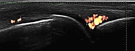
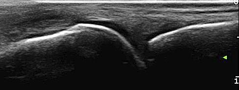
19 Where Do We Look for Synovitis? Recesses are lax capsular areas where fluid and synovial hypertrophy accumulate. Favorite joints and PD sensitivity: MCP : Dorsal > Volar PIP : Volar > Dorsal Wrist: Dorsal Elbow: Lateral and Annular Recess > Posterior > Anterior Knee: Parapatellar > Parameniscal >> Suprapatellar MTP Dorsal recesses contain small amounts of fluid normally

20 Synovitis MCP


21 Grade 2 or 3? Within recess or synovial hypertrophy?
22 Which color should the Pixels be in?
23 Kaeley et al Tocilizumab study Blue color for Power Doppler Detailed scoring system B-mode and Doppler
24 Scores Pediatric reduced joint power Doppler US (PDUS) score validity, feasibility, reliability and sensitivity to change compared with a 44 joints PDUS assessment in JIA 42 children with active JIA requiring modified therapy greyscale (GS) synovitis and power Doppler signal 4-point semiquantitative scale calculation of US composite indices and US composite joint counts Collado-P et al Rheumatology 2013
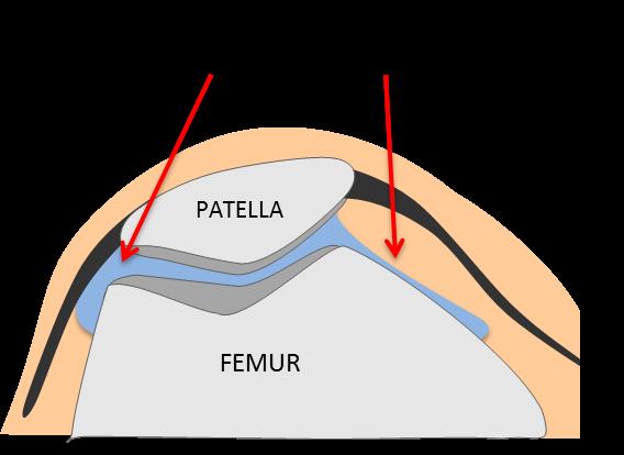
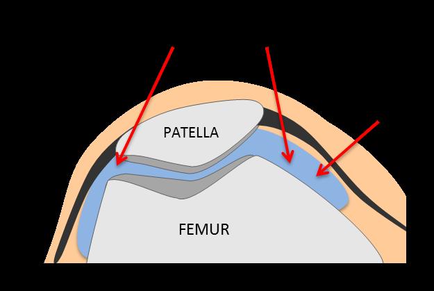
25 PedSynS 4-point semiquantitative scale of GS synovitis 0 absence, normal joint recess 1 mild, synovitis filling the joint recess between periarticular epiphyses change from angle-shaped to a plateau-shaped recess 2 moderate, convex shape of the joint recess without extension over the bone diaphysis 3 marked, convex shape of the joint recess with extension to at least one of the bone diaphyses Collado-P et al Rheumatology 2013


26 The problem: Grade 0 0 absence, normal joint recess 1 mild, synovitis filling the joint recess between periarticular epiphyses, change from angle-shaped to a plateau-shaped recess Grade 1 2 moderate, convex shape of the joint recess without extension over the bone diaphysis 3 marked, convex shape of the joint recess with extension to at least one of the bone diaphyses Grade 2 Grade 3 Collado P, et al. Rheumatology 2013;52:
27 Score suggestion Grade 0 = normal Grade 1 = mild, synovitis filling the joint recess between periarticular epiphyses that leads to a change from the angle-shaped recess to a plateau-shaped recess Grade 2 = convex shape of the recess without extension to the bone diaphysis nor to the whole length of a short bone Grade 3 = convex shape of the recess with extension to the bone diaphysis or the whole length of a short bone
28 Power Doppler Score 0 absence, no synovial flow 1 mild, single-vessel signal 2 moderate, confluent vessels 3 marked, vessel signals in more than half of the Intraarticular area Tenosynovitis and bursitis were scored using a dichotomous assessment (0, absence; 1, presence) in both GS and PD scoring systems Collado-P et al Rheumatology 2013
29 Current Pediatric Projects 1) Omeract ongoing global scoring system 2) PRES 3) PANLAR enthesis 4) CARRA joint by joint 5) Collaborative projects using 12 joint score
30 1"#"Overview"of"Joints"and"Images" Joint Imaging specification Total Number of Images Elbow A sagittal scan over the humeroulnar, humeroradial and dorsal elbow joint will be done. 3 images in B-Mode and 3 images in Doppler-Mode, total 6 images. Wrist A sagittal scan over the dorsal aspect of the medial and ulnar part of the wrist will be done and the radiocarpal, midcarpal as well as the carpometacarpal joints will be evaluated. The radioulnar joint will be evaluated as well in an additional scan. A dorsal transverse scan of the wrist will be done to evaluate the extensor tendon sheaths Fingers The subject will be in a sitting position with the hands palm-side down or dorsum-side down (depending on whether dorsal or volar assessment is done) in a neutral position on an examination table. A sagittal scan over the dorsal and volar aspect of the MCP 2 and 3 joint will be done. In addition a transverse scan on the dorsal and volar aspect will be done for MCP 2 and 3. Knee The subject will be in a supine position with the lower extremities parallel to each other. A measurement will be taken in 30 degrees flexed position. The measurement will be done sagittally over the suprapatellar recess, with the probe positioned cranial to the superior edge of the patella. Subsequently a medial and lateral parapatellar scan will be done. Ankle The subject will be in supine position with the knee in 90 degrees flexion. A sagittal scan over the medial aspect of the tibiotalar joint will be done. In addition in 3 transverse scans the tendon sheath and cross sectional area of the Tibialis anterior, Extensor Hallucis Longus, Extensor Digitorum at the level of the talar cartilage, the Tibialis Posterior, Flexor Digitorum, Flexor Hallucis Longus at the level of the sustentaculum and the Peroneus Tendons at the level of the supramalleolar and trochlea (where they separate) will be assessed. In a separate medial and lateral scan the subtalar joint will be assessed. Finally the talonavicular joint will be assessed in a sagittal scan. 4 images in B-Mode and 4 images Doppler-Mode, total 8 images. 8 images in B-Mode and 8 images Doppler-Mode, total 16 images 3 images in B-Mode and 3 images Doppler-Mode, total 6 images 7 images in B-Mode and 7 images Doppler-Mode, total 14 images
31 Grade"1" "mild" Distension of synovial recess originating from the radiocarpal or midcarpal joint or both with the superiorborderoftherecessremainingparalleltotherespectivebonelineandfillinglessthan50%of thepotentialspacebelowthebetaline. Radius''''''''''''''Lunate'''''''''!!!!!!!!!!!!!!!!!!!!!!!!!!!!!!Capitate!!!!!!!!!!!!!!!!!!!!!!!!!!!!!!!!!! Metc Grade"2" "moderate" Distension of synovial recess originating from the radiocarpal or midcarpal joint or both with the superiorborderoftherecessconvexandfilling50%ormoreofthepotentialspacebelowthebetaline andremainingdeeptothebetaline.distensionofthe"recessmaybepresentintheradiocarpaljoint only,intheintercarpaljointonly,orinbothjoints. Radius''''''''''''''Lunate#########!!!!!!!!!!!!!!!!!!!!!!!!!!!!!!Capitate!!!!!!!!!!!!!!!!!!!!!!!!!!!!!!!!!! Metc Grade"3" "severe Distension of synovial recess originating from the radiocarpal or midcarpal joint or both with the superiorborderoftherecessconvexandextendingabovethebetaline. Radius''''''''''''''Lunate'''''''''!!!!!!!!!!!!!!!!!!!!!!!!!!!!!!Capitate!!!!!!!!!!!!!!!!!!!!!!!!!!!!!!!!!! Metc
32 Scoring Doppler Grade 0 No Doppler signals except for feeding vessels crossing synovial recesses. A feeding vessel is defined as a single vessel showing continuity in entering the bone/cartilage Grade 1 Single Signals (up to three) Grade 2 Confluent signals in less than 50 % of synovial hypertrophy/hyperplasia Grade 3 Confluent signals in more than 50 % of synovial hypertrophy/hyperplasia
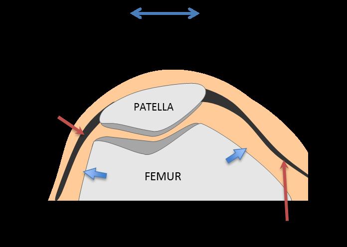
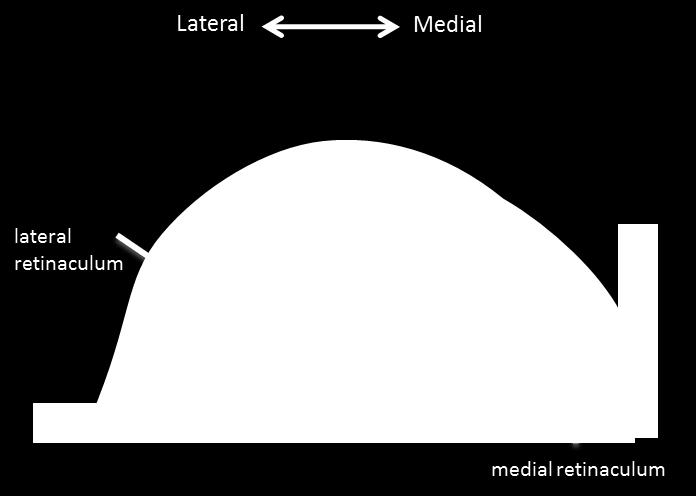


33 B6mode" Grade"0" Theparapatellarrecessesareempty.Aminimalbulgeofsynoviummaybefoundatthepatellofemoral jointline. " Grade"1" Synoviumfillstheproximalpartoftheparapatellarrecess " """""""""" " "
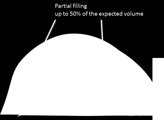
34 Grade"2" Synoviumfillspartoftheparapatellarrecessorextendsthroughouttherecessfilling<50%ofits expectedfullvolume. """"""""""""""""""" " " Grade"3"" Synoviumoccupies>50%oftheexpectedfullvolumeoftherecess " "
35 Enthesis Scores Adults Gandjbakhch et al. Arthritis Research Therapy 2011, 13:R188 RESEARCH ARTICLE Open Access Ultrasound in the evaluation of enthesitis: status and perspectives Frédérique Gandjbakhch 1, Lene Terslev 2, Fredrick Joshua 3, Richard J Wakefield 4, Esperanza Naredo 5 and Maria Antonietta D Agostino 6*, for OMERACT Ultrasound Task Force Huge variability in scores B-mode vs Doppler, Doppler in area of abnormal B-mode? Tendon, Enthesis, Bursae, other structures Inclusion of parameters of damage? Various sum scores weighing parameters differently
36 Scoring Enthesis Adults - Option Thickness <1 mm of increase exceeding the threshold of normal values grade 1 1 mm or greater but less than 2 mm of increase grade 2 2 mm or greater grade 3 Erosions max diameter (grade 1, >0 mm but <2 mm; grade 2, 2 mm and <3 mm; grade 3, 3 mm) Hypoechogenicity, enthesophytes, calcifications and bursa semiquantitative scores (mild changes: grade 1, moderate changes: grade 2 and severe changes: grade 3). Intraentheseal Doppler signals 0: no Doppler signal, 1: mild ( 2 punctiform Doppler signals with no confluent Doppler signal), 2: moderate (2 4 punctiform Doppler signal or 1 confluent Doppler signal), 3: marked (> 4 punctiform Doppler signals or > 1 confluent Doppler signal).
37 Pediatric Enthesis No consensus yet No need to include parameters of damage (Weiss Roth 2016) Focus on Doppler signals in area of B-mode abnormality Important in light of physiologic Doppler in Enthesis Scoring system: Semiquantitative scoring for Doppler signals

38
Ultrasound in Rheumatology
 Ultrasound in Rheumatology Alison Hall Consultant MSK Sonographer Research Institute for Primary Care & Health Sciences, Keele University Department of Rheumatology, Cannock Hospital, Royal Wolverhampton
Ultrasound in Rheumatology
 Arthritis Research UK Primary Care Centre Winner of a Queen s Anniversary Prize For Higher and Further Education 2009 Ultrasound in Rheumatology Alison Hall Consultant MSK Sonographer/Research Fellow Primary
Current Status in Pediatric Musculoskeletal Ultrasonography. Johannes Roth, MD PhD FRCPC RhMSUS
 1 Current Status in Pediatric Musculoskeletal Ultrasonography Johannes Roth, MD PhD FRCPC RhMSUS 2 Objectives Provide a rationale for the use of imaging and ultrasonography in particular in pediatric rheumatology
Sonographic appearance of chronic inflammatory rheumatism
 Sonographic appearance of chronic inflammatory rheumatism Poster No.: C-2237 Congress: ECR 2013 Type: Educational Exhibit Authors: H. Elfattach, F. Houari, O. Addou, M. Maaroufi, S. Tizniti ; 1 1 1 1 2
ELENI ANDIPA General Hospital of Athens G. Gennimatas
 ELENI ANDIPA General Hospital of Athens G. Gennimatas Technological advances over the last years have caused a dramatic improvement in ultrasound quality and resolution An established imaging modality
Reporting Ultrasound Findings and Diagnosis
 Reporting Ultrasound Findings and Diagnosis Rodina Nestorova MD Rheumatology Centre St. Irina, Sofia Bulgarian MSUS Society Basic MSU Course 14-16 Jan 2016 Plovdiv, Bulgaria ULTRASOUND REPORT COLLECTION
Sonographic assessment of adult and juvenile rheumatoid arthritis
 Sonographic assessment of adult and juvenile rheumatoid arthritis Poster No.: C-1485 Congress: ECR 2013 Type: Educational Exhibit Authors: C. A. S. Ruano, P. L. Pegado, J. M. G. Lourenco, P. Alves, L.
Table of contents. Foreword. Preface. 1 Introduction Historical Perspective 00
 Table of contents Foreword Preface 1 Introduction 00 1.1 Historical Perspective 00 2 Fundamentals of musculoskeletal ultrasound 00 2.1 Frequency and wavelength 00 2.2 Generating ultrasound waves 00 2.3
Introduction to Musculoskeletal Ultrasound. Disclosures. Evidence Based Medicine Key References 8/30/2017
 Introduction to Musculoskeletal Ultrasound Johannes Roth MD, PhD, FRCPC, RhMSUS Professor of Pediatrics University of Ottawa Gurjit S Kaeley MBBS, MRCP, RhMSUS Professor of Medicine Division Chief Director
EULAR 25th Jubilee EULAR Sonography Course Basic, Intermediate and Advanced Musculoskeletal Ultrasound (MSUS) in Rheumatology
 in Rheumatology") EULAR 25th Jubilee EULAR Sonography Course Basic, Intermediate and Advanced Musculoskeletal Ultrasound (MSUS) in Rheumatology Sunday 10th Wednesday 13th June 2018 Amsterdam, Netherlands ORGANISATION AND
EULAR 25th Jubilee EULAR Sonography Course Basic, Intermediate and Advanced Musculoskeletal Ultrasound (MSUS) in Rheumatology Sunday 10th Wednesday 13th June 2018 Amsterdam, Netherlands ORGANISATION AND
David Bong (Spain), MD Faculty member of the EULAR MSUS courses since 2011.
, MD Faculty member of the EULAR MSUS courses since 2011.") MUSCULOSKELTAL SONOGRAPHY COURSE Intermediate Level Sept 30 & Oct 1 & 2, 2016 Dubai, United Arab Emirates Organizing Office: Compass Conferences ENDORSED Organizers Ingrid Möller (Scientific Organizer)
MUSCULOSKELTAL SONOGRAPHY COURSE Intermediate Level Sept 30 & Oct 1 & 2, 2016 Dubai, United Arab Emirates Organizing Office: Compass Conferences ENDORSED Organizers Ingrid Möller (Scientific Organizer)
Ultrasound in Rheumatological Conditions:
 Ultrasound in Rheumatological Conditions: Status and Perspectives Nancy A. Chauvin, MD Assistant Professor of Radiology Director of Musculoskeletal Imaging The Children s Hospital of Philadelphia University
MUSCULOSKELTAL SONOGRAPHY COURSE Basic Level 4 6 November, 2016 Manama, Kingdom of Bahrain Organizing Office: Compass Conferences ENDORSED
 MUSCULOSKELTAL SONOGRAPHY COURSE Basic Level 4 6 November, 2016 Manama, Kingdom of Bahrain Organizing Office: Compass Conferences ENDORSED Organizers Möller (Scientific Organizer) Ayman Fahim (Organizer)
Schäfer et al. BMC Musculoskeletal Disorders 2013, 14:358
 Schäfer et al. BMC Musculoskeletal Disorders 2013, 14:358 RESEARCH ARTICLE Open Access Evaluation of the novel ultrasound score for large joints in psoriatic arthritis and ankylosing spondylitis: six month
Ligaments of Elbow hinge: sagittal plane so need lateral and medial ligaments
 Ligaments of Elbow hinge: sagittal plane so need lateral and medial ligaments Ulnar Collateral ligament on medial side; arising from medial epicondyle and stops excess valgus movement (lateral movement)
EULAR 25th Jubilee EULAR Sonography Course Basic, Intermediate and Advanced Musculoskeletal Ultrasound (MSUS) in Rheumatology
 in Rheumatology") EULAR 25th Jubilee EULAR Sonography Course Basic, Intermediate and Advanced Musculoskeletal Ultrasound (MSUS) in Rheumatology Sunday 10th Wednesday 13th June 2018 Amsterdam, Netherlands ORGANISATION AND
EULAR 25th Jubilee EULAR Sonography Course Basic, Intermediate and Advanced Musculoskeletal Ultrasound (MSUS) in Rheumatology Sunday 10th Wednesday 13th June 2018 Amsterdam, Netherlands ORGANISATION AND
"EULAR endorsed course"
 MUSCULOSKELTAL SONOGRAPHY COURSE InterMediate Level 15 17 September, 2017 Dubai, UAE Organizing Office: Compass Conferences "EULAR endorsed course" Organizers Ingrid Möller (Scientific Organizer) Esperanza
The use of musculoskeletal ultrasound (MSKUS) as a. Musculoskeletal Ultrasound as a Diagnostic and Prognostic Tool in Rheumatoid Arthritis
 as a. Musculoskeletal Ultrasound as a Diagnostic and Prognostic Tool in Rheumatoid Arthritis") 215 Musculoskeletal Ultrasound as a Diagnostic and Prognostic Tool in Rheumatoid Arthritis Manish Jain, M.D., and Jonathan Samuels, M.D. Abstract The use of musculoskeletal ultrasound (MSKUS) has increased
215 Musculoskeletal Ultrasound as a Diagnostic and Prognostic Tool in Rheumatoid Arthritis Manish Jain, M.D., and Jonathan Samuels, M.D. Abstract The use of musculoskeletal ultrasound (MSKUS) has increased
2019 RHEUMATOLOGIC ULTRASOUND (RhUS) CURRICULUM SUPPLEMENT TO THE AMERICAN COLLEGE OF RHEUMATOLOGY 2015 CORE CURRICULUM OUTLINE
 CURRICULUM SUPPLEMENT TO THE AMERICAN COLLEGE OF RHEUMATOLOGY 2015 CORE CURRICULUM OUTLINE") 2019 RHEUMATOLOGIC ULTRASOUND (RhUS) CURRICULUM SUPPLEMENT TO THE AMERICAN COLLEGE OF RHEUMATOLOGY 2015 CORE CURRICULUM OUTLINE TABLE OF CONTENTS I. MEDICAL KNOWLEDGE II. PATIENT CARE III. PRACTICE-BASED
2019 RHEUMATOLOGIC ULTRASOUND (RhUS) CURRICULUM SUPPLEMENT TO THE AMERICAN COLLEGE OF RHEUMATOLOGY 2015 CORE CURRICULUM OUTLINE TABLE OF CONTENTS I. MEDICAL KNOWLEDGE II. PATIENT CARE III. PRACTICE-BASED
Clinical Practice Guideline. Ultrasound in Rheumatological Settings. Version
 Clinical Practice Guideline Ultrasound in Rheumatological Settings Version 1.1.2017 November 2017 Table of Contents Abbreviations...3 Introduction...4 Diagnostic Musculoskeletal Ultrasound...6 Definition
Index. Note: Page numbers of article titles are in boldface type.
 Note: Page numbers of article titles are in boldface type. A ACJ. See Acromioclavicular joint (ACJ) Acromioclavicular joint (ACJ) procedures of, 557 559 Ankle and foot procedures of, 649 671 (See also
15 th EULAR SONOGRAPHY COURSE. June 08 th - 11 th, 2008 Paris, France. Basic, Intermediate and Advanced Musculoskeletal Ultrasound in Rheumatology
 15 th EULAR SONOGRAPHY COURSE Basic, Intermediate and Advanced Musculoskeletal Ultrasound in Rheumatology June 08 th - 11 th, 2008 Paris, France Societé française de rhumatologie Université de Versailles
Paediatric rheumatology
 Paediatric rheumatology Ultrasonography vs. clinical examination in children with suspected arthritis. Does it make sense to use poliarticular ultrasonographic screening? G. Filippou, L. Cantarini, I.
The Egyptian Journal of Hospital Medicine (October 2017) Vol. 69 (4), Page
 Vol. 69 (4), Page") The Egyptian Journal of Hospital Medicine (October 2017) Vol. 69 (4), Page 2294-2300 Role of Magnetic Resonance Imaging and Ultrasonography in Diagnosis and Follow Up Rheumatoid Arthritis in Hand and Wrist
The Egyptian Journal of Hospital Medicine (October 2017) Vol. 69 (4), Page 2294-2300 Role of Magnetic Resonance Imaging and Ultrasonography in Diagnosis and Follow Up Rheumatoid Arthritis in Hand and Wrist
EULAR 24th EULAR Sonography Course Basic, Intermediate and Advanced Musculoskeletal Ultrasound (MSUS) in Rheumatology
 in Rheumatology") EULAR 24th EULAR Sonography Course Basic, Intermediate and Advanced Musculoskeletal Ultrasound (MSUS) in Rheumatology Sunday 11th Wednesday 14th June 2017 Madrid, Spain ORGANISATION AND COMMITTEE Scientific
EULAR 24th EULAR Sonography Course Basic, Intermediate and Advanced Musculoskeletal Ultrasound (MSUS) in Rheumatology Sunday 11th Wednesday 14th June 2017 Madrid, Spain ORGANISATION AND COMMITTEE Scientific
"EULAR endorsed course"
 MUSCULOSKELTAL SONOGRAPHY COURSE Basic Level 20 22 July, 2018 Abu Dhabi, United Arab Emirates Organizing Office: Compass Conferences "EULAR endorsed course" Organizers Möller (Scientific Organizer) Esperanza
Scientific Programme
 Scientific Programme (preliminary version) Musculoskeletal Sonography Course for Rheumatologists Warszawa 2010 th st September 28 - October 01, 2010 Warszawa, Poland under scientific patronage of the European
Lateral Elbow Pathology
 Lateral Elbow Pathology Jon A. Jacobson, M.D. Professor of adiology Director, Division of Musculoskeletal adiology University of Michigan Disclosures: Consultant: Bioclinica Advisory Board: GE, Philips
The Elbow 3/5/2015. The Elbow Scanning Sequence. * Anterior Joint (The anterior Pyramid ) * Lateral Epicondyle * Medial Epicondyle * Posterior Joint
 * Lateral Epicondyle * Medial Epicondyle * Posterior Joint") Scanning Sequence * Anterior Joint (The anterior Pyramid ) * Lateral Epicondyle * Medial Epicondyle * Posterior Joint Anterior Elbow Pyramid Courtesy of Jay Smith, MD. Vice chair PMR Mayo Clinic Rochester,
Scanning Sequence * Anterior Joint (The anterior Pyramid ) * Lateral Epicondyle * Medial Epicondyle * Posterior Joint Anterior Elbow Pyramid Courtesy of Jay Smith, MD. Vice chair PMR Mayo Clinic Rochester,
Knee, Ankle, and Foot: Normal and Abnormal Features with MRI and Ultrasound Correlation. Disclosures. Outline. Joint Effusion. Suprapatellar recess
 Knee, Ankle, and Foot: Normal and Abnormal Features with MRI and Ultrasound Correlation Jon A. Jacobson, M.D. Professor of Radiology Director, Division of Musculoskeletal Radiology University of Michigan
Bulgarian Association for Musculoskeletal Ultrasound
 MUSCULOSKELETAL ULTRASOUND COURSE INTERMEDIATE LEVEL ORGANIZATION AND COMMITTEE Scientific Organiser Prof. Annamaria Iagnocco Local Organisers Prof Anastas Batalov Dr Rodina Nestorova Dr Plamen Todorov
26 th Sonography Course MSUS Intermediate
 26 th Sonography Course MSUS Intermediate Madrid, Spain Sunday, 9 th June Wednesday, 12 th June 2019 The 26 th EULAR Sonography Course will be run simultaneously at three levels, basic, intermediate and
Wrist and Ankle MRI of Patients With Juvenile Idiopathic Arthritis: Identification of Unsuspected Multicompartmental Tenosynovitis and Arthritis
 Pediatric Imaging Original Research Javadi et al. Wrist and nkle MRI of Patients With Juvenile Idiopathic rthritis Pediatric Imaging Original Research Sanaz Javadi 1 J. Herman Kan 1 Robert C. Orth 1 Marietta
I-A-1) Non-specific thickening of synovial membrane
 Non-specific thickening of synovial membrane") I-A-1) Non-specific thickening of synovial membrane Grayscale Metatarsal Power Doppler Dorsal aspect of metatarsophalangeal joint in right 1 st toe, longitudinal view Asterisks indicate non-specific thickening
I-A-1) Non-specific thickening of synovial membrane Grayscale Metatarsal Power Doppler Dorsal aspect of metatarsophalangeal joint in right 1 st toe, longitudinal view Asterisks indicate non-specific thickening
ARD Online First, published on June 7, 2005 as /ard
 ARD Online First, published on June 7, 2005 as 10.1136/ard.2005.037382 TITLE: Inter-observer reliability in musculoskeletal ultrasonography: results from a "Teach-the-Teachers" rheumatologist course AUTHORS:
M usculoskeletal ultrasonography is a diagnostic
 988 EXTENDED REPORT Standard reference values for musculoskeletal ultrasonography W A Schmidt, H Schmidt, B Schicke, E Gromnica-Ihle... See end of article for authors affiliations... Correspondence to:
Interobserver reliability in musculoskeletal ultrasonography: results from a Teach the Teachers rheumatologist course
 14 EXTENDED REPORT Interobserver reliability in musculoskeletal ultrasonography: results from a Teach the Teachers rheumatologist course E Naredo, I Möller, C Moragues, J J de Agustín, A K Scheel, W Grassi,
International Musculoskeletal Ultrasound Course MITOS
 Basic and Intermediate Levels th International Musculoskeletal Ultrasound Course MITOS Final Course Program 30 November - 2 December 2017 Wyndham Grand Athens Hotel Athens Greece www.synthesispco.com/mitoscourse2017
Practical use of musculoskeletal ultrasonography in rheumatoid arthritis: how to use this attractive but arduous tool in clinical practice
 International Journal of Clinical Rheumatology nogra- Practical use of musculoskeletal ultrasonography in rheumatoid arthritis: how to use this attractive but arduous tool in clinical practice Musculoskeletal
The Role of Ultrasonography in the Assessment of Rheumatic Diseases. Current and Potential Role of Ultrasonography in Rheumatology
 The Role of Ultrasonography in the Assessment of Rheumatic Diseases Karina D. Torralba, MD, MACM, CCD, Assistant Professor of Medicine, Keck School of Medicine, University of Southern California Objective:
Overview of musculoskeletal ultrasound for the clinical rheumatologist
 Overview of musculoskeletal ultrasound for the clinical rheumatologist S. Hassan Rheumatology Division, Rush University Medical Center, Chicago, USA. Sobia Hassan, MD, MRCP, RhMSUS Please address correspondence
Wrist and Hand Anatomy
 Wrist and Hand Anatomy Bone Anatomy Scapoid Lunate Triquetrium Pisiform Trapeziod Trapezium Capitate Hamate Wrist Articulations Radiocarpal Joint Proximal portion Distal portion Most surface contact found
Concise report RHEUMATOLOGY
 RHEUMATOLOGY Rheumatology 2012;51:2034 2038 doi:10.1093/rheumatology/kes124 Advance Access publication 30 July 2012 Concise report Head-to-head comparison of quantitative and semi-quantitative ultrasound
Occupational Therapy Assessment Guidelines
 Occupational Therapy Assessment Guidelines DATA BASE Patient's Main Concerns and Goals Patient's problems. - pq!p"*q., Vancouver " i,'" ~;a ;i 5 r,:!. H e a Lt h Prontof~trg u~cllness Rnsurrng car? Mary
Musculoskeletal Ultrasound Technical Guidelines. VI. Ankle
 European Society of MusculoSkeletal Radiology Musculoskeletal Ultrasound Technical Guidelines VI. Ankle Ian Beggs, UK Stefano Bianchi, Switzerland Angel Bueno, Spain Michel Cohen, France Michel Court-Payen,
The Biomechanics of the Human Upper Extremity-The Elbow Joint C. Mirzanli Istanbul Gelisim University
 The Biomechanics of the Human Upper Extremity-The Elbow Joint C. Mirzanli Istanbul Gelisim University Structure of The Elbow Joint A simple hinge joint, actually categorized as a trochoginglymus joint
Imaging. Introduction Systemic lupus erythematosus (SLE) is an autoimmune multisystem disorder
 is an autoimmune multisystem disorder") Clinical and Experimental Rheumatology 2009; 27: 00-00 Imaging Ultrasound imaging for the rheumatologist XXIV. Sonographic evaluation of wrist and hand joint and tendon involvement in systemic lupus erythematosus
Clinical and Experimental Rheumatology 2009; 27: 00-00 Imaging Ultrasound imaging for the rheumatologist XXIV. Sonographic evaluation of wrist and hand joint and tendon involvement in systemic lupus erythematosus
Ultrasonography for diagnosis, monitoring and treatment of tenosynovitis in patients with rheumatoid
 PHD THESIS DANISH MEDICAL JOURNAL Ultrasonography for diagnosis, monitoring and treatment of tenosynovitis in patients with rheumatoid arthritis Mads Ammitzbøll Danielsen This review has been accepted
Ultras ono graphic Evaluation of Rotator Cuff Tendons in Patients with Rheumatoid Arthritis
 Med. J. Cairo Univ., Vol. 83, No. 1, June: 395-399, 215 www.medicaljournalofcairouniversity.net Ultras ono graphic Evaluation of Rotator Cuff Tendons in Patients with Rheumatoid Arthritis HALA I. ELGENDY,
Unit of Rheumatology, Ospedale Sacro Cuore, Negrar, Verona, Italy; 2
 Comprehensive evaluation of finger flexor tendon entheseal soft tissue and bone changes by ultrasound can differentiate psoriatic arthritis and rheumatoid arthritis I. Tinazzi 1, D. McGonagle 2, A. Zabotti
6 th MUSCULOSKELETAL SONOGRAPHY COURSE. Belgrade, 5 th 7 th October, 2017 INTERMEDIATE LEVEL COURSE
 6 th MUSCULOSKELETAL SONOGRAPHY COURSE INTERMEDIATE LEVEL COURSE Belgrade, 5 th 7 th October, 2017 ORGANIZER Rheumatology Association of Serbia www.belgrade-course.org SCIENTIFICALLY ENDORSED BY eular
Difference Between Angle You Can Bend Your Left Wrist Back vs Your Right Wrist Jenna Priest Science Department Altoona High School January 25, 2017
 Difference Between Angle You Can Bend Your Left Wrist Back vs Your Right Wrist Jenna Priest Science Department Altoona High School January 25, 2017 Background 1- The wrist joint (also known as the radiocarpal
i;l Contents PART I INTRODUCTIOM TO GONIOMETRY, I ~haoter '1 Basic Conceots. 3 Chapter 2 Procedures, 19 Chapter 3 Validity and Reliability, 39
 w Contents i;l PART I INTRODUCTIOM TO GONIOMETRY, I ~haoter '1 Basic Conceots. 3 Goniometry, 3 Joint Motion, 4 Arthrokinematics, 4 Osteokinematics, 5 Planes and Axes, 5 Range of Motion, 6 Active Range
Guidelines for musculoskeletal ultrasound in rheumatology
 Ann Rheum Dis 2001;60:641 649 641 REVIEW Rheumatology and Clinical Immunology, Charité University Hospital, Humboldt University, Berlin, Germany M Backhaus G-R Burmester Rheumatology and Physical Medicine,
Musculoskeletal Ultrasound
 EFSUMB Course Book Student Edition Editors: Jan Tuma, Radu Badea, Christoph F. Dietrich Musculoskeletal Ultrasound Giorgio Tamborrini Ultrasound Center, Switzerland Corresponding author: KD Dr. med. Giorgio
MUSCULOSKELETAL SONOGRAPHY COURSE FOR RHEUMATOLOGISTS - BASIC LEVEL - June 27 th 29 th, 2013 Innsbruck, Austria
 MUSCULOSKELETAL SONOGRAPHY COURSE FOR RHEUMATOLOGISTS - BASIC LEVEL - June 27 th 29 th, 2013 Innsbruck, Austria GENERAL INFORMATION Course opening: Thursday, June 27 th, 2013 - h 12.00 Course closing:
Pragmatic ultrasound in the diagnosis of soft tissue rheumatic pain. Plamen Todorov
 Pragmatic ultrasound in the diagnosis of soft tissue rheumatic pain Plamen Todorov INTRODUCTION Soft tissue rheumatism: nonsystemic, focal pathological syndromes involving the periarticular structures.
June 26 th 28 th, 2014 Innsbruck, Austria
 MUSCULOSKELETAL SONOGRAPHY COURSE FOR RHEUMATOLOGISTS - INTERMEDIATE LEVEL - June 26 th 28 th, 2014 Innsbruck, Austria This course is scientifically endorsed by: GENERAL INFORMATION Course opening: Thursday,
ONLINE. J.A. Mendonça 1, M. Yazbek 1, I.M.M. Laurindo 2 and M.B. Bertolo 1
 Brazilian Journal of Medical and Biological Research Online Provisional Version ISSN 0100-879X This Provisional PDF corresponds to the article as it appeared upon acceptance. Fully formatted PDF and full
Early Rheumatoid Arthritis: AReview of MRI and Sonographic Findings
 outry et al. MRI and Sonography of Rheumatoid rthritis Musculoskeletal Imaging Pictorial Essay Nathalie outry 1 Mélanie Morel 1 René-Marc Flipo 2 Xavier Demondion 1,3 nne Cotten 1 outry N, Morel M, Flipo
Physical therapy of the wrist and hand
 Physical therapy of the wrist and hand Functional anatomy wrist and hand The wrist includes distal radius, scaphoid, lunate, triquetrum, pisiform, trapezium, trapezoid, capitate, and hamate. The hand includes
UNIT 7 JOINTS. Knee and Ankle Joints DR. ABDEL-MONEM A. HEGAZY
 UNIT 7 JOINTS Knee and Ankle Joints BY DR. ABDEL-MONEM A. HEGAZY (Degree in Bachelor of Medicine and Surgery with honor 1983, Dipl."Gynaecology and Obstetrics "1989, Master "Anatomy and Embryology "1994,
US finding of the shoulder (with live demonstration) 인제의대상계백병원 안재기
 인제의대상계백병원 안재기") US finding of the shoulder (with live demonstration) 인제의대상계백병원 안재기 Shoulder US Biceps tendon & Rotator Cuff Long Head of Biceps Tendon Subscapularis tendon Supraspinatus tendon Infraspinatus tendon Teres
US finding of the shoulder (with live demonstration) 인제의대상계백병원 안재기 Shoulder US Biceps tendon & Rotator Cuff Long Head of Biceps Tendon Subscapularis tendon Supraspinatus tendon Infraspinatus tendon Teres
The Elbow Scanning Protocol
 The Elbow Scanning Protocol Diagnostic Imaging of the Elbow: Introduction The elbow maybe considered as consisting of four quadrants, anterior, medial, lateral and posterior. Ultrasound would normally
EDUCATIONAL COURSE. MUSCULOSKELETAL ULTRASOUND in RHEUMATOLOGY BASIC COURSE. Course Coordinator Annamaria Iagnocco
 EDUCATIONAL COURSE 2019 MUSCULOSKELETAL ULTRASOUND in RHEUMATOLOGY BASIC COURSE Rome, March 21-23, 2019 Course Coordinator Annamaria Iagnocco COURSE PRESENTATION Musculoskeletal ultrasound in rheumatology
musculoskeletal system anatomy muscles of foot sheet done by: dina sawadha & mohammad abukabeer
 musculoskeletal system anatomy muscles of foot sheet done by: dina sawadha & mohammad abukabeer Extensor retinaculum : A- superior extensor retinaculum (SER) : originates from the distal ends of the tibia
Pragmatic use of US in intraarticular and periarticular procedures
 Pragmatic use of US in intraarticular and periarticular procedures Plovdiv, 13. January 2018 Dr. med. Giorgio Tamborrini-Schütz www.irheuma.com member of the EULAR Network of Imaging Training Centres Ulrasound
Joints of the upper limb II
 Joints of the upper limb II Prof. Abdulameer Al-Nuaimi E-mail: a.al-nuaimi@sheffield.ac.uk E. mail: abdulameerh@yahoo.com Elbow joint The elbow joint is connecting the upper arm to the forearm. It is classed
Connects arm to thorax 3 joints. Glenohumeral joint Acromioclavicular joint Sternoclavicular joint
 Connects arm to thorax 3 joints Glenohumeral joint Acromioclavicular joint Sternoclavicular joint Scapula Elevation Depression Protraction (abduction) Retraction (adduction) Downward Rotation Upward Rotation
The Elbow and the cubital fossa. Prof Oluwadiya Kehinde
 The Elbow and the cubital fossa Prof Oluwadiya Kehinde www.oluwadiya.com Elbow and Forearm Anatomy The elbow joint is formed by the humerus, radius, and the ulna Bony anatomy of the elbow Distal Humerus
Clarification of Terms
 Clarification of Terms The plantar aspect of the foot refers to the role or its bottom The dorsal aspect refers to the top or its superior portion The ankle and foot perform three main functions: 1. shock
Diagnostic utility of musculoskeletal ultrasound in patients with suspected arthritis a probabilistic approach
 Rezaei et al. Arthritis Research & Therapy 2014, 16:448 RESEARCH ARTICLE Open Access Diagnostic utility of musculoskeletal ultrasound in patients with suspected arthritis a probabilistic approach Hamed
Ultrasound Evaluation of Masses
 Ultrasound Evaluation of Masses Jon A. Jacobson, M.D. Professor of Radiology Director, Division of Musculoskeletal Radiology University of Michigan Disclosures: Consultant: Bioclinica Advisory Panel: GE,
Early diagnosis of Rheumatoid
 26 Original Article Diagnostic Accuracy of Ultrasonography in Detection of Destructive Changes in Small Joints of Hands in Patients of Rheumatoid Arthritis: A Comparison with Magnetic Resonance Imaging
Correspondence should be addressed to Zuhudha Hussain Manik;
 Scientifica Volume 2016, Article ID 5609132, 7 pages http://dx.doi.org/10.1155/2016/5609132 Research Article Ultrasound Assessment of Synovial Thickness of Some of the Metacarpophalangeal Joints of Hand
Ultrasound of the hand is sufficient to detect subclinical inflammation in rheumatoid arthritis remission: a post hoc longitudinal study
 Hammer et al. Arthritis Research & Therapy (2017) 19:221 DOI 10.1186/s13075-017-1428-4 RESEARCH ARTICLE Ultrasound of the hand is sufficient to detect subclinical inflammation in rheumatoid arthritis remission:
Reliability of different Doppler ultrasound quantification methods and devices in the assessment of therapeutic response in arthritis
 Rheumatology 2008;47:1521 1526 Advance Access publication 2 August 2008 doi:10.1093/rheumatology/ken318 Reliability of different Doppler ultrasound quantification methods and devices in the assessment
The US7 score is sensitive to change in a large cohort of patients with rheumatoid arthritis over 12 months of therapy
 OPEN ACCESS EXTENDED REPORT The US7 score is sensitive to change in a large cohort of patients with rheumatoid arthritis over 12 months of therapy Tina M Backhaus, 1,2 Sarah Ohrndorf, 2 Herbert Kellner,
September 14 th 16 th, 2017 Innsbruck, Austria
 MUSCULOSKELETAL SONOGRAPHY COURSE FOR RHEUMATOLOGISTS - BASIC LEVEL - September 14 th 16 th, 2017 Innsbruck, Austria This course is scientifically endorsed by: GENERAL INFORMATION Course opening: Thursday,
Shoulder Elbow Wrist/Hand
 Shoulder Elbow Wrist/Hand Randy E. Moore DC RDMS RMSK General Musculoskeletal Imaging, Inc. 1 Shoulder Tendinosis : 3 key Ultrasound Findings 1. Increased cellularity thickened and ACR inhomogeneous CLV
Musculoskeletal Ultrasound. Technical Guidelines SHOULDER
 Musculoskeletal Ultrasound Technical Guidelines SHOULDER 1 Although patient s positioning for shoulder US varies widely across different Countries and Institutions reflecting multifaceted opinions and
Levels of the anatomical cuts of the upper extremity RADIUS AND ULNA right
 11 CHAPTER 2 Levels of the anatomical cuts of the upper extremity AND right CUT 1 CUT 4 1 2 3 4 5 6 Isolated fixation of the radius is difficult at this level because of the anterolateral vessels and the
CLASSIFICATION OF JOINTS STRUCTURAL VS FUNCTIONAL
 CHAPTER 8 JOINTS CLASSIFICATION OF JOINTS STRUCTURAL VS FUNCTIONAL The most moveable type of joint is a 1) Synarthrosis 2) Amphiarthrosis 3) Diarthrosis FIBROUS JOINTS Figure 8.1 Fibrous joints. (a) Suture
Psoriatic arthritis: early ultrasound findings
 Psoriatic arthritis: early ultrasound findings Poster No.: C-0399 Congress: ECR 2014 Type: Educational Exhibit Authors: R. Persechino 1, L. Cristiano 1, A. Bartoloni 1, C. Cantone 2, A. Keywords: DOI:
CHAPTER 6: THE UPPER EXTREMITY: THE ELBOW, FOREARM, WRIST, AND HAND
 CHAPTER 6: THE UPPER EXTREMITY: THE ELBOW, FOREARM, WRIST, AND HAND KINESIOLOGY Scientific Basis of Human Motion, 12 th edition Hamilton, Weimar & Luttgens Presentation Created by TK Koesterer, Ph.D.,
Shane A. Shapiro, M.D. Assistant Professor, Orthopedic Surgery Mayo Clinic 2012 MFMER slide MFMER slide-3
 Ultrasound Foot and Ankle Pathology Disclosures None relevant Shane A. Shapiro, M.D. Assistant Professor, Orthopedic Surgery Mayo Clinic Florida @ShaneShapiroMD 2012 MFMER slide-2 Foot and Ankle Fundamentals
Utility of ultrasound joint counts in the prediction of rheumatoid arthritis in patients with very early synovitis
 Additional data (supplementary tables and fi gures) are published online only. To view these fi les please visit the journal online ( http:// ard.bmj.com ). 1 MRC Centre for Immune Regulation, School of
Disclosures. Ultrasonography. Conventional radiography (XR) Magnetic resonance imaging. Conventional CT 10/27/2013
 Magnetic resonance imaging. Conventional CT 10/27/2013") What I can see in my gout patient that I couldn t before: the role of advanced imaging in gout diagnosis and monitoring Disclosures ND is supported by the Health Research Council of New Zealand I have
What I can see in my gout patient that I couldn t before: the role of advanced imaging in gout diagnosis and monitoring Disclosures ND is supported by the Health Research Council of New Zealand I have
Research Article Ultrasound Findings in Hand Joints Involvement in Patients with Psoriatic Arthritis and Its Correlation with Clinical DAS28 Score
 Radiology Research and Practice Volume 215, Article ID 353657, 9 pages http://dx.doi.org/.1155/215/353657 Research Article Ultrasound Findings in Hand Joints Involvement in Patients with Psoriatic Arthritis
Introduction to Ultrasound Examination of the Hand and upper
 Introduction to Ultrasound Examination of the Hand and upper Emil Dionysian, M.D. Ultrasound of upper ext. Upside Convenient Opens another exam dimension Can be like a stethoscope Helps 3-D D visualization
3rd MUSCULOSKELETAL SONOGRAPHY COURSE FOR RHEUMATOLOGISTS - INTERMEDIATE LEVEL -
 3rd MUSCULOSKELETAL SONOGRAPHY COURSE FOR RHEUMATOLOGISTS - INTERMEDIATE LEVEL - Bucharest, Romania October, 13th-15th 2016 Organizer: Romanian Society of Rheumatology Prof. Dr. Ruxandra Ionescu Course
Superficial Lumps and Bumps: Ultrasound Assessment
 Posterior knee Superficial Lumps and Bumps: Ultrasound Assessment Walter Mak, MD Department of Medical Imaging St. Michael s Hospital SM SM MGas MGas MGas MGas Synovial lined Synovial cyst: extrusion of
Sonographic appearance of fluid in peripheral joints and bursae of healthy asymptomatic Chinese population
 Original Article Sonographic appearance of fluid in peripheral joints and e of healthy asymptomatic Chinese population Liyun Wang, Xi Xiang, Yuanjiao Tang, Yujia Yang, Li Qiu Department of Ultrasound,
Ultrasound in Rheumatoid Arthritis
 Review Med Ultrason 2013, Vol. 15, no. 3, 199-208 DOI: 10.11152/mu.2013.2066.153.cr1fc2 Ultrasound in Rheumatoid Arthritis Chiara Rizzo, Fulvia Ceccarelli, Angelica Gattamelata, Caterina Vavala, Guido
American Diagnostics Services, Inc., a division of MobilexUSA. Upper Extremity Musculoskeletal Ultrasound Protocol
 Upper Extremity Musculoskeletal Ultrasound Protocol INDICATION To evaluate patient s with suspicion of shoulder, arm, elbow, and wrist or hand musculoskeletal pathology. Indications may include but not
Combined Structural and Synovial Assessment for Improved Ultrasound Discrimination of Rheumatoid, Osteoarthritic, and Normal Joints: A Pilot Study
 The Open Rheumatology Journal, 2012, 6, 199-206 199 Open Access Combined Structural and Synovial Assessment for Improved Ultrasound Discrimination of Rheumatoid, Osteoarthritic, and Normal Joints: A Pilot
Benefits of Aspiration and Injection JOINT INJECTIONS. Injection Indications. Mechanism of Action 1/11/2016
 Benefits of Aspiration and Injection JOINT INJECTIONS Mark Niedfeldt, M.D. Medical College of Wisconsin Decrease or resolution of pain Decrease or resolution of inflammation Decrease or resolution of effusion