MELTING CORNEAL ULCERS IN HORSES: DIAGNOSIS AND TREATMENT METHODS
|
|
|
- Dylan Richards
- 5 years ago
- Views:
Transcription
1 Vet Times The website for the veterinary profession MELTING CORNEAL ULCERS IN HORSES: DIAGNOSIS AND TREATMENT METHODS Author : FERNANDO MALALANA Categories : Vets Date : October 7, 2013 FERNANDO MALALANA discusses the various therapy options available regarding this common equine optical ailment Summary Corneal ulcers are very common in horses. Although most heal very quickly with the appropriate treatment, some can be complicated by corneal stromal melting or keratomalacia, which occurs as a result of imbalances between naturally occurring proteinases and their inhibitors. These proteinases are produced by corneal epithelial cells, fibroblasts, inflammatory cells and microorganisms, and can result in the destruction of the corneal stroma in a very short period of time. Aggressive therapy is essential to prevent this corneal melting. In addition to antimicrobials and treatment for the secondary uveitis, proteinase inhibitors such as topical ethylenediamine tetra-acetic acid (EDTA), serum, N-acetylcysteine and/or tetracyclines are used to reduce the progression of the stromal melting, accelerate epithelial healing and minimise corneal scarring. In some cases, surgery is also necessary to debride necrotic material and provide structural and vascular support to the damaged cornea. Surgical options available include keratectomy, conjunctival autografts or amniotic membrane grafts. Key words keratomalacia, melting cornea, stroma, proteinase, horse 1 / 13
2 CORNEAL ulcers are very common in horses, probably as a result of the prominent location of the globe within the skull and their fight or flight nature. Most corneal ulcers heal very quickly with the appropriate treatment, but some can be complicated by corneal melting or keratomalacia. Melting ulcers can be very severe, progress rapidly and are potentially sightthreatening, therefore, they should be identified and managed promptly. The cornea is traditionally divided into four layers. From the outside to the inside, these layers are the epithelium, stroma, Descemet s membrane and endothelium. The thickest corneal layer is the stroma, which is composed of a three-dimensional mosaic of interconnected keratocytes in an extracellular matrix of proteoglycans and collagen fibres1. During normal corneal healing following injury, proteinases are produced that aid in removal of debris and remodelling of the corneal stroma2. These proteinases are produced by microorganisms, inflammatory cells, corneal epithelial cells and fibroblasts, and can be divided in two main groups matrix metalloproteinases (MMPs, such as MMP-2 and MMP-9) and serine proteinases (such as neutrophil elastase, NE). The cornea contains naturally occurring inhibitors of MMPs and serine proteinases corneal destruction occurs when there is an imbalance between proteinases and their inhibitors2,3. Diagnosis In early stages, melting ulcers may look very innocuous. Later, they tend to appear as a grey, mucoid or gelatinous (liquefaction) opacity (Figure 1). A complete eye examination must be carried out, including staining with fluorescein to detect corneal epithelial defects, tear film deficiencies or leakage from the anterior chamber (the Seidel test). Cytologic examination and microbiologic cultures of melting ulcers are imperative2. Samples for cytology can be obtained with a previously soaked microbiology swab, cytology brushes or the blunt end of the scalpel blade. The sample is then rolled onto a glass slide. Common stains include Gram and DiffQuik. Although results of the culture are often more sensitive than cytology, examination of Gram-stained slides can give immediate information about the causative agent, allowing prompt, more focused therapy2. Cultures should be obtained before application of any topical solution such as fluorescein or local anaesthetics as they contain preservatives that may impair culture results. Medical therapy for melting ulcers 2 / 13
3 Appropriate antimicrobial therapy Aggressive antimicrobial therapy should be immediately implemented, ideally based on the results of cytology and culture. Gram staining will allow a rapid, more adequate selection of antimicrobial while waiting for the results of culture and sensitivity. If this is not possible, the choice can be made empirically, based on the normal components of the conjunctival flora in horses4. Although uncommon in the UK, fungal keratitis can also occur and detection of fungal elements on cytology and/or culture should prompt rapid antifungal treatment. Further discussion on antimicrobial choice is beyond the scope of this article and the reader is directed to the excellent review by Matthews (2009)5. Control of uveitis Uveitis is a common consequence of corneal disease and should be appropriately managed to avoid damage to the internal structures of the globe. Symptomatic treatment is achieved with the topical use of cycloplegics (atropine one per cent) and systemic anti-inflammatories. Flunixin meglumine is anecdotally believed to be the most effective systemic NSAID and analgesic for ocular conditions, but its long-term use can delay the development of corneal vascularisation, which is important for corneal healing. One should aim to change to a different compound, such as phenylbutazone, as soon as the eye is comfortable enough, or flunixin and phenylbutazone can be used on alternate days. A study found the concentration of firocoxib in the aqueous humour of horses is superior to that of flunixin6 and, therefore, may provide another option for the treatment of uveitis. However, to the author s knowledge, firocoxib is only licensed in the UK for the treatment of musculoskeletal conditions in horses. Topical NSAIDs, such as diclofenac and flurbiprofen, can be used in cases of uveitis, secondary to corneal ulceration. However, their application can cause some degree of ocular discomfort, delayed corneal neovascularisation and their efficacy is questionable. Again, more detailed treatment of uveitis is beyond the scope of this article. Inhibition of proteinases Proteinase inhibitors have been recommended for treatment of corneal ulcers to reduce the progression of the stromal melting, accelerate epithelial healing and minimise corneal scarring1,3. Topical 0.2 per cent ethylenediamine tetra-acetic acid (EDTA) has been shown to cause a 99.4 per cent reduction in proteinase activity in the ocular surface3,7,8. EDTA is a calcium and zinc chelator 3 / 13
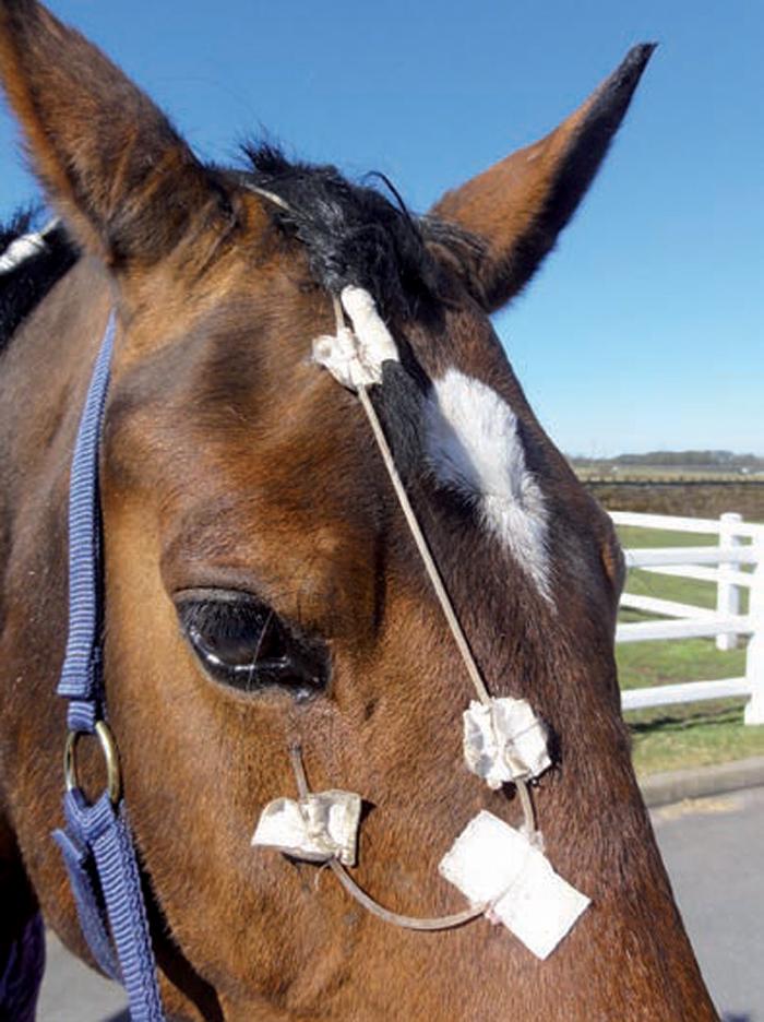
4 that inhibits MMP activity and can be easily prepared by adding sterile water to a commercial blood collection tube. Autologous serum can inhibit both MMPs and serine proteases, due to the presence of?1-antitrypsin and?2-macroglobulins3,7,9. Serum has been shown to decrease proteinase activity by 90 per cent. A long clotting time (120 minutes), a sharp centrifugation (3,000g for 15 minutes) and dilution with balanced salt solution improve the ability of serum eye drops to support the metabolism of corneal epithelial cells8. Serum contamination is unlikely, but it should, nevertheless, be closely monitored for changes in its turbidity that may indicate contamination. Plasma can also be used topically, but it lacks the beneficial platelet-derived growth factors found in serum. N-acetylcysteine is another MMP inhibitor, with an efficacy of 98.9 per cent3,7. However, its use should be minimised because it can disturb the stability of the tear film. Tetracyclines (doxycycline and oxytetracycline) also have an inhibitory effect on the MMPs on the ocular surface. Doxycycline was shown to reduce proteinase activity by 98.8 per cent3. Unfortunately, the availability of a topical ophthalmic preparation containing doxycycline is limited, however, a study by Baker et al (2008)10 showed detectable levels of doxycycline on the preocular tear film, following oral administration. Topically applied tetanus antitoxin (120units/ml) has also shown some anti-proteinase activity, compared in efficacy to serum and N-acetylcysteine11. Because these compounds use different mechanisms to inhibit various families of proteinases, combining several protease inhibitors (for example, alternating administration of serum with EDTA) may be beneficial3,7. Horses with corneal ulcers are often in pain and administration of topical treatment is usually difficult. In addition, keratomalacia cases require very frequent medication. Placement of a subpalpebral lavage system is therefore recommended (Figure 2). Various techniques and systems have been described. The author likes to use 8FR silicone tubing with a single hole and footplate. The subpalpebral lavage system can be placed in the superior or the ventral palpebral fornix. Higher concentration of medication into the cornea and anterior chamber is achieved when the system is placed in the superior fornix8, although the rate of complications is considered lower with a ventral placement. In severe cases, a constant delivery device can be attached to the subpalpebral lavage system, to 4 / 13
5 provide constant administration of medication to the corneal surface. Surgical therapy for melting ulcers Keratectomy Removing necrotic infected tissue by keratectomy speeds healing, encourages neovascularisation, minimises scarring and reduces the stimulus for uveitis1,2,8. This can be achieved with a cellulose swab, Colibri forceps and microsurgical corneal scissors or corneal dissector. Conjunctival autografts Conjunctival autografts or flaps are frequently used in equine ophthalmology for the treatment of deep, large or melting ulcers2,8,12. The ulcer should be stabilised with medical therapy, if possible, before the placement of the flap, to prevent digestion of the sutures. The conjunctival graft is best mobilised from the bulbar conjunctiva and then can be attached on to the cornea with absorbable 5/0 to 7/0 sutures. Two additional tension-relieving sutures are placed at the base of the graft. These grafts provide structural strength to the damaged cornea and, in addition, offer significant antibacterial, antifungal, antiviral, antiprotease and anticollagenase effects2,8,12. There are several types of conjunctival grafts. The rotational conjunctival pedicle graft is the most commonly used and is best harvested from the dorsal or temporal quadrants. The pedicle graft should be oriented vertically on the cornea, so as to minimise eyelid trauma to the corneal sutures and conjunctival graft12. The bridge pedicle graft is similar to the pedicle graft, but the conjunctiva is attached to both ends rather than just one. The hood or 180 graft can be used for peripheral lesions and the complete or 360 graft can be used when the whole cornea is affected 12. Amniotic membrane graft Equine amniotic membrane has been used in equine ophthalmology for the treatment of melting ulcers. Amnion consists of an epithelium, a thick basement membrane and an avascular stroma. Amniotic membrane has the ability to facilitate the migration and differentiation of the epithelial cells and the reinforcement of cellular adhesion. It also has the ability to modulate stromal scarring and decrease ocular surface inflammation, as well as having antiangiogenic and antimicrobial effects. It contains growth factors and several proteinase inhibitors13,14,15. Prior to its use, the placenta has to be harvested during a caesarean section. The amnion is then 5 / 13
6 separated from the allantois, placed on a nitrocellulose paper and stored frozen in Dulbecco s medium also containing antimicrobials. Most of the epithelium is lost in this process, so the graft material consists only of stroma and a basement membrane. When it is required, the amnion is thawed and rinsed with sterile saline, cut to size and applied to the corneal surface. If the graft is placed over the cornea with the stromal side down (inlay or graft technique), the amnion will adhere and be incorporated to the corneal stroma. If it is placed with the basement membrane facing the cornea (overlay or patch technique), epithelial cells will migrate along its surface and thus the amnion can be used as a bandage2, with the amnion expected to slough in seven to 10 days. A third technique (filling in or layering), which consists of placing multiple pieces of amnion trimmed to size and placed within the depth of the ulcer crater, is also described. In any case, the amniotic membrane is sutured to the cornea, with interrupted sutures of 5/0 to 7/0 absorbable material (Figure 3 ). Summary Corneal melting or keratomalacia is a relatively common complication following corneal ulceration in horses, as a result of imbalances between proteinases and their inhibitors. Rapid, aggressive medical or surgical treatment (or a combination of both) is often required to stop the progression of the disease, enhance the repair process and minimise scarring. Please note drugs mentioned within this article are appropriate to use under the cascade. References 1. Brooks D E and Matthews A G (2007). Equine ophthalmology. In Gelatt K N (ed), Veterinary Ophthalmology, Blackwell, Ames, IA. 2. Ollivier F J (2005). Medical and surgical management of melting corneal ulcers exhibiting hyperproteinase activity in the horse, Clinical Techniques in Equine Practice 4(1): Ollivier F J et al (2007). Proteinases of the cornea and preocular tear film, Veterinary Ophthalmology 10(4): Johns I C et al (2011). Conjunctival bacterial and fungal flora in healthy horses in the UK, Veterinary Ophthalmology 14(3): Matthews A G (2009). Ophthalmic antimicrobial therapy in the horse, Equine Veterinary Education 21(5): Hilton H G et al (2011). Distribution of flunixin meglumine and firo- Figure 3. Amnion membrane sutured to the corneal surface. coxib into aqueous humor of horses, J Vet Intern Med 25(5):1,127-1, Ollivier F J et al (2003). Evaluation of various compounds to inhibit activity of matrix metalloproteinases in the tear film of horses with ulcerative keratitis, American Journal of Veterinary Research 64(9): 1,081-1, Brooks D E (2010). Catastrophic ocular surface failure in the horse. In Annual Convention of the AAEP, Baltimore. 9. Brooks D E 6 / 13
7 (2004). Inflammatory stromal keratopathies: medical management of stromal keratomalacia, stromal abscesses, eosinophilic keratitis, and band keratopathy in the horse, Veterinary Clinics of North America: Equine Practice 20(2): Baker A et al (2008). Doxycycline levels in preocular tear film of horses following oral administration, Veterinary Ophthalmology 11(6): Haffner J C, Fecteau K A and Eiler H (2003). Inhibition of collagenase breakdown of equine corneas by tetanus antitoxin, equine serum and acetylcysteine, Veterinary Ophthalmology 6(1): Denis H M (2004). Equine corneal surgery and transplantation, Veterinary Clinics of North America: Equine Practice 20(2): Ollivier F J et al (2006). Amniotic membrane transplantation for corneal surface reconstruction after excision of corneolimbal squamous cell carcinomas in nine horses, Veterinary Ophthalmology 9(6): Plummer C E (2009). The use of amniotic membrane transplantation for ocular surface reconstruction: a review and series of 58 equine clinical cases ( ), Veterinary Ophthalmology 12(Suppl 1): Lassaline M E et al (2005). Equine amniotic membrane transplantation for corneal ulceration and keratomalacia in three horses, Veterinary Ophthalmology 8(5): / 13

8 Figure 1. Evidence of keratomalacia. The ulcer bed and margins have a grey, gelatinous appearance. 8 / 13
9 9 / 13
10 10 / 13

11 Figure 2a (above). Subpalpebral lavage system on the lower eyelid of a horse. 11 / 13

12 Figure 2b (below). Constant delivery devices can be connected to the lavage system, to allow continued administration of medication to the ocular surface. 12 / 13
13 Figure 3. Amnion membrane sutured to the corneal surface. 13 / 13 Powered by TCPDF (
Treating corneal ulceration in dogs part 2: deep ulcers
 Vet Times The website for the veterinary profession https://www.vettimes.co.uk Treating corneal ulceration in dogs part 2: deep ulcers Author : Mateusz Jaksz, Claudia Busse Categories : Companion animal,
MANAGING MELTING EYE ULCERS
 Vet Times The website for the veterinary profession https://www.vettimes.co.uk MANAGING MELTING EYE ULCERS Author : Anna Jennings Categories : Vets Date : January 18, 2010 Anna Jennings details effective
Proceedings of the World Small Animal Veterinary Association Sydney, Australia 2007
 Proceedings of the World Small Animal Sydney, Australia 2007 Hosted by: Next WSAVA Congress MANAGEMENT OF CORNEAL ULCERS IN SMALL ANIMALS Robin G Stanley, BVSc(Hons), FACVSc-Ophthalmology Animal Eye Care
Eye Care for Animals Micki Armour VMD DACVO THE CORNEA
 Eye Care for Animals Micki Armour VMD DACVO THE CORNEA ANATOMY 0.5-0.6mm thick 4 primary layers Epithelium (5-7 cell layers) Stroma (90% total thickness) Descemet s membrane Endothelium (1 layer) ANATOMY-
Ulcerative Keratitis (Type of Inflammation of the Cornea) Basics
 Basics") Ulcerative Keratitis (Type of Inflammation of the Cornea) Basics OVERVIEW Keratitis is inflammation of the cornea; the cornea is the clear outer layer of the front of the eye The corneal epithelium is
Ulcerative Keratitis (Type of Inflammation of the Cornea) Basics OVERVIEW Keratitis is inflammation of the cornea; the cornea is the clear outer layer of the front of the eye The corneal epithelium is
Corneal Ulcers. Andrew Enders, DVM Resident, Ophthalmology
 Corneal Ulcers Andrew Enders, DVM Resident, Ophthalmology Normal Corneal Anatomy Four layers Epithelium Stroma Descemet s membrane Endothelium Total thickness Canine 500-650 um Feline 500-700 um http://www.vetmed.ucdavis.edu/courses/vet_eyes/eye_path/
Some of the ophthalmic surgeries
 Some of the ophthalmic surgeries Some of the ophthalmic surgeries performed at the DMV Center. This document presents some types of the surgeries performed by the ophthalmology service at the DMV veterinary
Corneal Ulceration. Client Information Sheet Copyright Bilton Veterinary Centre All rights Reserved. What is the cornea?
 What is the cornea? Corneal Ulceration The cornea is the central clear part of the eye that is surrounded by the white of the eye called the Sclera. Looking through the cornea, you can see the coloured
INDOLENT ULCER IN BOXER. Dr n. wet. Przemysław K. Bryla Przychodnia weterynaryjna w Warszawie INTRODUCTION
 Dr n. wet. Przemysław K. Bryla Przychodnia weterynaryjna w Warszawie brylapik@wp.pl SUMMARY A case of indolent ulcer in a Boxer is described. An indolent ulcer is a ulcer which fails to heal in the expected
THE WEBINAR VET. K9 Ulcers: Drops, Cut or Refer. Guy Clare MA BVSc CertVOphthal E: 8/11/2016
 THE WEBINAR VET K9 Ulcers: Drops, Cut or Refer Guy Clare MA BVSc CertVOphthal E: info@ncvs.net.au 8/11/2016 Corneal anatomy; Corneal Ulcer Diagnosis; Corneal Ulcer Aetiology; The Boston Terrier; Indolent
Photodynamic therapy for IMMK in horses
 Photodynamic therapy for IMMK in horses Overview Immune mediated keratitis in horses Traditional treatment options Sustained release implant Photodynamic therapy Use in veterinary medicine Treatment for
Product Insert ProKera is approved by the US FDA (510K Approval) as a class II medical device.
 as a class II medical device.") Product Insert ProKera is approved by the US FDA (510K Approval) as a class II medical device. Effective Date: 2/23/2005 Version 1 ProKera is a corneal-epithelial device consisting of an ophthalmic conformer
Product Insert ProKera is approved by the US FDA (510K Approval) as a class II medical device. Effective Date: 2/23/2005 Version 1 ProKera is a corneal-epithelial device consisting of an ophthalmic conformer
I ve a drawer full of dressings i don t know how to use!
 I ve a drawer full of dressings i don t know how to use! Introduction: Originating from battlefield medicine much of what we use today is an evolution of material science combined with our understanding
CORNEAL CONDITIONS CORNEAL TRANSPLANTATION
 GENERAL INFORMATION CORNEAL CONDITIONS CORNEAL TRANSPLANTATION WHAT ARE CORNEAL CONDITIONS? The cornea is the clear outer layer of the eye. Shaped like a dome, it helps to protect the eye from foreign
Selected Diseases of the Cornea Dick Dubielzig July 20 th, 2009
 Selected Diseases of the Cornea Dick Dubielzig July 20 th, 2009 Canine Collagenolytic Keratitis Canine Collagenolytic Keratitis 270 cases (1.5%) Usually associated with suppurative keratitis Collagenolytic
Amniotic Membrane Transplantation In Ocular Surface Disorders
 Orginal Article Amniotic Membrane Transplantation In Ocular Surface Disorders Khalid Iqbal Talpur, Faiz Muhammad Halepota, Muhammad Pak J Ophthalmol 2005, Vol. 22 No. 3.................................................................................................
INSIGHT INTO RABBIT EYE DISEASES
 Vet Times The website for the veterinary profession https://www.vettimes.co.uk INSIGHT INTO RABBIT EYE DISEASES Author : ELISABETTA MANCINELLI Categories : Vets Date : August 12, 2013 ELISABETTA MANCINELLI
Eye Examination Techniques in Horses
 Eye Examination Techniques in Horses Dennis E. Brooks DVM, PhD Dip ACVO University of Florida brooksd@mail.vetmed.ufl.edu Basic Instruments How to tell the potential of vision? PLRs (retina, CN 2, chiasm,
The Ins and Outs of Ocular Trauma in the Horse. Noelle T. McNabb, DVM, DACVO. Peterson and Smith Equine Hospital. Ocala, Florida
 The Blown Up Eye The Ins and Outs of Ocular Trauma in the Horse Noelle T. McNabb, DVM, DACVO Peterson and Smith Equine Hospital Ocala, Florida The Prominent Equine Eye Vulnerable to trauma DESPITE protection
Case Study: Fuzz April 18th
 Case Study: Fuzz April 18th 33 year old Quarter Horse Had been battling corneal ulcer for several weeks before seeing us No foreign debris found Culture and cytology were taken. Started on topical antibiotics,
CONJUNCTIVITIS IN SMALL ANIMALS: DIAGNOSING AND TREATING CASES
 Vet Times The website for the veterinary profession https://www.vettimes.co.uk CONJUNCTIVITIS IN SMALL ANIMALS: DIAGNOSING AND TREATING CASES Author : James Oliver Categories : Vets Date : April 23, 2012
PRODUCTION ANIMAL AND EQUINE OPHTHALMOLOGY
 REF. NO. TITLE: C-VO.2 PRODUCTION ANIMAL AND EQUINE OPHTHALMOLOGY VALUE: 10 CREDITS NOTIONAL STUDY HOURS: 100 This module is intended to cover the theoretical knowledge of both Production Animal and Equine
Vol. 25, No. 3 March Veterinary Eye Care Birmingham, Alabama Kristina R. Vygantas, DVM, DACVO
 CE 196 V Vol. 25, No. 3 March 2003 Article #3 (1.5 contact hours) Refereed Peer Review Comments? Questions? Email: compendium@medimedia.com Web: VetLearn.com Fax: 800-556-3288 KEY FACTS The location and
Conjunctivitis in Cats
 Customer Name, Street Address, City, State, Zip code Phone number, Alt. phone number, Fax number, e-mail address, web site Conjunctivitis in Cats (Inflammation of the Moist Tissues of the Eye) Basics OVERVIEW
Around The Globe in 60 Minutes
 Around The Globe in 60 Minutes Around the GLOBE in Sixty Minutes Basic Ocular Anatomy, Examination, and Diagnostic Techniques Introduction Focusing on canine and feline ocular anatomy and basic examination
Ocular and periocular trauma
 Ocular and periocular trauma No financial disclosures. Tina Rutar M.D. Assistant Professor of Clinical Ophthalmology and Pediatrics Director, Visual Center for the Child University of California San Francisco
 Traumatic Cataract Orbital Wall Fracture Vitreous Hemorrhage Optic Disc Hemorrhage a) Amblyopia b) Strabismus c) Trauma Playing with other children Sports Fire works BB gun Injecting needles .
Dry Eye Assessment and Management Study ELIGIBILITY OCULAR EVALUATION FORM
 Page 1 of 13 BEFORE COMPLETING THE OCULAR EXAMINATION, YOU MUST BE ABLE TO ANSWER YES TO THE FOLLOWING QUESTIONS: Have you done MMP9? (SVonly) The Following are done at Baseline: Have you done Tear Osmolarity?
Degenerations. Conditions with cloudy cornea at birth or in infancy
 Dermoids The lesions are choristomas, which are congenital masses of tissue that have been dislocated from their normal position Limbal dermoids--overlapping the cornea and sclera, often inferotemporally
Childhood corneal neovascularization
 Miltos Balidis PhD, FEBOphth, ICOphth Sotiria Palioura MD,PhD Childhood corneal neovascularization Opacities Cornea clarity is essential for optimal vision at any age. In childhood, loss of corneal transparency
The Orbit. The Orbit OCULAR ANATOMY AND DISSECTION 9/25/2014. The eye is a 23 mm organ...how difficult can this be? Openings in the orbit
 The eye is a 23 mm organ...how difficult can this be? OCULAR ANATOMY AND DISSECTION JEFFREY M. GAMBLE, OD COLUMBIA EYE CONSULTANTS OPTOMETRY & UNIVERSITY OF MISSOURI DEPARTMENT OF OPHTHALMOLOGY CLINICAL
Specialist Referral Service Willows Information Sheets. Recurrent corneal erosions (indolent ulcers)
") Specialist Referral Service Willows Information Sheets Recurrent corneal erosions (indolent ulcers) A rabbit s cornea undergoing debridement under topical anaesthesia Recurrent corneal erosions (indolent
Specialist Referral Service Willows Information Sheets Recurrent corneal erosions (indolent ulcers) A rabbit s cornea undergoing debridement under topical anaesthesia Recurrent corneal erosions (indolent
Amniotic membrane transplantation (AMT) without the use of sutures/fibrin glue
 without the use of sutures/fibrin glue") Original article Amniotic membrane transplantation (AMT) without the use of sutures/fibrin glue Ajai Agrawal 1, VB Pratap 2 1 Department of Ophthalmology, Kalpana Chawla Government Medical College, Karnal,
Original article Amniotic membrane transplantation (AMT) without the use of sutures/fibrin glue Ajai Agrawal 1, VB Pratap 2 1 Department of Ophthalmology, Kalpana Chawla Government Medical College, Karnal,
Corneal specimens that influence clinical decisions
 Corneal specimens that influence clinical decisions Refractive surgery Corneal dystrophies Microbial infections J. Douglas Cameron, MD Chief, Ophthalmic Pathology Division Neuropathology Department Armed
Ophthalmology Wet Lab Notes - Kimberly Hsu, DVM, MSc, DACVO
 Ophthalmology Wet Lab Notes - Kimberly Hsu, DVM, MSc, DACVO If you have questions, please do not hesitate to call Dr. Hsu at Eye Care for Animals, St. Charles at 630-444-0393 or email at stcharlesinfo@eyecareforanimals.com
Differential Diagnosis of Conjunctivitis and Keratoconjunctivitis
 Differential Diagnosis of Conjunctivitis and Keratoconjunctivitis Dr. Victor Malinovsky 2006 Mechanical-Physical Trauma Corneal Abrasions Abrasions (interpalpebral/variable): a focal loss of epithelium
rhngf for neurotrophic keratitis first line
 September 2015 Horizon Scanning Research & Intelligence Centre rhngf for neurotrophic keratitis first line LAY SUMMARY This briefing is based on information available at the time of research and a limited
Grid keratotomy as a treatment for superficial nonhealing corneal ulcers in 10 horses
 Veterinary Ophthalmology (2007) 10, 3, 162 167 Blackwell Publishing Inc Grid keratotomy as a treatment for superficial nonhealing corneal ulcers in 10 horses A. Brünott,* M. H. Boevé and M. A. Velden*
Post-LASIK infections
 Post-LASIK infections By Mohamed El-moddather Assiss. Prof. and head of department of ophthalmology AL-Azhar unizersity Assuit LASIK has become a common refractive procedure and is generally considered
NovoSorb BTM. A unique synthetic biodegradable wound scaffold. Regenerating tissue. Changing lives.
 NovoSorb BTM A unique synthetic biodegradable wound scaffold Regenerating tissue. Changing lives. Overview NovoSorb BTM is a unique synthetic biodegradable wound scaffold that delivers good cosmetic and
Medicine HUMAN AMNIOTIC MEMBRANE FOR ACUTE SEVERE ALKALI BURN % VISUAL ACUITY RECOVERY
 Research and Science Today No. 2(10)/2015 Medicine HUMAN AMNIOTIC MEMBRANE FOR ACUTE SEVERE ALKALI BURN - 100 % VISUAL ACUITY RECOVERY Alina GHEORGHE * Monica Daniela POP 1 Calin- Petru TATARU 2 Constantin
Senile: flattening of vertical meridian, thinning of periphery, lack of luster
 Pterygia Etiology: triangular, fibrovascular, connective tissue overgrowths of bulbar conjunctiva onto cornea; distribution of ultraviolet energy- heat, wind, dust, dry atmosphere,higher prevalence nearer
NEW OPPORTUNITIES OF USING THERAPEUTICAL CONTACT LENSES IN OCULAR SURGERY
 NEW OPPORTUNITIES OF USING THERAPEUTICAL CONTACT LENSES IN OCULAR SURGERY Authors: Prof univ. dr. Adriana Stănilă, Dr. Elena Mihai, Dr. Adrian Teodoru, Dr. IonuŃ Costache The Clinical Department of Op
Therapeutic keratoplasty in
 Brit. J. Ophthal. (I 97 I) 55, 326 Therapeutic keratoplasty in Pseudomonas pyocyaneus corneal ulcers S. R. K. MALIK AND GURBAX SINGH Maulana Azad Medical College and Associated Irwin and G.B. Pant Hospital,
Principles of plastic and reconstructive surgery
 Plastic surgery - in general Principles of plastic and reconstructive surgery Dr. T. Németh, DVM, Ph.D, Diplomate ECVS Assoc. Professor and Head Definition: Surgical correction of morphological and/or
Pterygium Excision and Conjunctival-Limbal Autograft Transplantation: A Simplified Technique
 Pterygium Excision and Conjunctival-Limbal Autograft Transplantation: A Simplified Technique Kirti Nath Jha Professor of Ophthalmology Mahatma Gandhi Medical College & Research Institute,Pondy-Cuddalore
Postoperative follow up and treatment after refractive surgery
 Postoperative follow up and treatment after refractive surgery George Kontadakis, MD, MSc, PhD Institute of Vision and Optics and Department of Ophthalmology University of Crete Target of postoperative
WOUND CARE UPDATE. -Commonly Used Skin Substitute Products For Wound. -Total Contact Casting. Jack W. Hutter DPM, FACFAS, C. ped.
 WOUND CARE UPDATE -Commonly Used Skin Substitute Products For Wound Closure -Total Contact Casting Jack W. Hutter DPM, FACFAS, C. ped. Commonly Used Skin Substitute Products for Wound Closure why are they
Cornea & External Disease research at Moorfields
 Recruiting Research Studies Cornea & External Disease research at Moorfields Moorfields Eye Hospital wants to improve access to clinical research studies for all patients within the NHS and provide the
Keratoconjunctivitis sicca in canines diagnostic methods and routine testing
 Vet Times The website for the veterinary profession https://www.vettimes.co.uk Keratoconjunctivitis sicca in canines diagnostic methods and routine testing Author : JAMES OLIVER Categories : Vets Date
Original Research Article
 STUDY OF EPITHELIAL PHENOTYPE AFTER PTERYGIUM EXCISION BY USING CONJUNCTIVAL IMPRESSION CYTOLOGY. Dr. Sachin O. Agrawal*, Dr. Sudhir Pendke, Dr. Ravi Chauhan Department of Ophthalmology, Indira Gandhi
Tissue repair. (3&4 of 4)
") Tissue repair (3&4 of 4) What will we discuss today: Regeneration in tissue repair Scar formation Cutaneous wound healing Pathologic aspects of repair Regeneration in tissue repair Labile tissues rapid
Tissue repair (3&4 of 4) What will we discuss today: Regeneration in tissue repair Scar formation Cutaneous wound healing Pathologic aspects of repair Regeneration in tissue repair Labile tissues rapid
Appropriate Dressing Selection For Treating Wounds
 Appropriate Dressing Selection For Treating Wounds Criteria to Consider for an IDEAL DRESSING Exudate Management Be able to provide for moist wound healing by absorbing exudate or adding moisture Secure
Subject Index. Atopic keratoconjunctivitis (AKC) management 16 overview 15
 management 16 overview 15") Subject Index Acanthamoeba keratitis, see Infective keratitis Acute allergic conjunctivitis AKC, see Atopic keratoconjunctivitis Allergy acute allergic conjunctivitis 15 atopic keratoconjunctivitis 15
Subject Index Acanthamoeba keratitis, see Infective keratitis Acute allergic conjunctivitis AKC, see Atopic keratoconjunctivitis Allergy acute allergic conjunctivitis 15 atopic keratoconjunctivitis 15
Pinch and punch skin grafting: something not to be scared of
 Vet Times The website for the veterinary profession https://www.vettimes.co.uk Pinch and punch skin grafting: something not to be scared of Author : Sarah Boys Smith Categories : Equine, Vets Date : October
History- RCES. Recurrent Corneal Erosion Syndrome -update. Epidemiology. Etiology/Pathogenesis 12/3/2011
 History- RCES Recurrent Corneal Erosion Syndrome -update Bruce D. Gaynor, MD FI Proctor Foundation UCSF Recognized disease entity >100 years 1872- Hansen intermittent neuralgic vesicular keratitis antecedent
SCHEDULING STATUS Schedule 4 PROPRIETARY NAME AND DOSAGE FORM
 Page 1 of 5 SCHEDULING STATUS Schedule 4 PROPRIETARY NAME AND DOSAGE FORM FML Liquifilm Sterile Eye Suspension COMPOSITION FML Liquifilm Sterile Eye Suspension contains: Fluorometholone 1,0 mg/ml Liquifilm
John Rawstron Christchurch 2015
 John Rawstron Christchurch 2015 John Rawstron Christchurch 2015 Nasal and temporal pterygiae (medial and lateral) pingueculum Body Neck Head Cap/hood Iles de Fuchs Stocker s line Pathogenesis UV light
PATIENT INFORMATION ON CORNEAL GRAFT
 PATIENT INFORMATION ON CORNEAL GRAFT (TRANSPLANT) SURGERY M ANANDAN What is the cornea? The clear window of the eye approximately 0.5mm thick and 12mm across. It lies in front of the fluid filled anterior
Eye Trauma. Lid Laceration. Orbital Fracture
 Eye Trauma Lid Laceration The presence of a lid laceration, however insignificant, mandates careful exploration of the wound and examination of the globe. 1. Superficial lacerations parallel to the lid
Medical Affairs Policy
 Medical Affairs Policy Service: Corneal Treatments and Specialized Contact Lenses (Corneal remodeling, Corneal transplant, Corneal collagen crosslinking, Intrastromal Rings- INTACS, Keratoconus treatments,
The recurrence of pterygium after different modalities of surgical treatment
 Saudi Journal of Ophthalmology (2011) 25, 411 415 King Saud University Saudi Journal of Ophthalmology www.saudiophthaljournal.com www.ksu.edu.sa www.sciencedirect.com ORIGINAL ARTICLE The recurrence of
Mustard Gas Induced Ocular Surface Disorders
 Challenging Case Mustard Gas Induced Ocular Surface Disorders Section Editor: Alireza Baradaran-Rafii, MD Ophthalmic Research Center, Shahid Beheshti University of Medical Sciences, Tehran, Iran Sulfur
Non-ulcerative corneal disorders in the dog and cat
 Natasha Mitchell MVB DVOphthal MRCVS Non-ulcerative corneal disorders in the dog and cat Abstract: Non-ulcerative corneal disorders include any conditions of the cornea which do not uptake fluorescein
Trifluridine Ophthalmic Solution, 1% Sterile
 Trifluridine Ophthalmic Solution, 1% Sterile DESCRIPTION Trifluridine (also known as trifluorothymidine, F 3 TdR,F 3 T), is an antiviral drug for topical treatment of epithelial keratitis caused by herpes
Human Amniotic Membrane. KFAFH-Jeddah. KSA. Human Amniotic Membrane. Human Amniotic Membrane 3/12/2014
 بسم هللا الرحمن الرحيم KFAFH-Jeddah. KSA The role of in acute and chronic wounds Tauqeer Ahmad Malik MRCS-Ed Associate Consultant Diabetic Foot & Wound Care 5 March 2014 International spinal cord injury
Dressings do not heal wounds properly selected dressings enhance the body s ability to heal the wound. Progression Towards Healing
 Dressings in Wound Care: They Do Matter John S. Steinberg, DPM FACFAS Associate Professor, Department of Plastic Surgery Georgetown University School of Medicine Dressings do not heal wounds properly selected
Journal of Ophthalmic Medical Technology. Fuchs Dystrophy Amy Hischier
 Journal of Ophthalmic Medical Technology Volume 8, Number 1 October 2013 www.jomtonline.com Fuchs Dystrophy Amy Hischier Patient History: A 55 year old female complained that both of her eyes were red,
PAINFUL PAINLESS Contact lens user BOV
 Common Causes Allergies Infections Ocular Cornea, uveitis, endophthalmitis Orbital Orbital cellulitis Inflammation Uveitis Scleritis / episcleritis Glaucomas Trauma Foreign bodies Chemical injuries History
OPHTHALMOLOGY AND ULTRASOUND
 Vet Times The website for the veterinary profession https://www.vettimes.co.uk OPHTHALMOLOGY AND ULTRASOUND Author : JAMES OLIVER Categories : Vets Date : April 28, 2008 JAMES OLIVER discusses why ultrasound
EYE CARE PROTOCOL FOR PATIENTS IN ITU
 EYE CARE PROTOCOL FOR PATIENTS IN ITU Back to contents Developed by SUE LIGHTMAN PROFESSOR OF CLINICAL OPHTHALMOLOGY/CONSULTANT OPHTHALMOLOGIST MOORFIELDS EYE HOSPITAL Amended for UCLU ICU by Caroline
Management of acute ulcerative and necrotising herpes simplex and zoster keratitis with amniotic membrane transplantation
 1215 SCIENTIFIC REPORT Management of acute ative and necrotising herpes simplex and zoster keratitis with amniotic membrane transplantation A Heiligenhaus, H Li, E E Hernandez Galindo, J M Koch, K-P Steuhl,
Surgical Therapy. Tuesday, April 2, 13. Alessan"o Geminiani, DDS, MS
 Surgical Therapy Alessan"o Geminiani, DDS, MS Periodontal Flap: a surgical procedure in which incisions are made in the gingiva or mucosa to allow for separation of the epithelium and connective tissues
INDICATIONS For steroid responsive inflammation of the palpebral and bulbar conjunctiva, cornea, and anterior segment of the eye globe.
 Page 1 of 5 SCHEDULING STATUS Schedule 4 PROPRIETARY NAME AND DOSAGE FORM PRED FORTE Sterile Eye Suspension COMPOSITION PRED FORTE Sterile Eye Suspension contains: Prednisolone acetate 10 mg/ml Preservative:
VIROPTIC Ophthalmic Solution, 1% Sterile (trifluridine ophthalmic solution)
") VIROPTIC Ophthalmic Solution, 1% Sterile (trifluridine ophthalmic solution) PRODUCT OVERVIEW: VIROPTIC SOLUTION DESCRIPTION VIROPTIC is the brand name for trifluridine (also known as trifluorothymidine,
VIROPTIC Ophthalmic Solution, 1% Sterile (trifluridine ophthalmic solution) PRODUCT OVERVIEW: VIROPTIC SOLUTION DESCRIPTION VIROPTIC is the brand name for trifluridine (also known as trifluorothymidine,
Supplementary Online Content
 Supplementary Online Content Uchino Y, Uchino M, Yokoi N, et al. Alteration of tear mucin 5AC in office workers using visual display terminals: the Osaka Study. JAMA Ophthalmol. Published online June 5,
Humanity s Vision Is Our Focus. The Ahmed Glaucoma Valve
 Humanity s Vision Is Our Focus The Ahmed Glaucoma Valve Dr. A. Mateen Ahmed President - New World Medical New World Medical is a high tech medical device company whose goal is to help humanity lead a better
Nonulcerative Keratitis (Type of Inflammation of the Cornea) Basics
 Basics") Nonulcerative Keratitis (Type of Inflammation of the Cornea) Basics OVERVIEW Keratitis is inflammation of the cornea; the cornea is the clear outer layer of the front of the eye The corneal epithelium
Nonulcerative Keratitis (Type of Inflammation of the Cornea) Basics OVERVIEW Keratitis is inflammation of the cornea; the cornea is the clear outer layer of the front of the eye The corneal epithelium
Alert signs of ocular diseases in pets
 Alert signs of ocular diseases in pets Some ocular diseases can progress very quickly and lead to the loss of the vision and/or the loss of the eye. This document reviews the signs that should be an alert
Focus on Ophthalmology Inside the Eye of the Horse
 www.ivis.org Proceedings of the American Association of Equine Practitioners - Focus Meeting Focus on Ophthalmology Inside the Eye of the Horse Raleigh, NC, USA 2012 Next Focus Meetings: August 4-6, 2013
Human lamellar tendon graft in corneal surgery
 Human lamellar tendon graft in corneal surgery Armando Signorelli, Jr, MD, Carlos Roberto Signorelli, MD, Ernest Rifgatovich Muldashev, MD Refractive and Corneal surgery - 1993 - V.9(2) - P. 135-139 ABSTRACT
n Corneal epithelium is derived from surface ectoderm n Composed of stratified squamous epith. n 5% of total corneal thickness (50-90micro m thick)
") Cornea overview Dr. Sarita Tuladhar MD, Ophthalmology Gandaki Medical College Embryology CORNEA: n Corneal epithelium is derived from surface ectoderm n Corneal stroma, descement memb, bowman s layer,
Cornea overview Dr. Sarita Tuladhar MD, Ophthalmology Gandaki Medical College Embryology CORNEA: n Corneal epithelium is derived from surface ectoderm n Corneal stroma, descement memb, bowman s layer,
Histology of the Eye
 Histology of the Eye Objectives By the end of this lecture, the student should be able to describe: The general structure of the eye. The microscopic structure of:»cornea.»retina. EYE BULB Three coats
STAB INCISION GLAUCOMA SURGERY (SIGS)
") STAB INCISION GLAUCOMA SURGERY (SIGS) Dr. Soosan Jacob, MS, FRCS, DNB Senior Consultant Ophthalmologist, Dr. Agarwal's Eye Hospital, Chennai, India dr_soosanj@hotmail.com Videos available in Youtube channel:
STAB INCISION GLAUCOMA SURGERY (SIGS) Dr. Soosan Jacob, MS, FRCS, DNB Senior Consultant Ophthalmologist, Dr. Agarwal's Eye Hospital, Chennai, India dr_soosanj@hotmail.com Videos available in Youtube channel:
Slide 1. Slide 2. Slide 3. An EK For All Reasons: When and How to Perform DSAEK and DMEK. Financial Disclosure
 Slide 1 An EK For All Reasons: When and How to Perform DSAEK and DMEK M I C H A E L T A R A V E L L A, M D R I C H A R D D A V I D S O N, M D V I P U L S H A H, M D A A R O N W A I T E, M D Slide 2 Financial
(FIG.1) Landmarks of the external ear in dogs. (FIG.2) Anatomy of the ear.
 Landmarks of the external ear in dogs. (FIG.2) Anatomy of the ear.") SURGICAL ANATOMY of Ear (FIG.1) Landmarks of the external ear in dogs. (FIG.2) Anatomy of the ear. An aural (auricular) hematoma is a collection of blood within the cartilage plate of the ear. Suture placement
SURGICAL ANATOMY of Ear (FIG.1) Landmarks of the external ear in dogs. (FIG.2) Anatomy of the ear. An aural (auricular) hematoma is a collection of blood within the cartilage plate of the ear. Suture placement
WPS Medicare Policy Primer
 WPS Medicare Policy Primer Medicare Jurisdiction (J5 & J8) NE, KS, IA, MO, IN, & MI Retired skin substitute LCD 3/2016 Indications Apligraf Non-infected partial and full-thickness skin ulcers due to VSU
Ocular Urgencies and Emergencies
 Ocular Urgencies and Emergencies Pam Boyce, O.D., F.A.A.O. Boyce Family Eye Care, Ltd. 528 Devon Ave. Park Ridge, IL 60068 847-518-0303 Somebody s going to lose an eye Epidemiology 2.4 million ocular and
CORNEAL WARS: A NEW HOPE, CORNEAL WARS: OPACITY STRIKES BACK, CORNEAL WARS: RETURN OF THE TRANSPARENCY GUY CLARE
 CORNEAL WARS: A NEW HOPE, CORNEAL WARS: OPACITY STRIKES BACK, CORNEAL WARS: RETURN OF THE TRANSPARENCY GUY CLARE CORNEAL WARS GUY CLARE MA BVSc CertVOPhthal (COPYRIGHT) THE GROSS CORNEA THE CLEAR CORNEA
CLINICAL INPUT RESPONSES
 CLINICAL INPUT RESPONSES Additional Comments With regard to the 9 indications listed above, there was lower range confidence that there is adequate evidence demonstrating that sutureless fixation human
Cytoflex Barrier Membrane Clinical Evaluation
 Cytoflex Barrier Membrane Clinical Evaluation Historical Background Guided tissue regeneration is a well established concept in the repair of oral bone defects. The exclusion of soft tissue epithelial
Approaches to equine wound management: an update
 Vet Times The website for the veterinary profession https://www.vettimes.co.uk Approaches to equine wound management: an update Author : Justine Kane-Smyth Categories : Equine, Vets Date : August 17, 2015
2016 Week 2. Corneal Ulcer Culture Collection & Foreign Body Removal
 2016 Week 2 Corneal Ulcer Culture Collection & Foreign Body Removal Collection of corneal ulcer specimen Entering acuities Thorough SLE; anesthetic likely necessary for SLE & definitely for corneal ulcer
Your Ophthalmologist has prescribed you. Poly (carboxymethylglucose sulfate) Medical device. Patient Information
 Medical device. Patient Information") Your Ophthalmologist has prescribed you Poly (carboxymethylglucose sulfate) Medical device Patient Information Why is it so important to treat diseases of the cornea? The cornea plays a major role in sight.
Your Ophthalmologist has prescribed you Poly (carboxymethylglucose sulfate) Medical device Patient Information Why is it so important to treat diseases of the cornea? The cornea plays a major role in sight.
Focus on Ophthalmology Inside the Eye of the Horse
 www.ivis.org Proceedings of the American Association of Equine Practitioners - Focus Meeting Focus on Ophthalmology Inside the Eye of the Horse Raleigh, NC, USA 2012 Next Focus Meetings: August 4-6, 2013
Specialist Referral Service Willows Information Sheets. Corneal sequestrum
 Specialist Referral Service Willows Information Sheets Corneal sequestrum A large sequestrum in a Persian cat s left eye. There are blood vessels invading the cornea around it Corneal sequestrum What is
Review of local guidelines Contributes to CQC Fundamental Standard 9, 12
 Eye Care for Adults and Children on the Burns ICU (BICU), including eye care techniques Clinical Guideline Register No: 16024 Status: Public Developed in response to: Best practice Review of local guidelines
JMSCR Vol 07 Issue 04 Page April 2019
 www.jmscr.igmpublication.org Index Copernicus Value: 79.54 ISSN (e)-2347-176x ISSN (p) 2455-0450 DOI: https://dx.doi.org/10.18535/jmscr/v7i4.19 Original Research Article Use of Plasma Rich in Growth Factors
Various therapies for ocular surface diseases
 Romanian Journal of Ophthalmology, Volume 62, Issue 1, January-March 2018. pp:83-87 CASE REPORT Various therapies for ocular surface diseases Gheorghe Alina*, Rosoga Ancuţa Teodora**, Mrini Fildys*, Vărgău
A wide variety of membrane options
 A wide variety of membrane options BOVINE Bovine collagen membrane Neomem is a resorbable collagen membrane derived from highly purified type I collagen fibres from bovine Achilles tendon. The macromolecular