Title. CitationBritish journal of radiology, 89(1057): Issue Date Doc URL. Type. File Information. subtraction in rheumatoid wrist
|
|
|
- Baldric Robertson
- 5 years ago
- Views:
Transcription
1 Title Radiographic quantifications of joint space narrowin subtraction in rheumatoid wrist Author(s)Ichikawa, Shota; Kamishima, Tamotsu; Sutherland, Ken CitationBritish journal of radiology, 89(1057): Issue Date Doc URL Type article (author version) File Information Br J Radiol_89(1057).pdf Instructions for use Hokkaido University Collection of Scholarly and Aca
2 Blind title page Title; Radiographic Quantifications of Joint Space Narrowing Progression by Computer-based Approach Using Temporal Subtraction in Rheumatoid Wrist Short title; Radiographic Quantifications of JSN Progression in Rheumatoid Wrist 1
3 Abstract Objectives. To investigate a validity of the computer-based method using temporal subtraction in carpal joints of rheumatoid arthritis (RA) patients, which can detect the difference in joint space between two images as the joint space difference index (JSDI). Methods. The study consisted of 43 RA patients (39 women and 4 men) who underwent radiography at baseline and at 1 year. The joint space narrowing (JSN) of carpal joints on bilateral hand radiographs was assessed by our computer-based method, setting the Sharp/van der Heijde method as the standard of reference. We compared the JSDI of joints with JSN progression in the follow-up period with those without JSN progression. In addition, we examined whether there is a significant difference in JSDI in terms of laterality or topology of the joint. Results. The JSDI of joints with JSN progression was significantly higher than those without JSN progression (Mann-Whitney U test, P < 0.001). There was no statistically significant difference in the JSDI between left and right carpal joints, which was analyzed for 5 different joints altogether and each joint separately (Mann-Whitney U test, P > 0.05 respectively). There was statistically significant difference in JSDI among different joints (Kruskal-Wallis test, P=0.003). Conclusions. These results suggest that our computer-based method may be useful to 2
4 recognize the JSN progression on radiographs in rheumatoid wrist. Advances in knowledge. The computer-based temporal subtraction method can detect the JSN progression in the wrist, which is the single most commonly involved site in rheumatoid arthritis. 3
5 Introduction Rheumatoid arthritis (RA) is a chronic inflammatory disease characterized by joint swelling, joint tenderness and destruction of synovial joints, which leads to a progressive joint destruction resulting in severe disability 1. The optimal use of disease modifying antirheumatic drugs (DMARDs) 2,3 and the clinical application of several biologic agents 4,5 were facilitated in the last decade. In this context, remission has become a realistic goal in the treatment of early rheumatoid arthritis 6. The optimal adjustment of therapies and sensitive monitoring of the disease process are required to achieve this goal. Thus, quantifying the subtle structural changes with high sensitivity is of importance in assessment of therapeutic efficacy. Structural damage in RA has traditionally been assessed by conventional radiography. Although radiography is extensively used in clinical trials as the primary outcome measure, it requires a relatively long duration of follow-up to evaluate therapy effectiveness because it lacks sensitivity to change. Improvement in the ability to detect the subtle structural changes would therefore be a significant advance in clinical trials. Radiography is considered the gold standard for assessment of both disease progression and the effectiveness of treatment in RA 7, although ultrasound (US) and magnetic 4
6 resonance imaging (MRI) are under investigation. There are two main structural changes from RA visible on radiographs, which are bone erosion and joint space narrowing (JSN). Several visual scoring methods have been proposed to quantify the joint damage on the radiographs of RA patients. Of these, the Sharp scoring method, especially in the modified forms suggested by van der Heijde, is the most widely used to assess the bone erosion and JSN for joints of both hands and feet 8. However, traditional scoring methods are subjective and are not able to assess subtle changes with sufficient sensitivity. In addition, these methods are time consuming, require specialized training, and suffer from inter- and intra-reader variations 9. For routine use, ideal quantification methods would be performed by non-specialists who have not necessarily received any specialized training. In recent years, computer-based methods focusing on assessment of joint space widths have been developed to overcome the disadvantages of traditional scoring methods The computer-based methods provide a more sensitive, objective, quantitative, and reproducible measurement compared to assessment by visual scoring methods for JSN. However, these methods are applied to only finger joints such as metacarpophalangeal and proximal interphalangeal joints. Recently, we have developed the computer-based method using temporal subtraction technique for assessment of JSN which can detect the difference of joint space width 5
7 between two radiographs as the joint space difference index (JSDI). Although a previous study showed the relatively high sensitivity and specificity of the computer-based method for JSN progression in carpal joints as well as finger joint 15, it is unclear our computer-based method can quantify the JSN progression as is the case of Sharp/van der Heijde method in carpal joints. Our aim in this study was to investigate a validity of the computer-based method in rheumatoid wrist. Materials and methods Patients Forty-three RA patients (39 women and 4 men) treated with Tocilizmab (TCZ) and/or DMARDs were included in the study. Some patients had been pretreated with biological agents (7 patients with infliximab, 3 patients with etanercept, 1 patient with adalimumab, 1 patient with abatacept, and 3 patients with combination therapy). We recruited the patients who visited the local clinic for RA patients from October, 2008 to October, 2013 and were available for baseline and follow-up bilateral hand radiographs. No pre-selection regarding severity of RA was performed. Clinical and laboratory 6
8 characteristics of the patients at baseline are shown in Table 1. All patients satisfied the American College of Rheumatology revised 1987 criteria for RA 16. A portion of our patient population has been previously reported 15. The study was conducted in accordance with the Declaration of Helsinki and was approved by the local ethics committee. Informed consent was obtained from all patients. Radiograph Acquisition Radiographs were obtained at baseline and at 1 year follow-up with a median of 12 months. All plain radiographs of the bilateral hand were acquired at anterior-posterior view by an experienced X-ray technologist using digital X-ray equipment (Shimadzu UD150L-40E, Kyoto, Japan) under the following standardized conditions: X-ray aluminum filter with 1.5 mm thickness, film focus distance 1 m, tube voltage 40 kv, tube current 200 ma, exposure s. The X-ray beam centered on the midpoint between both hands at the level of the third metacarpophalangeal head. All radiographs were acquired by one radiological technologist under the same imaging conditions including positioning of the hand. In the computer-based analysis, all radiographs were 7
9 digitized as bit-mapped images with a 1 1 mm pixel size at 8-bit grayscale.. Radiographic visual assessment Each hand radiograph was scored using the Sharp/van der Heijde score (SvdH) for JSN by two experts (reader 1 and 2) who were blind to the other clinical information. The reader s professional situations were somewhat different: reader 1was experienced rheumatologist who was mainly working as a general practitioner; reader 2 was experienced radiologist who was also working as a researcher. The SvdH by reader 1 was considered as the standard of reference because reader 1 had more opportunity to assess radiographs using visual scoring method than reader 2. Interobserver reliability for the baseline, follow-up and delta SvdH (ΔSvdH) was assessed; here delta is the interval difference in the values between baseline and follow-up images. In this study, JSN for carpal joints was graded as follows: score 0 = normal; score 1 = focal or doubtful; score 2 = >50% of the original joint space; score 3 = <50% of the original joint space or subluxation; and score 4 = ankyloses or complete luxation 17. The readers scored the radiographs in pairs, in which bilateral hand radiographs of the same patient of both points in time are presented together. The order in time was known to the 8
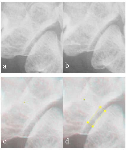
10 readers. Distribution of chronological changes in SvdH for JSN between baseline and follow-up images by reader 1 is shown in Table 2. Computer-based analysis for JSN progression The computer-based method can detect the difference of joint space width between baseline and follow-up images as the joint space difference index (JSDI). This method visualized interval JSN progression between the baseline and the follow-up image as red shadow (Fig.1). If there were no changes in joint space width between the baseline and the follow-up images, the joint space in the fused image was visualized as grey shadow (Fig.2). The JSDI is defined as the average absolute value of the difference of the pixel value in each pixel for baseline and follow-up images inside the region of the interest (ROI) (Fig.3). The details of the computer-based method are presented in the previous article 15. The computer-based method assessed the JSN progression of carpal joints (third carpometacarpal joint, fifth carpometacarpal joint, scaphoid-trapezium joint, scaphoid-capitate joint, and radius-scaphoid joint), setting the SvdH method as the standard of reference on bilateral hand radiographs. We excluded the fourth 9
11 carpometacarpal joints because of the difficulty in discriminating from adjoining bone during ROI placement for JSDI. To increase the homogeneity of the study sample, severely damaged (subluxation, ankylosed, and complete luxation) joints were also excluded based on the SvdH by each reader. Computer-based analysis was performed by a non-specialist who had not received specialized training in scoring of JSN and was blinded to other clinical information. Computer-based analysis was repeated twice, and intraobserver reliability was assessed based on each reader s score. The measurement procedure was performed as follows: First, the software read the baseline and follow-up images and fused them for every case. Second, the single reference bone of the two images was aligned visually. The chosen reference bone was as follows: third carpometacarpal joint, third metacarpal bone; fifth carpometacarpal joint, fifth metacarpal bone; scaphoid-trapezium joint, trapezium; scaphoid-capitate joint, capitate; and radius-scaphoid joint, radius. Third, the rectangular ROI sized 20 7 pixels was located manually in the center of the joint space with attention so that the edges of bones forming the joint were placed inside the ROI. At this time, the horizontal ROI borders were approximately parallel to the joint edges (Fig.1 d and Fig.2 d). Finally, information on each pixel value for the baseline and follow-up images inside the ROI was output to a text file that can be read by Microsoft Excel, and the JSDI was 10
12 calculated. We compared the JSDI of joints with JSN progression in the follow-up period (increase in SvdH) to those without JSN progression (no change in SvdH) based on each reader s score. Additionally, we examined whether there is a significant difference in JSDI in terms of laterality or topology of the joint based on each reader s score. In addition, a direct correlation of the JSDI with the ΔSvdH was evaluated. Data of the first time measurement were used for these analyses. Statistical analysis Statistical analyses were calculated with the use of the SPSS version 22.0 (IBM, Armonk, New York, USA), for Windows and the Excel program (Microsoft, Redmond, WA, USA). Intra- and interobserver reliabilities were estimated using calculations of intraclass correlation coefficients (ICC). The ICC ranged from 1 to +1. ICC values are interpreted as follows: <0.40, poor to fair agreement; 0.41 to 0.60, moderate agreement; 0.61 to 0.80, substantial agreement; 0.81 to 1.00, almost perfect agreement 18. Differences between two independent samples were examined using the Mann-Whitney U test. To assess the significance of differences in terms of topology, the Kruskal-Wallis 11
13 test was used. P-values less than 0.05 were considered statistically significant. Correlations between the JSDI and the ΔSvdH were examined using Spearman s rank correlation test. Results A total of 430 carpal joints on the bilateral hand radiographs in 43 patients were scored using SvdH method by reader 1 and 2. Interobserver reliability for baseline SvdH were in substantial agreement (ICC=0.695; 95% confidence interval [95% CI], ). Interobserver reliability for follow-up SvdH were in substantial agreement (ICC=0.678; 95% CI, ). Interobserver reliability for ΔSvdH was in moderate agreement (ICC=0.591; 95% CI, ). The computer-based method evaluated carpal joints twice on the bilateral hand radiographs in 43 patients, setting the SvdH method as the standard of reference. Out of 430 carpal joints, we targeted 355 and 365 carpal joints after excluding severely damaged 75 and 65 joints based on each reader s score. Intraobserver reliability for the JSDI based on the scores by reader 1 was in almost perfect agreement (ICC=0.967; 95% CI, ). Intraobserver reliability for the JSDI based on the scores by reader 2 12
14 was in almost perfect agreement (ICC=0.968; 95% CI, ). Based on the reader 1 s score, the medians of the JSDI for carpal joints were (inter-quartile range [IQR] , n=32) and 6.84 (IQR , n=323) with and without JSN progression, respectively. Both the ΔSvdH and JSDI of joints with JSN progression were significantly higher than those without JSN progression (P < respectively) (Table 3). While, based on reader 2 s score, they were 8.90 (IQR , n=40) and 7.01 (IQR , n=325) with and without JSN progression in carpal joints, respectively (P = 0.02). We next examined whether there is a significant difference in the JSDI andδsvdh in terms of laterality of the joint. There was no statistically significant difference in the JSDI and ΔSvdH between the left and right carpal joints based on reader 1 s score, which was analyzed for 5 different joints altogether and each joint separately (P > 0.05, respectively) (Table 4). There was no statistically significant difference in the JSDI in terms of laterality of the joints based on reader 2 s score (P > 0.05, respectively). Additionally, we examined whether there is a significant difference in the JSDI and Δ SvdH in terms of topology of the joint. There was no statistically significant difference in the ΔSvdH among different joints based on reader 1 s score (P = 0.393). However, there was statistically significant difference in the JSDI among different joints based on 13
15 reader 1 s score (P = 0.003) (Table 5). The JSDI of scaphoid-capitate joint was significantly lower than that of third carpometacarpal joint and scaphoid-trapezium joint (P = and 0.003, respectively). Based on reader 2 s score, there was statistically significant difference in the JSDI among different joints (P = 0.002). The JSDI of scaphoid-capitate joint was significantly lower than that of fifth carpometacarpal joint and scaphoid-trapezium joint (P = and 0.003, respectively). Finally, correlations between the JSDI and the ΔSvdH were examined. The mean and standard deviation (SD) JSDI (n; the number of joints) of joints for 0, 1, 2, 3 in reader 1 s ΔSvdH were 7.64±3.16 (n=323), 10.57±4.28 (n=19), 10.35±3.95 (n=10) and 11.42±0.18 (n=3), respectively. For this, the JSDI was not correlated with ΔSvdH (r = 0.877, P = 0.123) (Fig.4). The mean and SD JSDI of joints for 0, 1, 2, 3 in reader 2 s Δ SvdH were 7.93±3.36 (n=325), 9.07±4.76 (n=14), 9.84±3.85 (n=23) and 9.57±5.77 (n=3), respectively. For this, the JSDI was not correlated with ΔSvdH (r = 0.870, P = 0.130). Discussion In this study, we investigated a validity of the computer-based method for detecting JSN 14
16 progression in carpal joints by comparing the computer-based method with the SvdH method as the gold standard. The results of the computer-based method were consistent with those of the SvdH method in almost all examinations excluding assessment in terms of topology of the joints. The results indicate that our computer-based method can recognize the difference of joint space width on hand radiographs in carpal joints. Previous computer-aided analyses were validated only in finger joints such as metacarpophalangeal, proximal interphalangeal, or distal interphalangeal joints 10,12,19. However, assessment for JSN by traditional scoring methods such as the Sharp/van der Heijde score 17 and the Genant modified Sharp score 20 includes carpal joints. In addition, the carpal joint is a site of predilection for rheumatoid disease. Thus, it is preferred that computer-based methods are validated in not only finger joints but also in carpal joints. The advantage of our computer-based method is that it can detect JSN progression in carpal joints as well as finger joints. Furthermore, our method does not require highly trained personnel, as is the case for the traditional scoring methods. Other modalities (i.e., US and MRI) are available that directly visualize the active disease and feature a much better sensitivity in detecting the progression of RA 21. These modalities are thought to be better suited to monitor disease progression, and have increasingly been used as outcome measures in RA patients 18,22. While US and 15
17 MRI allow direct visualization of early inflammatory, conventional radiography is the pivotal method for diagnosing and monitoring structural joint damage such as JSN 21,23. Additionally, it is not only inexpensive, but also widely available and accepted. Although radiographs are usually assessed by established scoring methods, these methods suffer from intra- or inter- observer variations. Our data showed that interobserver reliability for SvdH were in substantial or moderate agreement, as assessed by ICC. In contrast, intraobserver reliability for the computer-based method was supported by high agreement. Consequently, the computer-based method could provide reproducible measurement of JSN progression. In a previous study, Angwin et al. reported no change in Sharp scores in 47% of their 245 patients with early RA after 2 years, but a significant reduction in joint space width using a different computer-based method 24. This report indicates that the computer-based method is more sensitive to the change in joint space width than traditional scoring methods. In this study, we cannot determine whether our computer-based method or the SvdH method is more sensitive because of the absence of a gold standard. However, our computer-based method could detect the chronological change of joint space width and is consistent with assessment by the SvdH method in almost all examinations. Additionally, our methods extracts the joint space difference 16
18 between two images by superimposing the images and therefore a slight joint space difference is detected more easily than if the images were to be observed side by side. Thus, our computer-based method may be useful as a computer-aided diagnosis tool and assist the assessment of JSN by a rheumatologist. The JSDI between progressive and stable joints revealed considerable overlap (Table 3). Although the JSDI of joints without JSN progression was expected to be 0 in theory, the median of it was 6.84 (IQR ) (overall carpal joint) in practice. Additionally, the JSDI of scaphoid-capitate joints was lower than that of other joints, showing statistically significant difference among different joints. These may be explained by the influence of different hand positions during imaging, variation in the x-ray beam angle, or progression of osteopenia. This result implied the scaphoid-capitate joints should be removed from a computer-based analysis, although further analysis is need to confirm this. Furthermore, there was no statically significant correlation between the JSDI and ΔSvdH, although showing high correlation coefficient. This was due to a relatively small number of joints with changes in SvdH, especially in ΔSvdH 3 (n=3). Several limitations of this study should be discussed. First, only a limited number of joints with JSN progression were studied (n=32 or 40). Therefore, we could validate for 5 different carpal joints altogether, but not for each joint separately. Additionally, we 17
19 could not reveal the precise relationship between the JSDI and ΔSvdH. Further study, with larger numbers of joints with JSN progression, is needed to prove that the JSDI were potential marker for assessment of disease progression. Second, no pre-selection regarding steroid therapy was considered. Treatment of RA with steroid therapy may increase susceptibility to osteoporosis but also suppresses inflammatory activity, which is a risk factor for osteoporosis in RA. Therefore, steroid therapy may have an influence on the JSDI inside the ROI. Finally, our computer-based method tends to be time consuming. The analysis time is around 3 minutes per joint. We will develop an automated computer-based method that automatically aligns the joint with only minimal human intervention and can evaluate JSN progression more easily and reproducibly. In conclusion, our computer-based method, which requires no special training or experience of traditional scoring methods, can detect the difference in joint space width between two radiographs as the joint space difference index (JSDI) in the rheumatoid wrist. Refinement of this method may enable us to obtain more sensitive, objective, quantitative, and reproducible information about JSN progression. Further study is needed to prove that this method is useful to quantify the JSN progression on radiographs in clinical trials. 18
20 References 1. Aletaha D, Neogi T, Silman AJ, Funovits J, Felson DT, Bingham CO, et al Rheumatoid arthritis classification criteria: An American College of Rheumatology/European League Against Rheumatism collaborative initiative. Arthritis and Rheumatism p Bijlsma JWJ, Weinblatt ME. Optimal use of methotrexate: the advantages of tight control. Annals of the rheumatic diseases p Visser K, van der Heijde D. Optimal dosage and route of administration of methotrexate in rheumatoid arthritis: a systematic review of the literature. Ann Rheum Dis. 2009;68(7): Smolen JS, Aletaha D, Koeller M, Weisman MH, Emery P. New therapies for treatment of rheumatoid arthritis. Lancet p Doan T, Massarotti E. Rheumatoid arthritis: an overview of new and emerging therapies. J Clin Pharmacol. 2005;45(7): Smolen JS, Aletaha D. What should be our treatment goal in rheumatoid arthritis today? Clin Exp Rheumatol. 2006; 7. Van der Heijde DM. Radiographic imaging: the gold standard for assessment of disease progression in rheumatoid arthritis. Rheumatology (Oxford). 2000;39 Suppl 1: Van der Heijde DMFM. Plain X-rays in rheumatoid arthritis: overview of scoring methods, their reliability and applicability. Baillieres Clin Rheumatol. 1996;10(3): Van Der Heijde D, Boonen A, Boers M, Kostense P, van Der Linden S. Reading radiographs in chronological order, in pairs or as single films has important implications for the discriminative power of rheumatoid arthritis clinical trials. Rheumatology (Oxford). 1999;38(12):
21 10. Finckh A, De Pablo P, Katz JN, Neumann G, Lu Y, Wolfe F, et al. Performance of an automated computer-based scoring method to assess joint space narrowing in rheumatoid arthritis: A longitudinal study. Arthritis Rheum. 2006;54(5): Pfeil A, Oelzner P, Bornholdt K, Hansch A, Lehmann G, Renz DM, et al. Joint damage in rheumatoid arthritis: assessment of a new scoring method. Arthritis Res Ther. BioMed Central Ltd; 2013;15(1):R Duryea J, Jiang Y, Zakharevich M, Genant HK. Neural network based algorithm to quantify joint space width in joints of the hand for arthritis assessment. Med Phys. 2000;27(5): Böttcher J, Pfeil A, Rosholm A, Petrovitch A, Seidl BE, Malich A, et al. Digital x-ray radiogrammetry combined with semiautomated analysis of joint space widths as a new diagnostic approach in rheumatoid arthritis: A cross-sectional and longitudinal study. Arthritis Rheum. 2005;52(12): Peloschek P, Langs G, Weber M, Sailer J, Reisegger M, Imhof H, et al. An automatic model-based system for joint space measurements on hand radiographs: initial experience. Radiology. 2007;245(3): Ichikawa S, Kamishima T, Sutherland K, Okubo T, Katayama K. Performance of computer-based analysis using temporal subtraction to assess joint space narrowing progression in rheumatoid patients. Rheumatol Int. Springer Berlin Heidelberg; 2015; 16. Arnett FC, Edworthy SM, Bloch DA, McShane DJ, Fries JF, Cooper NS, et al. The American Rheumatism Association 1987 revised criteria for the classification of rheumatoid arthritis. Arthritis Rheum. 1988;31(3): Van Der Heijde D. How to read radiographs according to the Sharp/van der Heijde method. Journal of Rheumatology p Kamishima T, Tanimura K, Shimizu M, Matsuhashi M, Fukae J, Kon Y, et al. Monitoring anti-interleukin 6 receptor antibody treatment for rheumatoid arthritis by quantitative magnetic resonance imaging of the hand and power Doppler ultrasonography of the finger. Skeletal Radiol. 2011;40(6):
22 19. Pfeil A, Renz DM, Hansch A, Kainberger F, Lehmann G, Malich A, et al. The usefulness of computer-aided joint space analysis in the assessment of rheumatoid arthritis. Joint Bone Spine. Elsevier Masson SAS; 2013;80(4): Genant HK. Methods of assessing radiographic change in rheumatoid arthritis. Am J Med. 1983;75(6A): Østergaard M, Pedersen SJ, Døhn UM. Imaging in rheumatoid arthritis - status and recent advances for magnetic resonance imaging, ultrasonography, computed tomography and conventional radiography. Best Practice and Research: Clinical Rheumatology. Elsevier Ltd; p Iagnocco a., Finucci A, Ceccarelli F, Perricone C, Iorgoveanu V, Valesini G. Power Doppler ultrasound monitoring of response to anti-tumour necrosis factor alpha treatment in patients with rheumatoid arthritis. Rheumatology. 2015; Ostergaard M, Szkudlarek M. Imaging in rheumatoid arthritis--why MRI and ultrasonography can no longer be ignored. Scandinavian journal of rheumatology p Angwin J, Lloyd A, Heald G, Nepom G, Binks M, James MF. Radiographic hand joint space width assessed by computer is a sensitive measure of change in early rheumatoid arthritis. J Rheumatol. 2004;31(6):
23 Tables Table 1. Clinical and laboratory characteristics of patients with RA at baseline Value Total no. of subjects included 43 Age, mean (range) years 58 (31-83) Sex, no. female/male 39/4 Duration of symptoms, median (IQR) months 65 (24-108) Follow-up time between first and second radiograph, median (IQR) months 12 (8.8-14) ESR, median (IQR) mm/hour 50 (32-80) CRP level, median (IQR) mg/dl 2.9 ( ) Swollen joint count, median (IQR) 15 (10-17) Tender joint count, median (IQR) 13 (10-23) VAS, median (IQR) 70 (56-77) DMARDs, no. None 2 Methotrexate 23 Mizoribine 1 Bucillamine 1 Salazosulfapyridine 1 Tacrolimus 1 Combination therapy 14 ESR = erythrocyte sedimentation rate; CRP = C-reactive protein; VAS = visual analog scale; IQR = inerquartile range; DMARDs= disease modifying antirheumatic drugs. 22
24 Table 2. Distribution of chronological changes in SvdH for JSN in carpal joints (n=355) Chronological changes of SvdH for JSN CM3 CM5 ST SC RS SvdH=Sharp/van der Heijde score; CM3=third carpometacarpal joint; CM5=fifth carpometacarpal joint; ST=scaphoid-trapezium joint; SC=scaphoid-capitate joint; RS=radius-scaphoid joint. 23
25 Table 3. Comparison of the JSDI/ΔSvdH between joints with and without JSN progression Joints with JSNP(+) Joints with JSNP(-) P value n median IQR n median IQR CM / / / / CM / / / / ST / / / / SC / / / / RS / / / / Carpal, overall / / / / <0.001 / <0.001 JSDI=joint space difference index; ΔSvdH=change of the Sharp/van der Heijde score; JSNP(+)=joint space narrowing progression; JSNP(-)=non-joint space narrowing progression; IQR=interquartile range; CM 3=third carpometacarpal joint; CM 5=fifth carpometacarpal joint; ST=scaphoid-trapezium joint; SC=scaphoid-capitate joint; RS=radius-scaphoid joint. 24
26 Table 4. Comparison of the JSDI/ΔSvdH between left and right joints Left joints Right joints P value n median IQR n median IQR CM / / / / / CM / / / / / ST / / / / / SC / / / / / RS / / / / / Carpal, overall / / / / / JSDI=joint space difference index; ΔSvdH=change of the Sharp/van der Heijde score; IQR=interquartile range; CM 3=third carpometacarpal joint; CM 5=fifth carpometacarpal joint; ST=scaphoid-trapezium joint; SC=scaphoid-capitate joint; RS=radius-scaphoid joint. 25
27 Table 5. Comparison of the JSDI/ΔSvdH among different carpal joints n median IQR P value CM / / CM / / ST / / / SC / / RS / / JSDI=joint space difference index; ΔSvdH=change of the Sharp/van der Heijde score; IQR=interquartile range; CM 3=third carpometacarpal joint; CM 5=fifth carpometacarpal joint; ST=scaphoid-trapezium joint; SC=scaphoid-capitate joint; RS=radius-scaphoid joint. 26
28 Figure Legends Figure 1 The scaphoid-trapezium joint of 57-year-old woman with rheumatoid arthritis with interval change in joint space width. Radiograph images of the scaphoid-trapezium joint for the right hand at baseline (a) and follow-up (b) are shown. These images correspond to a Sharp van der Heijde Score of 0 and 1, respectively. In the fused image (c), the joint space difference is visible as a red shadow. The rectangular ROI, with a size of 20 7 pixels, was located in the center of the joint space (d), and the chronological changes in the joint space width were measured as the joint space difference index (JSDI). The JSDI for this case was Figure 2 The scaphoid-trapezium joint of 61-year-old man with rheumatoid arthritis without interval change in joint space width. Radiograph images of the scaphoid-trapezium joint for the right hand at baseline (a) and follow-up (b) are shown. Both of these images correspond to a Sharp van der Heijde Score of 0. In the fused image (c), the difference in the joint space between the baseline and the follow-up images is not recognizable. The rectangular ROI, with a size of 20 7 pixels, was located in the center of the joint 27
29 space (d), and the chronological changes in the joint space width were measured as the joint space difference index (JSDI). The JSDI for this case was Figure 3 Calculation of the joint space difference index (JSDI). For each pixel with the coordinate indicated as x (column B) and y (column C), information on pixel value for the baseline and follow-up images inside region of interest (ROI) are shown in columns D and E, respectively. We subtracted the baseline pixel value from the follow-up pixel value in each pixel (column F) and calculated its absolute value (column G). Finally, we averaged these values and refer to this value as the JSDI (column I). Figure 4 Relationship of the JSDI and ΔSvdH. The JSDI were expressed as mean and standard deviation for each ΔSvdH. JSDI, joint space difference index; ΔSvdH, delta Sharp/van der Heijde score 28

30
31

32
33
The EULAR OMERACT rheumatoid arthritis MRI reference image atlas: the wrist joint
 i23 The EULAR OMERACT rheumatoid arthritis MRI reference image atlas: the wrist joint B Ejbjerg, F McQueen, M Lassere, E Haavardsholm, P Conaghan, P O Connor, P Bird, C Peterfy, J Edmonds, M Szkudlarek,
Rheumatoid arthritis 2010: Treatment and monitoring
 October 12, 2010 By Yusuf Yazici, MD [1] The significant changes in the way rheumatoid arthritis has been managed include earlier, more aggressive treatment with combination therapy. Significant changes
(For National Authority Use Only) Page:
 Page:") 2.0 Synopsis AbbVie Individual Study Table Referring to Part of Dossier: Name of Study Drug: Volume: HUMIRA 40 mg/0.8 ml for subcutaneous injection Page: (For National Authority Use Only) Name of Active
2.0 Synopsis AbbVie Individual Study Table Referring to Part of Dossier: Name of Study Drug: Volume: HUMIRA 40 mg/0.8 ml for subcutaneous injection Page: (For National Authority Use Only) Name of Active
Concise report RHEUMATOLOGY
 RHEUMATOLOGY Rheumatology 2012;51:2034 2038 doi:10.1093/rheumatology/kes124 Advance Access publication 30 July 2012 Concise report Head-to-head comparison of quantitative and semi-quantitative ultrasound
Aoyagi, Kiyoshi; Eguchi, Katsumi; K
 NAOSITE: Nagasaki University's Ac Title Author(s) Combination of MRI-detected bone ma arthritis classification criteria i rheumatoid arthritis Tamai, Mami; Kita, Junko; Nakashima Horai, Yoshiro; Okada,
Alexander Pfeil 1*, Peter Oelzner 1, Diane M. Renz 2, Andreas Hansch 3, Gunter Wolf 1 and Joachim Böttcher 4
 Pfeil et al. BMC Musculoskeletal Disorders (2015) 16:155 DOI 10.1186/s12891-015-0577-3 RESEARCH ARTICLE Open Access Is there a role for Digital X-ray Radiogrammetry as surrogate marker for radiological
Treat - to - Target Pathway Commissioning Chronic and Complex Care MIDLANDS RHEUMATOLOGY & MUSCULOSKELETAL (MSK) COMMISSIONING NETWORK
 COMMISSIONING NETWORK") Treat - to - Target Pathway Commissioning Chronic and Complex Care MIDLANDS RHEUMATOLOGY & MUSCULOSKELETAL (MSK) COMMISSIONING NETWORK Dr Bruce Kirkham Consultant Rheumatologist Guy s & St Thomas NHS Foundation
Treat - to - Target Pathway Commissioning Chronic and Complex Care MIDLANDS RHEUMATOLOGY & MUSCULOSKELETAL (MSK) COMMISSIONING NETWORK Dr Bruce Kirkham Consultant Rheumatologist Guy s & St Thomas NHS Foundation
Concordance with the British Society of Rheumatology (BSR) 2010 recommendations on eligibility criteria for the first biologic agent
 2010 recommendations on eligibility criteria for the first biologic agent") Concordance with the British Society of Rheumatology (BSR) 2010 recommendations on eligibility criteria for the first biologic agent Michela Frendo, John Paul Caruana Galizia, Andrew A Borg Abstract Aims:
Concordance with the British Society of Rheumatology (BSR) 2010 recommendations on eligibility criteria for the first biologic agent Michela Frendo, John Paul Caruana Galizia, Andrew A Borg Abstract Aims:
Smallest Detectable Difference in Radiological Progression
 Smallest Detectable Difference in Radiological Progression MARISSA LASSERE, MAARTEN BOERS, DÉSIRÉE van der HEIJDE, ANNELIES BOONEN, JOHN EDMONDS, ARIANE SAUDAN, and ARCO C. VERHOEVEN ABSTRACT. OMERACT
Radiography is the standard technique used to monitor the longterm progression of cartilaginous degradation and bone erosions in patients with rheumat
 Note: This copy is for your personal non-commercial use only. To order presentation-ready copies for distribution to your colleagues or clients, contact us at www.rsna.org/rsnarights. Philipp Peloschek,
Comparison of long-term clinical outcome with etanercept and adalimumab treatment of rheumatoid arthritis with respect to immunogenicity
 7 Comparison of long-term clinical outcome with etanercept and adalimumab treatment of rheumatoid arthritis with respect to immunogenicity Charlotte Krieckaert* Anna Jamnitski* Mike Nurmohamed Piet Kostense
Rheumatoid Arthritis. Ajay Bhatia Rheumatology Consultant Hillingdon Hospital
 Rheumatoid Arthritis Ajay Bhatia Rheumatology Consultant Hillingdon Hospital ajay.bhatia@thh.nhs.uk Rheumatoid Arthritis When to refer to secondary care? Why early referral is beneficial for the patient?
S tructural joint damage, a major outcome in
 i3 An introduction to the EULAR OMERACT rheumatoid arthritis MRI reference image atlas M Østergaard, J Edmonds, F McQueen, C Peterfy, M Lassere, B Ejbjerg, P Bird, P Emery, H Genant, P Conaghan... This
Maasa Hama Takeaki Uehara Kaoru Takase Atsushi Ihata Atsuhisa Ueda Mitsuhiro Takeno Kazuya Shizukuishi Ukihide Tateishi Yoshiaki Ishigatsubo
 Rheumatol Int (2012) 32:1327 1333 DOI 10.1007/s00296-011-1802-5 ORIGINAL ARTICLE Power Doppler ultrasonography is useful for assessing disease activity and predicting joint destruction in rheumatoid arthritis
Newer classification criteria 2010:How adequate is this to classify Rheumatoid Arthritis?
 Newer classification criteria 2010:How adequate is this to classify Rheumatoid Arthritis? DR MD MATIUR RAHMAN MBBS, MD, FCPS, FACR, Fellow APLAR Associate Professor, Medicine SSMC & Mitford Hospital New
J. van Aken* H. van Dongen* S. le Cessie F.C. Breedveld T.W.J. Huizinga. * both authors contributed equally
 CHAPTER Comparison of long term outcome of patients with rheumatoid arthritis presenting with undifferentiated arthritis or with rheumatoid arthritis: an observational cohort study J. van Aken* H. van
Day-to-day variation in Doppler activity in patients on stable etanercept treatment: an exploratory cohort study
 Research Article Day-to-day variation in Doppler activity in patients on stable etanercept treatment: an exploratory cohort study Aim: Doppler ultrasound was used to evaluate the day-to-day variation in
The Relationship Between Disease Activity and Radiologic Progression in Patients With Rheumatoid Arthritis
 ARTHRITIS & RHEUMATISM Vol. 50, No. 7, July 2004, pp 2082 2093 DOI 10.1002/art.20350 2004, American College of Rheumatology The Relationship Between Disease Activity and Radiologic Progression in Patients
Cover Page. The handle holds various files of this Leiden University dissertation.
 Cover Page The handle http://hdl.handle.net/1887/29578 holds various files of this Leiden University dissertation. Author: Krabben, Annemarie Title: Predictive factors for the development and disease course
The new ACR/EULAR classification criteria for RA: how are the new criteria performing in the clinic?
 RHEUMATOLOGY Rheumatology 2012;51:vi10 vi15 doi:10.1093/rheumatology/kes280 The new ACR/EULAR classification criteria for RA: how are the new criteria performing in the clinic? Vivian P. Bykerk 1,2 and
Targeted Ultrasound of the Fifth Metatarsophalangeal Joint in an Early Inflammatory Arthritis Cohort
 Arthritis & Rheumatism (Arthritis Care & Research) Vol. 61, No. 7, July 15, 2009, pp 1004 1008 DOI 10.1002/art.24564 2009, American College of Rheumatology CONTRIBUTIONS FROM THE FIELD Targeted Ultrasound
Citation for final published version:
 This is an Open Access document downloaded from ORCA, Cardiff University's institutional repository: http://orca.cf.ac.uk/97756/ This is the author s version of a work that was submitted to / accepted
Efficacy and Safety of Tocilizumab in the Treatment of Rheumatoid Arthritis and Juvenile Idiopathic Arthritis
 New Evidence reports on presentations given at EULAR 2010 Efficacy and Safety of Tocilizumab in the Treatment of Rheumatoid Arthritis and Juvenile Idiopathic Arthritis Report on EULAR 2010 presentations
Abatacept (Orencia) for active rheumatoid arthritis. August 2009
 for active rheumatoid arthritis. August 2009") Abatacept (Orencia) for active rheumatoid arthritis August 2009 This technology summary is based on information available at the time of research and a limited literature search. It is not intended to
Abatacept (Orencia) for active rheumatoid arthritis August 2009 This technology summary is based on information available at the time of research and a limited literature search. It is not intended to
Tomosynthesis of the Wrist and Hand in Patients With Rheumatoid Arthritis: Comparison With Radiography and MRI
 Musculoskeletal Imaging Original Research Aoki et al. Imaging of the Wrist and Hand in Rheumatoid Arthritis Musculoskeletal Imaging Original Research Takatoshi Aoki 1 Masami Fujii 1 Yoshiko Yamashita 1
Validation of Leiden Score in Predicting Progression of Rheumatoid Arthritis in Undifferentiated Arthritis in Indian Population
 Original Article Validation of Leiden Score in Predicting Progression of Rheumatoid Arthritis in Undifferentiated Arthritis in Indian Population Ghosh K, Chatterjee A 1, Ghosh S 2, Chakraborty S 3, Chattopadhyay
Received: 27 May 2003 Revisions requested: 26 Jun 2003 Revisions received: 14 Aug 2003 Accepted: 19 Aug 2003 Published: 1 Oct 2003
 Research article Etanercept versus etanercept plus methotrexate: a registrybased study suggesting that the combination is clinically more efficacious Ronald F van Vollenhoven 1, Sofia Ernestam 2, Anders
Magnetic Resonance Imaging of Inflammatory Lesions in the Spine in Ankylosing Spondylitis Clinical Trials: Is Paramagnetic Contrast Medium Necessary?
 Magnetic Resonance Imaging of Inflammatory Lesions in the Spine in Ankylosing Spondylitis Clinical Trials: Is Paramagnetic Contrast Medium Necessary? KAY-GEERT A. HERMANN, ROBERT B.M. LANDEWÉ, JÜRGEN BRAUN,
Clinical Policy: Etanercept (Enbrel) Reference Number: PA.CP.PHAR.250 Effective Date: 01/18 Last Review Date: 08/17 Line of Business: Medicaid
 Reference Number: PA.CP.PHAR.250 Effective Date: 01/18 Last Review Date: 08/17 Line of Business: Medicaid") Clinical Policy: (Enbrel) Reference Number: PA.CP.PHAR.250 Effective Date: 01/18 Last Review Date: 08/17 Line of Business: Medicaid Coding Implications Revision Log Description (Enbrel ) is tumor necrosis
Clinical Policy: (Enbrel) Reference Number: PA.CP.PHAR.250 Effective Date: 01/18 Last Review Date: 08/17 Line of Business: Medicaid Coding Implications Revision Log Description (Enbrel ) is tumor necrosis
Preventing the Progression From Undifferentiated Arthritis to Rheumatoid Arthritis: The Clinical and Economic Implications
 n report n Preventing the Progression From Undifferentiated Arthritis to Rheumatoid Arthritis: The Clinical and Economic Implications Michael H. Schiff, MD Introduction Over the past few years, the management
Bringing the clinical experience with anakinra to the patient
 Rheumatology 2003;42(Suppl. 2):ii36 ii40 doi:10.1093/rheumatology/keg331, available online at www.rheumatology.oupjournals.org Bringing the clinical experience with anakinra to the patient S. B. Cohen
Structural damage in rheumatoid arthritis as visualized through radiographs Désirée van der Heijde
 Structural damage in rheumatoid arthritis as visualized through radiographs Désirée van der Heijde University Hospital Maastricht, Maastricht, The Netherlands; and Limburg University Center, Diepenbeek,
An Overview of RAMRIQ: An Automated MRI Rheumatoid Arthritis Quantitative Assessment System
 Precision. Insight. Innovation. White Paper: An Overview of RAMRIQ: An Automated MRI Rheumatoid Arthritis Quantitative Assessment System At left: Volume of synovitis in the hand and wrist. Areas of enhancement
PsA. SIMPONI (golimumab) Rheumatoid arthritis. Psoriatic arthritis. Ankylosing spondylitis EFFICACY EFFICACY EFFICACY. QoL. QoL.
 Rheumatoid arthritis. Psoriatic arthritis. Ankylosing spondylitis EFFICACY EFFICACY EFFICACY. QoL. QoL.") RA Rheumatoid arthritis PsA Psoriatic arthritis AS Ankylosing spondylitis EFFICACY EFFICACY EFFICACY QoL QoL QoL SAFETY SAFETY SAFETY EXPERIENCE EXPERIENCE EXPERIENCE SUMMARY SUMMARY SUMMARY Copyright
RA Rheumatoid arthritis PsA Psoriatic arthritis AS Ankylosing spondylitis EFFICACY EFFICACY EFFICACY QoL QoL QoL SAFETY SAFETY SAFETY EXPERIENCE EXPERIENCE EXPERIENCE SUMMARY SUMMARY SUMMARY Copyright
C. Barnabe 1, E. Szabo 2, L. Martin 1, S.K. Boyd 2, S.G. Barr 1
 Quantification of small joint space width, periarticular bone microstructure and erosions using high-resolution peripheral quantitative computed tomography in rheumatoid arthritis C. Barnabe 1, E. Szabo
What is the additional value of MRI of the foot to the hand in undifferentiated arthritis to predict rheumatoid arthritis development?
 Dakkak et al. Arthritis Research & Therapy (2019) 21:56 https://doi.org/10.1186/s13075-019-1845-7 RESEARCH ARTICLE What is the additional value of MRI of the foot to the hand in undifferentiated arthritis
Impact of obesity on rheumatoid arthritis: Relation with disease activity, joint damage, functional impairment and response to therapy
 International Journal of Clinical Rheumatology Impact of obesity on rheumatoid arthritis: Relation with disease activity, joint damage, functional impairment and response to therapy Background and Aim
Clinical Policy: Certolizumab (Cimzia) Reference Number: PA.CP.PHAR.247 Effective Date: 01/18 Last Review Date: 08/17 Line of Business: Medicaid
 Reference Number: PA.CP.PHAR.247 Effective Date: 01/18 Last Review Date: 08/17 Line of Business: Medicaid") Clinical Policy: (Cimzia) Reference Number: PA.CP.PHAR.247 Effective Date: 01/18 Last Review Date: 08/17 Line of Business: Medicaid Coding Implications Revision Log Description (Cimzia ) is a tumor necrosis
Clinical Policy: (Cimzia) Reference Number: PA.CP.PHAR.247 Effective Date: 01/18 Last Review Date: 08/17 Line of Business: Medicaid Coding Implications Revision Log Description (Cimzia ) is a tumor necrosis
The Egyptian Journal of Hospital Medicine (October 2017) Vol. 69 (4), Page
 Vol. 69 (4), Page") The Egyptian Journal of Hospital Medicine (October 2017) Vol. 69 (4), Page 2294-2300 Role of Magnetic Resonance Imaging and Ultrasonography in Diagnosis and Follow Up Rheumatoid Arthritis in Hand and Wrist
The Egyptian Journal of Hospital Medicine (October 2017) Vol. 69 (4), Page 2294-2300 Role of Magnetic Resonance Imaging and Ultrasonography in Diagnosis and Follow Up Rheumatoid Arthritis in Hand and Wrist
Is synovial hypertrophy without Doppler activity sensitive to change? Post-hoc analysis from a rheumatoid arthritis ultrasound study
 Terslev et al. Arthritis Research & Therapy (2018) 20:224 https://doi.org/10.1186/s13075-018-1709-6 RESEARCH ARTICLE Open Access Is synovial hypertrophy without Doppler activity sensitive to change? Post-hoc
Remicade (Infliximab)
") Remicade (Infliximab) Policy Number: Original Effective Date: MM.04.016 11/18/2003 Line(s) of Business: Current Effective Date: HMO; PPO; QUEST Integration 07/26/2013 Section: Prescription Drugs Place(s)
Remicade (Infliximab) Policy Number: Original Effective Date: MM.04.016 11/18/2003 Line(s) of Business: Current Effective Date: HMO; PPO; QUEST Integration 07/26/2013 Section: Prescription Drugs Place(s)
RADIOLOGICAL ASSESSMENT IN PSORIATIC ARTHRITIS
 British Journal of Rheumatology 1998;37:760 765 RADIOLOGICAL ASSESSMENT IN PSORIATIC ARTHRITIS P. RAHMAN, D. D. GLADMAN,* R. J. COOK, Y. ZHOU, G. YOUNG and D. SALONEN Department of Medicine and Institute
DAS28 score vs. ultrasound examination for assessment of rheumatoid arthritis disease activity: comparison and discussion of pros and cons
 Review paper Reumatologia 215; 53, 4: 213 218 DOI: 1.5114/reum.215.53999 DAS28 score vs. ultrasound examination for assessment of rheumatoid arthritis disease activity: comparison and discussion of pros
Performance of the Ankylosing Spondylitis Disease Activity Score (ASDAS) in patients under biological therapies
 in patients under biological therapies") Performance of the Ankylosing Spondylitis Disease Activity Score (ASDAS) in patients under biological therapies 1. Introduction The Ankylosing Spondylitis Disease Activity Score (ASDAS) is a new instrument
Performance of the Ankylosing Spondylitis Disease Activity Score (ASDAS) in patients under biological therapies 1. Introduction The Ankylosing Spondylitis Disease Activity Score (ASDAS) is a new instrument
New Evidence reports on presentations given at EULAR Tocilizumab for the Treatment of Rheumatoid Arthritis
 New Evidence reports on presentations given at EULAR 2012 Tocilizumab for the Treatment of Rheumatoid Arthritis Report on EULAR 2012 presentations Tocilizumab monotherapy is superior to adalimumab monotherapy
Introduction ORIGINAL ARTICLE
 Mod Rheumatol (2007) 17:28 32 Japan College of Rheumatology 2007 DOI 10.1007/s10165-006-0532-0 ORIGINAL ARTICLE Hisashi Yamanaka Yoshiya Tanaka Naoya Sekiguchi Eisuke Inoue Kazuyoshi Saito Hideto Kameda
1.0 Abstract. Title. Keywords. Rationale and Background
 1.0 Abstract Title A Prospective, Multi-Center Study in Rheumatoid Arthritis Patients on Adalimumab to Evaluate its Effect on Synovitis Using Ultrasonography in an Egyptian Population Keywords Synovitis
Safety and effectiveness of biologic Disease-Modifying Antirheumatic Drugs in elderly patients with rheumatoid arthritis
 1. Title: Safety and effectiveness of biologic Disease-Modifying Antirheumatic Drugs in elderly patients with rheumatoid arthritis 2. Background: The population of older individuals with rheumatoid arthritis
Michael Ziegelasch 1*, Kristina Forslind 2,3, Thomas Skogh 1, Katrine Riklund 4, Alf Kastbom 1 and Ewa Berglin 5
 Ziegelasch et al. Arthritis Research & Therapy (2017) 19:195 DOI 10.1186/s13075-017-1403-0 RESEARCH ARTICLE Open Access Decrease in bone mineral density during three months after diagnosis of early rheumatoid
Annual Rheumatology & Therapeutics Review for Organizations & Societies
 Annual Rheumatology & Therapeutics Review for Organizations & Societies Comparative Effectiveness Studies of Biologics Learning Objectives Understand the motivation for comparative effectiveness research
Role of Ultrasound and MRI in Detection of Hand and Wrist Joints Erosions in Rheumatoid Arthritis Patients, Comparative Study
 Med. J. Cairo Univ., Vol. 83, No. 1, September: 615-620, 2015 www.medicaljournalofcairouniversity.net Role of Ultrasound and MRI in Detection of Hand and Wrist Joints Erosions in Rheumatoid Arthritis Patients,
OMERACT Rheumatoid Arthritis Magnetic Resonance Imaging Studies. Exercise 3: An International Multicenter Reliability Study Using the RA-MRI Score
 2002-945-1 OMERACT Rheumatoid Arthritis Magnetic Resonance Imaging Studies. Exercise 3: An International Multicenter Reliability Study Using the RA-MRI Score MARISSA LASSERE, FIONA McQUEEN, MIKKEL ØSTERGAARD,
Golimumab: a novel anti-tumor necrosis factor
 Golimumab: a novel anti-tumor necrosis factor Rossini M, De Vita S, Ferri C, et al. Biol Ther. 2013. This slide deck represents the opinions of the authors, and not necessarily the opinions of the publisher
A Modified Sharp Score Demonstrates Disease Progression in Established Psoriatic Arthritis
 Arthritis Care & Research Vol. 62, No. 1, January 15, 2010, pp 86 91 DOI 10.1002/acr.20018 2010, American College of Rheumatology ORIGINAL ARTICLE A Modified Sharp Score Demonstrates Disease rogression
Examining the prevalence of rheumatoid arthritis in data from the Clinical Practice Research Datalink
 Examining the prevalence of rheumatoid arthritis in data from the Clinical Practice Research Datalink Julian Gardiner, Michael Soljak, Department of Primary Care & Public Health Benjamin Ellis, Arthritis
Treatment of Rheumatoid Arthritis: The Past, the Present and the Future
 Treatment of Rheumatoid Arthritis: The Past, the Present and the Future Lai-Ling Winchow FCP(SA) Cert Rheum(SA) Chris Hani Baragwanath Academic Hospital University of the Witwatersrand Outline of presentation
Study of Bone Mineral Density (BMD) in Patients with Rheumatoid Arthritis and its Corelation with Severity of the Disease
 in Patients with Rheumatoid Arthritis and its Corelation with Severity of the Disease") 26 ORIGINAL ARTICLE Study of Bone Mineral Density (BMD) in Patients with Rheumatoid Arthritis and its Corelation with Severity of the Disease Liyakat Ali Gauri 1, Qadir Fatima 2, Sharanbasu Diggi 3, Asim
26 ORIGINAL ARTICLE Study of Bone Mineral Density (BMD) in Patients with Rheumatoid Arthritis and its Corelation with Severity of the Disease Liyakat Ali Gauri 1, Qadir Fatima 2, Sharanbasu Diggi 3, Asim
Efficacy of Anakinra in Bone: Comparison to Other Biologics
 Advances In Therapy Volume 19 No. 1 January/February 2002 Efficacy of Anakinra in Bone: Comparison to Other Biologics Stephen A. Paget, M.D. Hospital for Special Surgery Department of Rheumatic Disease
Guideline on clinical investigation of medicinal products for the treatment of rheumatoid arthritis
 14 December 2017 CPMP/EWP/556/95 Rev. 2 Committee for Medicinal Products for Human Use (CHMP) Guideline on clinical investigation of medicinal products for the treatment of Draft Agreed by Rheumatology-Immunology
A 3-page standard protocol to evaluate rheumatoid arthritis (SPERA): Efficient capture of essential data for clinical trials and observational studies
: Efficient capture of essential data for clinical trials and observational studies") A 3-page standard protocol to evaluate rheumatoid arthritis (SPERA): Efficient capture of essential data for clinical trials and observational studies T. Pincus Division of Rheumatology and Immunology,
A 3-page standard protocol to evaluate rheumatoid arthritis (SPERA): Efficient capture of essential data for clinical trials and observational studies T. Pincus Division of Rheumatology and Immunology,
High-resolution ultrasound (HRUS) of extrinsic carpal ligaments in patients affected by rheumatoid arthritis (RA)
 of extrinsic carpal ligaments in patients affected by rheumatoid arthritis (RA)") High-resolution ultrasound (HRUS) of extrinsic carpal ligaments in patients affected by rheumatoid arthritis (RA) Poster No.: C-1981 Congress: ECR 2011 Type: Scientific Paper Authors: L. M. Sconfienza
High-resolution ultrasound (HRUS) of extrinsic carpal ligaments in patients affected by rheumatoid arthritis (RA) Poster No.: C-1981 Congress: ECR 2011 Type: Scientific Paper Authors: L. M. Sconfienza
in patients with psoriatic arthritis
 Feasibility, reliability and sensitivity to change of four radiographic scoring methods in patients with psoriatic arthritis W. Tillett 1 MBChB, BSc, MRCP D. Jadon 1 MBBCh, MRCP G. Shaddick 2 MSc, PhD
APPLICATION FOR SUBSIDY BY SPECIAL AUTHORITY
 APPLICANT (stamp sticker acceptable) Page 1 Fm SA1620 Etanercept INITIAL APPLICATION - juvenile idiopathic arthritis Applications only from a named specialist rheumatologist. Approvals valid f 6 months.
A Slightly Dorsally Tilted Lunate on MRI can be Considered Normal
 )48( COPYRIGHT 016 BY THE ARCHIVES OF BONE AND JOINT SURGERY RESEARCH ARTICLE A Slightly Dorsally Tilted Lunate on MRI can be Considered Normal Anne-Carolin Döring, MD; Celeste L. Overbeek, MD; Teun Teunis,
Low field dedicated magnetic resonance imaging in untreated rheumatoid arthritis of recent onset
 770 Department of Internal Medicine C, Section of Rheumatology, Odense University, Denmark H Lindegaard P Junker Department of Radiology, Sønderborg Hospital J Vallø Graasten Gigthospital K Hørslev-Petersen
Repair in Rheumatoid Arthritis, Current Status. Report of a Workshop at OMERACT 8
 Repair in Rheumatoid Arthritis, Current Status. Report of a Workshop at OMERACT 8 DÉSIRÉE van der HEIJDE, ROBERT LANDEWÉ, JOHN T. SHARP for the OMERACT Subcommittee on Repair ABSTRACT. Repair of structural
CHAPTER 4. S. Hirata 1, 2 L. Dirven 1 Y. Shen 3 M. Centola 4 G. Cavet 3 W.F. Lems 5 Y. Tanaka 2 T.W.J. Huizinga 1 C.F. Allaart 1
 CHAPTER 4. A multi-biomarker based disease activity (MBDA) score system compared to a conventional disease activity score (DAS) system in the BeSt rheumatoid arthritis (RA) study S. Hirata 1, 2 L. Dirven
Computed tomography for the detection of thumb base osteoarthritis, comparison with digital radiography.
 Computed tomography for the detection of thumb base osteoarthritis, comparison with digital radiography. Poster No.: C-1981 Congress: ECR 2012 Type: Scientific Exhibit Authors: M. S. Saltzherr, J. W. van
S everal new disease modifying antirheumatic
 597 REVIEW Interpreting radiographic data in rheumatoid arthritis P A Ory... Plain film radiography is the preferred method for evaluating disease progression in rheumatoid arthritis and for establishing
Mitsuru Hanada 1, Masaaki Takahashi 2, Hiroki Furuhashi 1, Hiroshi Koyama 1 and Yukihiro Matsuyama 1. Research Article
 Research Article Annals of Clinical Biochemistry 2016, Vol. 53(5) 548 553! The Author(s) 2015 Reprints and permissions: sagepub.co.uk/journalspermissions.nav DOI: 10.1177/0004563215610142 acb.sagepub.com
Cover Page. The handle holds various files of this Leiden University dissertation.
 Cover Page The handle http://hdl.handle.net/1887/38506 holds various files of this Leiden University dissertation. Author: Nies, Jessica Annemarie Bernadette van Title: Early identification of rheumatoid
Adrenocorticotropic hormone gel in patients with refractory rheumatoid arthritis: a case series
 International Journal of Clinical Rheumatology For reprint orders, please contact: reprints@futuremedicine.com Adrenocorticotropic hormone gel in patients with refractory rheumatoid arthritis: a case series
Numerous studies have demonstrated that magnetic resonance imaging (MRI) is more sensitive for detection of
 is more sensitive for detection of") Reducing Invasiveness, Duration, and Cost of Magnetic Resonance Imaging in Rheumatoid Arthritis by Omitting Intravenous Contrast Injection Does It Change the Assessment of Inflammatory and Destructive
Reducing Invasiveness, Duration, and Cost of Magnetic Resonance Imaging in Rheumatoid Arthritis by Omitting Intravenous Contrast Injection Does It Change the Assessment of Inflammatory and Destructive
RADIOGRAPHY OF THE HAND, FINGERS & THUMB
 RADIOGRAPHY OF THE HAND, FINGERS & THUMB FINGERS (2nd 5th) - PA Projection Patient Position: Seated; hand ; elbow on IR table top Part Position: Fingers centered to IR unless protocol is Central Ray: Perpendicular
METHODS In the context of an indirect comparison metaanalysis between tocilizumab and other biological
 c Additional data are published online only at http://ard.bmj. com/content/vol69/issue1 Correspondence to: Professor M Boers, Department of Epidemiology and Biostatistics, VU University Medical Centre,
Early diagnosis of Rheumatoid
 26 Original Article Diagnostic Accuracy of Ultrasonography in Detection of Destructive Changes in Small Joints of Hands in Patients of Rheumatoid Arthritis: A Comparison with Magnetic Resonance Imaging
Measurement of Joint Space Width and Erosion Size
 OMERACT 7 Special Interest Group Measurement of Joint Space Width and Erosion Size JOHN T. SHARP, DESIREE van der HEIJDE, JANE ANGWIN, JEFF DURYEA, HEINS J. BERNELOT MOENS, JOHANNES W.G. JACOBS, JEAN-FRANCIS
OSTEOARTHRITIS OF THE TRAPEZIOSCAPHOID JOINT
 375 OSTEOARTHRITIS OF THE TRAPEZIOSCAPHOID JOINT A. CAROLINE PATTERSON Isolated osteoarthritis (OA) of the trapezioscaphoid (TS) joint is little recognized. Nine cases that were examined clinically and
Assessing synovitis based on dynamic gadolinium-enhanced MRI and EULAR-OMERACT scores of the wrist in patients with rheumatoid arthritis
 Assessing synovitis based on dynamic gadolinium-enhanced MRI and EULAR-OMERACT scores of the wrist in patients with rheumatoid arthritis W. Wojciechowski 1,2, Z. Tabor 3, A. Urbanik 2 1 Medical Centre
Scapholunate Ligament Lesions Imaging Which and when?
 Scapholunate Ligament Lesions Imaging Which and when? Kolo Frank Lesions to scapholunate ligament(sl) Most frequent cause of carpal instability Traumatic tears of SL ligament = most common ligament injury
The use of musculoskeletal ultrasound (MSKUS) as a. Musculoskeletal Ultrasound as a Diagnostic and Prognostic Tool in Rheumatoid Arthritis
 as a. Musculoskeletal Ultrasound as a Diagnostic and Prognostic Tool in Rheumatoid Arthritis") 215 Musculoskeletal Ultrasound as a Diagnostic and Prognostic Tool in Rheumatoid Arthritis Manish Jain, M.D., and Jonathan Samuels, M.D. Abstract The use of musculoskeletal ultrasound (MSKUS) has increased
215 Musculoskeletal Ultrasound as a Diagnostic and Prognostic Tool in Rheumatoid Arthritis Manish Jain, M.D., and Jonathan Samuels, M.D. Abstract The use of musculoskeletal ultrasound (MSKUS) has increased
Swollen joint count in psoriatic arthritis is associated with progressive radiological damage in hands and feet
 Swollen joint count in psoriatic arthritis is associated with progressive radiological damage in hands and feet P. Simon 1, C. Pföhler 2, R. Bergner 3, M. Schreiber 4, M. Pfreundschuh 1, G. Assmann 1 1
Open Access NY, USA. Keywords: HAQ, early RA, disease activity, DAS, cohort, correlation, longitudinal.
 Send Orders for Reprints to reprints@benthamscience.net 58 The Open Rheumatology Journal, 2013, 7, 58-63 Open Access The Relationship Between Function and Disease Activity as Measured by the HAQ and DAS28
Extended report. See Editorial pg, 241. Department of Rheumatology, Copenhagen University Hospitals at Hvidovre and Glostrup, Hvidovre, Denmark 2
 See Editorial pg, 241. 1 Copenhagen University Hospitals at Hvidovre and Glostrup, Hvidovre, Denmark 2 Department of Internal Medicine, Division of Rheumatology, Maastricht University Medical Center, Maastricht,
G Wells, 1 J-C Becker, 2 J Teng, 2 M Dougados, 3 M Schiff, 4 J Smolen, 5 D Aletaha, 6 P L C M van Riel 7. Extended report
 1 Department of Epidemiology and Community Medicine, University of Ottawa, Ottawa, Canada; 2 Bristol-Myers Squibb, Princeton, New Jersey, USA; 3 Paris-Descartes University, Medicine Faculty and UPRES-EA
Review. Structural progression in rheumatoid arthritis remission: can MRI predict it? Frederique Gandjbakhch*1 & Violaine Foltz1
 Structural in rheumatoid arthritis remission: can MRI predict it? Remission has become an achievable goal in rheumatoid arthritis (RA) thanks to a large choice of drug therapies and improvement of treatment
Vectra DA Blood Test for Rheumatoid Arthritis
 Medical Policy Manual Laboratory, Policy No. 67 Vectra DA Blood Test for Rheumatoid Arthritis Next Review: June 2019 Last Review: June 2018 Effective: August 1, 2018 IMPORTANT REMINDER Medical Policies
P T.Ishiguchi 1, S.Iwanami 2, S.Kawatsu 1, T.Ishigaki 1 and S.Koga 3
 Radiation Exposure by Routine Radiographic Examinations: Multicenter Study in Japan with Thermoluminescence Dosimetry and Estimation from the Radiographic Data T.Ishiguchi 1, S.Iwanami 2, S.Kawatsu 1,
A Web based Computer aided Diagnosis Tool for Bone Age Assessment:
 Society of Pediatric Radiology 2009 A Web based Computer aided Diagnosis Tool for Bone Age Assessment: Clinical Implementation and Lessons Learned K Ma*; P Moin, MD*; M Fleshman*; L Vachon, MD**; A Zhang,
Overweight and obesity affect clinical assessment of synovitis in rheumatoid arthritis: comparison of ultrasonography and clinical exam
 Overweight and obesity affect clinical assessment of synovitis in rheumatoid arthritis: comparison of ultrasonography and clinical exam J. Goossens, B. Coustet, E. Palazzo, P. Dieudé, S. Ottaviani Université
K. Laas 1, R. Peltomaa 1, K. Puolakka 2, H. Kautiainen 3, M. Leirisalo-Repo 1
 Early improvement of health-related quality of life during treatment with etanercept and adalimumab in patients with rheumatoid arthritis in routine practice K. Laas 1, R. Peltomaa 1, K. Puolakka 2, H.
T he ability to predict accurately prognosis at presentation
 555 EXTENDED REPORT MRI of the wrist in early rheumatoid arthritis can be used to predict functional outcome at 6 years N Benton, N Stewart, J Crabbe, E Robinson, S Yeoman, F M McQueen... See end of article
Disease activity and severity in early inflammatory arthritis predict hand cortical bone loss
 RHEUMATOLOGY Rheumatology 2010;49:1943 1948 doi:10.1093/rheumatology/keq181 Advance Access publication 23 June 2010 Original article Disease activity and severity in early inflammatory arthritis predict
Cover Page. The handle holds various files of this Leiden University dissertation.
 Cover Page The handle http://hdl.handle.net/1887/40654 holds various files of this Leiden University dissertation. Author: Stomp, W. Title: MR imaging in early rheumatoid arthritis : techniques and applications
Validity of HAMIS: A Test of Hand Mobility in Scleroderma
 Validity of HAMIS: A Test of Hand Mobility in Scleroderma Gunnel Sandqvist and Mona Eklund Objective. Hand Mobility in Scleroderma (HAMIS) is a hand function test for persons who have systemic sclerosis
Golimumab: In Combination with Methotrexate as Once Monthly Treatment for Moderate to Severe Rheumatoid Arthritis
 Clinical Medicine Reviews in Therapeutics Review Golimumab: In Combination with Methotrexate as Once Monthly Treatment for Moderate to Severe Rheumatoid Arthritis Lauren Keyser McCluggage 1 and Kelly Michelle
Rheumatoid arthritis (RA) is characterized by proliferative, hypervascularized synovitis, resulting in bone erosion, damage to cartilage, joint destru
 is characterized by proliferative, hypervascularized synovitis, resulting in bone erosion, damage to cartilage, joint destru") Note: This copy is for your personal, non-commercial use only. To order presentation-ready copies for distribution to your colleagues or clients, contact us at www.rsna.org/rsnarights. Clarissa Canella,
Note: This copy is for your personal, non-commercial use only. To order presentation-ready copies for distribution to your colleagues or clients, contact us at www.rsna.org/rsnarights. Clarissa Canella,
PDF hosted at the Radboud Repository of the Radboud University Nijmegen
 PDF hosted at the Radboud Repository of the Radboud University Nijmegen The following full text is a publisher's version. For additional information about this publication click this link. http://hdl.handle.net/2066/23314
Meta-analysis of long-term joint structural deterioration in minimally treated patients with rheumatoid arthritis
 Jansen et al. BMC Musculoskeletal Disorders (2016) 17:348 DOI 10.1186/s12891-016-1195-4 RESEARCH ARTICLE Open Access Meta-analysis of long-term joint structural deterioration in minimally treated patients
We are IntechOpen, the world s leading publisher of Open Access books Built by scientists, for scientists. International authors and editors
 We are IntechOpen, the world s leading publisher of Open Access books Built by scientists, for scientists 3,700 108,500 1.7 M Open access books available International authors and editors Downloads Our