Targeted Radionuclide Therapy:
|
|
|
- Brooke Atkinson
- 5 years ago
- Views:
Transcription
1
2
3 Targeted Radionuclide Therapy: Current status and potentials for future improvements Flavio Forrer
4 The described research in this thesis was performed at the Department of Nuclear Medicine, University Hospital Basel, Switzerland (Head: Prof. Dr. Jan Müller-Brand) and at the Department of Nuclear Medicine, Erasmus MC, Rotterdam, The Netherlands (Head: Prof. Dr. Eric P. Krenning) ISBN: Flavio Forrer All rights reserved. Printed by: Optima Grafische Communicatie, Rotterdam
5 Targeted Radionuclide Therapy: Current status and potentials for future improvements Receptor Radionuclidentherapie: Huidige status en mogelijkheden voor verbetering in de toekomst Proefschrift ter verkrijging van de graad van doctor aan de Erasmus Universiteit Rotterdam op gezag van de rector magnificus Prof.dr. S.W.J. Lamberts en volgens besluit van het College voor Promoties. De openbare verdediging zal plaatsvinden op woensdag 12 december 2007 om uur door Flavio Forrer geboren te Basel
6 Promotiecommissie Promotoren Overige leden Prof.dr.ir. M. de Jong Prof.dr. H.R. Maecke Prof.dr. E.P. Krenning Prof.dr.ir. H. H. Weinans Prof.dr. A.J. van der Lelij
7 Imagination is more important than knowledge. For knowledge is limited to all we now know and understand, while imagination embraces the entire world, and all there ever will be to know and understand. (Albert Einstein)
8
9 CONTENTS 1. Introduction 9 2. Current clinical status of peptide receptor radionuclide therapy on the basis of DOTAOTC 2A 2B Targeted Radionuclide Therapy with 90 Y-DOTATOC in Patients with Neuroendocrine Tumors. Treatment with 177 Lu-DOTATOC of patients with Relapse of Neuroendocrine Tumors after Treatment with 90 Y-DOTATOC Dosimetry 3A 3B Dosimetric comparison of two somatostatin analogues in patients: A comparison of 111 In-DOTATOC and 111 In-DOTATATE: Biodistribution and Dosimetry in the same Patients with Metastatic Neuroendocrine Tumours. Bone Marrow Dosimetry in Peptide Receptor Radionuclide Therapy with [ 177 Lu-DOTA 0,Tyr 3 ]octreotate Preclinical models for future improvement of peptide receptor radionuclide therapy 4A 4B In vivo radionuclide uptake quantification using a multi-pinhole SPECT system to predict renal function in small animals. From Outside to Inside? Dose-dependent Renal Tubular Damage after High Dose Peptide Receptor Radionuclide Therapy in Rats Measured with in vivo 99m Tc-DMSA-SPECT and Molecular Imaging Summary and Conclusions Samenvatting en Conclusies Acknowledgements, Curriculum vitae, List of Publications 113
10
11 CHAPTER 1 INTRODUCTION Adapted from: Flavio Forrer, Roelf Valkema, Dik J. Kwekkeboom, Marion de Jong, Eric P. Krenning Best Practice & Research Clinical Endocrinology & Metabolism 2007;21:
12
13 INTRODUCTION 11 In targeted radionuclide therapy the goal is to deliver the highest radioactivity possible to the target cell while the absorption of the radioactivity in non-target tissue should be as low as achievable. Usually, this goal is reached by coupling the radionuclide to a vector which recognises a structure, e.g. receptor, on the target cell. By far the most established combination is the somatostatin receptor (sst) and radiolabeled somatostatin analogues. The majority of neuroendocrine tumours feature a strong over-expression of the somatostatin receptors (sst), mainly subtype 2 (sst 2 ). Somatostatin receptors are attractive targets for radiolabelled peptides since the density of sst on tumours is vastly higher than on non tumour tissue [1,2]. In addition to the favourable receptor distribution, sst 2 internalises into the cell after a ligand bound to the receptor. Consequently, radioactivity delivered by the vector is captured in the target cell after binding [3]. Development of Peptide Receptor Radionuclide Therapy Somatostatin receptor scintigraphy was introduced in the late 1980s and after the development of [Indium-111-DTPA 0 ]-octreotide ([ 111 In-DTPA 0 ]-octreotide) this radiolabelled hormone analogue became the gold standard for staging sst-positive neuroendocrine tumours [4,5]. Since then many improvements concerning the peptide and the radiolabelling were made. Nowadays, somatostatin analogues labelled with positron emitters are available. The use of these compounds with an integrated PET/CT camera provides a highly valuable combination of physiological and anatomical information [6-8]. The high tumour to non-tumour ratio that can be achieved with radiolabelled somatostatin analogues resulted in attempts to treat patients with metastatic, sst-positive, neuroendocrine tumours with these drugs. In turn, diagnostic scans with radiolabelled somatostatin analogues are not only used for staging of patients, but also to identify suitable candidates for peptide receptor radionuclide therapy (PRRT) and for the monitoring of the therapy. The first therapy studies using radiolabelled somatostatin analogues were performed with high dosages of 111 In-octreotide which was available for diagnostic purposes at that time [9-12]. Later on peptides with higher receptor affinity were developed and conjugated with the 1,4,7,10-tetraazacyclododecane-1,4,7,10-tetraacetic acid (DOTA) chelator, which allowed stable labelling with the pure, high energy beta-emitter Yttrium-90 ( 90 Y). A number of studies using [ 90 Y-DOTA 0,Tyr 3 ]octreotide ( 90 Y-DOTATOC), [ 90 Y-DOTA]lanreotide and [ 90 Y- DOTA 0,Tyr 3 ]octreotate have been published [13-19]. In a next step, studies using the intermediate energy beta emitter Lutetium-177 ( 177 Lu) were presented [20-23]. Currently several studies using different radionuclides, peptides and treatment protocols are performed in different centres. The detailed results of the various studies are reported below. Radionuclides Over the past decade, the most frequently used radionuclides in PRRT with somatostatin analogues were Indium-111 ( 111 In), Yttrium-90 ( 90 Y), and Lutetium-177 ( 177 Lu). These radionuclides have different physical characteristics which will influence the effects of the therapy. I.e. different particles are emitted at different energies resulting in various tissue penetration ranges. The peptide is conjugated with a chelator which forms a stable complex with these three radionuclides. Beside the gamma-radiation, which makes 111 In suitable for imaging with a gamma-camera, 111 In emits Auger electrons. Auger electrons are low energy electrons with a short tissue penetration range of μm. The first clinical therapy trials
14 12 CHAPTER 1 were performed with [ 111 In-DTPA 0 ]-octreotide [9,10,24]. In contrast, 90 Y is a pure betaemitter. The electrons are emitted with a relatively high energy (E max = 2.28 MeV) resulting in a tissue penetration range of up to 12 mm. Therefore, a pronounced cross fire effect is found when using 90 Y. On the one hand the cross fire effect is beneficial since it allows to irradiate tumour cells which are not directly targeted by the radiopharmaceutical. On the other hand, the long range of the 90 Y beta-particles appears to be less favourable concerning kidney toxicity [25]. The third radionuclide used frequently for PRRT, 177 Lu, emits intermediate energy beta-particles with an E max = 0.5 MeV resulting in tissue penetration range of up to 2 mm. In addition, 177 Lu has two gamma peaks at 113 and 208 kev which makes it suitable for imaging with a gamma camera as well. Imaging can be used for posttherapeutic dosimetry [20-23]. Another difference between these radionuclides is their physical half-life. Although the influence of the physical half-life is not fully understood yet, it is very likely that it influences the therapeutic as well as the secondary effects. For 111 In and 90 Y it is almost identical with 2.8 and 2.7 days, respectively, whereas the physical half-life of 177 Lu is more than double (6.7 days). Somatostatin analogues used for PRRT The two known natural somatostatins consist of 14 or 28 amino acids, respectively. As neurotransmitter with endocrine and paracrine functions in vivo and are rapidly degraded by peptidases. The serum half life of these peptides in blood is approximately 2 minutes which is too short to qualify natural somatostatin as a radiopharmaceutical [26]. The breakthrough in somatostatin receptor imaging and consecutively in therapy was made when the octapeptide octreotide was radiolabelled [4]. This small peptide is metabolically more stable. It has a plasma half-life of approx. 1.7 hours. Initially the non-radiolabelled octreotide was developed to be used as a drug inhibiting the secretion of growth hormone, which is one of the physiological actions of somatostatin [27]. Five different subtypes of sst are known (sst 1 to sst 5 ). Not all subtypes are equally important for PRRT [28]. For neuroendocrine tumours sst 2 appears to be the most important subtype [29]. Octreotide has a high affinity for sst 2, a lower affinity for sst 3 and sst 5 and no affinity for sst 1 and sst 4 (Table 1) [29-31]. Modifications of octreotide, like the conjugation with a chelator can provoke a change in the affinity profile. Remarkably, the same holds true when identical conjugated peptides are labelled with different radionuclides [29].
15 INTRODUCTION 13 Table 1 Affinity profiles (IC 50 ) for human sst1 sst5 receptors of a series of somatostatin analogues Peptide sst 1 sst 2 sst 3 sst 4 sst 5 Somatostatin ±0.3 (19) 2.7±0.3 (19) 7.7±0.9 (15) 5.6±0.4 (19 4.0±0.3 (19) Octreotide >10,000 (5) 2.0±0.7 (5) 187±55 (3) >1,000 (4) 22±6 (5) DTPA-octreotide >10,000 (6) 12±2 (5) 376±84 (5) >1,000 (5) 299±50 (6) In-DTPA-octreotide >10,000 (5) 22±3.6 (5) 182±13 (5) >1,000 (5) 237±52 (5) DOTA-TOC >10,000 (7) 14±2.6 (6) 880±324 (4) >1,000 (6) 393±84 (6) Y-DOTA-TOC >10,000 (4) 11±1.7 (6) 389±135 (5) >10,000 (5) 114±29(5) DOTA-LAN >10,000 (7) 26±3.4 (6) 771±229 (6) >10,000 (4) 73±12 (6) Y-DOTA-LAN >10,000 (3) 23±5 (4) 290±105 (4) >10,000 (4) 16±3.4 (4) DOTA-OC >10,000 (3) 14±3 (4) 27±9 (4) >1,000 (4) 103±39 (3) Y-DOTA-OC >10,000 (5) 20±2 (5) 27±8 (5) >10,000 (4) 57±22 (4) Ga-DOTA-TOC >10,000 (6) 2.5±0.5 (7) 613 ±140 (7) >1,000 (6) 73±21 (6) Ga-DOTA-OC >10,000 (3) 7.3±1.9 (4) 120±45 (4) >1,000 (3) 60±14 (4) DTPA-[Tyr 3 ]-octreotate >10,000 (4) 3.9±1 (4) >10,000 (4) >1,000 (4) >1,000 (4) DOTA-[Tyr 3 ]-octreotate >10,000 (3) 1.5±0.4 (3) >1,000 (3) 453±176 (3) 547±160 (3) In-DTPA-[Tyr 3 ]-octreotate >10,000 (3) 1.3±0.2 (3) >10,000 (3) 433±16 (3) >1,000 (3) Y-DOTA-[Tyr 3 ]-octreotate >10,000 (3) 1.6±0.4 (3) >1,000 (3) 523±239 (3) 187±50 (3) Ga-DOTA-[Tyr 3 ]-octreotate >10,000 (3) 0.2±0.04 (3) >1,000 (3) 300±140 (3) 377±18 (3) All values are IC 50 ± SEM in nm. The number of experiments is in parentheses. Reported after Reubi et al. [31] The introduction of small changes in amino acids of octreotide created a batch of peptides with different affinity profiles for the different receptor subtypes (Table 1) [29]. The peptides used most frequently in PRRT are discussed in more detail in the next paragraphs. Clinical studies A number of Phase I and II therapy studies using different somatostatin analogues, different radionuclides, and different treatment protocols have been published to date. The numerous variables, including different patient characteristics, make it nearly impossible to compare the results of these studies properly. However, it became evident that the kidneys and / or the bone marrow are the major dose limiting organs for this treatment. Studies using [ 111 In-DTPA 0 ]octreotide [ 111 In-DTPA 0 ]octreotide, developed initially for diagnosis [4], was the first radiolabelled somatostatin analogue used for PRRT. In several studies, the total cumulative dose ranged from 3.1 to GBq [9-12]. The number of objective responses according to WHO or SWOG criteria was low. Valkema et al. reported the outcome in 50 patients with sst-positive tumours, including 26 patients with gastroenteropancreatic (GEP) tumours [8]. All patients had documented progressive disease (PD) at the time of inclusion. From the 26 patients with GEP tumours, 15 (58%) achieved a stabilisation of their disease (SD) and 2 (8%) achieved a minor remission (MR), defined as a reduction of tumour mass between 25% and 50%. These 17 patients (65%) were considered to have benefited from the therapy.
16 14 CHAPTER 1 Anthony and colleagues reported a trial including 26 evaluable patients with GEP tumours [11]. A partial remission (PR) was found in 2 patients (8%) and 21 patients (81%) achieved stabilisation (SD) of their disease. However, this study did not use WHO or SWOG criteria to define the outcome. In a smaller study, including 12 patients with GEP tumours, Buscombe et al. reported results with a follow up of at least 6 months after the last therapy cycle [12]. In 7 patients (58%) SD was found, 2 patients (17%) achieved a PR and 3 patients (25%) remained progressive despite therapy according to RECIST criteria. Although the number of objective responses was rather small, these results were encouraging, especially when seen in the context of the results that can be achieved with other therapy modalities like chemotherapy [32]. Nevertheless it appeared that the anti-tumour effect of [ 111 In-DTPA 0 ]octreotide is not ideal for macroscopic tumours. Experimental data collected in rats, suggested that high doses of [ 111 In-DTPA 0 ]octreotide can inhibit the growth of sst 2 positive liver metastases after the injection of tumour cells into the portal vein [33]. These results indicated that [ 111 In-DTPA 0 ]octreotide might be particularly effective in micro-metastases. However, no clinical studies that confirmed these findings are available. Studies using [ 90 Y-DOTA 0,Tyr 3 ]octreotide ( 90 Y-DOTATOC), [ 90 Y-DOTA]lanreotide and [ 90 Y-DOTA 0,Tyr 3 ]octreotate In order to improve the anti-tumour effect, subsequent studies were performed with 90 Y labelled somatostatin analogues. With the introduction of 90 Y the need of a new chelator arose since it cannot be bound in a sufficient stable way by DTPA [34]. 90 Y as well as 177 Lu (see below) is a bone seekers, i.e. free radionuclides would accumulate in the bone which consecutively would lead to a high absorbed dose to the bone marrow. DOTA is the most frequently used chelator in PRRT. DOTA has the ability to bind 90 Y as well as 177 Lu stably under various conditions [35]. An overview over the most important PRRT studies using 90 Y is given in Table 2.
17 INTRODUCTION 15 Table 2 Peptide receptor radionuclide therapy with 90 Y- and 177 Lu-labelled somatostatin analogues in patients with neuroendocrine tumours. Authors n PD at time of inclusion [ 90 Y-DOTA 0,Tyr 3 ]octreotide ( 90 Y-DOTATOC) Otte et al N/I 0 [13] (6%) Waldherr 34/ et al. [14] (84%) (3%) (24%) Waldherr 37/ et al. [15] (100%) (3%) (19%) Bodei 6 21 N/I 0 et al. [17] (29%) c Valkema 41/ et al. [41] (76%) (7%) [ 90 Y-DOTA]-lanreotide Virgolini 39/ et al. [18] (100%) [ 90 Y-DOTA 0,Tyr 3 ]octreatate Baum d 67/75 28 d 75 0 et al. [19,44] (89%) (37%) [ 177 Lu-DOTA 0,Tyr 3 ]octreotate ( 177 Lu-DOTATATE) Kwekkeboom 55/ (25%) et al. [22] (43%) (2%) Response a CR PR MR b SD PD CR+PR 1 N/I 14 (88%) (6%) 4 N/I 23 (62%) (11%) 6 3 N/I (70%) (8%) 4 N/I 11 (52%) (19%) 7 33 (61%) 10 (19%) (13%) 8 (20%) N/I 24 (19%) 17 (44%) 14 (36%) 39 d (52%) 8 d (11%) 44 (34%) 22 (17%) 1/16 (6%) 10/37 (27%) 8/37 (22%) 6/21 (29%) 4/54 (7%) 0/39 (0%) 28/75 d (37%) 35/131 (27%) N/I, not indicated. a Criteria of tumour response (SWOG / WHO): CR (complete remission), no evidence of disease; PR (partial remission), >50%reduction in tumour size; SD (stable disease), ±25% reduction or increase in tumour size; PD (progressive disease), >25% increase in tumour size. b Modification of SWOG criteria including MR (minor remission), between 25 and 50% reduction in tumour size. c R. Valkema, personal communication, d Criteria for tumor response are not published in this study The research group at Basel University reported the first clinical results in 1997 [36]. In this study 10 patients with sst-positive tumours were included. Two (20%) achieved a PR after treatment with [ 90 Y-DOTA 0,Tyr 3 ]octreotide ( 90 Y-DOTATOC). In the following studies patients were treated with either 6.0 or 7.4 GBq/m 2 90 Y-DOTATOC. The objective response rates (OR) (defined as CR + PR) were 27% (10 out of 37 patients) and 22% (8 out of 37 patients) respectively [14,15]. In another study from the same group, including 116 patients, who were treated with 6.0 to 7.4 GBq/m 2 90 Y-DOTATOC an OR of 27% was found [16]. This study is reported in chapter 2a. The research group from the European Cancer Institute in Milan also published several studies using 90 Y-DOTATOC [17,37-40]. In the most recent study [40] Bodei et al. reported the results of 141 patients with various sst-positive tumours. An OR was found in 26 out of 113 patients with progressive disease before therapy (23%) and in 9 out of 28 patients (32%) with stable disease before therapy. However, the results were not subdivided for different tumour types. In a study reported in more detail [17], 40 patients with sst- positive tumours were included. The patients were treated in 2 cycles with a cumulative doses ranging from 5.9
18 16 CHAPTER 1 to 11.1 GBq. In the group of patients with GEP tumours the OR rate was 29% (6 out of 21 patients). Eleven out of 21 patients (52%) achieved a stabilisation of their disease and 4 (19%) remained progressive. The goal of a multicentre phase I study, performed in Rotterdam, Brussels, and Tampa, was to determine the maximum tolerated injected activity in a single or in four cycles [41-43]. Escalating doses of 90 Y-DOTATOC up to 9.3 GBq/m 2 as a single injection and up to 14.8 GBq/m 2 in four cycles were administered in 60 patients. Fifty-four patients could be treated with their maximum allowed activity. From these patients 4 (7%) achieved a PR, in 7 patients (13%) a minor response was found, and 33 (61%) had SD. The median time to progression was not reached at 26 months after the last treatment cycle. However, the maximum tolerated injected activity could not be determined since, based on 86 Y-DOTATOC dosimetry, the dose to the red marrow would be too high. Another 90 Y labelled peptide, 90 Y-DOTA-lanreotide, was investigated in a European multicentre trial (MAURITIUS). In total 39 patients with GEP tumours were treated with a cumulative dose ranging from 1.9 to 8.6 GBq [18]. Minor remissions were found in 8 out of these 39 patients (20%) and 17 patients had SD (44%). Recently data have been published of a study using the 90 Y labelled [DOTA 0,Tyr 3 ]octreotate [19,44]. However, the treatment schemes are very inconsistent and the evaluation of benefit is not defined. The results reported are an objective response rate (PR) of 37% (28 out of 75) and a stabilisation of the disease in 39 out of 75 patients (52%). In the same study the intraarterial use of [ 90 Y-DOTA 0,Tyr 3 ]octreotate in 5 patients is described. However, no detailed results for this application are available and all data have not been confirmed by another research group. The first long term follow up and survival data for 90 Y-DOTATOC were published by Valkema et al. [45]. In this study 58 patients were treated in a dose escalating study with 1.7 to 32.8 GBq of 90 Y-DOTATOC. The response rates were comparable to other studies using 90 Y labelled somatostatin analogues, but in addition to the encouraging response rates a significant longer overall survival (36.7 months) was shown compared to a group treated with [ 111 In-DTPA 0 ]octreotide (median survival 12.0 months) [9]. Although the relevant patient characteristics did not show significant differences, the patients treated with [ 111 In- DTPA 0 ]octreotide had a somewhat lower Karnofsky Performance Status, which might have slightly influenced the results. The use of different protocols, peptides and the difficulties in the comparison of the patients included makes it virtually impossible to compare the results of these therapy-studies with 90 Y labelled peptides. Nevertheless, the results with ORs rates up to 37% and the suggested prolonged overall survival represent an improvement in therapeutic effectiveness compared to the studies with [ 111 In-DTPA 0 ]octreotide.


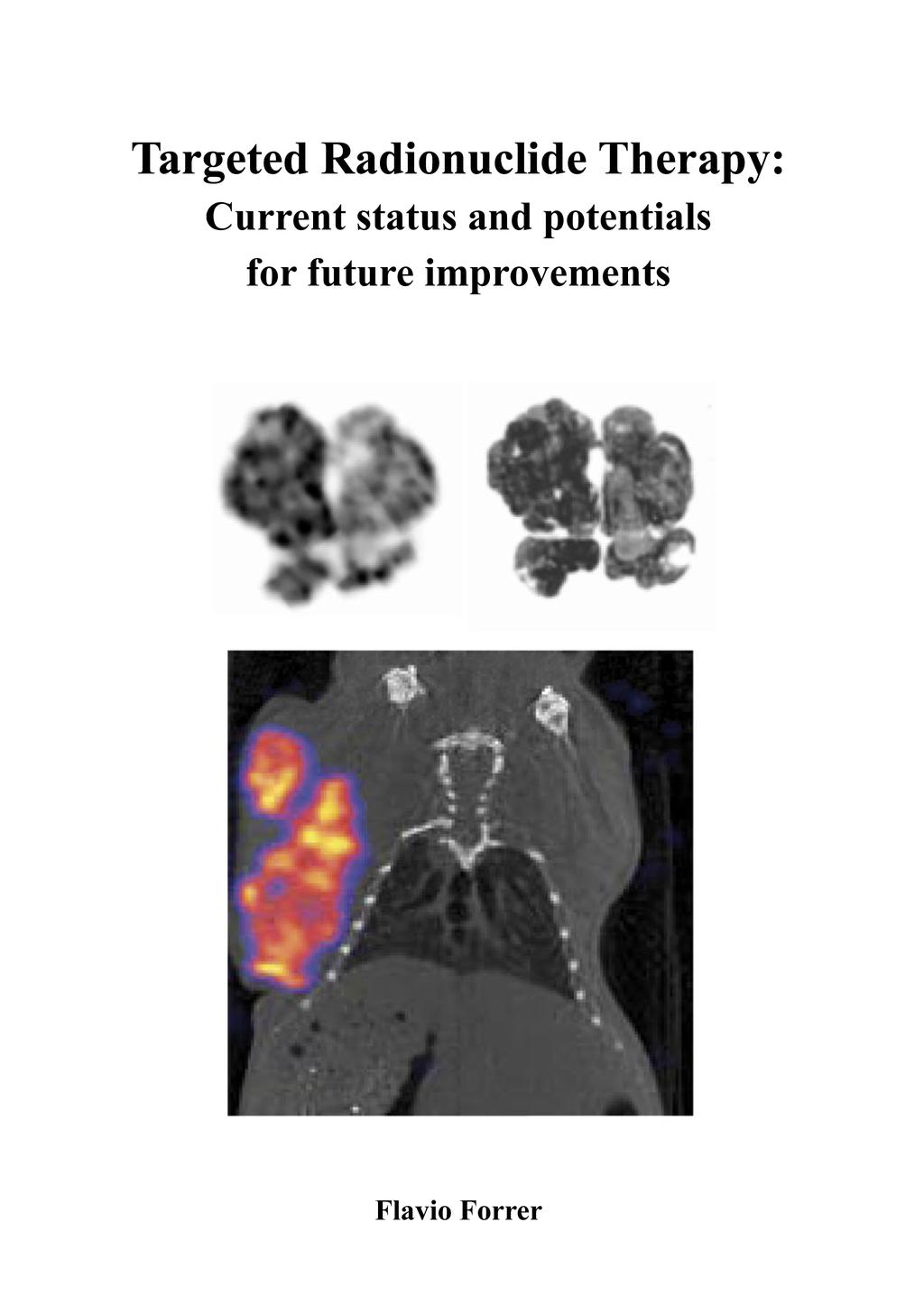
19 INTRODUCTION 17 Figure 1 1a 1b 1a 1b Planar scintigraphic scan of the abdomen 46 hours after the injection of 7.4 GBq 90 Y-DOTATOC and 111 MBq 111 In-DOTATOC in a patient with a neuroendocrine tumor of the pancreas with liver metastases. The scan was performed after the first treatment. Planar scintigraphic scan of the abdomen 46 hours after the injection of 7.4 GBq 90 Y-DOTATOC and 111 MBq 111 In-DOTATOC in the same patient shown in figure 1a. The scan was performed after the second treatment. Scintigraphically a clear reduction of the primary tumor and the liver metastases can be seen. Figure 2 2a 2b 2a Pretherapeutic CT-scan of the patient shown in figure 1. Multiple liver metastases can be seen. 2b CT-scan 3 month after the second treatment of the same patient. Liver metastases are not demonstrated anymore.
20 18 CHAPTER 1 Studies using [ 177 Lu-DOTA 0,Tyr 3 ]octreotate ( 177 Lu-DOTATATE) and [ 177 Lu- DOTA 0,Tyr 3 ]octreotide ( 177 Lu-DOTATOC) In 2003 the first results from a study using a 177 Lu labelled peptide were published [20]. In this study 35 patients with neuroendocrine GEP tumours were treated with escalating dosages of [ 177 Lu-DOTA 0,Tyr 3 ]octreotate ( 177 Lu-DOTATATE) up to a final cumulative dose of GBq. The effects of the therapy could be evaluated in 34 patients. Three months after the last treatment cycle 1 CR, 12 PR, 14 SD and 7 PD (including 3 patients who died during therapy) were found. This equals an objective response rate, i.e. PR or CR, of 38%. A later update of this study in 76 patients essentially confirmed these results [21]. In the follow-up the median time to progression in the patients that had at least SD after the treatment was not reached at 25 months from the beginning of the therapy. In a more recent evaluation of 131 GEP tumour patients these outcomes were confirmed again and a median time to progression of more than 36 months was found [22]. The latest evaluation of these patients with focus on long term outcome revealed an overall survival which appears to be even longer than the results published for 90 Y-DOTATOC (D.J. Kwekkeboom, personal communication 2006). However, influences resulting from different patient characteristics can currently not be ruled out. So far only one study was published using 177 Lu-DOTATOC [23]. One of the inclusion criteria was relapse after 90 Y-DOTATOC treatment and only one therapy cycle was administrated because of the pretreatment. Yet effectiveness of the treatment could be demonstrated, but because of the different setting the results of the outcome can not be compared. The detailed results are shown in chapter 2b. Side Effects and Toxicity Generally PRRT can be regarded as a relatively safe treatment and severe side effects are rare, especially when compared with side effects in studies using chemotherapy [46-48]. The side effects in PRRT can be divided into acute side-effects and more delayed effects caused by radiation toxicity. The acute effects occurring at the time of injection up to a few days after therapy include nausea, vomiting and increased pain at tumour sites (approximately 30% of the patients), symptoms that were reported after treatments with all radionuclides [5,11,14]. These side effects are generally mild, can be controlled by symptomatic treatment. In patients treated with 177 Lu-DOTATATE mild hair loss was reported [5], however hair growth had normalised at follow up 3 to 6 months after the treatment. Beside these minor side effects severe toxicity may occur as a result of the radiation absorbed dose in healthy organs. The organs at risk are mainly the kidneys, the bone marrow and to a lower extend the liver. Haematological toxicity Essentially all studies investigating PRRT report haematological toxicity. It appears that the absorbed radiation dose to the bone marrow is mainly caused by the circulation of the radioactivity in the blood [49], which limits the options to reduce the absorbed dose. Severe haematological toxicity (> grade 2 for haemoglobin, white blood cells and platelets) was reported in a maximum of 15% of the patients treated [11,13,14,17,21,41]. In general the decrease in blood counts was transient. Blood transfusions were needed only occasionally and
21 INTRODUCTION 19 patients recovered fully. More serious side effects were reported from a study where 3 out of 50 patients developed a myelodysplastic syndrome (MDS) after treatment with total cumulative doses higher than 100 GBq 111 In-octreotide [9]. In a dose escalating phase I study with 90 Y-DOTATOC to determine the maximum tolerated dose (MTD), one patient developed a MDS two years after PRRT [41]. In a recently updated record of roughly 500 patients treated with [ 177 Lu-DOTA 0,Tyr 3 ]octreotate, 3 patients developed a MDS (D.J. Kwekkeboom, personal communication) and a recent update of roughly 700 patients treated with 90 Y-DOTATOC showed that 2 patients developed a MDS (J. Mueller-Brand, personal communication). Two out of these 5 cases (one in each group) were most probable related to prior chemotherapy. Generally, the definition of the cause for these MDS cases is difficult because most of the patents that were included into PRRT trials are pretreated, many of them with chemotherapy and external beam radiation. Currently a maximum absorbed radiation dose to the bone marrow of 2Gy is assumed to be safe [20]. However, most studies lack long term follow up data which makes them ineffective to estimate long term risks. Haematological toxicity following PRRT is frequent but generally mild and transient. MDS may occur, but the limited data on long term follow up does not allow a reliable, precise estimation of the risk to date. Renal Toxicity Conjugated peptides are predominantly cleared by the kidneys. Although the major part of the radiopharmaceutical is excreted into the urine, partial reabsorption in the tubular cells can lead to a considerable radiation dose to the kidneys [25,49,50]. It was shown recently that the localization of the radiopeptide in the kidney is not homogeneous, but predominantly in the cortex where it follows a striped pattern, with most of the radioactivity centred in the inner cortical zone [51]. This pattern of up-take results in different dose distributions for different radionuclides [25]. The reabsorption of radiolablled somatostatin is mediated by the multiligand scavenger receptor megalin [52]. The high capacity of megalin challenges the reduction or blockade of renal reabsorption of the radiolabelled somatostatin analogues. However, it was proven that the co-administration of amino acids, especially arginine and lysine, significantly reduces the renal uptake of radiopeptides [53,54]. Gelatine based plasma expander were recently shown to reduce renal uptake of diagnostic [ 111 In-DTPA 0 ]octreotide efficiently in animals and patients [55,56]. However, the benefit in patients during PRRT remains to be proven. Another promising approach might be the use of amifostine [57,58]. Amifostine is the first drug investigated for PRRT that does not aim at reducing the renal uptake but which acts as a radical scavenger to reduce systemically the toxic effects of the radiation on normal tissue. Because amifostine is acting by a different mechanism, a combination with drugs that reduce the renal uptake appears most promising. Combinations of different drugs to reduce renal uptake are worth being tested as well. Preliminary results of a combination of gelatine based plasma expander and amino acids showed very promising results in rats (unpublished data). In a phase I study to define the MTD of 90 Y-DOTATOC that was performed without amino acids co-administration 2 out of 16 patients developed renal toxicity grade IV [13]. Renal biopsies of patients treated with 90 Y-DOTATOC that developed renal toxicity revealed mainly thrombotic microangiopathy and abnormalities in the tubules, histological changes comparable to the changes that occur after external beam radiation [59]. Despite the coadministration of amino acids a number of later studies using 90 Y-labelled peptides reported renal toxicity [60-63]. The MTD with amino acid co-infusion for 90 Y-DOTATOC was defined
22 20 CHAPTER 1 as 7.4 GBq/m 2 body surface in this study. Nevertheless a case of late onset renal toxicity after less than 7.4 GBq/m 2 was reported [60]. It was shown that individual dosimetry can be helpful to avoid kidney failure [62]. An absorbed dose of 23 Gy to the whole kidney is generally accepted to be safe. However, this value is derived from external beam radiation (fractions of 2 Gy) [64] and is therefore not indisputable. In contrast to external beam radiation the physical characteristics of the radiation in PRRT is different, applying radiation in a very low dose rate over a long period of several days. In contrast to the use of 90 Y labelled somatostatin analogues, no renal toxicity was reported after the therapeutic use of very high doses of [ 111 In-DTPA 0 ]octreotide [9]. For 177 Lu- DOTATATE one patient out of a group of 201 patients was reported who developed renal insufficiency [21]. This is an indication that the physical characteristics of the radionuclide have a significant impact on renal toxicity. While the Auger electrons emitted by 111 In have a range of approximately 5 μm in tissue, the maximum range of the 90 Y electrons can be up to 12 mm. Auger electrons emitted within the tubular cells do not reach the radiosensitive glomeruli [65]. Several studies investigated kidney toxicity after PRRT more detailed [62,65,66]. It was shown that together with the total absorbed dose to the kidney, the dose volume, fractionation rate and clinical parameters like hypertension, diabetes and age play an important role for the development of kidney failure. Especially the fractionation influences the specific biologic efficacy of internally deposited radiation strongly [66]. If renal toxicity occurs it can not be regarded as fixed kidney damage. It appears rather that the loss of function is a continuous process with a defined pace of progression that can be expressed as loss of clearance per year [65]. Liver Toxicity Beside the fact that most patients who are treated with PRRT suffer from liver metastases, physiological uptake in normal liver tissue also occurs after administration of radiolabelled somatostatin analogues. The sum of this physiological uptake and the dose to the normal liver from the specific uptake in liver metastases can result in a considerable radiation absorbed dose to the liver [49]. However, since the tumour load in the liver shows a high interpatient variability, it is difficult to generalise radiation absorbed doses to the liver. In a study using [ 111 In-DTPA 0 ]octreotide three out of 27 patients showed a temporary increase in liver enzymes corresponding to a grade 3 liver toxicity (WHO) [11]. All three patients had a liver tissue replacement of more than 75% of their hepatic parenchyma by metastases and treatment associated necrosis on the computed tomography scans was suggested. A significant increase in liver enzymes after the administration of 90 Y-DOTATOC was reported in two studies [41,67]. Valkema et al. reported one transient grade 3 toxicity in a group of 60 patients treated with 90 Y-DOTATOC in a phase I study [41]. In another study, 15 patients with known liver metastases (of whom 12 had extensive liver involvement, defined as 25% or more) from neuroendocrine tumours were treated with three cycles of 120 mci (4.4 GBq) each [67]. In four of these 15 patients, one or more of the three liver enzymes that were measured (serum aspartate aminotransferase, alanine aminotransferase and alkaline phosphatase) increased. Increase was defined as at least one grade, according to the WHO criteria, from baseline to final follow-up measurement (4-6 weeks post cycle 3). It was concluded that patients with diffuse sst-positive hepatic metastases could be treated with a
23 INTRODUCTION 21 cumulative administered activity of 360 mci (13.2 GBq) of 90 Y-DOTATOC with only a small chance of developing mild acute or subacute hepatic injury. In the group of patients treated with 177 Lu-DOTATATE, significantly increased liver function parameters (grade 4 liver toxicity) was evident in two patients after the first cycle of treatment (D.J. Kwekkeboom, personal communication, 2004). In summary, liver toxicity is very rare and if it occurs it is mostly mild and reversible. However, extensive liver metastases seem to be a risk factor for liver impairment after PRRT. Especially in these patients though, it is difficult to distinguish between real toxicity caused by radiation from effects by the metastases themselves. Dosimetry In order to improve the efficacy of PRRT and to limit toxicity, appropriate dosimetry helps to choose an injected activity that delivers an optimal radiation absorbed dose to the tumor while the dose to normal organs does not exceed defined limits. Additionally, dosimetry is mandatory to characterize a new compound properly in patients, especially when it is foreseen for therapy. The basic principles of dosimtery in Nuclear Medicine are explained in chapter 3a on the basis of a comparison in patients of the two most frequently used peptides for PRRT (DOTA-TOC and DOTAT-[Tyr 3 ]-octreotate. The dose limiting organs in PRRT are usually the kidneys and / or the bone marrow. Especially dosimetry of the bone marrow is very challenging since the bone marrow is not a solid organ. The definition of the volume and mass is associated with many sources of error. For radiolabeled somatostatin analogues, there are several models which are used to calculate the absorbed radiation dose to the bone marrow. Most often the residence time of the radiopharmaceutical in the bone marrow - a value that is mandatory for dosimetry - is calculated from the residence time in the blood. A correction factor is added depending on the vector used for treatment [49]. This method was validated by bone marrow aspirations for antibodies but not for radiopeptides [68,69]. Reliable dose estimation for the bone marrow is mandatory for several reasons. In order to achieve a maximum anti-tumor effect, patients should be treated with the highest justifiable dose of the radiopharmaceutical that does not cause serious toxicity. Many studies with radiolabeled somatostatin analogues showed that the toxicity is generally mild and transient [13,15,20,22]. It should however not be neglected that in a phase 1 study with [ 111 In- DTPA 0 ]octreotide 3 out of 50 patient developed a myelodysplastic syndrome (MDS) which was probably related to the therapy [9]. Calculations from these data resulted in an estimated radiation absorbed dose for the bone marrow of approximately 3 Gy. In another study with [ 177 Lu-DOTA 0,Tyr 3 ]octreotate, one MDS was observed in a patient who had had chemotherapy with alkylating agents 2 years before study entry [21]. In the latest update of our own records of roughly 500 patients treated with [ 177 Lu-DOTA 0,Tyr 3 ]octreotate, 3 patients (including the patient mentioned before) developed a MDS (unpublished data). To avoid MDS, a maximum absorbed dose of 2 Gy to the bone marrow is generally accepted [20]. Nevertheless even if this limit is not exceeded the risk for the patient to develop a MDS can not be excluded completely, but an accurate estimation of the absorbed dose to the bone marrow will help to find an adequate dosage.
24 22 CHAPTER 1 For daily practise the method to estimate the absorbed dose to the bone marrow has to be easily applicable and should not cause a lot of discomfort to the patient. This is given with the method to calculate the residence time in the bone marrow from the blood. However, taking a bone marrow sample is probably the most reliable method for bone marrow dosimetry. A detailed comparison of different methods to calculate the absorbed radiation dose to the bone marrow compared with a bone marrow aspiration is made in chapter 3b. Current Clinical Practice In symptomatic patients at the time of diagnosis metastatic disease is present in 90% and surgical cure is not possible [70]. nevertheless it remains an important cornerstone in the management of these tumours. Beside surgery, radiofrequency ablation (RFA) or chemoembolisation is a minimal invasive treatment option when the disease is limited to the liver or when the tumour load in the liver is very high. Several small series have shown good responses [71-73]. However, RFA and chemo-embolisation are not systemic approaches and will not treat extrahepatic (occult) disease. In patients with metastasised neuroendocrine tumours in whom surgery is no longer an option, PRRT appears to be the most effective therapeutic option with limited side-effects. Conventional chemotherapy and external radiotherapy either alone or in a variety of permutations are of minimal efficacy and should be balanced against the decrease in quality of life often caused by such agents [73]. Non-radiolabelled somatostatin analogues, particularly in a subcutaneous depot formulation are effective in symptom alleviation and improvement of quality of life but their effect on tumour burden is very limited [73]. A randomised controlled study of PRRT with other treatment modalities is lacking though. The wait-and-see approach often still remains the mainstay of initial management in patients with unresectable disease. The rationale for this approach is found in the natural course of well-differentiated GEP tumours. Tumours can be indolent for many years and the well-being of patients, even with metastasised tumours, can be unchanged for a long period. However, the reported studies on PRRT clearly indicate that patients with documented progressive disease or a substantial increase in symptoms benefit in a high percentage from this therapy. The recognition of the possible benefit of PRRT for patients with GEP tumours is increasing, but its implementation within the whole therapeutic array is rather poor. The fact that PRRT is a relatively new therapeutic modality may be one of the contributing factors. Another factor is the lack of approved radiopharmaceuticals. Several reasons account for this: beside increased governmental demands, and therapy-related costs, it has to be kept in mind that neuroendocrine tumours are rather rare which limits the interest of the pharmaceutical industry to invest specifically into these tumours. Indications for Peptide Receptor Radionuclide Therapy The approach of PRRT with radiolabelled somatostain analogues allows theoretically treating all sst-positive tumours. However, due to the potential morbidity of PRRT patients should be selected carefully. Incurability by surgery is an absolute prerequisite for the inclusion of a patient. In addition, the presence of a sufficient high density of sst has to be proven by means of scintigraphy. Usually this will be an 111 In-octreotide scintigraphy with sufficient tumour uptake. Recently PET with radiolabelled somatostatin analogues became available and might be used alternatively However, these PET methods are available only at a few centres, the radiopharmaceuticals are not FDA approved yet and the methods have yet to be formally validated. Inclusion criteria for most studies were tumour uptake equal or higher than liver
25 INTRODUCTION 23 uptake on the 111 In-octreotide scintigraphy [74]. High uptake on 111 In-octreotide scintigraphy has been shown to correlate with tumour regression after PRRT [20]. Because of the potential, renal and haematological toxicity of PRRT patients need to fulfil certain minimal criteria in addition. Blood and kidney function parameters have to be checked before therapy. Details are given in table 3. Bone metastases, present only in a minority of the patients, are not an exclusion criteria. However it seems that bone and cystic lesions respond in a more protracted way than the common solid liver metastases although no formal analysis to this end are available. Table 3 Criteria for peptide receptor radionuclide therapy in patients with neuroendocrine tumours Inclusion Sufficient tumour uptake on (tumour uptake liver uptake) 111 In-octreotide scintigrams Haematology: Haemoglobin 5.0 mmol/l White blood cell count /l Platelet count /l Kidney function: Creatinin (serum) 150 μmol/l or creatinine clearance 40 ml/min Karnofsky Performance Status 50 Life expectancy > 3 months Written informed consent Exclusion Chemotherapy within 6 weeks prior to the start of treatment Pregnancy/lactation Distinct restricted liver function Timing of Therapy The best time point to initiate PRRT in patients with malignant neuroendocrine tumours remains uncertain up to now. The stage of disease at the time of diagnosis is highly variable. It ranges from a small localised primary tumour to advanced or even end-stage disease with limited liver function and ascites. In addition, the variation in tumour-differentiation results in
26 24 CHAPTER 1 highly variable rates of progression. In a study in which the relationship between delay of diagnosis, extent of disease and survival in 115 patients with carcinoid was studied, a mean delay in the diagnosis of 66 months was found [75]. It was concluded that the diagnosis of carcinoid is difficult, and therefore a delay of diagnosis by physicians is common. Strikingly, the delay of the diagnosis did not correlate with the extent of the disease. However, the extent of the disease did correlate with survival. Patients with primary tumours and lymph node metastases were less likely to die of carcinoid disease than patients with hepatic metastases, carcinomatosis or extra-abdominal metastases. Although there are no guidelines yet for the initiation of PRRT there are certain hints that the treatment is more effective when given in an earlier stage. The degree of liver involvement is inversely related to the chance of remission [22]. In a trial with [ 111 In-DTPA 0 ]octreotide it was reported that a beneficial effect of PRRT is less likely in end-stage patients than in patients with less tumour burden and in better general conditions [9,45]. Furthermore, it was clearly shown that patients benefit in quality of life after PRRT [15,76]. This justifies treating all symptomatic patients that fulfil the inclusion criteria and are not responding to treatment with non-radiolabelled somatostatin analogues (anymore). Another argument for an earlier treatment is the fact that neuroendocrine tumour can dedifferentiate over time. Dedifferentiation is commonly associated with a decrease in sst density. In turn PRRT using radiolabelled somatostatin analogues will be less effective or even impossible. The administration of PRRT in an early stage does not exclude patients from a later repetition of the treatment. It was shown recently that patients can be retreated. A good response after the first treatment cycles was found to be a positive predictor for the effectiveness of the retreatment [23]. A randomised study comparing the long term survival of patients with malignant, unresectable neuroendocrine tumours that undergo PRRT compared to a wait and see strategy is lacking. Keeping in mind the latest follow up data of patients treated with 177 Lu- DOTATATE (median time to progression > 36 months) [20] makes however such a study disputable from an ethical point of view. Future Developments Future research to improve PRRT with radiolabelled somatostatin analogues consists of 5 main directions. Improving the vehicle, i.e. the peptide, is highly interesting. Many new somatostatin analogues with a higher affinity for sst 2 or with a wider affinity for several sst subtypes were already introduced into the preclinic [77]. Simultaneously investigations have been made to improve the delivery of the radiopharmaceutical to the target. This includes the way of application, e.g. intra-arterially, at a slower rate or fractionated, as well as the improvement of the availability of the target by different peptide concentrations or by modulation of the receptor with drugs [78,79]. Furthermore the most suitable radionuclide will have to be defined. In preclinical studies, comparing 90 Y and 177 Lu it appeared that 90 Y was more effective for bigger tumours while with 177 Lu less relapses occurred when treating smaller lesions [42,80]. Beside the effects on the tumour, the different physical properties cause differences in microdosimetry which in turn will influence the toxicity profile of a compound [25]. Beside the commonly used 90 Y
27 INTRODUCTION 25 and 177 Lu a number of other radionuclides with different physical characteristics including alfa-emitters are under investigation [81,82]. In external beam radiation the application of radio-sensitizers to improve the anti-tumour effect of the radiation is established [83]. In PRRT, the introduction of combination therapies will open a whole new field of research to improve the treatment. A multi-centre trial with 177 Lu-DOTATATE and Capecitabine was initiated recently (E.P. Krenning, personal communication, 2006). Finally, the improvement of the toxicity-profile of the current radiopeptides is an important issue. Especially with respect to the reduction of the kidney uptake, several studies were recently published and also other strategies to reduce radiation toxicity in general are under investigation [55-58]. An important step towards an improved toxicity profile will be a better understanding of the low dose rate irradiation. Currently, the generally accepted maximum tolerated absorbed doses to normal organs are still derived from external beam radiation [64]. There is emerging evidence in the literature of a low dose hypersensitivity phenomenon where at low doses and at very low dose rates, a significantly increased cell kill is found compared with high dose rates and compared with what would be predicted from the classical linear quadratic model [84,85]. Other Peptides for new Peptide Receptor Radionuclide Therapies Beside radiolabelled somatostatin analogues a number of newly developed peptides were introduced lately, targeting different receptors [86]. Bombesin is just one example of these new peptides. Bombesin is a well characterized 14 amino acid neuropeptide binding (among others) to the gastrin-releasing-peptide (GRP) receptor. Several bombesin derivatives with high affinity for the GRP receptor have been developed, analogous to somatostatin, labelled with an array of radionuclides [87]. Overexpression of GRP receptors was found on many neoplasms, especially on prostate and breast cancer. Remarkably the GRP receptor is not expressed on healthy prostate tissue. This might give the chance to distinguish in vivo between benign and malignant prostate nodules by means of receptor imaging and in a second step to apply PRRT to GRP receptor-positive tumours. Gastrin analogues have a high affinity for the Cholecystokinin (CCK) B-receptor which is overexpressed e.g. on medullary thyroid carcinomas. To date eight patients with advanced metastatic disease were injected in a dose-escalation study with potentially therapeutic activities of a 90 Y-labelled minigastrin derivative at 4-6-weekly intervals with GBq/m 2 per injection for a maximum of four injections. Hematologic and renal were identified as the dose-limiting toxicities. Two patients experienced partial remissions, 4 stabilization of their previously rapidly progressing disease [88]. Clinical Summary PRRT has been proven to be an effective and safe treatment alternative for sst-positive, unresectable neuroendocrine tumours. Currently the maximum tolerated dose is defined by the dose to the critical organs, kidney and bone marrow. It is likely that the dose can be increased in future by the introduction of new protective agents, different treatment schemes and radionuclides.
28 26 CHAPTER 1 The present data in the literature do not allow defining the most suitable peptide and radionuclide for the treatment of neuroendocrine tumours. Especially concerning the radionuclide there is emerging evidence that a combination of nuclides with different physical characteristics might be more effective. The principle of targeted treatment has a number of obvious advantages over unspecific systemic treatments. Therefore PRRT holds great promise for the future. Preclinical Studies As mentioned previously, new approaches could potentially improve PRRT further but a number of questions still need to be answered. Although results from preclinical studies can not always be translated easily to the clinics, preclinical evaluation and test of new compounds or new strategies will remain a corner stone to improve PRRT. Before new drugs or treatment strategies are evaluated in patients, they are usually tested first in preclinical studies. Animal experiments are of high relevance in this stage of the development. To date mainly biodistribution studies were performed. This is associated with a large number of animals that are needed to investigate different processes and function at different time points. Over the last years dedicated small animal imaging devices gained increasing influence on preclinical research. Particularly single photon emission computed tomography (SPECT) and positron emission tomography (PET) as tools for molecular imaging were proven to be valuable e.g. to follow physiological processes in an animal over time. Small animal SPECT/CT For our research we had a dedicated small animal SPECT/CT at our disposal [89]. The camera was a four headed multiplexing multi-pinhole camera. Each head is fitted with an application-specific tungsten collimator with nine pinholes. The rat apertures, e.g., comprise a total of 36 2-mm-diameter pinholes imaging a cylindrical field of view that is 60 mm in diameter by 24 mm in length. These rat apertures provide a reconstructed resolution below 1.6 mm at 140 kev, with an average sensitivity of 1,100 cps/mbq across the field of view (FOV). The axial FOV is extended using a step-and-shoot helical scan of the animal, with the user defining a range from 24 to 270 mm according to the region to be imaged. Accordingly the mouse high resolution apertures comprise of 1-mm-diameter pinholes resulting in a resolution in the sub millimeter range.

29 INTRODUCTION 27 Figure 3 Whole body SPECT/CT of a PC3- tumor-bearing mouse 24 hours after the injection of 50 MBq 111 In-Bombesin.
30 28 CHAPTER 1 After the acquisition, the data are reconstructed iteratively with the HiSPECT (Bioscan Inc., Washington D.C., USA) software, a dedicated ordered subsets-expectation maximization (OSEM) software package for multiplexing multi-pinhole reconstruction. The camera is calibrated with a phantom, approximately of the size of the animals, filled with a known activity of 99m Tc such that voxel values in the reconstruction provide a proper estimate of the activity level without further calculation. Regions of interest (ROI) can be drawn manually around the object of interest; the 3D activity distribution within the ROI is then summed to determine the radioactivity. No correction for scatter or attenuation is performed because the quantification factor also corrects for attenuation within the animal. Quantification is performed with the INTERVIEW XP (Mediso Ltd., Budapest, Hungary) software. The absolute in vivo quantification is probably the most important tool to evaluate new tracers. The detailed evaluation of the accuracy of this function is described in chapter 4a. In various studies the capabilities of the system were evaluated (unpublished data). Depending on the injected activity and the imaging time a very high resolution for static images can be achieved. An example of a standard SPECT/CT of a tumor bearing mouse is shown in Figure 3. Using long scanning times and high resolution apertures allow even to resolve inhomogeneities of tracer uptake within the tumor caused by inhomogeneous distribution of receptors. These images can be correlated very well with ex vivo autoradiograms. An example is shown in figure 4. Figure 4 Correlation between ex vivo autoradiograms and in vivo SPECT slices of a somatostatin receptor positive rat pancreatic tumor (CA20948) after the injection 111 In-Octreoscan. Beside this high resolution that is provided by pinhole SPECT, the multi-pinhole technique results in sensitivity comparable to parallel-hole collimators. On the one hand this allows injecting relatively small amounts of radioactivity and on the other hand it allows keeping the acquisition times or the time per projection respectively low. In turn this gives a very good temporal resolution. We investigated dynamically rat kidney function with 99m Tc-MAG3 and tumor uptake of 111 In-Octreoscan in a somatostatin receptor positive tumor in a rat. The time per scan could be kept as low as 60 seconds per scan. Up to 45 scans were acquired per study. The resulting time activity curves as well as an example of subsequent MIP images of a 99m Tc-MAG3 study are shown in figure 3.
31 INTRODUCTION 29 Figure 5 5a 5b 5c Activity [MBq] t [s] left kidney right kidney 5a. Dynamic in vivo SPECT images (MIP data) of rat kidneys after the injection of 40 MBq 99m Tc-MAG3. 5b. In vivo renograms of rat kidneys generated with the NanoSPECT/CT after the injection of 40 MBq 99m Tc-MAG3. Activity in the tumor [MBq] c. Dynamic measurement of 111 In-Octreoscan tumor uptake in a somatostatin receptor positive rat pancreatic tumor (CA20948). The data are generated with the NanoSPECT/CT in vivo Time [min] The variety of tracers available allows to look at different physiological functions in one animal or to follow a process over time. In a therapy study with tumor bearing rats we were able to monitor the animals over time. Five days after injection the biodistribution of the therapeutic [ 177 Lu-DOTA 0,Tyr 3 ]-octreotide was documented. Approximately 4 weeks after the treatment the response to the therapy was assessed by an 111 In-Octreoscan scintigraphy and finally before sacrificing the animals the kidney function was monitored with 99m Tc- DMSA. An example of these three scans in one animal is shown in figure 6.

32 30 CHAPTER 1 Figure 6 SPECT images of the identical rat with different tracers at different time points as indicated. In a first study, taking advantage of the NanoSPECT/CT we investigated mechanism of kidney damage in rats during high dose PRRT [90]. Besides small animal imaging a number of other molecular imaging methods along with histology using different staining were applied. The results of this study are shown in chapter 4b. In conclusion we believe that small animal imaging will strongly influence further preclinical research. It allows to follow processes over time or to monitor different functions in a single animal at the same time which will reduces the number of animals required and in turn it will save costs as well. Additionally, the situation as it is given in a patient is reflected better when different functions can be monitored in one animal simultaneously and as true in vivo investigation.
33 INTRODUCTION 31 References 1. Reubi JC. Peptide receptors as molecular targets for cancer diagnosis and therapy. Endocr Rev. 2003;24(4): Reubi JC, Waser B, Schaer JC, Laissue JA. Somatostatin receptor sst1-sst5 expression in normal and neoplastic human tissues using receptor autoradiography with subtypeselective ligands. Eur J Nucl Med. 2001;28(7): Reubi JC, Waser B, Liu Q et al. Subcellular distribution of somatostatin sst2a receptors in human tumors of the nervous and neuroendocrine systems: membranous versus intracellular location. J Clin Endocrinol Metab. 2000;85: Krenning EP, Kwekkeboom DJ, Bakker WH et al. Somatostatin receptor scintigraphy with [ 111 In-DTPA-d-Phe1]- and [ 123 I-Tyr 3 ]-octreotide: the Rotterdam experience with more than 1000 patients, Eur J Nucl Med 1993;20: Kwekkeboom DJ, Krenning EP and de Jong M. Peptide receptor imaging and therapy. J Nucl Med 2000;41: Hofmann M, Maecke H, Borner R et al. Biokinetics and imaging with the somatostatin receptor PET radioligand (68)Ga-DOTATOC: preliminary data. Eur J Nucl Med. 2001;28: Maecke HR, Hofmann M & Haberkorn U. (68)Ga-labeled peptides in tumor imaging. J Nucl Med. 2005;46 Suppl 1: 172S-178S. 8. Wester HJ, Schottelius M, Scheidhauer K et al. PET imaging of somatostatin receptors: design, synthesis and preclinical evaluation of a novel 18F-labelled, carbohydrated analogue of octreotide. Eur J Nucl Med Mol Imaging. 2003;30: Valkema R, De Jong M, Bakker WH et al. Phase I study of peptide receptor radionuclide therapy with [In-DTPA]octreotide: the Rotterdam experience. Semin Nucl Med 2002;32: McCarthy KE, Woltering EA and Anthony LB. In situ radiotherapy with 111 Inpentetreotid. State of the art and perspectives. Q J Nucl Med 2000;44: Anthony LB, Woltering EA, Espenan GD et al. Indium-111-pentetreotide prolongs survival in gastroenteropancreatic malignancies. Semin Nucl Med 2002;32: Buscombe JR, Caplin ME and Hilson AJ, Long-term efficacy of high-activity 111 Inpentetreotide therapy in patients with disseminated neuroendocrine tumors, J Nucl Med 2003;44: Otte A, Herrmann R, Heppeler A. et al. Yttrium-90 DOTATOC: first clinical results, Eur J Nucl Med. 1999;26: Waldherr C, Pless M, Maecke HR et al. The clinical value of [ 90 Y-DOTA]-dPhe 1 -Tyr 3 - octreotide ( 90 Y-DOTATOC) in the treatment of neuroendocrine tumours: a clinical phase II study. Ann Oncol. 2001;12: Waldherr C, Pless M. Maecke HR et al. Tumor response and clinical benefit in neuroendocrine tumors after 7.4 GBq (90)Y-DOTATOC, J Nucl Med. 2002;43: Forrer F, Waldherr C, Maecke HR, Mueller-Brand J. Targeted radionuclide therapy with 90 Y-DOTATOC in patients with neuroendocrine tumors. Anticancer Res. 2006;26: Bodei L, Cremonesi M, Zoboli S et al. Receptor-mediated radionuclide therapy with 90 Y-DOTATOC in association with amino acid infusion: a phase I study. Eur J Nucl Med Mol Imaging. 2003;30: Virgolini I, Britton K, Buscombe J et al., In- and Y-DOTA-lanreotide: results and implications of the MAURITIUS trial, Semin Nucl Med. 2002;32:
34 32 CHAPTER Baum RP, Söldner J, Schmüching M, and A. Niesen. Peptidrezeptorvermittelte Radiotherapie (PRRT) neuroendokriner Tumoren Klinischen Indikationen und Erfahrung mit 90 Yttrium-markierten Somatostatinanaloga, Der Onkologe 2004;10: Kwekkeboom DJ, Bakker WH, Kam BL et al. Treatment of patients with gastro-enteropancreatic (GEP) tumours with the novel radiolabelled somatostatin analogue [(177)Lu- DOTA(0),Tyr(3)]octreotate. Eur J Nucl Med Mol Imaging 2003;30: Kwekkeboom DJ, Bakker WH, Teunissen JJ et al. Treatment with Lu-177-DOTA-Tyr 3 - octreotate in patients with neuroendocrine tumors: interim results, Eur J Nucl Med Mol Imaging 2003;30: (supplement 2), p. S231 (abstract). 22. Kwekkeboom DJ, Teunissen JJ, Bakker WH et al. Radiolabelled somatostatin analog [ 177 Lu-DOTA O, Tyr 3 ]octreotate in patients with endocrine gastro entero pancreatic tumors. J Clin Oncol 2005; 23: Forrer F, Uusijarvi H, Storch D et al. Treatment with 177 Lu-DOTATOC of patients with relapse of neuroendocrine tumors after treatment with 90 Y-DOTATOC. J Nucl Med. 2005;46: Krenning EP, Kooij PP, Bakker WH et al., Radiotherapy with a radiolabeled somatostatin analogue, [ 111 In-DTPA-d-Phe 1 ]-octreotide. A case history, Ann N Y Acad Sci 1994;733: Konijnenberg MW, Bijster M, Krenning EP, and De Jong M. A stylized computational model of the rat for organ dosimetry in support of preclinical evaluations of peptide receptor radionuclide therapy with (90)Y, (111)In, or (177)Lu. J Nucl Med. 2004;45: Scarpignato C, Pelosini I. Somatostatin analogs for cancer treatment and diagnosis: an overview. Chemotherapy. 2001;47 Suppl 2: Lamberts SW, van der Lely AJ, de Herder WW, and Hofland LJ. Octreotide. N Engl J Med. 1996;334: Reubi JC, Schaer JC, Laissue JA and Waser B. Somatostatin receptors and their subtypes in human tumors and in peritumoral vessels. Metabolism. 1996;45(Suppl 1): Reubi JC, Schar JC, Waser B, et al. Affinity profiles for human somatostatin receptor subtypes SST1-SST5 of somatostatin radiotracers selected for scintigraphic and radiotherapeutic use. Eur J Nucl Med 2000;27: Bruno JF, and Berelowitz M. Somatostatin receptors: orphan that found family and function, Mol Cell Neurosci 1993;4: Abstract 31. Yamada Y, Kagimoto S, Kubota A, et al. Cloning, functional expression and pharmacological characterization of a fourth (hsstr4) and a fifth (hsstr5) human somatostatin receptor subtype. Biochem Biophys Res Commun 1993;195: Oberg K and Eriksson B. Endocrine tumours of the pancreas. Best Pract Res Clin Gastroenterol. 2005;19: Slooter GD, Breeman WA, Marquet RL, et al. Anti-proliferative effect of radiolabelled octreotide in a metastases model in rat liver. Int J Cancer. 1999;81: Mardirossian G, Wu C, Hnatowich DJ. The stability in liver homogenates of indium-111 and yttrium-90 attached to antibody via two popular chelators. Nucl Med Biol. 1993;20: Liu S. The role of coordination chemistry in the development of target-specific radiopharmaceuticals. Chem Soc Rev. 2004;33: Otte A, Jermann E, Behe M et al. DOTATOC: a powerful new tool for receptormediated radionuclide therapy. Eur J Nucl Med. 1997;24:
35 INTRODUCTION Chinol M, Bodei L, Cremonesi M and Paganelli G. Receptor-mediated radiotherapy with Y-DOTA-DPhe-Tyr-octreotide: the experience of the European Institute of Oncology Group. Semin Nucl Med 2002;32: Paganelli G, Bodei L, Handkiewicz Junak D et al. 90 Y-DOTA-d-Phe 1 -Try 3 -octreotide in therapy of neuroendocrine malignancies. Biopolymers 2002;66: Paganelli G, Zoboli S, Cremonesi M et al. Receptor-mediated radiotherapy with 90 Y- DOTA-d-Phe 1 -Tyr 3 -octreotide. Eur J Nucl Med 2001;28: Bodei L, Cremonesi M, Grana C et al. Receptor radionuclide therapy with (90)Y- [DOTA](0)-Tyr(3)-octreotide ((90)Y-DOTATOC) in neuroendocrine tumours. Eur J Nucl Med Mol Imaging 2004;31: Valkema R, Pauwels S, Kvols L et al. Long-term follow-up of a phase 1 study of peptide receptor radionuclide therapy (PRRT) with ( 90 Y-DOTA 0,Tyr 3 )octreotide in patients with somatostatin receptor positive tumours, Eur J Nucl Med 2003;30 (supplement 2): 232p. (Abstract). 42. de Jong M, Valkema R, Jamar F et al. Somatostatin receptor-targeted radionuclide therapy of tumors: preclinical and clinical findings. Semin Nucl Med 2002:32; Smith MC, Liu J, Chen T et al. OctreoTher: ongoing early clinical development of a somatostatin-receptor-targeted radionuclide antineoplastic therapy. Digestion 2000;62 (supplement 1): Baum RP, Soldner J, Schmucking M and Niesen A. Intravenous and intra-arterial peptide receptor radionuclide therapy (PRRT) using Y-90-DOTA-Tyr3-octreotate (Y- 90-DOTA-TATE) in patients with metastatic neuroendocrine tumors. Eur J Nucl Med 2004;31 (supplement 2): S238p (abstract). 45. Valkema R, Pauwels S, Kvols LK, et al. Survival and response after peptide receptor radionuclide therapy with [90Y-DOTA0,Tyr3]octreotide in patients with advanced gastroenteropancreatic neuroendocrine tumors. Semin Nucl Med. 2006;36: Oberg K,Norheim I,Lundqvist G et al :Cytotoxic treatment in patients with malignant carcinoid tumors.response to streptozocin alone or in combination with 5--FU.Acta Oncol 1987;26 : Engstrom PF,Lavin PT and Moertel CG:Streptozocin plus fluorouracil versus doxorubicin therapy for metastatic carcinoid tumor.j Clin Oncol. 1984;2: Moertel CG and Hanley JA:Combination chemotherapy trials in metastatic crcinoid tumor and the malignant carcinoid syndrome. Cancer Clin Trials. 1979;2: Cremonesi M, Ferrari M, Zoboli S, et al. Biokinetics and dosimetry in patients administered with (111)In-DOTA-Tyr(3)-octreotide: implications for internal radiotherapy with (90)Y-DOTATOC. Eur J Nucl Med 1999;26: Forrer F, Uusijarvi H, Waldherr C, et al. A comparison of 111 In-DOTATOC and 111 In- DOTATATE: biodistribution and dosimetry in the same patients with metastatic neuroendocrine tumours. Eur J Nucl Med Mol Imaging. 2004;31: De Jong M, Valkema R, Van Gameren A, et al. Inhomogeneous localization of radioactivity in the human kidney after injection of [(111)In-DTPA]octreotide.J Nucl Med. 2004;45: de Jong M, Barone R, Krenning E,et al. Megalin is essential for renal proximal tubule reabsorption of (111)In-DTPA-octreotide. J Nucl Med. 2005;46: Behr TM, Sharkey RM, Sgouros G, et al. Overcoming the nephrotoxicity of radiometallabeled immunoconjugates: improved cancer therapy administered to a nude mouse model in relation to the internal radiation dosimetry. Cancer. 1997;80(12 Suppl):
36 34 CHAPTER Rolleman EJ, Valkema R, de Jong M, et al. Safe and effective inhibition of renal uptake of radiolabelled octreotide by a combination of lysine and arginine, Eur J Nucl Med Mol Imaging 2003;30: van Eerd JE, Vegt E, Wetzels JF, et al. Gelatin-based plasma expander effectively reduces renal uptake of 111 In-octreotide in mice and rats. J Nucl Med. 2006;47: Vegt E, Wetzels JF, Russel FG, et al. Renal uptake of radiolabeled octreotide in human subjects is efficiently inhibited by succinylated gelatin. J Nucl Med. 2006;47: Forrer F, Rolleman E, Valkema R, Bernard B, Melis M, Bijster M, Krenning E, de Jong M. Amifostine is most promising in protecting renal function during radionuclide therapy with [Lu-177-DOTA 0,Tyr 3 ]octreotate. J Nucl Med. 2006; 47 (Supplement 1):43P (Abstract). 58. Rolleman EJ, Forrer F Bernard B, Bijster M, Vermeij M, Valkema R, Krenning EP, de Jong M. Amifostine protects rat kidneys in peptide receptor radionuclide therapy with [ 177 Lu-DOTA 0,Tyr 3 ]octreotate. Eur J Nucl Med Mol Imaging 2006 submitted 59. Moll S, Nickeleit V, Mueller-Brand J et al. A new cause of renal thrombotic microangiopathy: yttrium 90-DOTATOC internal radiotherapy. Am J Kidney Dis. 2001;37: Cybulla M, Weiner SM, and Otte A. End-stage renal disease after treatment with 90Y- DOTATOC. Eur J Nucl Med 2001;28: Stoffel MP, Pollok M, Fries J, and Baldamus CA. Radiation nephropathy after radiotherapy in metastatic medullary thyroid carcinoma. Nephrol Dial Transplant 2001;16: Barone R, Borson-Chazot F, Valkema R, et al. Patient-specific dosimetry in predicting renal toxicity with 90 Y-DOTATOC: relevance of kidney volume and dose rate in finding a dose effect relationship. J Nucl Med 2005;46: 99S 106S. 63. Otte A, Mueller-Brand J, Dellas S, et al. Yttrium-90-labelled somatostatin-analogue for cancer treatment, Lancet. 1998;351: Emami B, Lyman J, Brown A, et al. Tolerance of normal tissue to therapeutic irradiation. Int J Radiat Oncol Biol Phys. 1991;21: Valkema R, Pauwels SA, Kvols LK et al. Long-term follow-up of renal function after peptide receptor radiation therapy with 90 Y-DOTA 0,Tyr 3 -octreotide and 177 Lu-DOTA 0, Tyr 3 -octreotate, J Nucl Med 2005;46 Suppl 1: 83S 91S. 66. Pauwels S, Barone R, Walrand S et al. Practical dosimetry of peptide receptor radionuclide therapy with (90)Y-labeled somatostatin analogs. J Nucl Med. 2005;46 Suppl 1:92S-8S. 67. Bushnell D, Menda Y, Madsen M, et al. Assessment of hepatic toxicity from treatment with 90Y-SMT 487 (OctreoTher(TM)) in patients with diffuse somatostatin receptor positive liver metastases. Cancer Biother Radiopharm ;18: Siegel JA, Wessels BW, Watson EE, et al. Bone marrow dosimetry and toxicity for radioimmunotherapy. Antibody Immunoconjugates and Radiopharm 1990;3: Sgouros G. Bone marrow dosimetry for radioimmunotherapy: theoretical considerations. J Nucl Med 1993;34: Raut C, Kulke M, Glickman J, et al. Carcinoid tumors. Curr Probl Surg. 2006;43: Berber E, Flesher N, and Siperstein AE. Laparoscopic radiofrequency ablation of neuroendocrine liver metastases. World J Surg. 2002;26: Hellman P, Ladjevardi S, Skogseid B, et al. Radiofrequency tissue ablation using cooled tip for liver metastases of endocrine tumors. World J Surg. 2002;26: Modlin IM, Latich I, Kidd M, et al. Therapeutic options for gastrointestinal carcinoids. Clin Gastroenterol Hepatol. 2006;4:
37 INTRODUCTION Krenning EP, de Jong M, Kooij PP, et al. Radiolabelled somatostatin analogue(s) for peptide receptor scintigraphy and radionuclide therapy. Ann Oncol. 1999;10 Suppl 2: S Toth-Fejel S and Pommier RF. Relationships among delay of diagnosis, extent of disease, and survival in patients with abdominal carcinoid tumors. Am J Surg. 2004;187: Teunissen JJ, Kwekkeboom DJ and Krenning EP. Quality of life in patients with gastroenteropancreatic tumors treated with [ 177 Lu-DOTA 0,Tyr 3 ]octreotate. J Clin Oncol. 2004;22: Ginj M, Chen J, Walter MA, et al. Preclinical evaluation of new and highly potent analogues of octreotide for predictive imaging and targeted radiotherapy. Clin Cancer Res. 2005;11: Breeman WA, De Jong M, Visser TJ, et al. Optimising conditions for radiolabelling of DOTA-peptides with 90 Y, 111 In and 177 Lu at high specific activities. Eur J Nucl Med Mol Imaging. 2003;30: Froidevaux S, Hintermann E, Torok M, et al. Differential regulation of somatostatin receptor type 2 (sst 2) expression in AR4-2J tumor cells implanted into mice during octreotide treatment. Cancer Res ;59 : de Jong M, Breeman WAP, Valkema R, et al. Combination Radionuclide Therapy Using 177 Lu- and 90 Y-Labeled Somatostatin Analogs. J Nucl Med 2005;46 Suppl 1: 13S-17S. 81. Uusijarvi H, Bernhardt P, Rosch F, et al. Electron- and positron-emitting radiolanthanides for therapy: aspects of dosimetry and production. J Nucl Med. 2006;47: Norenberg JP, Krenning BJ, Konings IR, et al. 213 Bi-[DOTA 0, Tyr 3 ]octreotide peptide receptor radionuclide therapy of pancreatic tumors in a preclinical animal model. Clin Cancer Res. 2006;12: van Putten JW, Price A, van der Leest AH, et al. A phase I study of gemcitabine with concurrent radiotherapy in stage III, locally advanced non-small cell lung cancer. Clin Cancer Res. 2003;9: Joiner MC, Marples B, Lambin P et al. Low-dose hypersensitivity: current status and possible mechanisms. Int J Radiat Oncol Biol Phys 2001; 49: Collis SJ, Schwaninger JM, Ntambi AJ et al. Evasion of early cellular response mechanisms following low level radiation-induced DNA damage. J Biol Chem. 2004;279: Reubi JC, Macke HR, and Krenning EP. Candidates for peptide receptor radiotherapy today and in the future. J Nucl Med. 2005;46 Suppl 1: 67S-75S. 87. Smith CJ, Volkert WA, and Hoffman TJ. Gastrin releasing peptide (GRP) receptor targeted radiopharmaceuticals: a concise update. Nucl Med Biol. 2003;30: Behe M, and Behr TM. Cholecystokinin-B (CCK-B)/gastrin receptor targeting peptides for staging and therapy of medullary thyroid cancer and other CCK-B receptor expressing malignancies. Biopolymers. 2002;66: Forrer F, Valkema R, Bernard B, Schramm NU, Hoppin JW, Rolleman E, Krenning EP, de Jong M. In vivo radionuclide uptake quantification using a multi-pinhole SPECT system to predict renal function in small animals. Eur J Nucl Med Mol Imaging. 2006;33: Forrer F, Rolleman E, Bijster M, Melis M, Bernard B, Krenning EP, de Jong M. From Outside to Inside? Dose dependent Renal Tubular Damage after high-dose Peptide Receptor Radionuclide Therapy in Rats measured with in vivo 99m Tc-DMSA-SPECT and Molecular Imaging. Cancer Biother Radiopharm. 2007;22:40-9.
38
39 CHAPTER 2 A. TARGETED RADIONUCLIDE THERAPY WITH 90 Y-DOTATOC IN PATIENTS WITH NEUROENDOCRINE TUMORS Flavio Forrer, Christian Waldherr, Helmut R. Maecke, Jan Mueller-Brand Anticancerresearch 2006;26(1B):
40
41 A. TARGETED RADIONUCLEDE THERAPY WITH 90 Y-DOTATOC 39

42 40 CHAPTER 2

43 A. TARGETED RADIONUCLEDE THERAPY WITH 90 Y-DOTATOC 41

44 42 CHAPTER 2
45 A. TARGETED RADIONUCLEDE THERAPY WITH 90 Y-DOTATOC 43
46
47 CHAPTER 2 B. TREATMENT WITH 177 LU-DOTATOC OF PATIENTS WITH RELAPSE OF NEUROENDOCRINE TUMORS AFTER TREATMENT WITH 90 Y-DOTATOC Flavio Forrer, Helena Uusijärvi, Daniel Storch, Helmut R. Maecke, Jan Mueller-Brand Journal of Nuclear Medicine 2005:46;
48
49 B. 177 LU-DOTATOC AFTER 90 Y-DOTRATOC 47 Treatment with 177 Lu-DOTATOC of Patients with Relapse of Neuroendocrine Tumors After Treatment with 90 Y-DOTATOC Flavio Forrer, MD 1 ; Helena Uusijärvi, MSc 2 ; Daniel Storch, PhD 3 ; Helmut R. Maecke, PhD 3 ; and Jan Mueller-Brand, MD 1 1 Institute of Nuclear Medicine, University Hospital, Basel, Switzerland; 2 Department of Radiation Physics, Göteborg University, Göteborg, Sweden; and 3 Division of Radiological Chemistry, University Hospital, Basel, Switzerland Therapy with [ 90 Y-DOTA 0, Tyr 3 ]-octreotide (DOTATOC, where DOTA tetraazacyclododecane tetraacetic acid and TOC D-Phe-c(Cys-Tyr-D-Trp-Lys-Thr-Cys)-Thr(ol)) is established for the treatment of metastatic neuroendocrine tumors. Nevertheless, many patients experience disease relapse, and further treatment may cause renal failure. Trials with 177 Lu-labeled somatostatin analogs showed less nephrotoxicity. We initiated a prospective study with 177 Lu-DOTATOC in patients with relapsed neuroendocrine tumors after 90 Y-DOTATOC treatment. Methods: Twenty-seven patients, pretreated with 90 Y-DOTA- TOC, were included. The mean time between the last treatment with 90 Y-DOTATOC and 177 Lu-DOTATOC was mo (SD). All patients were injected with 7,400 MBq of 177 Lu-DOTA- TOC. Restaging was performed after 8 12 wk. Hematotoxicity or renal toxicity of World Health Organization grade 1 or 2 was not an exclusion criterion. Results: Creatinine levels increased significantly, from mol/l to mol/l (P ), after 90 Y-DOTATOC therapy. The mean hemoglobin level dropped from to g/l (P ) after 90 Y-DOTATOC therapy. 177 Lu-DOTATOC therapy was well tolerated. No serious adverse events occurred. The mean absorbed doses were mgy for the whole body, Gy for the kidneys, and 61 5 mgy for the red marrow. After restaging, we found a partial remission in 2 patients, a minor response in 5 patients, stable disease in 12 patients, and progressive disease in 8 patients. Mean hemoglobin and creatinine levels did not change significantly. Conclusion: 177 Lu-DOTA- TOC therapy in patients with relapse after 90 Y-DOTATOC treatment is feasible, safe, and efficacious. No serious adverse events occurred. Key Words: 177 Lu-DOTATOC; 90 Y-DOTATOC; radionuclide therapy; somatostatin; neuroendocrine tumors J Nucl Med 2005; 46: Received Dec. 20, 2004; revision accepted Apr. 7, For correspondence or reprints contact: Flavio Forrer, MD, Institute of Nuclear Medicine, University Hospital Basel, Petersgraben 4, CH-4031 Basel, Switzerland. fforrer@uhbs.ch Treatment options for metastatic neuroendocrine tumors are limited. Trials with long-acting somatostatin analogs (octreotide or lanreotide), interferon-, or chemotherapy, mostly 5-fluorouracil based, have shown rather low response rates with regard to cytoreduction (1 3). However, somatostatin analogs inhibit flushing, diarrhea, and other symptoms of the carcinoid syndrome (4,5). A retrospective case series in 1996 suggested that survival has increased since the introduction of somatostatin analogs (6). In the last few years, treatment strategies with radiolabeled somatostatin analogs have shown more convincing results (7 13). The 3 most investigated radiopharmaceuticals in clinical trials are [ 111 In-diethylenetriaminepentaacetic acid (DTPA) 0 ]-octreotide, [ 90 Y-DOTA 0, Tyr 3 ]-octreotide (DOTATOC, where DOTA tetraazacyclododecane tetraacetic acid and TOC D-Phe-c(Cys-Tyr-D-Trp-Lys-Thr-Cys)-Thr(ol)), and [ 177 Lu-DOTA 0, Tyr 3, Thr 8 ]-octreotide (DOTATATE) (7 13). Initial studies with high activities of [ 111 In-DTPA 0 ]-octreotide were encouraging. Although partial remissions were not found, favorable effects on symptoms were reported. Many patients in poor clinical condition were included (12,13). For the other 2 radiopeptides, a high overall response rate and distinct improvement in quality of life could be demonstrated (10,14). Although the results with these radiolabeled somatostatin analogs seem promising, relapses occur after a certain time in many patients (15), and further treatment with 90 Y-DOTATOC can cause renal failure (16). According to data in the literature, the median time to progression after treatment with 90 Y-DOTATOC is 30 mo (17,18). For 177 Lu-DOTATATE, the median time to progression had not been reached at 25 mo after the start of therapy (19). In comparison to 90 Y, which is a high-energy, pure -emitter (E max, 2.25 MeV), 177 Lu is a low-energy -emitter (maximum electron energy [E max ], MeV) with a small -component that is suitable for scintigraphic imaging (133 kev [6.5%]; 208 kev [11%]) without using a radionuclide surrogate. Small peptides such as DOTATOC are reabsorbed by the proximal tubules of the kidneys (20). The
50 48 CHAPTER 2 damage that can occur after treatment with 90 Y-DOTATOC is in the glomeruli. It is conceivable that the length of the -particles influences kidney toxicity. This hypothesis is supported by animal experiments (21). Renal toxicity has been identified as the dose-limiting factor of 90 Y-DOTATOC therapy (9). In a study with [ 177 Lu-DOTA 0, Tyr 3, Thr 8 ]-octreotide, no nephrotoxicity was found (11). Although no long-term outcome data concerning nephrotoxicity after treatment with 90 Y-DOTATOC or 177 Lu-DOTATATE are available, we assumed that 177 Lu might be less nephrotoxic than 90 Y. In vitro, a higher affinity to the somatostatin receptor subtype 2 was demonstrated for Y (III) -DOTATATE than for Y (III) -DOTATOC (22). However, because in humans a better tumor-to-kidney-ratio was found for 111 In-DOTATOC than for 111 In-DOTATATE (23), we decided to use DOTA- TOC as a DOTA-peptide conjugate labeled to 177 Lu in patients with relapse. We initiated a prospective feasibility study with 177 Lu- DOTATOC in patients with relapse of neuroendocrine tumors after successful treatment with 90 Y-DOTATOC. Because of the assumption that 177 Lu-DOTATOC is less nephrotoxic than 90 Y-DOTATOC, we did not consider World Health Organization (WHO) grade 1 or 2 renal toxicity, based on creatinine levels, to be an exclusion criterion, nor were patients with WHO grade 1 or 2 hematotoxicity excluded. Human data for 177 Lu-DOTATATE show promising results and a tolerable toxicity for injected activities of around GBq ( mci) in patients who are not pretreated with peptide receptor mediated radionuclide therapy (11). But for 177 Lu-DOTATOC, we could find no human data in the literature. Because our patients were pretreated with peptide receptor mediated radionuclide therapy, and because no dosimetric data were available, we started with a relatively low injected activity. We treated all patients with a fixed activity of 7,400 MBq (200 mci). MATERIALS AND METHODS The study was approved by the local ethical committee and the Swiss authorities. All patients gave written informed consent. Patients Twenty-seven patients (17 men and 10 women) were included. The mean age ( SD) was 58 9 y. All patients had a histologically confirmed metastatic neuroendocrine tumor, which was progressive at the time of treatment. The progression was demonstrated by CT or ultrasound in all patients. All patients were pretreated with 90 Y-DOTATOC and benefited from this treatment. Benefit was defined as complete remission, partial remission, minor response, or stable disease according to the WHO standard criteria. For the partial remissions in our collective, the mean time to progression was mo. Many patients were pretreated with surgery, chemotherapy, octreotide, or interferon as well. Details are listed in Table 1. Pretherapeutically, all patients underwent staging with CT, 111 In-pentetreotide scintigraphy (OctreoScan; Mallinckrodt, Inc.), complete blood counts, and blood chemistry. The findings of 111 In-octreotide scintigraphy were strongly positive in all patients. None of the patients had been treated with the long-acting somatostatin analogs octreotide (Sandostatin LAR; Novartis) or lanreotide (Somatuline; Ipsen) during at least the last 6 wk before receiving 177 Lu-DOTATOC or with short-acting octreotide (Sandostatin s.c.; Novartis) during the last 3 d before receiving 177 Lu- DOTATOC. Radiotracer DOTATOC was synthesized as previously described (24). For radiolabeling DOTATOC, we used lyophilized kits containing DOTATOC, gentisic acid, inositol, and sodium ascorbate (ph 5.0). We added 7,400 MBq of 177 LuCl 3 (IDB Holland BV) to the lyophilized DOTATOC kits and heated them for 30 min at 95 C. After they had been cooled to room temperature, a quality control check was performed using an analytic high-performance liquid chromatograph (model 1050; Hewlett Packard) with a radiometric detector (model LB 506 C1; Berthold). Additionally, the labeling yield was determined by separation of bound and free 177 Lu 3 using Sep-Pak C18 cartridges (Waters). After 177 Lu-DOTATOC had been loaded onto the cartridge, the free 177 Lu was eluted with sodium acetate buffer (0.4 mol/l, ph 5.0), and bound 177 Lu- DOTATOC was then eluted with methanol. Each fraction was measured on a -counter. Treatment The patients were hospitalized for 3 d in accordance with the legal requirements for radioactivity control. A single, fixed-activity treatment protocol was used. The injected activity was 7,400 MBq of 177 Lu-DOTATOC. An infusion of 2,000 ml of an amino acid solution (Ringer s lactated Hartmann solution, Proteinsteril [B. Braun Medical AG] HEPA 8%, Mg 5-Sulfat [B. Braun Medical AG]) to inhibit tubular reabsorption of the radiopeptide was started 30 min before administration of the radiopharmaceutical and was continued until up to 3 h after administration of the radiopharmaceutical (20,25,26). Imaging and Dosimetry Imaging was performed with a dual-head Prism 2000 XP camera (Picker) using parallel-hole, medium-energy, general-purpose collimators. The windows were centered over both 177 Lu photon peaks (113 and 208 kev) with a window width of 20%. In 4 patients, whole-body scans for dosimetry were obtained immediately and at 4, 24, and 28 h after injection. The acquisition time for the whole-body scans was 15 min. In all other patients, wholebody scans and spot images were obtained after 24 and 28 h for control of biodistribution. To determine blood clearance, we drew blood samples from 4 patients at 5, 10, 30, and 60 min and at 12, 4, 24, and 28 h after injection. Radioactivity in blood was measured with a -counter (Cobra II; Canberra-Packard). For dosimetric calculations, regions of interest were drawn manually on the whole-body scans from anterior and posterior projections. Those parts of the kidneys showing tumor infiltration or superimposition were excluded from the evaluation of organ uptake. The Odyssey XP program (Philips Electronics N.V.) was used. Background regions were placed close to the regions of interest for background correction. The geometric mean value between anterior and posterior was taken and corrected for attenuation and physical decay. Whole-body activity acquired immediately after injection was defined as 100% of the injected activity.
51 B. 177 LU-DOTATOC AFTER 90 Y-DOTRATOC 49 TABLE 1 Patient Characteristics Patient no. Sex Age (y) Diagnosis Date of diagnosis Classification 3 mo after 90 Y- DOTATOC Pretreatments (except 90 Y-DOTATOC) Number of 90 Y- DOTATOC treatments Total dose of 90 Y- DOTATOC/m 2 Months since last treatment with 90 Y- DOTATOC 1 F 71 Neuroendocrine tumor of small bowel Nov 01 Stable disease M 55 Neuroendocrine tumor of pancreas Jun 97 Partial remission S, Oct, Ch M 63 Neuroendocrine tumor of pancreas Dec 00 Stable disease Ch, INF F 74 Neuroendocrine tumor of appendix Jan 00 Stable disease S F 55 Neuroendocrine tumor of pancreas Feb 01 Partial remission S F 60 Neuroendocrine tumor with unknown primary Dec 99 Partial remission Oct F 59 Neuroendocrine tumor of stomach Nov 00 Minor response S F 51 Neuroendocrine tumor of rectum Aug 95 Partial remission S M 56 Neuroendocrine tumor of small bowel June 02 Partial remission Oct M 60 Neuroendocrine tumor of small bowel Mar 99 Partial remission S F 65 Neuroendocrine tumor of pancreas Dec 98 Partial remission Oct, INF M 38 Neuroendocrine tumor with unknown primary Mar 98 Stable disease Oct, Ch, 186 Re-HEDP M 58 Neuroendocrine tumor with unknown primary May 01 Minor response S M 54 Neuroendocrine tumor of small bowel Oct 00 Minor response S M 63 Neuroendocrine tumor of pancreas (insulinoma) Sep 01 Partial remission Oct M 76 Neuroendocrine tumor of pancreas Nov 98 Partial remission S M 49 Neuroendocrine tumor of pancreas Dec 01 Partial remission Oct M 43 Neuroendocrine tumor of pancreas Feb 01 Partial remission S, Ch F 66 Neuroendocrine tumor of pancreas Aug 96 Partial remission S M 49 Neuroendocrine tumor of rectum Jun 00 Partial remission S, Oct, INF F 46 Neuroendocrine tumor of unknown origin, most Jan 97 Stable disease Oct likely insulinoma 22 M 51 Neuroendocrine tumor of small bowel Apr 00 Stable disease S M 65 Neuroendocrine tumor of small bowel Feb 02 Stable disease S F 60 Neuroendocrine tumor of small bowel Oct 99 Stable disease S M 65 Neuroendocrine tumor of bronchus Mar 99 Partial remission Ch M 50 Neuroendocrine tumor of pancreas May 98 Stable disease S, Oct, INF, Ch M 51 Neuroendocrine tumor of pancreas (gastrinoma) Feb 00 Stable disease S S surgery; Oct octreotide (long- or short-acting) or lanreotide; Ch chemotherapy; INF interferon.
52 50 CHAPTER 2 Data were expressed as percentage injected activity as a function of time. The resulting time activity data were fitted to a monoexponential curve for the whole-body clearance and to a biexponential curve for the kidneys to calculate residence time. Published radiation dose factors were used to calculate the absorbed doses (27). The activity in blood was fitted to a biexponential curve to determine the residence time in blood. The dose to the red marrow was calculated from the residence time in blood, assuming no specific uptake, a uniform distribution of activity, and clearance from red marrow equal to that from blood. A correction factor of 1 was used as described by Cremonesi et al. (28). Evaluation of Results and Assessment of Clinical Benefit Pretherapeutically, patients underwent disease staging. Eight to 12 wk after peptide receptor mediated radionuclide therapy, tumor growth and tumor response were monitored by CT or ultrasound. Tumor response was defined according to the WHO standard criteria. In addition, complete blood cell and platelet counts were obtained every 2 wk for at least 8 wk or until resolution of nadir. Side effects were scored according to the WHO criteria. Statistics Paired t testing was used to determine statistical significance. Differences at the 95% confidence level (P 0.05) were considered significant. RESULTS The study included 27 patients with metastasized tumors, 11 of whom had neuroendocrine pancreatic tumors and 16, neuroendocrine tumors of other sites (7 of the small bowel, 4 of unknown primary, 2 of the rectum, 1 of the stomach, 1 of the bronchus, and 1 of the appendix). Detailed patient characteristics are listed in Table 1. Evaluation of Long-Term Outcome After 90 Y-DOTATOC Therapy All patients had progressive disease before 90 Y-DOTA- TOC therapy and before 177 Lu-DOTATOC therapy. One criterion for inclusion into this study was benefit from 90 Y-DOTATOC therapy. Of the 27 patients studied, we found a partial remission in 14, a minor response in 3, and stable disease in 10 at 3 mo after the last treatment with 90 Y-DOTATOC. The mean time between the last treatment with 90 Y- DOTATOC and the treatment with 177 Lu-DOTATOC was mo (range, 4 32 mo). Before therapy with 90 Y-DOTATOC, the mean hemoglobin level was g/l, the mean thrombocyte level was /L, and the mean creatinine level was mol/l. Before treatment with 177 Lu-DOTATOC, the level of hemoglobin was significantly lower: g/l (P ). The thrombocyte counts ( /L) were lower as well but did not show significant changes. Creatinine levels increased to mol/l. The difference was significant (P ), although a high SD was seen. Details are listed in Table 2. Labeling of 177 Lu-DOTATOC The quality control testing of 177 Lu-DOTATOC was done using 2 independent systems; the labeling efficiency was determined by analytic high-performance liquid chromatography and ranged from 99% to 100%. When the labeling yield was less than 99.5%, DTPA (1 mmol/l, ph 7.4) was added. Dosimetry Dosimetric calculations were performed on 4 patients and resulted in a mean whole-body absorbed dose of mgy. The mean absorbed dose to the kidney was Gy, and that to the red marrow was 61 5 mgy. Treatment with 177 Lu-DOTATOC The treatment was well tolerated. No severe adverse events occurred. Nausea and vomiting within the first 24 h after treatment occurred in 8 patients (30%). All cases of nausea and vomiting could be treated successfully with domperidone and ondansetron. Some increase of pain at the site of the tumor was experienced by 5 patients (19%) within the first 48 h after treatment. All cases could be controlled with analgesics. No other nonhematologic toxicity was found. As expected. 177 Lu-DOTATOC showed a high specific uptake in somatostatin receptor positive tumors. The -component of 177 Lu allowed acquisition of scintigraphic images of a high level of quality (Fig. 1A). At the time of restaging, we found no change in creatinine levels. With these findings, late nephrotoxicity cannot be excluded definitely. But if nephrotoxicity arises, an increase in creatinine levels has usually been found 3 mo after treatment (16). Before treatment, 9 patients had grade 1 anemia and 1 had grade 2. Eight to 12 wk after treatment, 8 patients had grade 1 anemia, 1 had grade 2, and 1 had grade 3. The mean level of thrombocytes decreased significantly, from to /L (P 0.01). Details are listed in Table 2. Eight to 12 wk after treatment, 8 patients did not show a benefit from peptide receptor mediated radionuclide therapy and continued to have progressive disease. Nineteen patients (70%) showed a benefit: 12 with stabilization of the disease, 5 with a minor response, and 2 with partial remission. Scans of patient 9, with a minor response, are shown in Figure 1, and corresponding anatomic images are shown in Figure 2. According to the referring physicians, the general condition of the patients improved for 15 (56%), remained the same for 11 (41%), and decreased for only 1 (4%). The subgroup of patients who achieved partial remission after 90 Y-DOTATOC (n 14) included 2 with partial remission, 5 with a minor response, and 7 with stable disease after 177 Lu-DOTATOC treatment. In no patient of this subgroup did the disease remain progressive. The overall time of follow-up was 4 17 mo (mean, mo). The time of remission (stable disease, minor response, or partial remission) ranged from 4 to 13
53 B. 177 LU-DOTATOC AFTER 90 Y-DOTRATOC 51 TABLE 2 Blood Values and Clinical Results Patient no. Before 90 Y- DOTATOC treatment Hemoglobin (g/l) Thrombocytes ( 10 9 /L) Creatinine ( mol/l) Before 177 Lu- DOTATOC treatment After 177 Lu- DOTATOC treatment Before 90 Y- DOTATOC treatment Before 177 Lu- DOTATOC treatment After 177 Lu- DOTATOC treatment Before 90 Y- DOTATOC treatment Before 177 Lu- DOTATOC treatment After 177 Lu- DOTATOC treatment Clinical result after treatment with 177 Lu-DOTATOC Stable disease Stable disease Progressive disease Progressive disease Partial remission Stable disease Progressive disease Minor response Minor response Minor response Minor response Stable disease Progressive disease Progressive disease Partial remission Stable disease Minor response Stable disease Stable disease Stable disease Stable disease Progressive disease Stable disease Progressive disease Stable disease Progressive disease Stable disease Mean SD
 Scan obtained 24 h after injection of 7,400 MBq of 177 Lu-DOTATOC shows several abdominal metastases (liver, spleen, and lymph nodes).")
.")
54 52 CHAPTER 2 FIGURE 1. Anterior whole-body scans of patient 9. (A) Scan obtained 24 h after injection of 7,400 MBq of 177 Lu-DOTATOC shows several abdominal metastases (liver, spleen, and lymph nodes). (B) Scan obtained 6 h after injection of 185 MBq 111 In- Octreoscan 6 mo after treatment with 7,400 MBq of 177 Lu- DOTATOC shows a decrease in tumor load. Especially, a reduction of liver metastases can be seen. mo (mean, mo). Presently, 8 patients are still without disease progression; therefore, the overall time to progression will increase further. DISCUSSION The labeling of 177 Lu-DOTATOC was straightforward, and its application was safe. No serious adverse events occurred. The group of 27 patients was selected from patients treated earlier with 90 Y-DOTATOC; all showed stable disease, a minor response, or partial remission after treatment but experienced relapse rather early and a short time to progression ( mo) The time to progression after treatment with 90 Y-DOTATOC in these patients was shorter than has been reported in the literature (18). The absorbed doses to normal organs, especially to the kidneys, were low. In previous clinical trials, a cumulative absorbed dose to the kidneys of 23 or 27 Gy was taken as the maximum tolerated dose (11,26,29). But these values are controversial (30) because they are derived from external-beam radiation (31) with a potentially different mechanism. The low absorbed doses are compatible with the fact that no increase of creatinine levels was found. When the clinical results after 177 Lu-DOTATOC are correlated with the clinical results after 90 Y-DOTATOC, a good response after 90 Y-DOTATOC (partial remission in our patients) is obviously a positive prognostic factor for further radionuclide treatment. Some tumors seem to be especially suited for peptide receptor mediated radionuclide therapy. Two reasons are possible: There could be a high density of somatostatin receptors leading to a high radiation-absorbed dose, or there could be some tumors that are more radiosensitive than others. The general condition of the patients was not scaled before treatment with 177 Lu-DOTATOC but was worse than before the first treatment with 90 Y-DOTATOC because all patients had a longer history of illness and experienced progression after remission or stabilization after 90 Y-DOTATOC therapy. The total amount of injected activity (fixed activity, 7,400 MBq of 177 Lu- DOTATOC) was rather low because we included patients with an increased serum creatinine level or with a diminished hemoglobin level. The toxicity in patients with increased creatinine or diminished hemoglobin levels was not different from that in patients with normal values. We found no severe toxicity and, especially, no increase of creatinine levels. Therefore, we conclude that the treatment with 177 Lu-DOTATOC in cases of relapse after treatment with 90 Y-DOTATOC is feasible and safe. Clinical improvement could be observed, and most patients benefited from the treatment. FIGURE 2. CT scans of patient 9. (A) Nine months after treatment with 90 Y- DOTATOC and 4 wk before treatment with 177 Lu-DOTATOC, CT scan shows multiple liver metastases. (B) Corresponding CT scan 4 mo after treatment with 177 Lu- DOTATOC shows minor response.
55 B. 177 LU-DOTATOC AFTER 90 Y-DOTRATOC 53 With regard to the radiobiologic mechanisms of 177 Lu and 90 Y, the combination of the 2 radionuclides could improve the biologic efficiency. The high-energy -emitter 90 Y deposits high doses to tumors and also to areas with low target protein expression and to heterogeneous tumor tissue. Because of the strong crossfire effect, parts of the tumor that either are poorly differentiated and therefore have a low density of somatostatin receptors or are poorly vascularized can be reached. 177 Lu, on the other hand, seems to have more favorable physical characteristics for the treatment of small tumors (32 34). Another mechanism that is not well defined is the socalled low-dose hypersensitivity-inducible radioresistance hypothesis as described by Joiner et al. (35). The administration of only a low absorbed dose at a low dose rate might be more effective in inducing tumor cell death than are higher absorbed doses. CONCLUSION Treatment with 177 Lu-DOTATOC of patients who were pretreated with 90 Y-DOTATOC is feasible and appears to be safe even when patients present with grade 1 or 2 hematotoxicity or nephrotoxicity. Clinical response at a low injected activity is promising. A good response after treatment with 90 Y-DOTATOC is a positive predictor for successful treatment with 177 Lu-DOTATOC. ACKNOWLEDGMENTS We thank all supporting personnel of the Division of Radiologic Chemistry and the Institute of Nuclear Medicine for their expert help and effort, and we gratefully thank Martin Speiser and Marlies Meury for technical assistance and nursing. We are indebted to Daniela Biondo, Priska Preisig, Nadia Mutter, Pia Powell, and Stefan Good for nuclear pharmacy support. This work was supported by the Swiss National Science Foundation (grant /97) and was performed within the COST B12 action. REFERENCES 1. Faiss S, Pape UF, Bohmig M, et al. International lanreotide and interferon alfa study group: prospective, randomized, multicenter trial on the antiproliferative effect of lanreotide, interferon alfa, and their combination for therapy of metastatic neuroendocrine gastroenteropancreatic tumors. J Clin Oncol. 2003;21: Kaltsas GA, Mukherjee JJ, Isidori A, et al. Treatment of advanced neuroendocrine tumours using combination chemotherapy with lomustine and 5-fluorouracil. Clin Endocrinol (Oxf). 2002;57: Moertel CG, Kvols LK, O Connell MJ, et al. Treatment of neuroendocrine carcinomas with combined etoposide and cisplatin: evidence of major therapeutic activity in the anaplastic variants of these neoplasms. Cancer. 1991;68: Dharmsathaphorn K, Sherwin RS, Cataland S, et al. Somatostatin inhibits diarrhea in the carcinoid syndrome. Ann Intern Med. 1980;92: Vinik A, Moattari AR. Use of somatostatin analog in management of carcinoid syndrome. Dig Dis Sci 1989;34(suppl 3):14S 27S. 6. Anthony LB, Martin W, Delbeke D, et al. Somatostatin receptor imaging: predictive and prognostic considerations. Digestion. 1996;57(suppl 1): Waldherr C, Pless M, Maecke H, et al. Tumor response and clinical benefit in neuroendocrine tumors after 7.4 GBq 90 Y-DOTATOC. J Nucl Med. 2002;43: Paganelli G, Bodei L, Handkiewicz Junak D, et al. 90 Y-DOTA-D-Phe 1 -Try 3 -octreotide in therapy of neuroendocrine malignancies. Biopolymers. 2002;66: Otte A, Herrmann R, Heppeler A, et al. Yttrium-90 DOTATOC: first clinical results. Eur J Nucl Med. 1999;26: Waldherr C, Pless M, Maecke HR, et al. The clinical value of [ 90 Y-DOTA]-D- Phe 1 -Tyr 3 -octreotide ( 90 Y-DOTATOC) in the treatment of neuroendocrine tumours: a clinical phase II study. Ann Oncol. 2001;12: Kwekkeboom D, Bakker W, Kam BLR, et al. Treatment of patients with gastro-entero-pancreatic (GEP) tumours with the novel radiolabelled somatostatin analogue [ 177 Lu-DOTA 0,Tyr 3 ]octreotate. Eur J Nucl Med. 2003;30: Anthony LB, Woltering EA, Espanan GD, et al. Indium-111-pentetreotide prolongs survival in gastroenteropancreatic malignancies. Semin Nucl Med. 2002;32: Valkema R, de Jong M, Bakker WH, et al. Phase I study of peptide receptor radionuclide therapy with [ 111 In-DTPA 0 ]octreotide: the Rotterdam experience. Semin Nucl Med. 2002;32: Teunissen J, Kwekkeboom D, Krenning E. Quality of life in patients with gastro-entero-pancreatic tumors treated with [ 177 Lu-DOTA 0,Tyr 3 ]octreotate. J Clin Oncol. 2004;22: Krenning EP, Kwekkeboom DJ, Valkema, et al. Peptide receptor radionuclide therapy. Ann N Y Acad Sci. 2004;1014: Moll S, Nickeleit V, Mueller-Brand J, et al. A new cause of renal thrombotic microangiopathy: Yttrium-90-DOTATOC internal radiotherapy. Am J Kidney Dis. 2001;37: Valkema R, Pauwels S, Kvols L, et al. Long-term follow-up of a phase 1 study of peptide receptor radionuclide therapy (PRRT) with [ 90 Y-DOTA 0,Tyr 3 ]- octreotide in patients with somatostatin receptor positive tumours [abstract]. Eur J Nucl Med Mol Imaging. 2003;30(suppl 2):S Kwekkeboom DJ, Mueller-Brand J, Paganelli G, et al. Overview of results of peptide receptor radionuclide therapy with 3 radiolabeled somatostatin analogs. J Nucl Med. 2005;46(suppl 1):62S 66S. 19. Kwekkeboom DJ, Bakker WH, Teunissen JJM, et al. Treatment with Lu-177- DOTA-Tyr 3 -octreotate in patients with neuroendocrine tumors: interim results [abstract]. Eur J Nucl Med Mol Imaging. 2003;30(suppl 2):S Behr TM, Goldenberg DM, Becker W. Reducing the renal uptake of radiolabeled antibody fragments and peptides for diagnosis and therapy: present status, future prospects and limitations. Eur J Nucl Med. 1998;25: Konijnenberg MWE, Bijster M, Krenning E, et al. A stylized computational model of the rat for organ dosimetry in support of preclinical evaluations of peptide receptor radionuclide therapy with 90 Y, 111 In, or 177 Lu. J Nucl Med. 2004;45: Reubi J, Schaer J, Waser B, et al. Affinity profiles for human somatostatin receptor subtypes SST1-SST5 of somatostatin radiotracers selected for scintigraphic and radiotherapeutic use. Eur J Nucl Med. 2000;27: Forrer F, Uusijärvi H, Waldherr C, et al. A comparison of 111 In-DOTATOC and 111 In-DOTATATE: biodistribution and dosimetry in the same patients with metastatic neuroendocrine tumours. Eur J Nucl Med Mol Imaging. 2004;31: Wild D, Schmitt JS, Ginj M, et al. DOTA-NOC, a high-affinity ligand of somatostatin receptor subtypes 2, 3 and 5 for labelling with various radiometals. Eur J Nucl Med Mol Imaging. 2003;30: Rolleman EJ, Valkema R, de Jong M, et al. Safe and effective inhibition of renal uptake of radiolabelled octreotide by a combination of lysine and arginine. Eur J Nucl Med Mol Imaging. 2003;30: Jamar F, Barone R, Mathieu I, et al. 86 Y-DOTA 0 -D-Phe 1 -Tyr 3 -octreotide (SMT487): a phase 1 clinical study pharmacokinetics, biodistribution and renal protective effect of different regimens of amino acid co-infusion. Eur J Nucl Med Mol Imaging. 2003;30: RADAR medical procedure radiation dose calculator and consent language generator. Stanford Dosimetry, LLC, Web site. Available at: doseinfo-radar.com/radardoseriskcalc.html. Accessed June 6, Cremonesi M, Ferrari M, Zoboli S, et al. Biokinetics and dosimetry in patients administered with 111 In-DOTA-Tyr 3 -octreotide: implications for internal radiotherapy with 90 Y-DOTATOC. Eur J Nucl Med. 1999;26: Helisch A, Forster GJ, Reber H, et al. Pre-therapeutic dosimetry and biodistribution of 86 Y-DOTA-Phe 1 -Tyr 3 -octreotide versus 111 In-pentetreotide in patients with advanced neuroendocrine tumours. Eur J Nucl Med Mol Imaging. 2004;31: Forrer F, Mueller-Brand J, Maecke H. Pre-therapeutic dosimetry with radiolabelled somatostatin analogues in patients with advanced neuroendocrine tumours. Eur J Nucl Med Mol Imaging. 2005;32: Emami B, Lyman J, Brown A, et al. Tolerance of normal tissue to therapeutic irradiation. Int J Radiat Oncol Biol Phys. 1991;21: Bernhardt P, Forssell-Aronsson E, Jacobsson L, et al. Low-energy electron emitters for targeted radiotherapy of small tumours. Acta Oncol. 2001;40: de Jong M, Breeman WA, Bernard BF, et al. [ 177 Lu-DOTA 0,Tyr 3 ] octreotate for somatostatin receptor-targeted radionuclide therapy. Int J Cancer. 2001;92: Capello A, Krenning EP, Breeman WA, et al. Tyr 3 -octreotide and Tyr 3 -octreotate radiolabeled with 177 Lu or 90 Y: peptide receptor radionuclide therapy results in vitro. Cancer Biother Radiopharm. 2003;18: Joiner MC, Marples B, Lambin P, et al. Low-dose hypersensitivity: current status and possible mechanisms. Int J Radiat Oncol Biol Phys. 2001;49:
56
57 CHAPTER 3 A. A COMPARISON OF 111 IN-DOTATOC AND 111 IN-DOTATATE: BIODISTRIBUTION AND DOSIMETRY IN THE SAME PATIENTS WITH METASTATIC NEUROENDOCRINE TUMOURS Flavio Forrer, Helena Uusijärvi, Christian Waldherr, Marta Cremonesi, Peter Bernhardt, Jan Mueller-Brand, Helmut R. Maecke European Journal of Nuclear Medicine and Molecular Imaging 2004;31:
58
59 A. 111 IN-DOTATOC VS. 111 IN-DOTATATE 57 A comparison of 111 In-DOTATOC and 111 In-DOTATATE: biodistribution and dosimetry in the same patients with metastatic neuroendocrine tumours F. Forrer 1, H. Uusijärvi 2, C. Waldherr 1, M. Cremonesi 3, P. Bernhardt 2, J. Mueller-Brand 1, H. R. Maecke 4 1 Institute of Nuclear Medicine, University Hospital, Basel, Switzerland 2 Department of Radiation Physics, Göteborg University, Gothenburg, Sweden 3 Divisione di Medicina Nucleare, Istituto Europeo di Oncologia, Milan, Italy 4 Division of Radiological Chemistry, University Hospital, Basel, Switzerland Received: 19 December 2003 / Accepted: 18 March 2004 / Published online: 10 June 2004 Springer-Verlag 2004 Abstract. [Yttrium-90-DOTA-Tyr 3 ]-octreotide (DOTA- TOC) and [ 177 Lu-DOTA-Tyr 3 -Thr 8 ]-octreotide (DOTA- TATE) are used for peptide receptor-mediated radionuclide therapy (PRMRT) in neuroendocrine tumours. No human data comparing these two compounds are available so far. We used 111 In as a surrogate for 90 Y and 177 Lu and examined whether one of the 111 In-labelled peptides had a more favourable biodistribution in patients with neuroendocrine tumours. Special emphasis was given to kidney uptake and tumour-to-kidney ratio since kidney toxicity is usually the dose-limiting factor. Five patients with metastatic neuroendocrine tumours were injected with 222 MBq 111 In-DOTATOC and 111 In- DOTATATE within 2 weeks. Up to 48 h after injection, whole-body scans were performed and blood and urine samples were collected. The mean absorbed dose was calculated for tumours, kidney, liver, spleen and bone marrow. In all cases 111 In-DOTATATE showed a higher uptake (%IA) in kidney and liver. The amount of 111 In- DOTATOC excreted into the urine was significantly higher than for 111 In-DOTATATE. The mean absorbed dose to the red marrow was nearly identical. 111 In- DOTATOC showed a higher tumour-to-kidney absorbed dose ratio in seven of nine evaluated tumours. The variability of the tumour-to-kidney ratio was high and the significance level in favour of 111 In-DOTATOC was P= In five patients the pharmacokinetics of 111 In- DOTATOC and 111 In-DOTATATE was found to be comparable. The two peptides appear to be nearly equivalent for PRMRT in neuroendocrine tumours, with minor advantages for 111 In/ 90 Y-DOTATOC; on this basis, we shall F. Forrer ( ) Institute of Nuclear Medicine, University Hospital, Petersgraben 4, 4031 Basel, Switzerland fforrer@uhbs.ch Tel.: , Fax: continue to use 90 Y-DOTATOC for PRMRT in patients with metastatic neuroendocrine tumours. Eur J Nucl Med Mol Imaging (2004) 31: DOI /s Introduction Somatostatin receptors have been identified in high density on neuroendocrine tumours as well as on tumours of the central nervous system, the breast, the lung and the lymphatic tissue [1]. To demonstrate the presence of somatostatin receptors in vivo, scintigraphy with radiolabelled somatostatin analogues such as [ 111 In-DTPA-D- Phe 1 ]-octreotide (Octreoscan) has become the gold standard [2]. Peptide receptor-mediated radionuclide therapy (PRMRT) has been used for several years in the treatment of progressive, metastasised, somatostatin receptorpositive tumours. Both somatostatin analogues, [ 90 Y- DOTA-Tyr 3 ]-octreotide (DOTATOC) and [ 177 Lu-DOTA- Tyr 3 -Thr 8 ]-octreotide (DOTATATE), have been used for this purpose and have shown encouraging results [3 6]. Recently, Kwekkeboom et al. showed that 177 Lu- DOTATATE had an up to fourfold higher tumour uptake than [ 111 In-DTPA]-octreotide in six patients [7]. Moreover, Reubi et al. have shown that Y (III) -DOTATATE has an approximately sevenfold higher binding affinity to the somatostatin receptor subtype 2 (hsst2) compared with Y (III) -DOTATOC [8], which suggests that 111 In/ 90 Y- DOTATATE would show a higher tumour uptake in patients. However, no patient data comparing these two compounds are available so far. Therefore, the aim of this study was to compare the pharmacokinetics of 111 In-DOTATOC and 111 In- DOTATATE in the same patients. Special emphasis was placed on the mean absorbed doses for tumour, kidney
60 58 CHAPTER 3 and bone marrow since the dose-limiting organs for PRMRT with radiolabelled somatostatin analogues are usually the kidneys and the bone marrow [3, 4, 9]. In addition, 111 In-DOTATOC/ 111 In-DOTATATE was taken as a surrogate for 90 Y-DOTATOC/ 90 Y-DOTATATE and the calculated mean absorbed doses of 90 Y-DOTATOC were compared with the doses of this radiopeptide known from the literature [10 13]. Table 1. Patient details Patient Age (yrs) Histology and primary tumour 1 69 Neuroendocrine tumour of the pancreas 2 65 Neuroendocrine tumour of unknown origin 3 73 Neuroendocrine tumour of unknown origin 4 53 Neuroendocrine tumour of unknown origin 5 49 Neuroendocrine tumour of the pancreas Materials and methods Patients. Five male patients (age 49 73, mean 62 years) (Table 1) with known metastatic neuroendocrine tumours were injected with 222 MBq 111 In-DOTATOC and 222 MBq 111 In-DOTATATE, with an interval of 2 weeks between the administrations. In three patients, 111 In-DOTATATE was injected first, while in two, 111 In- DOTATOC was injected first. In two patients the primary tumour was in the pancreas. In three patients the origin of the disease was unknown. None of the patients had received medication with somatostatin analogues (Octreotide s/c or LAR, Novartis Pharma; Lanreotide, Ipsen Ltd.) within the 8 weeks before the examinations. All patients had a histologically confirmed neuroendocrine tumour and had been treated with 90 Y-DOTATOC before. There were at least 14 months between the last therapy and the beginning of the study (14 25 months, mean months). Metastatic disease had been confirmed in all cases by recent morphological imaging with magnetic resonance imaging (MRI), computed tomography (CT) or ultrasonography. Based on these examinations, the tumour volumes were calculated. The study was approved by the Swiss authorities and by the local ethical committee (Ethikkommission beider Basel). All patients gave informed consent. Radiopharmaceuticals. Both somatostatin analogues, DOTATOC and DOTATATE, were synthesised in house according to a previously published procedure [8, 14] and radiolabelled with 111 In as published previously [15]. 111 In was purchased from Tyco Healthcare (Petten, The Netherlands). The labelling yield and the radiopharmaceutical purity were checked using C 18 reversed-phase high-performance liquid chromatography. Imaging. All images were acquired with a dual-head gamma camera Picker Prism 2000 XP (Philips, Eindhoven, The Netherlands). The windows were centred over both 111 In photon peaks (245 and 172 kev) with a width of 20%. Parallel-hole, medium-energy general-purpose collimators were used. For both compounds, the same protocol was followed: dynamic imaging up to 20 min post injection with a field of view over the kidneys and liver from the posterior projection (80 images, 15 s per image). Whole-body scans were obtained 1, 2, 4, 24 and 48 h after injection. The acquisition time for all whole-body scans was 15 min. Measurement of radioactivity in blood and urine. Blood samples were drawn 10, 20, 30 and 60 min and 2, 4, 24 and 48 h after injection. Urine was collected in four intervals: 0 2, 2 4, 4 24 and h after injection. Radioactivity in blood and urine was measured with a gamma counter (Cobra II Autogamma, Packard, A Canberra Company). Pharmacokinetics and dosimetry. Regions of interest (ROIs) were drawn manually on the whole-body scans from the anterior and posterior projections for the whole body, the kidneys, the spleen, the liver, the bladder and tumour lesions. The Odyssey XP program was used. Background regions were placed close to the ROIs for background correction. Parts of the organs showing tumour infiltration or superimposition were excluded from the evaluation of organ uptake. The geometric mean value, between anterior and posterior, was taken and corrected for attenuation and physical decay. Whole-body activity acquired 1 h after injection was defined as 100% of the injected activity (IA). The patients did not empty the bladder during this period. All data for the whole body, organs and tumour lesions were expressed in %IA. A compartment model as described previously was used to calculate the residence time from the time-activity data resulting from the scans [10]. The activity in blood was fitted by three exponential curves. The residence times were determined using these data and the respective half-lives of 111 In and 90 Y. Assuming no specific uptake in the red marrow, a uniform distribution of the activity, and that the red marrow clearance was the same as in blood, the dose to the red marrow was calculated with a correction factor of 1 from the residence time in blood as published previously [10]. Statistics. Paired t test was used to determine statistical significance. Differences at the 95% confidence level (P<0.05) were considered significant. Results Patients showed no clinical adverse reactions and no side-effects after the intravenous injection of 111 In- DOTATOC or 111 In-DOTATATE. Pharmacokinetic studies Figure 1 displays the mean plasma radioactivity (and standard deviation) expressed as %IA. The clearance of both peptides was fast. The time-activity in blood could be fitted by three exponential curves. In all patients and for both radiopeptides, the activity in blood decreased to less than 10% within the first 4 h. 111 In-DOTATOC showed a somewhat slower clearance initially. After 24 and 48 h, a slightly higher amount of 111 In-DOTATATE was found in the blood of all patients. The mean residence time (τ) was h (SD±0.19 h) for 111 In-DOTA- TOC and h (SD±0.32 h) for 111 In-DOTATATE. Therefore, the mean absorbed dose to the red marrow was not significantly different between the two compounds (Table 2). Only a small interpatient variability was found (Table 3).
 for 90 Y-DOTATOC (TOC) and 90 Y-DOTATATE (TATE) derived from biodistribution data in five patients using the 111 In-labelled peptides Patient Kidneys Liver Spleen Red")
61 A. 111 IN-DOTATOC VS. 111 IN-DOTATATE 59 Table 2. Mean absorbed doses (mgy/mbq) for 90 Y-DOTATOC (TOC) and 90 Y-DOTATATE (TATE) derived from biodistribution data in five patients using the 111 In-labelled peptides Patient Kidneys Liver Spleen Red Marrow TOC TATE TOC TATE TOC TATE TOC TATE Mean ± SD 2.84± ± ± ± ± ± ± ±0.03 P value Table 3. Comparison of mean absorbed doses (±SD) (mgy/mbq) for 90 Y-DOTATOC derived from 111 In-DOTATOC and 86 Y-DOTATOC Forrer et al. this study Cremonesi et al. [10] Förster et al. [11] Krenning et al. [12] Derived from 111In-DOTATOC 111In-DOTATOC 86Y-DOTATOC 86Y-DOTATOC Kidney 2.84 (±0.64) 3.31 (±2.22) 2.73 (±1.41) 2.1 (±0.78) Liver 0.92 (±0.35) 0.72 (±0.57) 0.66 (±0.15) Spleen 6.57 (±5.25) 7.62 (±6.30) 2.32 (±1.97) 1.83 (±1.45) Red marrow 0.17 (±0.02) 0.03 (±0.01) (±0.002) 0.11 (±0.06) Fig. 1. Blood clearance expressed as percentage of the injected activity (%IA) in the blood (mean ± SD) Fig. 2. Cumulative activity excreted into the urine expressed as %IA (mean ± SD) Cumulative activity excreted into the urine was higher for 111 In-DOTATOC in all samples and all patients (Fig. 2). For all periods (0 2, 2 4, 4 24 and h) the difference was significant (P<0.05). Biodistribution and dosimetry The distribution pattern of 111 In-DOTATATE was initially comparable to the pattern using 111 In-DOTATOC. In four of the five patients a distinct specific uptake in tumour sites was seen after approximately 2 min. Also, there was fast visualisation of the liver, kidneys and spleen. The fifth patient showed no tumour uptake with either compound due to an impressive decrease in tumour load after 90 Y- DOTATOC therapy. In this patient, only two small liver metastases <1 cm were found on a recent CT scan. We found higher mean absorbed doses to the kidneys and liver for 111 In-DOTATATE. The calculated difference for 90 Y, when taking 111 In as a surrogate, was significant in the liver (P<0.05) but not in the kidneys (P=0.135). The dose to the spleen showed a high interpatient variability. Although in three patients the mean absorbed dose to the spleen for 111 In-DOTATATE was higher (Table 2), the difference did not reach significance (P=0.205). The calculated absorbed doses for 90 Y- DOTATOC (taking 111 In-DOTATOC as the surrogate) for the various organs are comparable with the doses known from the literature [10 12]. Our values, along with literature data, are shown in Table 3.

62 60 CHAPTER 3 Fig. 3. Whole-body scans of patient no. 2, 4 h after injection of 222 MBq 111 In-DOTATOC (left) and 111 In-DOTATATE (right). Better visualisation of the tumours in the liver can be seen in 111 In-DOTATOC Table 4. Mean absorbed tumour doses (mgy/mbq) for 90 Y-DOTATOC and 90 Y-DOTATATE derived from biodistribution data in nine tumours using the 111 In-labelled peptides and the respective tumour-to-kidney ratio Patient Lesion (localisation) Tumour doses (mgy/mbq) Tumour-to-kidney ratio DOTATOC DOTATATE DOTATOC DOTATATE 1 1 (ln) (li) (li) (li) (b) (li) (li) (li) (b) Mean ± SD 16.01± ± ± ±4.36 P value (ln), Abdominal lymph node; (li), liver; (b), bone Overall, nine metastases could be evaluated in scintigraphic images and correlated with a lesion on CT, MRI or sonography. In five of the nine lesions, a somewhat higher mean absorbed dose (mgy/mbq) was found for 111In-DOTATOC; however, high variability between the lesions was observed (Table 4). Since most often the dose-limiting organ in therapy with radiopeptides is the kidneys, the absorbed dose ratio of lesion to kidneys will determine the therapeutic window. Due to the high variability in mean absorbed doses in the tumours, we found a high variability in the dose ratios as well. However, in seven out of nine lesions, 111 In-DOTATOC showed a higher ratio and the difference in mean values almost reached significance (P=0.065) (Table 4). In two patients, whole-body scans 4, 24 and 48 h after injection showed better visualisation of liver metastases with 111 In-DOTATOC. In the other three patients, the scans were visually identical. The better demarcation was due to the lower uptake in the normal liver. Findings in one of the patients with better demarcation of the liver metastases with 111 In-DOTATOC are shown in Fig. 3. Discussion In this study, both radiopeptides, 111 In-DOTATOC and 111 In-DOTATATE, showed the expected high specific uptake in somatostatin receptor-positive tissue. Visually, the results obtained with the two compounds were com-
63 A. 111 IN-DOTATOC VS. 111 IN-DOTATATE 61 parable, although better visualisation of some liver metastases was found with 111 In-DOTATOC. The dosimetric analyses showed small differences between the radiopeptides, but a significantly higher mean absorbed dose to the liver was found for 111In-DOTATATE, and a favourable tumour-to-kidney ratio (P=0.065) was calculated for 111 In-DOTATOC. These findings were unexpected since data from animal models have shown a more favourable biodistribution for DOTATATE-derived radiopeptides [16]. Because the total administered therapeutic dose with radiolabelled somatostatin analogues is determined by tumour-to-kidney mean absorbed dose ratios (and/or tumour-to-red marrow absorbed dose ratios), these ratios are the most important parameters for therapeutic success. Slightly better results were found for 111In-DOTATOC in three of the four patients with welldefined uptake in metastases. In this study, we used 111 In as a surrogate for 90 Y, as we assume high similarity between the tracers although differences in pharmacological parameters have been shown if DOTATOC is labelled with 67 Ga/ 68 Ga instead of 111 In [15]. In addition, de Jong et al. [17] showed differences in the biodistribution of 111 In-DOTATOC and 90Y-DOTATOC in rats bearing the CA tumour. On the other hand, Froidevaux et al. [18] showed an essentially similar performance of 111 In-DOTATOC and 90 Y-DOTATOC when using the AR4-2J bearing mouse model. The mean absorbed doses calculated for 90 Y-DOTA- TOC are comparable with the absorbed doses published in the literature so far [9 13] (Table 3), confirming the accuracy of our methodology. Although the absorbed doses calculated for the red marrow were higher than the doses reported by Cremonesi et al. [10] and Förster et al. [11], they are comparable with the doses published by Krenning et al. [12] (Table 3). A high interpatient absorbed tumour dose variability was found, which is not unexpected as receptor densities vary markedly among patients and tumours. This fact is well known from the literature [10 13]. A better tumour-to-kidney absorbed dose ratio can be achieved by co-infusion of amino acids, especially lysine and arginine [13, 19 21]. It is unclear whether the results obtained in comparing 111 In-DOTATOC and 111 In- DOTATATE would be the same if the measurements were to be performed with co-infusion of amino acids. The difference in charge (positive overall charge of 111 In- DOTATOC and neutral charge of 111 In-DOTATATE) might lead to different results with regard to the kidney absorbed dose after amino acid co-infusion. The accuracy of the absolute values obtained by organ dosimetry using gamma-scintigraphy may still be limited owing to many potential sources of error. However, since the main aim of this study was to compare two compounds in the same patients with the same methods, this would not have affected the reliability of the findings. We could not confirm the assumption, based on animal experiments, that 90 Y-DOTATATE may have more favourable characteristics for PRMRT compared with 90Y-DOTATOC. Therefore, we will continue treatment with 90 Y-DOTATOC. Acknowledgements. The authors wish to thank all supporting personnel of the Division of Radiological Chemistry, especially P. Powell, and the Institute of Nuclear Medicine, especially I. Gutierrez, for their expert help and effort. This work was supported by the Swiss National Foundation (project 3100 AO ) and was performed within the COST B12 action. We also wish to thank Drs. M. Konijnenberg (Tyco Healthcare, Petten, The Netherlands) and H. Roser and Prof. J. Roth (Division of Medical Physics, University Hospital Basel) for valuable discussions. References 1. Reubi JC, Laissue JA. Multiple actions of somatostatin in neoplastic disease. Trends Pharmacol Sci 1995;16: Krenning EP, Kwekkeboom DJ, Bakker WH et al. Somatostatin receptor scintigraphy with [ 111 In-DTPA-D-Phe 1 ]- and [ 123 I- Tyr 3 ]-octreotide: the Rotterdam experience with more than 1000 patients. Eur J Nucl Med 1993;20: Waldherr C, Pless M, Maecke H, Schumacher T, Crazzolara A, Nitzsche E, Haldemann A, Mueller-Brand J. Tumor response and clincical benefit in neuroendocrine tumors after 7.4 GBq 90 Y-DOTATOC. J Nucl Med 2002;43: Kwekkeboom D, Bakker W, Kam BLR, Teunissen J, Kooij P, Herder W, Feelders R, Eijck C, Jong M, Srinivasan A, Erion J, Krenning E. Treatment of patients with gastro-entero-pancreatic (GEP) tumours with the novel radiolabelled somatostatin analogue [ 177 Lu-DOTA 0, Tyr 3 ]octreotate. Eur J Nucl Med 2003;30: Otte A, Herrmann R, Heppeler A, Behe M, Jermann E, Powell P, Maecke H, Mueller J. Yttrium-90 DOTATOC: first clinical results. Eur J Nucl Med 1999;26: Paganelli G, Bodei L, Handkiewicz Junak D, Rocca P, Papi S, Lopera Sierra M, Gatti M, Chinol M, Bartolomei M, Fiorenza M, Grana C. 90 Y-DOTA-D-Phe 1 -Tyr 3 -octreotide in therapy of neuroendocrine malignancies. Biopolymers 2002;66: Kwekkeboom DJ, Bakker WH, Kooij PP, Konijnenberg MW, Srinivasan A, Erion JL, Schmidt MA, Bugaj JL, de Jong M, Krenning EP. [ 177 Lu-DOTA 0 Tyr 3 ]octreotate: comparison with [ 111 In-DTPA 0 ]octreotide in patients. Eur J Nucl Med 2001; 28: Reubi J, Schaer J, Waser B, Wenger S, Heppeler A, Schmitt J, Maecke H. Affinity profiles for human somatostatin receptor subtypes SST1 SST5 of somatostatin radiotracers selected for scintigraphic and radiotherapeutic use. Eur J Nucl Med 2000; 27: Bodei L, Cremonesi M, Zoboli S, Grana C, Bartolomei M, Rocca P, Caracciolo M, Maecke H, Chinol M, Paganelli G. Receptor-mediated radionuclide therapy with 90 Y-DOTATOC in association with amino acid infusion: a phase I study. Eur J Nucl Med Mol Imaging 2003;30: Cremonesi M, Ferrari M, Zoboli S, Chinol M, Stabin M, Orsi F, Maecke H, Jermann E, Robertson C, Fiorenza M, Tosi G, Paganelli G. Biokinetics and dosimetry in patients administered with 111 In-DOTA-Tyr 3 -octreotide: implications for internal radiotherapy with 90 Y-DOTATOC. Eur J Nucl Med 1999; 26:877 86
64 62 CHAPTER Förster GJ, Engelbach M, Brockmann J, Reber H, Buchholz H-G, Maecke HR, Rösch F, Herzog H, Bartenstein P. Preliminary data on biodistribution and dosimetry for therapy planning of somatostatin receptor positive tumours: comparison of 86Y-DOTATOC and 111 In-DTPA-octreotide. Eur J Nucl Med 2001;28: Krenning EP, de Jong M, Jamar F, Valkema R, Kwekkeboom DJ, Kvols LK, Smith C, Pauwels E. Somatostatin receptortargeted radiotherapy of tumors: preclinical and clinical findings. In: Lamberts S, Dogliotti L, eds. The expanding role of octreotide I: advances in oncology. Bristol: BioScientifica; 2002: Jamar F, Barone R, Mathieu I, Walrand S, Labar D, Carlier P, De Camps J, Schran H, Chen T, Smith MC, Bouterfa H, Valkema R, Krenning EP, Kvols LK, Pauwels S. 86 Y-DOTA 0 - D-Phe 1 -Tyr 3 -octreotide (SMT 487) a phase 1 clinical study: pharmacokinetics, biodistribution and renal protective effect of different regimens of amino acid co-infusion. Eur J Nucl Med Mol Imaging 2003;30: Wild D, Schmitt JS, Ginj M, Maecke HR, Bernard BF, Krenning E, De Jong M, Wenger S, Reubi JC. DOTA-NOC, a high-affinity ligand of somatostatin receptor subtypes 2, 3 and 5 for labelling with various radiometals. Eur J Nucl Med Mol Imaging 2003;30: Heppeler A, Froidevaux S, Mäcke HR, Jermann E, Béhé M, Powell P, Hennig M. Radiometal-labelled macrocyclic chelator-derivatised somatostatin analogue with superb tumourtargeting properties and potential for receptor-mediated internal radiotherapy. Chem Eur J 1999;5: Erion J, Schmidt M, Wilhelm R, Achilefu S, Srinivasan A. Biodistribution and radiotherapy studies using samarium-153 and lutetium-177 DTPA conjugates of Y 3 -Octreotate. J Nucl Med 1999;40(Suppl): de Jong M, Bakker WH, Krenning EP, Breeman WA, van der Pluijm ME, Bernard BF, Visser TJ, Jermann E, Béhé M, Powell P, Maecke HR. Yttrium-90 and indium-111 labelling, receptor binding and biodistribution of [DOTA 0,D-Phe 1,Tyr 3 ]- octreotide, a promising somatostatin analogue for radionuclide therapy. Eur J Nucl Med 1997;24: Froidevaux S, Eberle AN, Christe M, Sumanovski L, Heppeler A, Schmitt JS, Eisenwiener K, Beglinger C, Maecke HR. Neuroendocrine tumor targeting: study of novel galliumlabeled somatostatin radiopeptides in a rat pancreatic tumor model. Int J Cancer 2002;98: Bernard BF, Krenning EP, Breeman WA, Rolleman EJ, Bakker WH, Visser TJ, Macke H, de Jong M. D-Lysine reduction of indium-111 octreotide and yttrium-90 octreotide renal uptake. J Nucl Med 1997;38: Behr TM, Goldenberg DM, Becker W. Reducing the renal uptake of radiolabeled antibody fragments and peptides for diagnosis and therapy: present status, future prospects and limitations. Eur J Nucl Med 1998;25: Rolleman EJ, Valkema R, de Jong M, Kooij PP, Krenning EP. Safe and effective inhibition of renal uptake of radiolabelled octreotide by a combination of lysine and arginine. Eur J Nucl Med Mol Imaging 2003;30:9 15
65 CHAPTER 3 B. BONE MARROW DOSIMETRY IN PEPTIDE RECEPTOR RADIONUCLIDE THERAPY WITH [ 177 LU-DOTA 0,TYR 3 ]OCTREOTATE Flavio Forrer, Eric P. Krenning, Bert F.Bernard, Mark Konijnenberg, Peter P. Kooij, Willem H. Bakker, Jaap J. M. Teunissen, Marion de Jong, Kirsten van Lom, Wouter W. de Herder, Dik J. Kwekkeboom
66
67 B. BONE MARROW DOSIMETRY IN PRRT 65 Abstract Purpose: Adequate dosimetry is mandatory for effective and safe peptide receptor radionuclide therapy. The radiation-dose to the bone marrow can be calculated from the residence time of the radiopeptide in the blood, but it might be underestimated since stem cells express somatostatin-receptors. We verified the blood-method by comparing the results with bone marrow aspirations. Also, we compared other models, taking into account the radioactivity in source organs and the remainder of the body. Methods: Bone marrow aspirates were drawn in 15 patients after treatment with [ 177 Lu-DOTA 0,Tyr 3 ]octreotate. Radioactivity in the bone marrow was compared with radioactivity in the blood drawn simultaneously. The nucleated cell fraction was isolated from the bone marrow aspirate and radioactivity was measured. Furthermore, the absorbed dose to the bone marrow was calculated from the remainder of the body, with and without the additional radioactivity from source organs. All results were correlated to the change in platelet counts 6 weeks after treatment. Results: Strong linear correlation and high agreement in measured radioactivities between bone marrow aspirates and blood was found (r=0.914, p<0.001). No correlation between any of the calculated absorbed doses and the change in platelets was found. The best relation was found for the radioactivity in the nucleated cells of the bone marrow aspirate. Conclusions: There is a high agreement between the radioactivities in the bone marrow aspirate and blood. Neither the models had a significant correlation with the change in platelet counts. For the prediction of haematological toxicity many other factors may be crucial as well. Keywords: Dosimetry, bone marrow, [ 177 Lu-DOTA 0,Tyr 3 ]octreotate, therapy, somatostatin receptor
68 66 CHAPTER 3 Introduction Peptide receptor radionuclide therapy (PRRT) with radiolabeled somatostatin analogues, such as [ 90 Y-DOTA 0,Tyr 3 ]octreotide ( 90 Y-DOTATOC) or [ 177 Lu-DOTA 0,Tyr 3 ]octreotate, is an effective treatment in patients with metastatic neuroendocrine tumours [1-6]. The doselimiting organs with this treatment are usually either the kidneys or the bone marrow [7-10]. Haematological toxicity due to the radiation absorbed dose to the bone marrow occurs with most radionuclide therapies, like for instance therapy with 186 Re-hydroxyethylidene diphosphonate (HEDP) for bone metastases of prostate carcinoma, therapy with radiolabeled antibodies for Non-Hodgkin Lymphoma, and 131 I-metaiodobenzylguanidine (MIBG) therapy for neuroblastoma. The bone marrow toxicity with 186 Re-HEDP is mainly due to the bone seeking nature of this radiopharmaceutical, whereas bone marrow toxicity with radiolabeled antibodies or 131 I-MIBG results from the circulation of the radioactivity in the blood and its retention in organs, tumours and in the remainder of the body [11-14]. For radiolabeled somatostatin analogues, the absorbed dose to the bone marrow is estimated with the aid of the MIRD scheme [15, 16]. Contributions to the red marrow dose arise from the activity in the marrow tissue itself (self-dose), from activity in source organs, and/or activity in the whole body (cross dose) : D rm = Ã rm S rm rm + Σ Ã h S rm h + Ã rb S rm rb h with S(RM RM) the self-dose S-factor and S(RM RB) the cross-dose S-factor for the radiation from the remainder of the body. The residence time of the radiopharmaceutical in the bone marrow τ RM is calculated from the residence time in the blood. Taking into consideration the red marrow to blood activity concentration ratio (RMBLR), a correction factor can be added depending on the vector used for treatment [17]. This method was validated by bone marrow aspirations for antibodies but not for radiopeptides [18, 19]. In addition, especially for radiolabeled antibodies several more sophisticated methods have been developed recently, taking into account the patient-specific bone marrow concentration and contribution of other organs [14, 16, 19-21]. Relevant results were shown using 86 Y as a surrogate for 90 Y to calculate the residence time in the red marrow by PET before treatment with 90 Y-DOTATOC. A region of interest (ROI) was drawn around a segment of the thoracic spine and rescaled for the whole red marrow mass using the standard fraction of active red marrow present in the thoracic spine [22]. Recently a comparable method showing relevant results as well was presented for iodinated antibodies. Instead of a PET scan patients underwent a SPECT/CT during therapy [23]. Acquiring a CT and using an integrated SPECT/CT camera allows placing the ROIs anatomically more accurately than using a SPECT alone. This is especially important in radiopharmaceuticals, e.g. most radiopeptides, which do not present sufficiently high bone marrow uptake to be indisputably identified on the scans. In addition the CT provides an attenuation map that can be used to apply an attenuation and scatter correction. With three SPECT scans at different time points after therapy we investigated the feasibility of this method in a therapeutic setting. The method calculating the absorbed radiation dose in the bone marrow from the residence time in the blood is the easiest to apply in daily clinical practice. It deals with the assumption
69 B. BONE MARROW DOSIMETRY IN PRRT 67 of no specific uptake of the radiolabel in the bone marrow. Since certain haematological cells (lymphocytes, monocytes) and haematopoietic progenitor cells express somatostatin receptors, mainly subtype 2 [24-26], this assumption may result in an underestimation of the absorbed radiation dose to the bone marrow. It is not known whether in patients a significantly higher radioactivity in the bone marrow, compared to the blood, may be present [17]. Also, the method using the whole body retention, with or without taking into account the contribution from the blood and/or source organs to calculate the bone marrow residence time could be more accurate. An overview of the parameters required and the advantages and disadvantages of the most commonly used methods for bone marrow dosimetry is given in Table 1. Reliable dose estimation for the bone marrow is mandatory for several reasons. In order to achieve a maximum anti-tumour effect, patients should be treated with the highest justifiable dose of the radiopharmaceutical that does not cause serious toxicity. Many studies with radiolabeled somatostatin analogues showed that the toxicity is generally mild and transient [1-3, 6]. It should however not be neglected that in a phase 1 study with [ 111 In- DTPA 0 ]octreotide 3 out of 50 patient developed a myelodysplastic syndrome (MDS) which was probably related to the therapy [27]. Calculations from these data resulted in an estimated radiation absorbed dose for the bone marrow of approximately 3 Gy. In another study with [ 177 Lu-DOTA 0,Tyr 3 ]octreotate, one MDS was observed in a patient who had had chemotherapy with alkylating agents 2 years before study entry [28]. In the latest update of our own records of roughly 500 patients treated with [ 177 Lu-DOTA 0,Tyr 3 ]octreotate, 3 patients (including the patient mentioned before) developed MDS (unpublished data). To avoid hypoplasia, a maximum absorbed dose of 2 Gy to the bone marrow is generally accepted [29, 30]. At a total body dose of 2 Gy the probability for developing leukaemia is approximately 2% [31]. In radioiodine therapy of metastatic thyroid cancer a threshold of 2 Gy to the blood as surrogate for bone marrow has been maintained since the pioneering work of Benua and co-workers in 1962 [32]. More recent work has set this limit to even 3 Gy, by using more patient-specific dosimetry techniques [33]. Nevertheless even if this limit is not exceeded the risk for the patient to develop a MDS can not be excluded completely, but an accurate estimation of the absorbed dose to the bone marrow will help to find an adequate dosage. To perform bone marrow dosimetry in the clinical routine, the method has to be easily applicable. We compared the radioactivity in the bone marrow aspirate with the radioactivity in the blood. In addition we determined the difference of radioactivity in the nucleated cell fraction of the bone marrow, including the stem cells, versus that in the blood to demonstrate a potential difference which could be attributed to the specific binding of the radiopeptide to somatostatin receptor-positive cells of the bone marrow. Finally the calculated absorbed doses to the bone marrow calculated from the blood, the remainder of the body, with or without taking into account the contribution from the blood and/or source organs, and from the SPECT scans were correlated with the change in the platelets counts 6 weeks after the treatment.
70 68 CHAPTER 3 Table 1 Calculation method for the absorbed dose to the red marrow From the blood From the remainder of the body From a bone marrow aspiration Experimental data to be collected / measured Approx. 5 blood samples Urine; Images at approx. 3 time points One bone marrow aspiration Advantages Easy and cheap to perform; low discomfort level for patients; no interobserver variability Non invasive Real information from the bone marrow; no interobserver variability Disadvantages / uncertainties Use of standard volumes for blood Urine collection is a source of errors; time consuming; interobserver variability; difficult quantitative determination The time dependency is not known; highly invasive; discomfort for the patients; time consuming; costly From PET-scans with a ROI around a segment of the thoracic spine From SPECT/CTscans with a ROI around a segment of the thoracic spine PET scans at approx. 3 time points SPECT/CT scans at approx. 3 time points Non invasive Non invasive PET surrogate necessary; can only be done pretherapeutically; additional examination; not quantitative concerning partial volume effect, scattering SPECT/CT necessary; ; not quantitative concerning partial volume effect, scattering; validated only for radiolabeled antibodies Table 1 gives an overview over the parameters that need to be collected / measured and the advantages and disadvantages for the two most common methods to calculate the absorbed dose to the red marrow in peptide receptor radionuclide therapy. In addition the same information is given for the calculation of the absorbed dose by a bone marrow aspiration. For antibodies a combination of the first and second method have been performed (13).
71 B. BONE MARROW DOSIMETRY IN PRRT 69 Material and Methods Patients Fifteen patients with somatostatin receptor-positive neuroendocrine tumours were studied. All patients were admitted to our clinic for PRRT with [ 177 Lu-DOTA 0,Tyr 3 ]octreotate and fulfilled the inclusion criteria as previously described [6]. None of the patients had known bone metastases. All patients gave written informed consent to participate in the study, which was approved by the medical ethical committee of the hospital. Comparison of the radioactivity in the bone marrow and nucleated cell fraction with the radioactivity in the blood In addition to the examinations, the treatment, and the scans that are performed routinely after the first treatment cycle, patients underwent a bone marrow aspiration from their iliac crest 4 days (7 patients), 7 days (7 patients) or 8 days (1 patient) after the treatment. A rough differential count of cells was performed on the bone marrow samples to prove the presence of a sufficient number of bone marrow cells and the samples were analyzed for haematological abnormalities. The volume of the aspirate was recorded and the radioactivity was measured in a gamma counter (Perkin Elmer, Groningen, the Netherlands). One patient (aspiration 4 days after treatment) was excluded because no bone marrow could be aspirated. In all patients, a blood sample was drawn simultaneously to determine the radioactivity in the blood. Part of the blood samples and of the bone marrow samples were purified for the mononuclear cells including stem cells (bone marrow only). The samples (blood: 8-48 mg; 26 ± 12.2 mg (mean ± SD); bone marrow: 2-49 mg; 22 ± 16.6 mg) were diluted with 5 ml Dispase (Roche Diagnostics, Almere, the Netherlands) and mixed for 20 minutes to suspend the cells. Then the samples were diluted with 50 ml phosphate buffered saline (PBS) (ph = 7.4) and in total 3 times centrifuged for 10 minutes at 2500 rpm ( 60xg) each time. In between, the samples were washed with 50 ml PBS after every step. The mononuclear cells, including the stem cells in the samples from the bone marrow, were isolated by Ficoll Paque gradient sedimentation (density = g/ml) (GE Healthcare Europe GmbH, Diegem, the Netherlands). Afterwards the weight of the cell pellets was determined and the radioactivity was measured in a gamma counter. Bone marrow dosimetry For all patients the cumulated activity in the bone marrow was estimated with different methods: The distribution in the remaining tissue of the body besides organ and tumour uptake is assumed to be homogeneously distributed in the remainder of the body [34]. In addition, cross radiation from organs and tumours as well as blood can be taken into account. Lastly, the activity concentration in the red marrow can be assumed to be equal to the activity concentration measured in the plasma [35]. Also, quantitative imaging of the radioactivity uptake in the thoracic spine at several timepoints can give an estimate of the total bone marrow kinetics by rescaling these values according to the average fraction of active marrow in the observed regions [36]. Bone marrow dosimetry using different models At three different time points between 24 and 168 hours after the administration of [ 177 Lu- DOTA 0,Tyr 3 ]octreotate, scans of tumour deposits, liver, kidney, spleen and bladder were acquired using a dual head camera (Picker Prism 2000 XP, Philips, Eindhoven, The
72 70 CHAPTER 3 Netherlands). For all quantitative analyses of planar scans and SPECT, only the higher energy peak of 177 Lu was considered, i.e. the energy window was set at 208 kev ± 10%. Regions of interest were drawn manually with the Odysse XP software around visible tumours, the liver, the kidneys, the spleen and the bladder. The radioactivity in these organs and in the tumours was determined as described previously [37]. Attenuation correction was applied using the data from the pretherapeutical CT scan. Urine was collected up to 24 hours after treatment and the radioactivity was measured in the gamma counter. From these data the radioactivity in the remainder of the body (i.e. total body minus source organs and minus excreted radioactivity) was calculated, taking into account the fact that the radioactivity was overestimated since the urine was collected for 24 hours only. In 2 patients the determination of radioactivity in the remainder was not possible. Assuming the bone marrow being part of the remainder of the body, the absorbed dose to the bone marrow was calculated. Also, the contribution of source organ irradiation and/or blood radioactivity was additionally investigated. The bone marrow dosimetry using the residence time of the radiopeptide in the blood was based on at least 5 blood samples per patient drawn between 0 and 168 hours post injection. Assuming no specific uptake in the red marrow, a uniform distribution of the activity, and that the red marrow clearance was the same as in blood, the dose to the red marrow was calculated. Additionally the residence time of the radiopharmaceutical in tumour and organs (liver, kidneys and spleen) was calculated from planar scans at three different time points between 24 and 168 hours after the administration of [ 177 Lu-DOTA 0,Tyr 3 ]octreotate. Due to overlap of these tumours and organs in all patients, this was regarded as one single source. Using OLINDA, the cross-dose from this source was added to the absorbed dose to the red marrow calculated from the blood. The results were compared with the dose calculated from the blood alone and it was correlated with the change in platelets as well. Bone marrow dosimetry using SPECT scans In addition, at three different time points between 24 and 168 hours after treatment, SPECT scans of the thorax were acquired (energy window 208 kev ± 10%, Matrix 128 x 128, 120 projections, 20 sec/projection). Correlation with the haematological response Six weeks after the treatment, blood was drawn from 13 patients to determine haematological toxicity after the treatment. For one patient no blood results were available. All values were correlated with the decrease in platelet counts expressed as percentage of the pretherapeutic value. The results of the bone marrow (full bone marrow and nucleated cell fraction) radioactivity were corrected for physical decay in order to compare the inter-individual values of the samples drawn at different time points. Similarly, calculated absorbed dose to the bone marrow from the remainder of the body, and from the residence time in the blood (with and without the dose from the source organs) were correlated with the decrease of platelet counts. Statistics To correlate the results, Pearson s correlation coefficient was calculated. A p-value 0.05 was considered significant. Results The treatment with [ 177 Lu-DOTA 0,Tyr 3 ]octreotate was well tolerated by all patients and no serious adverse events occurred. The injected radioactivity ranged from 7.26 to 7.75 GBq (7.47 ± 0.10 GBq; mean ± SD). On the post-therapeutic scans all patients showed the
.")
73 B. BONE MARROW DOSIMETRY IN PRRT 71 expected distribution of the radiopharmaceutical with specific uptake in all known tumours. No patient had known or visible bone metastases. The bone marrow aspiration was uneventful. One patient had a dry tap. The volume of bone marrow that was aspirated in the other patients ranged from 1 to 9.2 g (5.3 ± 2.9 g). Simultaneously a tube of blood of 2.8 to 7.3 g (5.8 ± 1.3 g) was drawn. Smears were made from the bone marrow aspirate. A differential count was performed to establish the numbers of immature (bone marrow) and mature nucleated cells. The fraction of immature cells ranged from 25 to 80% (51 ± 15%) indicating that the purified aspirations consisted of a considerable amount of bone marrow and that the contamination with blood was moderate. A typical example of a purified bone marrow smear is shown in Figure 1. Figure 1 Typical example of a smear of one of the bone marrow aspirations. It shows a mixture of immature nucleated cells originating from the bone marrow as well as red blood cells and mature nucleated cells originating from peripheral blood. The radioactivity in the full bone marrow samples ranged from 850 to 4473 Bq/ml (2216 ± 899 Bq/ml). The radioactivity in the blood ranged from 1077 to 6451 Bq/ml (2437 ± 1324 Bq/ml). Fitting the correlation line through the origine (0,0) as it seems right from a theoretical point of view, a strong, significant, linear correlation between the radioactivity determined in the blood and in the bone marrow aspirate was found (y=1.13x, r = 0.90, p < 0.001) (Figure 2). This results in a mean Red Marrow over Blood Ratio of 0.88 (value not significantly different from 1). Both qualitatively and quantitatively the results showed strong agreement over a whole range of activities at the three different time points.
74 72 CHAPTER 3 Figure Activity Blood [Bq/ml] Activity Bone Marrow Aspirate [Bq/ml] Correlation between the activities measured in the bone marrow aspirate and the activities measured in the blood at the same time points in Bq/ml. The straight line is the linear regression line: r = 0.914, p < The slope of the regression line is 1.35 indicating that also the absolute values of the measured activities are comparable. The absorbed dose to the bone marrow calculated from the residence time in the blood, from the residence time in the remainder of the body, from both and from both together with the cross radiation from source organs as well as the change in platelets counts after 6 weeks are listed in table 2. The contribution of the remainder of the body to the red marrow dose was substantial in relation to the contribution of the blood alone, whereas the additional contribution from source organs was relatively insignificant.
75 B. BONE MARROW DOSIMETRY IN PRRT 73 Table 2 Patient A B C D Drop in platelet counts 6 weeks after therapy [%] A: Absorbed dose to the red marrow [mgy] after the injection of 7400 MBq [ 177 Lu-DOTA 0,Tyr 3 ]octreotate, calculated from the residence time in the blood B: Absorbed dose to the red marrow [mgy] after the injection of 7400 MBq [ 177 Lu-DOTA 0,Tyr 3 ]octreotate, calculated from the residence time in remainder of the body C: Absorbed dose to the red marrow [mgy] after the injection of 7400 MBq [ 177 Lu-DOTA 0,Tyr 3 ]octreotate, calculated as a combination of the dose to the red marrow from the blood and the remainder of the body D: Absorbed dose to the red marrow [mgy] after the injection of 7400 MBq [ 177 Lu-DOTA 0,Tyr 3 ]octreotate, calculated as a combination of the dose to the red marrow from the blood, the remainder of the body and taking into account the cross radiation from source organs (tumours, liver, kidneys and spleen) No or merely very faint uptake in the bone marrow could be seen on the SPECT scans of the thorax. Bone marrow to background (ROI placed into the lungs) ratios between 1 and 1.8 were found. Therefore it was not possible to place a ROI reliably and consequently no absorbed dose to the bone marrow was calculated from SPECT scans. An example of transaxial SPECT slices of one patient is shown in Figure 3.
![74 CHAPTER 3 Figure 3 Typical example of transverse SPECT slices through the thorax, obtained 24h after therapy with 7.4 GBq [ 177 Lu-DOTA 0,Tyr 3 ]octreotate.](/docs-images/91/106911280/images/76-0.jpg "Note that no clear uptake over the spine area is found.")
76 74 CHAPTER 3 Figure 3 Typical example of transverse SPECT slices through the thorax, obtained 24h after therapy with 7.4 GBq [ 177 Lu-DOTA 0,Tyr 3 ]octreotate. Note that no clear uptake over the spine area is found. No correlation was found between any of the calculated absorbed doses to the bone marrow and the decrease in platelets, expressed as percentage of the pre-treatment value. The relation between the activity in the full bone marrow aspirate and the drop in platelet counts was poor (r = 0.35, p = 0.24) (Figure 4). However, the relation between the absorbed doses calculated with the different methods and the decrease in platelet counts was less significant. The correlations were r=0.07 for the absorbed dose calculated from the blood and r=0.12 for the absorbed dose calculated from the remainder of the body. Taking into account both doses resulted in r=0.07 and including the cross radiation from source organs resulted in r=0.06. Figure 4 Drop in platelet count [%] Activity BM-Aspirate [Bq/ml] Correlation between the radioactivity per ml in the bone marrow aspirate with the drop of thrombocytes expressed as % of drop from the value measured before the treatment. The activities measured in the bone marrow were decay corrected to the theoretical value at day 8. The straight line is the linear regression line: r = 0.35, p = 0.24.
77 B. BONE MARROW DOSIMETRY IN PRRT 75 The ratio of radioactivity in the isolated mononuclear cell fraction of the bone marrow to the isolated mononuclear cell fraction of the blood, obtained at the same day, yielded highly variable results. We found ratios ranging from 0.29 to 536 (59.3 ± 143.6). In three patients a higher radioactivity could be found in the mononuclear cells isolated from the blood whereas 11 patients showed a higher radioactivity in the mononuclear cells isolated from the bone marrow. For 11 patients the radioactivity in the isolated mononuclear cell fraction, microscopically containing a mixture of immature and mature cells, could be weight-corrected. In the other 3 patients, the isolated fraction could not be weighed reliably, because of the small number of isolated cells. Weight-corrected ratios of the radioactivity in the isolated cell fraction of the bone marrow to the isolated cell fraction of the blood ranged from 0.54 to (19.4 ±38.6). Only one patient showed a higher radioactivity in the isolated cell fraction per gram of the blood. The correlation coefficient between the radioactivity per gram in the nucleated cell fraction of the bone marrow aspirate and the drop in platelet counts after the treatment was r = 0.51 (p = 0.13) (Fig 5). From our study this was the best relation that could be obtained. Figure 5 Drop in platelet count [%] Activity in the Mononuclear Cell Fracion isolated from the Bone Marrow Aspirate [Bq/g] Correlation between the radioactivity per gram measured in the isolated mononuclear cell fraction of the bone marrow aspirate with the drop of platelet counts after the treatment. The straight line is the linear regression line: r = 0.51, p = Discussion Accurate bone marrow dosimetry is mandatory for safe PRRT. All methods available have certain disadvantages: aspiration of bone marrow is costly, time consuming and it is an invasive procedure associated with a high level of discomfort for the patients. The diverse models also all have their theoretical limitations: Calculating the dose to the red marrow from the residence time in the blood deals with the assumption that no specific binding of the radiolabeled somatostatin analogue occurs in the bone marrow. Also, the determination of the radioactivity in the remainder of the body depends on accuracy in urine collection, which is subject to errors. Besides these considerations, it should be considered that at present none of
78 76 CHAPTER 3 the diverse models to estimate the dose to the bone marrow has been actually confirmed with toxicity data, be it thrombocytopenia or the occurrence of MDS. Apart from the risk for the patients to develop MDS the most essential issue of bone marrow dosimetry is to predict or better to avoid severe haematological toxicity caused by PRRT. However, the dose to the red marrow is only one of several factors influencing the haematological toxicity after PRRT. A high inter patient variability in the haematological response after PRRT has been found and also previous therapies can highly influence the results [22, 29]. Using different radiopharmaceuticals, some investigators have found a correlation between haematological toxicity and injected dose of radioactivity [13, 14], whereas others did not find such a correlation [20]. It is possible that additional factors like age and sex of the patients might influence the haematological toxicity as well although in a trial with radiolabeled antibodies there was only a minor influence of these factors [38]. In 2000, Blumenthal and colleagues already reported that plasma levels of FLT3-L help to predict haematological toxicity after radioimmunotherapy [39]. In 2003 this was confirmed by Siegel et al. for radioimmunotherapy with iodinated anti-cea antibodies [40]. However, no studies taking this into account are published for PRRT. Nevertheless, the importance of introducing biological parameters into treatment planning is indisputable [41]. Promising results for predicting the haematological response were obtained by using ROI surrounding a section of the thoracic spine for determining the absorbed dose to the red marrow. In these studies the absorbed dose was calculated pretherapeutically with the positron emitter 86 Y before treatment with [ 90 Y-DOTA 0 -Tyr 3 ]octreotide [22, 42]. Yttrium-86 can be regarded as the ideal surrogate for 90 Y and offers a high resolution when using a PET-scanner. However, imaging with 86 Y is currently only available in specialized centres because despite the need of a cyclotron to produce 86 Y, the reconstruction of the PET data requires sophisticated correction algorithms. There is certain evidence in literature that imaging with 86 Y might overestimate doses, particularly if close to dense tissue as is the spine [43, 44]. Besides, dosimetry using 86 Y will be a gold standard only for treatments with 90 Y because in PRRT the radionuclide used might influence the receptor affinity and consequently the biodistribution of the compound [45]. Moreover, the relatively short physical half life of 86 Y (14.7 h) does not allow to follow the activity over several days which is important for the planning of the treatment with 177 Lu. Despite these drawbacks of 86 Y it should be emphasized that a very good dose response curve was found for predicting the haematological toxicity after [ 90 Y-DOTA 0,Tyr 3 ]octreotide treatment [22, 42]. So far one group found interesting results when calculating the absorbed radiation dose to the bone marrow from scans for radiolabeled antibodies using an integrated SPECT/CT camera [23]. The results are still lacking of confirmation by other groups and no results using this method with radiopeptides have been published so far. A validation of this method for radiopeptides is needed. Remarkably no bone marrow dosimetry was feasible on SPECT scans in our setting because none or only very faintly visible uptake in the bone marrow was present on the scans (Fig. 3). On the one hand this is an indication that the biodistribution is not identical for [ 86 Y-DOTA 0,Tyr 3 ]octreotide and [ 177 Lu-DOTA 0,Tyr 3 ]octreotate. On the other hand this underlines the need of a different, reliable method for [ 177 Lu- DOTA 0,Tyr 3 ]octreotate dosimetry. The lower uptake in the bone marrow of radiopeptides compared to radiolabeled antibodies might be a drawback for this method in PRRT in general. Nevertheless it is an interesting method that should be evaluated further using a SPECT/CT.
79 B. BONE MARROW DOSIMETRY IN PRRT 77 We found a high correlation between the radioactivity in the blood and in the bone marrow aspirate during PRRT with [ 177 Lu-DOTA 0,Tyr 3 ]octreotate. The most important explanation for the high congruence between the radioactivity measured in the blood and in the bone marrow aspirate is that the amount of stem cells in a bone marrow aspirate is low and that most of the aspirate consists of blood. On the other hand, the high volume of blood in the bone marrow aspiration reflects that the blood contributes most of the radiation absorbed dose to the bone marrow. Taking into consideration the path length of the common therapeutic radionuclides in the millimetre range and the structure of the bone marrow it is evident that the radiation from the blood will reach all bone marrow structures. Calculating the radioactivity in the remainder of the body on the other hand deals with the assumption of a homogeneous distribution of all the activity that is not excreted or absorbed in one of the source organs. As stated in the introduction, a homogenous distribution of the activity in the bone marrow has to be assumed in order to apply the blood-method for dosimetry. Again the high agreement between the radioactivity in the blood, where the activity is distributed homogeneously, and in the bone marrow aspirates indicates that this assumption might count in the case of small peptides. However, it is not possible to prove this assumption with these data. Measuring the radioactivity in the blood is simple, accurate and the discomfort for the patients is limited. Since the time radioactivity curve in blood usually is fitted by a bi-exponential or three exponential curve [17, 37], a sum of five blood samples appears to be reasonable [46] whereas the time points of drawing the blood should be chosen depending on the biokinetics of the vector and the half life of the radionuclide. The relation between the calculated absorbed doses to red marrow using different methods and the decrease in platelet counts is disappointing. A number of reasons may account for this. The absorbed doses were compared with only one post therapeutic platelet count. This platelet count, six weeks after the treatment, does not reflect the nadir of each patient. Another explanation could be the relatively small number of patients that was studied. Moreover, probably the most important reason is that the response of an individual patient to PRRT is not only related to the radiation absorbed dose in the bone marrow but also to the pretherapeutical status of the bone marrow. Especially previous, potentially haematocytotoxic treatments can influence the response after the treatment. At this point no conclusions can be drawn between the calculation of the radiation absorbed dose in the bone marrow and the risk of developing MDS. However, developing MDS is probably also related to the pretherapeutic status of the bone marrow and previous treatments. The field of bone marrow dosimetry is a large and very difficult field. Beside all factors mentioned previously that may influence the dosimetry of an individual patient, many other problems have to be faced. The bone marrow is not a solid organ and simply the determination of the mass is virtually impossible. As for all internal radiotherapy treatments the dose rate in PRRT is low. Most values that deal with the maximum tolerated dose of healthy organs are derived fom external beam radiation with a much higher dose rate. The influence of such physical properties is not well understood and may as well highly influence the results of internal dosimetry and the biological response. Calculating the absorbed dose to the bone marrow from both the blood and the remainder of the body appears to be a cautious, but feasible method. Adding the radiation dose derived
80 78 CHAPTER 3 from organs and tumours seems relatively insignificant. However, since the correlation with the drop in platelets is poor, more attention should be paid to other factors that might influence the haematological response after PRRT in the future. Also, because of the lack of correlation between any of the calculations for the bone marrow dose and the haematological response, no conclusions can be drawn from this study as to which theoretical model is adequate. Such testing of models requires dose escalation studies for which approval of medical ethical committees may prove impossible. Acknowledgements Support was provided by the Swiss National Science Foundation and the Novartis Foundation. The authors wish to thank all the supporting personnel of the Department of Nuclear Medicine and the Department of Internal Medicine for their help and effort. We also wish to thank Dr. Stephan Walrand (Nuclear Medicine Center, Catholic University of Louvain, Brussels, Belgium) for highly valuable discussions. References 1. Waldherr C, Pless M, Maecke H, Schumacher T, Crazzolara A, Nitzsche EU, et al. Tumor response and clincical benefit in neuroendocrine tumors after 7.4 GBq 90 Y-DOTATOC. J Nucl Med 2002; 43: Kwekkeboom DJ, Bakker WH, Kam BL, Teunissen JJ, Kooij PP, de Herder WW et al. Treatment of patients with gastro-entero-pancreatic (GEP) tumours with the novel radiolabelled somatostatin analogue [ 177 Lu-DOTA 0,Tyr 3 ]octreotate. Eur J Nucl Med 2003;30: Otte A, Herrmann R, Heppeler A, Behe M, Jermann E, Powell P, et al. Yttrium-90 DOTATOC: first clinical results. Eur J Nucl Med 1999;26: Forrer F, Uusijarvi H, Storch D, Maecke HR, Mueller-Brand J. Treatment with 177 Lu- DOTATOC of patients with relapse of neuroendocrine tumors after treatment with 90 Y- DOTATOC. J Nucl Med 2005;46: Kwekkeboom DJ, Mueller-Brand J, Paganelli G, Anthony LB, Pauwels S, Kvols LK, et al. Overview of results of peptide receptor radionuclide therapy with 3 radiolabeled somatostatin analogs. J Nucl Med 2005;46 Suppl 1:62S-66S. 6. Kwekkeboom DJ, Teunissen JJ, Bakker WH, Kooij PP, de Herder WW, Feelders RA, et al. Radiolabeled somatostatin analog [ 177 Lu-DOTA 0,Tyr 3 ]octreotate in patients with endocrine gastroenteropancreatic tumors. J Clin Oncol 2005;23: Paganelli G, Bodei L, Handkiewicz Junak D, Rocca P, Papi S, Lopera Sierra M, et al. 90 Y- DOTA-D-Phe 1 -Try 3 -octreotide in therapy of neuroendocrine malignancies. Biopolymers. 2002;66: Moll S, Nickeleit V, Mueller-Brand J, Brunner FP, Maecke HR, Mihatsch MJ. A new cause of renal thrombotic microangiopathy: Yttrium 90-DOTATOC internal radiotherapy. Am J Kidney Dis 2001;37: Valkema R, Pauwels SA, Kvols LK, Kwekkeboom DJ, Jamar F, de Jong M, et al. Longterm follow-up of renal function after peptide receptor radiation therapy with (90)Y- DOTA(0),Tyr(3)-octreotide and (177)Lu-DOTA(0), Tyr(3)-octreotate. J Nucl Med 2005;46 Suppl 1:83S-91S. 10. Paganelli G, Bodei L, Handkiewicz Junak D, Rocca P, Papi S, Lopera Sierra M, Gatti M, Chinol M, Bartolomei M, Fiorenza M, Grana C. 90Y-DOTA-D-Phe1-Try3-octreotide in therapy of neuroendocrine malignancies. Biopolymers. 2002;66(6):393-8.
81 B. BONE MARROW DOSIMETRY IN PRRT Lam MG, de Klerk JM, van Rijk PP. 186 Re-HEDP for metastatic bone pain in breast cancer patients. Eur J Nucl Med Mol Imaging. 2004;31 Suppl 1:S Stabin MG, Brill AB. Monoclonal antibodies in the treatment of hematologic malignancies: radiation dosimetry aspects. Curr Pharm Biotechnol. 2001;2: Matthay KK, Panina C, Huberty J, Price D, Glidden DV, Tang HR, et al. Correlation of tumor and whole-body dosimetry with tumor response and toxicity in refractory neuroblastoma treated with (131)I-MIBG. J Nucl Med Nov;42: Lim SM, DeNardo GL, DeNardo DA, Shen S, Yuan A, O'Donnell RT, et al. Prediction of myelotoxicity using radiation doses to marrow from body, blood and marrow sources. J Nucl Med. 1997;38: Siegel JA, Wessels BW, Watson EE, Stabin MG, Vriesendorp HM, Bradley EW, et al. Bone marrow dosimetry and toxicity for radioimmunotherapy. Antibody Immunoconjugates and Radiopharm 1990;3: Wessels BW, Bolch WE, Bouchet LG, Breitz HB, DeNardo GL, Meredith RF, et al. Bone marrow dosimetry usinf blood-based models for radiolabeled antibody therapy: a multiinstitutional comparison. J Nucl Med 2004; 45: Cremonesi M, Ferrari M, Zoboli S, Chinol M, Stabin MG, Orsi F, et al. Biokinetics and dosimetry in patients administered with (111)In-DOTA-Tyr(3)-octreotide: implications for internal radiotherapy with (90)Y-DOTATOC. Eur J Nucl Med 1999;26: Sgouros G. Bone marrow dosimetry for radioimmunotherapy: theoretical considerations. J Nucl Med 1993;34: Shen S, DeNardo SJ, Richman CM, Yuan A, Siantar CH, O'Donnell RT, et al. Planning time for peripheral blood stem cell infusion after high-dose targeted radionuclide therapy using dosimetry. J Nucl Med 2005;46: Vallabhajosula S, Goldsmith SJ, Hamacher KA, Kostakoglu L, Konishi S, Milowski MI, et al. Prediction of myelotoxicity based on bone marrow radiation-absorbed dose: radioimmunotherapy studies using 90 Y- and 177 Lu-labeled J591 antibodies specific for prostate-specific membrane antigen. J Nucl Med. 2005;46: Vallabhajosula S, Goldsmith SJ, Kostakoglu L, Milowsky MI, Nanus DM, Bander NH. Radioimmunotherapy of prostate cancer using 90 Y- and 177 Lu-labeled J591 monoclonal antibodies: effect of multiple treatments on myelotoxicity. Clin Cancer Res 2005;11: Pauwels S, Barone R, Walrand S, Borson-Chazot F, Valkema R, Kvols LK, et al. Practical dosimetry of peptide receptor radionuclide therapy with (90)Y-labeled somatostatin analogs. J Nucl Med 2005;46 Suppl 1:92S-98S. 23. Boucek JA, Turner JH. Validation of prospective whole-body bone marrow dosimetry by SPECT/CT multimodality imaging in 131 I-anti-CD20 rituximab radioimmunotherapy of non-hodgkinrsquos lymphoma. Eur J Nucl Med Mol Imaging 2005;32: Reubi JC, Waser B, Schaer JC, Laissue JA. Somatostatin receptor sst1-sst5 expression in normal and neoplastic human tissues using receptor autoradiography with subtypeselective ligands. Eur J Nucl Med 2001;28: Lichtenauer-Kaligis EG, Dalm VA, Oomen SP, Mooij DM, van Hagen PM, Lamberts SW, et al. Differential expression of somatostatin receptor subtypes in human peripheral blood mononuclear cell subsets. Eur J Endocrinol 2004;150: Oomen SP, van Hennik PB, Antonissen C, Lichtenauer-Kaligis EG, Hofland LJ, Lamberts SW, et al. Somatostatin is a selective chemoattractant for primitive (CD34(+)) hematopoietic progenitor cells. Exp Hematol 2002;30: Valkema R, De Jong M, Bakker WH, et al. Phase I study of peptide receptor radionuclide therapy with [In-DTPA]octreotide: the Rotterdam experience. Semin Nucl Med 2002;32:
82 80 CHAPTER Kwekkeboom DJ, Bakker WH, Teunissen JJM, Kooij PP, Krenning EP. Treatment with Lu-177-DOTA-Tyr3-octreotate in patients with neuroendocrine tumors: interim results [abstract]. Eur J Nucl Med Mol Imaging. 2003;30(suppl 2):S Kwekkeboom DJ, Bakker WH, Kam BL, et al. Treatment of patients with gastro-enteropancreatic (GEP) tumours with the novel radiolabelled somatostatin analogue [ 177 Lu- DOTA(0),Tyr3]octreotate. Eur J Nucl Med Mol Imaging 2003;30: ICRP publication 41, Nonstochastic effects of ionizing radiation. Pergamon Press, Oxford, Coleman CN, Blakely WF, Fike JR, MacVittie TJ, Metting NF, Mitchell JB, et al., Molecular and cellular biology of moderate-dose (1-10 Gy) radiation and potential mechanisms of radiation protection: report of a workshop at Bethesda, Maryland, December 17-18, 2001.Radiat Res. 2003;159: Benua RS, Cicale NR, Sonenberg M, Rawson RW. The relation of radioiodine dosimetry to results and complications in the treatment of metastatic thyroid cancer. AJR 1962;87: Dorn R, Kopp J, Vogt H, Heidenreich P, Carroll RG, and Gulec SA. Dosimetry-Guided Radioactive Iodine Treatment in Patients with Metastatic Differentiated Thyroid Cancer: Largest Safe Dose Using a Risk-Adapted Approach. J Nucl Med 2003; 44: Stabin MG, Siegel JA, Sparks RB, Eckerman KF, and Breitz HB. Contribution to red marrow absorbed dose from total body activity: a correction to the MIRD method. J Nucl Med 2001; 42: Sgouros G. Bone marrow dosimetry for radioimmunotherapy: theoretical considerations. J Nucl Med. 1993; 34: Sgouros G, Stabin M, Erdi Y, Akabani G, Kwok C, Brill AB, Wessels B. Red marrow dosimetry for radiolabeled antibodies that bind to marrow, bone, or blood components. Med Phys. 2000; 27: Forrer F, Uusijarvi H, Waldherr C, Cremonesi M, Bernhardt P, Mueller-Brand J. A comparison of (111)In-DOTATOC and (111)In-DOTATATE: biodistribution and dosimetry in the same patients with metastatic neuroendocrine tumours. Eur J Nucl Med Mol Imaging. 2004;31: Juweid ME, Zhang CH, Blumenthal RD, Hajjar G, Sharkey RM, Goldenberg DM. Prediction of hematologic toxicity after radioimmunotherapy with (131)I-labeled anticarcinoembryonic antigen monoclonal antibodies. J Nucl Med. 1999;40: Blumenthal RD, Lew W, Juweid M, Alisauskas R, Ying Z, Goldenberg DM. Plasma FLT3-L levels predict bone marrow recovery from myelosuppressive therapy. Cancer. 2000;88: Siegel JA, Yeldell D, Goldenberg DM, Stabin MG, Sparks RB, Sharkey RM et al. Red marrow radiation dose adjustment using plasma FLT3-L cytokine levels: improved correlations between hematologic toxicity and bone marrow dose for radioimmunotherapy patients. J Nucl Med Jan;44(1): Sgouros G. Dosimetry of internal emitters. J Nucl Med. 2005;46 Suppl 1:18S-27S. 42. Walrand S, Barone R, Jamar F, De Camps J, Krenning EP, Valkema R, et al. Red marrow 90 Y-OctreoTher dosimetry estimated using 86 Y-OctreoTher PET and biological correlates [Abstract]. Eur J Nucl Med Mol Imaging 2002;29(suppl. 1):301S. 43. Pentlow KS, Finn RD, Larson SM. Erdi YE, Beattie BJ, Humm JL. Quantitative Imaging of Yttrium-86 with PET. The Occurrence and Correction of Anomalous Apparent Activity in High Density Regions. Clin Positron Imaging. 2000;3: Buchholz HG, Herzog H, Forster GJ, Reber H, Nickel O, Rosch F, et al. PET imaging with yttrium-86: comparison of phantom measurements acquired with different PET scanners before and after applying background subtraction. Eur J Nucl Med Mol Imaging. 2003;30:
83 B. BONE MARROW DOSIMETRY IN PRRT Reubi JC, Schar JC, Waser B, Wenger S, Heppeler A, Schmitt JS, et al. Affinity profiles for human somatostatin receptor subtypes SST1-SST5 of somatostatin radiotracers selected for scintigraphic and radiotherapeutic use. Eur J Nucl Med 2000;27: Siegel JA, Thomas SR, Stubbs JB Stabin MG, Hays MT, Koral KF, et al. MIRD pamphlet no. 16: Techniques for quantitative radiopharmaceutical biodistribution data acquisition and analysis for use in human radiation dose estimates. J Nucl Med 1999;40:37S-61S.
84
85 CHAPTER 4 A. IN VIVO RADIONUCLIDE UPTAKE QUANTIFICATION USING A MULTI-PINHOLE SPECT SYSTEM TO PREDICT RENAL FUNCTION IN SMALL ANIMALS Flavio Forrer, Roelf Valkema, Bert Bernard, Nils U. Schramm, Jack W Hoppin, Edgar Rolleman, Eric P. Krenning, Marion de Jong European Journal of Nuclear Medicine and Molecular Imaging 2006;33:
86
87 A. IN VIVO QUANTIFICATION WITH SMALL ANIMAL SPECT 85 In vivo radionuclide uptake quantification using a multi-pinhole SPECT system to predict renal function in small animals F. Forrer 1, R. Valkema 1, B. Bernard 1, N. U. Schramm 2, J. W. Hoppin 2, E. Rolleman 1, E. P. Krenning 1, M. de Jong 1 1 Department of Nuclear Medicine, Erasmus MC Rotterdam, Dr. Molewaterplein 40, 3015 GD Rotterdam, The Netherlands 2 Central Institute for Electronics, Research Centre Jülich, Jülich, Germany Received: 27 January 2006 / Accepted: 19 May 2006 / Published online: 11 July 2006 Springer-Verlag 2006 Abstract. Purpose: In vivo quantification of radiopharmaceuticals has great potential as a tool in developing new drugs. We investigated the accuracy of in vivo quantification with multi-pinhole single-photon emission computed tomography (SPECT) in rats. Methods: Fifteen male Lewis rats with different stages of renal dysfunction were injected with 50 MBq 99m Tcdimercaptosuccinic acid. Four to six hours after injection, SPECT of the kidneys was acquired with a new fourheaded multi-pinhole collimator camera. Immediately after imaging the rats were sacrificed and the kidneys were counted in a gamma-counter to determine the absorbed activity. SPECT data were reconstructed iteratively and regions of interest (ROIs) were drawn manually. The absolute activity in the ROIs was determined. Results: Uptake values ranging from 0.71% to 21.87% of the injected activity were measured. A very strong linear correlation was found between the determined activity in vivo and ex vivo (r 2 =0.946; slope m=1.059). Conclusion: Quantification in vivo using this multipinhole SPECT system is highly accurate. Keywords: Animal SPECT In vivo quantification 99m Tc-DMSA Renal uptake Eur J Nucl Med Mol Imaging (2006) 33: DOI /s F. Forrer ()) Department of Nuclear Medicine, Erasmus MC Rotterdam, Dr. Molewaterplein 40, 3015 GD Rotterdam, The Netherlands fforrer@uhbs.ch Tel.: , Fax: Introduction In vivo quantification of radiopharmaceuticals has great potential as a tool in the development of new drugs [1]. Accurate in vivo quantification allows the user to determine the uptake of a radiopharmaceutical without sacrificing the animal. Another consequence of accurate in vivo quantification is that a physiological process can be followed in the same animal over time. Treatment with radiolabelled somatostatin analogues has become the treatment of choice for patients with metastatic, somatostatin receptor-positive neuroendocrine tumours [2]. Such treatments, when repeated, result in a high absorbed radiation dose in the kidney, which in turn may cause renal failure [3]. To investigate the effect of different agents on kidney protection during peptide receptor radionuclide therapy (PRRT) in animals, a tool to quantify the renal damage after PRRT is needed. Renal damage after PRRT occurs mainly in the glomeruli and proximal tubules of the kidneys [4]. 99m Tc-dimercaptosuccinic acid (DMSA) is a marker for tubular function [5]. After glomerular filtration, 99m Tc-DMSA is taken up by functional tubular cells. In this preliminary study we investigated the accuracy of in vivo quantification of 99m Tc-DMSA uptake in rat kidneys with the NanoSPECT, a new multi-pinhole four-headed camera, after induction of different levels of kidney damage by high-dose PRRT. In several small-animal SPECT systems a very high resolution has been demonstrated previously [6, 7]. The NanoSPECT is a small-animal SPECT system which has been shown to greatly improve sensitivity while achieving high, even submillimetre, resolution [8]. Some previous studies have used semi-quantitative quantification methods [9, 10], though to date only one small-animal SPECT study has shown accuracy of absolute quantification in vivo. This study, however, was performed with the Linoview system using a different acquisition technique, and only two animals were scanned [11]. Our aim was to investigate the accuracy of in vivo quantification over a broad range of activity concentrations in the animals.

88 86 CHAPTER 4 Materials and methods The animal experiments were performed in compliance with the regulations of the institution and with generally accepted guidelines governing such work. Radiopharmaceuticals [ 177 Lu-DOTA 0,Tyr 3 ]octreotate was synthesised and labelled as described previously [12]. The 99m Tc-DMSA kit was purchased from GE Healthcare (Roosendaal, the Netherlands) and labelled according to the indicated procedure. Animal studies Fifteen male Lewis rats were treated with different activities of [ 177 Lu-DOTA 0,Tyr 3 ]octreotate. The aim was to deliver renal irradiation with different activities in order to induce different levels of renal dysfunction. Two rats were not injected with the radiopeptide and served as a control group. The activity injected in the other 13 rats ranged from 278 to 555 MBq. Between 105 and 146 days after [ 177 Lu-DOTA 0,Tyr 3 ]octreotate injection, the rats received 50 MBq 99m Tc-DMSA i.v., and 4 6 h after 99m Tc-DMSA injection, SPECT was acquired as described below. Immediately after the imaging procedure, the animals were sacrificed, the left kidney was removed and the activity in the kidney was determined in a gamma-counter (Perkin Elmer, Groningen, the Netherlands). Beforehand the gamma-counter had been calibrated with different volumes and activities to exclude errors caused by volume effects or dead time. Animal SPECT (NanoSPECT) and software SPECT imaging was performed with a four-headed multiplexing multi-pinhole NanoSPECT (Bioscan Inc., Washington D.C.) (Fig. 1). Each head is fitted with an application-specific tungsten collimator with nine pinholes. For this study we imaged with the rat apertures, which comprise a total of 36 2-mm-diameter pinholes imaging a cylindrical field of view that is 60 mm in diameter by 24 mm in length. These rat apertures provide a reconstructed resolution below 1.6 mm at 140 kev, with an average sensitivity of 1,100 cps/mbq across the field of view (FOV). The axial FOV is extended using a step-and-shoot helical scan of the animal, with the user defining a range from 24 to 270 mm according to the region to be imaged. The energy peak for the camera was set at 140 kev. The window width was ±10%. The rats were scanned 4 6 h after the injection of approximately 50 MBq 99m Tc-DMSA. An acquisition time of 30 s per view was chosen, resulting in acquisition times ranging from 6 to 9 min per animal. After the acquisition, the data were reconstructed iteratively with the HiSPECT (Bioscan Inc., Washington D.C., USA) software, a dedicated ordered subsets-expectation maximisation (OSEM) software package for multiplexing multi-pinhole reconstruction. The NanoSPECT is calibrated with a phantom, approximately of the size of the animals, filled with a known activity of 99m Tc such that voxel values in the reconstruction provide a proper estimate of the activity level without further calculation. A region of interest (ROI) was drawn manually around both kidneys; the 3D activity distribution within the ROI was then summed to determine the uptake. Because of the favourable biodistribution of 99m Tc- DMSA, limited to the kidneys, the ROI could be drawn generously to Fig. 1. The NanoSPECT (Bioscan Inc., Washington D.C., USA), a commercially available four-headed multiplexing multi-pinhole camera prevent partial volume effects at the edges. No correction for scatter was performed. All measured activities were corrected for decay and expressed as % injected activity (%IA). The injected activity was determined by measuring the syringe in a dose calibrator before and after injection of the animal. The difference was defined as the injected activity. Quantification of the ROI is performed with the INTERVIEW XP (Mediso Ltd., Budapest, Hungary) software. Statistical analyses Linear regression was performed with the values calculated with SPECT plotted against those collected with the gamma-counter. The square of the correlation factor (r 2 ) was then calculated to provide some measure of the results. Results In all rats, both kidneys could be visualised. The spatial resolution was very high. Differentiation between the parenchyma characterised by tracer accumulation and the cold regions indicative of the renal basin was easily possible over a broad range of activity concentrations. Scans of two animals with different activities in the kidneys are demonstrated in Figs. 2 and 3. As a consequence of the high contrast, the ROI around the kidney could be placed indisputably.
, coronal (b) and sagittal (c) slices of a rat kidney acquired by multi-pinhole SPECT, 6 h after the injection of 50 MBq 99m Tc-DMSA. Of note is the very high resolution.")

89 A. IN VIVO QUANTIFICATION WITH SMALL ANIMAL SPECT 87 Fig. 2. Transaxial (a), coronal (b) and sagittal (c) slices of a rat kidney acquired by multi-pinhole SPECT, 6 h after the injection of 50 MBq 99m Tc-DMSA. Of note is the very high resolution. Clear differentiation is possible between the parenchyma and the renal basin. In the left kidney, 21.87%IA was determined by SPECT Over the kidneys of the healthy animals with the highest 99m Tc-DMSA uptake, we achieved count rates of approx. 1,500 cps per detector. The maximum capacity of the detectors is specified by the manufacturer to be 50,000 cps. Therefore effects of dead time can be excluded. Uptake values of 0.71% to 20.77%IA were determined in the gamma-counter. By comparison, SPECT values ranging from 0.74 to 21.87%IA in the left kidney were measured. We found a very good linear correlation between the values determined by the gamma-counter and the values determined by SPECT. The square of the correlation factor was r 2 =0.946 and the slope of the correlation line was m=1.059 (Fig. 4). Both qualitatively and quantitatively, the results showed strong agreement over a whole range of activities. The results determined by SPECT for the left and the right kidney in the same animal were nearly identical. No difference >1.5%IA was found (data not shown). Discussion We found a strong linear correlation between the two methods determining the absolute activity of 99m Tc-DMSA in the kidney. The slope of the regression line and the correlation factor were close to 1, indicating that the two methods produce nearly identical results. We assumed the determination in the gamma-counter to be the gold standard, though both modalities used in this study have some inherent variance. With these results, we have shown that it is possible to perform absolute quantification of activity in vivo with the NanoSPECT. These results will influence our future planning of animal studies. We have now demonstrated that this system is capable of quantifying radiopharmaceuticals in vivo. Thus, we can follow physiological processes in the same animal over time, i.e., we are able to perform longitudinal studies. In addition to saving animals, following a function Fig. 3. Transaxial (a), coronal (b) and sagittal (c) slices of a rat kidney acquired by multi-pinhole SPECT. The experimental setting and acquisition mode were as in Fig. 2. This rat had severe renal damage after receiving 555 MBq [ 177 Lu-DOTA 0,Tyr 3 ]octreotate 125 days earlier. In the left kidney, 3.08%IA was determined by SPECT
90 88 CHAPTER 4 Acknowledgements. Support was provided by the Swiss National Science Foundation, the Novartis Foundation (Switzerland), the Dutch Organisation for Scientific Research (ZonMw) (the Netherlands) and the Alexander von Humboldt Foundation. The authors wish to thank all supporting personnel of the Department of Nuclear Medicine, Erasmus MC, Rotterdam, and especially Marleen Melis, for their expert help and effort. References Fig. 4. Percentage injected activity of 99m Tc-DMSA in the left kidney of rats with different levels of kidney damage. The x-axis indicates the activity determined in the left kidney by the gammacounter and the y-axis indicates the activity determined in the left kidney by SPECT. The straight line is the linear regression line for these values in one animal over time is closer to the situation encountered in clinical practice. In our study we used 99m Tc-DMSA, which is filtered by the glomeruli and actively reabsorbed by the functional tubule cells. This tracer is very suitable for quantification as the activity is strictly limited to the organ of interest. The volume of the ROI did not influence the results substantially, since nearly no activity was located outside the kidneys (data not shown). Further investigations will have to show how reliable the results are when more background activity is present, e.g. in tumours. When more background activity is present, the shape and size of the ROI will strongly influence the results, which in turn will increase inter-observer variability. In the next generation of the NanoSPECT, an X-ray CT will be implemented to provide anatomical information that in turn will enable the imager to better define ROIs. We conclude that in our setting, in vivo quantification with the NanoSPECT is highly accurate, resulting in precise determination of the absolute activity in an ROI over a broad range of activities of 99m Tc-DMSA in rat kidneys. 1. Habraken JBA, de Bruin K, Shehata M, Booij J, Bennink R, van Eck Smit BL, et al. Evaluation of high-resolution pinhole SPECT using a small rotating animal. J Nucl Med 2001;42: Kwekkeboom DJ, Mueller-Brand J, Paganelli G, Anthony LB, Pauwels S, Kvols LK, et al. Overview of results of peptide receptor radionuclide therapy with 3 radiolabeled somatostatin analogs. J Nucl Med 2005;46:62S 66S 3. Forrer F, Uusijärvi H, Storch D, Maecke HR, Mueller-Brand J. Treatment with 177 Lu-DOTATOC of patients with relapse of neuroendocrine tumors after treatment with 90 Y-DOTATOC. J Nucl Med 2005;46: Behr TM, Sharkey RM, Sgouros G, Blumenthal RD, Dunn RM, Kolbert K, et al. Overcoming the nephrotoxicity of radiometal-labeled immunoconjugates. Cancer 1997;80(12 Suppl): Kabasakal L, Turkmen C, Ozmen O, Alan N, Onsel C, Uslu I. Is furosemide administration effective in improving the accuracy of determination of differential renal function by means of technetium-99m DMSA in patients with hydronephrosis. Eur J Nucl Med Mol Imaging 2002;29: Acton PD, Kung HF. Small animal imaging with high resolution single photon emission tomography. Nucl Med Biol 2003;30: Beekman FJ, van der Have F, Vastenhouw B, van der Linden AJ, van Rijk PP, Burbach JP, et al. U-SPECT-I: a novel system for submillimeter-resolution tomography with radiolabeled molecules in mice. J Nucl Med 2005;46: Lackas C, Schramm NU, Hoppin JW, Engeland U, Wirrwar A, Halling. T-SPECT: a novel imaging technique for small animal imaging. IEEE Trans Nucl Sci 2005;52: Constantinesco A, Choquet P, Monassier L, Israel-Jost V, Mertz L. Assessment of left ventricular perfusion, volumes, and motion in mice using pinhole gated SPECT. J Nucl Med 2005;46: Acton PD, Choi SR, Plossl K, Kung HF. Quantification of dopamine transporters in the mouse brain using ultra-high resolution single-photon emission tomography. Eur J Nucl Med Mol Imaging 2002;29: Walrand S, Jamar F, de Jong M, Pauwels S. Evaluation of novel whole-body high-resolution rodent SPECT (Linoview) based on direct acquisition of linogram projections. J Nucl Med 2005;46: Kwekkeboom DJ, Bakker WH, Kooij PP, Konijnenberg MW, Srinivasan A, Erion JL, et al. [ 177 Lu-DOTA 0 Tyr 3 ]octreotate: comparison with [ 111 In-DTPA 0 ]octreotide in patients. Eur J Nucl Med 2001;28:
91 CHAPTER 4 B. FROM OUTSIDE TO INSIDE? DOSE- DEPENDENT RENAL TUBULAR DAMAGE AFTER HIGH-DOSE PEPTIDE RECEPTOR RADIONUCLIDE THERAPY IN RATS MEASURED WITH IN VIVO 99M TC-DMSA- SPECT AND MOLECULAR IMAGING Flavio Forrer, Edgar Rolleman, Magda Bijster, Marleen Melis, Bert Bernard, Eric P. Krenning, Marion de Jong Cancer Biotherapy & Radiopharmaceuticals 2007;22:40-49
92
93 B. RENAL TUBULAR DAMAGE AFTER PRRT IN RATS 91 CANCER BIOTHERAPY & RADIOPHARMACEUTICALS Volume 22, Number 1, 2007 Mary Ann Liebert, Inc. DOI: /cbr From Outside to Inside? Dose-Dependent Renal Tubular Damage After High-Dose Peptide Receptor Radionuclide Therapy in Rats Measured with In Vivo 99m Tc-DMSA-SPECT and Molecular Imaging Flavio Forrer, Edgar Rolleman, Magda Bijster, Marleen Melis, Bert Bernard, Eric P. Krenning, and Marion de Jong Department of Nuclear Medicine, Erasmus MC Rotterdam, The Netherlands ABSTRACT In peptide receptor radionuclide therapy (PRRT), the dose-limiting organ is, most often, the kidney. However, the precise mechanism as well as the exact localization of kidney damage during PRRT have not been fully elucidated. We studied renal damage in rats after therapy with different amounts of [ 177 Lu- DOTA 0,Tyr 3 ]octreotate and investigated 99m Tc-DMSA (dimercaptosuccinic acid) as a tool to quantify renal damage after PRRT. Experimental Design: Twenty-nine (29) rats were divided into 3 groups and injected with either 0, 278, or 555 MBq [ 177 Lu-DOTA 0,Tyr 3 ]octreotate, leading to approximately 0, 46, and 92 Gy to the renal cortex. More than 100 days after therapy, kidney damage was investigated using 99m Tc- DMSA single-photon emission computed tomography (SPECT) autoradiography, histology, and blood analyses. Results: In vivo SPECT with 99m Tc-DMSA resulted in high-resolution ( 1.6-mm) images. The 99m Tc-DMSA uptake in the rat kidneys was inversely related with the earlier injected activity of [ 177 Lu- DOTA 0,Tyr 3 ]octreotate and correlated inversely with serum creatinine values. Renal ex vivo autoradiograms showed a dose-dependent distribution pattern of 99m Tc-DMSA. 99m Tc-DMSA SPECT could distinguish between the rats that were injected with 278 or 555 MBq [ 177 Lu-DOTA 0,Tyr 3 ]octreotate, whereas histologic damage grading of the kidneys was nearly identical for these 2 groups. Histologic analyses indicated that lower amounts of injected radioactivity caused damage mainly in the proximal tubules, whereas as well the distal tubules were damaged after high-dose radioactivity. Conclusions: Renal damage in rats after PRRT appeared to start in a dose-dependent manner in the proximal tubules and continued to the more distal tubules with increasing amounts of injected activity. In vivo SPECT measurement of 99m Tc-DMSA uptake was highly accurate to grade renal tubular damage after PRRT. Key words: PRRT, renal damage, [ 177 Lu-DOTA 0,Tyr 3 ]octreotate, 99m Tc-DMSA, animal SPECT INTRODUCTION Peptide receptor radionuclide therapy (PRRT) with radiolabeled somatostatin analogs has be- Address reprint requests to: Flavio Forrer; Department of Nuclear Medicine, Erasmus MC Rotterdam; Dr. Molewaterplein 40, NL-3015 GD Rotterdam, The Netherlands; Tel.: ; Fax: fforrer@uhbs.ch come an important tool in the management of neuroendocrine tumors. Convincing results were found for both objective tumor response and quality of life. 1 4 During PRRT, using somatostatin analogs labeled with -emitters, such as 90 Y and 177 Lu, usually the kidney is the dose-limiting organ. 5,6 Although the major part of the radiopharmaceutical is excreted into the urine, the partial 40
94 92 CHAPTER 4 reabsorption in the tubular cells leads to a considerable radiation dose to the radiosensitive kidneys. 7,8 It was shown recently that the localization of the radiopeptide in the kidney is not homogeneous, but predominately in the cortex, where it forms a striped pattern, with most of the radioactivity centered in the inner cortical zone. 9 In the only study that included biopsies of human kidneys after PRRT, mainly thrombotic microangiopathy was found despite minor tubular atrophy and interstitial fibrosis. 10 In addition, it is known that PRRT can lead to radiation nephritis. 11,12 The precise mechanism of renal damage, however, is not fully known, nor has the localization of the most pronounced damage yet been identified. The potential coherence of the inhomogeneous distribution of radioactivity in the kidney and the localization of damage is highly relevant, as a number of radionuclides potentially suitable for therapy are available. The beta-emitters, 90 Y, 177 Lu, and 131 I, are widely used for therapies with a number of vectors. 13,14 Several therapy studies were performed with the Auger-emitter, 111 In, and several new radionuclides, including alphaemitters, are under investigation. 15,16 The different physical characteristics of these radionuclides result in a different tissue penetration range of the therapeutic particles and will, therefore, lead to a different distribution pattern of absorbed radiation dose in the kidney. 17 The aim of this study was to investigate rat kidneys more than 100 days after injections of different amounts of [ 177 Lu-DOTA 0,Tyr 3 ]octreotate. The highest activity injected was intended to induce severe kidney damage. The kidneys were investigated with a number of methods: in vivo autoradiography, histologic analysis with different staining methods and measurement of serum creatinine to get a complete overview of function and morphology. As it is known that radiopeptides are absorbed partially in the tubular epithelial cells, in vivo single-photon emission computed tomography (SPECT) with 99m Tc-DMSA (dimercaptosuccinic acid) as a marker for renal tubular damage, 18,19 were acquired with a dedicated animal SPECT camera (NanoSPECT Bioscan Inc.; Washington, DC., USA), that allows absolute in vivo quantification of renal 99m Tc-DMSAuptake. 20 The 99m Tc-DMSA scintigrams were performed to evaluate the value of this tracer in the follow-up of renal function after PRRT in rats in order to develop a sensitive method to follow renal function over time. MATERIALS AND METHODS Animal experiments were performed in compliance with the regulations of the institution and with generally accepted guidelines governing such work. Radiopharmaceuticals [ 177 Lu-DOTA 0,Tyr 3 ]octreotate was synthesized and labeled as described previously. 21 The 99m Tc- DMSA kit was purchased from GE Healthcare (Buckinghamshire, United Kingdom) and labeled according to the indicated procedure. Animal Studies Twenty-nine (29) young, male Lewis rats (Harlan; Horst, The Netherlands), with a body weight of g, were divided into 3 groups. The control group consisted of 9 rats. Ten (10) rats were intravenously (i.v.) injected with 278 MBq [ 177 Lu-DOTA 0,Tyr 3 ]octreotate and 10 rats with 555 MBq [ 177 Lu-DOTA 0,Tyr 3 ]octreotate. In 20 rats (5 controls, 7 of the 278 MBq group and 8 of the 555 MBq group), SPECT scans with 99m Tc-DMSA were acquired. Because renal damage is late toxicity, the scans were acquired between 109 and 146 days after the injection of 177 Lu-DOTA 0,Tyr 3 ]octreotate. The 99m Tc- DMSA uptake in the kidneys was quantified, after which an autoradiogram of the 99m Tc-DMSA uptake was performed in 6 rats (2 from each group). Kidneys from all animals were analyzed histologically. Animal SPECT (NanoSPECT) and Software SPECT imaging was performed with a fourheaded multiplexing multipinhole NanoSPECT. Each head was outfitted with an application-specific tungsten collimator with 9 pinholes. For this study, we imaged with rat apertures that were comprised of a total of 36 2-mm diameter pinholes imaging a cylindrical field of view that was 60 mm in diameter by 24 mm in length. These rat apertures provided a reconstructed resolution below 1.6 mm at 140 kev, with an average sensitivity of 1100 cps/mbq across the field of view (FOV). The images were acquired in a step-and-shoot helical scan-mode, which allowed to image a defined range from 24 to 270 mm, according to the region to be imaged. The energy-peak for the camera was set at 140 kev. The window width was 10%. The
95 B. RENAL TUBULAR DAMAGE AFTER PRRT IN RATS 93 rats were scanned 4 6 hours after the injection of 50 MBq 99m Tc-DMSA. An acquisition time of 30 seconds per projection was chosen, resulting in total acquisition times ranging from 6 to 9 minutes per animal. The data were reconstructed iteratively with the HiSPECT software (Bioscan), a dedicated ordered subsets-expectation maximisation (OSEM) software package for multiplexing multipinhole reconstruction. The NanoSPECT was calibrated with a phantom, approximately of the size of the animals, filled with a known activity of 99m Tc, such that voxel values in the reconstruction provided a proper estimate of the activity level without further calculation. A volume of interest (VOI) was drawn manually around both kidneys; the three-dimensional (3D) activity distribution within the VOI was then summed to determine the uptake. Because of the favourable biodistribution of 99m Tc- DMSA, limited to the kidneys, the VOI could be drawn generously to prevent partial-volume effects at the edges. All measured activities were corrected for decay and expressed as percent injected activity (%IA). The IA was determined by measuring the syringe in a dose-calibrator before and after injection of the animal. The difference was defined as the IA. The quantification of the VOI was performed with INTERVIEW XP software (Mediso Ltd.; Budapest, Hungary). After imaging, the rats were sacrificed. Autoradiography In 6 animals (2 from each group), after euthanasia, 1 kidney was removed, quickly frozen on liquid nitrogen cooled isopentane, and processed further for autoradiography. The tissue was embedded in TissueTek (Sakura; Zoeterwoude, The Netherlands) and processed for cryosectioning, as described previously. 22 Briefly, tissue sections (10- m) were mounted on glass slides. The sections were exposed to SR phosphor imaging screens (Packard Instruments Co.; Canberra CT) for 1 day in radiographic cassettes. The screens were analyzed using a Cyclone phosphor imager Table 1. Criteria for the Histological Kidney Damage Score Grade Overview Glomeruli Tubules 1 More or less normal Apoptotic cells in the Apoptotic cells aspect endothelium Rough protein staining High cell count Inflammatory infiltrate Little dilated glomeruli Normal BM No protein cylinders 2 Dilation of tubules Like grade 1 More apoptotic cells Damaged tubule cells More pronounced dilation BM thickened Little protein cylinders in tubules Regenerating cells (mitotic activity) 3 Stronger dilated tubules Vascular lumina Flat epithelium, partly Cell-rich infiltrate smaller, few total loss of epithelium Regenerating tubules erythrocytes Strong dilation PAS: thickened BM Sometimes shrinkage Inflammatory infiltrate PAS: protein cylinders Regeneration present Protein cylinders More pronounced BM thickening 4 Heavily dilated tubules Like Grade 3 Like Grade 3, but more Heavily thickened BM More optical empty empty cylinders Protein cylinders space owing to shrinkage Periferal fibrosis of glomeruli BM, basal membrane
96 94 CHAPTER 4 and a computer-assisted OptiQuant image processing system (Packard). Histology Immediately after removing them from the animal, the kidneys were fixed in 10% neutral buffered formalin, trimmed, and processed by standard techniques for embedding in paraffin. Four-micron (4- m) sections were cut and stained with haematoxylin-eosin (HE) or periodic acid-schiff reagent (PAS). The microscopic renal damage score (RDS) was graded, blinded to the treatment protocol ranging from 0 (no damage) to 4 (severe damage). The criteria for these grades are listed in Table 1. The PAS-stained sections were used for better differentiation between proximal and distal tubules. Blood Analyses At the day of sacrifice ( days after inclusion), blood samples were drawn by cardiac puncture from a total of 19 animals (6 controls, 6 of the 278 MBq group, and 7 of the 555 MBq group). Blood chemistry and hematological parameters were determined by standard hospital analysis procedures. Statistics To correlate the results, the Pearson s correlation coefficient was calculated. The Student s t test was used to test for significance of differences. A p-value 0.05 was considered significant. RESULTS The administration of [ 177 Lu-DOTA 0,Tyr 3 ]octreotate to the rats was straightforward. No acute discomfort was observed in the rats treated. After inclusion, the body weight of the rats from all the groups dropped slightly, by not more than 5%. After this initial decline, the body weight of the A B C Figure 1. Body weight [g] (mean standard deviation) of the rats treated with 0 MBq [ 177 Lu-DOTA 0,Tyr 3 ]octreotate (A), with 278 MBq [ 177 Lu-DOTA 0,Tyr 3 ]octreotate (B), and with 555 MBq [ 177 Lu-DOTA 0,Tyr 3 ]octreotate (C).

![(A) is from a control animal, (B) is from an animal 115 days after therapy with 278 MBq [ 177 Lu-DOTA 0,Tyr 3 ]octreotate, and (C) is from an animal 109 days after](/docs-images/91/106911280/images/97-1.jpg "therapy with 555 MBq [ 177 Lu-DOTA 0,Tyr 3 ]octreotate. SPECT, single-photon emission computed tomography; DMSA, dimercaptosuccinic acid.")


97 B. RENAL TUBULAR DAMAGE AFTER PRRT IN RATS 95 A B C Figure 2. Coronal SPECT slices of rat kidneys acquired 4 6 hours after the injection of 50 MBq 99m Tc-DMSA. The slices correspond to the kidneys in Figures 3 and 4. (A) is from a control animal, (B) is from an animal 115 days after therapy with 278 MBq [ 177 Lu-DOTA 0,Tyr 3 ]octreotate, and (C) is from an animal 109 days after therapy with 555 MBq [ 177 Lu-DOTA 0,Tyr 3 ]octreotate. SPECT, single-photon emission computed tomography; DMSA, dimercaptosuccinic acid. control group rats increased continuously, as expected. In contrast, the body weight of the rats treated with 278 MBq initially increased slightly and then remained stable, whereas the body weight of the rats treated with 555 MBq initially increased and then dropped approximately 70 days after PRRT (Fig. 1A 1C). By SPECT with 99m Tc-DMSA, in all rats, both kidneys could be visualized, although kidneys in the group injected with 555 MBq were only faintly visible. The spatial resolution of the images was high, with a spatial resolution below 1.6 mm. Differentiation between functional parenchyma, characterized by tracer accumulation and the cold regions indicative of the renal pelvis, was easily determined (Fig. 2A C). The renal uptake of 99m Tc-DMSA was significantly different between the 3 groups (all p 0.01). The mean values standard deviation (SD) were %IA for the control group, %IA for the A B C Figure 3. Renal autoradiograms after an in vivo injection of 50 MBq 99m Tc-DMSA. The rats were sacrificed 6 hours after injection. (A) shows the autoradiogram of a control animal with a normal radioactivity distribution. (B) shows the autoradiogram of an animal 115 days after therapy with 278 MBq [ 177 Lu-DOTA 0,Tyr 3 ]octreotate. (C) shows the autoradiogram of an animal 109 days after therapy with 555 MBq [ 177 Lu-DOTA 0,Tyr 3 ]octreotate. The images correspond to the kidneys in Figures 2 and 4. DMSA, dimercaptosuccinic acid.
 of coronal histologic slices of rat")


 is from an animal 115 days after therapy with 278 MBq [ 177")
![Lu-DOTA 0,Tyr 3 ]octreotate, showing a decrementation of tubular](/docs-images/91/106911280/images/98-4.jpg "cells with necrotic epithelial cells, inflammatory infiltration,")
 is from an animal 109 days after therapy with 555 MBq [ 177")
98 96 CHAPTER 4 A B C Figure 4 (A C). Low-power overviews (3 ) of coronal histologic slices of rat kidneys, stained with hematoxylin and eosin. The slices correspond to Figures 2 and 3. A B C Figure 5. Histologic slices of the cortex and outer medulla of rat kidneys, PAS stained, magnification 20. (A) shows a normal histology from a control animal. (B) is from an animal 115 days after therapy with 278 MBq [ 177 Lu-DOTA 0,Tyr 3 ]octreotate, showing a decrementation of tubular cells with necrotic epithelial cells, inflammatory infiltration, and intact distal tubules. (C) is from an animal 109 days after therapy with 555 MBq [ 177 Lu-DOTA 0,Tyr 3 ]octreotate. Inflammatory infiltration, nearly complete decrementation of morphologically normal tubular cells, and protein leakage to the tubular volume can be seen.
Peptide Receptor Radionuclide Therapy using 177 Lu octreotate
 Peptide Receptor Radionuclide Therapy using 177 Lu octreotate BLR Kam, Erasmus Medical Centre, Rotterdam DJ Kwekkeboom, Erasmus Medical Centre, Rotterdam Legal aspects As 177 Lu-[DOTA 0 -Tyr 3 ]octreotate
Erasmus Experience. Lu-DOTA-octreotate PRRT
 Erasmus Experience 177 Lu-DOTA-octreotate PRRT Mark Konijnenberg Erasmus MC, Rotterdam, Netherlands METRO/MRT meeting 2015, NPL, Teddington Leading questions? 1. For which MRT procedure dosimetry is used?
Dosimetry and radiobiology for Peptide Receptor Radionuclide Therapy
 Dosimetry and radiobiology for Peptide Receptor Radionuclide Therapy Short-ranged particle emitters for targeted radionuclide therapy require specific dosimetry and radiobiology Mark Konijnenberg Melodi
Neuroendocrine Tumors Positron Emission Tomography (PET) Imaging and Peptide Receptor Radionuclide Therapy
 Imaging and Peptide Receptor Radionuclide Therapy") Neuroendocrine Tumors Positron Emission Tomography (PET) Imaging and Peptide Receptor Radionuclide Therapy Lawrence Saperstein, M.D. Assistant Professor of Radiology and Biomedical Imaging Chief, Nuclear
Neuroendocrine Tumors Positron Emission Tomography (PET) Imaging and Peptide Receptor Radionuclide Therapy Lawrence Saperstein, M.D. Assistant Professor of Radiology and Biomedical Imaging Chief, Nuclear
Targeted Radionuclide Therapy with 90 Y-DOTATOC in Patients with Neuroendocrine Tumors
 Targeted Radionuclide Therapy with 90 Y-DOTATOC in Patients with Neuroendocrine Tumors FLAVIO FORRER 1, CHRISTIAN WALDHERR 1, HELMUT R. MAECKE 2 and JAN MUELLER-BRAND 1 1 Institute of Nuclear Medicine
Physical Bases : Which Isotopes?
 Physical Bases : Which Isotopes? S. Gnesin Institute of Radiation Physics, Lausanne University Hospital, Lausanne, Switzerland 1/53 Theranostic Bruxelles, 2 Octobrer 2017 Theranostic : use of diagnostic
Dosimetry in Nuclear Medicine Therapies
 Dosimetry in uclear Therapies F. Forrer Institut of uclear University ospital Basel uclear Therapies Radioiodine Phosphonates Metabolites (e.g. 131 I-mIBG) Radiopeptides Radioimmunotherapy Radiosynoviothesis
Patient information file
 Internal irradiation of neuroendocrine tumors with Yttrium-90-DOTATOC, a radiolabeled somatostatin analogue Patient information file Ladies and Gentlemen You are diagnosed with a neuroendocrine tumor and
Chapter 10. Summary, conclusions and future perspectives
 Chapter 10 Summary, conclusions and future perspectives 10.1 SUMMARY In this thesis, a new tumor imaging tracer in nuclear medicine is studied. This 123 tracer, L-3-[ I]Iodo-alpha-methyl-tyrosine (IMT),
Targeted radionuclide therapy for neuroendocrine tumours
 NEUROENDOCRINE TUMOURS Targeted radionuclide therapy for neuroendocrine tumours V J Lewington 1 Nuclear Medicine, Southampton University Hospitals NHS Trust, Tremona Road, Southampton SO16 6YD, UK (Requests
Lu 177-Dotatate (Lutathera) Therapy Information
 Therapy Information") Lu 177-Dotatate (Lutathera) Therapy Information Information for Lu 177-dotatate therapy also known as Lutathera, for the treatment of metastatic midgut neuroendocrine tumor and other metastatic neuroendocrine
Lu 177-Dotatate (Lutathera) Therapy Information Information for Lu 177-dotatate therapy also known as Lutathera, for the treatment of metastatic midgut neuroendocrine tumor and other metastatic neuroendocrine
Peptide Receptor Radionuclide Therapy (PRRT) of NET
 of NET") Peptide Receptor Radionuclide Therapy (PRRT) of NET Dr. Tuba Kendi Associate Prof of Radiology, Mayo Clinic, Rochester, MN 2014 MFMER slide-1 Relevant Financial Relationship(s) None Off Label Usage None
Peptide Receptor Radionuclide Therapy (PRRT) of NET Dr. Tuba Kendi Associate Prof of Radiology, Mayo Clinic, Rochester, MN 2014 MFMER slide-1 Relevant Financial Relationship(s) None Off Label Usage None
Lu-DOTATATE PRRT dosimetry:
 177 Lu-DOTATATE PRRT dosimetry: From theory to practice Silvano Gnesin Medical Physics department Institute of Radiation Physics, Lausanne University Hospital, Lausanne, Switzerland Gwennaëlle Marin Medical
Quantitative Theranostics in Nuclear Medicine
 Quantitative Theranostics in Nuclear Medicine M. Lassmann Klinik und Poliklinik für Nuklearmedizin Direktor: Prof. Dr. A. Buck Contents What is Theranostics? Potential Targets Basic Principles of Quantitative
THERANOSTICS clinical aimshots in surgical warfare against well-differentiated neuroendocrine neoplasms
 Featured Article Page 1 of 8 THERANOSTICS clinical aimshots in surgical warfare against well-differentiated neuroendocrine neoplasms ieter Hörsch 1, Harshad R. Kulkarni 2, Richard P. Baum 2 1 Internal
Preclinical imaging and therapy. Marion de Jong
 Preclinical imaging and therapy Marion de Jong Content Introduction Preclinical Imaging Preclinical Therapy to raise awareness about problems related to translation of animal studies Radiopharmaceuticals
Diabetes mellitus and its effects on all cause mortality after radiopeptide therapy for neuroendocrine tumors
 Journal of Nuclear Medicine, published on September 15, 2016 as doi:10.2967/jnumed.116.180687 Diabetes mellitus and its effects on all cause mortality after radiopeptide therapy for neuroendocrine tumors
Nuclear oncology using SPECT and PET is able to show
 Molecular Imaging as In Vivo Molecular Pathology for Gastroenteropancreatic Neuroendocrine Tumors: Implications for Follow-Up After Therapy Eric P. Krenning, MD, PhD 1,2 ; Roelf Valkema, MD, PhD 1 ; Dik
Theranostics in Nuclear Medicine
 Theranostics in Nuclear Medicine Patrick FLAMEN, MD, PhD Head Nuclear Medicine Institut Jules Bordet Université Libre de Bruxelles (U.L.B.) n Theranostics in Nuclear Medicine n A form of (nuclear) diagnostic
Peptide receptor radiation therapy (PRRT) with radiolabeled
 with radiolabeled") Long-Term Follow-Up of Renal Function After Peptide Receptor Radiation Therapy with Y-DOTA 0,Tyr 3 -Octreotide and Lu-DOTA 0, Tyr 3 -Octreotate Roelf Valkema, MD 1 ; Stanislas A. Pauwels, MD 2 ; Larry
Long-Term Follow-Up of Renal Function After Peptide Receptor Radiation Therapy with Y-DOTA 0,Tyr 3 -Octreotide and Lu-DOTA 0, Tyr 3 -Octreotate Roelf Valkema, MD 1 ; Stanislas A. Pauwels, MD 2 ; Larry
Case Report. Ameya D. Puranik, MD, FEBNM; Harshad R. Kulkarni, MD; Aviral Singh, MD; Richard P. Baum, MD, PhD ABSTRACT
 Case Report 8-YEAR SURVIVAL WITH A METASTATIC THYMIC NEUROENDOCRINE TUMOR: EMPHASIS ON REDEFINING TREATMENT OBJECTIVES USING PERSONALIZED PEPTIDE RECEPTOR RADIONUCLIDE THERAPY WITH 177 Lu- AND 90 Y-LABELED
MEDICAL POLICY EFFECTIVE DATE: 06/21/07 REVISED DATE: 05/14/08, 04/16/09, 03/18/10, 03/17/11, 03/15/12, 02/21/13, 02/20/14, 02/19/15
 MEDICAL POLICY PAGE: 1 OF: 6 If the member's subscriber contract excludes coverage for a specific service it is not covered under that contract. In such cases, medical policy criteria are not applied.
Small-cell lung cancer (SCLC) accounts for 15% to 18% of
 accounts for 15% to 18% of") BRIEF REPORT Brief Report on the Use of Radiolabeled Somatostatin Analogs for the Diagnosis and Treatment of Metastatic Small-Cell Lung Cancer Patients Martina Sollini, MD,* Daniela Farioli, MS,* Armando
BRIEF REPORT Brief Report on the Use of Radiolabeled Somatostatin Analogs for the Diagnosis and Treatment of Metastatic Small-Cell Lung Cancer Patients Martina Sollini, MD,* Daniela Farioli, MS,* Armando
Pathophysiology and management of hemostatic alterations in cirrhosis and liver transplantation Arshad, Freeha
 University of Groningen Pathophysiology and management of hemostatic alterations in cirrhosis and liver transplantation Arshad, Freeha IMPORTANT NOTE: You are advised to consult the publisher's version
Dosimetry in Targeted Radionuclide Therapy: The Bad Berka Dose Protocol Practical Experience
 10.5005/jp-journals-10028-1058 REVIEW ARTICLE Dosimetry in Targeted Radionuclide Therapy: The Bad Berka Dose Protocol Practical Experience Christiane Schuchardt, Harshad Kulkarni, Carolin Zachert, Richard
Individualised Treatment Planning for Radionuclide therapy (Molecular Radiotherapy)
") Individualised Treatment Planning for Radionuclide therapy (Molecular Radiotherapy) FMU/ICRP workshop 2017 Glenn Flux Royal Marsden Hospital & Institute of Cancer Research Sutton UK ICRP Task Group 101
Individualised Treatment Planning for Radionuclide therapy (Molecular Radiotherapy) FMU/ICRP workshop 2017 Glenn Flux Royal Marsden Hospital & Institute of Cancer Research Sutton UK ICRP Task Group 101
MEDICAL POLICY SUBJECT: PEPTIDE RECEPTOR RADIONUCLIDE THERAPY (PRRT)
") MEDICAL POLICY SUBJECT: PEPTIDE RECEPTOR PAGE: 1 OF: 6 If a product excludes coverage for a service, it is not covered, and medical policy criteria do not apply. If a commercial product (including an Essential
MEDICAL POLICY SUBJECT: PEPTIDE RECEPTOR PAGE: 1 OF: 6 If a product excludes coverage for a service, it is not covered, and medical policy criteria do not apply. If a commercial product (including an Essential
A New Proposal for Metabolic Classification of NENs Stefano Severi IRST Meldola Italy
 RADIONUCLIDE THERAPY AND ALLIED SCIENCE President: Giovanni Paganelli Chairman: Maria Salvato Baltimore USA Domenico Barone Meldola Italy A New Proposal for Metabolic Classification of NENs Stefano Severi
Somatostatin receptor-based imaging and therapy of gastroenteropancreatic neuroendocrine tumors
 REVIEW Endocrine-Related Cancer (2010) 17 R53 R73 Somatostatin receptor-based imaging and therapy of gastroenteropancreatic neuroendocrine tumors Dik J Kwekkeboom 1, Boen L Kam 1, Martijn van Essen 1,
Peptide receptor radionuclide therapy with radiolabelled somatostatin analogues Bodei, Lisa
 University of Groningen Peptide receptor radionuclide therapy with radiolabelled somatostatin analogues Bodei, Lisa IMPORTANT NOTE: You are advised to consult the publisher's version (publisher's PDF)
Standard therapy in patients with differentiated (follicular
 tapraid4/zl7-jnmj/zl7-jnmj/zl710105/zl71499d04a thoenyj S 22 5/24/05 17:11 Art: 010926 Input-tam(tam) Peptide Receptor Radionuclide Therapy for Non Radioiodine-Avid Differentiated Thyroid Carcinoma Jaap
Radiopharmacy. Prof. Dr. Çetin ÖNSEL. CTF Nükleer Tıp Anabilim Dalı
 Prof. Dr. Çetin ÖNSEL CTF Nükleer Tıp Anabilim Dalı What is Nuclear Medicine? Nuclear Medicine is the branch of medicine concerned with the use of radionuclides in the study and the diagnosis of diseases.
Lutetium-DOTA TATE Treatment of inoperable GEP NETs
 Logo 177 Lutetium-DOTA TATE Treatment of inoperable GEP NETs Dr. Augusto Llamas-Olier. Nuclear medicine department. Dr. Maria Cristina Martínez*, Dr. Alfonso Lozano** and Dr. Augusto Llamas-Olier*. *Nuclear
Multimodality Imaging of Anatomy and Function in Coronary Artery Disease. Joanne D. Schuijf
 Multimodality Imaging of Anatomy and Function in Coronary Artery Disease Joanne D. Schuijf The research described in this thesis was performed at the departments of Cardiology and Radiology of the Leiden
lutetium ( 177 Lu) oxodotreotide 370MBq/mL solution for infusion (Lutathera ) SMC No 1337/18 Advanced Accelerator Applications
 oxodotreotide 370MBq/mL solution for infusion (Lutathera ) SMC No 1337/18 Advanced Accelerator Applications") lutetium ( 177 Lu) oxodotreotide 370MBq/mL solution for infusion (Lutathera ) SMC No 1337/18 Advanced Accelerator Applications 8 June 2018 The Scottish Medicines Consortium (SMC) has completed its assessment
lutetium ( 177 Lu) oxodotreotide 370MBq/mL solution for infusion (Lutathera ) SMC No 1337/18 Advanced Accelerator Applications 8 June 2018 The Scottish Medicines Consortium (SMC) has completed its assessment
The role of the general practitioner in the care for patients with colorectal cancer Brandenbarg, Daan
 University of Groningen The role of the general practitioner in the care for patients with colorectal cancer Brandenbarg, Daan IMPORTANT NOTE: You are advised to consult the publisher's version (publisher's
Patient information and consent form concerning: Radiotherapy with lutetium-octreotate as treatment for patients with neuroendocrine tumors
 Nuclear Medicine Patient information and consent form concerning: Radiotherapy with lutetium-octreotate as treatment for patients with neuroendocrine tumors Direct dial +31-10-4635963 Fax number +31-10-4635997
Session 6 NEW TECHNIQUES IN RADIATION TREATMENT. Chairman : Françoise MORNEX
 Session 6 NEW TECHNIQUES IN RADIATION TREATMENT Chairman : Françoise MORNEX INTERNAL IRRADIATION FOR ENDOCRINE TUMORS Emmanuel DESHAYES INTERNAL IRRADIATION FOR ENDOCRINE TUMORS Dr Emmanuel DESHAYES Nuclear
Molecular Imaging Guided Therapy: The Perfect Storm. David M Schuster, MD Emory University Department of Radiology Atlanta, GA
 Molecular Imaging Guided Therapy: The Perfect Storm David M Schuster, MD Emory University Department of Radiology Atlanta, GA Talk can be found at radiology.emory.edu Let s start with a case 74 year
New Horizons in radionuclide therapy. John Buscombe Royal Free Hospital
 New Horizons in radionuclide therapy John Buscombe Royal Free Hospital Date for you diary Interested in Radionuclide imaging and therapy using antibodies and peptides IRIST 2008 Krakow Poland 18-21 June
RADIOIMMUNOCONJUGATES
 RADIOIMMUNOCONJUGATES TOSITUMOMAB (BEXXAR ) I. MECHANISM OF ACTION Tositumomab and Iodine I 131 tositumomab is an antineoplastic radioimmunotherapeutic monoclonal antibody-based regimen composed of the
Therapy: An introduction Prof John Buscombe
 Therapy: An introduction Prof John Buscombe What is radionuclide therapy Different terms used Unsealed sources Defined for radiation protection and legal reasons Internal radiotherapy Could be confused
SARCOPHAGINE CHELATORS AND COPPER ISOTOPES FOR IMAGING AND THERAPY
 SEEING IS BELIEVING SARCOPHAGINE CHELATORS AND COPPER ISOTOPES FOR IMAGING AND THERAPY Sydney Vital and STEaM Neuroendocrine Tumour Preceptorship May 2018 Amos Hedt Head of Clinical Development 1 CLARITY
METROLOGY TO SUPPORT INNOVATION IN MOLECULAR RADIOTHERAPY. Glenn Flux
 METROLOGY TO SUPPORT INNOVATION IN MOLECULAR RADIOTHERAPY Glenn Flux Head of Radioisotope Physics Royal Marsden Hospital & Institute of Cancer Research Sutton UK CGPM 2018 glenn.flux@icr.ac.uk Overview
Review of Gastrointestinal Carcinoid Tumors: Latest Therapies
 Review of Gastrointestinal Carcinoid Tumors: Latest Therapies Arvind Dasari, MD, MS Department of Gastrointestinal Medical Oncology The University of Texas MD Anderson Cancer Center Houston, TX, USA Neuroendocrine
Somatostatin receptor agonists and antagonists Melpomeni Fani
 Somatostatin receptor agonists and antagonists Melpomeni Fani Clinic of Radiology and Nuclear Medicine University of Basel Hospital, Switzerland Somatostatin and somatostatin receptors Human Somatostatin
Prior Authorization Review Panel MCO Policy Submission
 Prior Authorization Review Panel MCO Policy Submission A separate copy of this form must accompany each policy submitted for review. Policies submitted without this form will not be considered for review.
SIRT in Neuroendocrine Tumors
 SIRT in Neuroendocrine Tumors Marnix G.E.H. Lam, MD PhD Professor of Nuclear Medicine AVL Amsterdam UMC Utrecht ENETS Center of Excellence, The Netherlands Disclosure of speaker s interests Consultant
Y90 SIRT Therapy Dosimetric Aspects
 Y90 SIRT Therapy Dosimetric Aspects David Chee-Eng Ng MBBS, BSc, MSc, MRCP, FAMS, FRCP (Edin) Head and Senior Consultant, Department of Nuclear Medicine and PET Singapore General Hospital Adjunct Assistant
Option D: Medicinal Chemistry
 Option D: Medicinal Chemistry Basics - unstable radioactive nuclei emit radiation in the form of smaller particles alpha, beta, positron, proton, neutron, & gamma are all used in nuclear medicine unstable
International Radiation Protection Association 12 th International Congress Buenos Aires, Argentina October 19-24, Seminar I
 International Radiation Protection Association 12 th International Congress Buenos Aires, Argentina October 19-24, 2008 Seminar I IRPA 12 Radiological Protection of Patient in Nuclear Medicine Ana María
Austin Radiological Association Ga-68 NETSPOT (Ga-68 dotatate)
") Austin Radiological Association Ga-68 NETSPOT (Ga-68 dotatate) Overview Ga-68 dotatate binds to somatostatin receptors, with highest affinity for subtype 2 receptors (sstr2). It binds to cells that express
Austin Radiological Association Ga-68 NETSPOT (Ga-68 dotatate) Overview Ga-68 dotatate binds to somatostatin receptors, with highest affinity for subtype 2 receptors (sstr2). It binds to cells that express
Bone targeted radionuclide therapy. Val Lewington Royal Marsden Hospital, London
 Bone targeted radionuclide therapy Val Lewington Royal Marsden Hospital, London Bone malignancy Primary bone tumours Bone malignancy Primary bone tumours - chemotherapy, surgery & external beam radiotherapy
Clinical Trials? John Buscombe
 Clinical Trials? John Buscombe Introduc7on Look at the role of clinical trials in radionuclide therapy Understand some of the terms used in clinical trials Look at two recent examples Commercial trial
Color Codes Pathology and Genetics Medicine and Clinical Pathology Surgery Imaging
 Saturday, November 5, 2005 8:30-10:30 a. m. Poorly Differentiated Endocrine Carcinomas Chairman: E. Van Cutsem, Leuven, Belgium 9:00-9:30 a. m. Working Group Sessions Pathology and Genetics Group leaders:
Palliative treatment of bone metastases with samarium-153
 APPROVED BY: Z. Yang Page 1 of 5 Palliative treatment of bone metastases with samarium-153 Primary Indications: Rationale: To treat bone pain resulting from osteoblastic metastases as defined by bone scan.
Chapter 19: Radionuclide Therapy
 Chapter 19: Radionuclide Therapy Slide set of 40 slides based on the chapter authored by G. Flux and Y. Du of the publication (ISBN 978 92 0 143810 2): Nuclear Medicine Physics: A Handbook for Teachers
University of Groningen. Cost and outcome of liver transplantation van der Hilst, Christian
 University of Groningen Cost and outcome of liver transplantation van der Hilst, Christian IMPORTANT NOTE: You are advised to consult the publisher's version (publisher's PDF) if you wish to cite from
Citation for published version (APA): Sinkeler, S. J. (2016). A tubulo-centric view on cardiorenal disease [Groningen]
![Citation for published version (APA): Sinkeler, S. J. (2016). A tubulo-centric view on cardiorenal disease [Groningen]](/thumbs/90/101590599.jpg "Citation for published version (APA): Sinkeler, S. J. (2016). A tubulo-centric view on cardiorenal disease [Groningen]") University of Groningen A tubulo-centric view on cardiorenal disease Sinkeler, Steef Jasper IMPORTANT NOTE: You are advised to consult the publisher's version (publisher's PDF) if you wish to cite from
University of Groningen A tubulo-centric view on cardiorenal disease Sinkeler, Steef Jasper IMPORTANT NOTE: You are advised to consult the publisher's version (publisher's PDF) if you wish to cite from
IART and EBRT, an innovative approach
 External beam radiotherapy and nuclear medicine therapy: which kind of integration? Mahila Ferrari mahila.ferrari ferrari@ieo.it A different NM and RT integration... Radionuclide therapy combined to EBRT
In Vivo Measurement and Characterization of a Novel Formulation of [ 177 Lu]-DOTA-Octreotate
![In Vivo Measurement and Characterization of a Novel Formulation of [ 177 Lu]-DOTA-Octreotate](/thumbs/78/76864037.jpg "In Vivo Measurement and Characterization of a Novel Formulation of [ 177 Lu]-DOTA-Octreotate") In Vivo Measurement and Characterization of a Novel Formulation of [ 177 Lu]-DOTA-Octreotate Dale L Bailey 1,*, Thomas M Hennessy 2, Kathy P Willowson 3, E Courtney Henry 3, David LH Chan 1, Alireza Aslani
In Vivo Measurement and Characterization of a Novel Formulation of [ 177 Lu]-DOTA-Octreotate Dale L Bailey 1,*, Thomas M Hennessy 2, Kathy P Willowson 3, E Courtney Henry 3, David LH Chan 1, Alireza Aslani
University of Groningen. A geriatric perspective on chronic kidney disease Bos, Harmke Anthonia
 University of Groningen A geriatric perspective on chronic kidney disease Bos, Harmke Anthonia IMPORTANT NOTE: You are advised to consult the publisher's version (publisher's PDF) if you wish to cite from
Radionuclid Therapy. dr. Erzsébet Schmidt Dept. of Nuclear Medicine University of Pécs
 Radionuclid Therapy dr. Erzsébet Schmidt Dept. of Nuclear Medicine University of Pécs Therapy Principle of radionuclid therapy - carrying a cytotoxic agent, such as radionuclid, direct to an aberrant cell
SELECTIVE INTERNAL RADIATION THERAPY FOR TREATMENT OF LIVER CANCER
 SELECTIVE INTERNAL RADIATION THERAPY FOR TREATMENT OF LIVER CANCER SIR-Spheres*: A New Treatment Option for Non-Resectable Liver Tumors Treatment Overview SIRT: Selective Internal Radiation Therapy Concept
Multimodality imaging in chronic coronary artery disease Maureen M. Henneman
 Multimodality imaging in chronic coronary artery disease Maureen M. Henneman 080237 Henneman boek.indb 1 03-11-2008 10:56:25 The studies described in this thesis were performed at the Department of Cardiology
Peptide receptor radionuclide therapy with radiolabelled somatostatin analogues Bodei, Lisa
 University of Groningen Peptide receptor radionuclide therapy with radiolabelled somatostatin analogues Bodei, Lisa IMPORTANT NOTE: You are advised to consult the publisher's version (publisher's PDF)
Joint ICTP-IAEA Advanced School on Internal Dosimetry. Trieste, April 2010
 Joint ICTP-IAEA Advanced School on Internal Dosimetry Trieste, 12-16 April 2010 Dosimetry in PRRT: what for Dosimetry has the purpose to address %&!"# '!&!"#!(%)* $ *!$+ Most Used Radiopeptides for PRRT
University of Groningen. Understanding negative symptoms Klaasen, Nicky Gabriëlle
 University of Groningen Understanding negative symptoms Klaasen, Nicky Gabriëlle IMPORTANT NOTE: You are advised to consult the publisher's version (publisher's PDF) if you wish to cite from it. Please
The utility of peptide receptor radionuclide therapy
 CONTINUING EDUCATION Dosimetry in Peptide Radionuclide Receptor Therapy: A Review* Marta Cremonesi 1, Mahila Ferrari 1, Lisa Bodei 2, Giampiero Tosi 1, and Giovanni Paganelli 2 1 Division of Medical Physics,
Echocardiographic evaluation of left ventricular function in ischemic heart disease. Sjoerd A. Mollema
 Echocardiographic evaluation of left ventricular function in ischemic heart disease Sjoerd A. Mollema The studies described in this thesis were performed at the Department of Cardiology of the Leiden University
Cover Page. The handle holds various files of this Leiden University dissertation.
 Cover Page The handle http://hdl.handle.net/1887/42781 holds various files of this Leiden University dissertation. Author: Elbaz M.S.M.M. Title: Three-dimensional in-vivo intra-cardiac vortex flow from
Nuclear Medicine and PET. D. J. McMahon rev cewood
 Nuclear Medicine and PET D. J. McMahon 150504 rev cewood 2018-02-15 Key Points Nuclear Medicine and PET: Imaging: Understand how Nuc Med & PET differ from Radiography & CT by the source of radiation. Be
Radioembolization with Lipiodol for the Treatment of Hepatocellular Carcinoma and Liver Metastases
 Radioembolization with Lipiodol for the Treatment of Hepatocellular Carcinoma and Liver Metastases Pr Francesco Giammarile CHLS Lyon Faculté de Lyon Sud «Aut tace aut loquere meliora silentio» Malignant
Citation for published version (APA): van der Paardt, M. P. (2015). Advances in MRI for colorectal cancer and bowel motility
: van der Paardt, M. P. (2015). Advances in MRI for colorectal cancer and bowel motility") UvA-DARE (Digital Academic Repository) Advances in MRI for colorectal cancer and bowel motility van der Paardt, M.P. Link to publication Citation for published version (APA): van der Paardt, M. P. (2015).
UvA-DARE (Digital Academic Repository) Advances in MRI for colorectal cancer and bowel motility van der Paardt, M.P. Link to publication Citation for published version (APA): van der Paardt, M. P. (2015).
University of Groningen. Prediction and monitoring of chronic kidney disease Schutte, Elise
 University of Groningen Prediction and monitoring of chronic kidney disease Schutte, Elise IMPORTANT NOTE: You are advised to consult the publisher's version (publisher's PDF) if you wish to cite from
University of Groningen. Pelvic Organ Prolapse Panman, Chantal; Wiegersma, Marian
 University of Groningen Pelvic Organ Prolapse Panman, Chantal; Wiegersma, Marian IMPORTANT NOTE: You are advised to consult the publisher's version (publisher's PDF) if you wish to cite from it. Please
Case 4: Disseminated bone metastases from differentiated follicular thyroid cancer
 Case 4: Disseminated bone metastases from differentiated follicular thyroid cancer Giuliano Mariani Regional Center of Nuclear Medicine, University of Pisa Medical School, Pisa (Italy) Disseminated bone
PET/CT imaging and RIT of prostate cancer. Kirsten Bouchelouche, MD, DMSc PET & Cyclotron Unit Rigshospitalet, Copenhagen Denmark
 PET/CT imaging and RIT of prostate cancer Kirsten Bouchelouche, MD, DMSc PET & Cyclotron Unit Rigshospitalet, Copenhagen Denmark Prostate cancer Prostate cancer is the most common malignancy in men Imaging
Ga68 Imaging. Roland HUSTINX Division of Nuclear Medicine and Oncologic Imaging Centre Hospitalier Universitaire de Liège Belgium
 Ga68 Imaging Roland HUSTINX Division of Nuclear Medicine and Oncologic Imaging Centre Hospitalier Universitaire de Liège Belgium 68 Ga Produced by a 68 Ge/ 68 Ga generator Decays by positron emission
THERANOSTICS MOLEKULARE BILDGEBUNG MITTELS PET/CT
 THERANOSTICS MOLEKULARE BILDGEBUNG MITTELS PET/CT UND RADIOREZEPTORTHERAPIE VON NET Richard P. Baum Klinik für Molekulare Radiotherapie / Zentrum für Molekulare Bildgebung (PET/CT) ENETS Center of Excellence,
GI CARCINOID Dr Mussawar Iqbal Consultant Oncologist Hull and East Yorkshire Hospitals NHS Trust
 GI CARCINOID Dr Mussawar Iqbal Consultant Oncologist Hull and East Yorkshire Hospitals NHS Trust Introduction Carcinoid was old term, introduced in 1906 by German pathologist Cancinoma like More recent
FMU-ICRP Workshop on Radiological Protection in Medicine Current Status in Radionuclide Therapy Tuesday, October 3, 2017 Makoto Hosono, MD PhD Kindai
 FMU-ICRP Workshop on Radiological Protection in Medicine Current Status in Radionuclide Therapy Tuesday, October 3, 2017 Makoto Hosono, MD PhD Kindai University Faculty of Medicine, Osaka, Japan Current
Radiolabeled somatostatin analogs such as 111 In-DTPAoctreotide
 Radiation Dose Distribution in Human Kidneys by Octreotides in Peptide Receptor Radionuclide Therapy Mark Konijnenberg 1, Marleen Melis 2, Roelf Valkema 2, Eric Krenning 2, and Marion de Jong 2 1 Research
University of Groningen. Ablation of atrial fibrillation de Maat, Gijs
 University of Groningen Ablation of atrial fibrillation de Maat, Gijs IMPORTANT NOTE: You are advised to consult the publisher's version (publisher's PDF) if you wish to cite from it. Please check the
MEDICAL POLICY SUBJECT: PEPTIDE RECEPTOR RADIONUCLIDE THERAPY (PRRT)
") MEDICAL POLICY SUBJECT: PEPTIDE RECEPTOR RADIONUCLIDE THERAPY (PRRT) POLICY NUMBER: 7.01.78 CATEGORY: Technology Assessment EFFECTIVE DATE: 06/21/07 REVISED DATE: 05/14/08, 04/16/09, 03/18/10, 03/17/11,
MEDICAL POLICY SUBJECT: PEPTIDE RECEPTOR RADIONUCLIDE THERAPY (PRRT) POLICY NUMBER: 7.01.78 CATEGORY: Technology Assessment EFFECTIVE DATE: 06/21/07 REVISED DATE: 05/14/08, 04/16/09, 03/18/10, 03/17/11,
The role of the general practitioner during treatment and follow-up of patients with breast cancer Roorda-Lukkien, Carriene
 University of Groningen The role of the general practitioner during treatment and follow-up of patients with breast cancer Roorda-Lukkien, Carriene IMPORTANT NOTE: You are advised to consult the publisher's
Selection Criteria and Insertion of SIRT into HCC Treatment Guidelines
 Selection Criteria and Insertion of SIRT into HCC Treatment Guidelines 2 nd Asia Pacific Symposium on Liver- Directed Y-90 Microspheres Therapy 1st November 2014, Singapore Pierce Chow FRCSE PhD SIRT in
Specialised Services Policy CP66: 68-gallium DOTA- peptide scanning for the Management of Neuroendocrine Tumours (NETs)
") Specialised Services Policy CP66: Management of Neuroendocrine Tumours (NETs) Document Author: Assistant Planner for Cancer and Blood Executive Lead: Director of Quality and Nursing Approved by: Management
Specialised Services Policy CP66: Management of Neuroendocrine Tumours (NETs) Document Author: Assistant Planner for Cancer and Blood Executive Lead: Director of Quality and Nursing Approved by: Management
Radionuclide Therapy. Prof. Dr. Çetin Önsel. Cerrahpaşa Medical School Department of Nuclear Medicine. Radionuclide Therapy
 Prof. Dr. Çetin Önsel Cerrahpaşa Medical School Department of Nuclear Medicine Principles of radionuclide therapy (1) The radionuclide has to be concentrated at the site of the abnormality (tumor) with
University of Groningen. ADHD and atopic diseases van der Schans, Jurjen
 University of Groningen ADHD and atopic diseases van der Schans, Jurjen IMPORTANT NOTE: You are advised to consult the publisher's version (publisher's PDF) if you wish to cite from it. Please check the
Case Report Metastatic Insulinoma Managed with Radiolabeled Somatostatin Analog
 Case Reports in Endocrinology Volume 2013, Article ID 252159, 4 pages http://dx.doi.org/10.1155/2013/252159 Case Report Metastatic Insulinoma Managed with Radiolabeled Somatostatin Analog Ricardo Costa,
PRRT in Management of NETs. Ioannis Karfis, MD PhD Assistant Head of Clinic Nuclear Medicine Dept IJB, Brussels
 PRRT in Management of NETs Ioannis Karfis, MD PhD Assistant Head of Clinic Nuclear Medicine Dept IJB, Brussels THERAPEUTIC TARGETS in NENs Pavel M: Neuroendocrinology 2013;97:99-112 THERAPEUTIC ARENA in
Radionuclide Therapy: History, Present and Future Promise. History of Radionuclide (RN) Therapy. History of RN Therapy 8/2/2017
 Therapy. History of RN Therapy 8/2/2017") Radionuclide Therapy: History, Present and Future Promise Bennett S. Greenspan, MD, MS Professor, Dept. of Radiology, MCG / AU President, SNMMI AAPM Annual Meeting, Denver, CO History Symposium 8/02/2017
Radionuclide Therapy: History, Present and Future Promise Bennett S. Greenspan, MD, MS Professor, Dept. of Radiology, MCG / AU President, SNMMI AAPM Annual Meeting, Denver, CO History Symposium 8/02/2017
original article introduction original article
 Annals of Oncology 21: 787 794, 2010 doi:10.1093/annonc/mdp372 Published online 15 October 2009 Efficacy of radionuclide treatment DOTATATE Y-90 in patients with progressive metastatic gastroenteropancreatic
Targeted Alpha Particle Therapy: Imaging, Dosimetry and Radiation Protection
 Targeted Alpha Particle Therapy: Imaging, Dosimetry and Radiation Protection Michael Lassmann Klinik und Poliklinik für Nuklearmedizin Direktor: Prof. Dr. A. Buck Targeted Therapy Basic Principles 2 Influence
Using PET/CT in Prostate Cancer
 Using PET/CT in Prostate Cancer Legal Disclaimer These materials were prepared in good faith by MITA as a service to the profession and are believed to be reliable based on current scientific literature.
University of Groningen. Physical activity and cognition in children van der Niet, Anneke Gerarda
 University of Groningen Physical activity and cognition in children van der Niet, Anneke Gerarda IMPORTANT NOTE: You are advised to consult the publisher's version (publisher's PDF) if you wish to cite
Molecular understanding of tamoxifen resistance in breast cancer. Renée de Leeuw
 Molecular understanding of tamoxifen resistance in breast cancer Renée de Leeuw Omslag ontworpen door: Theophile Suijkerbuijk (www.theophile.nl) Molecular understanding of tamoxifen resistance in breast
Renal function affects absorbed dose to the kidneys and haematological toxicity during 177 Lu-DOTATATE treatment
 Eur J Nucl Med Mol Imaging (2015) 42:947 955 DOI 10.1007/s00259-015-3001-1 ORIGINAL ARTICLE Renal function affects absorbed dose to the kidneys and haematological toxicity during 177 Lu-DOTATATE treatment