Implementation of Edge Detection Technique for Identification and Evaluation of Brain Tumor Metrics in MR Image through Lab VIEW
|
|
|
- Clifford Hood
- 5 years ago
- Views:
Transcription
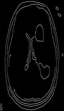
1 Implementation of Edge Detection Technique for Identification and Evaluation of Brain Tumor Metrics in MR Image through Lab VIEW S. Sunita Ratnam 1, D. Suresh Kumar 2, K. Madhukar 2 1 Department of Physics and Electronics, Shadan Degree College for Women, Hyderabad, Telangana, India 2 Department of Physics, Osmania Universiy, Hyderabad, Telangana, India Abstract : In this paper the authors present the development of an algorithm based on the Edge Detection Technique for Automatic Detection of Brain Tumors in MR Images of the Brain and their Quantification. The basic Edge detection technique is primarily based on the abrupt changes in the intensities between the tumor and the brain regions, identified through gradient operations. This is implemented through Vision Assistant tool box in Lab VIEW 12. The authors have proposed an algorithm which is implemented through Canny Edge Detector. The results of the measurements of maximum width, maximum length and the areas of the tumor and the brain obtained by the algorithm on the Ground truth image are in good agreement within ± 3% error, establishes that the proposed algorithm by the authors yields reliable results. Hence, this algorithm is implemented on 12 different patients through Lab VIEW. The results of the analysis are automatically evaluated and metricized. Keywords: Edge detection technique, Ground Truth Image, Image Segmentation, Brain Tumor Detection, Quantification, Lab VIEW, Vision Assistant. 1. Introduction As per the statistics released by the American Brain Tumour Association, several millions of people world over are affected by Brain Cancer [1-3]. The conditions of other developing and under-developed countries are pathetically alarming. Hence, there is a dire necessity for faster, accurate and automatic diagnostic system for the identification and evaluation of brain tumors. Early detection of brain tumor is important for a radiotherapy planning. As the MRI images exhibit complex characteristics it is indeed necessary to perform the image enhancement and segmentation process [4-5]. Recently, computerized methods in the area of nuclei detection segmentation and classification are extensively being used [6-8]. Medical image segmentation had inherited complex problems for the proper diagnosis of brain disorders [9]. The Digital Image Processing has provided the means to overcome these limitations in the analysis [10]. This is extensively used for identification of Brain Tumors in MR Images [11]. Considering these aspects, the authors have taken up the task for developing highly reliable algorithms for this proposed analysis. The present research work is carried out by making use of Vision Assistant Tool in LabVIEW software with Edge Detection and segmentation methods [12]. The algorithm detects the edge patterns and segmentation of tumor and brain are efficiently. This is usually identified through local linear gradient operators like Canny, Sobel, Laplace, etc [13]. In the present investigation, the authors have incorporated Canny Edge Detector [14] into their algorithm for automatic identification of Brain Tumours in MR Images and for the evaluation of their metrics of the physical parameters like the maximum width, maximum length and the areas of the tumor and the brain. 2. Methodology and Analysis In medical diagnosis the segmentation of the MRI images is preferred for quality segmentation [15]. The process of a portioning a digital image into multiple segments is called segmentation. By segmentation the image can be converted into a finite number of non-overlapping regions with respect to some characteristics (gray value, texture etc.,). Some tumors may deform surrounding structures in the brain because of the mass effect or oedema. The presence of artifacts and noise in the brain tumor images increases the difficulty in segmentation process [16]. Most of the segmentation algorithms depend on one of the two basic properties of intensity values: Discontinuity and Similarity. The segmentation process is mainly used to locate objects and the boundaries of different regions of an image and it is implemented using Edge detection technique. Edge based methods are focussed on detecting the contour of the image [17]. Canny Edge Detector [18] is used in the present investigation for this purpose. Lab VIEW based methods are being extensively used in the area of Digital image processing for automatic detection, segmentation and quantification [17] of brain tumors. Digital image processing enables faster and precise detection of the tumors. Edge extraction based schemes provide an efficient detection result against variations in the illuminations. The Edge Detection is achieved by Spatial Gradient Operation on pixel intensities of Red, Green and Blue components in the image plane defined by equations (1, 2 & 3).... (1)... (2) Paper ID: SUB
![... (3) For smoothening the image the relation (4) & (5) are employed [15]. Where (4)... (5) The intensity gradient g, the magnitude m and the direction of the edge pixels in the image plane.](/docs-images/88/114965599/images/2-1.jpg "They are presented in equations (6) & (7).... (6)... (7) The Canny Edge detector achieves low error rate as the edges detected are as close to the true edges.")
2 ... (3) For smoothening the image the relation (4) & (5) are employed [15]. Where (4)... (5) The intensity gradient g, the magnitude m and the direction of the edge pixels in the image plane. They are presented in equations (6) & (7).... (6)... (7) The Canny Edge detector achieves low error rate as the edges detected are as close to the true edges. The Edge points are also well localized with respect to the centre of the true edge. It also avoids the detection of multiple edges. The Canny Edge detection algorithm is presented in Figure 1. The work of the edge detection decides the result of the final processed image. Although many edge detection and evaluation methods have been developed in the past it is still a challenging task. Conventionally edge is detected according to some forward algorithms like Sobel, Prewitt and Robert, but in theory they all belong to the high pass filter [19] category. Hence, they are not suitable for analysing noisy medical images. Hence it may be difficult to distinguish the exact edge from noise or trivial geometric features. Hence Canny, presented the well known three criteria of edge detectors namely accurate detection, excellent localization, low spurious response and showed that the optimal detection for an isolated step edge. Based on these criteria the Canny edge detector first smoothens the image to eliminate the noise [20] and then locates the edges. 3. Implementation of Algorithm through Vision Assistant 3.1 Pre-processing The multi modal MRI images used by the radiologists cannot provide complementary data on tumors. Hence image enhancement [21, 22] operation improves the quality of the images such as contrast, brightness characteristics, reducing the noise level and by sharpening the image to extract the fine detail of the tumor for further processing. It also reduces noise of the image. This improves the quality of the image and accuracy in detecting the tumor [23]. It performs filtering of noise and other artifacts in the image and sharpens the edges in the image. Figure 1: Edge Detection Technique Algorithm 3.2 Color Plane (Green Plane) Extraction In bio-medical applications Red color interacts with the human blood and may not yield good result. Similarly, as Blue color is a darker shade, some of the information may hide-out. So Blue is not preferred. Hence, the green color plane [11] is selected, because most of the image information is contained in the Green Plane. 3.3 Histogram The Histogram [24] is a graphical representation of the total intensity distribution in a digital image. One can judge the entire tonal distribution at a glance and can manually fix the threshold value. 3.4 Filters (Low Pass) The image enhancement is carried out in spatial domain. A low pass linear filter is used to eliminate the low frequency noise. This filter produces smoother, softer looking images from a sharper original image. Thus smoothens skin lines, blemishes and noise pixels [20]. 3.5 Canny Edge Detector Canny Edge detector plays an important role to obtain the detailed information about the infected area of the brain. It identifies the contour of the region of interest (ROI) which is highly advantageous to identify the location of the tumor. This is most efficient edge detector. The advantageous is based on smoothening the image through the Gaussian filter to reduce the noise and then apply the local gradient operation in X and Y directions. The edge point is defined as the point of maximum strength in the direction of the gradient (magnitude). Paper ID: SUB
![3.6 Thresholding International Journal of Science and Research (IJSR) Thresholding is a very simple approach for segmentation [24] in computer vision and image analysis.](/docs-images/88/114965599/images/3-1.jpg "As the computational complexity is low thresholding based scheme is considered for real time computer vision systems.")

3 3.6 Thresholding International Journal of Science and Research (IJSR) Thresholding is a very simple approach for segmentation [24] in computer vision and image analysis. As the computational complexity is low thresholding based scheme is considered for real time computer vision systems. Thresholding scheme is broadly classified into two categories as Contextual and non-contextual, depends on the gray level distribution of the image. The Contextual thresholding, depends on the second order gray level statistics or co-occurrance matrix of the image and the noncontextual threshold depends on the Gray level distribution of the image. The input gray image is converted into a binary format. The selection of a threshold value from the image using Histogram is important for segmentation. The vision assistant script for identification of the tumor is shown in Figure 2. The Lab VIEW block diagram generated for the Canny Edge Detector is presented in Figure 3. Figure 2: The Vision Assistant script (Edge detection) Figure 3: Lab VIEW Block Diagram 4. Measurements Edge Detection Technique developed by the authors is implemented in the Lab VIEW using Vision Assistant toolkit. The maximum width, maximum length and the area of the tumour and the brain are automatically quantified [25, 26] by using the Polygon Tool in pixels. The Edge Detection algorithm yielded the results with a maximum error of ±4%. 5. Results The algorithm developed is implemented on Ground Truth images [27] and the results of the measurements obtained are presented in Table 1. The algorithm is implemented for analyzing the MR Images of 12 different patients through LabVIEW. The results of this analysis are presented in Table 2 and the resulting output images are presented in Table 3 and also graphically presented in Figure 4. Paper ID: SUB


4 Original MR Image International Journal of Science and Research (IJSR) Table 1: The resultant processed images of different patients Color Plane Filter Thresholding Measurements Extraction GT P-1 P-2 P-3 P-4 P-5 Paper ID: SUB


5 P-6 P-7 P-8 P-9 P-10 P-11 Dual P-12 Paper ID: SUB
 of Area widths lengths Areas Tumor (w) Tumor (l) (a) Brain (L) (A) (w/ W) (l / L) (a / A) pixels Pixels pixels pixels pixels GT 54")
6 Table 2A: Comparison of Tumor Index on GT with Edge Detection Algorithm Max. Max. length Tumor Max.Width of Max. length Brain Ratio of Ratio of Ratio of Width of of Area Brain (W) of Area widths lengths Areas Tumor (w) Tumor (l) (a) Brain (L) (A) (w/ W) (l / L) (a / A) pixels Pixels pixels pixels pixels GT Algorithm Patient code Table 2B: Estimation of Error in Tumor Index GT Algorithm Error % Error % Error % w/w l/l (a/a) w/w l/l (a/a) (Width) (Length) (Area) GT = Doctor s Data. Algorithm = GT analysed through Authors developed Edge Detection algorithm. All the images taken for the present analysis are of high resolution of 32-bits. Max. Width of Tumor (w) Table 3: Estimation of Tumor Index for different patients Max. Max. length Width of of Brain Brain Area (A) Brain (L) (W) Max. length Tumor Area of Tumor (l) (a) Ratio of widths (w/ W) Ratio of lengths (l / L) Ratio of Areas (a / A) Pixels Pixels Pixels Pixels Pixels Pixels GT P P P P P P P P P P P-11A P-11B P Conclusions By analysing the MR Images of the brain, through the algorithm the authors could achieve the objective of Automatic Identification and Quantification of Brain Tumours. This greatly relieves the doctors from the work pressure of examining and interpreting the huge amount of brain tumor images which can be entrusted to the junior doctors equipped with the software developed by the authors. Only critical cases which require the guidance of the expert will be referred to them. This facilitates the expert doctors to concentrate on more important research. 7. Future Scope The research can be further directed towards generating brief reports from the results obtained it can also can be extended to 3D Image processing. 8. Acknowledgements Figure 5: Metrics of the tumor and the brain The authors thank the Head, Faculty members and coscholars of Physics Department, Osmania University, Hyderabad for their encouragement and valuable suggestions in carrying out my research work. Paper ID: SUB
7 References International Journal of Science and Research (IJSR) [1] A. Jemal, R. Siegel, E. Ward, Y. Hao, J. Xu and M. J. Thun, Cancer statistics, 2009, CA Cancer J. Clin. Vol. 59, pp , [2] World Health Organization WHO Cancer, Fact sheet No. 297, Feb 2009, [3] American Brain Tumor Association. Brain tumor statistics retrieved from [4] Kimmi Verma, Aru Mehrotra, Vijayeta Pandey, Shardendu Singh, Image processing techniques for the enhancement of brain tumor patterns, International Journal of Advanced Research in Electrical, Electronics and Instrumentation Engineering, Vol.2, Issue -4, April [5] Rohan Kandwal, Ashok Kumar, An Automated System for Brain Tumor detection and segmentation, International Journal of Advanced research in Computer Science and Software Engineering, ISSN ; x, Volume 4, Issue 3, March [6] Sivaramakrishnan, Dr. M. Kaman, A novel based approach for extraction of brain tumor in MRI images using soft computing techniques, International Journal of Advanced Research in Computer and Communication Engineering, vol-2, Issue-4, April [7] D. Bhattacharyya and T.H. Kim, Brain tumor detection using MRI image analysis, Commun. Comput. Inform. Sci. Vol. 151, pp , [8] Moon. N, Bullitt. E, Van Leemput K, Gerig. G., Automatic Brain and Tumor Segmentation, MICCAI, LNCS 2489 : , [9] Rohan Khandwal and Ashok Kumar, An automated system for Brain Tumor Detection and Segmentation, International Journal of Advanced Research in Computer Science and Software Engineering, Volume- 4, Issue-3, March [10] Nagalkar V.J. Asole S.S. Brain Tumour detection using digital image processing, Journal of Signal and Image Processing, Volume 3, Issue - 3 ISSN : , (2012). [11] Gauri P. Anandgaonkar, Ganesh S. Sable, Detection and Identification of Brain tumor in Brain MR Images using Fuzzy C-Means Segmentation, International Journal of Advanced Research in Computer and Communication Engineering, Vol.-2, Issue 10, October [12] K. Sudha Rani, A.Swapna Priya, Dr. T.C. Sarma and Dr.K.Satya Prasad, LabVIEW based Brain Tumor Area and length detection in CT and MRI scan images, 2 nd International conference on Computing, Engineering and Information Technology (ICCEIT) 20 th Sept., 2013 [13] Narkhede Sachin, Dr.Deven Shah, Prof. Vaishalikhairnar, Prof. Sujatakadu, Brain tumor detection based on bilateral symmetry information, Vol,-4, Issue-6 (Version 3), June 2014, pp [14] Dina Aboul Dahab, Samy S.A. Ghoniemy, Gamal M. Selim, Automated Brain tumor detection and identification using image processing and probabilistic neural network techniques, International journal of image processing and visual communication, Volume- 1, Issue-2, October [15] Samir kumar bandhyopadhyay, Tuhin Utsab Paul, Automatic Segmentation of Brain tumor from multiple images of brain MRI, Volume-2, Issue-1, January [16] Bhausaheb Shinde, Dnyandeo Mhaske, Machindra Patare, A. R. Dani, Study of noise and filtering techniques in medical images, (IJESAT),Vol.-3, No.1 Jan-Feb [17] N. Senthilkumaran and R. Rajesh, A study on Edge Detection Methods for Image Segmentation, Proceedings computer science (ICMCS 2009, Vol. 1, pp ), [18] Pratibha Sharma, Manoz Diwakar, Sangam Choudhary, Application of Edge Detection for Brain Tumour detection, Volume 58, No. 16 (2012). [19] Prof. B. K. Saptalakar, Miss. Rajeshwari. H, Segmentation based detection of Brain tumour, International journal of computer and electronics research volume-2, Issue-1, February [20] Bhausaheb Shinde, Dnyandeo Mhaske, Machindra Patare, A. R. Dani, Apply different filtering techniques to remove the speckle noise using medical images, International Journal of Engineering research and Applications, Vol-2, Issue -1,Jan-Feb 2012, pp [21] Poonam, Jyotika Pruthi, Review of Image Processing Techniques for Automatic Detection of Tumor in Human Brain, International Journal of Computer Science and Mobile Computing, IJCSMC, Vol.2, Issue 11, November 2013, pg [22] Bhausaheb Shinde, Dnyandeo Mhaske, Machindra Patare, A. R. Dani, Study of Image Processing, Enhancement and Restoration, IJCSI,Vol.-8, Issue- 6,Nov [23] Manor K. Kowari and Sourabh Yadav, Brain tumor detection and segmentation using histogram thresholding, International journal of engineering and advanced technology, IJEAT ISSN: , Vol.- 1 Issue -4, Journal India, April [24] Shiping Zhu, Xi Xia, Qingrong Zhang, Kamel Bellouata An Image Segmentation in Image Processing Based on Threshold Segmentation. Third International Conference on Signal Image Technologies and Internet based System, SITIS [25] Varsha Kshirsagar, Prof. Jagruti Panchal, Segmentation of Brain Tumour and its Area Calculation, International Journal of Advanced Research in Computer science and Software Engineering, ISSN : x, Vol. 4, Issue 5, May [26] Chunyan Jiang, Xinhua Zhang, Wanjun Huang, Christoph Meinel, Segmentation and Quantification of Brain Tumor, IEEE International Conference on Virtual Environment, Human-Computer interfaces and Measurement Systems, USA, 12-14, July 2008 [27] E. D. Gelasca, B. Obara, D. Fedorov, K. Kvilekval and B. Manjunath, A biosegmentation benchmark for evaluation of bioimage analysis methods. BMC Bioinform, Vol. 10, pp. 1 12, Paper ID: SUB
A Survey on Brain Tumor Detection Technique
 (International Journal of Computer Science & Management Studies) Vol. 15, Issue 06 A Survey on Brain Tumor Detection Technique Manju Kadian 1 and Tamanna 2 1 M.Tech. Scholar, CSE Department, SPGOI, Rohtak
ANALYSIS AND DETECTION OF BRAIN TUMOUR USING IMAGE PROCESSING TECHNIQUES
 ANALYSIS AND DETECTION OF BRAIN TUMOUR USING IMAGE PROCESSING TECHNIQUES P.V.Rohini 1, Dr.M.Pushparani 2 1 M.Phil Scholar, Department of Computer Science, Mother Teresa women s university, (India) 2 Professor
Brain Tumor Detection using Watershed Algorithm
 Brain Tumor Detection using Watershed Algorithm Dawood Dilber 1, Jasleen 2 P.G. Student, Department of Electronics and Communication Engineering, Amity University, Noida, U.P, India 1 P.G. Student, Department
An efficient method for Segmentation and Detection of Brain Tumor in MRI images
 An efficient method for Segmentation and Detection of Brain Tumor in MRI images Shubhangi S. Veer (Handore) 1, Dr. P.M. Patil 2 1 Research Scholar, Ph.D student, JJTU, Rajasthan,India 2 Jt. Director, Trinity
Keywords Fuzzy Logic, Fuzzy Rule, Fuzzy Membership Function, Fuzzy Inference System, Edge Detection, Regression Analysis.
 Volume 6, Issue 3, March 2016 ISSN: 2277 128X International Journal of Advanced Research in Computer Science and Software Engineering Research Paper Available online at: www.ijarcsse.com Modified Fuzzy
Unsupervised MRI Brain Tumor Detection Techniques with Morphological Operations
 Unsupervised MRI Brain Tumor Detection Techniques with Morphological Operations Ritu Verma, Sujeet Tiwari, Naazish Rahim Abstract Tumor is a deformity in human body cells which, if not detected and treated,
Tumor Detection In Brain Using Morphological Image Processing
 Abstract: - Tumor Detection In Brain Using Morphological Image Processing U.Vanitha 1, P.Prabhu Deepak 2, N.Pon Nageswaran 3, R.Sathappan 4 III-year, department of electronics and communication engineering
BRAIN TUMOR DETECTION AND SEGMENTATION USING WATERSHED SEGMENTATION AND MORPHOLOGICAL OPERATION
 BRAIN TUMOR DETECTION AND SEGMENTATION USING WATERSHED SEGMENTATION AND MORPHOLOGICAL OPERATION Swe Zin Oo 1, Aung Soe Khaing 2 1 Demonstrator, Department of Electronic Engineering, Mandalay Technological
Image Enhancement and Compression using Edge Detection Technique
 Image Enhancement and Compression using Edge Detection Technique Sanjana C.Shekar 1, D.J.Ravi 2 1M.Tech in Signal Processing, Dept. Of ECE, Vidyavardhaka College of Engineering, Mysuru 2Professor, Dept.
Segmentation of Tumor Region from Brain Mri Images Using Fuzzy C-Means Clustering And Seeded Region Growing
 IOSR Journal of Computer Engineering (IOSR-JCE) e-issn: 2278-0661,p-ISSN: 2278-8727, Volume 18, Issue 5, Ver. I (Sept - Oct. 2016), PP 20-24 www.iosrjournals.org Segmentation of Tumor Region from Brain
BraTS : Brain Tumor Segmentation Some Contemporary Approaches
 BraTS : Brain Tumor Segmentation Some Contemporary Approaches Mahantesh K 1, Kanyakumari 2 Assistant Professor, Department of Electronics & Communication Engineering, S. J. B Institute of Technology, BGS,
EARLY STAGE DIAGNOSIS OF LUNG CANCER USING CT-SCAN IMAGES BASED ON CELLULAR LEARNING AUTOMATE
 EARLY STAGE DIAGNOSIS OF LUNG CANCER USING CT-SCAN IMAGES BASED ON CELLULAR LEARNING AUTOMATE SAKTHI NEELA.P.K Department of M.E (Medical electronics) Sengunthar College of engineering Namakkal, Tamilnadu,
Automatic Classification of Breast Masses for Diagnosis of Breast Cancer in Digital Mammograms using Neural Network
 IJSTE - International Journal of Science Technology & Engineering Volume 1 Issue 11 May 2015 ISSN (online): 2349-784X Automatic Classification of Breast Masses for Diagnosis of Breast Cancer in Digital
Enhanced Detection of Lung Cancer using Hybrid Method of Image Segmentation
 Enhanced Detection of Lung Cancer using Hybrid Method of Image Segmentation L Uma Maheshwari Department of ECE, Stanley College of Engineering and Technology for Women, Hyderabad - 500001, India. Udayini
Edge Detection Techniques Based On Soft Computing
 International Journal for Science and Emerging ISSN No. (Online):2250-3641 Technologies with Latest Trends 7(1): 21-25 (2013) ISSN No. (Print): 2277-8136 Edge Detection Techniques Based On Soft Computing
MRI Image Processing Operations for Brain Tumor Detection
 MRI Image Processing Operations for Brain Tumor Detection Prof. M.M. Bulhe 1, Shubhashini Pathak 2, Karan Parekh 3, Abhishek Jha 4 1Assistant Professor, Dept. of Electronics and Telecommunications Engineering,
Keywords MRI segmentation, Brain tumor detection, Tumor segmentation, Tumor classification, Medical Imaging, ANN
 Volume 5, Issue 4, 2015 ISSN: 2277 128X International Journal of Advanced Research in Computer Science and Software Engineering Research Paper Available online at: www.ijarcsse.com An Improved Automatic
Cancer Cells Detection using OTSU Threshold Algorithm
 Cancer Cells Detection using OTSU Threshold Algorithm Nalluri Sunny 1 Velagapudi Ramakrishna Siddhartha Engineering College Mithinti Srikanth 2 Velagapudi Ramakrishna Siddhartha Engineering College Kodali
Extraction of Blood Vessels and Recognition of Bifurcation Points in Retinal Fundus Image
 International Journal of Research Studies in Science, Engineering and Technology Volume 1, Issue 5, August 2014, PP 1-7 ISSN 2349-4751 (Print) & ISSN 2349-476X (Online) Extraction of Blood Vessels and
International Journal for Science and Emerging
 International Journal for Science and Emerging ISSN No. (Online):2250-3641 Technologies with Latest Trends 8(1): 7-13 (2013) ISSN No. (Print): 2277-8136 Adaptive Neuro-Fuzzy Inference System (ANFIS) Based
Automatic Hemorrhage Classification System Based On Svm Classifier
 Automatic Hemorrhage Classification System Based On Svm Classifier Abstract - Brain hemorrhage is a bleeding in or around the brain which are caused by head trauma, high blood pressure and intracranial
Clustering of MRI Images of Brain for the Detection of Brain Tumor Using Pixel Density Self Organizing Map (SOM)
") IOSR Journal of Computer Engineering (IOSR-JCE) e-issn: 2278-0661,p-ISSN: 2278-8727, Volume 19, Issue 6, Ver. I (Nov.- Dec. 2017), PP 56-61 www.iosrjournals.org Clustering of MRI Images of Brain for the
IOSR Journal of Computer Engineering (IOSR-JCE) e-issn: 2278-0661,p-ISSN: 2278-8727, Volume 19, Issue 6, Ver. I (Nov.- Dec. 2017), PP 56-61 www.iosrjournals.org Clustering of MRI Images of Brain for the
Bapuji Institute of Engineering and Technology, India
 Volume 4, Issue 1, January 2014 ISSN: 2277 128X International Journal of Advanced Research in Computer Science and Software Engineering Research Paper Available online at: www.ijarcsse.com A Segmented
International Journal of Advanced Research in Computer Science and Software Engineering
 Volume 2, Issue 8, August 2012 ISSN: 2277 128X International Journal of Advanced Research in Computer Science and Software Engineering Research Paper Available online at: www.ijarcsse.com A Novel Approach
IJRE Vol. 03 No. 04 April 2016
 6 Implementation of Clustering Techniques For Brain Tumor Detection Shravan Rao 1, Meet Parikh 2, Mohit Parikh 3, Chinmay Nemade 4 Student, Final Year, Department Of Electronics & Telecommunication Engineering,
MEM BASED BRAIN IMAGE SEGMENTATION AND CLASSIFICATION USING SVM
 MEM BASED BRAIN IMAGE SEGMENTATION AND CLASSIFICATION USING SVM T. Deepa 1, R. Muthalagu 1 and K. Chitra 2 1 Department of Electronics and Communication Engineering, Prathyusha Institute of Technology
A Pictorial Review and an Algorithm for the Determination of Sickle Cell Anemia
 International Journal of Engineering and Advanced Technology (IJEAT) ISSN: 2249 8958, Volume-5 Issue-2, December 2015 A Pictorial Review and an Algorithm for the Determination of Sickle Cell Anemia Hariharan.S,
Comparative Analysis of Canny and Prewitt Edge Detection Techniques used in Image Processing
 Comparative Analysis of Canny and Prewitt Edge Detection Techniques used in Image Processing Nisha 1, Rajesh Mehra 2, Lalita Sharma 3 PG Scholar, Dept. of ECE, NITTTR, Chandigarh, India 1 Associate Professor,
Detection and Classification of Brain Tumor using BPN and PNN Artificial Neural Network Algorithms
 Available Online at www.ijcsmc.com International Journal of Computer Science and Mobile Computing A Monthly Journal of Computer Science and Information Technology IJCSMC, Vol. 4, Issue. 4, April 2015,
Performance evaluation of the various edge detectors and filters for the noisy IR images
 Performance evaluation of the various edge detectors and filters for the noisy IR images * G.Padmavathi ** P.Subashini ***P.K.Lavanya Professor and Head, Lecturer (SG), Research Assistant, ganapathi.padmavathi@gmail.com
Detection of Glaucoma and Diabetic Retinopathy from Fundus Images by Bloodvessel Segmentation
 International Journal of Engineering and Advanced Technology (IJEAT) ISSN: 2249 8958, Volume-5, Issue-5, June 2016 Detection of Glaucoma and Diabetic Retinopathy from Fundus Images by Bloodvessel Segmentation
Research Article. Automated grading of diabetic retinopathy stages in fundus images using SVM classifer
 Available online www.jocpr.com Journal of Chemical and Pharmaceutical Research, 2016, 8(1):537-541 Research Article ISSN : 0975-7384 CODEN(USA) : JCPRC5 Automated grading of diabetic retinopathy stages
Brain Tumor Detection and Segmentation in MR images Using GLCM and. AdaBoost Classifier
 2015 IJSRSET Volume 1 Issue 3 Print ISSN : 2395-1990 Online ISSN : 2394-4099 Themed Section: Engineering and Technology Brain Tumor Detection and Segmentation in MR images Using GLCM and ABSTRACT AdaBoost
Early Detection of Lung Cancer
 Early Detection of Lung Cancer Aswathy N Iyer Dept Of Electronics And Communication Engineering Lymie Jose Dept Of Electronics And Communication Engineering Anumol Thomas Dept Of Electronics And Communication
PERFORMANCE ANALYSIS OF BRAIN TUMOR DIAGNOSIS BASED ON SOFT COMPUTING TECHNIQUES
 American Journal of Applied Sciences 11 (2): 329-336, 2014 ISSN: 1546-9239 2014 Science Publication doi:10.3844/ajassp.2014.329.336 Published Online 11 (2) 2014 (http://www.thescipub.com/ajas.toc) PERFORMANCE
EXTRACT THE BREAST CANCER IN MAMMOGRAM IMAGES
 International Journal of Civil Engineering and Technology (IJCIET) Volume 10, Issue 02, February 2019, pp. 96-105, Article ID: IJCIET_10_02_012 Available online at http://www.iaeme.com/ijciet/issues.asp?jtype=ijciet&vtype=10&itype=02
Brain Tumor segmentation and classification using Fcm and support vector machine
 Brain Tumor segmentation and classification using Fcm and support vector machine Gaurav Gupta 1, Vinay singh 2 1 PG student,m.tech Electronics and Communication,Department of Electronics, Galgotia College
Earlier Detection of Cervical Cancer from PAP Smear Images
 , pp.181-186 http://dx.doi.org/10.14257/astl.2017.147.26 Earlier Detection of Cervical Cancer from PAP Smear Images Asmita Ray 1, Indra Kanta Maitra 2 and Debnath Bhattacharyya 1 1 Assistant Professor
Brain Tumor Detection and Segmentation In MRI Images
 Brain Tumor Detection and Segmentation In MRI Images AbhijithSivarajan S 1, Kamalakar V. Thakare 2, Shailesh Kathole 3, Pramod B. Khamkar 4, Danny J. Pereira 5 Department of Computer Engineering, Govt.
International Journal of Advance Research in Engineering, Science & Technology
 Impact Factor (SJIF): 3.632 International Journal of Advance Research in Engineering, Science & Technology e-issn: 2393-9877, p-issn: 2394-2444 (Special Issue for ITECE 2016) An Efficient Image Processing
COMPUTER AIDED DIAGNOSTIC SYSTEM FOR BRAIN TUMOR DETECTION USING K-MEANS CLUSTERING
 COMPUTER AIDED DIAGNOSTIC SYSTEM FOR BRAIN TUMOR DETECTION USING K-MEANS CLUSTERING Urmila Ravindra Patil Tatyasaheb Kore Institute of Engineering and Technology, Warananagar Prof. R. T. Patil Tatyasaheb
IJREAS Volume 2, Issue 2 (February 2012) ISSN: LUNG CANCER DETECTION USING DIGITAL IMAGE PROCESSING ABSTRACT
 ISSN: LUNG CANCER DETECTION USING DIGITAL IMAGE PROCESSING ABSTRACT") LUNG CANCER DETECTION USING DIGITAL IMAGE PROCESSING Anita Chaudhary* Sonit Sukhraj Singh* ABSTRACT In recent years the image processing mechanisms are used widely in several medical areas for improving
LUNG CANCER DETECTION USING DIGITAL IMAGE PROCESSING Anita Chaudhary* Sonit Sukhraj Singh* ABSTRACT In recent years the image processing mechanisms are used widely in several medical areas for improving
A CONVENTIONAL STUDY OF EDGE DETECTION TECHNIQUE IN DIGITAL IMAGE PROCESSING
 Available Online at www.ijcsmc.com International Journal of Computer Science and Mobile Computing A Monthly Journal of Computer Science and Information Technology IJCSMC, Vol. 3, Issue. 4, April 2014,
Extraction and Identification of Tumor Regions from MRI using Zernike Moments and SVM
 I J C T A, 8(5), 2015, pp. 2327-2334 International Science Press Extraction and Identification of Tumor Regions from MRI using Zernike Moments and SVM Sreeja Mole S.S.*, Sree sankar J.** and Ashwin V.H.***
Automated Brain Tumor Segmentation Using Region Growing Algorithm by Extracting Feature
 Automated Brain Tumor Segmentation Using Region Growing Algorithm by Extracting Feature Shraddha P. Dhumal 1, Ashwini S Gaikwad 2 1 Shraddha P. Dhumal 2 Ashwini S. Gaikwad ABSTRACT In this paper, we propose
Study And Development Of Digital Image Processing Tool For Application Of Diabetic Retinopathy
 Study And Development O Digital Image Processing Tool For Application O Diabetic Retinopathy Name: Ms. Jyoti Devidas Patil mail ID: jyot.physics@gmail.com Outline 1. Aims & Objective 2. Introduction 3.
BRAIN TUMOR SEGMENTATION USING K- MEAN CLUSTERIN AND ITS STAGES IDENTIFICATION
 ABSTRACT BRAIN TUMOR SEGMENTATION USING K- MEAN CLUSTERIN AND ITS STAGES IDENTIFICATION Sonal Khobarkhede 1, Poonam Kamble 2, Vrushali Jadhav 3 Prof.V.S.Kulkarni 4 1,2,3,4 Rajarshi Shahu College of Engg.
ANN BASED IMAGE CLASSIFIER FOR PANCREATIC CANCER DETECTION
 Singaporean Journal of Scientific Research(SJSR) Special Issue - Journal of Selected Areas in Microelectronics (JSAM) Vol.8.No.2 2016 Pp.01-11 available at :www.iaaet.org/sjsr Paper Received : 08-04-2016
Brain Tumor Detection Using Morphological And Watershed Operators
 Brain Tumor Detection Using Morphological And Watershed Operators Miss. Roopali R. Laddha 1, Dr. Siddharth A. Ladhake 2 1&2 Sipna College Of Engg. & Technology, Amravati. Abstract This paper presents a
LUNG NODULE DETECTION SYSTEM
 LUNG NODULE DETECTION SYSTEM Kalim Bhandare and Rupali Nikhare Department of Computer Engineering Pillai Institute of Technology, New Panvel, Navi Mumbai, India ABSTRACT: The Existing approach consist
Detection of Lung Cancer Using Marker-Controlled Watershed Transform
 2015 International Conference on Pervasive Computing (ICPC) Detection of Lung Cancer Using Marker-Controlled Watershed Transform Sayali Satish Kanitkar M.E. (Signal Processing), Sinhgad College of engineering,
COMPUTERIZED SYSTEM DESIGN FOR THE DETECTION AND DIAGNOSIS OF LUNG NODULES IN CT IMAGES 1
 ISSN 258-8739 3 st August 28, Volume 3, Issue 2, JSEIS, CAOMEI Copyright 26-28 COMPUTERIZED SYSTEM DESIGN FOR THE DETECTION AND DIAGNOSIS OF LUNG NODULES IN CT IMAGES ALI ABDRHMAN UKASHA, 2 EMHMED SAAID
Extraction of Texture Features using GLCM and Shape Features using Connected Regions
 Extraction of Texture Features using GLCM and Shape Features using Connected Regions Shijin Kumar P.S #1, Dharun V.S *2 # Research Scholar, Department of Electronics and Communication Engineering, Noorul
SAPOG Edge Detection Technique GUI using MATLAB
 SAPOG Edge Detection Technique GUI using MATLAB Poonam Kumari 1, Sanjeev Kumar Gupta 2 Software Engineer, Devansh Softech Consultancy Services Pvt. Ltd., Agra, India 1 Director, Devansh Softech Consultancy
Implementation of Clustering Techniques For Brain Tumor Detection
 Implementation of Clustering Techniques For Brain Tumor Detection Shravan Rao 1, Meet Parikh 2, Mohit Parikh 3, Chinmay Nemade 4 Student, Final Year, Department Of Electronics & Telecommunication Engineering,
Semi-automatic Thyroid Area Measurement Based on Ultrasound Image
 Semi-automatic Thyroid Area Measurement Based on Ultrasound Image Eko Supriyanto, Nik M Arif, Akmal Hayati Rusli, Nasrul Humaimi Advanced Diagnostics and Progressive Human Care Research Group Research
Diagnosis System for the Detection of Abnormal Tissues from Brain MRI.
 Diagnosis System for the Detection of Abnormal Tissues from Brain MRI Arshad Javed 1, 2, Abdulhameed Rakan Alenezi 1, Wang Yin Chai 2, Narayanan Kulathuramaiyer 2 1 Faculty of Computer Science and Information,
International Journal of Computational Science, Mathematics and Engineering Volume2, Issue6, June 2015 ISSN(online): Copyright-IJCSME
: Copyright-IJCSME") Various Edge Detection Methods In Image Processing Using Matlab K. Narayana Reddy 1, G. Nagalakshmi 2 12 Department of Computer Science and Engineering 1 M.Tech Student, SISTK, Puttur 2 HOD of CSE Department,
Various Edge Detection Methods In Image Processing Using Matlab K. Narayana Reddy 1, G. Nagalakshmi 2 12 Department of Computer Science and Engineering 1 M.Tech Student, SISTK, Puttur 2 HOD of CSE Department,
Implementation of Brain Tumor Detection using Segmentation Algorithm & SVM
 Implementation of Brain Tumor Detection using Segmentation Algorithm & SVM Swapnil R. Telrandhe 1 Amit Pimpalkar 2 Ankita Kendhe 3 telrandheswapnil@yahoo.com amit.pimpalkar@raisoni.net ankita.kendhe@raisoni.net
Automated Detection Of Glaucoma & D.R From Eye Fundus Images
 Reviewed Paper Volume 2 Issue 12 August 2015 International Journal of Informative & Futuristic Research ISSN (Online): 2347-1697 Automated Detection Of Glaucoma & D.R Paper ID IJIFR/ V2/ E12/ 016 Page
AUTOMATIC BRAIN TUMOR DETECTION AND CLASSIFICATION USING SVM CLASSIFIER
 AUTOMATIC BRAIN TUMOR DETECTION AND CLASSIFICATION USING SVM CLASSIFIER 1 SONU SUHAG, 2 LALIT MOHAN SAINI 1,2 School of Biomedical Engineering, National Institute of Technology, Kurukshetra, Haryana -
Threshold Based Segmentation Technique for Mass Detection in Mammography
 Threshold Based Segmentation Technique for Mass Detection in Mammography Aziz Makandar *, Bhagirathi Halalli Department of Computer Science, Karnataka State Women s University, Vijayapura, Karnataka, India.
BONE CANCER DETECTION USING ARTIFICIAL NEURAL NETWORK
 ISSN: 0976-2876 (Print) ISSN: 2250-0138(Online) BONE CANCER DETECTION USING ARTIFICIAL NEURAL NETWORK 1 Asuntha A, 2 Andy Srinivasan 1 Department of Electronics and Instrumentation Engg., SRM Institute
CALCULATION of the CEREBRAL HEMORRHAGE VOLUME USING ANALYSIS of COMPUTED TOMOGRAPHY IMAGE
 CALCULATION of the CEREBRAL HEMORRHAGE VOLUME USING ANALYSIS of COMPUTED TOMOGRAPHY IMAGE Cory Amelia* Magister of Physics, Faculty of Science and Mathematics, Diponegoro University, Semarang, Indonesia
A Fusion Technique Based on Image - Statistical Analysis for Detection of Throat Cancer Types
 A Fusion Technique Based on Image - Statistical Analysis for Detection of Throat Cancer Types Adnan Al-Bashir Hashemite University Department of Industrial Engineering P.O. Box 150459, Zarqa 13115, Jordan
Automatic Detection of Brain Tumor Using K- Means Clustering
 Automatic Detection of Brain Tumor Using K- Means Clustering Nitesh Kumar Singh 1, Geeta Singh 2 1, 2 Department of Biomedical Engineering, DCRUST, Murthal, Haryana Abstract: Brain tumor is an uncommon
Detection of microcalcifications in digital mammogram using wavelet analysis
 American Journal of Engineering Research (AJER) e-issn : 2320-0847 p-issn : 2320-0936 Volume-02, Issue-11, pp-80-85 www.ajer.org Research Paper Open Access Detection of microcalcifications in digital mammogram
Research Article Volume 6 Issue No. 3
 DOI 10.4010/2016.601 ISSN 2321 3361 2016 IJESC Research Article Volume 6 Issue No. 3 Multifractal Texture Estimation for Detection and Segmentation of Brain Tumors with the Source and Age of the Tumor
Brain Tumor Detection from MRI Images using Fuzzy C-Means Segmentation
 Brain Tumor Detection from MRI Images using Fuzzy C-Means Segmentation Jinal A. Shah 1, S. R. Suralkar 2 ME Student, E&TC Department, SSBTs COET Bambhori, Jalgaon, India 1 HOD, E&TC Department, SSBTs COET
Development of novel algorithm by combining Wavelet based Enhanced Canny edge Detection and Adaptive Filtering Method for Human Emotion Recognition
 International Journal of Engineering Research and Development e-issn: 2278-067X, p-issn: 2278-800X, www.ijerd.com Volume 12, Issue 9 (September 2016), PP.67-72 Development of novel algorithm by combining
Edge Detection Operators: Peak Signal to Noise Ratio Based Comparison
 I.J. Image, Graphics and Signal, 2014, 10, 55-61 Published Online September 2014 in MECS (http://www.mecs-press.org/) DOI: 10.5815/ijigsp.2014.10.07 Edge Detection Operators: Peak Signal to Noise Ratio
Automated Preliminary Brain Tumor Segmentation Using MRI Images
 www.ijcsi.org 102 Automated Preliminary Brain Tumor Segmentation Using MRI Images Shamla Mantri 1, Aditi Jahagirdar 2, Kuldeep Ghate 3, Aniket Jiddigouder 4, Neha Senjit 5 and Saurabh Sathaye 6 1 Computer
A new Method on Brain MRI Image Preprocessing for Tumor Detection
 2015 IJSRSET Volume 1 Issue 1 Print ISSN : 2395-1990 Online ISSN : 2394-4099 Themed Section: Engineering and Technology A new Method on Brain MRI Preprocessing for Tumor Detection ABSTRACT D. Arun Kumar
Optimization Technique, To Detect Brain Tumor in MRI
 Optimization Technique, To Detect Brain Tumor in MRI Depika Patel 1, Prof. Amit kumar Nandanwar 2 1 Student, M.Tech, CSE, VNSIT Bhopal 2 Computer Science andengineering, VNSIT Bhopal Abstract- Image Segmentation
Brain Tumor Segmentation of Noisy MRI Images using Anisotropic Diffusion Filter
 Available Online at www.ijcsmc.com International Journal of Computer Science and Mobile Computing A Monthly Journal of Computer Science and Information Technology IJCSMC, Vol. 3, Issue. 7, July 2014, pg.744
2D-Sigmoid Enhancement Prior to Segment MRI Glioma Tumour
 2D-Sigmoid Enhancement Prior to Segment MRI Glioma Tumour Pre Image-Processing Setyawan Widyarto, Siti Rafidah Binti Kassim 2,2 Department of Computing, Faculty of Communication, Visual Art and Computing,
International Journal of Computer Sciences and Engineering. Review Paper Volume-5, Issue-12 E-ISSN:
 International Journal of Computer Sciences and Engineering Open Access Review Paper Volume-5, Issue-12 E-ISSN: 2347-2693 Different Techniques for Skin Cancer Detection Using Dermoscopy Images S.S. Mane
Brain Tumor Detection Using Neural Network
 International Journal of Science and Modern Engineering (IJISME) ISSN: 2319-6386, Volume-1, Issue-9, August 2013 Brain Tumor Detection Using Neural Network Pankaj Sapra, Rupinderpal Singh, Shivani Khurana
Implementation of Automatic Retina Exudates Segmentation Algorithm for Early Detection with Low Computational Time
 www.ijecs.in International Journal Of Engineering And Computer Science ISSN: 2319-7242 Volume 5 Issue 10 Oct. 2016, Page No. 18584-18588 Implementation of Automatic Retina Exudates Segmentation Algorithm
Image Processing of Eye for Iris Using. Canny Edge Detection Technique
 Image Processing of Eye for Iris Using Canny Edge Detection Technique D. Anitha 1, M. Suganthi 2 & P. Suresh 3 1 Department of IT, Muthayammal Engineering College, Rasipuram, Tamilnadu. 2 Department of
Segmentation and Analysis of Cancer Cells in Blood Samples
 Segmentation and Analysis of Cancer Cells in Blood Samples Arjun Nelikanti Assistant Professor, NMREC, Department of CSE Hyderabad, India anelikanti@gmail.com Abstract Blood cancer is an umbrella term
Brain Tumor Detection Using Image Processing.
 47 Brain Tumor Detection Using Image Processing. Prof. Mrs. Priya Charles, Mr. Shubham Tripathi, Mr.Abhishek Kumar Professor, Department Of E&TC,DYPIEMR,Akurdi,Pune, Student of BE(E&TC),DYPIEMR,Akurdi,Pune,
Lung Cancer Detection using Image Processing Techniques
 Lung Cancer Detection using Image Processing Techniques 1 Ayushi Shukla, 2 Chinmay Parab, 3 Pratik Patil, 4 Prof. Savita Sangam 1,2,3 Students, Department of Computer Engineering, SSJCOE Dombivli, Maharashtra,
AUTOMATIC MEASUREMENT ON CT IMAGES FOR PATELLA DISLOCATION DIAGNOSIS
 AUTOMATIC MEASUREMENT ON CT IMAGES FOR PATELLA DISLOCATION DIAGNOSIS Qi Kong 1, Shaoshan Wang 2, Jiushan Yang 2,Ruiqi Zou 3, Yan Huang 1, Yilong Yin 1, Jingliang Peng 1 1 School of Computer Science and
A Review of Techniques for Lung Cancer Detection
 International Journal of Current Engineering and Technology E-ISSN 2277 4106, P-ISSN 2347 5161 2015 INPRESSCO, All Rights Reserved Available at http://inpressco.com/category/ijcet Review Article A Review
ACCELERATING EMPHYSEMA DIAGNOSIS ON LUNG CT IMAGES USING EMPHYSEMA PRE-DETECTION METHOD
 ACCELERATING EMPHYSEMA DIAGNOSIS ON LUNG CT IMAGES USING EMPHYSEMA PRE-DETECTION METHOD 1 KHAIRUL MUZZAMMIL BIN SAIPULLAH, 2 DEOK-HWAN KIM, 3 NURUL ATIQAH ISMAIL 1 Lecturer, 3 Student, Faculty of Electronic
TWO HANDED SIGN LANGUAGE RECOGNITION SYSTEM USING IMAGE PROCESSING
 134 TWO HANDED SIGN LANGUAGE RECOGNITION SYSTEM USING IMAGE PROCESSING H.F.S.M.Fonseka 1, J.T.Jonathan 2, P.Sabeshan 3 and M.B.Dissanayaka 4 1 Department of Electrical And Electronic Engineering, Faculty
Computer based delineation and follow-up multisite abdominal tumors in longitudinal CT studies
 Research plan submitted for approval as a PhD thesis Submitted by: Refael Vivanti Supervisor: Professor Leo Joskowicz School of Engineering and Computer Science, The Hebrew University of Jerusalem Computer
A New Approach For an Improved Multiple Brain Lesion Segmentation
 A New Approach For an Improved Multiple Brain Lesion Segmentation Prof. Shanthi Mahesh 1, Karthik Bharadwaj N 2, Suhas A Bhyratae 3, Karthik Raju V 4, Karthik M N 5 Department of ISE, Atria Institute of
Available online at ScienceDirect. Procedia Computer Science 93 (2016 )
") Available online at www.sciencedirect.com ScienceDirect Procedia Computer Science 93 (2016 ) 431 438 6th International Conference On Advances In Computing & Communications, ICACC 2016, 6-8 September 2016,
Available online at www.sciencedirect.com ScienceDirect Procedia Computer Science 93 (2016 ) 431 438 6th International Conference On Advances In Computing & Communications, ICACC 2016, 6-8 September 2016,
Detection of Glaucoma using Cup-to-Disc Ratio and Blood Vessels Orientation
 International Journal of Scientific Research in Computer Science, Engineering and Information Technology 2018 IJSRCSEIT Volume 3 Issue1 ISSN : 2456-3307 Detection of Glaucoma using Cup-to-Disc Ratio and
Brain Tumor Segmentation Based On a Various Classification Algorithm
 Brain Tumor Segmentation Based On a Various Classification Algorithm A.Udhaya Kunam Research Scholar, PG & Research Department of Computer Science, Raja Dooraisingam Govt. Arts College, Sivagangai, TamilNadu,
IMPROVED BRAIN TUMOR DETECTION USING FUZZY RULES WITH IMAGE FILTERING FOR TUMOR IDENTFICATION
 IMPROVED BRAIN TUMOR DETECTION USING FUZZY RULES WITH IMAGE FILTERING FOR TUMOR IDENTFICATION Anjali Pandey 1, Dr. Rekha Gupta 2, Dr. Rahul Dubey 3 1PG scholar, Electronics& communication Engineering Department,
DETECTING DIABETES MELLITUS GRADIENT VECTOR FLOW SNAKE SEGMENTED TECHNIQUE
 DETECTING DIABETES MELLITUS GRADIENT VECTOR FLOW SNAKE SEGMENTED TECHNIQUE Dr. S. K. Jayanthi 1, B.Shanmugapriyanga 2 1 Head and Associate Professor, Dept. of Computer Science, Vellalar College for Women,
Automatic Detection of Malaria Parasite from Blood Images
 Volume 1, No. 3, May 2012 ISSN 2278-1080 The International Journal of Computer Science & Applications (TIJCSA) RESEARCH PAPER Available Online at http://www.journalofcomputerscience.com/ Automatic Detection
Edge Detection Techniques Using Fuzzy Logic
 Edge Detection Techniques Using Fuzzy Logic Essa Anas Digital Signal & Image Processing University Of Central Lancashire UCLAN Lancashire, UK eanas@uclan.a.uk Abstract This article reviews and discusses
LOCATING BRAIN TUMOUR AND EXTRACTING THE FEATURES FROM MRI IMAGES
 Research Article OPEN ACCESS at journalijcir.com LOCATING BRAIN TUMOUR AND EXTRACTING THE FEATURES FROM MRI IMAGES Abhishek Saxena and Suchetha.M Abstract The seriousness of brain tumour is very high among
AN ALGORITHM FOR EARLY BREAST CANCER DETECTION IN MAMMOGRAMS
 AN ALGORITHM FOR EARLY BREAST CANCER DETECTION IN MAMMOGRAMS Isaac N. Bankman', William A. Christens-Barryl, Irving N. Weinberg2, Dong W. Kim3, Ralph D. Semmell, and William R. Brody2 The Johns Hopkins
Computer-Aided Quantitative Analysis of Liver using Ultrasound Images
 6 JEST-M, Vol 3, Issue 1, 2014 Computer-Aided Quantitative Analysis of Liver using Ultrasound Images Email: poojaanandram @gmail.com P.G. Student, Department of Electronics and Communications Engineering,
A Quantitative Performance Analysis of Edge Detectors with Hybrid Edge Detector
 A Quantitative Performance Analysis of Edge Detectors with Hybrid Edge Detector Bunil Kumar Balabantaray 1*, Om Prakash Sahu 1, Nibedita Mishra 1, Bibhuti Bhusan Biswal 2 1 Product Design and Development