REGULATION OF APOPTOSIS AT THE POINT OF CYTOCHROME C IN DISEASE MODELS. Yolanda Y. Huang
|
|
|
- Darlene Hall
- 6 years ago
- Views:
Transcription
1 REGULATION OF APOPTOSIS AT THE POINT OF CYTOCHROME C IN DISEASE MODELS Yolanda Y. Huang A dissertation submitted to the faculty of the University of North Carolina at Chapel Hill in partial fulfillment of the requirements of the degree of Doctor of Philosophy in the Department of Cell and Developmental Biology Chapel Hill 2009 Approved by: Mohanish Deshmukh, PhD Douglas Cyr, PhD Scott Hammond, PhD Norman Sharpless, MD W. Kimryn Rathmell, MD/PhD
2 ABSTRACT Yolanda Y. Huang Regulation of Apoptosis at the Point of Cytochrome c in Disease Models (Under the direction of Dr. Mohanish Deshmukh) Apoptosis is a tightly regulated genetic process which governs the ability of a cell to undergo death under various developmental and pathological stimuli. The apoptotic pathway is essential not only for the development of organisms but also for maintaining homeostasis. However, in pathological conditions, aberrant regulation of apoptosis can cause disarray of normal cell physiology. In the mitochondrial-dependent, intrinsic apoptotic pathway, death signals converge to the mitochondria where Bcl-2 family members regulate the release of cytochrome c from the mitochondria. Cytosolic cytochrome c binds to Apaf-1 and induces the formation of the apoptosome complex, which in turn activates caspases that are responsible for the execution of cell death. Despite having similar core apoptotic components, postmitotic neurons are found to have a more stringent regulation on apoptosis as compared to mitotic cells. It is beneficial for postmitotic neurons to evolve mechanisms that restrict apoptosis because of their limited regenerative potential and their need to last the lifetime of the organism. Here, I demonstrate the differential sensitivity of mitotic primary brain tumors and post-mitotic non-malignant neural ii
3 tissues to the activation of apoptosis at the point of cytochrome c. Cytochrome c induces rapid caspase activation in brain tumor tissue but not the surrounding normal neural tissues. This difference in response to cytochrome c-mediated death is attributed to differential expression of Apaf-1. In addition, this work suggests that direct activation of apoptosis at the point of cytochrome c can be utilized as an adjuvant treatment for various brain tumors. In this work, I also show that the lack of XIAP in postmitotic neurons make them more vulnerable to a mitochondrial damaging stimulus in an animal model of ALS (amyotrophic lateral sclerosis). This result strengthens and validates the role of endogenous XIAP as a safety brake in postmitotic neurons that prevent unwanted caspase activation and induction of cell death in situations of accidental cytochrome c release secondary to mitochondrial damage. Moreover, this study suggests that mutations in XIAP that reduce its caspase inhibition function can be a risk factor to the development of neurodegenerative diseases in humans. iii
4 ACKNOWLEDGEMENTS I would like to thank the past and present members of the Deshmukh lab, as daily interactions with these peer mentors have been fruitful both scientifically and personally. I am also thankful for all the individuals who have contributed to this body of work with their expertise and their invaluable discussions as well as those who help edited this dissertation. Most importantly, I would like to thank my mentor, Dr. Mohanish Deshmukh, along with other members of my committee who have taken interests in me and given me sound advice and support during my graduate career. Last but not least, I want to thank my family who has always been there for me. iv
5 TABLE OF CONTENTS i. LIST OF FIGURES..viii ii. LIST OF ABBREVIATIONS...x I. CHAPTER ONE: INTRODUCTION..1 A. Overview of Apoptosis Core apoptotic machinery 2 2. Intrinsic apoptotic pathway: Bcl-2 family proteins Intrinsic apoptotic pathway: apoptosome formation and caspase activation Regulation of Apoptosis: focus on apoptosome and caspase activation 10 a. Apoptosome regulation.10 b. IAPs and IAP inhibitors 13 B. Physiologic and Pathologic Apoptosis Apoptosis in development..17 a. Apoptosis in neural development..17 b. Regulation of neuronal apoptosis Apoptosis in diseases..23 a. Apoptosis in cancers..23 b. Apoptosis in acute neurological disorders.25 c. Apoptosis in chronic neurodegenerative diseases.27 i. Alzheimer s disease 27 v
6 ii. Huntington s disease..29 iii. Parkinson s disease...31 iv. Amyotrophic lateral sclerosis 32 v. Caspase catalytic activity is important in neurodegenerative diseases...35 d. Apoptosis in cardiac pathology.36 C. Figures and Legends 41 II. CHAPTER TWO: Differential Apaf-1 Levels Allow Cytochrome c to Induce Apoptosis in Brain Tumors but not in Normal Neural Tissues.51 A. Abstract.52 B. Introduction..52 C. Results Multiple types of neurons become resistant to cytochrome c upon maturation Cytochrome c induces robust caspase activation in brain tumor cells Endogenous mouse models of high-grade astrocytoma and medulloblastoma demonstrate selective cytochrome c-induced caspase activation in tumor tissue Apaf-1 expression levels determine the differential sensitivity to cytochrome c in normal and malignant brain tissue Levels of Apaf-1 in normal and malignant brain tissue are transcriptionally regulated D. Discussion..60 E. Material and Methods...64 F. Figures and Legends...69 III. CHAPTER THREE: Lack of XIAP Confers Vulnerability to Neurons.79 A. Introduction.. 80 vi
7 B. Results XIAP-deficient sympathetic neurons undergo normal developmental apoptosis XIAP-deficient neurons are more vulnerable to mutant SOD1-induced death XIAP deficiency alone does not affect developmental apoptosis of lumbar motor neurons and its functionality at older age XIAP-deficiency confers an early onset and more severe neurodegenerative symptoms in SOD1 G93A transgenic mice A polymorphism of XIAP found in human exhibits differential ability to inhibit cytochrome c-mediated death..88 C. Discussion. 89 D. Material and Methods.92 E. Figures and Legends 96 IV. APPENDICES A. Appendix A: The Role of Endogenous Smac in Cardiomyocyte Apoptosis.110 Figures and Legends 115 B. Appendix B: Mechanisms Involved in Cytochrome c Degradation..123 Figures and Legends 130 V. REFERENCES vii
8 LIST OF FIGURES Figure 1.1: The apoptotic pathway in C. elegans and its mammalian homolog..41 Figure 1.2: Bcl-2 family of proteins regulate the release of cytochrome c from the mitochondria...43 Figure 1.3: The intrinsic pathway of apoptosis 45 Figure 1.4: The structure of X-linked inhibitor of apoptosis protein (XIAP)..47 Figure 1.5: Coupling of increased effectiveness of XIAP and reduced Apaf-1 levels in postmitotic cell types restricts apoptosis 49 Figure 2.1: Cytochrome c is incapable of activating caspases and inducing apoptosis in mature neurons Figure 2.2: Brain cancer cells are hypersensitive to cytochrome c-induced apoptosis...71 Figure 2.3: Brain tumors from mouse models of high-grade astrocytoma and medulloblastoma display sensitivity to cytochrome c-mediated apoptosis 73 Figure 2.4: A marked increase in Apaf-1 causes the increased sensitivity of brain tumor tissues to cytochrome c-mediated apoptosis Figure 2.5: Transcriptional regulation of Apaf-1 mrna levels contributed by E2F1 77 Figure 3.1: Developmental apoptosis occurs normally in sympathetic neurons of XIAPdeficient mice.96 Figure 3.2: XIAP-deficient neurons are more vulnerable to SOD1-induced apoptosis..98 Figure 3.3: XIAP-deficient mice have normal developmental apoptosis in lumbar motor neurons and exhibit no motor symptoms Figure 3.4: XIAP-deficiency confers an early onset and more severe neurodegenerative symptoms in SOD1 G93A transgenic mice..102 Figure 3.5: A polymorphism of human XIAP at amino acid 423 has differential ability to inhibit cytochrome c-mediated death..104 Supplemental Figure 3.1: SOD1 G93A transgenic mice in XIAP-deficient background show no difference in survival compared to SOD1 G93A transgenic mice viii
9 Supplemental Figure 3.2: A decrease of XIAP and an increase of Apaf-1 in spinal cord of 5- month-old SOD1 G93A transgenic mice..108 Figure A.1: Truncated Bid (tbid) induced caspase-mediated cell death in rat cardiomyocytes.115 Figure A.2: Generation of pshuttle 2/TO plasmid vector for tbid-gfp adenovirus production 117 Figure A.3: Truncated Bid (tbid) induced apoptosis is not dependent on Smac Figure A.4: Endogenous Smac levels are similar in myotubes and cardiomyocytes. 121 Figure B.1: Loss of cytochrome c staining in cardiomyocytes expressing tbid-gfp Figure B.2: Degradation of cytochrome c in Apaf-1 knockout mouse embryonic fibroblasts (MEF) Figure B.3: Cytochrome c is degraded by the proteasome upon release into the cytosol..134 Figure B.4: The construction and testing of yeast two hybrid bait vector.136 Figure B.5: Lysine 29 linkage of ubiquitin targets cytochrome c for degradation in sympathetic neurons.138 ix
10 LIST OF ABBREVIATIONS Aβ: AD: ALS: APAF-1: APP: ARC: BACE: BCL: BH: BIR: CARD: CAS: CED: CNS: dadp: datp: EGL: ETNA: HCA: HD: HSP: IAP: IBM: NBD: β-amyloid Alzheimer s disease Amyotrophic lateral sclerosis Apoptotic protease-activating factor-1 Amyloid precursor protein Apoptosis repressor with caspase recruitment domain β-site APP-cleaving enzyme B cell lymphoma Bcl-2 homology Baculoviral IAP Repeat Caspase activation and recruitment domain Cellular apoptosis susceptibility protein Cell death abnormal Central nervous system Deoxy adenosine diphosphate Deoxy adenosine triphosphate Egg laying defect Embryonic telencephalic naïve Apaf-1 Hepatocellular carcinoma antigen Huntington s disease Heat shock protein Inhibitor of apoptosis protein IAP-binding motif Nucleotide binding domain x
11 NGF: PD: PHAPI: PNS: PPP: PS: RING: SOD: UBA: XIAP: Nerve growth factor Parkinson s disease Putative HLA-DR-associated protein Peripheral nervous system Pentose phosphate pathway Presenilin Really interesting new gene Superoxide dismutase Ubiquitin-associated X-lined inhibitor of apoptosis protein xi
12 CHAPTER ONE: INTRODUCTION
13 A. Overview of Apoptosis Apoptosis, a term coined by Drs. Kerr, Wyllie and Currie in 1972 in their seminal article published in British Journal of Cancer, describes a form of cell death that occurs normally within organisms and is distinct from that seen during traumatic acute injury (Kerr, Wyllie et al. 1972). The phenomenon of apoptosis was discovered well before the given nomenclature, as Dr. Carl Vogt documented its existence in 1842 while studying the development of the tadpole of midwife toads (Clarke and Clarke 1996). Histological characterization of apoptosis shows cell shrinkage, nuclear fragmentation and chromatin condensation. But one of the distinguishing and important features of apoptosis is the clearing of cellular debris, also known as apoptotic bodies, through the engulfment by macrophages. As a result, this type of cell death does not elicit an immune response or effect the neighboring cellular environment (Danial and Korsmeyer 2004). This regulated process of death is essential for an organism s development and homeostasis. During the developmental period of an organism, apoptosis is implicated in the elimination of vestigial organs, organogenesis and remodeling of tissues. For instance, the disappearance of the interdigital webbing during embryogenesis is important for the separation of digits for mammals. Also, it is understood that during early development, the nervous system produces an excess number of neurons, eighty percent of which undergo apoptosis prior to adulthood (Davies 2003). In the immune system, negative selection of non-functional and autoreactive B and T lymphocytes results in the apoptotic death and removal of these cells (Marsden and Strasser 2003). In addition to the examples of developmental apoptosis, this pathway is also critical for the maintenance of homeostasis in an adult organism. The number of cells is kept within certain limits through both division and death. Daily mitotic activity is offset by apoptosis such that proliferation and death is in balance (Vaux and Korsmeyer 1999). 2
14 While apoptosis is important in normal physiology for development and homeostasis, it also plays a role in variety of diseases. Pathological stimuli can induce aberrant regulation of apoptosis that contributes to the development of disease. For example, increased apoptosis can cause excessive loss of cells and lead to a decrease in the functionality of organs, such as in cases of stroke, spinal cord injury and many neurodegenerative diseases (Vila and Przedborski 2003). On the other hand, the evasion of apoptosis by cancerous cells, along with unregulated proliferation, can facilitate tumorigenesis (Hanahan and Weinberg 2000; Green and Evan 2002). While apoptosis was first described more than a hundred years ago and was appreciated for its vital role in development and diseases, the biochemical pathway that governs this process was discovered only decades ago. In the 1980s and 1990s, the core molecular components of apoptosis were revealed and showed that a highly conserved pathway exists from nematodes to mammals, with higher organisms developing a more diversified regulation. The original work which demonstrated apoptosis in the nematode Caenorhabditis elegans led to the 2002 Nobel Prize for Drs. Brenner, Sulston and Horvitz. 1. Core apoptotic machinery Dr. Sulston s work of tracing cells during the development of an adult C. elegans hermaphrodite showed that exactly 131 out of the total 1090 cells undergo programmed cell death (Sulston, Schierenberg et al. 1983). This death is lineage-specific and reproducible. Mutagenesis screens in C. elegans carried on in Dr. Horvitz s lab demonstrated that a deficiency in either ced-3 (cell death abnormal) or ced-4 genes would rescue the 131 cells that normally die during development (Ellis and Horvitz 1986; Yuan and Horvitz 1990), suggesting that both Ced- 3
15 3 and Ced-4 are required for programmed cell death to occur. The anti-apoptotic gene ced-9 was discovered first through a gain-of-function mutagenesis screen that prevented all somatic cell death, while a loss of function mutation of this gene later was shown to cause excessive cell death which leads to embryonic lethality (Hengartner and Horvitz 1994). Under normal conditions in C. elegans, Ced-9 sequesters Ced-4 at the mitochondria and prevents the interaction between Ced-4 and Ced-3. Without the activity of Ced-9, Ced-4 is capable of activating Ced-3 to subsequently induce death. In conditions where cells are destined to die, Egl-1(Egg-laying defect) is transcriptionally upregulated. Increased Egl-1 expression antagonizes Ced-9 anti-apoptotic activity, as Egl-1 binds to Ced-9 and induces a conformational change in Ced-9 making it no longer able to bind Ced-4. The release of Ced-4 from Ced-9 sequestration activates programmed cell death through the activity of Ced-3 (Vaux, Haecker et al. 1994; Danial and Korsmeyer 2004) (see Figure 1.1). The discovery of the core apoptotic players in C. elegans led researchers in search of mammalian homologues of these proteins. A family of cysteine proteases, termed caspases, was discovered through gene sequence homology to ced-3. The first caspase identified, termed interleukin-1β-converting enzyme (ICE), also known as caspase-1, was capable of inducing death in mammalian cells when overexpressed, as was the ced-3 gene (Yuan, Shaham et al. 1993). The closest mammalian homologue of Ced-3 is CPP32, also known as caspase-3 (Fernandes-Alnemri, Litwack et al. 1994). As the activation of Ced-3 requires interaction with Ced-4, Dr. Xiaodong Wang and his group developed a biochemical assay to identify proteins that are able to cleave and activate caspase-3. From this assay, a protein termed apoptotic protease activating factor-1 (Apaf-1) was discovered as the mammalian homologue of Ced-4 (Zou, Henzel et al. 1997). Interestingly, 4
16 Apaf-1 has evolved to be more complex than Ced-4, as this protein contains Ced-3 homologous domain at the N-terminus followed by Ced-4 homologous domain, and ending with multiple WD40 repeats at the C-terminus. In addition to Apaf-1, in the mammalian system there are two other proteins that are required in conjunction with Apaf-1 to activate caspase-3, one of which is caspase-9 (Liu, Kim et al. 1996; Li, Nijhawan et al. 1997), suggesting an amplification loop of caspase cleavage is needed to induce apoptosis. However, the most surprising finding from these series of experiments was the identification of cytochrome c as one of the death promoting proteins. This result was unexpected as cytochrome c was a well studied mitochondrial protein known for its role in the electron transport chain for ATP generation and thus, this protein is essential for life (Reed 1997; Ow, Green et al. 2008). The finding that the cytosolic localization of cytochrome c promotes the death of a cell was groundbreaking. Despite the fact that the apoptotic pathway is conserved from nematodes to mammals, the mammalian apoptotic pathway has evolved to be more complex and regulated. The multimeric structure formed by cytochrome c, Apaf-1 and caspase- 9 to activate caspase-3 will be discussed later. 2. Intrinsic apoptotic pathway: Bcl-2 family proteins In C. elegans, the anti-apoptotic Ced-9 and pro-apoptotic Egl-1 are the brake and gas for the initiation of apoptosis, respectively. From sequence homology, it was deduced that the previously known Bcl-2 (B cell lymphoma-2) tumor suppressor gene was the mammalian counterpart of Ced-9 (Hengartner and Horvitz 1994). Bcl-2 belongs to a family of proteins that contain the Bcl-2 homology (BH) domain and is crucial for the regulation of the intrinsic 5
17 mammalian apoptotic pathway (Chao and Korsmeyer 1998). Apoptotic signals, such as growth factor withdrawal, DNA damage and endoplasmic reticulum stress, all converge to the mitochondria and regulate the activity of anti- and pro-apoptotic proteins in the Bcl-2 family resulting in the release of cytochrome c from the mitochondria (Bratton and Cohen 2001; Talapatra and Thompson 2001; Breckenridge, Germain et al. 2003; Hammerman, Fox et al. 2004) (Figure 1.2). Proteins of the Bcl-2 family can be categorized to three groups. First, there are the antiapoptotic family members, which include Bcl-2, Bcl-xL, Bcl-w, and Mcl-1. These proteins contain four BH domains, in the order BH4, BH3, BH1 and BH2 from N- to C-terminus. These proteins function to sequester the pro-apoptotic members of the Bcl-2 family such that apoptosis is prevented (Chao and Korsmeyer 1998; Adams 2001). The other two groups in the Bcl-2 family proteins are both pro-apoptotic, but subdivided due to differences in their structure and function. The multidomain members of the proapoptotic Bcl-2 family members are Bax and Bak, which have conserved BH3, BH1, and BH2 domains. Normally, Bax is found in the cytosol whereas Bak is at the mitochondria and held inactive by Bcl-xL and Mcl-1 (Suzuki, Youle et al. 2000; Willis, Chen et al. 2005). Activation of Bax/Bak induces a conformational change and promotes its homo-oligomerization and insertion into the mitochondrial outer membrane. Although the exact mechanisms of Bax/Bak activation and pore-forming membrane insertion are still under intense investigation, the consequences of these actions are the permeabilization of the mitochondria and the release of cytochrome c along with other proteins that reside in the mitochondrial intermembrane space (Scorrano and Korsmeyer 2003). The unique role of Bax and Bak as the only proteins capable of releasing cytochrome c was demonstrated in cells doubly deficient of Bax and Bak. In such cells 6
18 apoptotic stimuli were incapable of inducing death, as cytochrome c was never translocated to the cytosol to initiate the process of caspase-3 activation (Wei, Zong et al. 2001). The last group of proteins in the Bcl-2 family contains only a BH3 domain, and therefore is known as the BH3-only family. Members of this family include Bid, Bad, Bim, Blk, Bmf, DP5/Hrk, PUMA, and Noxa amongst others (Chao and Korsmeyer 1998). Like Egl-1 in C. elegans, the activities of these proteins are induced in response to apoptotic signals by various mechanisms, such as transcriptional upregulation or posttranslational modification. Interestingly, different apoptotic signals use a combination of specific molecules in this family to deliver the death message. For instance, upon DNA damage, PUMA and Noxa levels are upregulated through increased transcriptional activity (Oda, Ohki et al. 2000; Nakano and Vousden 2001). With growth factor deprivation, the induction of Bim and DP5 is seen (Imaizumi, Tsuda et al. 1997; Putcha, Moulder et al. 2001; Whitfield, Neame et al. 2001; Gilley, Coffer et al. 2003). Nevertheless, there remains a great deal of controversy as for the exact actions of these BH3- only proteins in activating Bax/Bak upon a death stimulus. The prevailing and more dominate view is that BH3-only proteins can act through two ways. First, certain sensitizer BH3-only proteins can indirectly activate Bax and Bak by binding to anti-apoptotic Bcl-2 family members and sequestering them from their interaction with Bax and Bak. Without anti-apoptotic molecules holding Bax and Bak in the inactive form, the multidomain Bax/Bak can be activated to cause cytochrome c release. Second, BH3-only proteins such as Bid, Bim and PUMA are known as activators since they can directly interact with and activate Bax/Bak. A recent structural study demonstrated a new interacting site on Bax, which is distinct from the canonical binding groove for anti-apoptotic Bcl-2 molecules, where Bim is able to bind directly and induce a conformational change in Bax (Gavathiotis, Suzuki et al. 2008) (Figure 1.2). 7
19 3. Intrinsic apoptotic pathway: apoptosome formation and caspase activation The main function of the anti-apoptotic and pro-apoptotic Bcl-2 family member protein ensemble is to regulate the translocation of cytochrome c. In the mitochondria of healthy cells, cytochrome c is vital to life as it is involved in energy production. However, in response to an apoptotic signal, the release of cytochrome c to the cytosol is also crucial since it initiates apoptosome formation and caspase activation (Ow, Green et al. 2008). As mentioned previously, in vitro studies showed that cytochrome c, Apaf-1, and caspase-9, the main components of the apoptosome complex, work in concert to activate caspase-3 in the mammalian system. In C. elegans, the only trigger needed to activate Ced-3 and initiate cell death is the loss of binding between Ced-4 and Ced-9. However, the mammalian homologue of Ced-4, Apaf-1, has developed additional regulatory mechanisms that must be overcome before Apaf-1 is competent to interact with caspase-9 to activate caspase-3 (Figure 1.3). Apaf-1 is a 130kDa protein that contains a N-terminal caspase activation and recruitment domain (CARD), followed by a nucleotide binding domain (NBD) and an oligomerization domain, and ending with 12 repeats of the WD40 domain at the C-terminus (Li, Nijhawan et al. 1997; Shi 2002). Under normal conditions in healthy cells, Apaf-1 is in an auto-inhibited confirmation, as Apaf-1 folds over on itself such that the accessibility of the N-terminal CARD and NBD is blocked by the C-terminal WD40 domains. When cytochrome c translocates to the cytosol, it can bind to the WD40 domains of Apaf-1 and expose the CARD and NBD, thereby releasing the auto-inhibition (Li, Nijhawan et al. 1997; Hu, Ding et al. 1998). In addition to cytochrome c binding, a round of datp hydrolysis and an exchange of dadp to datp are 8
20 required for Apaf-1 to acquire a conformation which allows monomeric Apaf-1 to oligomerize and form an active apoptosome structure (Kim, Du et al. 2005; Riedl, Li et al. 2005). The apoptosome is composed of seven subunits of Apaf-1 arranged in a wheel-like structure where the CARD domains form the central hub of the wheel and cytochrome c is at the outer edge of the wheel bound to the WD40 repeats of Apaf-1 (Acehan, Jiang et al. 2002; Yu, Acehan et al. 2005). At the center site of the apoptosome the CARD domains of Apaf-1 cluster, providing a docking site for interaction with the CARD domains of procaspase-9. In effect, the assembly of the apoptosome functions to recruit and activate procaspase-9. Although the exact mechanism of caspase-9 activation remains elusive, based on structural and biochemical studies, it is generally believed that cleavage of the pro-domain of procaspase-9 is not the method of activation (Bao and Shi 2007). In fact, non-cleavable caspase- 9 mutants retain their catalytic activity (Stennicke, Deveraux et al. 1999). Since apoptosomeassociated caspase-9 demonstrates a 1000-fold increase in its ability to cleave and activate caspase-3 versus monomeric capsase-9 (Rodriguez and Lazebnik 1999), the recruitment of caspase-9 to the apoptosome is crucial for its activation. A model of induced proximity was introduced, which stated that the recruitment of procaspase-9 to the apoptosome induces its dimerization and leads to its activation (Renatus, Stennicke et al. 2001; Boatright, Renatus et al. 2003; Boatright and Salvesen 2003). However, a later study showed that while forced dimerization of caspase-9 leads to increased activity compared to monomeric caspase-9, the constitutively dimeric caspase-9 still had considerably far less catalytic activity when compared to apoptosome-associated caspase-9 (Chao, Shiozaki et al. 2005). A revised induced conformation model builds on the induced proximity model but adds a clause that in addition 9
21 to dimerization, a conformational change of caspase-9 which occurs when associated with the apoptosome is required for its full catalytic activity (Chao, Shiozaki et al. 2005). While there still remains much to learn about the exact mechanism of procaspase-9 activation, the mechanism by which procaspase-3 is activated is generally agreed upon. Catalytically inactive zymogens of caspase-3 are present as homodimers with the active site buried within the dimer. The activation of procasapse-3 by caspase-9 cleavage results in a rearrangement of the structure such that the catalytic site is exposed (Shi 2002). Once caspase-3 is activated, it cleaves various substrates leading to hallmark events of apoptosis and eventual cell demise (Fischer, Janicke et al. 2003). For example, caspase-3 can cleave ICAD (inhibitor of caspase-activated deoxyribonuclease) and release its inhibition on CAD. Once free from inhibition, CAD can translocate to the nucleus and cleave DNA between nucleosomes, resulting in the classic DNA laddering seen in apoptotic cells (Enari, Sakahira et al. 1998; Sakahira, Enari et al. 1998). As activation of caspases leads to rapid cell death, there are regulatory mechanisms in place to ensure that the caspase activation cascade occurs only under appropriate conditions. Caspase-9, which is categorized as an initiator caspase, along with caspases-1, -2, -8, -10, requires recruitment to multimeric complexes for activation. Once activated, the initiator caspase-9 cleaves downstream caspase-3, which belongs to the executioner caspases along with caspases-6, -7. The mechanisms to regulate caspase activity are discussed below. 4. Regulation of apoptosis: focus on apoptosome and caspase activation a. Apoptosome regulation 10
22 The assembly and activity of the apoptosome can be regulated in several ways. First, assembly of the apoptosome can be inhibited through modification of its main components, such as altered function of cytochrome c or Apaf-1. Other possibilities include modulation by proteins and molecules that are not core components of the apoptosome complex. The initial step in apoptosome formation is the binding of cytosolic cytochrome c to Apaf-1. Cytochrome c protein is initially synthesized in the cytosol and then is imported in the mitochondria where the presence of the enzyme heme lyase catalyzes the attachment of a heme group to the protein. The heme-attached cytochrome c, also known as holocytochrome c, resides in the intermembrane space of the mitochondria and functions to transfer electrons between integral protein complexes during oxidative phosphorylation (Ow, Green et al. 2008). Upon an apoptotic stimulus, the release of holocytochrome c to the cytosol initiates apoptosome formation. Interestingly, the apo form of cytochrome c that lacks the heme does not have the ability to induce apoptosis as it is not able to induce Apaf-1 oligomerization. Moreover, overexpression of apocytochrome c can act as a dominant negative and prevent Bax-mediated apoptosis (Martin and Fearnhead 2002; Martin, Nguyen et al. 2004). Modifications on the heme group in cytochrome c are also capable of affecting its pro-apoptotic function, as reduced cytochrome c is inefficient as compared to oxidized cytochrome c at activating caspases (Hancock, Desikan et al. 2001; Suto, Sato et al. 2005). Posttranslational modification on cytochrome c can also influence its ability to promote apoptosome assembly. Despite high conservation between yeast and mammalian cytochrome c, yeast cytochrome c is incapable of substituting for mammalian cytochrome c to induce the activation of caspases. This inability of yeast cytochrome c to promote apoptosis is due in part to the tri-methylation of lysine 72 residue, as methylation of mammalian cytochrome c on this 11
23 residue abolished its apoptotic activity (Kluck, Ellerby et al. 2000). However, it is unclear whether mammalian cells utilize methylation of this residue to regulate the apoptotic function of cytochrome c in vivo. Potentially, there could be other posttranslational modifications on cytochrome c that modulate apoptosome function. In Appendix B, I discuss preliminary studies examining the ubiquitylation of cytochrome c, which might affect the binding of cytochrome c to Apaf-1 thereby preventing apoptosome assembly. Regulation of Apaf-1 expression can also modulate apoptosome activity. For example, epigenetic silencing of Apaf-1 by methylation of its promoter region is seen in leukemia to prevent the activation of apoptosis and facilitate cancerous growth (Fu, Bertoni et al. 2003). In healthy postmitotic cells, such neurons, cardiomyocytes and skeletal muscle, a transcriptional downregulation of Apaf-1 increases the effectiveness of XIAP (X-linked inhibitor of apoptosis; which will be discussed later) to render these terminally differentiated cells refractory to cytochrome c-mediated caspase activation and cell death (Potts, Singh et al. 2003; Wright, Linhoff et al. 2004; Potts, Vaughn et al. 2005; Smith, Huang et al. 2009). In addition to the expression of Apaf-1, the localization of Apaf-1 can also influence the sensitivity to apoptosis. In Burkitt lymphoma cell lines, most Apaf-1 is found to be localized to the plasma membrane associated potentially with lipid rafts. This sequestration of Apaf-1 results in decreased levels of Apaf-1 in the cytosolic fraction and causes these cells to be resistant to cytochrome c-induce apoptosis (Sun, Orrenius et al. 2005). In addition to modulating the components of the apoptosome, other proteins and compounds can influence the formation and activity of the apoptosome. For example, heat shock protein 27 (HSP 27) can bind and sequester cytochrome c and thereby negatively affect apoptosome formation (Bruey, Ducasse et al. 2000). HSP70 and HSP90 can also regulate the 12
24 apoptosome by binding to Apaf-1. The former prevents the processing of caspase-9 on the apoptosome while the latter potentially block the binding of cytochrome c to Apaf-1 (Beere, Wolf et al. 2000; Pandey, Saleh et al. 2000; Saleh, Srinivasula et al. 2000). Other aspects of apoptosome formation and activity can be inhibited, such as the binding of Aven to Apaf-1 to prevent Apaf-1 oligomerization or the binding of TUCAN to procaspase-9 to block caspase-9 activation (Chau, Cheng et al. 2000; Pathan, Marusawa et al. 2001). The law of ying-yang dictates if there are protein inhibitors of apoptosome assembly and activity, then protein activators are likely to exist as well. Indeed, hepatocellular carcinoma antigen 66 (HCA66) binds to Apaf-1 directly and increases the recruitment of caspase-9 (Piddubnyak, Rigou et al. 2007). NAC, comprised of domains of CARD and NBD similar to Apaf-1, can form a large complex with Apaf-1 through CARD domain interactions and enhances caspase-9 recruitment and activation (Chu, Pio et al. 2001). Although the mechanism is not entirely clear, putative HLA-DR-associated protein (PHAPI) can also enhance caspase-9 activation at the apoptosome (Jiang, Kim et al. 2003). PHAPI with the assistance of CAS (cellular apoptosis susceptibility protein) and HSP70 accelerates nucleotide exchange on Apaf-1 and prevents inactive Apaf-1/cytochrome c aggregation (Kim, Jiang et al. 2008). Moreover, the increased expression of PHAPI in breast cancers sensitized these tumors to cytochrome c- mediated apoptosis (Schafer, Parrish et al. 2006). b. IAPs and IAP inhibitors As the activation of caspases, the main executioners in the apoptotic process, leads eventually to cell death, it is therefore crucial for a cell to develop mechanisms to regulate the 13
25 activity of these enzymes. One family of proteins, termed inhibitor of apoptosis proteins (IAPs), functions as endogenous inhibitors of caspases. The identification of the IAP family of proteins initially came from studying the interaction between viruses and their host cells. A viral IAP protein was discovered in baculovirus to have the capability of preventing the host insect cells from undergoing apoptotic death (Crook, Clem et al. 1993). This allows for viral replication to proceed safely in the host cell environment. This discovery led to the uncovering of similar mammalian proteins which all contain the baculovirus IAP repeat (BIR) domain. Mammalian IAPs include XIAP, ciap1, ciap2, ML-IAP, NAIP, and the more distant members of Survivin, and Bruce (Salvesen and Duckett 2002). While overexpression studies showed that many proteins of the IAP family were able to bind to caspases and inhibit apoptosis, recently these results, mostly obtained from in vitro studies with overexpressed and tagged protein, have been challenged. It is believed that XIAP is the most potent direct inhibitor of caspases while other family members may inhibit apoptosis through other mechanisms (Eckelman, Salvesen et al. 2006). XIAP consists of three N-terminal BIR domains followed by a ubiquitin-associated (UBA) domain and a C-terminal RING (really interesting new gene) domain (Gyrd-Hansen, Darding et al. 2008) (Figure 1.4). The RING domain provides XIAP with E3 ubiquitin ligase activity and allows XIAP to degrade target proteins (Yang, Fang et al. 2000; Suzuki, Nakabayashi et al. 2001; Morizane, Honda et al. 2005). While it has been shown that XIAP can ubiquinate itself and caspases in vitro, it is less understood whether this ubiquitin ligase activity of XIAP is essential in regulating apoptosis in living cells. The primary method by which XIAP prevents caspase activation is through direct binding to caspases, blocking their catalytic site. XIAP can bind to and inhibit initiator caspase- 9 as well as executioner caspase-3, -7 (Figure 1.4). Structural data reveal the binding of XIAP to 14
26 caspases occurs at two interaction sites (Scott, Denault et al. 2005; Eckelman, Salvesen et al. 2006), one for inhibiting the activity of caspases and another for stabilization, which involves the IAP-binding motif (IBM) of caspases to be situated into a negatively charged groove formed by the BIR domain of XIAP. The binding of XIAP to caspase-9 relies on the BIR3 domain of XIAP to interact with the N-terminal exposed IBM on cleaved caspase-9 for stabilization. In addition, the flanking region following the BIR3 domain of XIAP forms a helical structure that packs against the dimerization interface of caspase-9, therefore, forcing caspase-9 to become monomers and causes its inactivation (Sun, Cai et al. 2000; Shiozaki, Chai et al. 2003). Inactivation of caspase- 3 and caspase-7 by XIAP utilizes a different method, in which the N-terminal linker region of XIAP BIR2 domain stretches across and occludes the active site of caspase-3. The stabilization of the structure is maintained by BIR2 domain of XIAP and the IBM of capases-3, -7 (Chai, Shiozaki et al. 2001; Huang, Park et al. 2001; Riedl, Renatus et al. 2001). These structural data showed that XIAP blocks caspase activation not only through an IBM-interacting groove but also with another inhibitory element. Based on these requirements, it is implied that other IAP family members cannot directly inhibit caspases. The IAPs that are most structurally similar to XIAP are ciap1 and ciap2. Despite having BIR domains that are capable of stabilizing the interaction with caspases, ciap1 and ciap2 lack the crucial residues found in the secondary inhibitory element of XIAP which are responsible for inactivating caspases. These results suggest that physiologically, ciap1 and ciap2 may not inhibit caspases through direct binding (Eckelman and Salvesen 2006). Interestingly, two endogenous mammalian IAPs inhibitors, Smac/DIABLO and Omi/HtrA2, have been identified (Du, Fang et al. 2000; Verhagen, Ekert et al. 2000; Suzuki, 15
27 Imai et al. 2001; Hegde, Srinivasula et al. 2002; Martins, Iaccarino et al. 2002; van Loo, van Gurp et al. 2002). Both proteins are located in the intermembrane space of the mitochondria. Once targeted to the mitochondria, the N-terminus of these proteins which specifies mitochondrial localization are cleaved off, exposing a new N-terminus sequence that contains an IAP-binding motif (IBM) as seen in caspases (Vaux and Silke 2003). Smac monomers can directly bind to XIAP BIR3 domain and disrupt the interaction of XIAP with caspase-9 through direct competition, while Smac dimers can bind to XIAP BIR2 domain and destabilize caspases- 3, -7 and XIAP interaction (Liu, Sun et al. 2000; Wu, Chai et al. 2000; Srinivasula, Hegde et al. 2001). While the ability of Smac to sensitize cells to apoptosis is dependent on its IAP-binding activity, structural and biochemical data reveal that HtrA2 may promote death through its serine protease activity (Li, Srinivasula et al. 2002). Therefore, it is still debated as to whether HtrA2 is a bona fide IAP inhibitor. In addition to in vitro work which has demonstrated the capability of Smac to inhibit XIAP, overexpression of exogenous Smac is capable of relieving the inhibition of endogenous IAPs in sympathetic neurons, cardiomyocytes and myotubes to induce death (Potts, Singh et al. 2003; Potts, Vaughn et al. 2005; Smith, Huang et al. 2009). However, it is less clear whether endogenous Smac plays a role in living cells. A previous study showed that in sympathetic neurons, endogenous Smac released by hydrogen peroxide did not sensitize these cells to death (Potts, Singh et al. 2003). In contrast, in terminally differentiated myotubes, endogenous Smac release from the mitochondria by truncated Bid (tbid) can induce death (Smith, Huang et al. 2009). One possibility is that the role of endogenous Smac is cell type dependent. In Appendix A, I will describe the results I have obtained regarding the function of endogenous Smac in cardiomyocytes. 16
28 B. Physiologic and Pathologic Apoptosis 1. Apoptosis in development a. Apoptosis in neural development The intrinsic pathway of apoptosis is involved in the development of the organism, and its crucial role in the morphogenesis of the nervous system is unequivocally demonstrated by mouse genetic knockout studies. Mice deficient in caspase-3, caspase-9 and Apaf-1 individually all display severe brain malformations and perinatal lethality (Kuida, Zheng et al. 1996; Cecconi, Alvarez et al. 1998; Kuida, Haydar et al. 1998; Yoshida, Kong et al. 1998). Massive defects in the central nervous system (CNS) development, such as protrusion of brain masses, of these embryonic mice is attributed to a reduction of apoptosis in the neuroepithelial progenitor cells situated at the ventricular zone. These data strongly suggest that the apoptosome pathway is crucial in the regulation of neural precursor numbers and a disruption can cause gross morphological defects. Moreover, to date, only caspase-3 and caspase-9 null mutants, amongst the caspase proteases, showed severe defects of programmed cell death in the nervous system, signifying the importance of the mitochondrial-dependent, apoptosome-mediated pathway of apoptosis in the developing nervous system, as well as placing a more prominent role of caspase- 3 as the main executioner caspase. In addition to organ development, apoptosis can occur during tissue remodeling, as is seen in the peripheral nervous system (PNS). Dated to experiments in the 50s and 60s, Dr. Rita Levi-Montalcini and Dr. Viktor Hamburger demonstrated that sensory and sympathetic nerve outgrowth is strongly dependent on a secreting factor by tumors (Levi-Montalcini 1987). This molecule was later isolated and coined as NGF nerve growth factor. NGF promotes survival 17
29 by binding to cell surface TrkA receptors, causing TrkA dimerization and transphosphorylation of its intracellular catalytic domain. The phosphorylation of the TrkA receptor in turn activates the downstream PI-3-kinase survival pathway (Crowder and Freeman 1998). The necessity of NGF for the growth and survival of these peripheral neurons was illustrated by experiments showing that blocking the accessibility to NGF with antiserum was sufficient to cause death (Levi-Montalcini and Booker 1960; Levi-Montalcini 1964). Therefore, the target field theory postulates that neurons which do not successfully compete for trophic factors secreted by organs which they innervate would undergo apoptosis whereas neurons that receive trophic factors would survive. Indeed, during the first postnatal week of rodents, the number of sympathetic neurons in the superior cervical ganglion declined significantly (Oppenheim 1991). This decreased in neuronal number is thought to be a result of developmental apoptosis that takes place to eliminate excess neurons produced during embryogenesis and to eventually match the population size of the neurons to their innervating targets. The nervous system is an extremely complex tissue that is comprised of various neuronal cell types along with supporting cells. Primary sympathetic neurons have been used extensively as a model system to study neuronal development and apoptosis in culture and therefore are well characterized. The molecular events causing NGF deprivation-induced apoptosis in sympathetic neurons have been studied, as well as those in response to other various apoptotic stimuli such as DNA damage, ER stress and others (Eilers, Whitfield et al. 1998; Park, Morris et al. 1998; Rideout, Zang et al. 2001; Besirli and Johnson 2003; Besirli, Wagner et al. 2005; Wyttenbach and Tolkovsky 2006; Smith and Deshmukh 2007; Vaughn and Deshmukh 2007). Only in the past decade, the use of primary neuronal cultures has advanced our knowledge of neuronal apoptosis and clearly showed a distinction as how these cells regulate their apoptotic 18
30 pathway as compared to mitotic cell lines. Multiple restrictions in the apoptotic pathway are developed in neurons to ensure their long term survival. A review of these findings is described in the next section. b. Regulation of neuronal apoptosis The function of pro-apoptotic Bax and Bak are deemed redundant, as both molecules control the release of cytochrome c by oligomerization and forming membrane pores in the mitochondria. Moreover, the activation of either Bax or Bak in response to an apoptotic signal is sufficient to induce cell death in mitotic cells (Wei, Zong et al. 2001). However, in various neuronal cell types, sympathetic neurons included, the sole deletion of Bax is sufficient and capable of preventing the release of cytochrome c and the activation of caspases under various apoptotic stimuli (Deckwerth, Elliott et al. 1996; Miller, Moulder et al. 1997; Johnson, Xiang et al. 1998; Cregan, MacLaurin et al. 1999; Besirli, Deckwerth et al. 2003; Smith and Deshmukh 2007). These results suggest that Bak does not seem to play a role in neuronal apoptosis, which is consistent with the fact that Bak deficiency in neurons does not offer any additional protection to apoptosis. This elimination of Bax/Bak redundancy in neurons was later found out to be caused by an insertion of 20 base pairs in a coding region of neuronal Bak. This insertion causes a translation frameshift giving a premature stop codon and results in a truncated protein (Sun, Yu et al. 2001). This isoform, N-Bak (neuron-specific isoform of Bak), is a BH-3 only protein due to the loss of BH1 and BH2 domains found normally at the C-terminus of Bak and therefore, unlike normal Bak, N-Bak does not cause apoptosis when overexpressed in NGFmaintained sympathetic neurons (Sun, Yu et al. 2001; Sun, Yu et al. 2003; Uo, Kinoshita et al. 19
31 2005). Therefore, the elimination of Bax/Bak redundancy and thereby the sole dependence on Bax for causing cytochrome c release in neurons is seen as a mechanism for restricting apoptosis. In addition to examining restrictions upstream of cytochrome c release, several studies have examined apoptotic regulation after the point of cytochrome c in neurons. Some have argued that cells become irreversibly committed to die upon the release of cytochrome c as it is accompanied by the loss of mitochondrial membrane potential (McCarthy, Rubin et al. 1997; Brunet, Gunby et al. 1998); therefore, questioning the importance of regulating apoptosis after cytochrome c release. It may be true for many cell types that the release of cytochrome c signifies a point of no return, where the cells are committed to death even in the absence of caspase activation. However, interestingly, sympathetic neurons retain mitochondrial membrane potential for a period of time after cytochrome c release. In the case of NGF deprivation, if NGF is restored after the release of cytochrome c but prior to the loss of mitochondrial membrane potential in the presence of a caspase inhibitor, sympathetic neurons are able to recover and remain alive (Martinou, Desagher et al. 1999; Deshmukh, Kuida et al. 2000). Therefore, I argue that the regulation of apoptosis even after the point of cytochrome c remains an interesting topic to examine. Below, I will introduce two known mechanisms and a third potential mechanism for restricting apoptosis at or after the point of cytochrome c release in neurons. During the examination of NGF deprivation-induced apoptosis in sympathetic neurons, it was found that two separate steps are required to induce death in neurons. One requirement is the release of cytochrome c as it is the trigger to initiate the intrinsic, mitochondrial-dependent apoptotic pathway. By inhibiting cytochrome c release, either through Bax deletion or by 20
32 addition of a protein translation inhibitor cycloheximide, in part to prevent induction of BH3- only Bim expression, apoptosis can be prevented under NGF withdrawal. Another required step was a competence pathway induced by NGF deprivation as introduction of cytosolic cytochrome c alone directly did not induce death in sympathetic neurons (Deshmukh and Johnson 1998; Deshmukh, Du et al. 2002). Later, it was found that NGF withdrawal induces competence through relieving XIAP s inhibitory effects on caspases by degrading XIAP protein levels in neurons (Potts, Singh et al. 2003). While in vitro studies showed that XIAP can readily inhibit caspases, these studies in postmitotic neurons were the first to demonstrate a role for endogenous XIAP. In addition, other terminally differentiated cells such as cardiomyocytes and skeletal myotubes use endogenous XIAP to actively block caspase activation in the presence of cytosolic cytochrome c (Potts, Vaughn et al. 2005; Smith, Huang et al. 2009). The increased effectiveness of XIAP in sympathetic neurons as compared to other mitotic cells is not due to an increase in XIAP protein as initially expected, but rather due to a decrease in the ratio of Apaf-1 to XIAP. It is the Apaf-1 expression and apoptosome activity that mediates XIAP s ability to inhibit caspases (Wright, Linhoff et al. 2004). In mitotic cells, high Apaf-1 levels induce efficient apoptosome formation and the resulting massive caspase activation cannot be inhibited by XIAP. However in neurons, as well as other postmitotic cells such as cardiomyocytes and myotubes, decreased Apaf-1 levels render endogenous XIAP to be more proficient at blocking caspase activation (Figure 1.5). This regulation of apoptosis by XIAP is developed as sympathetic neurons mature. Sympathetic neurons from embryo day 16 (E16) mice have high levels of Apaf-1 and remain sensitive to cytochrome c-mediated death while those from postnatal day 3 (P3) animals exhibit low Apaf-1 levels and thus, engage the XIAP regulation (Wright, Linhoff et al. 2004) (Figure 1.5). In Chapter 2, I will describe that other 21
33 neuronal types, including those in the CNS, also display differential levels of Apaf-1 during early and late stages of development. This stringent regulation of apoptosis imposed by XIAP led to the examination, discussed in Chapter 3, of whether XIAP-deficient neurons will become more vulnerable to mitochondrial damaging insults that result in cytochrome c release, as the XIAP brake that prevents unwanted caspase activation is no longer there. Recently, our lab demonstrated a mechanism of restricting apoptosis in neurons directly at the point of cytochrome c. It was shown that the direct release of endogenous cytochrome c using an activated BH3-only protein, tbid, was insufficient to induce apoptosis even in the absence of XIAP inhibition (Vaughn and Deshmukh 2008). It was later found that the redox environment in neurons restricts apoptosis by regulating the ability of cytochrome c to activate the apoptosome-mediated pathway. Neurons heavily utilize glucose and increase flux through the pentose phosphate pathway (PPP) resulting in a reduced cellular environment. This reduced environment is thought to prohibit cytochrome c-mediated caspase activation as the reduced form of cytochrome c, unlike the oxidized form, has diminished ability of inducing apoptosis (Pan, Voehringer et al. 1999; Hancock, Desikan et al. 2001; Suto, Sato et al. 2005). In the situation of NGF deprivation, the cellular environment of these neurons switched from a reduced state to an oxidized state, thus allowing cytochrome c-mediate death. These results link the metabolic state of sympathetic neurons to regulation of apoptosis at the point of cytochrome c. Another potential mechanism in regulating apoptosis at the point of cytochrome c would be strictly monitoring the level of this pro-apoptotic molecule in the cytosol. Upon an apoptotic stimulus, the translocation of cytochrome c from the mitochondria to the cytosol has been reported in most cell lines. However, reported over a decade ago, accumulation of cytosolic 22
34 cytochrome c was not observed after an apoptotic signal in sympathetic neurons if caspase activation was blocked, and instead there was a complete loss of mitochondrial cytochrome c immunostaining pattern (Deshmukh and Johnson 1998). Other reports showed that cytochrome c can be degraded via the ubiquitin-proteasome pathway in Apaf-1 deficient neural precursor cells, also known as ETNA (embryonic telencephalic naïve Apaf-1) cells (Cozzolino, Ferraro et al. 2004; Ferraro, Pulicati et al. 2008). Together, these reports along with the observation that postmitotic cardiomyocytes also do not show cytosolic cytochrome c accumulation led to the preliminary experiments detailed in Appendix B which aim to dissect the molecular machinery involved in cytochrome c degradation and its physiological relevance. 2. Apoptosis in diseases The dysregulation of apoptosis is implicated in various disease states. Abnormal increases in apoptosis are seen in neurological diseases and cardiomyopathies whereas too little apoptosis are observed in cancers and autoimmune diseases (Stefanis, Burke et al. 1997; Hanahan and Weinberg 2000; Honig and Rosenberg 2000; Siegel, Chan et al. 2000; Fadeel and Orrenius 2005; Foo, Mani et al. 2005; Kitsis and Mann 2005). As projects in this dissertation examine apoptotic regulation in the models of CNS neoplasm, neurodegenerative diseases, and in cardiomyocytes, I will review apoptosis in these systems in particular. a. Apoptosis in cancers Cancer involves an abnormal growth of cells that proliferate in an uncontrollable fashion and impinge on normal tissue function, either initially at the site of growth or eventually at secondary sites in the body through metastases. Accumulated mutations enable cancer cells to 23
35 acquire intrinsic replicative potential and self-sufficient growth signal while disregarding environmental anti-growth signals. In addition, evasion of apoptosis in part contributes to tumorigenesis (Hanahan and Weinberg 2000; Green and Evan 2002). Without blocking the apoptotic pathway, cancer cells are likely to undergo cell death due to genome instability or detachment from extracellular matrix during metastasis, as these events trigger activation of apoptosis in normal cells (Griffiths, Clarke et al. 1997; Funk 1999; Bertram 2000; Hood and Cheresh 2002; Nojima 2004; Valentijn, Zouq et al. 2004). To prevent apoptosis, cancer cells can adopt mechanisms that inactivate pro-apoptotic genes or enhance anti-apoptotic gene function. For example, to inhibit cytochrome c release, the manipulation of Bcl-2 family members is observed in various cancers (Reed 1996). The classical anti-apoptotic Bcl-2 was discovered in follicular lymphoma where its expression is highly upregulated (Tsujimoto, Cossman et al. 1985; Tsujimoto, Jaffe et al. 1985) while the loss of function of pro-apoptotic Bcl-2 family members such as Bax and Bak are detected in hematopoietic malignancies and colon cancers to prevent apoptosis (Meijerink, Mensink et al. 1998; Caligo, Ghimenti et al. 2000; Rashmi, Kumar et al. 2005). In addition, defective apoptotic machinery in cancers can arise downstream of cytochrome c release to inhibit apoptosome function and caspase activation. Apoptosome formation can be affected by a decrease in or an absence of Apaf-1 activity as seen in ovarian cancer, leukemia and melanoma (Jia, Srinivasula et al. 2001; Soengas, Capodieci et al. 2001; Wolf, Schuler et al. 2001; Furukawa, Sutheesophon et al. 2005). Also, caspase activation can be inhibited by IAP overexpression. Increased expression of XIAP and ML-IAP are observed in acute myelogenous leukemia and melanoma, respectively (Tamm, Kornblau et al. 2000; Vucic, Stennicke et al. 2000). 24
36 Different cancers utilize different mechanisms for inhibiting the apoptotic pathway to facilitate tumorigenesis. In addition, a defective apoptotic pathway in cancers renders them resistant to chemo and radiation therapies, as both of these treatment modalities rely on activating the intrinsic apoptotic pathway in cells (Fulda and Debatin 2006). Therefore, several therapeutic strategies that attempt to reactivate apoptosis are in clinical trials for the treatment of cancers; these include small molecule antagonists of anti-apoptotic Bcl-2 family proteins, as well as small molecule Smac mimetics that would relieve IAP inhibition on caspases (Zheng 2001; Beauparlant and Shore 2003; Sun, Nikolovska-Coleska et al. 2004; van Delft, Wei et al. 2006; Zobel, Wang et al. 2006; Deng, Carlson et al. 2007; Sun, Nikolovska-Coleska et al. 2008). However, these specialized treatments would only be beneficial when we understand the apoptotic regulation in various cancer cells. In Chapter 2, I will examine the sensitivity of different CNS neoplasms, mainly medulloblastoma and high-grade astrocytoma, to apoptosis when activated at the point of cytochrome c. Tumors that are resistant to chemo and radiation therapy due to a blockage of cytochrome c release could benefit from direct apoptosome activation via cytosolic cytochrome c delivery given that the apoptosome machinery is intact and functional. Also, in Chapter 2, I will explore whether the surrounding brain parenchyma of CNS tumors is resistant to cytochrome c-mediated death as implied by previous work from the lab showing that PNS sympathetic neurons are insensitive to cytosolic cytochrome c (Potts, Singh et al. 2003). This would make the delivery of cytosolic cytochrome c to be an attractive strategy as it may potentially target the CNS tumors and spare the surrounding normal brain tissues. b. Apoptosis in acute neurological disorders 25
37 In general, after ischemic and traumatic injury to the brain and spinal cord, the pattern of cell death is a combination of necrosis and apoptosis (Choi 1996; Linnik 1996; Snider, Gottron et al. 1999; Back and Schuler 2004). In ischemia, necrotic cell death is seen at the core of the infarction due to the abrupt biochemical collapse. The histological features of necrosis are mitochondrial and nuclear swelling, dissolution of organelles, as well as rupture of nuclear and cytoplasmic membranes. Due to the spillage of cytosolic contents into the extracellular space, necrotic cell death is accompanied by inflammatory responses. In contrast, the cell death associated with the penumbra of the infarction or injury site is apoptotic in nature as the degree of ischemic deprivation is not as severe due to collateral blood supply. Bcl-2 family members are implicated in acute ischemic injury. Mice deficient in proapoptotic BH3-only proteins (e.g. Bad or Bim) or multidomain Bax all show decreased loss of hippocampal brain parenchyma versus wildtype control mice in the neonatal hypoxia-ischemia model (Gibson, Han et al. 2001; Ness, Harvey et al. 2006). Similarly, transgenic mouse with Bcl- 2 overexpression is able to rescue brain infarction volume from middle cerebral artery occlusion as compared to wildtype (Martinou, Dubois-Dauphin et al. 1994). In addition to the involvement of Bcl-2 family proteins, cytochrome c translocation and activation of several different caspases are observed in animal models of cerebral ischemia, thus providing further evidences for the occurrence of apoptosis in acute neurological injury (Namura, Zhu et al. 1998; Antonawich 1999; Ouyang, Tan et al. 1999; Velier, Ellison et al. 1999). Administration of a synthetic pan caspase inhibitor z-vad-fmk was capable of reducing the infarct volume in focal hypoxia-ischemia model (Hara, Friedlander et al. 1997). Minocycline, a tetracycline that crosses the blood-brain barrier and inhibits the activity of capases-1, -3, also show neuroprotective effect in models of neonatal hypoxia-ischemia injury, adult global and 26
38 focal ischemia injury (Yrjanheikki, Keinanen et al. 1998; Yrjanheikki, Tikka et al. 1999; Arvin, Han et al. 2002). Together, these studies demonstrate a clear role of apoptosis in acute neurological disorders. c. Apoptosis in chronic neurodegenerative diseases The role of apoptosis remains controversial in various neurodegenerative diseases, as it is difficult to detect apoptotic cells in chronic pathological situations for several reasons. First, apoptosis is a relatively rapid occurring process that results in the clearing of apoptotic cells by macrophages, or microglia in the brain. Hence, post-mortem studies of neurodegenerative diseases in humans only allows for the examination at the end point of the disorders, which probably do not show a significant level of apoptosis unless the clearance by microglia is somehow inhibited, resulting in the accumulation of apoptotic cells. Also, studies from these terminal stages yield little information about the signals that might initiate cell death in these pathological conditions. Second, as most of these chronic neurodegenerative diseases are progressive in nature, the amount of cell death at a given time may be low thus giving only few morphologically identifiable apoptotic cells. Therefore, what we know about the potential mechanisms of cell death is obtained from studies using cultured neuronal cells and animal models that mimic the human disease. In this section, I will briefly review the link between apoptosis and several neurodegenerative diseases with a special emphasis on amyotrophic lateral sclerosis (ALS). i. Alzheimer s Disease 27
39 Mild cognitive impairment is an initial symptom in Alzheimer s disease (AD) patients which later progresses to a loss of higher cognitive functions with severe dementia. The symptoms of AD are correlated to the death of neurons in the hippocampus, amygdala and entorhinal cortex, which are involved in learning and memory. Histopathological studies show amyloid β peptides in the extracellular senile plaques and aggregation of hyperphosphorylated tau in intracellular cytoplasmic neurofibrillary tangles of AD brains (Burns and Iliffe 2009). These pathologies seen in human AD brains led to several different hypotheses indicating that the precipitating factor of the disease may be due to the accumulation of either Aβ peptides that are generated from amyloid precursor protein (APP) metabolism or the microtubule-associated protein tau. Detection of caspases and increased expression of pro-apoptotic Bcl-2 family proteins, such as Bak and Bad in human brains of Alzheimer s disease patients are evidences suggesting that apoptosis occurs in AD (Desjardins and Ledoux 1998; Kitamura, Shimohama et al. 1998). Furthermore, cultured cortical neurons exhibit apoptotic cell death upon Aβ peptide treatment and the deletion of Bax, caspase-2, or caspase-12 in neuronal cells is capable of protecting cells from Aβ-induced death (Nakagawa, Zhu et al. 2000; Selznick, Zheng et al. 2000; Troy, Rabacchi et al. 2000). In a transgenic mouse model, APP23, where an APP mutation found in a Swedish family is expressed under the mouse Thy1 promoter, selective neuronal loss is detected in CA1 hippocampal region, but not in the neocortex, as compared to wildtype animals (Calhoun, Wiederhold et al. 1998). In 22-month-old double transgenic mice of presenilin (PS) and APP23, CA1 hippocampal neurons degenerate more significantly in comparison to non-transgenic mice (Sadowski, Pankiewicz et al. 2004), and it is further demonstrated that morphological apoptotic 28
40 nuclei and activated caspase-3 staining are present in aging PS/APP mice indicating that apoptosis is in part responsible for the neuronal loss (Yang, Kumar et al. 2008). ii. Huntington s Disease Huntington s disease (HD) is a genetically inherited disease that is caused by the unstable expansion of CAG repeats in exon 1 of the IT15 gene that encodes for the protein huntingtin (Rubinsztein, Barton et al. 1993). It is a fatal disorder characterized by motor dysfunction with hyperkinetic involuntary movements, cognitive decline and psychiatric disturbance. These symptoms arise mainly due to a selective degeneration of the medium spiny GABAergic projection neurons in the striatum. The age of onset of symptoms is inversely proportional to the number of CAG repeats in the IT15 gene, which corresponds to the number of glutamine in the huntingtin protein (Berman and Greenamyre 2006; Walker 2007). In normal population, the number of CAG repeats is in the range of 6 to 35. An increased risk of the disease is seen in people with 36 to 41 CAG repeats and those with 42 or above would unquestionably display disease symptoms. To date, there is no cure to completely cease or reverse the disease progression (Berman and Greenamyre 2006; Walker 2007). In cultured cells, mutant huntingtin can induce characteristic features of apoptosis such as cytochrome c release and caspase activation, eventually leading to cell death (Li, Lam et al. 2000; Jana, Zemskov et al. 2001). Interestingly, the amount of cell death induced is proportional to the number of the CAG repeats (Wang, Mitsui et al. 1999). This link between polyglutamine repeats and apoptosis is strengthened by the fact that viral introduction of long CAG repeats in adult rat brain induces apoptosis and leads to cell death in vivo (Senut, Suhr et al. 2000; de Almeida, Ross et al. 2002). 29
41 Mouse models of HD have also been used to determine the role of apoptosis in disease progression. One of the most well known transgenic mouse models of HD is the R6/2 line, where these mice express only exon 1 of the IT15 gene with >100 CAG repeats (Mangiarini, Sathasivam et al. 1996). These mice recapitulate the human HD disease courses with loss of body weight, as well as progressive decline in motor and cognitive functions, such that they die before reaching 4 months of age (Mangiarini, Sathasivam et al. 1996; Carter, Lione et al. 1999; Murphy, Carter et al. 2000). In brain regions of these mice as well as in human HD patients, cytochrome c immunostaining show cytosolic localization and activated caspase-9 is detected (Kiechle, Dedeoglu et al. 2002), suggesting the engagement of the apoptosome-mediated apoptotic pathway. Moreover, functional assays depict a more significant activation of various caspases during the late-stages of R6/2 mice while compared to age-matched wildtype controls (Kiechle, Dedeoglu et al. 2002). Upregulation of several BH3 family members are seen and a small, but significant rescue in survival is detected when R6/2 mice are crossed with Bcl-2 overexpressing mice (Zhang, Ona et al. 2003). Despite these studies that strongly suggest the involvement of apoptosis in HD animal models, ultrastructural analysis of neurons in affected brain region does not show characteristic apoptotic morphology in R6/2 mice at various time points. Therefore, there is some uncertainty with regards to the role of apoptosis in HD pathogenesis. Additionally, a knock-in mouse model exists where a pathological number of polyglutamine repeats is targeted to replace the wildtype murine huntingtin homologue. This knock-in mouse model is potentially a more faithful representation of the genetic background of HD patients. Surprisingly, these mice show no neuronal cell loss via stereology counts, however there is a loss of striatal volume (Lin, Tallaksen-Greene et al. 2001). Conflicting results in 30
42 various genetic mouse models illustrate the difficulty to determine the extent of apoptosis in the pathogenesis of HD. iii. Parkinson s Disease As the 2 nd most common neurodegenerative disease worldwide after AD, Parkinson s disease (PD) patients exhibit symptoms of resting tremor, slowness of movement, postural instability and rigidity. These symptoms are mostly due to the loss of dopaminergic neurons in the substantia nigra. Treatment with levodopa and dopamine agonists can only relieve symptoms but do not slow or cease the progression of the disease (Temlett 1996). A neuropathological hallmark of the disease is the presence of intracellular inclusions, also known as Lewy bodies. One of the important components in Lewy bodies is α-synuclein, which either by point mutation or multiplication can cause an autosomal dominant, familial form of PD (Spillantini, Schmidt et al. 1997). However, the more frequent cause of autosomal dominant PD is mutations in leucinerich repeat kinase 2 (LRRK2). Loss of function in other genes, such as Parkin (an E3 ligase), PINK1 (PTEN-induced putative kinase 1) and DJ-1 cause autosomal recessive Parkinsonism. While familial forms of PD constitute only 5-10% of all cases, studying the pathogenesis as a result of monogenic mutations nevertheless provides an understanding of the etiology of PD (Yao and Wood 2009). One fascinating discovery led to the use of systematic administration of low dose of MPTP (1-methyl-4-phenyl-1,2,3,6-tetahydropyridine) in mice as a model to study PD. It was found that contaminating MPTP caused acute PD in a group of illicit drug users who were injecting synthetic opioid drugs. MPTP itself is not toxic; but it readily crosses the blood brain barrier and is metabolized to the active toxic compound that is selectively taken up by 31
43 dopaminergic neurons, thus causing rapid onset of PD symptoms (Vila and Przedborski 2003). In the MPTP mouse model of PD, morphologically apoptotic dopaminergic neurons are observed (Tatton and Kish 1997). In addition, Bax is significantly upregulated in dopamine-containing neurons of the substantia nigra in these MPTP-infused mice (Vila, Jackson-Lewis et al. 2001). This result correlates with the finding in brain tissues of human PD patients where immunostaining of Bax is positive as compared to controls (Hartmann, Michel et al. 2001). However, the key role of Bax in MPTP-induced neurotoxicity is most strongly illustrated with the result that Bax-deficient mice are resistant to the toxicity of MPTP. Involvement of other molecules in the intrinsic apoptotic pathway are demonstrated in the MPTP model of PD, as overexpression of Bcl-2 or expression of dominant negative Apaf-1 protect dopaminergic neurons against MPTP-induced cell death (Offen, Beart et al. 1998; Yang, Matthews et al. 1998; Mochizuki, Hayakawa et al. 2001). Furthermore, increased activity of executioner caspase-3 is found in the substantia nigra of people with PD as compared to controls (Hartmann, Hunot et al. 2000). Consistent with notion that caspase activation is important in PD, transgenic mice overexpressing a general caspase inhibitor p35 in neurons show decreased amount of MPTPinduced dopaminergic cell death and lesser extent of striatal dopaminergic nerve fiber depletion (Viswanath, Wu et al. 2001). iv. Amyotrophic Lateral Sclerosis Amyotrophic lateral sclerosis (ALS), or Lou Gehrig s disease, is characterized by a rapid and progressive loss of motor neurons in the cortex, brainstem and spinal cord. Patients with the disease initially exhibit symptoms related to muscle weakness and atrophy, showing problems with moving, chewing, swallowing and speaking. Three to five years after the onset of symptoms, patients usually die of respiratory failure. It is estimated that 5,000 people in the 32
44 United States are diagnosed with ALS each year (Wong, Rothstein et al. 1998; Cluskey and Ramsden 2001). Most ALS cases are sporadic with no clear genetic causes or associated risk factors, however about 10% of all ALS cases are inherited in an autosomal dominant pattern. Twenty percent of these familial ALS cases are found to result from genetic mutations in the enzyme Cu/Zn superoxide dismutase (SOD1) (Rosen 1993). SOD1 neutralizes superoxide radicals, byproducts of normal cellular processes, by converting them to oxygen and hydrogen peroxide. Nevertheless, the disease etiology of ALS is not believed to be caused by the absence of SOD1 enzymatic activity (Reaume, Elliott et al. 1996), but rather from a gain-of-function activity associated with mutations in SOD1. Transgenic mouse models that overexpress mutant forms of SOD1 recapitulate clinical symptoms in ALS patients, thus establishing mutant SOD1 expression as a valid model for the study of ALS (Boillee, Vande Velde et al. 2006). However, the specific mechanism as to how mutant SOD1 can cause death in motor neurons remains unknown. To date, the selective vulnerability of motor neurons is thought to arise from a combination of abnormalities including mitochondrial dysfunction, oxidative damage, protein misfolding, excitotoxicity, defective axonal transport, inflammation, and insufficient growth factor signaling (Cluskey and Ramsden 2001; Boillee, Vande Velde et al. 2006). Importantly, increasing studies are examining the non-cell autonomous effect of mutant SOD1 toxicity on motor neurons (Clement, Nguyen et al. 2003; Boillee, Yamanaka et al. 2006; Di Giorgio, Carrasco et al. 2007; Nagai, Re et al. 2007). Research from many laboratories has established an involvement of apoptosis in ALS progression, especially during the final stages. Reports show caspase-3 activation in motor neurons concurrent with neuronal death (Li, Ona et al. 2000; Pasinelli, Houseweart et al. 2000) and blockage of the cascade of caspase activation with pharmacological or genetic means 33
45 demonstrate beneficial effects. Specifically, long-term delivery of zvad-fmk, a synthetic pancaspase inhibitor, by intrathecal administration prolongs the lifespan of human G93A SOD1 transgenic (SOD1 G93A -Tg) mice (Li, Ona et al. 2000). Likewise, minocycline, which inhibits caspase activity, shows neuroprotection in SOD1 G93A -Tg mice by delaying onset and extending survival (Zhu, Stavrovskaya et al. 2002). Moreover, increasing expression of the anti-apoptotic factor Bcl-2 slows disease onset and increases survival of SOD1 G93A -Tg mice by several weeks (Kostic, Jackson-Lewis et al. 1997). A detailed study, by Guegan et al., on the involvement of the mitochondrial-dependent apoptotic pathway in SOD1 G93A -Tg mice (Guegan, Vila et al. 2001) reveal a release of mitochondrial cytochrome c in the spinal cords of SOD1 G93A -Tg mice as early as the asymptomatic stage (1-2 month old) and increasingly in the early symptomatic phase (approximately 3 months). Activation of procaspase-9, and -7, as well as cleavage of XIAP are reported at the end stage of disease (approximately 5 months). Interestingly, there is a significant lag between the time of cytochrome c release and activation of caspases (Guegan, Vila et al. 2001), suggesting a potential blockage in this cascade. In addition, vacuolated mitochondrial remnants are seen in spinal motor neurons of both patients and transgenic mouse lines (SOD1 G93A and SOD1 G37R ) well before the development of any disease symptoms and degeneration of neurons (Dal Canto and Gurney 1994; Wong, Pardo et al. 1995; Kong and Xu 1998). Taken together, these results suggest that early insults to the mitochondria cause cytochrome c release but that this event does not immediately activate the apoptotic pathway. One reason for this could be that endogenous XIAP effectively inhibits caspase activation in these neurons. In Chapter 3, I will use the hsod1 G93A mouse model of ALS as a means of inducing mitochondrial stress to examine whether XIAP-deficient neurons are more vulnerable to mitochondrial insults as compared to WT neurons. 34
46 v. Caspase catalytic activity is important in neurodegenerative diseases As with most neurodegenerative diseases, the role of key apoptotic molecules has been demonstrated despite the presence or absence of morphological apoptotic features. However, it is unclear whether apoptosis accompanies the neuronal dysfunction or is likely one of the end results to accumulative stresses on these selected neuronal populations. Thus, it is conceivable that apoptosis is not the primary abnormality in these neurodegenerative disease, but rather a secondary effect. Despite being highly debatable whether apoptosis is the main pathway of neuronal cell loss in these neurodegenerative diseases, an interesting trend that emerges from these studies is the possibility that players in the apoptotic pathway, rather than apoptosis per se, may perhaps be important for the pathogenesis of these diseases. For example, in Huntington s Disease, the protein huntingtin is shown to be cleaved by caspases and the smaller cleaved fragments may be responsible for the toxicity (Wellington, Ellerby et al. 1998; Kim, Yi et al. 2001). Either a pan caspase inhibitor or the generation of caspase cleavage resistant huntingtin can relieve such cellular toxicity (Wellington, Singaraja et al. 2000). Furthermore, not only observed in vitro, tissues from animal models of HD and post mortem brain of human HD patients also showed caspase cleaved huntingtin products, which precedes the onset of neurodegeneration in animal models (Wellington, Ellerby et al. 2002). However, wildtype huntingtin is also cleaved by caspases and observed in vitro and normal human brain tissue (Kim, Yi et al. 2001; Wellington, Ellerby et al. 2002), suggesting that the pathogenesis is not due to the presence of the cleavage event itself, but rather the resulting fragments of the mutant huntingtin polyglutamine repeats. 35
47 Another example of apoptotic players potentially involved in pathogenesis but not through apoptosis is reported in Alzheimer s disease (AD). As mentioned before, hyperphosphorylaton of tau and its aggregation in intracellular cytoplasmic neurofibrillary tangle is one of the hypotheses of the causation of AD. Tau is reported to be cleaved by caspases-1, -3, -6 and -7 (Canu, Dus et al. 1998; LeBlanc, Liu et al. 1999; Gamblin, Chen et al. 2003). Caspase- 3 cleaved tau facilitates nucleation-dependent filament formation and therefore more rapidly assembles into tau filament than wildtype tau (Gamblin, Chen et al. 2003; Rissman, Poon et al. 2004). Moreover, caspase-3 cleaved tau induces cell death in neuroblastoma cells (Chung, Song et al. 2001). In addition to tau, the amyloid precursor protein (APP) is also a substrate for caspases (Barnes, Li et al. 1998; Gervais, Xu et al. 1999; Pellegrini, Passer et al. 1999; Weidemann, Paliga et al. 1999). As with caspase-3 cleaved tau, both the N-terminus APP C31 and the C31 peptide are toxic to neuronal cell lines (Dumanchin-Njock, Alves da Costa et al. 2001; Galvan, Chen et al. 2002; Nishimura, Uetsuki et al. 2002). Interestingly, apoptotic conditions elevate the β-amyloid (Aβ) peptide production in cells and this phenomenon is not related to caspase cleavage of APP despite Aβ being derived from APP (Tesco, Koh et al. 2003). It is shown later that caspase-3 cleaves a substrate related to the degradation of β-secretase (also known as β-site APP-cleaving enzyme, BACE), which is responsible along with γ-secretase for the production of Aβ. The stabilization of BACE indirectly by capsase-3 activity resulted in the elevated Aβ generation (Tesco, Koh et al. 2007). Together, these data allude to the possibility that low level of caspase activity that does not result in apoptosis may perhaps result in chronic cytotoxicity by amplifying toxic products that are already implicated in disease progression. d. Apoptosis in cardiac pathology 36
48 The mature mammalian heart is comprised of supportive fibrovascular connective tissues and terminally differentiated cardiomyocytes, which are a population of cells that remain constant throughout the life of the organism. Only during fetal and early perinatal period, where myocardial morphogenesis growth is taking place, do differentiated, primitive cardiomyocytes remain in a mitotically active state. Shortly after birth, cell division of cardiomyocytes stops as these cells exit the cell cycle and become terminally differentiated (Zak 1974; Rumyantsev 1977). Despite intense research, it is still unclear what genetic mechanisms regulate the exit of cardiomyocytes from the cell cycle. It is thought that the myocardium have relatively limited regenerative ability as cardiomyocytes have arrested in the G0/G1 phase, though recent identification of cardiac stem cells in specialized niches sparks debate and future investigation for the potential of myocardium renewal after injury (Anversa, Kajstura et al. 2006; Urbanek, Cesselli et al. 2006). As acute myocardial infarction (AMI) is a major cause of morbidity and mortality worldwide, major research efforts are devoted in understanding the mechanisms governing the dysfunction and loss of cardiomyocytes during AMI in the hopes of preventing such events from happening. AMI occurs due to the thrombosis of an atherosclerotic coronary artery that supplies the heart. Similar to ischemic brain injury, the presence of apoptotic nuclei and DNA fragmentation are found in the myocardium bordering the infarct core of human patients with cardiomyopathy (Olivetti, Quaini et al. 1996; Abbate, Biondi-Zoccai et al. 2002; Baldi, Abbate et al. 2002). In addition, the presence of apoptosis is seen in cultured cardiac myocytes, isolated perfused hearts or in animal models of ischemia-reperfusion injury (Gottlieb, Burleson et al. 1994; Cheng, Li et al. 1995; Teiger, Than et al. 1996; Long, Boluyt et al. 1997). 37
49 Complementary to these observational studies, manipulation of the key apoptotic players in animal models of ischemia solidify the involvement of apoptosis in these conditions. Transgenic mouse with overexpression of anti-apoptotic Bcl-2 in heart tissue are protected from ischemia-reperfusion injury as reduced infarct size and functional improvement are reported (Brocheriou, Hagege et al. 2000; Chen, Chua et al. 2001). Moreover, administration of various caspase inhibitors were capable of attenuating injury caused by ischemia and functional recovery of the myocardium is observed (Holly, Drincic et al. 1999; Zhao, Morris et al. 2003; Chandrashekhar, Sen et al. 2004). Knowing the fact that apoptosis occurs in cardiac pathologies, it is therefore important to understand the mechanisms in governing cardiomyocytes apoptosis, which may differ than those regulating other tissue types. As cardiomyocytes are terminally differentiated, the loss of cardiomyocytes cannot be compensated from cell proliferation. Since these cells need to remain functional during the life of the organism, it is reasonable to expect that cardiomyocytes are more resistant to apoptosis than mitotic cells. Indeed, increased restriction on apoptosis in cardiomyocytes are observed though various mechanisms. First, a change in the level of Apaf-1, a core component in the apoptosome machinery, is reported in cardiomyocytes, thus rendering these cells insensitive to cytochrome c-mediated death. One report attributes to the complete absence of Apaf-1 in cardiomyocytes, while another study states a low level of Apaf-1 is present (Sanchis, Mayorga et al. 2003; Potts, Vaughn et al. 2005). It is believed that Apaf-1 is still present at a low level in cardiomyocytes as these cells are capable of undergoing death induced by cytochrome c when the XIAP inhibition is relieved (Potts, Vaughn et al. 2005). Therefore, similar to postmitotic neurons as well as skeletal muscles, cardiomyocytes has a low ratio of Apaf-1 to XIAP, yielding them an innate environment to restrict caspase activation. 38
50 Besides alternation in key components of the apoptotic pathway, another possible way of increased resistance could be due to the presence of inhibitors of the apoptotic pathway selectively seen in heart tissues. This idea is supported by the identification and characterization of ARC, apoptosis repressor with caspase recruitment domain (Koseki, Inohara et al. 1998). Human ARC is selectively expression in cardiac tissue and skeletal muscle. The N-terminus CARD domain of ARC can interact with caspases-2, -8, and C. elegans Ced-3 and functionally it is capable of inhibiting apoptosis induced by those caspases. The role of ARC with the mitochondrial death pathway is also demonstrated since ARC can prevent Bax activation and cytochrome c release in hydrogen peroxide-treated H9c2 cells (Gustafsson, Tsai et al. 2004) and that loss of endogenous ARC can cause cytochrome c release in H9c2 cells (Ekhterae, Lin et al. 1999). It is therefore not surprising that transduction of ARC has a cardioprotective effect to subsequent global ischemia and reperfusion (Gustafsson, Sayen et al. 2002). Moreover, the heart from cardiac-specific ARC transgenic mouse showed better functional recovery of contractile performance during reperfusion after ischemic damage as compared to wildtype (Pyo, Nah et al. 2008). The physiological importance of ARC is demonstrated because levels of ARC decrease when cardiomyocytes undergo apoptosis. For example, stimulated with hydrogen peroxide or anoxia in H9c2 cells, the increased level of p53 was capable of initiating apoptosis by transcriptionally downregulating ARC (Li, Lu et al. 2008). Also, in doxorubicin-induced cardiotoxicity, ARC mrna and protein levels are declined and inhibition of ARC protein degradation in doxorubicin-treated condition enhances cellular survival (An, Li et al. 2009). Other regulators exist in cardiomyocytes as heart lysates activate caspases less efficiently in an in vitro assay compared to liver lysates (Samali, O'Mahoney et al. 2007). Subsequent complementation analysis suggests a presence of an inhibitor as the mixture of heart and liver 39
51 lysates together diminished the capable of liver lysates itself alone in activating caspases. In the process of identifying the inhibitor, it is found that the physiological level of ATP is maintained at 5-10mM in heart tissues as compared to approximately 1mM in liver tissue. Consistent with a previous report, this high concentration of ATP in heart restricts apoptosome assembly and subsequent caspase activation (Chandra, Bratton et al. 2006), suggesting this mechanism can render cardiac tissue more resistant to apoptosis. However, the study fails to identify a particular molecule that inhibits caspases activity in heart lysates (Samali, O'Mahoney et al. 2007). All these evidences imply that apoptosis in cardiomyocyte may be unique and there remains much to be uncovered about the apoptotic pathway in cardiomyocytes and its regulation. As mentioned before, these terminally differentiate cardiomyocytes adopt a similar mechanism as neurons and myotubes in having the XIAP brake to inhibit cytochrome c-mediated death. Smith et al. was the first to demonstrate a role of endogenous Smac in overcoming the XIAP inhibition in myotubes (Smith, Huang et al. 2009). In Appendix A, I will examine the role of endogenous Smac in cardiomyocytes to determine whether it has the same capability of relieving the XIAP brake as seen in myotubes. 40
52 C. Figures and Legends Figure 1.1 The apoptotic pathway in C. elegans and its mammalian homolog Developmental apoptotic signals induce the transcriptional upregulation of Egl-1, which can bind to Ced-9. The interaction between Egl-1 and Ced-9 displaces Ced-4 from Ced-9 sequestration. Free Ced-4 can then translocation from the mitochondria to the perinuclear region and induce the activation of Ced-3 that is responsible for cell death. Mammalian homologues of Egl-1, Ced-9, Ced-4 and Ced-3 are pro-apoptotic BH3-only proteins, anti-apoptotic Bcl-2, Apaf-1 and caspase- 3, respectively. 41
53 Figure
54 Figure 1.2 Bcl-2 family of proteins regulate the release of cytochrome c from the mitochondria Apoptotic stimuli can induce the activity of pro-apoptotic BH3-only proteins and lead to the activation and oligomerization of the multidomain pro-apoptotic molecules Bax and Bak. Bax and Bak form pores in the mitochondria outer membrane and cause the release of cytochrome c. Pro-apoptotic BH3-only proteins can function through two ways. One method is to directly activate Bax and Bak. Alternatively, pro-apoptotic BH3-only sensitizer proteins can bind to and sequester away the anti-apoptotic members such as Bcl-2, Bcl-xL and Mcl-1 which are normally bound to Bax and Bak. 43
55 Figure 1.2 Apoptotic Signals Sensitizer BH3-only e.g. Bad, Noxa, Bik, Hrk Activator BH3-only e.g. Bid, Bim, PUMA Multidomain Anti-Apoptotic e.g. Bcl-2, Bcl-xL, Mcl-1 Multidomain Pro-Apoptotic e.g. Bax, Bak 44
56 Figure 1.3 The intrinsic pathway of apoptosis A, Once cytochrome c is released from the mitochondria, it binds to the adaptor protein Apaf-1 and causes Apaf-1 to unfold and expose the caspase activation and recruitment domain (CARD) and the nucleotide binding domain (NBD). A conformational change dependent on datp hydrolysis and exchange on Apaf-1 induce the oligomerization of Apaf-1 to from a heptamer complex termed the apoptosome. The apoptosome, a wheel-like structure, with the CARD domains of Apaf-1 at the center recruits procaspase-9 and induces its autoactivation. Activated caspase-9 can proceed to cleave and activate executioner caspase-3 and caspase-7. The activity of caspases can be inhibited by the inhibitor of apoptosis protein XIAP. 45
57 Figure 1.3 Adapted from Cain et. al. Biochime
58 Figure 1.4 The structure of X-linked inhibitor of apoptosis protein (XIAP) XIAP consists of three baculovirus IAP repeat (BIR) domain at the N-terminus followed by a newly identified ubiquitin-associated (UBA) domain and a C-terminal really interesting new gene (RING) domain. The BIR2 and BIR3 domains and their linker regions of XIAP are responsible for the binding to caspase-3 and caspase-9 respectively. The RING domain of XIAP gives the protein the activity of a E3 ubiquitin ligase. 47
59 Figure BIR1 BIR2 BIR3 UBA RING 497 Caspase-3 Caspase-9 48
60 Figure 1.5 Coupling of increased effectiveness of XIAP and reduced Apaf-1 levels in postmitotic cell types restricts apoptosis In comparison to mitotic cells, post-mitotic cell types such as neurons, cardiomyocytes and myotubes are resistant to cytochrome c-mediated death. The low levels of Apaf-1 present in these terminally differentiated cells result in reduced apoptosome formation and caspase activation. Hence, endogenous XIAP levels are sufficient to effectively block this low level of caspase activity. In contrast, mitotic cells which have high levels of Apaf-1 result in robust apoptosome formation and increased caspase activation. Similar levels of endogenous XIAP cannot successfully inhibit caspase activity; therefore, mitotic cells readily undergo apoptosis with cytosolic cytochrome c. 49

61 Figure
62 CHAPTER TWO: Differential Apaf-1 Levels Allow Cytochrome c to Induce Apoptosis in Brain Tumors but not in Normal Neural Tissues
63 A. Abstract Brain tumors are typically resistant to conventional chemotherapeutics, most of which initiate apoptosis upstream of mitochondrial cytochrome c release. In this study, we demonstrate that directly activating apoptosis downstream of the mitochondria, with cytosolic cytochrome c, kills brain tumor cells but not normal brain tissue. Specifically, cytosolic cytochrome c is sufficient to induce apoptosis in glioblastoma and medulloblastoma cell lines. In contrast, primary neurons from the cerebellum and cortex are remarkably resistant to cytosolic cytochrome c. Importantly, tumor tissue from mouse models of both high-grade astrocytoma and medulloblastoma display hypersensitivity to cytochrome c when compared to surrounding brain tissue. This differential sensitivity to cytochrome c is attributed to high Apaf-1 levels in the tumor tissue as compared to low Apaf-1 levels in the adjacent brain tissue. These differences in Apaf-1 abundance correlate with differences in the levels of E2F1, a previously identified activator of Apaf-1 transcription. Chromatin immunoprecipitation assays reveal that E2F1 binds the Apaf-1 promoter specifically in tumor tissue, suggesting that E2F1 contributes to the expression of Apaf-1 in brain tumors. Together, these results demonstrate an unexpected sensitivity of brain tumors to post-mitochondrial induction of apoptosis. Moreover, they raise the possibility that this phenomenon could be exploited therapeutically to selectively kill brain cancer cells while sparing the surrounding brain parenchyma. B. Introduction Primary brain tumors arise from cells intrinsic to the brain and intracranial cavity. While these tumors account for only 2% of cancers, they cause a disproportionate share of cancer- 52
64 related morbidity and mortality (Ries, Eisner et al. 2004). Despite resection in conjunction with chemoradiation, the five-year survival rate for glioblastoma, the most common histologic subtype, remains only 3% (CBTRUS 2004). Although survival rates for childhood medulloblastoma are better, long-term neurological deficits secondary to radiation therapy remains a significant problem (Rutkowski and Kaufman 2004). Therefore, therapeutic strategies that selectively induce apoptosis in brain tumors while sparing surrounding neural tissue could offer significant clinical promise. Apoptosis is a form of programmed cell death required for proper embryonic development and tissue homeostasis. Aberrant signaling allows malignant cells to evade apoptosis, thus fostering tumor progression (Hanahan and Weinberg 2000). In the intrinsic pathway of apoptosis, death-inducing signals converge upon the mitochondria, causing release of cytochrome c. Cytosolic cytochrome c binds to Apaf-1, leading to recruitment of procaspase-9 and formation of the apoptosome. Apoptosome-mediated activation of caspase-9 activates executioner caspases-3 and -7, which promote cell death (Danial and Korsmeyer 2004). Cytosolic cytochrome c is sufficient to induce apoptosis in many dividing cells including fibroblasts, HEK293, and HeLa (Liu, Kim et al. 1996; Li, Srinivasan et al. 1997). In contrast, differentiated sympathetic neurons are highly resistant to apoptosis induced by cytochrome c (Wright, Linhoff et al. 2004). This differential susceptibility to cytochrome c-induced death in cycling cells and neurons led us to hypothesize that activating apoptosis with cytochrome c might selectively induce death in dividing brain tumor cells while sparing neurons in the brain parenchyma. However, this idea was tempered by the fact that various tumors have been shown to differ markedly in their sensitivity to cytochrome c. While ovarian cancers and melanomas appear resistant to cytochrome c-induced apoptosis (Soengas, Capodieci et al. 2001; Wolf, 53
65 Schuler et al. 2001), breast cancers are hypersensitive to cytochrome c (Schafer, Parrish et al. 2006). We show here that despite the remarkable resistance of mature neurons and brain tissues to cytochrome c, both high-grade astrocytoma and medulloblastoma are susceptible to cytochrome c-mediated apoptosis. Importantly, although normal brain exhibits nearly undetectable levels of Apaf-1, we demonstrate that brain tumors express high levels of Apaf-1 through transcriptional induction of Apaf-1 mrna. These results identify direct activation of the apoptosome as a potential chemotherapeutic strategy for brain tumors that would eliminate cancer cells while sparing surrounding neural tissue. C. Results Multiple types of neurons become resistant to cytochrome c upon maturation We recently reported that decreased Apaf-1-dependent apoptosome activity, which accompanies neuronal differentiation, renders sympathetic neurons resistant to cytochrome c- mediated apoptosis (Wright, Linhoff et al. 2004). To determine whether the development of cytochrome c resistance is seen in other neurons, including those in the CNS, we examined neurons from the dorsal root ganglion (DRG), cerebellum and cortex. Since these neurons mature at different times, we chose two time points for each neuronal type, corresponding to early and late stages of differentiation. Sensory neurons from the DRG were isolated from embryonic day 15 (E15) and postnatal day 2 (P2) mice. Microinjection of cytochrome c into E15 DRG neurons after 1 day in culture (E16 equivalent) induced extensive death within 3 hours. 54
66 In contrast, P2 DRG neurons injected after 1 day in culture (P3 equivalent) were remarkably resistant to cytochrome c (Figure 2.1A). To examine the sensitivity of cerebellar granule neurons (CGN) and cortical neurons to cytochrome c, we utilized a cell-free assay, as the small size of these neurons is unsuitable for microinjection. In this assay, addition of cytochrome c to cytosolic lysates (extracts) prepared from either primary tissue or cultured cells can recapitulate caspase-dependent apoptosis (Liu, Kim et al. 1996). Although cytochrome c induced robust caspase activation in extracts of P5 CGN maintained one day in culture (P6 equivalent), no significant caspase activation was detected in extracts of P5 CGN maintained 14 days in culture (P19 equivalent) (Figure 2.1B). Next, we examined whether cortical extracts exhibited a similar resistance to cytochrome c with maturation. Addition of cytochrome c was sufficient to activate caspases in cortical extracts from E16 but not P12 mice (Figure 2.1C). Together, these results show that our previous observations in sympathetic neurons, in which cytochrome c sensitivity is dramatically decreased upon maturation, can be generalized to multiple neuronal cell types, including those of the CNS. To determine whether the resistance to cytochrome c upon neuronal maturation correlated with Apaf-1 downregulation, we examined components of the apoptotic machinery in early and late stages of neuronal differentiation. Immunoblot analysis confirmed that in all neuronal cell types examined, Apaf-1 levels were high in early-stage neurons but markedly decreased with maturation (Figure 2.1D). 55
67 Cytochrome c induces robust caspase activation in brain tumor cells Unlike in neurons, in many dividing cells the introduction of cytosolic cytochrome c induces apoptosis. This difference prompted us to investigate whether brain tumors would be sensitive to cytochrome c while surrounding neural tissue would be resistant. We first confirmed that components of the apoptotic machinery were present in extracts from neuroblastoma (SH- SY5Y), medulloblastoma (UW228, D341MED, MCD1), and glioblastoma (MGR3, MGR1, D54MG, D247MG, H392) cell lines (Figure 2.2A). Next, we found that cytochrome c elicited robust caspase activation in all of the brain tumor cell line-derived extracts, but not in extracts of mouse cortex or cerebellum (Figure 2.2B). As an alternative to working with cultured cells, we examined whether human brain tumor cells grown subcutaneously in immunocompromised mice also exhibited cytochrome c sensitivity. Xenograft extracts were prepared from human medulloblastoma (D341MED), human glioma from adults (D54MG, U87MG) and children (H2159MG, D456MG), as well as from ependymomas (EP528, EP612). Consistent with the cultured cell data, addition of cytochrome c to the xenograft extracts elicited marked caspase activation. In contrast, extract from adult human cortex did not induce caspase activation upon cytochrome c addition (Figure 2.2C). Endogenous mouse models of high-grade astrocytoma and medulloblastoma demonstrate selective cytochrome c-induced caspase activation in tumor tissue To extend relevance of these results to brain tumor models where spontaneously-forming lesions within the brain more accurately mimic human disease, we examined whether 56
68 cytochrome c could induce caspase activation in tumors from both high-grade astrocytoma and medulloblastoma mouse models. These models enabled us to compare tumor tissue with surrounding neural tissue from the same animal. In the high-grade astrocytoma model, mice have been engineered to achieve somatic prb inactivation and constitutive K-ras G12D activation with or without PTEN deletion, specifically in adult astrocytes. Tumors from these mice have been histopathologically characterized as predominantly anaplastic astrocytoma (WHO grade III) or glioblastoma (WHO grade IV) (Q. Zhang et. al., unpublished data). Extracts from these tumors exhibited strong caspase activation upon addition of cytochrome c, while extracts prepared from adjacent neural tissue did not (Figure 2.3A). Next, we examined the ability of cytochrome c to activate caspases in tumors from patched heterozygous mice that develop medulloblastoma (Goodrich, Milenkovic et al. 1997; Oliver, Read et al. 2005). Caspases were activated in medulloblastoma extracts following cytochrome c addition, while no caspase activation was detected in extracts of adjacent cerebellar tissue (Figure 2.3B). Consistent with apoptosome-mediated apoptosis, caspase-9 processing was observed in both high-grade astrocytoma and medulloblastoma extracts supplemented with cytochrome c, but not in extracts prepared from adjacent neural tissue (Figure 2.3C). These data illustrate the potential of cytochrome c to activate caspases selectively in brain tumors in vivo. Apaf-1 expression levels determine the differential sensitivity to cytochrome c in normal and malignant brain tissue In considering the molecular basis for the differential cytochrome c sensitivity of brain tumor and normal brain tissue, we reflected on our earlier observations that cytochrome c 57
69 resistance in differentiated sympathetic neurons was due to low Apaf-1 levels (Wright, Linhoff et al. 2004). Low Apaf-1 expression was also observed in mature cerebellar and cortical neurons (Figure 2.1D) (Yakovlev, Ota et al. 2001). In contrast, Apaf-1 expression was clearly evident in the brain cancer cell lines (Figure 2.2B). Importantly, Apaf-1 immunoblotting revealed markedly higher Apaf-1 protein levels in both high-grade astrocytoma and medulloblastoma tumors as compared to adjacent neural tissues (Figure 2.4A). A similar difference was observed in human high-grade gliomas as compared to normal human cortex (Figure 2.4D). To investigate whether differences in Apaf-1 expression were responsible for the differential sensitivity to cytochrome c, we added recombinant Apaf-1 protein to extracts prepared from late-stage CGNs, mouse cortex and cerebellum. While no caspase activation was observed with cytochrome c alone, the addition of Apaf-1 and cytochrome c was sufficient to induce caspase activation (Figure 2.4B). Likewise, human cortical extracts showed caspase activation with cytochrome c and Apaf-1 but not with cytochrome c alone (Figure 2.4E). We wished to determine whether the low levels of Apaf-1 were sufficient to activate caspases in the mature brain if caspase inhibition by the inhibitor of apoptosis proteins (IAPs) was relieved. Addition of Smac, an IAP inhibitor, to extract from wild-type (WT) adult mouse cortex did not promote increased caspase activation (Figure 2.4C). Additionally, extracts of XIAP-/- and WT adult mouse cortex displayed similar resistance to cytochrome c (and similar sensitivity upon Apaf-1 addition) (Figure 2.4C). These data illustrate that the low levels of Apaf-1 in adult mouse cortex (Figure 2.4C) and cerebellum (data not shown) could not induce caspase activation even upon inactivation or removal of IAPs. 58
70 Levels of Apaf-1 in normal and malignant brain tissue are transcriptionally regulated Having found that levels of Apaf-1 protein underlie the observed sensitivity to cytochrome c, we examined whether this difference could be traced back to transcriptional regulation. Quantitative reverse transcriptase polymerase chain reaction (RT-PCR) revealed that Apaf-1 mrna was significantly more abundant in medulloblastoma than in adjacent cerebellum (Figure 2.5A). Importantly, Apaf-1 mrna levels in isolated medulloblastoma cells were comparable to developing P7 cerebellum, which is comprised of granule cell precursors; levels in both being significantly higher than in mature cerebellum (Figure 2.5A). We then investigated Apaf-1 mrna abundance in human astrocytomas by analyzing data from published gene profiling studies available in the Oncomine Gene Profiling Database. Analysis of data from Sun et al. (Sun, Hui et al. 2006) demonstrated a statistically significant increase in Apaf-1 mrna levels in glioblastoma as compared with brain from epilepsy patients (Figure 2.5B). From two additional studies (Freije, Castro-Vargas et al. 2004; Phillips, Kharbanda et al. 2006), relative Apaf-1 mrna expression was increased in glioblastoma (grade IV astrocytoma) compared with grade III astrocytoma (Figure 2.5B). Similarly, a human tissue dot blot revealed a marked increase in Apaf-1 protein expression from a low-grade astrocytoma to a glioblastoma (Figure 2.5C). These data suggest not only that Apaf-1 expression is differentially regulated in normal versus tumor cells, but also that Apaf-1 expression increases with increasing tumor grade. To elucidate the mechanism of Apaf-1 mrna upregulation in brain tumors, we examined the levels of E2F1 and p53, two previously identified transcriptional activators of Apaf-1 (Fortin, Cregan et al. 2001; Moroni, Hickman et al. 2001; Furukawa, Nishimura et al. 59
71 2002). Levels of E2F1, but not p53, were consistently upregulated in tumor tissues and low in adjacent brain tissues (Figure 2.5D). Additionally, many tumors, including two of the brain tumor lines we analyzed (MGR1 and MCD1), have mutations in p53 (Moore, Dillon-Carter et al. 1996). We therefore focused on determining whether E2F1 was associated with the Apaf-1 promoter in brain tumors. Indeed, chromatin immunoprecipitation (ChIP) assays demonstrated that E2F1 specifically associates with the Apaf-1 promoter in mouse medulloblastoma tissue and in the human glioblastoma cell line MGR3 (Figure 2.5E). In aggregate, the coordinated upregulation of E2F1 and Apaf-1 in brain tumor cells, the previously reported ability of E2F1 to drive Apaf-1 transcription, and the ability of E2F1 to bind Apaf-1 promoter in brain tumors, all suggest that E2F1 contributes to Apaf-1 expression in brain tumors. D. Discussion Low Apaf-1 levels offer protection from cytochrome c-dependent apoptosis in differentiated neurons and neural tissue Resistance to cytochrome c-induced apoptosis in neuronally-differentiated rat pheochromocytoma PC12 cells and in differentiated sympathetic neurons has been reported (Wright, Linhoff et al. 2004). In this study we show that this striking development of resistance to cytochrome c during maturation is seen in multiple types of neurons, including those of the CNS. Specifically, we demonstrate this resistance in isolated late-stage neurons (Figure 2.1) and in extracts from adult mouse cortex, cerebellum (Figure 2.2B) and human cortex (Figure 2.2C). We have examined the mechanistic basis for this neuronal resistance to cytochrome c- mediated apoptosis and identified a link between Apaf-1 expression levels and the 60
72 developmental state of a neuron. As neurons mature they dramatically decrease their levels of Apaf-1. Reconstitution with recombinant Apaf-1 protein in late-stage neurons and mature neural tissue (Figure 2.4B,E) restores sensitivity to cytochrome c-induced apoptosis, thus providing strong evidence that downregulation of Apaf-1 is the critical factor underlying the observed apoptotic resistance. Similarly, other studies in rodent brain (Yakovlev, Ota et al. 2001; Madden, Donovan et al. 2007) and mouse retina (Donovan and Cotter 2002) have reported that neuronal maturation leads to inhibition of apoptosis and that this parallels a decrease in Apaf-1 expression. We theorize that the reduction in Apaf-1 levels accompanying neuronal maturation may be a way of restricting unwanted apoptosis in differentiated neurons, in which long-term survival is necessary. Thus, upregulation of Apaf-1 is predicted to be necessary and sufficient for these neurons to undergo cytochrome c-mediated apoptosis under pathological conditions. Indeed, during DNA damage-induced neuronal death (Fortin, Cregan et al. 2001; Vaughn and Deshmukh 2007) and after fluid percussion-induced traumatic brain injury, Apaf-1 levels were markedly increased (Yakovlev, Ota et al. 2001). Brain tumor susceptibility to cytochrome c-induced apoptosis Although inhibition of apoptosis is a hallmark of cancer, different cancers employ distinct mechanisms to serve this purpose. In some instances, cancer cells evade apoptosis by preventing mitochondrial cytochrome c release in response to apoptotic stimuli. Other tumors display resistance to cytoplasmic cytochrome c due to defective apoptosome formation (Johnstone, Ruefli et al. 2002; Schafer and Kornbluth 2006). In contrast, we have previously 61
73 shown that breast cancers are actually hypersensitive to cytochrome c-induced apoptosis relative to normal mammary epithelial cells (Schafer, Parrish et al. 2006). Given this unexpected phenomenon in breast cancer cells, we decided to investigate the sensitivity of primary brain tumors to cytochrome c-induced apoptosis. Using cultured human brain tumor cells (Figure 2.2B), human brain cancer-derived xenograft tumors (Figure 2.2C), and in vivo mouse models of high-grade astrocytoma and medulloblastoma (Figure 2.3), we found that, unlike their normal counterparts, brain tumors are susceptible to cytochrome c-induced apoptosis. Our mouse model data confirm this differential sensitivity between tumor tissue and adjacent neural tissue despite common genetic alterations in both tissues in the engineered mice. Although the sensitivity of breast and brain cancers to cytochrome c is superficially similar, the underlying mechanisms governing this sensitivity appear to be entirely distinct. Specifically, breast cancer cytochrome c hypersensitivity reflects overexpression of the apoptosome activator PHAPI, without alterations in levels of core apoptosome components (Schafer, Parrish et al. 2006). However, we report here that brain tumor sensitivity to cytochrome c is controlled through elevation of Apaf-1 expression relative to the extremely low levels present in mature neurons and neural tissue (Figure 2.4). Moreover, this difference in Apaf-1 is transcriptionally regulated (Figure 2.5A). Of note, Oncomine analysis of publicly available microarray data suggests that Apaf-1 mrna levels are not only higher in glioblastoma relative to normal brain, but also that Apaf-1 mrna levels increase with increasing tumor grade (Figure 2.5B). It may be that since Apaf-1 transcription can be regulated by E2F1, increased Apaf-1 levels are an inexorable consequence of the increased E2F1 levels associated with (and in part responsible for) increased proliferation in tumor cells. According to this model, we would expect elevated Apaf-1 levels in poorly- 62
74 differentiated, highly proliferative brain tumors, which we did indeed observe in comparing glioblastoma (grade IV astrocytoma) with well-differentiated grade II astrocytoma (Figure 2.5C). Furthermore, expression of E2F1 has recently been shown to be sufficient to cause brain tumors in mice (Olson, Johnson et al. 2007). Here we show that brain tumors harbor high levels of E2F1 while levels in normal brain are quite low (Figure 2.5D). Furthermore, our ChIP studies suggest a physiological role for E2F1 in promoting Apaf-1 transcription in brain tumors (Figure 2.5E). Although Apaf-1 can be regulated at the transcriptional level, it has been reported previously that Apaf-1 translation initiates via an internal ribosomal entry segment (IRES) (Coldwell, Mitchell et al. 2000). One known factor in IRES-mediated Apaf-1 translation, nptb, is expressed in neuronal cell lines (Mitchell, Spriggs et al. 2003; Boutz, Stoilov et al. 2007). Therefore, keeping Apaf-1 protein levels low in mature neurons may critically depend on keeping Apaf-1 mrna levels low. It is attractive to speculate that neurons are poised to translate Apaf-1 should the message be produced, for example, under conditions of neuronal damage where reinstatement of Apaf-1-dependent apoptosis might be desirable. Apoptosome activation as a therapeutic strategy In aggregate, our data show that activating apoptosis with cytochrome c induces caspase activation in brain tumors but not in mature neural tissue. We have demonstrated that this differential sensitivity to cytochrome c is due to a transcriptionally regulated difference in Apaf- 1 levels. While apoptotic resistance upstream of mitochondrial cytochrome c release likely 63
75 renders brain tumors refractory to standard chemotherapeutics, our results show that they remain sensitive to apoptosis induced by cytochrome c. Exploiting this vulnerability by directly activating the apoptosome with peptides or small molecules that mimic cytochrome c is therefore an attractive therapeutic approach for cancer cells that maintain functionally active apoptosome components. Importantly, our results from extracts of neural tissue, which are comprised of both neurons and glia, suggest that like mature neurons, glia are also likely to be resistant to cytochrome c. Therefore we believe that the development of a cytochrome c mimetic would be particularly beneficial in the context of brain tumors where it would selectively induce apoptosis in tumor cells while sparing adjacent brain tissue. Because local delivery of a cytochrome c mimetic would be necessary to avoid potential systemic side effects, wafer implant technology would be one feasible approach. During brain tumor excision, gel wafers embedded with chemotherapeutics are inserted into the space previously occupied by tumor, resulting in slow release of drug precisely in the region of persisting malignant cells (Fleming and Saltzman 2002). Ongoing studies are focused on the development of a cytochrome c mimetic that could be delivered in such a manner to eliminate brain tumor cells without harming surrounding neural tissue. E. Material and Methods Cell culture and microinjection 64
76 Primary neurons from the dorsal root ganglion and the cerebellum were cultured as described (Miller and Johnson 1996; Molliver, Wright et al. 1997). SH-SY5Y neuroblastoma cells (gift from Dr. Daniel Sanchis, Universitat de Lleida, Spain) were maintained in a 1:1 mixture of DMEM and Ham s F12 supplemented with 10% FBS. Glioblastoma lines, MGR1and MGR3 (gifts from Dr. Francis Ali-Osman, Duke University), were maintained in low glucose DMEM supplemented with 10% FBS. Medulloblastoma lines UW228 (gift from Dr. John Silber, University of Washington) and MCD1 (gift from Dr. William Freed, NIH) were maintained in DMEM supplemented with non-essential amino acids, L-glutamine, and 10% FBS. Remaining glioblastoma and medulloblastoma lines were obtained from the Duke University Preston Robert Tisch Brain Tumor Center and maintained in RPMI 1640 supplemented with 10% FBS. Sensory neurons from the dorsal root ganglion were microinjected using 10µg/µl cytochrome c as described (Wright, Linhoff et al. 2004). The microinjection solution contained 100mM KCl, 10mM KPi, ph 7.4, and 4mg/ml rhodamine dextran to mark injected cells. Cell viability was determined by counting rhodamine-positive cells with intact, phase-bright cell bodies. Extract preparation Cytosolic extracts from cultured neurons were prepared as previously described (Wright, Linhoff et al. 2004). Brain tumor cell lines, xenograft tumors and human cortical tissues were harvested, washed with cold PBS, and pelleted. Pellets were resuspended in hypotonic lysis buffer (20 mm HEPES [ph 7.5], 10 mm KCl, 1.5 mm MgCl 2, 1 mm EDTA, 1 mm EGTA, 1 mm dithiothreitol [DTT], 1 mm phenylmethylsulfonyl fluoride [PMSF], 5 µg/ml leupeptin, 5 µg/mlaprotinin) with 250mM sucrose, and incubated for 15 minutes on ice. Tissues were homogenized using a 0.5mL Bellco glass homogenizer, centrifuged for 30 min at 14,000 rpm (Eppendorf 5415C) at 4 C and the supernatant preserved as extract. Extracts were quantitated 65
77 using the Bradford method. Xenograft tumors, human cortical tissue, and high grade gliomas were a gift of the Tisch Brain Tumor Center at Duke University. Tissues were diced on ice into 1mm 3 pieces and extracts were prepared as above. Medulloblastoma tumor cells were isolated for RT-PCR as described (Oliver, Read et al. 2005). Caspase assays Assays were performed as described (Wright, Linhoff et al. 2004; Schafer, Parrish et al. 2006). In brief, extracts were incubated with 10µM of either mammalian or yeast cytochrome c and 1mM datp at 37 C for 30min before addition of the fluorogenic substrate, Ac-DEVD-afc (Biomol). Alternatively, extracts alone or with 8µM cytochrome c were incubated as above before addition of the colorimetric substrate Ac-DEVD-pNA (Biomol). Immunoblotting Antibodies used include: anti-caspase-9 (M0543; MBL International Corporation and 9504; Cell Signaling), anti-procaspase 3 (9665; Cell Signaling), anti-apaf-1(13f11 and 2E12; Alexis), anti-p53 (DO1, Santa Cruz), anti-e2f1 (C-20, Santa Cruz), anti-α-tubulin (T9026; Sigma), anti-β-actin (A5316; Sigma). Either Alexa Flour secondary antibodies were used with the LI-Cor Odyssey IR Imaging System or HRP-conjugated secondary antibodies (Pierce Chemical Co.) along with ECL-Plus detection system (Amersham Biosciences). Protein array of human astrocytomas was from BioChain Institute (A ). Real Time RT-PCR RNA was isolated using the small scale RNAqueous Kit and treated with DNAse I (Ambion). For RT-PCR, first-strand cdna was synthesized with an oligo-dt primer by adding 66
78 ~300g RNA with SuperScript III Reverse Transciptase (Invitrogen). Real-time PCR was performed using iq SYBR Green Supermix (BioRad), 10M forward and reverse primers (sequences available upon request), and 5ng of cdna. Real-time quantitation was performed using a BioRad icycler iq System (BioRad). Data was normalized to β 2-microglobulin, and fold change was determined using the 2 -CT method (Livak and Schmittgen 2001). For RT- PCR agarose gel analysis, reactions performed as above except with iq Supermix. Oncomine microarray data analysis Three independent gene profiling studies (Freije, Castro-Vargas et al. 2004; Phillips, Kharbanda et al. 2006; Sun, Hui et al. 2006) publicly available on the Oncomine Cancer Profiling Database ( were used to investigate Apaf-1 mrna levels. The resulting data were analyzed as described by Turley et al. (Turley, Finger et al. 2007). Briefly, the mean Apaf-1 expression and the SD were calculated for each study. Differences in Apaf-1 expression between epileptic patient brain and glioblastoma were displayed using a standard box and whisker plot. For data from the other two studies (Freije, Castro-Vargas et al. 2004; Phillips, Kharbanda et al. 2006), we calculated the standard difference in means of Apaf-1 mrna expression between grade III and grade IV astrocytomas using the statistical program, Comprehensive Meta-analysis (Biostat, Inc.). Chromatin Immunoprecipitation Assay ChIP was performed using the EpiQuik Tissue Chromatin Immunoprecipitation Kit (Epigentek, P-2003). DNA was purified using QIAquick PCR purification kit (Qiagen), and PCR was performed using iq Supermix (BioRad). Approximately 2% of the input chromatin 67
79 and 7% of the ChIP samples were used as template in each case (primer sequences available upon request). Amplicons were visualized with ethidium bromide in 2.5% agarose gels. 68
80 Figure 2.1 Cytochrome c is incapable of activating caspases and inducing apoptosis in mature neurons. A, E16 and P3 neurons from dorsal root ganglia (DRG) were microinjected with 10 µg/µl cytochrome c and rhodamine dextran (for visualization). Data shown are neuronal survival at times after injection and are mean ± SEM of three independent experiments. In corresponding images, arrows indicate microinjected cells. Cytosolic lysates from cerebellar granule neurons (CGN) (B) and whole cortex (CTX) (C), at early and late stages of neuronal maturation were assessed for caspase activation after the addition of 10µM cytochrome c. Caspase activation was monitored via cleavage of DEVD-afc. Yeast cytochrome c, which cannot bind Apaf-1 (8), was added to extracts as a negative control. D, Immunoblotting shows protein levels of Apaf-1, caspase-9 and caspase-3 in DRG, CGN and whole cortex at early and late stages of neuronal differentiation. 69


81 Figure
82 Figure 2.2 Brain cancer cells are hypersensitive to cytochrome c-induced apoptosis. A, Protein levels of Apaf-1, caspase-9 and caspase-3 were examined in human brain tumor cell lines by immunoblotting. SY5Y: neuroblastoma; UW228, D341MED and MCD1: medulloblastoma; MGR3, MGR1, D54MG, D247MG and H392: glioblastoma, B, Extracts from human brain tumor cell lines or mouse neural tissue were supplemented with 8µM cytochrome c. Caspase activation was monitored via cleavage of Ac-DEVD-pNA. Data shown are mean ± SEM of three independent experiments. mctx: mouse cortex; mcer: mouse cerebellum. C, Extracts from human non-neoplastic temporal cortex and xenograft tumors were assessed for their ability to activate caspases as in (A). U87MG and D54MG: adult glioma; H2159MG and D456MG: pediatric glioma; D341MED: medulloblastoma; EP528 and EP612: ependymoma. 71

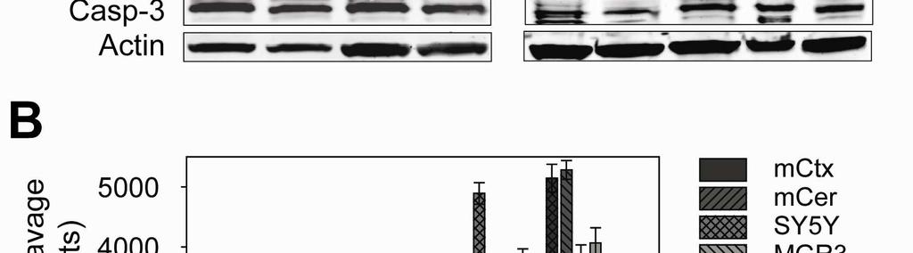
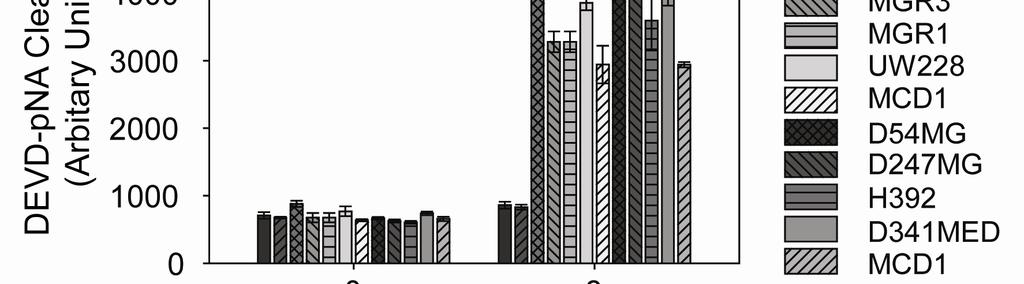

83 Figure
84 Figure 2.3: Brain tumors from mouse models of high-grade astrocytoma and medulloblastoma display sensitivity to cytochrome c-mediated apoptosis. A and B, Extracts, prepared from tumor tissues and adjacent neural tissues of two brain tumor mouse models, highgrade astrocytoma (A) and medulloblastoma (B), were supplemented with cytochrome c and caspase activation was monitored. C, Immunoblotting shows caspase-9 cleavage in extracts treated in (A) and (B). Astro.: Astrocytoma; Adj. Cer.: adjacent cerebellum. 73

85 Figure
86 Figure 2.4: A marked increase in Apaf-1 causes the increased sensitivity of brain tumor tissues to cytochrome c-mediated apoptosis. A, Immunoblots demonstrating Apaf-1 protein levels in high-grade astrocytoma (Astro.) and medulloblastoma (Med.) relative to their respective adjacent neural tissue (Cer.: cerebellum). Quantitation of Apaf-1 (mean ± SEM of three independent experiments) is shown. B, Caspase activation in extracts from mouse cortex, cerebellum and P19 CGNs were assessed in the presence of no cytochrome c, 8µM cytochrome c, or 8µM cytochrome c along with 1µg of recombinant Apaf-1. C, In vitro assay assessing caspase activation in mouse cortical extracts when IAPs were inactivated (by Smac addition) or when XIAP was genetically removed (XIAP-/-). D, Immunoblots showing relative Apaf-1 levels in human cortex (Ctx) versus four samples of high-grade gliomas, as well as E, caspase activation assay on human cortical extracts in the presence of no cytochrome c, 8µM cytochrome c, or 8µM cytochrome c along with 1µg of recombinant, Apaf-1. 75
87 Figure
88 Figure 2.5 Transcriptional regulation of Apaf-1 mrna levels contributed by E2F1 A, Quantitative analysis from RT-PCR shows the fold changes of Apaf-1 mrna from dissected medulloblastoma and adjacent cerebellar tissues. Cells isolated from medulloblastoma tumor (Med.) were compared with normal P7 and adult cerebellar tissue. Data display the fold changes of Apaf-1 mrna relative to normal P7 cerebellum, which is arbitrarily set as 1, and a corresponding agarose gel is shown for the PCR. B, Oncomine analysis of three independent gene profiling studies, with data from Sun et al. (Sun, Hui et al. 2006) used to compare Apaf-1 mrna expression levels in brain from epilepsy patients and in glioblastoma (*, p < , independent two-tailed t test), and with data from Phillips et al. (Phillips, Kharbanda et al. 2006) and Freije et al. (Freije, Castro-Vargas et al. 2004) analyzed using the Comprehensive Metaanalysis software to plot the standard difference in means along with the 95% confidence intervals for Apaf-1 mrna in Grade III Astrocytoma (corresponds to 0 on the X-axis) compared with Glioblastoma (positive values indicate an increase in Apaf-1 expression in glioblastoma versus grade III astrocytoma). C, A human tissue dot blot with duplicated samples demonstrates Apaf-1 levels in grade II astrocytoma and glioblastoma. Areas within the white circles represent sample location. D, Immunoblotting shows levels of E2F1 and p53 in brain tumor tissues versus adjacent brain tissue from mouse models of medulloblastoma and high-grade astrocytoma. E, Chromatin immunoprecipitation (ChIP) assay demonstrates E2F1 asssociation with Apaf-1 promoter in human glioblastoma cell line, MGR3, and in mouse medulloblastoma tissue. 77
89 Figure
90 CHAPTER THREE: Lack of XIAP Confers Vulnerability to Neurons
91 A. Introduction Most studies on the regulation of apoptosis have been explored in mitotic cell lines. Only recently has there been an appreciation to utilize primary cells to discover how this intricate apoptotic pathway can be modulated in different cell types (Deckwerth and Johnson 1993; Deshmukh and Johnson 1997; Tanabe, Eguchi et al. 1998; Contestabile 2002; Kitsis and Mann 2005). In particular, neurons, cardiomyocytes and myotubes cells of postmitotic nature are more accurately depicted by primary cell cultures, rather than immortalized cell lines that remain in the cell cycle and continue to proliferate. Recent studies with primary cultures have pointed to differences in apoptosis regulation between postmitotic and mitotic cells (Koseki, Inohara et al. 1998; Deshmukh, Kuida et al. 2000; Wright, Linhoff et al. 2004). For example, a more stringent regulation of apoptosis is imposed in developed postmitotic neurons as compared to immature neurons. This increased restriction observed in mature, terminally differentiated neurons is beneficial as these cells have limited regenerative capacity and need to last a lifetime of an organism. In all cells, the crucial mediators of apoptosis are caspases, a family of cysteine aspartate proteases (Cohen 1997; Degterev, Boyce et al. 2003). When a mammalian cell receives an apoptotic signal and is committed to die, the integrity of mitochondria is lost and cytochrome c, a protein that resides in the mitochondrial intermembrane space, is released into the cytosol to trigger a downstream cascade that leads to caspase activation. Cytochrome c once in the cytosol binds to its adaptor protein, Apaf-1 and forms a multimeric complex termed the apoptosome. Procaspase-9 is then recruited to the apoptosome and thus facilitates its autoactivation. Activated caspase-9 in turn can cleave and activate procaspase-3 and procaspase-7, which cleave various cellular proteins that ultimately lead to cell demise (Danial and Korsmeyer 2004). 80
92 Mitotic cells, such as fibroblasts, HEK293 and HeLa, die rapidly when cytochrome c is introduced in the cytosol, as cytochrome c serves as an initiating factor to induce the apoptosome-mediated apoptotic pathway (Liu, Kim et al. 1996; Li, Srinivasan et al. 1997). Surprisingly however, postmitotic sympathetic neurons in culture are remarkably resistant to microinjected cytosolic cytochrome c (Wright, Linhoff et al. 2004). This differential sensitivity to cytochrome c-mediated death between mitotic cells and postmitotic neurons is attributed to the increased effectiveness of endogenous X-linked inhibitor of apoptosis protein (XIAP) in postmitotic neurons (Potts, Singh et al. 2003). Therefore, to induce death in primary cultures of neurons, not only is the release of cytochrome c required, but the XIAP inhibition on caspases also needs to be overcome. XIAP belongs to the IAP family, which also includes ciap-1, ciap-2, and more distant members of NAIP, ILP2, ML-IAP, Survivin and Bruce (Salvesen and Duckett 2002). Despite being ubiquitously expressed in all tissue, the importance of endogenous XIAP in restricting cytochrome c-dependent apoptosis has been demonstrated only in cultures of postmitotic cell types, such as sympathetic neurons, cardiomyocytes and myotubes (Potts, Singh et al. 2003; Potts, Vaughn et al. 2005; Smith, Huang et al. 2009). While enthusiasm of endogenous XIAP playing a crucial role in vivo is challenged by the fact that XIAP knockout mice display no apparent phenotype (Harlin, Reffey et al. 2001),this is not unexpected based on our hypothesis that XIAP functions as a safety break that prevents caspases from being activated if accidental mitochondrial damage results in the release of cytochrome c. Thus, the genetic deletion of XIAP alone is indeed not anticipated to exhibit a phenotype. A prediction of our hypothesis is that XIAP deficiency would make neurons more vulnerable to mitochondrial insults that inadvertently causes release of cytochrome c. 81
93 To test our hypothesis, we examine the survival and functionality of postmitotic neurons in the presence and absence of XIAP under a mitochondrial damaging insult of mutant Cu/Zn superoxide dismutase (SOD1) expression in vitro and in vivo. Mutations in SOD1 are associated with familial amyotrophic lateral sclerosis (fals), a fatal paralytic condition due to a progressive loss of spinal cord motor neurons (Rosen 1993). The exact mechanism of how mutant SOD1 causes the disease remains elusive; though it is proposed that mutant SOD1 has a gain-of-function property (Cluskey and Ramsden 2001; Boillee, Vande Velde et al. 2006). Our rationale for using the SOD1 G93A transgenic mice as an animal model in this study was based on the fact that mitochondrial damage and cytochrome c release are observed in this model of neurodegeneration (Dal Canto and Gurney 1994; Wong, Pardo et al. 1995; Kong and Xu 1998; Guegan, Vila et al. 2001; Xu, Jung et al. 2004). In addition, cytochrome c translocation from the mitochondria to the cytosol of spinal cord is reported to occur well before any detection of activated caspases and development of symptoms. These results suggest that XIAP may play a critical role in restricting activation of apoptosis in young, asymptomatic SOD1 G93A transgenic (Tg) mice. Furthermore, the relative ease of examining symptoms of hindlimb paralysis in SOD1 G93A -Tg mice using simple, straightforward behavioral tests facilitates our analysis of results. Consistent with the results that XIAP serves as a safety brake in cultured sympathetic neurons, we find that XIAP-deficient neurons are indeed more vulnerable than wildtype neurons to mutant SOD1-induced death in cell culture, and, most importantly in the animal model. Behavioral studies showed that XIAP-deficient, SOD1 G93A mice developed symptoms earlier, and with more severity than in the SOD1 G93A transgenic mice alone. These findings are the first to show an important role of XIAP in vivo and identify its function as a safety brake in the 82
94 apoptotic pathway that becomes engaged under stress condition when there is accidental cytochrome c release to prevent unwanted caspase activation. Together, these results show that the loss of XIAP can be a risk factor that would increase susceptibility to neurodegenerative diseases that exhibit mitochondria disruption and cytochrome c release. B. Result XIAP-deficient sympathetic neurons undergo normal development apoptosis Mice deficient in XIAP are born with no gross phenotypic abnormalities and live a normal lifespan (Harlin, Reffey et al. 2001). However, XIAP-deficient sympathetic neurons in culture (equivalent of postnatal day 5-7; P5-7) are reported to be more susceptible to cytochrome c-induced cell death compared to their wildtype (WT) counterpart (Wright, Linhoff et al. 2004). To examine more closely whether a lack of XIAP influences the normal developmental programmed cell death (PCD) of sympathetic neurons in vivo, we evaluated neuronal counts of the superior cervical ganglia (SCG) from either WT or XIAP knockout mice at P6-7. Developmental PCD occurs at different time points for different subpopulations of neurons, therefore, it is important to evaluate neuronal numbers at a time when the normal PCD is near completion to accurately assess the process. In rodents, sympathetic neurons in the SCG, undergo significant decline in neuronal numbers during the first postnatal week (Oppenheim 1991). We found that SCG neuronal counts in WT and XIAP-deficient mice were not significantly different (Figure 3.1), suggesting that XIAP deficiency does not affect the extent of PCD during sympathetic neuronal development. This finding is consistent with our previous 83
95 report that WT and XIAP-deficient sympathetic neurons in culture show no difference in the extent or kinetics of death after trophic factor withdrawal (Potts, Singh et al. 2003). XIAP-deficient neurons are more vulnerable to mutant SOD1-induced death Unlike mitotic cells, sympathetic neurons are remarkably resistant to microinjection of cytosolic cytochrome c due to the strict inhibition of caspases by XIAP in neurons. In contrast to wildtype neurons, the XIAP-deficient neurons are sensitive to cytosolic cytochrome c microinjection (Potts, Singh et al. 2003). To test whether XIAP deficiency also confers vulnerability to neurons that encounter a mitochondrial damaging insult, we microinjected a plasmid containing a human mutation of G93A SOD1 (SOD1 G93A ) along with a GFP plasmid into WT and XIAP-deficient P4-5 sympathetic neurons. Mutant SOD1 G93A was chosen for the initial evaluation in vitro, as these data would complement the later animal studies with SOD1 G93A transgenic mice. Whereas 60% of WT neurons survive from the expression of SOD1 G93A, the XIAP-deficient neurons showed only approximately 36% survival rate at 72 hours after microinjection (Figure 3.2A). Likewise, A4V mutant SOD1, a fals-associated mutation that causes the most aggressive clinical progression in human patients, was also seen to induce a more significant death in XIAP-deficient neurons than in WT neurons (Figure 3.2B). To validate that the mutant SOD1-induced death is mediated through the apoptotic pathway, we further examine the immunostaining pattern of cytochrome c in sympathetic neurons injected with SOD1 G93A in the presence of a caspase-inhibitor, Q-VD-OPH. We find that cytochrome c staining is absence in sympathetic neurons expressing SOD1 G93A while normal neurons exhibit the distinct mitochondrial pattern of cytochrome c staining with high intensity 84
96 (Figure 3.2C). These results indicate that, as anticipated, SOD1 G93A expression caused the translocation of cytochrome c from the mitochondria to the cytosol in sympathetic neurons. Together, these results show that an absence of the XIAP safety brake through genetic deletion indeed renders neurons more susceptible to mutant SOD1-induced apoptotic death. XIAP deficiency alone does not affect developmental apoptosis of lumbar motor neurons and its functionality at older age Our results show that XIAP-deficient neurons in culture are more sensitive to mutant SOD1-induced death. To examine whether XIAP deficiency also makes neurons more vulnerable to neurodegeneration in vivo, we crossed the SOD1 G93A transgenic mice with the XIAP-deficient mice and examined the outcome of SOD1 G93A expression in WT or XIAP deficient background. Transgenic mice expressing SOD1 G93A has been widely studied and characterized as a mouse model of ALS. These mice progressively show motor deficit of hindlimb paralysis (Weydt, Hong et al. 2003). In order to eliminate potential cofounding factor of XIAP deficiency alone having an effect on the development of lumbar motor neuron (MN), which innervates the hind limbs, we first investigated lumbar MN counts after developmental PCD in WT and XIAP-deficient mice. Nissl-stained lumbar MNs from WT and XIAP-deficient mice at P6-7, a time well after the normal developmental PCD in lumbar MNs, which occurs during late embryonic period (Oppenheim 1986), did not show a difference in cell numbers (Figure 3.3A). This result indicate that the lack of XIAP alone does not affect lumbar motor neuron developmental PCD, as in the case of sympathetic neurons. 85
97 Despite the fact that no behavioral phenotype was noted in the initial characterization of XIAP-deficient mice (Harlin, Reffey et al. 2001), we wanted to confirm that the absence of XIAP alone, without any additional stress stimulus, would also not cause any motor deficits at an older age when SOD1 G93A transgenic mice demonstrated motor symptoms. Indeed, XIAPdeficient mice around 20 weeks of age show no difference in their ability of motor coordination compared to the WT mice as the time for the two genotypes of mice to remain on the accelerating rotarod were similar (Figure 3.3B). Furthermore, in assessing for paw grip strength, both WT and XIAP-deficient mice were capable of holding onto the metal cage bar against gravity, as the measurement of latency to fall in the 60-second-trial show similar result with no statistical difference (Figure 3.3C). Together, these data illustrated that XIAP deficiency alone did not induce any changes in the developmental PCD of lumbar MNs and no deficits in motor functions were observed. XIAP-deficiency confers an early onset and more severe neurodegenerative symptoms in SOD1 G93A transgenic mice To test the hypothesis that XIAP deficiency would make neurons more vulnerable to a mitochondrial damaging insult in an animal model, we generated SOD1 G93A transgenic animals in WT and XIAP-deficient genetic background. With XIAP being on the X chromosome, we crossed female XIAP heterozygous (XIAP+/-) with male SOD1 G93A transgenic (Tg) mice to obtain offspring with the desirable phenotypes of SOD1 G93A -Tg and XIAP-deficient/ SOD1 G93A - Tg. Wildtype animals were used as internal controls. There was no difference between the survival of XIAP-deficient/ SOD1 G93A -Tg and SOD1 G93A -Tg mice (Supplemental Figure 3.1). 86
98 To gain a comprehensive view of the disease progression, mice of the three genotypes were assessed weekly starting at 10 weeks of age for their motor function. In the accelerating rotarod test, XIAP-deficient/ SOD1 G93A -Tg mice displayed a significant, earlier onset of motor coordination deficit starting at 17 weeks of age, a time when the SOD1 G93A -Tg or WT mice exhibited no phenotype (Figure 3.4A). Moreover, between weeks, XIAP-deficient/ SOD1 G93A -Tg mice showed a more severe phenotype than SOD1 G93A -Tg mice. The rate of disease progression was however similar between the two groups. Beyond 20 weeks of age, there was no significant difference in rotarod performance between SOD1 G93A -Tg mice in either WT or XIAP-deficient background. This lack of difference in the performance between XIAPdeficient/ SOD1 G93A -Tg mice and SOD1 G93A -Tg mice beyond 20 weeks of age could be due to the decreased XIAP level in the spinal cord of SOD1 G93A -Tg mice (Supplemental Figure 3.2), thus allowing motor neuron death. A similar trend was observed in the wire hang test when assessing the paw grip strength of these animals (Figure 3.4B). At 17 weeks of age, XIAP-deficient/SOD1 G93A -Tg mice showed an early decline in their ability to grip onto the metal bar to prevent themselves from falling from the grid. During weeks, XIAP-deficient/SOD1 G93A -Tg mice continued to demonstrate a significant incapability in the wire hang test compared to the SOD1 G93A -Tg mice. However, measurements past 20 weeks of age showed comparable impairment in both groups. Together, these behavioral experiments demonstrated that a XIAP deficient background indeed renders SOD1 G93A -Tg mice more susceptible to the development of disease symptoms, as XIAPdeficient/ SOD1 G93A -Tg mice exhibited motor defects that was more severe and appeared earlier than seen in the SOD1 G93A -Tg mice. 87
99 A polymorphism of XIAP found in humans exhibits differential ability to inhibit cytochrome c-mediated death A prediction based on our animal studies, is that any polymorphisms in XIAP that reduces its ability to inhibit caspases could be a potential risk factor that increases sensitivity to neurodegenerative diseases. In searching through the Single Nucleotide Polymorphism (SNP) database, we found that a glutamine to proline change at the amino acid residue 423 (listed in dbsnp as rs ) of XIAP has been reported in humans to occur at a high frequency (40%). To test whether this particular amino acid change has an effect on the anti-apoptotic function of XIAP, we generated the XIAP variants of 423Q and 423P, introduced these into XIAP-deficient neurons, and assessed for the sensitivity of these neurons to undergo cytochrome c -mediated death. Previous data showed that restoring wild type XIAP is capable of rescuing XIAPdeficient sympathetic neurons from cytochrome c-mediated death. However, these experiments were conducted with relatively high levels of XIAP expressing plasmid (200 ng/µl) (Potts, Singh et al. 2003). To allow for the detection of any subtle differences between these two XIAP variants in their ability to inhibit caspases, we injected the XIAP expressing plasmids into neurons at low concentration (2 ng/µl). Our results show that the 423Q variant of XIAP was able to protect XIAP-deficient neurons to cytosolic microinjection of cytochrome c when compared to GFP expressing plasmid as a control (Figure 3.5A). 40.3% of cells survived after 3 hours of cytochrome c injection in the 423Q variant of XIAP expressed condition as compared to a 13.3% of cell survival in GFP expressed condition. In contrast, the 423P variant of XIAP, was incapable of rescuing XIAP-deficient sympathetic neurons from cytochrome c-mediated death at these concentrations, as only a 13.1% of cell survival was observed (Figure 3.5A). 88
100 Correspondingly, XIAP-deficient neurons expressing the 423Q variant of XIAP exhibit phasebright, intact cell membrane 3 hours after cytochrome c injection while those with 423P XIAP expression displayed a distended cellular membrane and loss of contrast in the phase photo (Figure 3.5B). Together, these results show that the naturally occurring polymorphism at amino acid 423 in XIAP exhibits a differential capability of regulating cytochrome c-mediated apoptosis, and thus has the potential to be a risk factor for increased susceptibility to neurodegenerative diseases in humans. C. Discussion Our lab had previously shown that sympathetic neurons are remarkably resistant to cytosolic cytochrome c microinjection, and only upon the removal of XIAP through either genetic deletion or addition of exogenous Smac protein (an inhibitor of IAPs) can cytosolic cytochrome c then be capable of inducing cell death in these neurons (Potts, Singh et al. 2003). This established the importance of endogenous XIAP in providing a stringent regulation of apoptosis in neurons, a feature that is arguably beneficial to these postmitotic cells as they have limited proliferative potential and are needed to last for a lifetime of an organism. Therefore, in mammalian neurons, caspase activation requires not only the induction of cytochrome c release but also the relief of XIAP inhibition; having either one alone being insufficient to cause apoptosis. XIAP is therefore anticipated to be engaged as a safety brake in neurons to protect against any accidental caspase activation if cytochrome c is unexpectedly released into the cytosol from any mitochondrial damage. 89
101 In this study, we showed that neurons lacking the XIAP brake indeed become more vulnerable to the toxic stimulus of mutant SOD1 expression that causes mitochondrial damage and cytochrome c release (Figure 3.2). Importantly, our in vivo studies of WT and XIAPdeficient mice stressed by SOD1 G93A -Tg expression also showed that a lack of XIAP confers vulnerability to the pathological insult (Figure 3.4). The SOD1 G93A -Tg mouse model of ALS has been extensively characterized and studies have shown that the neuronal dysfunction and death observed in the SOD1 G93A -Tg mouse model are in part due to the activation of the intrinsic apoptotic pathway. Interestingly, early vacuolization and mitochondrial damage in motor neurons of SOD1 G93A -Tg mice are observed well before the onset of disease and any activation of apoptosis (Dal Canto and Gurney 1994; Wong, Pardo et al. 1995; Kong and Xu 1998; Guegan, Vila et al. 2001; Xu, Jung et al. 2004). Corresponding to the early observation of mitochondrial alteration, cytochrome c was also shown to be released from the mitochondria into the cytosol. Activation of caspases and features of apoptosis, however, were observed only during the late stage of the disease (Guegan, Vila et al. 2001; Tun, Guo et al. 2007). This discrepancy between early mitochondrial dysfunction with the release of cytochrome c and late involvement of caspase activation made the SOD1 G93A -Tg mice an excellent model to test the role of endogenous XIAP in vivo. Here we found that SOD1 G93A -Tg mice in a XIAP-deficient background exhibited an early decline in motor function and continued to show a more severe phenotype as compared to SOD1 G93A -Tg mice in a WT background (Figure 3.4A, B). Despite the strict inhibition of caspases by XIAP, apoptosis does occur in neurons during developmental periods and under pathological insults. Under these conditions when neurons become committed to die, the function of XIAP appears to be inhibited to allow for an apoptotic death. There are several ways by which the effectiveness of XIAP inhibition on caspases can be 90
102 diminished. First, the levels of functional XIAP can be decreased through its transcriptional downregulation and/or degradation (Perrelet, Perrin et al. 2004; Tun, Guo et al. 2007). In a physiological setting, the withdrawal of tropic factor can cause a decrease in XIAP mrna level to allow for apoptosome-mediated caspase activation as seen in the development of sympathetic neurons (Potts, Singh et al. 2003). Recently, S-nitrosylation of XIAP was shown to be another way by which XIAP activity could be inhibited (Tsang, Lee et al. 2009). Lastly, a high ratio of Apaf-1 to XIAP can also overcome the XIAP brake, as high levels of Apaf-1 result in robust apoptosome formation and increase caspase activation that cannot be successfully inhibited by XIAP (Wright, Linhoff et al. 2004). In the case of fals-associated SOD1 G93A -Tg mice, XIAP is reported to be cleaved or reduced in levels at the end stage of disease when caspase activation is seen concomitantly (Ishigaki, Liang et al. 2002; Tokuda, Ono et al. 2007). In this study, we showed that, at the end stage of disease, the level of XIAP protein in the spinal cord is indeed decreased (Supplemental Figure 3.2). Intriguingly, we also found Apaf-1 protein levels to be elevated in spinal cord at the late stage of SOD1 G93A -Tg mice compared to age-matched WT mice (Supplemental Figure 3.2). The increase in Apaf-1 and the decrease in XIAP together would tilt the Apaf-1: XIAP ratio more significantly favoring apoptosis to occur in these mice. Additionally, the reactivation of Apaf-1 in SOD1 G93A -Tg mice during the late stage suggests that there is indeed sufficient Apaf-1 for these mature neurons to undergo apoptosis as mature neurons otherwise have been shown to be incapable of activating caspases even with the removal of XIAP because of restricted Apaf-1 expression (Wright, Smith et al. 2007). Although we used the ALS mouse model here to demonstrate an important function of XIAP for neuroprotection, XIAP deficiency could be a risk factor of other neurological pathologies as well. For example, in human patients with Parkinson s disease (PD), XIAP is 91
103 shown to be S-nitrosylated, which affects its ability to inhibit caspases (Tsang, Lee et al. 2009). Nitrosative stress that impairs the function of the prosurvival XIAP protein partly contributes to PD pathogenesis, implicating that XIAP deficiency may be a risk factor that may accelerate the disease course of PD. In addition to neurodegenerative diseases, a lack of XIAP has also been recently shown to increase apoptosis and tissue loss following neonatal brain injury of hypoxic ischemia. Together, these studies and ours highlight the importance of XIAP in regulating activation of caspases in neurons in vivo. Our functional studies of an XIAP polymorphism at amino acid 423 showed that the 423Q variant of XIAP is more capable of inhibiting cytochrome c-mediated death than the 423P variant (Figure 3.5). One prediction of these results is that individuals more prone to neurodegenerative diseases may have a higher allelic frequency of 423P than 423Q in XIAP, because of the reduced capability of 423P to inhibit apoptosis. Maintaining a delicate balance of XIAP s effectiveness could be critical, as a decreased in XIAP activity can be a risk factor to neurodegenerative diseases. However, excessive XIAP activity is also not favorable, as increased XIAP can inhibit apoptosis and facilitate tumorigenesis. Therefore, we speculate that the polymorphism of XIAP at amino acid 423 may exist because of the evolutional pressure to strike a balance between increased or reduced XIAP activity to maintain homeostasis for optimum organismal survival. D. Material and Methods Immunoblotting 92
104 Western blots were performed as previously described (Potts, Singh et al. 2003). Primary antibodies used include: anti-apaf1 (Alexis); anti-xiap (R&D); anti-α-tubulin (T9026; Sigma); anti-β-actin (A5316; Sigma). HRP-conjugated secondary antibodies purchased from Pierce Chemical Co. were used along with ECL-Plus detection system (GE Healthcare). Densitometry was performed using NIH ImageJ software and normalized to the levels of a loading control protein on the representative Western blot. Primary sympathetic neuron cultures Primary sympathetic neurons from the superior cervical ganglia were dissected out from P0-1 neonatal XIAP-deficient mice and WT littermates. Ganglia were treated with 1mg/ml collagenase and 2.5mg/ml trypsin for 30 minutes respectively at37 C. Afterwards, dissociation of cells was accomplished by passing superior cervical ganglia through a fire-polished glass pipet and the cells were then plated on rat tail collagen-coated 35mm culture dishes. Cells were maintained in MEM with Earle s alts supplemented with 50ng/ml NGF, 10% FBS, 2mM glutamine, 100U/ml penicillin, 100µg/ml streptomycin, 20µM flourodeoxyuridine, 20µM uridine and 3.3µg/ml aphidicolin. Microinjection and Quantitation of Cell Survival Microinjection of cells was done using a Narashigi micromanipulator mounted on a Leica inverted florescent microscope. For DNA microinjection, twenty to fifty cells were injected in each experiment with 200ng/µl of the indicated plasmid and 50ng/µl enhanced GFP (Clontech). Only in situation specifically listed, the low concentration (2ng/µl) of plasmid was used. The microinjection buffer contains 100mM KCL, 10mM KPi, ph 7.4. Cells were injected and allowed to express the plasmid DNA for 12 hours prior to experimentation. Cell survival was 93
105 evaluated by counting clearly identifiable cells with intact morphology. Surviving cells in culture were counted at the indicated times and expressed as a percentage of the number of cells assessed at the designated 0 hr, which would be 12 hours after DNA microinjection and immediately after protein injection. This method of assessing survival correlates well with other cell survival assays such as trypan blue exclusion and staining with calcein AM (Potts, Singh et al. 2003). Animals SOD1 G93A -Tg mice were obtained from the Jackson Laboratory (Bar Harbor, ME) and XIAP knockout mice were a gift from Dr. Craig Thompson (University of Pennsylvania). Because XIAP is a X chromosome-linked gene, to generate littermates of WT, SOD1 G93A -Tg and XIAPdeficient/ SOD1 G93A -Tg animals, XIAP+/- females were crossed with SOD1 G93A -Tg males. Genotypes of all animals were confirmed with PCR. All procedures for generating the tissues used in this study were approved by the Animal Care and Use Committees at UNC-CH and compiled with the National Institutes of Health Guidelines for the Care and Use of Laboratory Animals. Histology and neuron quantification P6-7 animals were anesthetized by hypothermia followed by perfusion with Bouin s solution. The body without internal organs were immersed in Bouin s solution for several days and processed for paraffin sectioning. Procedures were similar for adult mice but anesthesia was performed using Avertin at 250mg/g body weight of the mice. Serial transverse sections (6-8um) were obtained from postnatal and adult mice, stained with either hematoxylin-eosin H&E) or thionin. Neurons were counted blind in every 10 th section for both lumbar spinal cord and 94
106 superior cervical ganglia using a method previously validated against a stereological optical dissector method (Clarke and Oppenheim 1995). Mice Behavioral Testing Mice were tested on an accelerating rotarod (Ugo Basile, Stoelting Co., Wood Dale, IL) to assess for motor coordination. Revolutions per minute (Rpm) was initially set at 3 and progressively increased to a maximum of 30rpm at the end of a 5-min-trial. Mice were first given 3 training trials with 45 seconds between each trial at the age of 5-6 weeks and then were tested weekly. Latency to fall or invert off from the top of the rotating barrel were recorded. Those mice that were unable to stay on the rotarod for at least 120 second during the test trial were given a second trial and the longest latency from two trials per test session was taken. To assess for paw grip strength, mice were placed on a large metal cage lid and the lid was gently shaken before a 180-degree-inversion. Latency for mice to lose the grip of the hindlimbs and to fall from the lid was measured. A maximum trial length of 90 seconds was given to each mouse. 95
107 Figure 3.1 Developmental apoptosis occurs normally in sympathetic neurons of XIAPdeficient mice Counts of Nissl-stained sympathetic neurons of the superior cervical ganglia (SCG) were obtained from P6-7 XIAP-deficient and wildtype (WT) mice. Data shown are mean ± SD from animals of n 4. 96
108 Figure 3.1 Neuronal Counts Superior Cervical Ganglia (SCGs) WT XIAP KO Genotype 97
109 Figure 3.2 XIAP-deficient neurons are more vulnerable to SOD1-induced apoptosis XIAP-deficient and wildtype (WT) postnatal day 4 (P4) sympathetic neurons were injected with a plasmid expressing either G93A (A) or A4V (B) mutant form of SOD1 (200ng/µl) along with a plasmid expressing GFP (50ng/µl). Twelve hours after injection, GFP expressing neurons were counted and the number was set as a hundred percent. Cell survival was assessed seventy-two hours after injection. Data plotted are means ± SD of three or more experiments. C, The levels and localization of cytochrome c in G93A SOD1 expressing neuron were examined via immunofluorescence. Arrow points to a G93A SOD1 expressing neuron that has no cytochrome c staining pattern while the arrowhead points to a GFP-expressing healthy, normal neuron which exhibits a typical mitochondrial staining pattern. 98
 C SOD1 (G93A) + GFP GFP Percent Survival 80 60 40 20 0 XIAP+/+ XIAP-/-")
110 Figure 3.2 A 100 SOD1 (G93A) C SOD1 (G93A) + GFP GFP Percent Survival XIAP+/+ XIAP-/- GFP B 100 SOD1 (A4V) Cty c Percent Survival Hoechst 0 XIAP+/+ XIAP-/- 99
111 Figure 3.3 XIAP-deficient mice have normal developmental apoptosis in lumbar motor neurons and exhibit no motor symptoms A, Counts of Nissl-stained lumbar motor neurons (MNs) were obtained from P6-7 XIAPdeficient and wildtype (WT) mice. Data shown are mean ± SD from animals of n 4. Motor coordination (B) and paw grip strength (C) of XIAP-deficient and wildtype (WT) mice beyond 20 weeks of age were assessed on an accelerating rotarod and a wire hang test, respectively. B, Latency to fall or invert off from the top of the rotating barrel was recorded. Mice which were unable to stay on the rotarod for at least 120 seconds were given a second trial and the longest latency from the two trials was taken. C, Latency to fall from the inverted wire grid was recorded and the maximum trial length was 60 seconds. Data shown are means (±SEM) for each group. 100
112 Figure 3.3 A Neuronal Counts Lumbar Motor Neurons WT XIAP KO Genotype B Latency to Fall (sec) Rotarod anaylsis WT XIAP KO Genotype C Latency to Fall (sec) Wire hang test WT XIAP KO Genotype 101
113 Figure 3.4 XIAP-deficiency confers an early onset and more severe neurodegenerative symptoms in SOD1 G93A transgenic mice Motor coordination (A) and paw grip strength (B) of SOD1 G93A transgenic mice in XIAPdeficient and wildtype (WT) background from 10 weeks of age were assessed every week on an accelerating rotarod and a wire hang test, respectively. Wildtype mice were also assessed as an internal control. A, Latency to fall or invert off from the top of the rotating barrel was recorded. Mice which were unable to stay on the rotarod for at least 120 seconds were given a second trial and the longest latency from the two trials was taken. B, Latency to fall from the inverted wire grid was recorded and the maximum trial length was 90 seconds. Data shown are means (±SEM) for each group. *p<0.05, comparison to wildtype group, **p<0.05, comparison to both other groups. 102
114 Figure 3.4 A B Latency on Rotarod (sec) Latency to Fall (sec) Wildtype SOD1 SOD1/XIAP Age in Weeks Wildtype SOD1 SOD1/XIAP ** ** ** ** ** * * * * ** ** * * Age in Weeks * ** ** * * 103
115 Figure 3.5 A polymorphism of human XIAP at amino acid 423 has differential ability to inhibit cytochrome c-mediated death A, XIAP-deficient postnatal day 4 (P4) sympathetic neurons were injected with a plasmid expressing either 423Q XIAP or 423P XIAP (2ng/µl) along with a GFP plasmid (50ng/µl), or with a GFP plasmid alone. Twenty-four hours after plasmid injection, 10mg/ml cytochrome c protein was injected into GFP expressing neurons. Cell survival of GFP expressing neurons was assessed 3 hours after cytosolic cytochrome c injection. Data shown are mean ± SD of three or more experiments. Unpaired student t-test reported p<0.05 when comparing 423Q XIAP expressing neurons to either GFP expressing or 423P XIAP expressing neurons. B, Arrow points to neurons expressing either 423Q or 423P XIAP and injected with cytochrome c. Photographs were taken 3 hours after cytosolic cytochrome c microinjection. 104


116 Figure 3.5 B XIAP 423Q XIAP 423P A Phase 60 % Survival GFP 0 GFP 423Q Cyt c 423P 105
117 Supplemental Figure 3.1 SOD1 G93A transgenic mice in XIAP-deficient background show no difference in survival compared to SOD1 G93A transgenic mice Kaplan-Meier survival curves of XIAP-deficient/SOD1 G93A transgenic mice, SOD1 G93A transgenic mice, and WT mice. 106
118 Supplement Figure Survival (%) Wildtype SOD1 SOD1/XIAP Age (Days)
119 Supplemental Figure 3.2 A decrease of XIAP and an increase of Apaf-1 in spinal cord of 5- month-old SOD1 G93A transgenic mice Spinal cords from SOD1 G93A transgenic mice and its wildtype littermate were collected at 1 or 5 month of age. Levels of XIAP and Apaf-1 proteins in spinal cord lysates were assessed via Western analysis. β-actin serves as a loading control. 108

120 Supplement Figure
121 IV. APPENDICES Appendix A: The Role of Endogenous Smac in Cardiomyocyte Apoptosis In this dissertation, I have examined the importance of XIAP in regulating neuronal apoptosis after the point of cytochrome c release. In Chapter 3 in particular, I described and validated that XIAP acts as a safety mechanism to protect neurons from accidental cytochrome c release and unwanted caspase activation. The effectiveness of endogenous XIAP in postmitotic neurons to stringently regulate their death is beneficial as these terminally differentiated cells cannot further proliferate in stressful situations and need to last for the lifetime of an organism. This idea of postmitotic neurons restricting their apoptotic pathway to ensure for long term survival led to the examination of whether other postmitotic cell types, such as cardiomyocytes and myotubes, also exhibit similar restriction to apoptosis. Indeed, published work from our laboratory showed that despite the differences in morphology and functionality amongst neurons, cardiomyocytes and myotubes, these postmitotic cells all engage the XIAP safety brake and are resistant to cytochrome c-mediated death (Potts, Singh et al. 2003; Smith, Huang et al. 2009). In this section, I describe experiments in which I examined whether endogenous Smac, shown to bind and inhibit IAPs in vitro, can overcome the XIAP-mediated postcytochrome c inhibition in cardiomyocytes. Smac was initially identified in mammalian cells as a protein that enhanced caspase activation in a biochemical assay (Du, Fang et al. 2000; Srinivasula, Datta et al. 2000), and was subsequently found to be a XIAP binding protein (Verhagen, Ekert et al. 2000). The immature form of Smac protein is synthesized in the cytosol but then relocates to the mitochondria of
122 healthy cells via an N-terminal mitochondrial targeting sequence. Processing of Smac protein occurs in the mitochondria, where the mitochondrial localizing sequence is cleaved off, exposing a new N-terminus with the first four amino acids being AVPI. During an apoptotic insult, this mature form of Smac can be released from the mitochondria, along with other mitochondrial intermembrane proteins such as cytochrome c, and interact with XIAP in the cytosol. In vitro studies have shown that the N-terminal AVPI can bind to XIAP and abrogate its ability to inhibit caspases (Liu, Sun et al. 2000). Although Smac is a potent inhibitor of XIAP in vitro, the role of endogenous Smac remains elusive. In the three postmitotic cell types examined (neurons, cardiomyocytes, and myotubes), ectopic injection of AVPI-Smac but not the mutant form of MVPI-Smac, is capable of overcoming the XIAP inhibition on caspases to allow cytochrome c to induce apoptosis. However, the release of endogenous Smac from the mitochondria, interestingly, has different effects in neurons and myotubes. Using hydrogen peroxide to induce release of endogenous cytochrome c and Smac from the mitochondria, NGF-maintained wildtype sympathetic neurons are still incapable of undergoing cell death, as determined by low active capsase-3 staining (Potts, Singh et al. 2003). In contrast, using truncated Bid (tbid), a pro-apoptotic BH3-only protein, to release endogenous cytochrome c, Smac and other mitochondrial factors, wildtype myotubes undergo a rapid caspase-mediated cell death. The importance of endogenous Smac for relieving XIAP in myocytes is unquestionably demonstrated as Smac-deficient myotubes are completely resistant to tbid-mediated death (Smith, Huang et al. 2009). Thus, despite having XIAP as a common mechanism for inhibiting caspase activation in these postmitotic cells, the ability of endogenous Smac to overcome the XIAP inhibitory effect differs between neurons and myotubes. 111
123 Therefore, I conducted experiments to examine whether endogenous Smac plays a role in cardiomyocyte apoptosis. To examine whether the release of endogenous mitochondrial factors, including Smac and cytochrome c, overcome XIAP inhibition and allow cardiomyocytes to die, rat cardiomyocytes were transfected with tbid-gfp plasmid or GFP plasmid alone and examined six hours after transfection. By immunohistochemistry, 70% of tbid-transfected rat cardiomyocytes showed active caspase-3 staining while only 15% exhibited active caspase-3 staining in the GFPtranfected cardiomyocytes (Figure A.1A). In addition, as illustrated in Figure A.1B, tbidtransfected rat cardiomyocytes became rounded and die whereas those in the presence of the pan caspase inhibitor z-vad-fmk remained alive. Together, these data suggest that the release of endogenous mitochondrial factors in cardiomyocytes, including Smac and cytochrome c, is capable of inducing an apoptotic cell death similar to myotubes. If Smac is responsible for overcoming XIAP inhibition upon tbid-mediated cardiomyocyte death, then one would predict that cardiomyocytes isolated from Smac-deficient mice would be resistant to cell death while wildtype cardiomyocytes would die. Mouse cardiomyocytes, unlike those from rats, are not transfectable and are also sensitive to the act of microinjection. Therefore, to express tbid in mouse cardiomyocytes, I generated an adenovirus expressing tbid-gfp. Due to the inherent toxicity of tbid expression, I designed a recombinant virus of Ad-tBid-GFP that is responsive to TetR regulation and can be amplified in 293TREx cells without cytotoxicity (Figure A.2). The MluI-XbaI fragment of pcdna4/to (Invitrogen) containing a CMV promoter with TetO 2 was ligated into a MluI-NheI-digested pshuttle2 (BD Bioscience) to create pshuttle2/to. tbid-egfp was PCR amplified with DraI-XbaI ends from a plasmid obtained from Dr. Doug Green and cloned into pshuttle2/to. Subsequent steps in 112
124 generating the recombinant virus were according to manufacturer's instructions of BD Bioscience Adeno-X Expression System. Surprisingly, Smac-deficient mouse cardiomyocytes infected with tbid-gfp adenovirus died in a similar fashion as wildtype cardiomyocytes when examined by time-lapse microscopy over a 12-hour-period (Figure A.3). Truncated Bid-infected wildtype mouse cardiomyocytes, as expected, showed only 20% survival and could be rescued by a pan caspase inhibitor z-vadfmk. Interestingly, Smac-deficient mouse cardiomyocytes also showed a similar survival rate to wildtype cardiomyocytes at 23% when infected with tbid-gfp adenovirus. The death of Smacdeficient cardiomyocytes upon tbid expression is apoptotic as it can be blocked by the addition of z-vad-fmk (Figure A.3). Therefore, unlike myotubes, endogenous Smac is not essential for overcoming XIAP inhibition in cardiomyocytes. Western analysis of Smac protein levels showed that myotubes and cardiomyocytes expressed comparable levels of Smac (Figure A.4) despite differences in the capability of endogenous Smac to relieve XIAP function in these two cell types. Intriguingly, the fact that Smac-deficient cardiomyocytes undergo an apoptotic tbidmediated death is indicative of a potential mitochondrial factor in cardiomyocytes that is able to relieve XIAP inhibition. One possibility is that another endogenous mitochondrial XIAP inhibitor, HtrA2, could be the protein in question (Suzuki, Imai et al. 2001; Hegde, Srinivasula et al. 2002). However, some have questioned whether the main function of HtrA2 is to inhibit IAPs, which thereby lessens the possibility that endogenous HtrA2 is the mechanism by which XIAP is inhibited in cardiomyocytes (Li, Srinivasula et al. 2002). An interesting future direction would be to identify the mitochondrial factor(s) responsible for overcoming XIAP and allowing tbid-dependent apoptosis in neonatal mouse cardiomyocytes. To address this in an unbiased 113
125 approach, one can isolate purified mitochondria from heart and skeletal muscle tissues of neonatal mice. Recombinant tbid protein can be added to the mitochondria to release all mitochondrial proteins from these two tissue types. Then using 2D gel electrophoresis along with mass spectrometer analysis, mitochondrial factors that are differentially expressed between the two tissues can then be identified. These factors can then be tested in cell culture assays for their ability to inhibit XIAP. 114
126 Figure A.1 Truncated Bid (tbid) induced caspase-mediated cell death in rat cardiomyocytes A, Rat cardiomyocytes were transfected with tbid-gfp or GFP alone. The percentage of transfected cells expressing active caspase 3 was determined 6 hours after transfection by immunohistochemistry. B, Photographs of rat cardiomyocytes 6 hours after transfection with tbid-gfp in the presence or absence of the caspase inhibitor z-vad-fmk (zvad). 115
127 Figure A.1 116
128 Figure A.2 Generation of pshuttle 2/TO plasmid vector for tbid-gfp adenovirus production The promoter of pshuttle 2 (BD Bioscience) is replaced by the promoter of pcdna4/to (Invitrogen) to create the vector pshuttle 2/TO. The newly synthesized pshuttle 2/TO has two tetracycline operator sequences (TetO 2 ) between the TATA box of the CMV promoter and the transcriptional start site. The TetO 2 sequence itself has no effect on expression. However, when the tetracycline repressor protein is present in 293TREx cells, it effectively binds the TetO 2 sites and blocks transcription initiation. Reminding steps of adenovirus generation is outlined in manufacturer s manual ( 117
129 Figure A.2 CMV+2TetO 2 promoter from pcdna4/to plasmid P CMV 2X TetO 2 Promoter replacement 118
130 Figure A.3 Truncated Bid (tbid) induced apoptosis is not dependent on Smac Wild type (wt) or Smac-deficient (Smac-/-) cardiomyocytes were infected with a tbid-gfp adenovirus in the presence or absence of z-vad-fmk (zvad). Cell survival was determined by morphology over a 12 hour period using time-lapse microscopy. 119

131 Figure A.3 120
132 Figure A.4 Endogenous Smac levels are similar in myotubes and cardiomyocytes Protein levels of Smac were examined by Western blot of whole cell lysates from Smacdeficient dermal fibroblasts (Smac-/- DF), myotubes, cardiomyocytes and sympathetic neurons (neurons). Tubulin serves as a loading control. Densitometry of Smac protein levels are normalized to tubulin levels of the representative blot. 121


133 Figure A.4 122
134 Appendix B Mechanisms Involved in Cytochrome c Degradation The key components of the apoptosome pathway have been discovered through biochemical studies done mostly using mitotic cell lines. Only recently there has been an appreciation of how primary cell types may have additional regulatory mechanisms to control the apoptotic pathway. Previous work from our laboratory has shown that various postmitotic cells, such as sympathetic neurons, cardiomyocytes and myotubes all engage a XIAP regulatory brake to inhibit cytochrome c-mediated caspase activation while mitotic cell lines die rapidly in response to cytosolic cytochrome c (Potts, Singh et al. 2003; Potts, Vaughn et al. 2005; Smith, Huang et al. 2009). In addition, several additional mechanisms for restricting apoptosomemediated cell death are found in the sympathetic neuron model system. First, mature sympathetic neurons are found to silence the expression of Apaf-1 through chromatin modification. Thus, only upon chromatin de-repression and E2F1-dependent transcriptional upregulation of Apaf-1 can these mature neurons undergo apoptosis (Wright, Smith et al. 2007). Second, direct release of endogenous cytochrome c from sympathetic neurons is found to be incapable of inducing cell death even with the relief of the XIAP brake (Vaughn and Deshmukh 2008). NGF-maintained sympathetic neurons maintain a reducing environment such that it is not conducive for the endogenously released cytochrome c to activate the apoptotic pathway as the reduced form of cytochrome c is less efficient than the oxidized form to induce apoptosis. In this section, I describe experiments that myself and others in the lab have conducted to investigate a novel mechanism of regulating the apoptotic pathway at the point of cytochrome c. During apoptosis, cytochrome c translocates from the mitochondria to the cytosol, where it accumulates and binds to the adaptor protein Apaf-1 to form the apoptosome. The assembly of 123
135 the apoptosome leads to downstream activation of caspase zymogens and subsequently causes the demise of the cell (Danial and Korsmeyer 2004). The event of cytochrome c translocation can be visualized by immunofluorescence. Under normal conditions in healthy cells, cytochrome c is localized to mitochondria displaying a punctate pattern of staining. In most cells after an apoptotic treatment, the normal mitochondrial pattern of cytochrome c staining is loss and cytochrome c is seen to be distributed diffusely within the cytosol. Interestingly, our lab has reported that sympathetic neurons upon exposure to various apoptotic stimuli, such as NGF withdrawal or etoposide, have no apparent cytosolic accumulation of cytochrome c (Potts, Singh et al. 2003; Vaughn and Deshmukh 2007). The absence of cytochrome c staining in sympathetic neurons after an apoptotic stimulus is due to proteasome-mediated degradation of cytochrome c as the addition of proteasome inhibitors such as lactacystin or MG132 results in the accumulation of cytosolic cytochrome c (data not shown). This efficient process of cytochrome c clearance in postmitotic neurons could be a potential mechanism of inhibiting apoptosis in situations of mitochondrial damage when cytochrome c may be released accidentally into the cytosol. Interestingly, while assessing cardiomyocyte apoptosis following tbid-gfp adenovirus infection as described in Appendix A, I found that the pattern of cytochrome c release in mouse cardiomyocytes was similar to that of sympathetic neurons (Figure B.1). After 24 hours infection with adenovirus expressing tbid-gfp, in the presence of a caspase inhibitor z-vadfmk, GFP-expressing cardiomyocytes exhibits no apparent cytochrome c staining, whereas neighboring cells not infected with Ad-tBid-GFP have punctate, mitochondrial cytochrome c staining. This suggests that both neurons and cardiomyocytes utilize a similar mechanism of degrading cytochrome c to restrict apoptosis. 124
136 The phenomenon of cytochrome c degradation has also been described by others. In an Apaf-deprived neural precursor cell line, ETNA-/-, treatment with either tunicamycin or actinomycin D caused release of cytochrome c from the mitochondria, but no accumulation of cytochrome c was detected by immunofluorescence and Western analysis. However, with the addition of proteasome inhibitor MG132, levels of cytosolic cytochrome c were restored (Cozzolino, Ferraro et al. 2004; Ferraro, Pulicati et al. 2008). Furthermore, the involvement of the ubiquitin-proteasome system was confirmed by the ubiquitylation of cytochrome c, but the study failed to identify the lysine residue on which cytochrome c is ubiquitinated, as it was not Lys 6, 8 or 9 tested in the study (Ferraro, Pulicati et al. 2008). As primary neurons and cardiomyocytes are limited in quantity and not conducive to biochemical studies, I wanted to find a cell line that would exhibit the same phenomenon of cytochrome c degradation in the cytosol upon an apoptotic stimulus. It was unclear whether such a cell line existed besides the neural precursor cells ETNA-/-, since most cells such as HeLa and HEK293 show the typical cytosolic accumulation of cytochrome c. However, I was able to find that Apaf-1 knockout mouse embryonic fibroblasts (MEFs) fit the criteria. After 16 hours of treatment with a pan kinase inhibitor staurosporine, cytochrome c staining of Apaf-1 knockout MEFs showed cells with three distinct staining patterns (Figure B.2A). First, cells that have not yet released cytochrome c displayed mitochondrial pattern as seen in untreated cells. Second, some cells released the protein and demonstrated a diffuse, bright, cytosolic staining of cytochrome c. The last pattern showed a complete lack of cytochrome c staining, but the presence of living cells was detected by the Hoechst nuclear staining. To complement immunofluorescence, Western analysis of Apaf-1 knockout MEF treated for 22 hours with staurosporine showed a decrease in cytochrome c levels as compared to untreated cells. In 125
137 addition, the proteasome inhibitor MG132 was capable of blocking the degradation of cytochrome c and restoring the level in staurosporine-treated cells (Figure B.2B). To illustrate that Apaf-1 knockout MEFs indeed release cytochrome c from the mitochondria to the cytosol upon multiple apoptotic stimuli, cells were treated with either staurosporine or etoposide (a topoisomerase II inhibitor) to induce death and after 18 hours of treatment, the heavy membrane and the cytosolic fraction of proteins in the cells were examine by Western analysis (Figure B.3). Untreated Apaf-1 knockout MEFs retained all cytochrome c in the heavy membrane fraction which contains mitochondria, while staurosporine- or etoposidetreated cells displayed no cytochrome c in either fractions suggesting that cytochrome c is released from the mitochondria and degraded in the cytosol. Moreover, addition of proteasome inhibitors, MG132 or lactacystin, blocked the degradation of cytochrome c in staurosporine- or etoposide-treated Apaf-1 knockout MEFs and restored the level of cytochrome c in the cytosolic fraction. Together, these data showed that Apaf-1 knockout MEFs provide a valuable system to study proteasome-mediated degradation of cytochrome c. In our lab, we are interested in identifying the responsible E2 ubiquitin conjugating enzyme, as well as the E3 ubiquitin ligase that recognizes cytochrome c as a target for proteasome-mediated degradation. To address which E3 ubiquitin ligase binds to cytochrome c, I took a non-biased approach to find cytochrome c binding partners using a yeast two hybrid system. A schematic of the process is outlined in Figure B.4. Briefly, a bait vector was constructed by cloning human cytochrome c cdna into EcoRI-BamHI-digested pgbkt7 vector (BD Clontech MATCHMAKER) with Trp as the selection marker. No toxicity was seen when the bait vector was transformed into the AH109 yeast strain (provided by Dr. A. Paul Barns) as the growth of yeast colonies on SD/-Trp plate was comparable to that of yeast transformed with a 126
138 control vector (data not shown). In addition, there was no auto-activation with the bait vector alone as colonies seen on SD/-Trp/X-gal plate did not show blue coloration (data not shown). The screen for cytochrome c-interacting proteins is currently in progress and is carried out by the company Creative BioLab using a human fetal brain cdna library (Cat. No: ). The 76-residue ubiquitin protein can exist as either a monomer or polymer through isopeptide-chain linkages on the seven different lysine residues present in the protein (Fang and Weissman 2004). The post-translational modification of a protein by either monoubiquitin or structurally distinct polyubiquitin chains dictates the intracellular signals (Sun and Chen 2004). In fact, the covalent conjugation of ubiquitin (Ub) to a protein can govern various biological processes, such as DNA damage tolerance, trafficking and is not limited to a fate of proteolysis. For example, modification of a protein by polyubiquitin containing Lys 48 linkages is the principle signal for proteasomal delivery while other lysine linkages of ubiquitin may mediate other responses. Therefore I was interested in determining which lysine linkage is present within ubiquitin and was responsible for the degradation of cytochrome c. To address this, I injected sympathetic neurons with various HA-Ub plasmids, provided by Dr. Cam Patterson s lab, which have either one or more lysine resides mutated to arginine to abolish the particular lysine linkage capability. Afterwards, I assessed for cytochrome c status after 48 hours of nerve growth factor (NGF) withdrawal in the presence of a pan caspase inhibitor z-vad-fmk. As expected, expression of HA-Ub WT did not affect the degradation of cytochrome c, as there was no apparent cytochrome c staining in these neurons (Figure B.5). I predicted that blocking the canonical Lys 48 linkage by expression of HA-Ub K48R mutants would block the degradation of cytochrome c in NGF-deprived neurons. Surprisingly, there was little or close to no cytochrome c remaining in neurons injected with HA-Ub K48R (Figure B.5), suggesting that another lysine 127
139 chain linkage is responsible for the proteasome delivery signal. Similarly, with HA-Ub K63R expression and NGF deprivation, neurons did not show any cytochrome c staining (data not shown), suggesting that the Lys 63 linkage is not the primary signal for degradation. Since it is not believed that Lys 63-linked ubiquitin targets proteins for degradation, this result was expected. Interestingly, neurons expressing HA-Ub K29R exhibited an intense cytochrome c staining after NGF withdrawal, as did neurons expressing either HA-Ub K29R-K48R double mutant or HA-Ub K29R-K48R-K63R triple mutant (Figure B.5). Together, these preliminary data suggest that Lys29 linkage on Ub, and not the Lys48 linkage, may be the primary polyubiquitin chain important for targeting cytochrome c for proteasome-mediated degradation. Besides examining the type of polyubiquitin chain involved in the ubiquitin-proteasome pathway, there is an interest in finding which lysine residue on cytochrome c contributes to the isopeptide covalent conjugation to ubiquitin. We expect the ubiquitylation and subsequent degradation of cytochrome c to be a mechanism for preventing apoptosome-mediated cell death. But in addition to this degradation, one attractive hypothesis is that ubiquitylation of cytochrome c may occur on a residue which is important for the binding to Apaf-1. Therefore once ubiquitin is bound to cytochrome c, its binding to Apaf-1 could be eliminated, thereby preventing apoptosome formation while simultaneously inducing cytochrome c degradation. To test this hypothesis, I made constructs of FLAG-Cyt-c with mutations in key lysine residues that are known to be involved in the interaction between cytochrome c and Apaf-1. Future experiments could use these mutant cytochrome c constructs and test whether ubiquitylation occurs on cytochrome c in an in-vitro ubiquitin assay system where Apaf-1 knockout MEFs are transfected with FLAG-Cyt-c constructs and HA-Ub WT. 128
140 These preliminary experiments start to address an interesting phenomenon of cytochrome c degradation and examine the machinery involved. However, more extensive work would be carried on beyond these initial observations by the lab to elucidate the exact mechanism. 129
141 Figure B.1 Loss of cytochrome c staining in cardiomyocytes expressing tbid-gfp Mouse cardiomyocytes were infected with adenovirus expressing tbid-efp in the presence of a caspase inhibitor, zvad-fmk. After 24 hours, cytochrome c and GFP were examined by immunofluorescence. Arrow points to the location of a cardiomyocyte expressing GFP. 130


142 Figure B.1 tbid-gfp Cytochrome c Nucleus 131
143 Figure B.2 Degradation of cytochrome c in Apaf-1 knockout mouse embryonic fibroblasts (MEF) A, Apaf-1 deficient MEF was treated with 1µM staurosporine (STS) in the presence of a pan caspase inhibitor zvad-fmk. After 16 hours of treatment, the levels of cytochrome c, as well as its localization, were determined by immunofluorescence. A yellow arrow points to a cell that has no cytochrome c staining, while a green arrow illustrates a cell that has intense, diffuse staining of cytochrome c. B, Western analysis of cytochrome c levels from whole lysates of Apaf-1 knockout MEF untreated or treated with 1µM staurosporine (STS). Cells were treated with STS for 22 hours in the presence or absence of a proteasome inhibitor, MG132. Tubulin serves as a loading control. 132


144 Figure B.2 A. B. Cyt c Untreated STS Untreated +STS +STS +MG132 Cyt c Tubulin Hoechst 133
145 Figure B.3 Cytochrome c is degraded by the proteasome upon release into the cytosol Apaf-1 deficient MEFs were treated with either 1µM staurosporine (STS) or 20 µm etoposide (Etop) for 18 hours in the presence or the absence of proteasome inhibitors, MG132 or lactacystin (Lact). Cellular fractionation was performed on Apaf-1 knockout MEFs and cytochrome c level was examined in the heavy membrane fraction (m) and the cytosolic fraction (c) via Western analysis. 134
146 Figure B.3 +STS +Etop Untreated +MG132 +Lact. +MG132 +Lact. m c m c m c m c m c m c m c Cyt c LDH COX IV 135
147 Figure B.4 The construction and testing of yeast two hybrid bait vector Human cytochrome c cdna was cloned into the empty bait vector pgbkt7 via the EcoRI and BamHI restriction enzyme sites. Prior to yeast two hybrid library screening for cytochrome c binding partner, the constructed bait vector containing cytochrome c was tested for potential toxicity and auto-activation that would invalid the approach 136
148 Figure B.4 Bait Construction Testing for Autoactivation & Toxicty Human cyt c (NM_018947) Library Screening Adapted from Clontech ( 137
149 Figure B.5 Lysine 29 linkage of ubiquitin targets cytochrome c for degradation in sympathetic neurons Postnatal day 4-5 sympathetic neurons were microinjected with HA tagged wildtype ubiquitin (Ub-WT) or mutant ubiquitin with one or more lysine residue(s) that was/were converted to arginine. A plasmid expressing GFP was also injected into sympathetic neurons to mark cells expressing the plasmids. Twenty four hours after injection, sympathetic neurons were deprived of NGF in the presence of a pan caspase inhibitor zvad-fmk. Cytochrome c levels were determined by immunofluorescence 48 hours after NGF withdrawal. Arrows point to neurons that lost cytochrome c staining. In contrast, arrowheads illustrate neurons that retained intense, diffuse, cytosolic staining of cytochrome c. 138


150 Figure B.5 Ub-WT Ub-K48R Ub-K29R Ub-K29/48R Ub-K29/48/63R Hoechst Cyt c GFP 139
POSTMITOCHONDRIAL REGULATION OF APOPTOSIS IN NEURONS AND CANCER CELLS
 POSTMITOCHONDRIAL REGULATION OF APOPTOSIS IN NEURONS AND CANCER CELLS Allyson Evans Vaughn A dissertation submitted to the faculty of the University of North Carolina at Chapel Hill in partial fulfillment
Programmed Cell Death (apoptosis)
") Programmed Cell Death (apoptosis) Stereotypic death process includes: membrane blebbing nuclear fragmentation chromatin condensation and DNA framentation loss of mitochondrial integrity and release of
Programmed Cell Death (apoptosis) Stereotypic death process includes: membrane blebbing nuclear fragmentation chromatin condensation and DNA framentation loss of mitochondrial integrity and release of
Mechanisms of Cell Death
 Mechanisms of Cell Death CELL DEATH AND FORMATION OF THE SEMICIRCULAR CANALS Carol M. Troy August 25, 2008 FROM: Fekete et al., Development 124: 2451 (1997) PHENOMENOLOGY OF CELL DEATH I. DEVELOPMENT A.
Introduction to pathology lecture 5/ Cell injury apoptosis. Dr H Awad 2017/18
 Introduction to pathology lecture 5/ Cell injury apoptosis Dr H Awad 2017/18 Apoptosis = programmed cell death = cell suicide= individual cell death Apoptosis cell death induced by a tightly regulated
Apoptotic Pathways in Mammals Dr. Douglas R. Green
 Apoptotic Pathways in Mammals Douglas R. Green 1 Apoptosis A form of cell death that is defined morphologically, and features a number of biochemical events Programmed cell death Cell death that occurs
Apoptosis Chapter 9. Neelu Yadav PhD
 Apoptosis Chapter 9 Neelu Yadav PhD Neelu.Yadav@Roswellpark.org 1 Apoptosis: Lecture outline Apoptosis a programmed cell death pathway in normal homeostasis Core Apoptosis cascade is conserved Compare
#19 Apoptosis Chapter 9. Neelu Yadav PhD
 #19 Apoptosis Chapter 9 Neelu Yadav PhD Neelu.Yadav@Roswellpark.org Why cells decide to die? - Stress, harmful, not needed - Completed its life span Death stimulation or Stress Cell Survival Death Functions
GMS 6644: Apoptosis. Introduction
 GMS 6644: Apoptosis Introduction (Feb. 15, 2006) Lei Xiao, Ph.D. Department of Anatomy & Cell Biology UF Shands Cancer Center ARB Rm R4-250, 846-1199, lxiao@ufl.edu Outline of the Lecture Different types
Apoptosome dysfunction in human cancer
 Apoptosis 2004; 9: 691 704 C 2004 Kluwer Academic Publishers Apoptosome dysfunction in human cancer K. M. Hajra and J. R. Liu Department of Obstetrics and Gynecology, University of Michigan Medical School,
Signaling Apoptosis. Scott André Oakes, M.D. Dept. of Pathology Univ. of Calif-San Francisco. Cyt c Release BAX/BAK. Apoptosome Formation
 Signaling Apoptosis Cyt c Release BAX/BAK Apoptosome Formation Caspase Activation Scott André Oakes, M.D. Dept. of Pathology Univ. of Calif-San Francisco Why do we need cell death? Sculpt Organs Control
Apoptotic cell signaling in cancer progression and therapyw
 Integrative Biology Dynamic Article Links Cite this: Integr. Biol., 2011, 3, 279 296 www.rsc.org/ibiology REVIEW ARTICLE Apoptotic cell signaling in cancer progression and therapyw Jessica Plati, a Octavian
Cell Quality Control. Peter Takizawa Department of Cell Biology
 Cell Quality Control Peter Takizawa Department of Cell Biology Cellular quality control reduces production of defective proteins. Cells have many quality control systems to ensure that cell does not build
#19 Apoptosis Chapter 9. Neelu Yadav PhD
 #19 Apoptosis Chapter 9 Neelu Yadav PhD Neelu.Yadav@Roswellpark.org Why cells decide to die? - Stress, harmful, not needed - Completed its life span Death stimulation or Stress Cell Survival Death Functions
p53 and Apoptosis: Master Guardian and Executioner Part 2
 p53 and Apoptosis: Master Guardian and Executioner Part 2 p14arf in human cells is a antagonist of Mdm2. The expression of ARF causes a rapid increase in p53 levels, so what would you suggest?.. The enemy
Molecular biology :- Cancer genetics lecture 11
 Molecular biology :- Cancer genetics lecture 11 -We have talked about 2 group of genes that is involved in cellular transformation : proto-oncogenes and tumour suppressor genes, and it isn t enough to
RAS Genes. The ras superfamily of genes encodes small GTP binding proteins that are responsible for the regulation of many cellular processes.
 ۱ RAS Genes The ras superfamily of genes encodes small GTP binding proteins that are responsible for the regulation of many cellular processes. Oncogenic ras genes in human cells include H ras, N ras,
34 Apoptosis Programmed cell death is vital to the health and development of multicellular organisms.
 Principles of Biology contents 34 Apoptosis Programmed cell death is vital to the health and development of multicellular organisms. Apoptosis is the reason we have separate fingers and toes. During embryonic
Part I Molecular Cell Biology
 1 Part I Molecular Cell Biology RNA Regulation: Advances in Molecular Biology and Medicine, First Edition. Edited by Robert A. Meyers. 2014 Wiley-VCH Verlag GmbH & Co. KGaA. Published 2014 by Wiley-VCH
NFκB What is it and What s the deal with radicals?
 The Virtual Free Radical School NFκB What is it and What s the deal with radicals? Emily Ho, Ph.D Linus Pauling Institute Scientist Department of Nutrition and Food Management Oregon State University 117
Signal Transduction Cascades
 Signal Transduction Cascades Contents of this page: Kinases & phosphatases Protein Kinase A (camp-dependent protein kinase) G-protein signal cascade Structure of G-proteins Small GTP-binding proteins,
Apoptosis-based Therapies: Mechanisms and Applications
 Apoptosis-based Therapies: Mechanisms and Applications Perspective Bcl-2 family members, potential usage of BH3 domains as drug targets Bcl-2/xL inhibitors - Antisense, inhibitors of protein-protein interactions,
Follicular Lymphoma. ced3 APOPTOSIS. *In the nematode Caenorhabditis elegans 131 of the organism's 1031 cells die during development.
 Harvard-MIT Division of Health Sciences and Technology HST.176: Cellular and Molecular Immunology Course Director: Dr. Shiv Pillai Follicular Lymphoma 1. Characterized by t(14:18) translocation 2. Ig heavy
Enzyme-coupled Receptors. Cell-surface receptors 1. Ion-channel-coupled receptors 2. G-protein-coupled receptors 3. Enzyme-coupled receptors
 Enzyme-coupled Receptors Cell-surface receptors 1. Ion-channel-coupled receptors 2. G-protein-coupled receptors 3. Enzyme-coupled receptors Cell-surface receptors allow a flow of ions across the plasma
The discovery of Bcl-2
 The discovery of Bcl-2 Bcl-2: B-cell lymphoma 2 The pro-survival subfamily of Bcl-2 protein family Cloning of Bcl-2 as the oncogene which is deregulated at t(14;18) lymphomas Pioneer works by Tsujimoto
Mitochondria in apoptosis. Jean-Claude Martinou, MD, Ph.D Department of cell biology University of Geneva Geneva, Switzerland
 Mitochondria in apoptosis Jean-Claude Martinou, MD, Ph.D Department of cell biology University of Geneva Geneva, Switzerland The dual role of mitochondria in life and death QuickTime and a TIFF (Uncompressed)
Supplementary Information
 Supplementary Information Significance of p53 dynamics in regulating apoptosis in response to ionizing radiation, and polypharmacological strategies Bing Liu 1,, Divesh Bhatt 1,, Zoltan N. Oltvai 2, Joel
Lecture 14 - The cell cycle and cell death
 02.17.10 Lecture 14 - The cell cycle and cell death The cell cycle: cells duplicate their contents and divide The cell cycle may be divided into 4 phases The cell cycle triggers essential processes (DNA
Apoptosis Oncogenes. Srbová Martina
 Apoptosis Oncogenes Srbová Martina Cell Cycle Control point Cyclin B Cdk1 Cyclin D Cdk4 Cdk6 Cyclin A Cdk2 Cyclin E Cdk2 Cyclin-dependent kinase (Cdk) have to bind a cyclin to become active Regulation
Complexity DNA. Genome RNA. Transcriptome. Protein. Proteome. Metabolites. Metabolome
 DNA Genome Complexity RNA Transcriptome Systems Biology Linking all the components of a cell in a quantitative and temporal manner Protein Proteome Metabolites Metabolome Where are the functional elements?
Cell cycle and apoptosis
 Cell cycle and apoptosis Cell cycle Definition Stages and steps Cell cycle Interphase (G1/G0, S, and G2) Mitosis (prophase, metaphase, anaphase, telophase, karyokinesis, cytokinesis) Control checkpoints
Regulation of Gene Expression in Eukaryotes
 Ch. 19 Regulation of Gene Expression in Eukaryotes BIOL 222 Differential Gene Expression in Eukaryotes Signal Cells in a multicellular eukaryotic organism genetically identical differential gene expression
shehab Moh Tarek ... ManarHajeer
 3 shehab Moh Tarek... ManarHajeer In the previous lecture we discussed the accumulation of oxygen- derived free radicals as a mechanism of cell injury, we covered their production and their pathologic
The inhibitors of apoptosis (IAPs) as cancer targets
 as cancer targets") Apoptosis (2007) 12:1543 1568 DOI 10.1007/s10495-007-0087-3 ORIGINAL PAPER The inhibitors of apoptosis (IAPs) as cancer targets Allison M. Hunter Æ Eric C. LaCasse Æ Robert G. Korneluk Published online:
Apoptosis (2007) 12:1543 1568 DOI 10.1007/s10495-007-0087-3 ORIGINAL PAPER The inhibitors of apoptosis (IAPs) as cancer targets Allison M. Hunter Æ Eric C. LaCasse Æ Robert G. Korneluk Published online:
BIOL 4374/BCHS 4313 Cell Biology Exam #1 February 13, 2001
 BIOL 4374/BCHS 4313 Cell Biology Exam #1 February 13, 2001 SS# Name This exam is worth a total of 100 points. The number of points each question is worth is shown in parentheses. Good luck! 1. (2) The
1. to understand how proteins find their destination in prokaryotic and eukaryotic cells 2. to know how proteins are bio-recycled
 Protein Targeting Objectives 1. to understand how proteins find their destination in prokaryotic and eukaryotic cells 2. to know how proteins are bio-recycled As a protein is being synthesized, decisions
mirna Dr. S Hosseini-Asl
 mirna Dr. S Hosseini-Asl 1 2 MicroRNAs (mirnas) are small noncoding RNAs which enhance the cleavage or translational repression of specific mrna with recognition site(s) in the 3 - untranslated region
Chapter 9. Cellular Signaling
 Chapter 9 Cellular Signaling Cellular Messaging Page 215 Cells can signal to each other and interpret the signals they receive from other cells and the environment Signals are most often chemicals The
Problem Set 8 Key 1 of 8
 7.06 2003 Problem Set 8 Key 1 of 8 7.06 2003 Problem Set 8 Key 1. As a bright MD/PhD, you are interested in questions about the control of cell number in the body. Recently, you've seen three patients
Overview: Conducting the Genetic Orchestra Prokaryotes and eukaryotes alter gene expression in response to their changing environment
 Overview: Conducting the Genetic Orchestra Prokaryotes and eukaryotes alter gene expression in response to their changing environment In multicellular eukaryotes, gene expression regulates development
Genome of Hepatitis B Virus. VIRAL ONCOGENE Dr. Yahwardiah Siregar, PhD Dr. Sry Suryani Widjaja, Mkes Biochemistry Department
 Genome of Hepatitis B Virus VIRAL ONCOGENE Dr. Yahwardiah Siregar, PhD Dr. Sry Suryani Widjaja, Mkes Biochemistry Department Proto Oncogen and Oncogen Oncogen Proteins that possess the ability to cause
Cell Cycle. Trends in Cell Biology
 Cell Cycle Trends in Cell Biology Cell Cycle The orderly sequence of events by which a cell duplicates its contents and divides into two Daughter Cells Activities of a cell from one cell division to the
Ch. 18 Regulation of Gene Expression
 Ch. 18 Regulation of Gene Expression 1 Human genome has around 23,688 genes (Scientific American 2/2006) Essential Questions: How is transcription regulated? How are genes expressed? 2 Bacteria regulate
Proteasomes. When Death Comes a Knock n. Warren Gallagher Chem412, Spring 2001
 Proteasomes When Death Comes a Knock n Warren Gallagher Chem412, Spring 2001 I. Introduction Introduction The central dogma Genetic information is used to make proteins. DNA RNA Proteins Proteins are the
7.06 Cell Biology EXAM #3 April 24, 2003
 7.06 Spring 2003 Exam 3 Name 1 of 8 7.06 Cell Biology EXAM #3 April 24, 2003 This is an open book exam, and you are allowed access to books and notes. Please write your answers to the questions in the
Robbins and Cotran Pathologic Basis of Disease 8th Edition Odabrana poglavlja
 Robbins and Cotran Pathologic Basis of Disease 8th Edition Odabrana poglavlja Apoptosis Apoptosis is a pathway of cell death that is induced by a tightly regulated suicide program in which cells destined
Cell Communication. Chapter 11. Overview: The Cellular Internet
 Chapter 11 Cell Communication Overview: The Cellular Internet Cell-to-cell communication is essential for multicellular organisms Biologists have discovered some universal mechanisms of cellular regulation
Biol403 MAP kinase signalling
 Biol403 MAP kinase signalling The mitogen activated protein kinase (MAPK) pathway is a signalling cascade activated by a diverse range of effectors. The cascade regulates many cellular activities including
Cancer. Throughout the life of an individual, but particularly during development, every cell constantly faces decisions.
 Cancer Throughout the life of an individual, but particularly during development, every cell constantly faces decisions. Should it divide? Yes No--> Should it differentiate? Yes No-->Should it die? Yes-->Apoptosis
Cancer. The fundamental defect is. unregulated cell division. Properties of Cancerous Cells. Causes of Cancer. Altered growth and proliferation
 Cancer The fundamental defect is unregulated cell division. Properties of Cancerous Cells Altered growth and proliferation Loss of growth factor dependence Loss of contact inhibition Immortalization Alterated
Cell Communication. Biology Eighth Edition Neil Campbell and Jane Reece. PowerPoint Lecture Presentations for
 Chapter 11 Cell Communication PowerPoint Lecture Presentations for Biology Eighth Edition Neil Campbell and Jane Reece Lectures by Chris Romero, updated by Erin Barley with contributions from Joan Sharp
Cell Communication. Chapter 11. Biology Eighth Edition Neil Campbell and Jane Reece. PowerPoint Lecture Presentations for
 Chapter 11 Cell Communication PowerPoint Lecture Presentations for Biology Eighth Edition Neil Campbell and Jane Reece Lectures by Chris Romero, updated by Erin Barley with contributions from Joan Sharp
Chapter 6. Antigen Presentation to T lymphocytes
 Chapter 6 Antigen Presentation to T lymphocytes Generation of T-cell Receptor Ligands T cells only recognize Ags displayed on cell surfaces These Ags may be derived from pathogens that replicate within
Cell Communication. Chapter 11. Biology Eighth Edition Neil Campbell and Jane Reece. PowerPoint Lecture Presentations for
 Chapter 11 Cell Communication PowerPoint Lecture Presentations for Biology Eighth Edition Neil Campbell and Jane Reece Lectures by Chris Romero, updated by Erin Barley with contributions from Joan Sharp
T cell maturation. T-cell Maturation. What allows T cell maturation?
 T-cell Maturation What allows T cell maturation? Direct contact with thymic epithelial cells Influence of thymic hormones Growth factors (cytokines, CSF) T cell maturation T cell progenitor DN DP SP 2ry
A particular set of insults induces apoptosis (part 1), which, if inhibited, can switch to autophagy. At least in some cellular settings, autophagy se
, which, if inhibited, can switch to autophagy. At least in some cellular settings, autophagy se") A particular set of insults induces apoptosis (part 1), which, if inhibited, can switch to autophagy. At least in some cellular settings, autophagy serves as a defence mechanism that prevents or retards
A particular set of insults induces apoptosis (part 1), which, if inhibited, can switch to autophagy. At least in some cellular settings, autophagy serves as a defence mechanism that prevents or retards
C-Phycocyanin (C-PC) is a n«sjfc&c- waefc-jduble phycobiliprotein. pigment isolated from Spirulina platensis. This water- soluble protein pigment is
 is a n«sjfc&c- waefc-jduble phycobiliprotein. pigment isolated from Spirulina platensis. This water- soluble protein pigment is") ' ^Summary C-Phycocyanin (C-PC) is a n«sjfc&c- waefc-jduble phycobiliprotein pigment isolated from Spirulina platensis. This water- soluble protein pigment is of greater importance because of its various
' ^Summary C-Phycocyanin (C-PC) is a n«sjfc&c- waefc-jduble phycobiliprotein pigment isolated from Spirulina platensis. This water- soluble protein pigment is of greater importance because of its various
Apoptosome Structure, Assembly, and Procaspase Activation
 Apoptosome Structure, Assembly, and Procaspase Activation Shujun Yuan 1 and Christopher W. Akey 1, * 1 Department of Physiology and Biophysics, Boston University School of Medicine, 700 Albany Street,
Eukaryotic Gene Regulation
 Eukaryotic Gene Regulation Chapter 19: Control of Eukaryotic Genome The BIG Questions How are genes turned on & off in eukaryotes? How do cells with the same genes differentiate to perform completely different,
The functional investigation of the interaction between TATA-associated factor 3 (TAF3) and p53 protein
 and p53 protein") THESIS BOOK The functional investigation of the interaction between TATA-associated factor 3 (TAF3) and p53 protein Orsolya Buzás-Bereczki Supervisors: Dr. Éva Bálint Dr. Imre Miklós Boros University of
THESIS BOOK The functional investigation of the interaction between TATA-associated factor 3 (TAF3) and p53 protein Orsolya Buzás-Bereczki Supervisors: Dr. Éva Bálint Dr. Imre Miklós Boros University of
Receptor mediated Signal Transduction
 Receptor mediated Signal Transduction G-protein-linked receptors adenylyl cyclase camp PKA Organization of receptor protein-tyrosine kinases From G.M. Cooper, The Cell. A molecular approach, 2004, third
G-Protein Signaling. Introduction to intracellular signaling. Dr. SARRAY Sameh, Ph.D
 G-Protein Signaling Introduction to intracellular signaling Dr. SARRAY Sameh, Ph.D Cell signaling Cells communicate via extracellular signaling molecules (Hormones, growth factors and neurotransmitters
Cancer. The fundamental defect is. unregulated cell division. Properties of Cancerous Cells. Causes of Cancer. Altered growth and proliferation
 Cancer The fundamental defect is unregulated cell division. Properties of Cancerous Cells Altered growth and proliferation Loss of growth factor dependence Loss of contact inhibition Immortalization Alterated
Homework Hanson section MCB Course, Fall 2014
 Homework Hanson section MCB Course, Fall 2014 (1) Antitrypsin, which inhibits certain proteases, is normally secreted into the bloodstream by liver cells. Antitrypsin is absent from the bloodstream of
Part-4. Cell cycle regulatory protein 5 (Cdk5) A novel target of ERK in Carb induced cell death
 A novel target of ERK in Carb induced cell death") Part-4 Cell cycle regulatory protein 5 (Cdk5) A novel target of ERK in Carb induced cell death 95 1. Introduction The process of replicating DNA and dividing cells can be described as a series of coordinated
Part-4 Cell cycle regulatory protein 5 (Cdk5) A novel target of ERK in Carb induced cell death 95 1. Introduction The process of replicating DNA and dividing cells can be described as a series of coordinated
Cell Communication. Chapter 11. Biology Eighth Edition Neil Campbell and Jane Reece. PowerPoint Lecture Presentations for
 Chapter 11 Cell Communication PowerPoint Lecture Presentations for Biology Eighth Edition Neil Campbell and Jane Reece Lectures by Chris Romero, updated by Erin Barley with contributions from Joan Sharp
Cell Injury MECHANISMS OF CELL INJURY
 Cell Injury MECHANISMS OF CELL INJURY The cellular response to injurious stimuli depends on the following factors: Type of injury, Its duration, and Its severity. Thus, low doses of toxins or a brief duration
Warner-Lambert/Parke-Davis Award Lecture
 American Journal of Pathology, Vol. 157, No. 5, November 2000 Copyright American Society for Investigative Pathology Warner-Lambert/Parke-Davis Award Lecture Mechanisms of Apoptosis John C. Reed From The
Practice Exam 2 MCBII
 1. Which feature is true for signal sequences and for stop transfer transmembrane domains (4 pts)? A. They are both 20 hydrophobic amino acids long. B. They are both found at the N-terminus of the protein.
Amino acids. Side chain. -Carbon atom. Carboxyl group. Amino group
 PROTEINS Amino acids Side chain -Carbon atom Amino group Carboxyl group Amino acids Primary structure Amino acid monomers Peptide bond Peptide bond Amino group Carboxyl group Peptide bond N-terminal (
Cell Communication. Chapter 11. Biology. Eighth Edition Neil Campbell and Jane Reece. PowerPoint Lecture Presentations for
 Chapter 11 Cell Communication PowerPoint Lecture Presentations for Biology Eighth Edition Neil Campbell and Jane Reece Lectures by Chris Romero, updated by Erin Barley with contributions from Joan Sharp
Signaling. Dr. Sujata Persad Katz Group Centre for Pharmacy & Health research
 Signaling Dr. Sujata Persad 3-020 Katz Group Centre for Pharmacy & Health research E-mail:sujata.persad@ualberta.ca 1 Growth Factor Receptors and Other Signaling Pathways What we will cover today: How
BIO360 Fall 2013 Quiz 1
 BIO360 Fall 2013 Quiz 1 1. Examine the diagram below. There are two homologous copies of chromosome one and the allele of YFG carried on the light gray chromosome has undergone a loss-of-function mutation.
Identification and characterization of genes responsive to apoptosis: Application of DNA chip technology and mrna differential display
 Histol Histopathol (2000) 15: 1271-1 284 http://www.ehu.es/histol-histopathol Histology and H istopat hology Cellular and Molecular Biology Invited Revie W Identification and characterization of genes
Cell Communication. Chapter 11. Biology. Eighth Edition Neil Campbell and Jane Reece. PowerPoint Lecture Presentations for
 Chapter 11 Cell Communication PowerPoint Lecture Presentations for Biology Eighth Edition Neil Campbell and Jane Reece Lectures by Chris Romero, updated by Erin Barley with contributions from Joan Sharp
Cbl ubiquitin ligase: Lord of the RINGs
 Cbl ubiquitin ligase: Lord of the RINGs Not just quite interesting - really interesting! A cell must be able to degrade proteins when their activity is no longer required. Many eukaryotic proteins are
Genetics and Genomics in Medicine Chapter 6 Questions
 Genetics and Genomics in Medicine Chapter 6 Questions Multiple Choice Questions Question 6.1 With respect to the interconversion between open and condensed chromatin shown below: Which of the directions
Signaling Vascular Morphogenesis and Maintenance
 Signaling Vascular Morphogenesis and Maintenance Douglas Hanahan Science 277: 48-50, in Perspectives (1997) Blood vessels are constructed by two processes: vasculogenesis, whereby a primitive vascular
Chemistry 107 Exam 4 Study Guide
 Chemistry 107 Exam 4 Study Guide Chapter 10 10.1 Recognize that enzyme catalyze reactions by lowering activation energies. Know the definition of a catalyst. Differentiate between absolute, relative and
Cell cycle and Apoptosis. Chalermchai Mitrpant
 Cell cycle and Apoptosis 2556 Chalermchai Mitrpant Overview of the cell cycle Outline Regulatory mechanisms controlling cell cycle Progression of the cell cycle Checkpoint of the cell cycle Phases of the
Introduction. Cancer Biology. Tumor-suppressor genes. Proto-oncogenes. DNA stability genes. Mechanisms of carcinogenesis.
 Cancer Biology Chapter 18 Eric J. Hall., Amato Giaccia, Radiobiology for the Radiologist Introduction Tissue homeostasis depends on the regulated cell division and self-elimination (programmed cell death)
Cell Biology Lecture 9 Notes Basic Principles of cell signaling and GPCR system
 Cell Biology Lecture 9 Notes Basic Principles of cell signaling and GPCR system Basic Elements of cell signaling: Signal or signaling molecule (ligand, first messenger) o Small molecules (epinephrine,
Enzymes Part III: regulation II. Dr. Mamoun Ahram Summer, 2017
 Enzymes Part III: regulation II Dr. Mamoun Ahram Summer, 2017 Advantage This is a major mechanism for rapid and transient regulation of enzyme activity. A most common mechanism is enzyme phosphorylation
Ras and Cell Signaling Exercise
 Ras and Cell Signaling Exercise Learning Objectives In this exercise, you will use, a protein 3D- viewer, to explore: the structure of the Ras protein the active and inactive state of Ras and the amino
REGULATION OF ENZYME ACTIVITY. Medical Biochemistry, Lecture 25
 REGULATION OF ENZYME ACTIVITY Medical Biochemistry, Lecture 25 Lecture 25, Outline General properties of enzyme regulation Regulation of enzyme concentrations Allosteric enzymes and feedback inhibition
Cells communicate with each other via signaling ligands which interact with receptors located on the surface or inside the target cell.
 BENG 100 Frontiers of Biomedical Engineering Professor Mark Saltzman Chapter 6 SUMMARY In this chapter, cell signaling was presented within the context of three physiological systems that utilize communication
Chapter 2 The Bcl-2 Family Proteins
 Chapter 2 The Bcl-2 Family Proteins Wen-Xing Ding and Xiao-Ming Yin Abstract The Bcl-2 family proteins are a group of evolutionarily conserved molecules that regulate apoptosis mainly at the site of mitochondria.
VII SYNOPSIS. Cancer is a disease, causes due to failure of control of growth and. cell proliferation mechanisms. There are genetic control systems,
 VII SYPSIS Cancer is a disease, causes due to failure of control of growth and cell proliferation mechanisms. There are genetic control systems, regulate the balance between cell birth and death in response
7.012 Quiz 3 Answers
 MIT Biology Department 7.012: Introductory Biology - Fall 2004 Instructors: Professor Eric Lander, Professor Robert A. Weinberg, Dr. Claudette Gardel Friday 11/12/04 7.012 Quiz 3 Answers A > 85 B 72-84
BIOL212 Biochemistry of Disease. Metabolic Disorders - Obesity
 BIOL212 Biochemistry of Disease Metabolic Disorders - Obesity Obesity Approx. 23% of adults are obese in the U.K. The number of obese children has tripled in 20 years. 10% of six year olds are obese, rising
Signal Transduction: G-Protein Coupled Receptors
 Signal Transduction: G-Protein Coupled Receptors Federle, M. (2017). Lectures 4-5: Signal Transduction parts 1&2: nuclear receptors and GPCRs. Lecture presented at PHAR 423 Lecture in UIC College of Pharmacy,
Molecular Cell Biology Problem Drill 16: Intracellular Compartment and Protein Sorting
 Molecular Cell Biology Problem Drill 16: Intracellular Compartment and Protein Sorting Question No. 1 of 10 Question 1. Which of the following statements about the nucleus is correct? Question #01 A. The
General information. Cell mediated immunity. 455 LSA, Tuesday 11 to noon. Anytime after class.
 General information Cell mediated immunity 455 LSA, Tuesday 11 to noon Anytime after class T-cell precursors Thymus Naive T-cells (CD8 or CD4) email: lcoscoy@berkeley.edu edu Use MCB150 as subject line
MOLECULAR CELL BIOLOGY
 1 Lodish Berk Kaiser Krieger scott Bretscher Ploegh Matsudaira MOLECULAR CELL BIOLOGY SEVENTH EDITION CHAPTER 13 Moving Proteins into Membranes and Organelles Copyright 2013 by W. H. Freeman and Company
KEY CONCEPT QUESTIONS IN SIGNAL TRANSDUCTION
 Signal Transduction - Part 2 Key Concepts - Receptor tyrosine kinases control cell metabolism and proliferation Growth factor signaling through Ras Mutated cell signaling genes in cancer cells are called
Propagation of the Signal
 OpenStax-CNX module: m44452 1 Propagation of the Signal OpenStax College This work is produced by OpenStax-CNX and licensed under the Creative Commons Attribution License 3.0 By the end of this section,
Transformation of Normal HMECs (Human Mammary Epithelial Cells) into Metastatic Breast Cancer Cells: Introduction - The Broad Picture:
 into Metastatic Breast Cancer Cells: Introduction - The Broad Picture:") Transformation of Normal HMECs (Human Mammary Epithelial Cells) into Metastatic Breast Cancer Cells: Introduction - The Broad Picture: Spandana Baruah December, 2016 Cancer is defined as: «A disease caused
Transformation of Normal HMECs (Human Mammary Epithelial Cells) into Metastatic Breast Cancer Cells: Introduction - The Broad Picture: Spandana Baruah December, 2016 Cancer is defined as: «A disease caused
Molecular Biology (BIOL 4320) Exam #2 May 3, 2004
 Exam #2 May 3, 2004") Molecular Biology (BIOL 4320) Exam #2 May 3, 2004 Name SS# This exam is worth a total of 100 points. The number of points each question is worth is shown in parentheses after the question number. Good
Molecular Biology (BIOL 4320) Exam #2 May 3, 2004 Name SS# This exam is worth a total of 100 points. The number of points each question is worth is shown in parentheses after the question number. Good
TRANSPORT PROCESSES. 1b. moving proteins into membranes and organelles
 1b. moving proteins into membranes and organelles SLIDE 1 A typical mammalian cell contains up to 10,000 different kinds of proteins. The vast majority of these proteins are synthesized by cytosolic ribosomes,
APOPTOSIS, NECROSIS AND CANCER. Dr. S. P. Pattanayak
 APOPTOSIS, NECROSIS AND CANCER Dr. S. P. Pattanayak LEARNING OBJECTIVES At the end of the lecture, students should be able to: Know the importance of cell death. Define various modes of cell death. Identify
Chapt 15: Molecular Genetics of Cell Cycle and Cancer
 Chapt 15: Molecular Genetics of Cell Cycle and Cancer Student Learning Outcomes: Describe the cell cycle: steps taken by a cell to duplicate itself = cell division; Interphase (G1, S and G2), Mitosis.