ABSTRACT. to discriminate precancerous tissue from other lesions by identifying ideal excitation wavelengths.
|
|
|
- Buddy Booth
- 5 years ago
- Views:
Transcription
1 GENDER MEDICINE/VOL. 9,NO. 1S, 2012 Oral Fluorescence Imaging Using 405-nm Excitation, Aiding the Discrimination of Cancers and Precancers by Identifying Changes in Collagen and Elastic Breakdown and Neovascularization in the Underlying Stroma Pierre Lane, PhD 1 ; Sylvia Lam, PhD 1 ; Michele Follen, MD, PhD 2 ; and Calum MacAulay, PhD 1 1 Department of Integrative Oncology, Section of Cancer Imaging, The British Columbia Cancer Research Centre, Vancouver, British Columbia, Canada; and 2 Department of Obstetrics and Gynecology, Drexel University of College of Medicine; Philadelphia, Pennsylvania ABSTRACT Background: Optical spectroscopy and imaging devices are being developed and tested for the screening and diagnosis of cancer and precancer in multiple organ sites. Objective: The aim of the study reported here is to optimize the capability of an optical imaging device to discriminate precancerous tissue from other lesions by identifying ideal excitation wavelengths. Methods: The studies reported here used a prototype of a direct fluorescence imaging device that uses 405-nm illumination to excite tissue. Results: There is ample evidence in the literature that 405 nm can distinguish oral cancers from normal tissue. Higher wavelengths may be necessary to differentiate potential confounding lesions, such as abrasions, burns, viral infections, inflammation, and gingivitis. Conclusions: Imaging at 405 nm could help doctors detect precancerous and cancerous oral lesions. Such imaging could be used by dentists, family practitioners, otorhinolaryngologists, general surgeons, obstetrician gynecologists, and internists, and could greatly increase the number of patients who have lesions detected in the precancerous phase. (Gend Med. 2012;9:S78 S82) 2012 Elsevier HS Journals, Inc. All rights reserved. Key words: cancer screening, fluorescence imaging, oral cancer diagnosis, oral intraepithelial neoplasia, sensitivity and specificity. Accepted for publication November 15, doi: /j.genm Elsevier HS Journals, Inc. All rights reserved /$ - see front matter S78
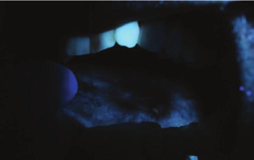
2 P. Lane et al. INTRODUCTION Almost all cancers are easier to treat successfully if caught early in their growth. This principal is particularly true for 85% of cancers that are epithelial in origin. Most of these cancers do not originate in fully malignant form. They typically arise from normal tissues that progress through one or more preinvasive pathologically recognizable steps, such as dysplasia or carcinoma in situ (CIS), for which there are limited detection methods. 1 Oral cancer is an especially tragic example of an epithelial cancer. 2 Although the mouth is easily accessible, oral cancers are often diagnosed at a late stage, beyond treatability, that brings with it high morbidity and mortality. Fluorescence spectroscopy and imaging may provide means of early detection and have been studied in many organ sites. 3 Robyler et al 4 reported on the use of 67 autofluoresence images that were taken of 56 patients who were divided into a training and validation set of 46 measurements each and applied to a test set of 21 images. An automated algorithm using 405-nm illumination discriminated normal from neoplastic tissue with a sensitivity of 96% and specificity of 96% in the training and in the validation sets and with a sensitivity of 100% and specificity of 91% in the test set. A previous report based on quantitative analyses suggested that imaging at this wavelength would be useful for diagnosis. 5 These results were confirmed by other investigators and found applicable to other organ sites This wavelength was shown to demonstrate intracellular activities of interest by several investigators This article presents the reader with a series of images showing the varied presentation of squamous cell carcinoma and dysplasia when observing these lesions with 405-nm excited direct fluorescence imaging, such as a clinician or dentist might see in an oral dysplasia referral clinic. We hope these images will be a useful visual aid as 405-nm excitation illumination fluorescence imaging devices are introduced into the market. MATERIALS AND METHODS Overview of Study Procedures Men and women who were 18 years of age were eligible to participate in the study, which began in 2008 and is ongoing. Patients are recruited from the oral cancer clinics of the British Columbia Cancer Research Centre and Agency. A research nurse or research assistant describes the study to eligible patients and written informed consent is obtained from those agreeing to participate, with emphasis that nonparticipation will not affect their care. A biomedical engineer assists with trial conduct, including assisting the provider with taking the photographic images included in the trial. All patients seen in the clinic receive: (1) a complete history and an oral cancer risk factors questionnaire, (2) a comprehensive head and neck examination, (3) a comprehensive oral white light examination, and (4) referrals for outstanding medical problems. Those patients in the trial undergo an additional examination using the device, which has violet light-excited fluorescence at 405 nm. Fluorescence images are taken of all lesions observed under white light or fluorescence. Suspicious areas are biopsied if the clinical impression suggest dysplasia or cancers. Each patient s history of lesions, including precancers and cancers are noted, and the clinical impression is entered into the database. Lesions that are biopsied are submitted for permanent section. All tissue biopsies are reviewed by histopathologists who are blinded to the imaging data and who specialize in oral cavity pathologic diagnosis. The study protocol was reviewed and approved by the Institutional Review Board at The British Columbia Cancer Agency. The studies were carried out at The British Columbia Cancer Agency, Vancouver, British Columbia, Canada. Fluorescence Imaging A handheld device emitting light at variable wavelengths was used. An image and detailed description of the imaging system can be found in Lane et al. 15 A light source with significant energy around 405 nm was positioned behind a highperformance, narrow band-pass interference filter designed to transmit predominately 405 nm light. This was used to excite the interior of the mouth, allowing the entire field of view of the camera to be illuminated. Each measurement takes approxi- S79
3 Gender Medicine mately an additional 3 minutes; however, documentation with photography adds an additional 2 minutes. A clinician using a similar device views the interrogated tissue through either filtered glasses or a variable magnification scope, both designed to highlight tissue fluorescence. Photographs presented here were taken using a filter to present the reader with an image akin to what a device operator sees. The total light exposure is less than the American National Standards Institute standards described in Roblyer et al. 4,5 All images were reviewed 4 times by 4 independent investigators blinded to the histology. These investigators included dentists, optical engineers, and clinicians familiar with fluorescence imaging. RESULTS One hundred nineteen patients participated in this study of 405 nm imaging. From each of these patients, several images were captured using fluorescence at 405 nm. Ninety percent of these patients had a concurrent biopsy at the time of their clinical visit; the remaining 10% were biopsied within 6 months of the time the image was taken. These images represent all surfaces areas in the oral cavity. Among the data set are 23 lesions of carcinoma (19 squamous and 2 verrucous); 2 CISs, 9 severe dyplasias, 18 moderate or mild dyplasias, 15 leukoplakias, 1 erythroplakia, 13 lichen planus, 5 hyperplasias, 2 mucositis or gingivitis, 10 traumatic lesions, 19 scars, 5 areas of pigmentation, including grafts and biopsy sites, and several other confounders (see Supplemental Appendix in the online version at doi: /j.genm ). For this article, we limited demonstrative images to invasive cancers, CIS, dyplasias, and erythroplakias and leukoplakias. The confounding lesions and scars will be published in a separate article outlining strategies for their separation from cancerous lesions. We chose 24 pairs of images (see Supplemental Figures 1 6 in the online version at doi: /j.genm ). On the left side, images are presented as seen by a clinician at the time of examination. On the right side, lesions are delineated to highlight regions of interest. Images of cancers, severe dysplasias, mild to moderate dysplasias, and 1 erythroplakia are presented. In most of these images, the delineated area is seen as a darker blue than the surrounding region. In some images, neovasculature can be noted. In those images where the delineated area is near the teeth, porphyrins can be noted, which fluoresce as pink, bright red, or pale green. There are several observations that can be made at 405 nm. The first observation is that areas of neoplasm that have outgrown their blood supply are often full of bacteria. Bacteria make porphyrins as a by-product, and porphyrins are seen as bright pink- and blue-colored areas using violet light fluorescence imaging. The dark areas demonstrate collagen and elastin breakdown in the stroma, and these areas can be very large, surrounding the white light visible cancers. Often, one can identify a plexus of vessels in the stroma that accompanies the breakdown of collagen and elastin. This neovascularization occurs as lesions grow; the process becomes increasingly deregulated in carcinogenesis. DISCUSSION Laser-induced fluorescence imaging has been used to identify precancerous and cancerous tissues for 20 years. When the field began, there were a limited number of lasers available: argon ion, helium neon, and helium cadmium. Besides being large and bulky instruments, they had limited lifetimes. Over the last 20 years, significant advances have made laser use more practical for clinical applications. Diode lasers at 405 nm have been used extensively in many forms of fluorescent microscopy. Their use has allowed cell biologists to image intracellular structures in the 20 to 50 nm range. They have also been used to turn on and off green fluorescent protein markers at the cellular level, facilitating a whole new field in science. This article shows a large number of images of 405 nm taken in a high-risk population in which there were a large percentage of dysplasic and neoplastic lesions. This gives the viewer some experience with images akin to the experience of the S80
4 P. Lane et al. imaging scientists and clinicians who performed the trials. It takes time to become accustomed to seeing the fluorescence images at this wavelength, and we provide a gallery of images for the viewer, to serve as a type of atlas. Most of the images show the lesion and an area of darkening surrounding it. Some of the images shown are quite dark in printed format. That is not so when viewed on the computer. This darkened image makes discrimination of the lesion harder when printed than when viewed on the computer. Early results suggested that the addition of the fluorescence examination at 405 nm was helpful in identifying characteristics indicative of precancer and cancer over that seen solely by comprehensive white light examination. Images of the violet-induced autofluorescence at 405 nm showed the deeper neovascularization of the stromal changes that accompanied the progression to neoplasia. Biologic studies of tissue slices demonstrated that there was loss of autofluorescence consistent with breakdown of collagen matrixes in the connective tissues as lesions progressed from normal to neoplastic. 3 Elastin breakdown is less well studied, but also strongly suspected in the carcinogenic pathway involving the epithelial stromal interface. Type 1 collagen is a major component of the extracellular matrix that exhibits both optical scattering and endogenous fluorescence. These optical properties render the study of the tumor microenvironment amenable to optical spectroscopy and imaging techniques. Further study of fluorescence imaging has the potential to improve clinical outcomes by giving providers more tools for the early detection of cancer. Similar technologies are being developed to employ fluorescence imaging in diverse organ sites, including the cervix, throat, and lungs. ACKNOWLEDGMENTS We would like to thank all the patients who selflessly participated in these studies in the interest of the advancement of medicine. This research would not have been possible without the contribution of Drs. Catherine Poh and Miriam Rosen of the British Columbia Cancer Research Agency and the University of British Columbia, who were responsible for collection and lesion demarcation shown in all of the image data. CONFLICTS OF INTEREST The symposium and publication of these proceedings were supported by: The National Institutes of Health, The Drexel University College of Medicine, The Helen I. Moorehead, MD Foundation, The Doris Willig, MD Foundation, The Institute for Women s Health and Leadership, and The Center for Women s Health Research at the Drexel University College of Medicine. Drs. Lane, Follen, and MacAulay and Lam have no financial interest in the device or company. This study was funded under a sponsored research agreement with Trimira, LLC. David A. Burns, Dr. Follen s husband, holds 8% interest in stock in Trimira. SUPPLEMENTAL MATERIAL Supplemental appendix and figures accompanying this article can be found in the online version at doi: /j.genm REFERENCES 1. World Health Organization. Accessed January 24, The Oral Cancer Foundation. The HPV connection. Accessed January 24, Richards-Kortum R, Sevick-Muraca. Quantitative optical spectroscopy for tissue diagnosis. Annu Rev Phys Chem. 1996;47: Roblyer D, Kurachi C, Stepanek V, et al. Objective detection and delineation of oral neoplasia using autofluorescence imaging. Cancer Prev Res (Phila). 2009;2: Roblyer D, Richards-Kortum R, Sokolov K, et al. Multispectral optical imaging device for in vivo detection of oral neoplasia. J Biomed Opt. 2008; 13: Da Costa RS, Andersson H, Wilson BC. Molecular fluorescence excitation-emission matrices rele- S81
5 Gender Medicine vant to tissue spectra. Photochem Photobiol. 2003;78: Andersson-Engels S, Elner A, Johansson J, et al. Clinical recording of laser-induced fluorescence spectra for evaluation of tumour demarcation feasibility in selected clinical specialties. Lasers Med Sci. 1991;6: Balasubramanian S, Elangovan V, Govindasamy S. Fluorescence spectroscopic identification of 7,12-dimethylbenz[a]anthracene-induced hamster buccal pouch carcinogenesis. Carcinogenesis. 1995;16: Farwell DG, Meier JD, Park Y, et al. Time-resolved fluorescence spectroscopy as a diagnostic technique of oral carcinoma: validation in the hamster buccal pouch model. Arch Otorhinolarygol Head Neck Surg. 2010;136: Lane P, Gilhuly T, Whitehead P, et al. Simple device for the direct visualization of oral-cavity tissue fluorescence. J Biomed Opt. 2006;11: Betzig E, Patterson GH, Sougrat R, et al. Imaging intracellular fluorescent proteins at nanometer resolution. Science. 2006;313: Melanson JE, Lucy CA. Violet (405) diode laser for laser induced fluorescence detection in capillary electrophoresis. The Royal Society of Chemistry. The Analyst. 2000;125: Beauregard KE, Lee K-D, Collier J, Swanson JA. ph-dependent perforation of macrophage phagoseomes by Listeriolysin O from Listeria moncytogenes. J Exp Med. 1997;186: Kirkpatrick ND, Hoying JB, Botting SK, et al. In vitro model for endogenous optical signatures of collagen. J Biomed Opt. 2006:11: Lane P, Follen M, MacAulay C. Has fluorescence spectroscopy come of age? A case series of oral precancers and cancers using white light, fluorescent light at 405 nm and reflected light at 545 nm using the Trimira Identafi Gender Med 2012;9:S25 S35. Address correspondence to: Michele Follen MD, PhD, Drexel University College of Medicine 245 N. 15th St., Philadelphia, PA Michele.Follen@Drexelmed.edu. S82
6 P. Lane et al. Supplemental Appendix 1. Study patients, date of measurement, location of lesions, and clinical impression Study ID Date Location Clinical Impression left lateral tongue 6mos scar for s/t of CIS right lateral tongue 1 yr old scar left ventral tongue 2 yr post s/t scar right lateral tongue 2.5 yr old scar, post s/t SCC right buccal mucosa 4 days wound, cheek biting lower left gingiva 6 mos post s/t graft right lateral tongue 6 mos post s/t scar for cancer left tongue background lichen planus right lateral tongue biopsy resident left buccal mucosa biteline right gingiva C1FV1 TB floor of mouth cancer, pre-surgery left tongue cancer, pre-surgery confounder left inside upper lip canker sore right buccal mucosa cheek biting right lower dentulus ridge CIS, concurrent bx left floor of mouth leukoplakia (D1-2) right posterior soft palate Squamous cell carcinoma right buccal mucosa confounder, contact lichenoid mucositis right lateral tongue mild dysplasia left lateral tongue moderate dysplasia right lateral tongue moderate dysplasia right lateral tongue moderate dysplasia right lateral tongue moderate dysplasia right lateral tongue moderate dysplasia right lateral tongue moderate to severe dysplasia left lateral tongue severe dysplasia floor of mouth C right tongue severe dysplasia left tongue severe dysplasia left lateral tongue dysplasia, hx of CIS right lateral tongue erythroplakia right lateral tongue fluorescence lost left lateral tongue geographic tongue tip of tongue geographic tongue left posterior tongue leukoplakia left ventral tongue leukoplakia left soft palate leukoplakia left ventral tongue leukoplakia right lateral tongue leukoplakia left tongue leukoplakia left lateral tongue leukoplakia left post maxillary retromolar leukoplakia right lateral tongue moderate dysplasia right buccal mucosa lichen planus right buccal mucosa lichen planus left posterior buccal mucosa lichen planus (continued) S82.e1
7 Gender Medicine Supplemental Appendix 1 (continued). Study ID Date Location Clinical Impression right lateral tongue lichen planus dorsal tongue lichen planus right buccal mucosa lichen planus left buccal mucosa lichen planus resident left gingival buccal lichen planus left buccal mucosa lichen planus, possible dysplasia right gingiva lichen planus, plaque type right buccal mucosa lichenoid dysplasia left lateral tongue mild dysplasia right lateral tongue mild dysplasia left lateral tongue mod dysplasia right lateral tongue mod dysplasia left lateral tongue mod dysplasia right lateral tongue mod dysplasia, leukoplakia left gingiva gingivitis right lateral tongue pigmentation left latetal tongue pigmentation right buccal mucosa pigmentation, lichen planus right ventral tongue post s/t scar right lateral tongue pre-surgery right tongue pre-surgery tongue pre-surgery right tongue pre-surgery left tongue pre-surgery left lateral tongue severe dyplasia right buccal mucosa red, white right tongue red, white right lateral tongue red, white leukoplakia right FOM residual lesion left ventral tongue scar left buccal mucosa scar right tongue scar right lateral tongue scar right lateral tongue scar left lateral tongue scar right lateral tongue scar right lateral tongue scar 3 mos rad tx severe dysplasia left lateral tongue scar, 6 mos post s/t CIS carcinoma in situ; hx history; SCC squamous cell carcinoma; s/t surgical treatment; rad tx radiotherapy. S82.e2


8 P. Lane et al. A B C D E F G H Supplemental Figure 1. Oral cavity illuminated at 405nm excitation. Areas of interest are delineated in panels on the right. S82.e3


9 Gender Medicine A B C D E F G H Supplemental Figure 2. Oral cavity illuminated at 405nm excitation. Areas of interest are delineated in panels on the right. S82.e4


10 P. Lane et al. A B C D E F G H Supplemental Figure 3. Oral cavity illuminated at 405nm excitation. Areas of interest are delineated in panels on the right. S82.e5


11 Gender Medicine A B C D E F G H Supplemental Figure 4. Oral cavity illuminated at 405nm excitation. Areas of interest are delineated in panels on the right. S82.e6


12 P. Lane et al. A B C D E F G H Supplemental Figure 5. Oral cavity illuminated at 405nm excitation. Areas of interest are delineated in panels on the right. S82.e7


13 Gender Medicine A B C D E F G H Supplemental Figure 6. Oral cavity illuminated at 405nm excitation. Areas of interest are delineated in panels on the right. S82.e8
ENHANCING DETECTION... ENHANCING YOUR PRACTICE
 ENHANCING DETECTION... ENHANCING YOUR PRACTICE Staggering Statistics on Oral Cancer 1 person dies from oral cancer each hour of every day The Mortality Rate for oral cancer has not decreased in over 30
Chapter 5. Oxygenated Hemoglobin Diffuse Reflectance Ratio for In Vivo Detection of oral Pre-cancer
 Chapter 5 Oxygenated Hemoglobin Diffuse Reflectance Ratio for In Vivo Detection of oral Pre-cancer This work is published in: JB0 (SPIE) 13(4):041306 (1-10), 2008 Oxygenated Hemoglobin Diffuse Reflectance
ACCURATE DIAGNOSIS IS THE ONLY TRUE CORNERSTONE ON WHICH RATIONAL TREATMENT CAN BE BUILT. C Noyek
 ACCURATE DIAGNOSIS IS THE ONLY TRUE CORNERSTONE ON WHICH RATIONAL TREATMENT CAN BE BUILT. C Noyek Oral diagnostics Definition of the discipline That area of dentistry, the which deals with gathering, recording
Oral Surgeons and the VELscope System: Partners in Early Detection & Diagnosis
 David Morgan, PhD Chief Science Officer LED Dental Inc. There is a growing trend within general dentistry stressing the importance of total oral health that is, not only health of the teeth and gums but
LARYNGEAL DYSPLASIA. Tomas Fernandez M; 3 rd year ENT resident, Son Espases University Hospital
 LARYNGEAL DYSPLASIA Tomas Fernandez M; 3 rd year ENT resident, Son Espases University Hospital INTRODUCTION Laryngeal cancer constitutes 1-2% of all malignancies diagnosed worldwide Survival is related
Fluorescence Spectroscopy: A New Approach in Cervical Cancer
 DOI 10.1007/s13224-012-0298-6 ORIGINAL ARTICLE : A New Approach in Cervical Cancer Pandey Kiran Pradhan Asima Agarwal Asha Bhagoliwal Ajay Agarwal Nidhi Received: 2 June 2008 / Accepted: 7 August 2012
Separating the Good from the Bad and Ugly The Use of Non-Invasive Optical and Molecular Tests in the Management of an Oral Lesion
 Separating the Good from the Bad and Ugly The Use of Non-Invasive Optical and Molecular Tests in the Management of an Oral Lesion Dr. Catherine Poh, DDS, PhD Cert. Oral Pathology, FRCD (C), DABOMP Clinician
Accepted 12 April 2006 Published online 18 September 2006 in Wiley InterScience ( DOI: /hed.
 CASE REPORT Dennis H. Kraus, MD, Section Editor DIRECT FLUORESCENCE VISUALIZATION OF CLINICALLY OCCULT HIGH-RISK ORAL PREMALIGNANT DISEASE USING A SIMPLE HAND-HELD DEVICE Catherine F. Poh, DDS, PhD, 1,2,3
ORIGINAL ARTICLE. Noninvasive Diagnosis of Oral Neoplasia Based on Fluorescence Spectroscopy and Native Tissue Autofluorescence
 ORIGINAL ARTICLE Noninvasive Diagnosis of Oral Neoplasia Based on Fluorescence Spectroscopy and Native Tissue Autofluorescence Ann Gillenwater, MD; Rhonda Jacob, DDS, MS; Ravi Ganeshappa, MD; Bonnie Kemp,
Introducing the VELscope Vx
 Introducing the VELscope Vx 888.541.4614 sales@leddental.com www.leddental.com Introducing the VELscope Vx What is the VELscope Vx? The VELscope Vx, the latest model release of LED Dental s VELscope technology,
Objective Detection and Delineation of Oral Neoplasia Using Autofluorescence Imaging
 Objective Detection and Delineation of Oral Neoplasia Using Autofluorescence Imaging Darren Roblyer, 1 Cristina Kurachi, 2 Vanda Stepanek, 3 Michelle D. Williams, 4 Adel K. El-Naggar, 4 J. Jack Lee, 5
Oral Cavity Cancer. Oral Cavity. Disclosures. Screening Methods for Early Oral Cancer
 Screening Methods for Early Oral Cancer M. Boyd Gillespie, M.D., M.Sc. UCSF Head & Neck Cancer Course San Francisco, CA November 8, 2014 Disclosures Paid consultant & Research Support on sleep apnea devices
Clinical Policy Bulletin: Oral Screening and Lesion Identification Systems
 Go Clinical Policy Bulletin: Oral Screening and Lesion Identification Systems Number: 0760 Policy *Please see amendment for Pennsylvania Medicaid at the end of this CPB. Additional Information Aetna considers
Finding Dangerous Mucosa
 Finding Dangerous Mucosa 2 Oral Cancer Squamous Cell Carcinoma Salivary Gland Adenocarcinoma Malignant Lymphoma Metastatic Carcinoma Sarcoma 4 Incidence of Cancer in the United States For Oral and Oropharyngeal
In Vivo Fluorescence Hyperspectral Imaging of Oral Neoplasia
 In Vivo Fluorescence Hyperspectral Imaging of Oral Neoplasia Darren Roblyer a, Cristina Kurachi b, Ann M. Gillenwater c, Rebecca Richards-Kortum a a Department of Bioengineering, Rice University, 6100
Chapter 7. Grading of Oral Mucosa by Curve-fitting of Corrected Autofluorescence Spectra
 Chapter 7 Grading of Oral Mucosa by Curve-fitting of Corrected Autofluorescence Spectra This work is submitted to: Head and Neck (Wiley-Blackwell), 2008. Grading of Oral Mucosa by Curve-fitting of Corrected
Oral Cancer FAQs. What is oral cancer? How many people are diagnosed with oral cancer each year?
 Oral Cancer FAQs What is oral cancer? Oral cancer or oral cavity cancer, is cancer that starts in the mouth. Areas affected by this type of cancer are the lips, the inside lining of the lips and cheeks
Diagnostic difficulties with lesions of the oral mucosa
 BDIAP London, November 2010 School of Clinical Dentistry University of Sheffield Diagnostic difficulties with lesions of the oral mucosa Paul M Speight Dept Oral & Maxillofacial Pathology University of
Update of the role of Human Papillomavirus in Head and Neck Cancer
 Update of the role of Human Papillomavirus in Head and Neck Cancer 2013 International & 12 th National Head and Neck Tumour Conference Shanghai, 11 13 Oct 2013 Prof. Paul KS Chan Department of Microbiology
الطلاوة = Leukoplakia LEUKOPLAKIA
 LEUKOPLAKIA Leukoplakia is a clinical term that refers to a predominantly white lesion of the oral mucosa that cannot be rubbed off or characterized by any other definable lesion or known disease. 130
Diagnostic aids of oral cancer
 Diagnostic aids of oral cancer The World Health Organization has clearly indentified prevention and early detection as major objectives in the control of the oral cancer. At the present time, screening
LEUKOPLAKIA Definition Epidemiology Clinical presentation
 LEUKOPLAKIA Definition Leukoplakia is the most common premalignant or "potentially malignant" lesion of the oral mucosa. Leukoplakia is a predominantly white lesion of the oral mucosa than cannot be clinicopathologically
WHITE LESIONS OF THE ORAL CAVITY - diagnostic appraisal & management strategies
 WHITE LESIONS OF THE ORAL CAVITY - diagnostic appraisal & management strategies * Joshy V.R ** Hari.S * Reader, Dept of Oral Pathology, Yenepoya Dental College, Yenepoya University, Mangalore 575 018.
LESIONS OF THE ORAL CAVITY ORAL CAVITY. Oral Cavity Subsites 4/10/2013 LIPS TEETH GINGIVA ORAL MUCOUS MEMBRANES PALATE TONGUE ORAL LYMPHOID TISSUES
 LESIONS OF THE ORAL CAVITY David I. Kutler, MD, FACS Associate Professor Division of Head and Neck Surgery Department of Otolaryngology HNS Weill Cornell Medical Center ORAL CAVITY LIPS TEETH GINGIVA ORAL
BC Cancer Cervix Screening 2015 Program Results. February 2018
 BC Cancer Cervix Screening 2015 Program Results BC Cancer Cervix Screening 2015 Program Results 2 Table of Contents BC Cancer Cervix Screening 2015 Program Results... 1 Table of Contents... 2 Program Overview...
Dysplasia, Mimics and Other Controversies
 Dysplasia, Mimics and Other Controversies Mary S. Richardson, MD Dept. of Pathology Medical University of South Carolina Charleston, SC Notice of Faculty Disclosure In accordance with ACGME guidelines,
Fluorescence Spectroscopy for In Vivo Characterization of Ovarian Tissue
 Lasers in Surgery and Medicine 29:128±135 (2001) Fluorescence Spectroscopy for In Vivo Characterization of Ovarian Tissue Molly Brewer, DVM, MD, MS, 1,2 Urs Utzinger, PhD, 3 Elvio Silva, MD, 4 David Gershenson,
That. Name QUIZ. 60 SEPTEMBER 2017 // dentaltown.com
 QUIZ Name That General dentists are first in the line of practitioners that patients see for an oral lesion evaluation; therefore, a sound understanding of oral mucosal diseases and their clinical presentation
Fluorescence Spectroscopy and Imaging for Photodetection of Cervical Intraepithelial Neoplasia
 Imaging for Photodetection of Cervical Intraepithelial Neoplasia Thomas Stepinac 1, Attila Major 2, Frank Lüdicke 2, Didier Goujon 1, Nora Dögnitz 1, Tanja Gabrecht 1, Norbert Lange 1, Hubert van den Bergh
Multispectral optical imaging device for in vivo detection of oral neoplasia
 Universidade de São Paulo Biblioteca Digital da Produção Intelectual - BDPI Departamento de Física e Ciências Materiais - IFSC/FCM Artigos e Materiais de Revistas Científicas - IFSC/FCM 2008-03 Multispectral
Oral Cancer Preven,on and Early Diagnosis
 Oral Cancer Preven,on and Early Diagnosis Prof. Surendra S. Shastri MD Technical Advisor IAEA impact Mission Ex Chair, Department of Preven,ve Oncology Head, WHO Collabora,ng Centre For Cancer Preven,on,
Oral cavity cancer accounts for approximately 3% of all malignancies and is a significant worldwide health problem.
 Oral cavity cancer accounts for approximately 3% of all malignancies and is a significant worldwide health problem. Majority are SCC ( 5-year survival rate only about 50-60% ) Many SCC arrive from premalignant
Premalignant lesions may expose to a promoting. factor & may be induced to undergo malignant. Carcinoma in situ displays the cytologic features of
 بسم رلاهللا Def. Premalignant lesions may expose to a promoting factor & may be induced to undergo malignant transformation. Carcinoma in situ displays the cytologic features of malignancy without invasion
Development of Low-Cost Photodynamic Therapy Device
 Vol. 112 (2007) ACTA PHYSICA POLONICA A No. 5 Proceedings of the International School and Conference on Optics and Optical Materials, ISCOM07, Belgrade, Serbia, September 3 7, 2007 Development of Low-Cost
Oral Cavity and Pharynx Cancer
 Oral Cavity and Pharynx Cancer Figure 18 Definition: Oral cancer begins in the mouth and can include the lips, cheeks, teeth, gums, the floor of the tongue, the roof of the mouth, and the front two-thirds
Oral Cancer Dr Christine Goodall Consultant Oral Surgeon University of Glasgow Dental School
 Oral Cancer Dr Christine Goodall Consultant Oral Surgeon University of Glasgow Dental School christine.goodall@glasgow.ac.uk Locations Lip, mouth, oropharynx Tongue, floor of mouth, buccal mucosa, palate,
Oral Cancer- Improving Early Detection
 Oral Cancer- Improving Early Detection GDC Recommended Subject Aims: To give an overview of the dental team's role in detecting the early signs of oral cancer; to give an overview of the risk factors associated
PREVENTION OF ORAL CANCER
 PREVENTION OF ORAL CANCER Oral cancer is increasing in incidence worldwide. Throughout the world, malignant neoplasms of the mouth and pharynx rate as the fifth most common cancer in men and the seventh
Quantitative Optical Spectroscopy of the Uterine Cervix: A cost effective way to detect and manage cervical disease
 Quantitative Optical Spectroscopy of the Uterine Cervix: A cost effective way to detect and manage cervical disease Nahida Chakhtoura, MD, Leo Twiggs, MD, Timothy DeSantis, MD Claudia Werner, MD, William
EVERYTHING YOU WANTED TO KNOW ABOUT. Robin Billet, MA, CTR, Head & Neck CTAP Member May 9, 2013
 EVERYTHING YOU WANTED TO KNOW ABOUT. Robin Billet, MA, CTR, Head & Neck CTAP Member May 9, 2013 Head and Neck Coding and Staging Head and Neck Coding and Staging Anatomy & Primary Site Sequencing and MPH
Chapter 8. 5-ALA Induced PpIX Fluorescence in Oral Cancer Detection In Vivo
 Chapter 8 5-ALA Induced PpIX Fluorescence in Oral Cancer Detection In Vivo This work is to be submitted to: Lasers in Surgery and Medicine (Wiley-Blackwell), 2008. 5-ALA Induced PpIX Fluorescence in Oral
CANCER IMAGING. Our Research Focus:
 Biennial Research Report 2005 2006 C A N C E R I M A G I N G B C C A CANCER RESEARCH CENTRE 675 West 10th Avenue, Vancouver, BC, V5Z 1L3 Telephone: (604) 675 8081 Our Research Focus: 4. In vivo tissue
Philip Chiu Associate Professor Department of Surgery, Prince of Wales Hospital The Chinese University of Hong Kong
 Application of Chromoendoscopy, NBI and AFI in Esophagus why, who, and how? Philip Chiu Associate Professor Department of Surgery, Prince of Wales Hospital The Chinese University of Hong Kong Cancer of
RESULT OF SURVEY OF 1705 LAWSUITS* ORAL CANER: A significant Public Health Concern. ORAL CANCER: Other Epidemiologic Facts
 FAILURE TO DIAGNOSE ORAL CANCER AND OTHER PATHOLOGIC CONDITIONS OF THE ORAL CAVITY Kalu U.E. Ogbureke, BDS, MSc, DMSc, JD, FDSRCS, FDSRCPS, FDSRCSEd, FRCPath Diplomate, American Board of Oral and Maxillofacial
Head and Neck Case 1 PATIENT HISTORY
 Head and Neck Case 1 PATIENT HISTORY Patient History May 7, 2007 Otolaryngology Head & Neck Subjective: Patient was recently seen by a dentist, who noted a roughness in his lower alveolus, and wanted to
Head and Neck Cancer How to recognize it in your office
 Head and Neck Cancer How to recognize it in your office Peter M Hunt, MD, FACS Associates in ENT/Head & Neck Surgery Director CHI Memorial Head & Neck and Melanoma Centers of Excellence September 8, 2018
Diseases of oral cavity
 Diseases of oral cavity Diseases of Teeth and Supporting Structures Inflammatory/Reactive Lesions Infections Oral Manifestations of Systemic Disease Precancerous and Cancerous Lesions Odontogenic Cysts
About Omics Group conferences
 About Omics Group OMICS Group International through its Open Access Initiative is committed to make genuine and reliable contributions to the scientific community. OMICS Group hosts over 400 leading-edge
The importance of raising awareness of oral cancer amongst our allied healthcare professionals: Case Study by Jessica Mann and Mili Doshi.
 The importance of raising awareness of oral cancer amongst our allied healthcare professionals: Case Study by Jessica Mann and Mili Doshi. Summary A 79-year-old woman was referred to the special care dental
Role of the Dental Hygienist in Oral Pathology. Role of the Dental Hygienist in Oral Pathology. Cancers of the Oral Cavity.
 Gum Gardeners Study Club April 25, 2016 Early Detection of Oral Cancer Cindy Kleinegger, DDS, MS NW Oral Pathology Tigard, OR nworalpathology.com Role of the Dental Hygienist in Oral Pathology Work closely
Autofluorescence spectroscopy for the classification of oral lesions
 Autofluorescence spectroscopy for the classification of oral lesions Rijksuniversiteit Groningen Autofluorescence spectroscopy for the classification of oral lesions PROEFSCHRIFT ter verkrijging van het
Head and neck cancer - patient information guide
 Head and neck cancer - patient information guide The development of reconstructive surgical techniques in the last 20 years has led to major advances in the treatment of patients with head and neck cancer.
Journal of American Science 2014;10(4)
") Detection of oral potentially malignant lesions among tobacco users; Identafi and Microlux versus histopathology Safia Al attas 1, Suzan Ibrahim 2, Zeinab Darwish 3, Hala Amer 4, Mona Hassan 5 1 Department
Detection of oral potentially malignant lesions among tobacco users; Identafi and Microlux versus histopathology Safia Al attas 1, Suzan Ibrahim 2, Zeinab Darwish 3, Hala Amer 4, Mona Hassan 5 1 Department
Research Article High-Resolution Optical Imaging of Benign and Malignant Mucosa in the Upper Aerodigestive Tract: An Atlas for Image-Guided Surgery
 International Scholarly Research Network ISRN Minimally Invasive Surgery Volume 2012, Article ID 364285, 9 pages doi:10.5402/2012/364285 Research Article High-Resolution Optical Imaging of Benign and Malignant
Oral Cavity. 1. Introduction. 1.1 General Information and Aetiology. 1.2 Diagnosis and Treatment
 Oral Cavity 1. Introduction 1.1 General Information and Aetiology The oral cavity extends from the lips to the palatoglossal folds and consists of the anterior two thirds of the tongue, floor of the mouth,
4Ps LUMPS AND BUMPS B.L.&T. BUMPS, LUMPS, AND TATTOOS. Most Common BUMP in the oral cavity Fibroma INTERDENTAL PAPILLAE LESIONS
 B.L.&T. BUMPS, LUMPS, AND TATTOOS LUMPS AND BUMPS DIFFERENTIAL DIAGNOSIS FOR LUMPS AND BUMPS Traumatic Fibroma Papilloma Epulis Fissuratum Inflammatory Papillary Hyperplasia Lesions of Attached Gingiva
Fluorescence spectroscopy and microscopy of cutaneous tumours correlation between micro- and macro- spectral measurements
 Fluorescence spectroscopy and microscopy of cutaneous tumours correlation between micro- and macro- spectral measurements E. Borisova 1, L. Avramov 1, M. Lomova 2, O. Semyachkina-Glushkovskaya 2, D. Gorin
A CASE OF A Huge Submandibular Pleomorphic Adenoma
 ISPUB.COM The Internet Journal of Head and Neck Surgery Volume 4 Number 2 S VERMA Citation S VERMA.. The Internet Journal of Head and Neck Surgery. 2009 Volume 4 Number 2. Abstract Pleomorphic adenoma
Applications in Dermatology, Dentistry and LASIK Eye Surgery using LASERs
 Applications in Dermatology, Dentistry and LASIK Eye Surgery using LASERs http://www.medispainstitute.com/menu_laser_tattoo.html http://www.life123.com/bm.pix/bigstockphoto_close_up_of_eye_surgery_catar_2264267.s600x600.jpg
Clinical behaviour of malignant transforming oral lichen planus
 EJSO 2002; 28: 838±843 doi:10.1053/ejso.2002.1302, available online at http://www.idealibrary.com on 1 Clinical behaviour of malignant transforming oral lichen planus M. D. Mignogna*, L. Lo Russo*, S.
The Oral Cavity. Image source:
 The Oral Cavity Anatomy Image source: http://anatomyforlayla.blogspot.co.za/2007/04/blog-post.html The major structures of the oral cavity are the lips, the teeth, the alveolar ridges (bony areas that
Head and Neck Cancer in FA: Risks, Prevention, Screening, & Treatment Options David I. Kutler, M.D., F.A.C.S.
 Head and Neck Cancer in FA: Risks, Prevention, Screening, & Treatment Options David I. Kutler, M.D., F.A.C.S. Associate Professor Division of Head and Neck Surgery Department of Otolaryngology-Head and
Proliferative Verrucous Leukoplakia of the Gingiva, Report of two Cases with Malignant Transformation
 Journal of Clinical and Anatomic Pathology Case Report Open Access Proliferative Verrucous Leukoplakia of the Gingiva, Report of two Cases with Malignant Transformation Nadereh Ghanee DMD, Selene Saraf
Multimodal Spectroscopic Tissue Scanner for diagnosis of ex vivo surgical specimens
 Multimodal Spectroscopic Tissue Scanner for diagnosis of ex vivo surgical specimens Collaborating researcher: Dr. Maryann Fitzmaurice (Case Western Reserve University, Cleveland, OH), Dr. Tulio Valdez
Evaluation and Management of Head and Neck Cancer in Patients with Fanconi anemia David I. Kutler, M.D., F.A.C.S.
 Evaluation and Management of Head and Neck Cancer in Patients with Fanconi anemia David I. Kutler, M.D., F.A.C.S. Residency Site Director Weill Cornell Medical Center Associate Professor Division of Head
The Optical Biopsy. Douglas S. Scherr
 Patricia Kuharic 66 The Optical Biopsy Douglas S. Scherr UROLOGY, WEILL CORNELL MEDICAL COLLEGE We want to create a better way to look inside the walls of a bladder for cancerous cells, eliminating the
Title: Advances in fluorescence imaging techniques to detect oral cancer and its precursors
 Title: Advances in fluorescence imaging techniques to detect oral cancer and its precursors Dongsuk Shin 1, Nadarajah Vigneswaran 2, Ann Gillenwater 3, Rebecca Richards Kortum 4 * *Author for correspondence
PH-04A: Clinical Photography Production Checklist With A Small Camera
 PH-04A: Clinical Photography Production Checklist With A Small Camera Operator Name Total 0-49, Passing 39 Your Score Patient Name Date of Series Instructions: Evaluate your Series of photographs first.
Advances in Endoscopic Imaging
 Advances in Endoscopic Imaging SGNA meeting February 20, 2010 Amar R. Deshpande, MD Asst Professor of Medicine Division of Gastroenterology University of Miami Miller School of Medicine Objectives To recognize
TITLE: Optical Spectroscopy and Multiphoton Imaging for the Diagnosis and Characterization of Hyperplasias in the Mouse Mammary Gland
 AD Award Number: W81XWH-04-1-0330 TITLE: Optical Spectroscopy and Multiphoton Imaging for the Diagnosis and Characterization of Hyperplasias in the Mouse Mammary Gland PRINCIPAL INVESTIGATOR: Melissa Caroline
MIRROR IMAGE MIRROR IMAGE. The future of cancer diagnostics may be closer than it appears
 MIRROR IMAGE MIRROR IMAGE The future of cancer diagnostics may be closer than it appears Advanced molecular techniques, fluorescence, and reconstruction of the tumor microenvironment. The many faces of
TSX-V: VRS OTCQX: VRSEF
 Verisante recently announced a breakthrough in the fight against oral and skin cancer with the announcement of a second phase prototype for the Company s Intelligent Multispectral Imaging Camera. Press
Original Research Article
 ASSESSMENT OF HUMAN PAPILLOMA VIRUS SUBTYPES BY POLYMERASE CHAIN REACTION AND THEIR IMPACT ON THE DEGREE OF DYSPLASIA IN ORAL LEUKOPLAKIA Submitted on: XXXX Dr. N. Kannan, Dr Teja Srinivas, Dr. Rakesh
Dental Supplement. Denturist. Ministry of Social Development and Poverty Reduction
 Dental Supplement Denturist Ministry of Social Development and Poverty Reduction TABLE OF CONTENTS Part A - Preamble - Dental Supplements - Denturist pages i - v The Preamble - Dental Supplements - Denturist
04/09/2018. Squamous Cell Neoplasia and Precursor Lesions. Agenda. Squamous Dysplasia. Squamo-proliferative lesions. Architectural features
 Squamous Cell Neoplasia and Precursor Lesions Jennifer L. Hunt, MD, MEd Aubrey J. Hough Jr, MD, Endowed Professor of Pathology Chair of Pathology and Laboratory Medicine University of Arkansas for Medical
Chromatography Vacuum Ultraviolet Spectroscopy
 Application Note Differentiation and Determination Differentiation and Determination of Fatty Acid Methyl of Fatty Esters Acid by Gas Methyl Chromatography Esters by Vacuum Gas Ultraviolet Spectroscopy
DETECTION OF BREAST & CERVICAL CANCER USING RAMAN SPECTROSCOPY
 DETECTION OF BREAST & CERVICAL CANCER USING RAMAN SPECTROSCOPY Binay Bhushan 1, Asima Pradhan 2 Post-Doctoral Fellow, Department of Physics, Indian Institute of Technology, Kanpur, India 1 Professor, Department
Handheld Radiofrequency Spectroscopy for Intraoperative Assessment of Surgical Margins During Breast-Conserving Surgery
 Last Review Status/Date: December 2016 Page: 1 of 6 Intraoperative Assessment of Surgical Description Breast-conserving surgery as part of the treatment of localized breast cancer is optimally achieved
Autofluorescence and Early Detection of Mucosal Lesions in Patients at Risk for Oral Cancer
 TECHNICAL STRATEGY Autofluorescence and Early Detection of Mucosal Lesions in Patients at Risk for Oral Cancer Alessandro Moro, SD, Francesco Di Nardo, MD, Roberto Boniello, SD, Tito M. Marianetti, SD,
OROPHYRENGEAL CANCERS
 OROPHYRENGEAL CANCERS INTRODUCTION 2 % 4 % of all malignant Tumors in west Asia India 40% Men ^ Age :Over 60 yrs 90% of all oral cancers results from Tobacco and Alcohol Pan (Betel Leaf,Nut, Lime), Reverse
Diseases of the vulva
 Diseases of the vulva 1. Bartholin Cyst - Infection of the Bartholin gland produces an acute inflammation within the gland (adenitis) and may result in an abscess. Bartholin duct cysts - Are relatively
NEOPLASMS OF THE SURFACE EPITHELIUM (KERATINOCYTES)
") NEOPLASMS OF THE SURFACE EPITHELIUM (KERATINOCYTES) Papillary Lesions Precancerous Lesions Keratinocyte Proliferations Carcinomas Melanotic Lesions Melanomas Normal Mucosa Keratin layer Spinous layer Basal
NEOPLASMS OF THE SURFACE EPITHELIUM (KERATINOCYTES) Papillary Lesions Precancerous Lesions Keratinocyte Proliferations Carcinomas Melanotic Lesions Melanomas Normal Mucosa Keratin layer Spinous layer Basal
Development and application of novel spectroscopic tools for breast cancer diagnosis
 Development and application of novel spectroscopic tools for breast cancer diagnosis LBRC researchers: Ramachandra Dasari, Jeon Woong Kang, Niyom Lue, Rishikesh Pandey, Nicolas Spegazzini External technology
Lesions & Lifestyles
 Lesions & Lifestyles attended a 3 hour Continuing Education Seminar on Oral Pathology presented by Nancy Dewhirst, RDH,BS on (date) at (location):. Course material is directly related patient care. Notes:
J L Jayanthi, 1 G U Nisha, 1 S Manju, 2 E K Philip, 2 P Jeemon, 3 K V Baiju, 4 V T Beena, 2 N Subhash 1
 Open Access To cite: Jayanthi JL, Nisha GU, Manju S, et al. Diffuse reflectance spectroscopy: diagnostic accuracy of a non-invasive screening technique for early detection of malignant changes in the oral
Squamous Cell Carcinoma of the Head and Neck (SCCHN)
") Squamous Cell Carcinoma of the Head and Neck (SCCHN) Part 1 Bruce M. Wenig, M.D. Dept. of Pathology & Laboratory Medicine Continuum Health Partners New York, NY College of American Pathologists 2004. Materials
Squamous Cell Carcinoma of the Head and Neck (SCCHN) Part 1 Bruce M. Wenig, M.D. Dept. of Pathology & Laboratory Medicine Continuum Health Partners New York, NY College of American Pathologists 2004. Materials
Abnormal Spectroscopy Scans May Presage Persistent or Progressive Cervical Dysplasia
 Abnormal Spectroscopy Scans May Presage Persistent or Progressive Cervical Dysplasia Leo B. Twiggs, M.D. Professor Emeritus University of Miami Miller School of Medicine Miami Florida USA Currently- Women
Cancer of the Oral Cavity
 The International Federation of Head and Neck Oncologic Societies Current Concepts in Head and Neck Surgery and Oncology Cancer of the Oral Cavity Ashok Shaha Principals of Management of Oral Cancer A)
Clinically Microscopically Pathogenesis: autoimmune not lifetime
 Vulvar Diseases: Can be divided to non-neoplastic and neoplastic diseases. The neoplastic diseases are much less common. Of those, squamous cell carcinoma is the most common. most common in postmenopausal
Journal of Oral & Dental Health
 Research Article Journal of Oral & Dental Health Audit on the Effectiveness of Oral Cancer Screening in Primary Care ISSN: 2573-8224 Junaid Iqbal Dental Implantology, University of Manchester, UK * Corresponding
FINE NEEDLE ASPIRATION OF ENLARGED LYMPH NODE: Metastatic squamous cell carcinoma
 Case Scenario 1 HNP: A 70 year old white male presents with dysphagia. The patient is a current smoker, current user of alcohol and is HPV positive. A CT of the Neck showed mass in the left pyriform sinus.
Oral Cancer. Online Course:
 Continuing Education Brought to you by Oral Cancer Course Author(s): Richard C. Jordan, DDS, PhD, FRCD(C) FRCPath CE Credits: 1 hour Intended Audience: Dentists, Dental Hygienists, Dental Assistants, Dental
Oral Cancer and Common Oral Lesions seen in HIV Seropositive Patients. Gwen Cohen Brown DDS, FAAOMP Professor New York City College of Technology
 Oral Cancer and Common Oral Lesions seen in HIV Seropositive Patients Gwen Cohen Brown DDS, FAAOMP Professor New York City College of Technology Program Objectives Recognize the oral health needs of the
PRINCESS MARGARET CANCER CENTRE CLINICAL PRACTICE GUIDELINES GYNECOLOGIC CANCER VULVAR
 PRINCESS MARGARET CANCER CENTRE CLINICAL PRACTICE GUIDELINES GYNECOLOGIC CANCER VULVAR Last Revision Date July 2015 1 Site Group: Gynecologic Cancer Vulvar Author: Dr. Stephane Laframboise 1. INTRODUCTION
Original Policy Date
 MP 2.04.03 Cervicography Medical Policy Section Medicine Issue 12:2013 Original Policy Date 12:2013 Last Review Status/Date Reviewed with literature search/12:2013 Return to Medical Policy Index Disclaimer
Clinical Policy Title: Fluorescence in situ hybridization for cervical cancer screening
 Clinical Policy Title: Fluorescence in situ hybridization for cervical cancer screening Clinical Policy Number: 01.01.02 Effective Date: April 1, 2015 Initial Review Date: January 21, 2015 Most Recent
Contents. 3 Diagnostic Tests and Studies Introduction Examination... 27
 Contents 1 Normal Anatomy... 1 1.1 Introduction... 1 1.2 Surface Landmarks... 1 1.3 Oral Mucosa... 3 1.4 Tongue... 5 1.5 Floor of Mouth... 6 1.6 Palate... 6 1.7 Dentition... 7 1.8 Temporomandibular Joint...
ORAL CANCER & EARLY DETECTION: A REVIEW
 Review Article International Journal of Dental and Health Sciences Volume 02,Issue 03 ORAL CANCER & EARLY DETECTION: A REVIEW Anshul Shah 1,Rushil Shah 2,Sonali Patel 3,Kuljit Singh 4,Sonali Chhabra 5
Effect of screening on oral cancer mortality in Kerala, India: a cluster-randomised controlled trial
 Effect of screening on oral cancer mortality in Kerala, India: a cluster-randomised controlled trial Rengaswamy Sankaranarayanan, Kunnambath Ramadas, Gigi Thomas, Richard Muwonge, Somanathan Thara, Babu