Faculty of Bioscience Engineering ACADEMIC YEAR
|
|
|
- Rachel Gilmore
- 6 years ago
- Views:
Transcription

1 Faculty of Bioscience Engineering ACADEMIC YEAR PATHOGENESIS OF GILL ASSOCIATED VIRUS DISEASE ON PACIFIC WHITE SHRIMP P. vannamei by NGUYEN THI THUY GIANG Promotor 1: Promotor 1: Tutor: Prof. Dr. Hans Nauwynk Prof. Dr. Patrick Sorgeloos Mathias Corteel Master s dissertation submitted in partial fulfillment of the requirements For the degree of Master of science in Aquaculture
2 COPYRIGHT The author and promoters give permission to put this thesis to disposal for consultation and to copy parts of it for personal use. Any other use falls under the limitations of copyright, in particular the obligation to explicitly mention the source when citing parts out of this thesis. Ghent, 24th August 2012 Promoter 1 Prof. Dr. Hans Nauwynck Promoter 2 Prof. Dr. Patrick Sorgeloos Author Nguyen Thi Thuy Giang
3 ACKNOWLEDGEMENTS Firstly, I would like to express my deepest gratitude to my promoter Prof. Dr. Hans Nauwynck for giving me an opportunity to work in the Laboratory of Virology, Faculty of Veterinary Medicine. I deeply appreciate to his valuable advices and positive suggestions during the entire experimental period as well as thesis writing. Secondly, I would like to express my sincere appreciation to Prof. Dr. Patrick Sorgeloos for accepting me as MSc student at the Laboratory of Aquaculture and Artemia Reference Centre (ARC), Ghent University, Belgium and also for being my second promoter. Also, I would like to express my gratefulness to my supervisor, PhD. Mathias Corteel for his enthusiasm guidance and constructive criticism. I am gratefully indebted for his great contribution in designing the experiment and correcting the thesis. I also wish to convey my special thanks to The Belgian Technical Cooperation agency (BTC) for awarding me a scholarship to follow the Master of Science degree program in Aquaculture at Ghent University. My deep gratitude is extended to all the members of staff at the ARC for their friendly behavior, valuable assistance and tireless effort to provide the best study conditions. My thanks also go to the technical staff at the Laboratory of Virology and Department of Morphology, Faculty of Veterinary Medicine, Ghent University, Belgium. I also would like to express my gratitude to my classmates Le Van Bao Duy and Nguyen Van Thao for the friendship and cooperation during my period of study. Special thanks to my friends living in Gent for their helpful support during the studyl period. Finally, the greatest appreciation to my most important persons in my life: my mom, dad, and my younger brother. Their generous spirit, kindness and continuous support have been incredibly helpful. If I failed to include your name above, but you helped me in one way or another, my sincere thanks goes out to you.
4 TABLE OF CONTENTS TABLE OF CONTENTS...i LIST OF FIGURES...iv LIST OF TABLES...vii ABSTRACT... viii CHAPTER 1 - INTRODUCTION... 1 CHAPTER 2 - LITERATURE REVIEW Aquaculture and shrimp culture industry Penaeus vannamei Taxonomy Morphology Habitat and biology Current major viral diseases and their impacts on shrimp culture Major viral diseases in shrimp culture Socio-economic impacts of shrimp viral diseases Environmental impacts Yellow head disease and the yellow head virus complex Historic emergence and distribution of YHV, GAV and other related viruses Taxonomy of yellow head complex viruses Morphology and properties of YHCV Host range and pathogenicity Transmission of YHCV Gross signs Target organs of YHCV Cytopathology Diagnostic methods Current viral disease management strategies i
5 Specific pathogen free (SPF) and specific pathogen resistance (SPR) shrimp stock Better management practice (BMP) New diagnostic methods Microbial intervention and immunity improvement Probiotics Immunostimulants Herbal immunostimulants Shrimp vaccine RNA interference (RNAi) Antimicrobial peptides Host - virus interaction Using ecological factors to reduce viral disease impacts CHAPTER 3 MATERIAL AND METHODS Time and location Experimental animals Virus Monoclonal antibody (MAb) for the detection of YHCV Preliminary test Time course pathogenesis study of GAV infection in P. vannamei Tissue processing and microtomy H&E staining Immunohistochemistry (IHC) Quantification of GAV-infected cells CHAPTER 4 - RESULTS Preliminary experiment Clinical signs of GAV disease ii
6 4.2. Time course pathogenesis study of GAV infection in P. vannamei Determination of the target organs of GAV in P. vannamei Quantification of GAV infection in the lymphoid organ in time Characterization of GAV infection patterns in P. vannamei Histopathology of GAV infection in the lymphoid organ of P. vannamei Histopathological features of GAV infection in other target organs CHAPTER 5 - DISCUSSION Clinical signs of GAV infection in P. vannamei Target organs of GAV infection in P. vannamei Quantification of GAV infection in the lymphoid organ in time Characterization of GAV infection patterns in P. vannamei Spheroid formation in the lymphoid organ and relation to chronic GAV infection Apoptosis and GAV infection CHAPTER 6 - CONCLUSIONS REFERENCES iii
7 LIST OF FIGURES Figure 1. Cultured shrimp production by world region from Figure 2. World shrimp culture (includingm. rosenbergii) by species from Figure3. Lateral view of the external anatomy of Penaeids (Bailey- Brock and Moss, 1992). 5 Figure 4. Lateral view of the internal anatomy of Penaeids... 6 Figure 5.Overal longitudinal view of the lymphoid organ of P. monodon. H & E stain, scale bar = 200µm. Ag: antennal gland; Gs: gastric sieve; Hp: hepatopancreas; Mus: muscle; Ov: ovary; Sin: haemal sinuse (Rusaini and Owens 2010)... 6 Figure 6.The life cycle of Penaeids. Eggs hatch within 16h after fertilization. The larval stages comprises nauplius (6 stages/2days), protozoea (3 stages/5 days), Mysis (3 stages/45days) and poslarvae (6-35 days). Transform from juvernile to sub-adult takes days. The completed sexual maturity occurs within 10 months (Bailey -Brock et al, 1992)... 7 Figure 7. History of the emergence of the major pathogens of farmed shrimp (Walker et al, 2009)... 8 Figure 8. Schematic drawing of a YHCV particle (Adapted from Walker and Cowley, 2001) Figure 9. Organization of the 26,235 nt (+)ss RNA YHCV genome (Cowley et al, 2002) Figure 10. Model of the infection and disease cycle of the yellow head complex viruses, as proposed by Walker et al, Figure 11. Gross signs of yellow head infection are characterized with a yellowish discoloration of the cephalothorax region. (Chantanachookin et al, 1993) Figure 12. A schematic diagram illustrating the pathway for YHV infection in lymphoid organ cells, as envisioned by Duangsuwan et al (2008) Figure 13. Gill of YHV infected shrimp stained with H&E in normal paraffin sections (A) and in rapidly fixed and stained (3hours) whole mounts (B). The densely stained purple inclusion can be easily recognized in infected gills. (Flegel, 2006) Figure 14. Hemocytes from normal and YHV infected shrimp. The disintegration of the nulcei is clearly evident in the YHV infected shrimp (Flegel, 2006) Figure 15. Immunohistochemistry stained GAV infected lymphoid organ tissue of P. monodon showed strong positive reations (brown) in lymphoid organs spheroid (arrows) using monoclonal antibody (MAb) Y19 against the nucleocapsid protein P20 (left) but no reaction with MAb V-3-2B specific for gp116 (right) (Soowannayan et al, 2003) iv
8 Figure 16. Lateral flow chromatographic strip using gold labeled MAb specific to the pathogenic type of YHV found in Thailand. (Flegel, 2007) Figure 17. World cultured shrimp production (thousand tonnes) was doubled from 1999 to 2006 and percent contribution of SPF P. vannamei has increased from 10% to 75% (Wyban, 2009) Figure 18. Experimental plan of the preliminary test to check whether the GAV virus stock production and antibody Y19 staining were successful Figure 19. Experimental plan for the time course pathogenesis study of GAV infection in P. vannamei Figure 20. Upon GAV injection, about 10% of infected shrimp (above) were anorectic with obviously empty midgut in comparison with control shrimp eating normally (below). Also note the pale, opaque coloration of the infected animal, compared to the transparent aspect of the normal shrimp Figure 21. Apart from being anorectic, this GAV-infected shrimp (above) showed a pale discoloration of the cephalothorax compared with the dark color cephalothorax of a control shrimp (below) Figure 22. GAV replication in cell cytoplasm Figure 23. The target organs of GAV infection in P. vannamei Figure 24. The evolution of GAV infection in the lymphoid organ in time in P. vannamei intramuscularly injected with a dose of 103 SID50. The amount of infection was expressed in percentage of lymphoid organ area positive for GAV signal as detected by IHC Figure 25. Time course of GAV infection in the lymphoid organ of P. vannamei up to 2 weeks post injection of a 103 SID50 dose Figure 26. GAV-positive signal was detected both in lymphoid organ spheroids (arrow) and tubules (arrowheads) Figure 27. GAV-positive (arrow) and negative (arrowhead) lymphoid organ spheroids Figure 28. Three forms of GAV infection in P. vannamei A, B, E and F: lymphoid organs; C: muscle; D: gills Figure 29. H&E staining showing the normal histological structures of lymphoid organ tubules (LOT) and absence of lymphoid organ spheroids (LOS) until 48 hpi after GAV inoculation. Numerous vacuoles as well as hemocyte aggregations were observed within the inter-tubular connective tissue of the lymphoid organ during the early stages of infection v
9 Figure 30. Histopathological features in the lymphoid organs of P. vannamei at 72 hpi were characterized by the formation of basophilic spheroids type A surrounded by capsular sheaths Figure 31. At 120 hpi, the LO tissue of GAV infected P. vannamei showed severe histopathological lesions including numerous pyknotic and karyorrhectic nuclei and vacuolated cells inside densely basophilic type B LOS Figure 32. By 168 hpi, numerous pyknotic and karyorrhectic nuclei and vacuolated cells could clearly be observed inside increasingly larger densely basophilic type B LOS Figure 33. By hpi, the highly basophilic LOS with an abundance of pyknotic and karyorrhectic nuclei and vacuolated cells could also be categorized as type C. The overall aspect of the LO has changed considerably from the normal situation by this time Figure 34. Signs of apoptosis with pyknotic (arrows) and karyorrhectic nuclei (arrowheads) in GAV-infected tissues of gills and hepatopancreas (right) in comparison with the same structures of uninfected tissues (left) vi
10 LIST OF TABLES Table 1. Estimated losses due to certain OIE-listed virus diseases since their emergence and/or discovery (Lightner, et al. 2012) Table 2. Natural and experimental host range for YHV vii
11 ABSTRACT The course of gill-associated virus (GAV) infections in P. vannamei has not been studied in detail up to today. The aim of the present study was to follow the pathogenesis of a wellcharacterized GAV stock in Specific pathogen free (SPF) Pacific white shrimp Penaeus (Lipopenaeus) vannamei under standardized conditions. Subadults(10-20 g n=20) were challenged by intramuscular injection with 50 µl/ shrimp of 103 SID50 of (GAV). During two weeks of time course experiment, neither mortality nor typical gross signs of yellow head disease (YHD) were observed. Abnormalities in GAV infected shrimp were anorexia, soft shell and pale color in just 10% of animals. By IHC, GAV infection in P. vannamei was systemic with positive signals occurring in the cytoplasm of cells in most major organs. The lymphoid organ was the primary target organ of GAV infection, with the earliest visibly positive signals at 24 hours post injection (hpi). Histopathology showed that lymphoid organ spheroids (LOS) formed from 72 hpi and remained until the end of the experiment at 336 hpi. At the later phase of infection (from 120 hpi onwards), signs of apoptosis or necrosis with pyknotic and karyorrhectic nuclei and vacuolated cells were commonly observed in the LOS, but infrequently in gills and interstitial connective tissue of hepatopancreas. GAV infections in the lymphoid organ cells of infected shrimp during the time course were quantified as percentage of positive (+) signal per area. GAV replicated slowly at the early phase of infection (from 0 to 48 hpi) but rapidly increased from 72 hpi with 17% infected area. The intensity of GAV infection reached the highest of 30% positive signal area at 120 hpi. Later, the infected areas were gradually reduced to about 27%, 25% and 22% at the time points of 168, 240 and 336 hpi, respectively. Overall, GAV infection in subadult P. vannamei appeared to progress subclinically but persisted chronically, mainly in lymphoid organ. Some of our histopathological and IHC data were conflicting with reports from other scientists. Together with some peculiar infection patterns were observed in this study, these findings warrant further investigations. viii
12 CHAPTER 1 - INTRODUCTION The Pacific white shrimp Penaeus (Lipopenaeus) vannamei has overcome the giant black tiger shrimp P. monodonto become the leading shrimp species in worldwide production contributing more than 50% of global farmed-shrimp production. (FAO, 2010) For example, in 2007, P. vannamei production accounted for more than 75% of global production (Wyban, 2009). Currently, P. vannamei has been the most cultured shrimp species in Asia and accounted for more than 95% of the total production in the Americas (Lightner, 2011). However, the global shrimp culture has faced with serious economic losses annually caused by disease outbreaks. Among of causative agents, virus is regarded as the most threatened accounted for 60% losses (Flegel, 2012). To date, about 20 viruses have been reported as shrimp pathogens, and six are listed by the World organization for animal health (= Office International des Epizooties or the OIE) due to causing notifiable diseases and devastating losses. The major viruses of concern are white spot syndrome virus (WSSV), Yellow head virus (YHV), Taura syndrome virus (TSV), Infectious hypodermal and haematopoietic necrosis virus (IHHNV), Infectious myonecrosis virus (IMNV), Gill associated virus (GAV), Monodon slow growth virus (MSGS) and Monodon baculovirus (MBV) (Walker et al., 2009). Yellow head complex virus (YHCV) - the causative agent of yellow head disease (YHD) is invertebrate nidovirus that is highly pathogenic for marine shrimp. From the first outbreak reported in Thailand in 1990, YHD has since been reported in most major shrimp farming countries in Asia, including India, Indonesia, Malaysia, Philippines, Sri Lanka, Vietnam and Taiwan (Flegel 2012). YHCV is an enveloped, rod-shaped (+) ssrna; to date, six genotypes of the virus were notified and classified into the new genus Okavirus, the new family Roniviridae, belonging to the order Nidovirales (Walker et al., 2005). Among them, yellow head virus - YHV (YHV-type 1) and gill associated virus - GAV (YHV-type 2) are considered as virulent pathogens of penaeid shrimp. The other four known genotypes in the complex have been detected only in healthy P. monodon in Asia and not known to be associated with disease. Gill associated virus (GAV) or YHV-type 2 is named following the disease outbreak in Australia in causing mortalities and losses in farmed P. monodon. Currently, GAV has been also founded in healthy shrimp with very high prevalence (reach 100% according areas) and deemed to be related to mid-crop mortality syndrome (MCMS) 1
13 outbreaks and decreased production of shrimp culture in Australia (Oanh et al., 2011; Munro et al., 2010). Although, yellow head disease outbreaks remain a serious concern for shrimp farmers, these pathogens are poorly understood with relatively little information yet available on either the epidemiology of YHD or the molecular biology of YHCV infection on penaeids (Sittidilokratna et al., 2006). Moreover, a little information is available on YHCV infection on P. vannamei, the most important and worldwide cultured species. Due to poorly comprehensive information, until now, there have been still no effective strategies to prevent and control YHD outbreaks. Although GAV is claimed to be far less virulent compared with the relative YHV-type 1 (Wijegoonawardane et al., 2008; Flegel, 2009), it still causes mortalities and losses for shrimp culture. Thus, it is essential to do research togive deeper and trustworthy information of YHCV in general and GAV infection on P. vannamei in particular. To accomplish the Master program, a study of Pathogenesis of gill associated virus disease on Pacific white shrimp Penaeus vannamei was conducted at Department of Virology, Faculty of Veterinary Medicine at Ghent University, from 3/2012-8/2012. In the study, the cause, development, and effects of a disease of GAV on P. vannamei are investigated. Detailed understanding of GAV infection may lead to the development of effective disease control strategies. General objective Study on pathogenesis of gill associated virus disease on Pacific white shrimp - P. vannamei Specific objectives Detect clinical signs and target organs of GAV infected shrimp Determine histopathological features of target tissues and organs in GAV infected shrimp Investigate histopathological progresses of GAV disease in the lymphoid organ of infected shrimp Virus quantification in the lymphoid organ of GAV infected shrimp 2

14 CHAPTER 2 - LITERATURE REVIEW 2.1. Aquaculture and shrimp culture industry Global fish and shellfish aquaculture production has grown substantially, from 32 million tonnes in 2000 to 60 million tonnes in And for 2011, the production has continued to increase to 63.6 million tonnes (FAO, 2012). The Global Aquaculture Alliance has predicted that the average annual growth rates will continue to increase in 2012 and Crustacean culture is an important part of the global fishery market due to the high value. For instance, in 2008, crustacean culture occupied only 9.5% of global aquaculture yield but accounted for 23.1% of total value (22.7 billion US$) (FAO, 2010). Among cultured crustacean species, shrimp is considered as the largest seafood commodity by value, accounting for 17% of total traded fishery products (Walker et al, 2010).The FAO and a GOAL survey estimate that the global production of farm-raised shrimp will reach 3.8 million tonnes in 2012 and 4 million tonnes in 2013 (Valderrama et al, 2012). Figure 1. Cultured shrimp production by world region from The Pacific white shrimp Penaeus (Litopenaeus) vannamei has been one of the most important and popular species in aquaculture due to fast growth, low production cost, potentiality for intensive culture and resistance to diseases. For instance, in 2008, the global production of P. vannamei accounted for 35% of all production coming from aquaculture (FAO 2010). The species has been mainly cultured in America since 1969 but after 2000, it 3
. From 2007, P.")
15 was introduced in Asia. By 2004, P. vannamei had overcome the Giant black tiger shrimp P. monodon to become the leading shrimp species in worldwide production contributing more than 50% of global farmed-shrimp production (reached 71% in 2011 according to FAO). From 2007, P. vannamei production accounted for more than 75% of Asian shrimp production and was the dominant species farmed in Thailand, China and Indonesia the world s three leading producing countries (Wyban 2009). Currently, in the Americas, P. vannamei accounts for more than 95% of the total shrimp production. (Lightner 2011) Figure 2. World shrimp culture (includingm. rosenbergii) by species from Penaeus vannamei Taxonomy The Pacific white-legged shrimp P. vannamei belongs to the Phylum Crustacea, in the Order Decapoda (Hickman et al, 2004). Kingdom: Phylum: Subphylum: Class: Subclass: Superorder: Order: Suborder: Animalia, Linnaeus 1758 Arthropoda, Latreille 1829 Crustacea, Brünnich 1772 Malacostraca, Latreille 1802 Eumalacostraca, Gröbben 1892 Eucarida, Calman 1904 Decapoda, Latreille 1802 Dendrobranchiata, Bate

16 Superfamily: Family: Genus: Sub -genus: Species: Penaeoidea, Rafinesque 1815 Penaeidae, Rafinesque 1815 Penaeus, Fabricius, 1798 Litopenaeus, Pérez Farfante 1969 Penaeus (Litopenaeus) vannamei, Boone Morphology The Pacific white shrimp can reach a maximum total length of 230 mm. Shrimp body is divided into two parts, the cephalothorax and the abdomen. The cephalothorax is a result of the fusion of the head with the thorax. The cephalothorax is protected by a shell called the carapace, and consists of 13 sections. These sections each have appendages, including the rostrum, a pair of compound eyes, a mouth with strong jaws (mandibles), a pair of antennae and antennule, maxillae, maxillipeds, and 5 pairs of walking legs (pereiopods). The abdomen consists of 6 abdominal segments, of which the first five bear a pair of swimming legs or swimmerets (pleopods).the sixth segment forms a tail fan (uropods) with a sharp spike in the middle (telson). Most organs are located in the cephalothorax, such as gills, heart, hepatopancreas, antennal gland, lymphoid organs and stomach. Only the gut and reproductive organs are in the abdomen. Figure3. Lateral view of the external anatomy of Penaeids (Bailey- Brock and Moss, 1992) 5


17 Figure 4. Lateral view of the internal anatomy of Penaeids Figure 5.Overal longitudinal view of the lymphoid organ of P. monodon. H & E stain, scale bar = 200µm. Ag: antennal gland; Gs: gastric sieve; Hp: hepatopancreas; Mus: muscle; Ov: ovary; Sin: haemal sinuse (Rusaini and Owens 2010) Habitat and biology The shrimp P. vannamei is a native American species distributed into the Eastern Pacific coast from Sonora, Mexico in the North, through Central and South America as far South as Tumbes in Peru, where the water temperature is almost warmer than 20 C all over the year. 6
18 Adult shrimp live and spawn offshore (up to 72 m depth) while larvae, migrate inshore to spend their stages of juveniles, adolescent and sub-adult near protected coastal areas such as estuaries, coastal lagoons and mangroves which serve as nursing areas rich in food sources (Bailey-Brock & Moss, 1992). Penaeids become mature at the age of 6-7 months when the males get the body weight at 20g and the females reach a little bit bigger 28g onwards. Mating and spawning typically occur during the night. The maximum number of eggs at a spawning is around 1 million per female. A female weighing g will spawn 100, ,000 eggs of approximately 0.22 mm in diameter. Hatching occurs about 16 hours after spawning and fertilization. The first stage larvae, nauplii, are intermittent swimmer and phototaxis. Nauplii do not feed, but live on their yolk reserves. The next larval stages (protozoea, mysis and early postlarvae respectively) are still planktonic, phytoplanktonic and zooplanktonic consumers, and carried towards the shore by tidal currents. The postlarvae (PL) change their planktonic habit about 5 days after molting into PL, actively move inshore and begin feeding on benthic detritus, worms, bivalves and crustaceans. In about 4 months, the juvenile will develop into sub-adult stage which starts at the onset of sexual maturity when the shrimp migrate from inshore to offshore - the spawning grounds. After next 4 months, the adult stage is reachable, which is characterized by the completion of sexual maturity. (Briggs et al, 2004) Figure 6.The life cycle of Penaeids. Eggs hatch within 16h after fertilization. The larval stages comprises nauplius (6 stages/2days), protozoea (3 stages/5 days), Mysis (3 stages/4-5days) and poslarvae (6-35 days). Transform from juvernile to sub-adult takes days. The completed sexual maturity occurs within 10 months (Bailey -Brock et al, 1992) 7

19 2.3. Current major viral diseases and their impacts on shrimp culture Major viral diseases in shrimp culture More than 20 viruses have been reported as shrimp pathogens, and six viruses of marine cultured shrimp are currently listed as notifiable by the World organization for animal health (OIE ) (OIE- 2012) (Walker and Winton 2010, Stentiford, et al. 2012). The major viruses of concern are white spot syndrome virus (WSSV), yellow head virus (YHV), taura syndrome virus (TSV), infectious hypodermal and haematopoietic necrosis virus (IHHNV), infectious myonecrosis virus (IMNV), gill-associated virus (GAV), monodon slow growth virus (MSGS) and monodon baculovirus (MBV) (Walker and Mohan 2009). Figure 7. History of the emergence of the major pathogens of farmed shrimp (Walker et al, 2009) The emergence and spread of shrimp diseases are natural and predictable consequences of the nature of shrimp aquaculture activities (Walker, et al. 2009). There is no denying of the significant contribution of shrimp aquaculture to socio-economic development in developing countries. However, the rapid expansion of global shrimp farming has led to considerable environmental disturbances, changes in the ecology of pathogens, the use of artificial feeds with potential risks of cross species transmission of pathogens, culture intensification, exposure to stress etc. All this factors resulted in an increased pathogenicity of existing infections, exposure to new pathogens and the rapid transmission and transboundary spread of diseases (Lightner, 1996) (Walker, et al. 2009, Walker, et al. 2010). Among major viral shrimp pathogens, WSSV, TSV and IMNV have certainly emerged via cross species transmission (Walker, et al. 2009). 8
20 In the first quarter of 2012, the emergence of a new disease - termed early mortality syndrome (EMS) was pronounced in both P. monodon and P. vannamei cultured in Asia (Lightner, et al 2012 b). The disease was detected in shrimp farms located in southern China, Hainan Island, Vietnam and Malaysia. The disease appears within 20 to 30 days of stocking ponds with postlarvae. Mortalities can approach 100% in severely affected ponds, where diseased shrimp become lethargic and anorexic with the hepatopancreas organ appeared atrophied and necrotic. The cause of EMS is under investigation Socio-economic impacts of shrimp viral diseases Infectious disease outbreaks have had devastating impacts on the shrimp farming industry due to lost crops, jobs and export revenue (Walker, et al. 2009, Senapin, et al. 2010, Walker, et al. 2010, Lightner 2011). Up to 2005, it was calculated that global production losses, caused by diseases over the preceding 15 years, had amounted to approximately US$15 billion, with about 80% occurring in Asia. The annual losses related to diseases in shrimp culture have been estimated to be about US$3 billion (Walker, et al. 2009). Among infectious diseases, those caused by viruses stand out as the biggest causes for concern. Due to the incurability of the diseases and the far more problematic management than bacterial disease, up to 60% of crops are lost (Flegel et al, 2008) (Flegel 2012). For instance, during the first 10 years after its emergence in 1992, WSSV caused an estimated US$6 billion of economic losses in Asia alone (Lightner 1999). In the Americas, only in 1999, the disease outbreak of WSSV cost about US$ 2 billion. The outbreaks of YHV in Thailand in caused a loss of US$500 million. At the same time, the emergence of TSV cost about US$3 billion for both Asia and Americas (Lightner 2011, Lightner, et al. 2012)(Walker, et al. 2010). The outbreaks of IMNV occurred in Americas and Asia in 2004 and 2006, respectively, were estimated the losses of more than US$1 billion (Lightner 2011, Lightner, et al. 2012). In 2010, aquaculture in China suffered production losses of 1.7 million tonnes caused by diseases, natural disasters and pollution. Disease outbreaks almost wiped out marine shrimp farming production in Mozambique in 2011 (FAO 2012). 9
21 Table 1. Estimated losses due to certain OIE-listed virus diseases since their emergence and/or discovery (Lightner, et al. 2012) The most disastrous effects were often observed at the moment when a new major pathogen emerged, with a remarkable pause in the industry expansion or a dramatic drop in the level of production. For example, in China, after the emergence of WSSV in 1993, the production dropped down from 150,000 tonnes in 1992 to under 50,000 tonnes in 1994, costing about US$420 million. Also WSSV disease outbreaks caused a significant fall in the shrimp production in Ecuador, from 135,000tonnes in 1998 to 50,000tonnes in 2001 (Walker, et al. 2009). Beyond the obvious direct negative effects on production and profitability, disease outbreaks also caused impacts on the income, food security, job security of farmers, especially for small scale farming. As a consequence, the sustainability of local communities and national economics were inevitably impacted and long term profitability remains under threat. (Walker, et al. 2010) Environmental impacts It is complicated to assess the total impact of aquatic animal diseases on the environment. However, two of the indirect environmental impacts of shrimp diseases are obviously evident. First, successive crop failures have led to the abandonment of cultured areas and the relocation of farming to new areas, which is causing big problems for coastal zones, especially mangrove habitat degradation. Second, the use of antibiotics, disinfectants and other chemicals to prevent, treat and control disease outbreaks have resulted in public concerns for pathogen resistance and pollution. 10
22 2.4. Yellow head disease and the yellow head virus complex Historic emergence and distribution of YHV, GAV and other related viruses Yellow head disease (YHD) was first described in 1991 in farmed P. monodon in central Thailand and subsequent disease outbreaks were reported from other shrimp farming countries in Asia, such as India, Indonesia, Malaysia, Philippines, Sri Lanka, Vietnam and Taiwan (OIE, 2009). The total losses of Asian shrimp industries, directly and indirectly was estimated to be about US$500 million (Lightner 2011). The causative agent was found and named yellow head virus (YHV) due to the remarkably typical gross sign of yellow/brownish discoloration of the cephalothorax and gills. The virus attacked early juvenile to sub adult shrimps and was soon considered as one of the most virulent shrimp viruses, causing total mortalities within 3-5 days of first appearance of visible signs of the disease. This original, highly virulent strain of YHV is now referred to as YHV type 1. In , a virus resembling YHV was first reported in a disease outbreak of Australian farmed juvenile and sub adult P. monodon(spann, Cowley, Walker and Lester 1997a). The clinical signs of the disease included a reddened colour of the body and gills, and mortality rose to100%. Although these signs were not the same as those previously reported for YHD in Thailand, the histological lesions and morphological virions were indistinguishable between the new causative agent and YHV. An extensive degeneration and cellular necrosis were observed in the lymphoid organ and gills, in which YHV-like virions and nucleocapsids were also detected at high levels. However, RT-PCR and sequencing proved that the new variant and YHV type 1 were closely related but different viruses (8085% nucleotide sequence identity) (Cowley, et al. 1999).The new virus was named gillassociated virus (GAV) and is now referred to as YHV type 2. In Australia, GAV has also been associated with other viruses such as Mourilyan virus (MoV), IHHNV, and spawner isolated mortality virus (SMV), and a disease called mid-crop mortality syndrome (MCMS) in P. monodon (Oanh, van Hulten, Cowley and Walker 2011). Besides, GAV was found at very high prevalence (close to 100%) in healthy wild and farmed shrimp populations (Cowley, et al. 2009)(Walker, et al. 2010). It is now thought that these viral infections cause disease in farmed shrimp through a complex interaction between the different viruses. This interaction is difficult to understand and predict, but is responsible for decreased production of P. monodon in Australian prawn farms (Munro, Callinan and Owens 2011). 11
23 Complicating things even more, another virus had been detected in the years before the discovery of GAV in healthy P. monodon in Australia (Spann, JE and RJG 1995). The virus was morphologically indistinguishable from the YHV type 1and 2, but infected shrimps showed no apparent typically signs of disease. Virions and nucleocapsids were observed only within the lymphoid organs from which the virus was named as lymphoid organ virus (LOV) (Spann, et al. 1995). LOV was regarded as non-pathogenic virus, not associated with disease and mortality. Genomic analysis showed a 98.9% nucleotide identity between GAV and LOV, and thus it was concluded that they were the same virus (Cowley, Dimmock, Spann and Walker 2000a). As a result, LOV was considered as a non-pathogenic relative of GAV (Spann, et al. 1995, Spann, et al. 1997a, Spann and Lester 1997b)(Wijegoonawardane, et al. 2008a). According to OIE reports, YHD seems to be limited to Asia - India - Pacific regions (OIE, 2009). There is no convincible evidence of YHD outbreaks in the Americas until now (Lightner 2011) though some authors reported that an avirulent yellow head like virus was found in both cultured and wild penaeid shrimp such as P. vannamei and P. stylirostris in Mexico (De La Rosa-Vélez, et al. 2006), (Castro-Longoria, Quintero-Arredondo, GrijalvaChon and Ramos-Paredes 2008), (Sánchez-Barajas, Liñán-Cabello and Mena-Herrera 2009). Also, it is likely that YHV was present in co-infections with WSSV during the outbreaks in US and Central American in However, Pantoja and Lightner argued that the diagnosis of YHLV was most probably due to misinterpretation of histopathological characteristic of the lymphoid organ necrosis caused by acute WSSV infection. (Pantoja and Lightner 2003) Apart of YHV type-1 and type-2 / GAV, four more genotypes of YHV have been identified in shrimp cultured in Asia (Thailand, Indonesia, Vietnam, India, Malaysia, Taiwan and Philippine) and Africa (Mozambique) (Wijegoonawardane, et al. 2008a). And even though they can be observed at high prevalence, no disease outbreaks have been related to them (Walker, et al. 2010). Because of the complexity of this situation, but the close genetic relation of the different strains, several authors have started to refer to a complex: the yellow head complex viruses (YHCV) or yellow head -like viruses (YHLV) (Wijegoonawardane, et al. 2008a). These six distinct genotypes (YHV type 1 - YHV type 6) are morphologically indistinguishable, but only types 1 ( classic YHD) and type 2 (GAV) are pathogenic, with type 1 being responsible for the majority of the crop losses (Wijegoonawardane, et al. 2008a). 12
. Then, upon the discovery that the genome is ssrna, it was suggested that the virus was either a rhabdovirus or a coronavirus (Wongteerasupaya, et al. 1995).")
24 Taxonomy of yellow head complex viruses Initially, the yellow head like viruses were mistakenly described as baculoviruses due to their size and enveloped rod-shaped appearance (Chantanachookin, et al. 1993). Then, upon the discovery that the genome is ssrna, it was suggested that the virus was either a rhabdovirus or a coronavirus (Wongteerasupaya, et al. 1995). Later, the virus was classified into the family Rhabdoviridae because the genome was reported to be negative in polarity (Loh, Tapay, Lu and Nadala Jr 1997, Nadala, Tapay and Loh 1997). Tang and Lightner (1999) found by in situ hybridization and sequence analysis that YHV had a positive strand RNA genome. In 2000, (Cowley, Dimmock, Spann and Walker) analyzed the ORF s in the genome, from which Cowley and Walker (2002b) concluded that the YHC should belong into the order Nidovirales. Now the complex is classified as the unique species members of Gill associated virus in the new genus Okavirus (based on the virus replication in the shrimp lymphoid or Oka organ) of the new family Roniviridae belonging to the order Nidovirales. (Cowley, Dimmock, Spann and Walker 2001, Mayo 2002);Walker et al 2005) Morphology and properties of YHCV The YHCV virion is rod-shaped, has an envelope which contains the glycoproteins Gp116 and Gp64, and is about nm x nm in size. The nucleocapsid is composed of the p20 nucleoprotein, has a helical symmetry with diameter of 20-30nm and surrounds the positive sense single-stranded RNA genome (Cowley, et al. (2001), (2002b), Jitrapakdee, et al. (2003)) Figure 8. Schematic drawing of a YHCV particle (Adapted from Walker and Cowley, 2001) 13
25 Yellow head virus replication was found in the cell cytoplasm of shrimp lymphoid organ, gills, haemocytes and connective tissues (Chantanachookin, et al. 1993)(Spann, et al. 1997a)(Tang, Spann, Owens and Lightner 2002). Free virions are also observed in intercellular spaces (Spann, et al. 1997a). The nucleocapsid of YHV becomes enveloped by passage through the endoplasmic reticulum or budding through the plasma membrane. (Duangsuwan et al. 2011) The complete genome of YHCV was mapped, and found to consist of 26,235 nucleotides with 4 ascertained open reading frames (ORF) (Cowley, et al. 2002b). ORF 1a and 1b encode non-structural enzymes involved in replication and transcription such as helicase, polymerase, exonuclease (Cowley, et al. (2000b); Sittidilokratna, et al. (2002)) ORF 2 was identified to encode the p20 nucleoprotein (Sittidilokratna, Dangtip, Cowley and Walker 2008). ORF 3 was assumed to encode gp64 and gp116. The function of ORF4 is still unclear. Figure 9. Organization of the 26,235 nt (+)ss RNA YHCV genome (Cowley et al, 2002) Host range and pathogenicity Only YHV type 1 and 2 (GAV) have been reported to be pathogenic topenaeid shrimp, but GAV is considered to be far less virulent compared to YHV type 1 (Wijegoonawardane, et al. (2008a); Cowley, et al. (2004)). P.monodon appears to be most likely the natural host of YHCV because most reports of the virus have been in P. monodon, cultured throughout the Asia - Indo Pacific areas, where is its natural distribution. So far, GAV in Australia has only been reported in P. monodon, where this species appears to be the only natural host (Walker et al, 2001). 14
26 The original source of the YHV type 1 infection in Thailand in 1991 remains unknown. However, some suggest that it might have already appeared earlier and caused serious disease problems in Indonesia, Malaysia, China and Philippine as well as the crash of P. monodon industry in Taiwan in (Lightner, 1996). It was also hypothesized that YHD in P. monodon is a result of cross-species transmission from a natural reservoir species such as metapenaeid, in which no disease occurs (Walker, et al. 2009). This hypothesis was supported by the survey performed by Ma, Overstreet and Jovonovich (2009). The data also showed that daggerblade grass shrimp, Palaemonetes pugio, could be a reservoir host for YHV. (Ma, et al. 2009) Other natural hosts of YHV infections have been reported, including: P. japonicus, P. merguiensis, P. setiferus, Metapenaeusensis, Palaemonstyliferus, Euphasiasuperba and Acetes spp.(flegel 1997)(Walker et al, 2001) (Longyant, et al. 2006) (Munro and Owens (2007b). Natural infection by YHCV was also observed in P. esculentus which was cocultivated with P. monodon. The infection in P. esculentus was reported to be chronic.(spann, McCulloch, Cowley, East and Walker 2003) Many other penaeid and palaemonid species have experimentally been found to be susceptible to YHCV, although infection and disease susceptibility varied significantly according to the species and age. Certain species of crabs and freshwater shrimp, as well as Artemia, have been found to be refractory to YHV infection. (Lu, Tapay, Brock and Loh (1994); Flegel (1997); Longyant, et al. (2006); Ma et al. (2009); Cowley, Hall, Cadogan, Spann and Walker (2002a)) The susceptibility of P. vannamei to YHCV infection and disease is not clear. Publications by OIE (2009) and Flegel (2006) mentioned that YHCV is not a major pathogen for P. vannamei cultured in Asia. Some reports have shown that P. vannamei can be infected with YHCV but disease and mortality were limited (Munro, et al. 2011). It was reported about the YHD outbreak causing mortalities in farmed P. vannamei in central Thailand in The estimated economic loss for those farms was about US$3 million, according to the Thai Animal Aquaculture Association (Senapin, et al. 2010). The original sources of this emergence have been unknown, but unlikely from the post-larvae as they were original from domesticated specific pathogen free stocks. Experimental YHV type 1 infected P. vanamei showed signs of disease as early as 2 days post injection and mortality reached 100% within 7 days post injection. (Lu, LM, PC, JA and RB 1995) In American penaeids however, typical signs of yellow head discoloration were not observed during laboratory challenges with 15
27 YHV type 1 or 2 (Lightner and Redman 1998).Similarly, YHV type 1 infected P.vannamei, in the disease outbreak in in Thailand, exhibited only faded overall body colour but accumulative mortalities up to 60-70% after days grow-out culture (Senapin, et al. 2010). However, histopathological examination also demonstrated typically clinical characteristics of YHD (Flegel 2006). In 2009, by reverse transcription nested polymerase chain reaction (RT-n PCR), a yellow head like virus was detected in intensive freshwater P. vannamei cultured Mexico, with the prevalence of 13% but absences of mortality were reported in these farms. (Sanchez-Barajas et al, 2009) Other experiments carried out in 4 species of P. monodon, P. japonicus, P. esculentus and P. merguiensis showed that different penaeid species have different susceptibility to YHCV infection (Spann, Donaldson, Cowley and Walker 2000). The results demonstrated that P. monodonis the most susceptible while P. japonicas is the least susceptibility. In several studies, YHV type 1was found to be highly pathogenic to P. monodon, causing 100% mortality within 6 to 14 days afterfeeding of infected P. monodon tissues. ((Spann, et al. 1995, Spann, et al. 1997a); (Vega, Degnan, Hall, Cowley and Wilson 2004); (Cowley, et al. 2000a); (Walker et al, 2001) Natural and experimental host range for YHV was performed in the following table. 16
28 Table 2. Natural and experimental host range for YHV (Lu et al. 1994; Flegel 1997; Walker et al.2001; Longyant et al.2006 and Munro et al 2007) Transmission of YHCV Horizontal transmission of YHCV was successfully performed by injection, immersion or digestion of infected shrimp tissues and by co- habitation with infected shrimp (Longyant, et al. (2006) ; Castro-Longoria, et al. (2008)). Walker et al, 2001 announced that occurrence of YHD was related to the viral load. YHCV extracted from healthy infected P. monodon and administered to uninfected shrimp caused infection but no mortality. However when the viral titer in the healthy shrimp reached an equivalent to that in the diseased shrimp, the disease occurred. There has been no official publication proving vertical transmission of YHCV. However, high levels of YHCV were detected by RT-PCR in spermatophores, seminal fluid and mature ovarian tissue (Cowley et al, 2002). However, nauplii and zoeae were negative for YHV infection. It was suggested that vertical transmission of YHV is associated with the egg 17
29 surface so that most of virus is lost when the larvae hatch. Moreover, Cowley et al, 2002 explained that absence of a developed lymphoid organ - considered as the primary replication site of YHCV, in early life stages of shrimps seemed to limit the potential infection in early larval stages. In spite of this, the identification of lymphoid organ spheroids (LOS) and YHCV particles showed that there was a degree of infection in P. monodon postlarvae. (Cowley, et al. 2002a). Figure 10. Model of the infection and disease cycle of the yellow head complex viruses, as proposed by Walker et al, 2001 The high prevalence of YHCV infections in healthy P. monodon throughout the Indo-Pacific areas has led to the hypotheses that YHCV must be maintained in the population in chronic, subclinical infections and vertically transmitted (Walker et al 2001; Spann et al, 2003). Moreover, physical stress induced by poor water quality or fluctuations of other environmental factors is thought to play a critical role as trigger for YHD outbreaks (Walker et al, 2009) Gross signs YHV type 1has been reported to infect cultured P. monodon from late postlarval stages onwards, but massive mortalities caused by YHD outbreaks were usually encountered from early to late juvenile stages to sub-adult 5-15 g in size, especially at days of grow-out 18
.")
30 culture (Lightner 1996). Natural disease outbreaks have only been reported in P. monodon up to 40g in size (Spann et al, 1997; Munro et al, 2007). The first gross sign in P. monodon, was a rapid increase in feed consumption during a few days, followed by a sudden cessation of feeding (Chantanachookin et al. 1993). Soon after the onset of anorexia, shrimp start to swim slowly and erratically near the edge of the pond. From3-5days after the first observed signs, mortalities rapidly accelerate, often leading to 100% mortality. One typical gross sign in dying shrimp due to YHV-type 1 was a pale-yellow appearance of the cephalothorax and gills, resulting from the discoloration of the hepatopancreas (Chantanachookin et al. 1993); (Cowley, et al. 1999). Figure 11. Gross signs of yellow head infection are characterized with a yellowish discoloration of the cephalothorax region. (Chantanachookin et al, 1993) The disease outbreaks caused by GAV/YHV-type 2 in P. monodon in Australia were reported without typical signs but common development of a reddish appearance, primarily in appendages, tail fan and mouth parts (Spann et al, 1997; Munro et al, 2010). It was also reported that the gills changed to pink color. Fouling of the gills and shell and tail rot were often observed in infected shrimp (Spann et al, 1997). Recently, GAV was found at very high prevalence (close to 100%) in healthy wild and farmed shrimp populations without disease signs. (Cowley, et al. (2000a); Walker et al, 2001, Cowley, et al. (2009); Munro, et al. (2011)) The other four genotypes of YHCV have only been reported in P. monodon which showed no apparent signs of YHD or other signs of disease (Walker et al, 2009 & 2010). 19
31 According to Lightner (1996), experimental YHV type 1 infections in P. vannamei and P. stylirostris did not result in typical signs of YHD, although the shrimp did die, they became pale when moribund. A similar result was reported by Tang et al (2002) when juvenile P. vannamei (1 2 g) were experimentally inoculated with GAV. The shrimp became moribund after 6 days post infection with GAV (originally from moribund P. monodon collected at GAV outbreaks in Australia), but 33% of shrimp did survive up to day 14 post injection. These data showed that GAV can also cause infection and mortality in P. vannamei, but the development of disease symptoms was different, and the mortality was less acute. Chronic infection of YHV was also reported in P. esculentus but no cross signs of the disease were observed (Spann, et al. 2003)(Munro, et al. 2007b) Target organs of YHCV YHCV infections were found in most of organs of shrimp, showing that the infection is systemic (Munro, et al. 2007b). The very first reports of YHD in P. monodon from Chantanachookin et al (1993) presented histological evidence of YHV type 1 infection in the lymphoid organ, gills and hepatopancreatic interstitial cells (Chantanachookin et al, 1993). Also, in experimentally YHV type 1 infected, moribund P. vannamei, the virus was found in 9 different organs including the gills, nerve cord, lymphoid organ, heart, midgut, hepatopancreas, muscleand eyestalk (Lu et al, 1995). Similarly, Tang, et al. (1999), by using in situ hybridization to detect YHV type 1 infection in P. vannamei, observed positive signals in tissue sections of lymphoid organ, cuticular epithelium, gills, connective tissues of hepatopancreas, heart, antennal gland, hematopoietic tissue, nerve tract, midgut cecum and muscle. Presence of YHV in haemolymph further confirmed the occurrence of a systemic infection (Cowley et al, 2000; Spann et al, 2003; Munro et al, 2007). According to Lu et al (1995), lymphoid organ and gills contained 10 to 800 times higher titers of YHV type 1 than the other tested tissues and organs. As a result of that, it was suggested that LO, gills and HST were the primary target organs for YHV type 1replication. Likewise, the lymphoid organ was considered as the main primary target organ of YHCV by several other scientists (Chantanachookin et al, 1993; Tang et al, 1999 and 2002; Wang CS et al, 1996; Wang and Chang, 2000; Cowley et al, 2001, Khanobdee, Soowannayan, Flegel, Ubol and Withyachumnarnkul (2002); Soowannayan, et al. (2003); Anantasomboon, Poonkhum, Sittidilokratna, Flegel and Withyachumnarnkul (2008); Duangsuwan, et al. (2008a), Duangsuwan, Tinikul, Chotwiwatthanakun, Vanichviriyakit and Sobhon (2008b)). 20
32 In GAV infected P. monodon, virus replication was found in cell cytoplasm, primarily in lymphoid organ, gills, haemocytes, hematopoietic tissue, connective tissue (Cowley et al, 2001), antennal gland and cuticular epithelium of stomach (Tang et al, 2002). But there was a different distribution of virus depending on chronic or acute infection. As reported by Spann et al (2003), in chronic GAV infected P. monodon, there was no evidence of GAV infection in other than in lymphoid organ cells, only in the cell cytoplasm within the spheroid bodies. In contrast, in acute infection, by ISH technique, GAV was detected in the connective tissues of most major organs and throughout the lymphoid organ (Spann et al, 2003). Also, RT-nested PCR detected GAV in male spermatophores at levels significantly higher than that detected in the lymphoid organ (Cowley et al, 2002). This finding supports the possibility that GAV transmits vertically Cytopathology YHCV replication was observed in the cell cytoplasm. By electron microscopy, observation of lymphoid organs from infected shrimp showed that numerous enveloped virions and nucleocapsids scattered or enclosed in the vesicles within the cytoplasm (Wang et el, 2000). The same results were reported by several scientists (Spann et al, 1997; Cowley et al, 2001, Wang et al, 2000; Duangsuwan et al, 2011). But it is still unclear how the viruses are assembled. Spann, et al. (1997b) assumed that the nucleocapsid of YHCV could become enveloped by passage through the endoplasmic reticulum or the virions invading the interstitial spaces of the lymphoid organ gained their envelope by passage through the plasma membrane. Moreover, the process of viral entry into the cell of YHCV is still poor understood. Duangsuwan et al (2011) suggested that the YHCV particles entered the cells by endocytosis, became uncoated and passed into the cytoplasm where the viral genomes were replicated and nuclecapsid proteins synthesized. Then, the nucleocapsid with genomes budded into rough endoplasmic reticulum (RER) and transferred through Golgi compartment where the envelopes were formed. Finally, the completely enveloped viral particles were released by exocytosis at the cell membrane. 21
33 Figure 12. A schematic diagram illustrating the pathway for YHV infection in lymphoid organ cells, as envisioned by Duangsuwan et al (2008). YHVtype-1 infected cells exhibited hypertrophic nuclei, pyknotic nuclei, and cytoplasmic basophilic inclusions (Chantanchookin et al, 1993; Flegel, 2006). The extensive abnormalities of the lymphoid organ such as nuclear malformation, cytoplasmic degradation and necrosis in the wall of tubules were reported as typical lesions due to YHCV infection (Chantanachookin et al, 1993; Spann et al, 1997 and 2003; Flegel et al, 1997; Tang et al, 2002; Khanobdee et al, 2008; Duangsuwan et al 2008 and 2011). The viral distribution in lymphoid cells (haemocytes and stromal cells) was earliest found by TEM at 18 hours post injection (Duangsuwan et al, 2011). However, the distribution of YHCV in the lymphoid organ varied between individuals (Spann et al, 2003). The lymphoid organ was found exclusively in penaeid shrimp, which was believed to play a central role in immune response against foreign materials and to function as a major phagocytic organ of penaeid shrimp (Duangsuwan, et al. (2008a), (2008b); Rusaini, et al. (2010)). In addition, the forming of lymphoid organ spheroid was observed in YHV infection shrimps (Spans et al, 2003; (Duangsuwan, et al. 2008b); Rusaini et al, 2010).Due to observation of lymphoid organ spheroid (LOS) during infection of several other shrimps, it was suggested that spheroid formation was indication of a non-specific defensive response ((Hasson, Lightner, Mohney, Redman and White 1999, Anggraeni and Owens 2000)(Duangsuwan, et al. 2008b)(Rusaini et al, 2010). Hypothesis was that spheroids were formed when phagocytes or haemocytes quarantine virus and accumulate in the haemal sinus between tubules. The 22
34 spheroid found in natural chronic infected shrimp and in survivors from acute infection suggested that active sequestration of virus exited and this could took an important role in modulation the spread and replication of virus in other tissues (Hasson et al, 1999; Spann et al, 2003). According to Hasson et al (1999), on the one hand, the virus could continue replicate within spheroid, then escape and enter the circulatory system and return to the lymphoid organ where they were isolated by phagocytes and new spheroids were formed. On the other hand, viruses were eliminated by apoptosis if infected cells within spheroid. The infection was cleared resulting in a normal lymphoid organ. Hence, it was suggested that the former process dominated in chronic YHCV infected shrimp. (Spann et al, 2003) Although the spheroids were common observed in viral and bacterial infection, their origin, mode of formation and relationship to lymphoid tubules have been still poor understood. Apoptosis and necrosis were evident in YHCV infected cells and tissues with pyknotic and karyorrhetic nuclei and densely basophilic inclusions in histological sections. (Spann et al, 1997; Khanobdee et al 2002) Results of GAV challenging P. monodon were presented that the lymphoid organ was necrotic and disorganized in the stromal matrix of tubule structure from the severe infection (Tang et al, 2002). Khanobdee et al (2002) found chromatin condensation and DNA fragmentation which were considered as characteristics of apoptosis in hemocytes, lymphoid organ and gills of infected shrimp. In acute infected shrimp, 50% of lymphoid organ displayed gapping between the tubules due to necrosis (Spann et al, 2003). The gills of disease shrimps displayed damages such as fusion of gill filaments, pyknotic and karyorrheticnulei and necrosis. The same pathological features were observed in other organs such as the interstitial tissues of the hepatopancreas, connective tissue underlying the midgut, hematopoietic tissue, heart and so on. (Khanobdee et al, 2002). The extensive deterioration of vital tissues such as hemolymph, gills, lymphoid organ, and heart suggested that many essential functions had been damaged and malfunctioned, which could have led to the collapse of vital functions followed by death (Khanobdee et al, 2002). However, no inflammation was detected in infected shrimp which suggested that cell death caused by YHCV infection may be due to apoptosis rather than necrosis. From the viral accommodation theory proposed by Flegel &Pasharawipas, apoptosis may be related to the phenomenon of tolerance in shrimp to YHCV infection (Anantasomboon et al, 2008) Flegel (2007), Flegel (2009), Flegel and Sritunyalucksana (2011). 23


35 Diagnostic methods The shrimp culture industry needs convenient, viable, trustworthy and cost-effective diagnostic techniques for the major circulating shrimp viruses. The fact that the YHV complex contains at least 6 different genotypes, which are morphologically and histopathologically indistinguishable, presents a problem. Only type-1 and type-2 are known to cause disease, and a positive diagnosis caused by the other types might unrightfully give the farmer the idea that the clinical problems of his shrimp are caused by YHCV. Therefore, the development of specific and sensitive diagnostic tools is an important objective for the disease control. In general, YHCV infection of shrimp can be recognized by densely basophilic inclusion bodies in gills by H&E staining or by hemolymph smears (Flegel, 2006) Figure 13. Gill of YHV infected shrimp stained with H&E in normal paraffin sections (A) and in rapidly fixed and stained (3hours) whole mounts (B). The densely stained purple inclusion can be easily recognized in infected gills. (Flegel, 2006) Figure 14. Hemocytes from normal and YHV infected shrimp. The disintegration of the nulcei is clearly evident in the YHV infected shrimp (Flegel, 2006) 24
36 Transmission electron microscopy (TEM) is considered as the golden standard test for visualization and confirmation of YHV infection. (Munro et al, 2007) However, due to time consuming and inevitable killing of samples, the method seems neither to be helpful for detection at early stage of infection or for screening hatchery broodstocks. To detect and differentiate 6 genotypes of YHV, especially for 2 virulent and pathogenic genotypes YHV and GAV, antibody ((Nadala Jr and Loh 2000); Sithigorngul, et al. (2000), Sithigorngul, et al. (2002); (Soowannayan, et al. 2003), in situ hybridization (ISH) (Tang et al 1999 and 2002, Spann et al, 2003), conventional RT-PCR (Cowley, et al. (1999), Cowley, et al. (2000a)), real time PCR (Dhar, Roux and Klimpel (2002); Vega, et al. (2004)), reverse transcription nested PCR (RT-nPCR) (Cowley, et al. 2004), and consensus RT-nested PCR (Wijegoonawardane, Cowley and Walker 2008b) have been designed and developed rapidly. The sensitive multiple RT nested PCR (RT-nPCR) have been successfully developed to co detect and distinguish YHV and GAV isolates. (Cowley, et al. 2004)It also reported to be helpful for examining suspected carriers of YHV and testing if they can transfer the virus to farmed shrimp (Cowley et al, 2004) (Flegel 2006). In 2010, a consensus real-time RT-PCR for detection of all genotypic variants of yellow head virus of penaeid shrimp was developed. The technique showed it to be 99.8% efficient and capable of detecting as few as 2.5 RNA copies reliably. As the test detects all six YHV complex genotypes and is extremely sensitive, capable of quantifying infection loads, it should prove useful for managing infections in shrimp hatchery broodstock. (Wijegoonawardane, Cowley and Walker 2010) Although the PCR techniques have proved themselves as highly sensitive for detection of YHV, there are still limitations for their widespread application such as requirement of special equipment, expensive reagents and well trained personnel (Munro et al, 2007) In situ hybridization using DNA probe targeted to regions in the ORF1 gene have allowed to cross detection of YHV type 1 and GAV (Tang et al, 1999 and 2002) However, the problem is that the viral RNA is very labile and easy to be damaged during specimen preparation, especially in a fixation step with Davidson s fixative (Flegel, 2006). According to Flegel (2006), Dr Walker s group has successfully developed a preservative solution (containing of 80% ethanol, 20% glycerol and 0.25% mercapto-ethanol) for field samples. With it, the samples are kept in reasonable length at room temperature. In addition to nucleic acid based detective methods for YHCV, antibody based tests such as immunohistochemistry (IHC), dot blot assay and lateral flow chromatographic assay are 25
 Immunological tests using monoclonal antibodies (MAbs) to a surface glycoprotein or nucleocapsid protein of YHV have recently detected and differentiated of YHV and GAV")
37 widely used. Being highly sensitive and specific, providing quick but accurate results, these techniques are ideal for on farm screening of disease and pathogen if using correctly. (Munro and Owens 2006) Immunological tests using monoclonal antibodies (MAbs) to a surface glycoprotein or nucleocapsid protein of YHV have recently detected and differentiated of YHV and GAV (Sithigorngul et al, 2000 and 2002; Soowannayan et al, 2003). Polyclonal antibodies (PAbs) and specific MAbs against GAV have been rapidly developed for detection of GAV by enzyme linked immunosorbent assay (ELISA) (Munro and Owens 2007a). Figure 15. Immunohistochemistry stained GAV infected lymphoid organ tissue of P. monodon showed strong positive reations (brown) in lymphoid organs spheroid (arrows) using monoclonal antibody (MAb) Y19 against the nucleocapsid protein P20 (left) but no reaction with MAb V-32B specific for gp116 (right) (Soowannayan et al, 2003) The lateral flow chromatographic assay - so called strip test is interesting because it is rapid result, cheap, convenient and simple to use at pond-side by farmers (Sithigorngul, et al. 2007) and (Sithigorngul, Rukpratanporn, Chaivisuthangkura, Sridulyakul and Longyant 2011) 26
 In 2005, Munro and Owen (Munro and Owens) developed a quantitative, low cost test based on haemagglutination (HA) using chicken erythrocytes to detect the viral load of GAV.")
38 Figure 16. Lateral flow chromatographic strip using gold labeled MAb specific to the pathogenic type of YHV found in Thailand. (Flegel, 2007) In 2005, Munro and Owen (Munro and Owens) developed a quantitative, low cost test based on haemagglutination (HA) using chicken erythrocytes to detect the viral load of GAV. Comparison with RT-nPCR and ELISA, it is suggested that HA has been a useful alternative for on farm detection of GAV (Munro et al, 2005, 2006 and 2007) Current viral disease management strategies Strategies for health management in shrimp are based on the preventive principles of pathogen exclusion and elimination, avoidance of stress factors and improvement of organism immunity because shrimps, as invertebrates, lack the vertebrate adaptive immune response that provides a mechanism for protection and application of vaccination against viruses. (Flegel et al, 2008; (Flegel 2012); (Walker et al, 2009); Lightner (2011), Lightner, et al. (2012)) Specific pathogen free (SPF) and specific pathogen resistance (SPR) shrimp stock The high quality seed plays a critical role in reducing the risk of crop failure. Thus SPF and SPR stocks have brought to the shrimp culture industries far more advantages in better health management, production improvement and sustainable development than ever before. In the past, using wild caught postlarvae or ones produced in hatcheries from wild caught broodstock has caused problems related to diseases and disease spread, which was blamable for crashes or reductions of the sustainable development of many shrimp culture industries (Flegel 2006) (Lightner 2011a). To improve the situation, shrimp stocks which are known to be free of the major pathogens have been commonly practiced in most shrimp culture 27
 are not present on shrimps at the start of grow-out.")
39 regions. Specific pathogen status refers to the absence of specific pathogens from a population of shrimp. The SPF shrimps are better than non - SPF ones by ensuring that pathogens (especially major viruses) are not present on shrimps at the start of grow-out. This brings a great advantage for keeping the pathogen out of production systems or below the levels of causing disease. (Moss, Arce, Otoshi, Doyle and Moss 2007, Moss, Moss, Arce, Lightner and Lotz 2012) (Lightner, 2011a) The development of specific pathogen free domesticated stock of P. vannamei was initiated in the US by US Marine Shrimp Farming Program (USMSPF) in early 1990s. The current list of specific pathogens has had 10 major viruses, in which seven one have been notified by OIE, including WSSV, YHCV, TSV, IHHNV, HPV, BP, MBV, BMN, IMNV and PvNV) as well as certain parasites and bacteria. (Lightner, 2011) Thanks to the program and widespread cultivation of the species, the global cultured shrimp industry has performed considerably sustainable increases in production and value. The global shrimp production has tripled over the last decade. (Wyban, 2009) Figure 17. World cultured shrimp production (thousand tonnes) was doubled from 1999 to 2006 and percent contribution of SPF P. vannamei has increased from 10% to 75% (Wyban, 2009) The domesticated SPF stocks have been also available for P. stylirostris and being developed for other shrimp species, such as P. chinensis and P. monodon. Recently, domestication and selective breeding of P. monodon in Australia have achieved promising successes. Stocks of P. monodon reared for five generations have had a 10 fold higher nauplii production than first generation broodstock. Shrimp breeding and selection for resistance to a specific pathogen have achieved success in P. vannamei against Taura syndrome virus (TSV) and in P. stylirostris against infectious hypodermal and hematopoietic necrosis virus (IHHNV) ( (Lightner, et al. 1998); (Moss, et 28
40 al. 2007)(Lightner, 2011a). An outstanding example was in US, within 5 year ( ) after using stocks against to TSV, the shrimp production has significantly increased from 1500 tonnes to 6150 tonnes (Wyban, 2009). TSV resistant SPF/SPR stocks have been recent reported to reach survival rate of % in laboratory challenge with four known TSV strains (Lightner, 2011a). But, the specific pathogen resistant terms do not mean that the shrimp resist to infection but that they do not become diseased after infection. Also, the fact is that the shrimp selected of tolerance for one pathogen does not seem to be resistant to another Moss, et al. (2007), Moss, et al. (2012). Understandings about mechanisms of viral resistance as well as genetic information have been essential for an effective selection program to develop the disease resistance of shrimp. Unfortunately, up to now, the development of shrimp stock resistant to WSSV, the most virulent pathogen, has got very little successful (Cock, Gitterle, Salazar and Rye 2009). In the near future, we can expect to see other advantages to come such as real disease resistance, fast growth and stress resistance of new selective shrimp strains. Also, to support the development of selection and breeding of shrimp and to protect the broodstock s right, triploid induction has been applied in four shrimp species: Ferrenopeaneus chinesis, P. japonicus, P. monodon and P. vannamei. The standard protocol has been still further developed but the initial research has performed promising results. (Sellars, Arce and Hertzler) Better management practice (BMP) To develop and implementation of practice, affordable and effective measures to reduce disease and environmental impacts, Better management practices (BMP) have developed for small holder farmers. BMP have provided guidance designed to increase efficiency productivity and profitability by reducing the risk of shrimp healthy problems and diseases, decreasing the environmental impacts, improving food safe and product quality and increasing sustainable development. (Walker et al, 2009) New diagnostic methods The availability of easy, convenient, rapid and reliable detection methods takes an important role in health management and disease outbreak control. The most rapid and sensitive PCR technique have been introduced, which takes about 3 hours to complete a test (Flegel, 2006). 29
41 Proteomics technologies have also played a major role due to assistance both in the development of new vaccines and diagnosis of diseases. (Rodrigues, Silva, Dias and Jessen) However, most of current trustworthy detective methods, such as PCR, are quite expensive, complicated and designed for highly trained people. Unskilled farmers need devices far more easily to use but still reliable enough. To meet these requirements, flow chromatographic immunodiagnostic strips based on labeled monoclonal antibodies have been designed and being commonly used due to highly specific, reasonably priced and so user friendly, which can be used pond- side by farmers. The strip test of YHCV using monoclonal antibody Y19 against the p20 nucleocapsid protein has been developed and available in The kit provides beneficial features such as simple, convenient, and rapid results without requirements of complex tools and special skills. (Sithigrongul et al, 2007) The core remain problem of PCR as well as DNA based detective methods are unable to detect a new emergent pathogen without specific designed probes. Hence, new costeffective and user friendly detection methods are essential to be urgently designed and developed. (Flegel et al 2008) Microbial intervention and immunity improvement Probiotics Probiotics - bio friendly agent have been common used in aquaculture in general, and in shrimp culture in particular (Mohapatra, Chakraborty, Kumar, DeBoeck and Mohanta 2012). The term probiotics was derived from the Greek words Pro and bios which mean life. Probiotics has defined as live micro-organisms which are administered in sufficient amounts to give a health benefits on the host (FAO, 2001) or introduced into the culture environment to increase water quality and to control pathogens by competitive exclusion and antimicrobial activities. (Balcázar, et al. 2006) ; (Mohapatra et al, 2012) Beyond these advantages, for shrimp culture, probiotics are well knows as playing important role in improving the innate immunity. Due to lack of adaptive immune responses, vaccination seemed to be so far successfully applied in shrimp. Probiotics have proved themselves as a great alternative to help the shrimp fighting against with pathogens as well as stress factors ((Balcázar, et al. 2006)). For example, the use of Bacillus sp strain 11 was reported providing protection by activating both cellular and humoral immune defenses of P. monodon(rengpipat, Rukpratanporn, Piyatiratitivorakul and Menasaveta 2000). Likewise, the supplement of Bacillus subtilis E20 (109 CFU/ml) into the larval rearing water of P. 30
42 vannamei showed a significant improvement of larval survival and development, stress tolerance and immune status. Also, remarkable increases of phenoloxidase and phagocytosis activities were observed in shrimp larvae (Liu, Chiu, Shiu, Cheng and Liu 2010). Immunity enhancement in shrimp by probiotics was also published by other scientists such as Gullian, Thompson and Rodriguez (2004) and Zokaeifar, et al. () Antiviral activity is also highlighted as a great advantage of probiotics. Some probiotics have been reported to have antiviral effects. Strains of Pseudomonas sp., Vibrio sp. and Aeromonas sp. performed antiviral activities against IHHNV and Bacilus spp. also reported positive effects against to WSSV. ((Bachère 2003); (Balcázar, et al. 2006) In addition, probiotics have proved to have no undesirable side effects to aquatic organisms, even by oral administration. (Mohapatra et al, 2012) Probiotics play a vital role in health management for sustainable shrimp culture development, especially currently chemotherapeutic agents have been restricted due to public concerns of antibiotic resistance and ecological effect. Probiotics has promised proved advantages: appetite and growth enhancement, disease prevention, stress reduction, and costly chemotherapy saving, especially antibiotics. (Mohapatra et al, 2012) Therefore, knowledge of the mode action of candidate probiotics is highly essential. More detailed and focused researches are necessary for betterment of future shrimp culture. (Flegel et al. 2008; Walker et al, 2009) Immunostimulants Immunostimulants are chemical substances that activate white blood cells (leukocytes), hence, render animals more resistant to infectious diseases, reducing the risk and loss of disease outbreaks (Raa, 2000). In shrimp, hemocytes have functions as macrophages, granulocytes and natural killer cells in vertebrates, which are responsible for antimicrobial products, phagocytosis, and removal of foreign particles. Due to lack of specific immune system, shrimp mainly rely on the innate immune system to fight with pathogens (Bachère 2000). Hence, immunostimulants play key roles in shrimp against pathogens, especially viruses. (Dalmo and Bøgwald 2008); (Zhao, Jiang and Zhang 2011)) Immunostimulants can be crude preparation of whole dead microbial cells (bacteria or yeast), cellular components of bacteria, fungi and yeasts, purified products from plants and microbes, pure chemicals. (Raa, 2000; Flegel et al, 2008) Laminarin, barley, glucan, lactoferrin, levamisole, lipopolysaccharides, curdlan, scleroglucan, zymosan, chitosan, β-glucans, dextran, lentinan, krestin, saponins, herbal 31
43 extracts, peptidoglycans and so forth, are common used in aquaculture. (Maqsood et al, 2011; Ringo et al 2012) Herbal immunostimulants Natural plant products have presented a viable alternative to antibiotics and other banned drugs due to being safer to aquatic animals and environment. Herbals and herbal extracts can be used in aquaculture not only as remedies but as growth promoters, stress resistance stimulators and preventatives of infections (Citarasu 2010). Many studies of immunestimulation and antimicrobial activities of herbal products in shrimp species have been published. Direkbusarakom et al (1995) used extraction of Phyllanthus spp. (P. amarus, P. debilis, P. pulcher, P. reticulatus and P. urinaria) to inactivate YHV in P. monodon. The virucidal activities of these extracts were tested by mixing the virus with plant extracts followed by incubation and injection into healthy shrimp. Antiviral activity was assessed by observation of the mortality rates of the injected shrimp. Extracts of P. amarus and P. urinaria provided the highest survival rate of % of YHV infected shrimp while the untreated challenged groups died 100% within 10days. The extracts also enhanced phagocytic activity in black tiger shrimp. The same scientists also reported that P. monodonfed with extract of Clinacanthusnutans had 95% survival rates when exposed to yellow head virus (YHV) compared to only 25% of survival in control group (Direkbusarakom et al., 1996). Administration of extracts of twenty medicine plants, such as Cyanodon dactylon, was reported to enhance the immunity of shrimp and efficiently protect the shrimp against WSSV infection. (Balasubramanian, Sarathi, Kumar and Hameed 2007); (Yogeeswaran, et al. 2012) Shrimp vaccine In Kuruma shrimp survivors from a WSSV outbreak in 2000, a factor, called a quasi immune response, was discovered in shrimp hemolymph related to prevent shrimp from dying caused by WSSV infection. (Venegas et al, 2000) Understanding of the quasi-immune response is still poor however it is true that the shrimp are protected from disease but not from infection. From the report, several studies have been carried out to exam the possibility of protecting shrimp from viral pathogens by using vaccine like substances or tolerines. (Flegel et al, 2008b)(Flegel, et al. 2011) A commercial product with trade name as SEMBVAC consisting whole particles of WSSV has been introduced. The vaccinated shrimp was reported tolerance to WSSV and sufferance less from disease (Bright Singh et al, 2005; Flegel et al, 2008). Other tolerines consist of 32
44 single or mixture of protein subunits of viral particles have been developed rapidly however most of publications have focused on vaccination against WSSV for common cultured shrimp species such as P. monodon, P. vannamei, P. chinensis, M. japonicas andcrayfish.(witteveldt, Cifuentes, Vlak and van Hulten 2004)(Haq, Vignesh and Srinivasan 2012) Recombinant WSSV envelope proteins (VP26, VP28, VP19, VP292 or VP466) have been used to design vaccines against WSSV and achieved successes, more or less. Particularly, VP28 gene was used to design DNA vaccine. The results of vaccination trials showed a significantly higher survival rate ( %) in vaccinated shrimp with VP28 when compared to control groups (100% mortality). The vaccinated shrimp showed significantly high level of prophenoloxidase and superoxide dismutase when compared to the control groups. The increase of innate immune response activities might be responsible for developing resistance against WSSV in DNA vaccinated shrimp (Kumar, Kawai and Akira 2011). However these vaccines provide such a short term protection. (Flegel et al, 2008; Walker et al, 2009) (Johnson, van Hulten and Barnes 2008) A rare study on recombination protein vaccination in P. monodon against GAV - RNA virus was carried by a group of Australian scientists in However, protein vaccination failed to protect the shrimp against GAV infection. The mortality was no significantly different between vaccinated groups and control ones. (Underwood, et al. 2010) Recently, it was found that a phagocytosis activating protein (PAP) gene could be able to activate phagocytosis of shrimp hemocytes and the immune cellular response of shrimp against pathogens. Experiments were conducted of intramuscular injection bacterially expressed PAP (PAP-phMGFP) into P.vannamei challenged by WSSV. The results presented the highest level of PAP and propo (prophenoloxidase) after 7 days post immunization performing the highest survive rate of 86.61%. Also, this immunization protected shrimp from other pathogens: YHV and V. harveyi. Unfortunately, no protection was observed after 30 days post injection. (Khimmakthong, Deachamag, Phongdara and Chotigeat 2011) However, up to now, no commercial product of protein based vaccine is available yet. (Haq, et al. 2012) RNA interference (RNAi) The development and application of nucleic acid base therapeutics for viral infection in cultured shrimp has showed significant promising as an efficient strategy for health 33
45 management and disease control. (Shekhar et al, 2009; (Hirono, Fagutao, Kondo and Aoki 2011)(Bartholomay, Loy, Dustin Loy and Harris 2012)(Labreuche and Warr) RNAi was first discovered in the nematode Caenorhabditiselegans (Hannon, 2002) then later in several other organisms. This is a critial pathway used by different organisms, such as arthropods, in gene expression regulation as well as in antiviral defense mechanism. RNAi is identifed as a response to existence of double straned RNA (dsrna), resulted in sequence specific silencing of a target gene. RNAi technology has recently applied experimentally by injection of dsrna or short interfering sirna homologous to viral mrna to efficiently protect shrimp from viral pathogens: YHV (Tirasophon, Roshorm and Panyim 2005, Tirasophon, Yodmuang, Chinnirunvong, Plongthongkum and Panyim 2007)(Yodmuang, Tirasophon, Roshorm, Chinnirunvong and Panyim 2006), WSSV (Xu, Han and Zhang 2007), TSV (Robalino, et al. 2007), Penaeus stylirostris denso virus (PstDBV) (Ho, Yasri, Panyim and Udomkit 2011) and IMNV (Loy, et al. 2012) It was reported that intramuscular injection of bacterially expressed dsrna was effective in protection P.monodon from GAV while oral delivery method was ineffective. Due to the RNAi response against GAV by injected dsrna targeted to multiple genome sites, it was suggested that this strategy can apply in enhancing protection against other single stranded RNA viruses in broodstocks. (Sellars, M, SJ, NM and JA 2011) Therefore, shrimp innate immunity enhancement induced by dsrna or sirna targeting to specific viral sequences, especially replication genes, is effective and advantaged in therapeutic treatment (Walker et al, 2009). However, remain obstacle to application of RNAi technology is practical, cost-effective delivery systems for commercial scales. (Bartholomay, et al. 2012) Antimicrobial peptides Up to now, more than 400 antimicrobial peptides were found in both prokaryotic and eukaryotic organisms, in which 50% comes from insects. Antimicrobial peptides are mostly cationic molecules and have small molecule weight ( 10kDa). In shrimp, antimicrobial peptides have been isolated from the haemocytes (Bachère 2003). The production of these peptides is induced in shrimp after they are infected by microbes as innate immune response as antibacterial, antifungal and antiviral agents (Bachère 2000, Bachère, Destoumieux and Bulet 2000,Bachère 2003). Member of the Pennaeidin family have been identified in P. vannamei and several other species of penaeids (Bachère, et al. 2000). It is suggested that the dual functions of penaeidins (chitin binding and antimicrobial 34
46 activity) are important in shrimp protection and wound healing, especially for molting stages (Bachère, et al. 2004). Due to antimicrobial activities, specific way of induction and their biological properties, antimicrobial peptides are potential for a new type of therapeutic agents and disease control methods. Being natural shrimp compounds and already present in shrimp, using antimicrobial peptides could not lead to the development of pathogen resistance. Bachère (2003); (Flegel et al, 2008) In addition, other kinds of proteins are assumed to be related in shrimp immune response to viral pathogens, such as C type lectin and interferon like protein. Understanding of these substance and their activities may lead to development of novel disease prevention methods for shrimp culture. (Flegel et al, 2008) (Flegel, et al. 2011) Host - virus interaction Understanding of the relationship between shrimp and viral pathogens is still poor, but progress is being made, both on molecular and genetic level. Evidence exists that viruses, such as WSSV and YHCV, can trigger massive apoptosis (programmed cell death) which leads to shrimp death (Sahtout et al, 2001; Khanobdee et al, 2002; Wongprasert et al, 2003). If the trigger for this process can be identified, there is a good chance that blocking it can prevent shrimp death and reduce impacts of disease. (Flegel et al, 2008) One phenomenon in shrimp that is commonly observed is that they can tolerate single and multiple viral infections without gross or histological signs of disease. (Flegel et al, 2004) The hypothesis of active viral accommodation in shrimp was first proposed by Flegel and Pasharawipas in 1998, but up to now, the mechanisms are still poor understood. Accommodation is characterized by the presence of an active mechanism to tolerate a pathogen and by the absence of an active defence against it. Recent research data suggested that such viral persistent infection could result in a specific memory that somehow reduce the severity of disease by preventing viral triggered apoptosis. Flegel (2007), Flegel (2009), Flegel, et al. (2011) Protection from a viral pathogen by infection of single or multiple other viruses have been investigated by several scientists. Tang et al, 2003 reported about the protection against WSD by persistent IHHNV infection in P. stylirostris. Likewise, protection from YHV infection in P. vannamei pre-infected with TSV was recently published (LF, KFJ and DV 2012). As a result of this, it is suggested that there is an existence of viral interference effects between TSV and YHV which could the sensible reason for the absence of YHV in the Americas. Hence, deeper knowledge of the molecular mechanisms behind shrimp tolerance to viral infections is essential to lead to a better 35
47 understanding of host-viral interaction and to the development of novel methods for viral disease control. (Flegel et al, 2008 and 2011) Using ecological factors to reduce viral disease impacts As in all systems, the relationship between environmental stress, immunological vitality and the virulence of viral pathogens is fundamental in shrimp culture (Flegel et al, 2008). De la Vega de la Vega, Hall, Degnan and Wilson (2006) et al (2006) reported that short-term hyperthermic treatment of P. monodon augmented expression of heat shock protein 70 (HSP70), a key indicator for environment stress events, and reduced GAV replication. Heat shock proteins have been found to be involved in the inhibition of viral replication and resistance to disease by functioning as molecular chaperones as well as mediating the humoral and cellular innate immune responses. The potential of the strategy to influence viruses in shrimp by manipulating the water temperature is further strengthened by the publication of several studies showing a similar protection against WSSV by high temperature ( 320C) (Granja, Aranguren, Vidal, Aragón and Salazar 2003); Rahman, et al. (2007a), Rahman, et al. (2007b)(Reyes, (WONGMANEEPRATEEP, et al. 2011). 36 Salazar and Granja 2007);
48 CHAPTER 3 MATERIAL AND METHODS Time and location The study was conducted between February and August Experiments and sample analysis were carried out in Laboratories of Virology and Morphology, Faculty of Veterinary Medicine, Ghent University. Experimental animals (Lito)penaeus vannamei were imported from Shrimp Improvement Systems, Florida, US. They were certified to be specific pathogen-free (SPF) by the USDA, Miami for the following pathogens: WSSV, YHLV, TSV, IHHNV, BP, MBV, BMN, IMNV, Microsporidians, Haplosporidians and NHP bacteria. The shrimp arrived in postlarval stage 10 and were reared in the recirculation system at the Laboratory of Aquaculture and Artemia Reference Center. During rearing water temperature was maintained at 27 ± 1 C, salinity at 35 ± 1 mg l-1 and ph at Total ammonia was kept below 0.5 mg l-1 and nitrite below 0.15 mg l 1. The room was illuminated 12 h per day by dimmed TL-light. During the first week, shrimp were fed twice a day with Artemia nauplii. After 3 weeks they were weaned onto commercial pelleted shrimp feed (CreveTec Grower which contents 36.9 % crude protein, 9.0 % lipid, 3.0 % crude fiber, 10.0 % ash and 1.0 % P from Aqua Bio JoosenLuyckx, Belgium), at a total rate of 5 % of their mean body weight. Before the viral challenge experiments, shrimp were transported to the Laboratory of Virology, Faculty of Veterinary Medicine, where experiments were carried out under biosafety conditions. Virus The AUS-96-Ref strain of GAV was kindly donated by Prof. Dr. Peter Walker from CSIRO, Australia. This mother stock was originally isolated from naturally infected P. monodon (Spann et al., 1997) and passaged once in P. monodon (Peter Walker, personal communication). 37
49 Once the virus had arrived to our laboratory, it was injected in SPF P. vannamei (n = 25, weight=20 g) for the production of a virus stock. For this, the GAV mother stock was diluted 10-1 in phosphate-buffered saline (PBS) with ph 7.4 and 50 µl of the dilution was intramuscularly (IM) injected into P. vannamei between the 3rd or 4th abdominal segment with an accurate syringe (P/N: 81001/00, 1710 LT, 100 µl, Hamilton Bonaduz) mounted with a 25 gauge needle. Injected shrimp were kept at 27±1 C in 50 l glass aquaria equipped with water heaters, mechanical filters (Eheim classic 2213, Germany) and continuous aeration. Artificial seawater at salinity of 35 g l-1 was prepared with artificial sea salt (Instant ocean, Aquarium systems, France) dissolved in distilled water. The aquaria were covered with glass plates to prevent contamination and shrimp jumping out. The shrimp were fed a restricted diet of 4 pellets twice per day to maintain water quality. The water quality was monitored regularly by measuring ionized ammonia (NH4+) using test kits. After allowing viral replication for 5 days, infected shrimp carcasses were collected and frozen at -70 C. Then, the presumed GAV-infected shrimp were thawed and the cuticle of carapace was removed using forceps. Collected tissues were placed in a petri dish and kept on ice before being minced finely with scalpel blades and weighed. The tissues were homogenized in PBS (in a volumetric proportion of 1:10) by an ultra-turrax at 5000 rpm, in a container introduced in a box of ice to avoid warming up of the suspension with the danger of inactivating the virus. To remove debris from the suspension, the homogenate was centrifuged at 5500Xg for 20 minutes in 4 C using a swinging bucket rotor. The supernatants were collected and pooled together in a new cold bottle. The pooled supernatants were homogenized and filtered through a 0.45µm membrane into a new, cold and sterile bottle. Finally, the filtered stock was aliquoted and frozen at -70 C. Monoclonal antibody (MAb) for the detection of YHCV MAb Y19 against the nucleocapsid protein P20 of YHCV (Sithigorngul et al, 2000 and 2002) was kindly provided by Prof. Dr. Paisarn Sithigorngul from the Department of Biology, Faculty of Science, Srinakharinwirot University, Bangkok, Thailand. 38
 ph 7.4 and injected IM into 12 SPF P. vannamei.")
50 Preliminary test In order to check whether the GAV virus stock production and antibody staining for GAV were successful, an inoculation with the newly produced GAV stock was first tested in a limited number of animals. The stock was diluted 1:3 in phosphate-buffered saline (PBS) ph 7.4 and injected IM into 12 SPF P. vannamei. Groups of 3 inoculated shrimp were kept at 27±1 C in 50 l aquaria and maintained as described for stock production. Shrimp were collected at 168 hours post injection (hpi) and processed for paraffin sectioning, H&E and immunohistochemistry staining (IHC) (Figure 18). SPF P. vannamei shrimp (n=12, g) Inoculation Route: intramuscular injection Dose: 1:3 dilution of GAV stock in PBS PBS only for control shrimp Volume: 50µl per shrimp Experimental conditions 3 shrimp per 50 l tank Temperature: 27±1 C Salinity: 35g l-1 Continuous aeration and mechanical filtration Commercial pellet : 4 pellets / shrimp ; twice per day Sampling at 168 hpi Fixation in Davidson's for 48 h ethanol 50% for 24 h Processing, parraffin embedding and sectioning H&E staining and Immunohistochemistry Aim2: Check if the antibody Y19 works Aim 1: Check if the GAV stock is infectious Figure 18. Experimental plan of the preliminary test to check whether the GAV virus stock production and antibody Y19 staining were successful. 39
51 Time course pathogenesis study of GAV infection in P. vannamei In order to follow-up the spread of GAV infection in time, an experiment was designed in which SPF P. vannamei were collected for immunostaining at different time points after injection with GAV (Figure 19). By the time of this experiment, in vivo titration experiments had shown that our GAV stock had a geometric mean infectious titer of SID50 ml-1. This stock was diluted in PBS to an inoculation dose of 103 SID50 for each shrimp. This was injected in a volume of 50 µl between the 3rd or 4th abdominal segment. Ten negative control shrimp were also included in the experiment and inoculated with 50 µl of PBS. Five virus-injected shrimp were collected for IHC and H&E at 0, 6, 12, 18, 24, 48, 72, 120, 168, 240 and 336 hpi. Five PBS-inoculated shrimp were collected for IHC and H&E at 72 and 336 hpi. 40
52 SPF P. vannamei shrimp (n=60, 10-20g) Inoculation Route: intramuscular injection Dose: 10-1 dilution of the virus stock containing 103 SID50 in 50 µl for challenged shrimp and PBS for control shrimp Experimental conditions Individual aquaria of 10 l Temperature: 27±1 C Salinity: 35g l-1 Continuous aeration Feed: 4 pellets / shrimp ; twice per day Clinical signs Sampling 5 shrimp at 0, 12, 18, 24, 48, 72, 120, 168, 240 and 336 hpi 5 control shrimp at 72 and 336 hpi Fixation in Davidson's for 48 h ethanol 50% for 24 h Processing, parraffin embedding and sectioning H&E staining Immunohistochemisty Identification of target organs Histopatholgy Quantification of infection Figure 19. Experimental plan for the time course pathogenesis study of GAV infection in P. vannamei. Each inoculated shrimp was kept individually in a 10 l aquarium, filled with artificial seawater at salinity of 35 g l-1, at 27±1 C and with continuous aeration. The aquaria were covered with Plexiglas plates to prevent shrimp jumping out and contamination. The shrimp were fed a restricted diet of 4 pellets per day to maintain water quality. The water quality was regularly monitored by measuring ionized ammonia (NH4+) using test kits, and water changes of 50% were performed when ammonia levels passed 0.5 mg l-1. 41
53 Tissue processing and microtomy In order to obtain paraffin sections for H&E and IHC staining, the methodology described by Bell & Lightner (1988) and Lightner (1996) was followed. Collected shrimp were fixed with Davidson s fixative. To ensure fast fixation, 10% of body volume of fixative was injected in the cephalothorax and tail, and a ventral incision through the cuticle was made to facilitate the entry of fixative. The shrimp were submersed in Davidson s fixative (10X volume of tissues) for 48 hours. After fixation, the samples were transferred into ethanol 50% until processed for paraffin embedding. After processing and embedding in paraffin, paraffin blocks were trimmed until the tissues had been reached. The blocks were then floated in formic acid for 15 min in order to decalcify the cuticle and reduce artifacts due to cutting. Sections (5 μm) were cut with a microtome and landed on a 50% ethanol bath at room temperature. Next they were moved to a distilled water bath at 46 C using uncoated glass slides. Once the sections were totally stretched on the water surface, they were fished up on silane-coated slides (A3648, Sigma-Aldrich, USA) and left to dry. Slides were finally heated on a warm plate for 1 h at 60 C and placed at 37 C overnight to ensure good attachment of the tissues sections to the slides. The next day they were either stained, or stored at room temperature. The entire cephalothorax of each shrimp was parasagittally sectioned. H&E staining H&E stainings were made by using the Linear Stainer II SAKURA automatic staining machine. The slides were passed through steps of xylene, a series of ethanol and water (100%, 95%, 80%, 75% and 50%) to deparaffinize and rehydrate the tissue. The slides were then stained with the nuclear dye (hematoxylin) and rinsed, then stained in the counterstain (eosin). They are then rinsed and run in the reverse manner (taken back through water, alcohol, and xylene) to dehydrate. It took about 1 minute 45 seconds for each step. After mounting with DPX, the slides were ready to observe by microscopes for detection pathological changes in tissues and cells of the shrimp. Immunohistochemistry (IHC) GAV infection in shrimp was analyzed by IHC according to the protocol of Sithrigorngul et al (2000) and Escobedo-Bonilla et al (2007). The first step was started with putting the slides including a positive control at 55 C for 30 minutes. Sections were deparaffinized and 42
54 rehydrated by placing in xylene, ethanol 100%, 95%, 70% and 50% in 5 minutes for each step. Then the slides were washed by Tris+NaCl in 5 minutes. The endogenous peroxidase was blocked by incubating the slides for 30 min at room temperature in a solution of 1% sodium azide and 0.02% hydrogen peroxidase in Tris buffer ph 7.4.Sections were incubated for 1 hour at 37 C with a 1:200 dilution of primary antibody Y19. After washing with Tris+NaCl buffer, the slides were incubated with a 1:200 dilution of biotinylated sheep antimouse IgG antibodies (RPN1001 Amersham Biosciences, UK) for 1 hour at 37 C. The slides were washed with Tris+NaCl buffer again before being incubated with 1:200 dilution of streptavidine biotinylated horseradish peroxidase complex (RPN1051 Amersham Biosciences, UK) for 30 minutes at room temperature. Colour development was made with 0.01% of 3, 3 - diaminobenzidine (DAB) (D8001 Sigma-Aldrich, Germany) in Tris buffer without NaCl for minutes. The slides were counter-stained with Gill s hematoxylin and then washed, dehydrated, cleared and mounted. The slides were observed by microscope for detecting target organs of GAV infection and virus quantification. Positive reactions for GAV were visualized by a brown coloration against the blue color of the hematoxylin. Quantification of GAV-infected cells GAV-infected cells were counted on IHC-stained slides by light microscope (Leica DM RBE, Germany) at a magnification of 400X. Previous studies quantifying virus infection in shrimp in our laboratory were based on counting individual infected cells (Escobedo-Bonilla et al., 2007; Rahman et al., 2007). However, in the present study, we found GAV positive signals diffusely spread throughout tissues and in large agglomerations of difficult to distinct cells. Hence, we designed an alternative method that quantified GAV infection as the area of positive signal per mm 2. We counted this area in the lymphoid organ and expressed it as a percentage of infected area of the total organ. The collected data were analyzed by Microsoft Office Excel

55 CHAPTER 4 - RESULTS 4.1. Preliminary experiment Clinical signs of GAV disease After completion of the in vivo titrations, it could be calculated back that -with a dilution of 1:3 of the stock- shrimp had been injected with a dose of SID50 of AUS-96-Ref GAV. With this dose, all challenged P. vannamei shrimp remained alive until 7 days post inoculation. GAV-infected animals did not show typical clinical signs of YHD with yellowish discoloration of the cephalothorax or reddish coloration of the body. Disease symptoms which were observed in GAV-infected animals included: anorexia, soft shell and pale color of the cephalothorax (Figures 20 and 21). However, these signs were only recorded in 10% of the inoculated animals. Often, these animals would be sick during the first few days of the experiment, recover and start to eat again after a few days. Figure 20. Upon GAV injection, about 10% of infected shrimp (above) were anorectic with obviously empty midgut in comparison with control shrimp eating normally (below). Also note the pale, opaque coloration of the infected animal, compared to the transparent aspect of the normal shrimp. IHC analysis of all GAV-inoculated shrimp in the preliminary test showed severe infections in most of organs. These shrimp were used as a positive control for the histopathology and IHC stainings in the time course study. 44

. In the majority of organs, GAV positive signals were mostly detected in the connective tissues.")
56 Figure 21. Apart from being anorectic, this GAV-infected shrimp (above) showed a pale discoloration of the cephalothorax compared with the dark color cephalothorax of a control shrimp (below) Time course pathogenesis study of GAV infection in P. vannamei During the 2-week time course study, no disease and mortality were observed in the shrimp Determination of the target organs of GAV in P. vannamei By IHC, GAV positive signals were observed in lymphoid organ, gills, hepatopancreas, cecum, hematopoietic tissue, connective tissue, nervous tissues, haemolymph vessels, stomach and cuticle epithelium, gonads, muscle, antennal gland, heart and appendages (Figure 23). In the majority of organs, GAV positive signals were mostly detected in the connective tissues. In the hepatopancreas, positive signals were observed in the interstitial connective tissues between the tubules and the organ capsule connective tissues. Hepatopancreas cells were negative for GAV. In the midgut caecae, epithelial cells were negative but spongy connective tissues were positive. The connective tissue of the stomach, directly adjacent to the basement membrane under the epithelial layer also gave positive signals. Few reactions were detected with the cuticular epithelium of stomach, but it did appear that the virus had spread in the epithelium in some foci. Connective cells around the ventral ganglion were also positively detected. In the heart, the connective tissues of the pericard gave a positive signal while myocardial cells were negative. The connective tissues in the antennal gland and between the muscle bundles in the head region gave positive reactions, sometimes very 45

57 strong. In haematopoietic tissues, positive cells were detected. Strong positive signals were observed in the central axis and the epithelial pillar cells of the primary and secondary filaments of the gills. On a cellular level, GAV virus replication was detected in cell cytoplasm but not in the nucleus (Figure 22). Figure 22. GAV replication in cell cytoplasm 46


58 47 Cecum Hepatopancreas Gills Lymphoid organ


59 48 Hemolymph vessel Ganglion Connective tissue Hematopoietic tissue


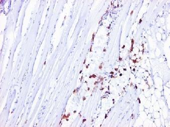


60 49 Stomach Muscle Gonad Epithelium tissue


61 Antennal gland Heart Appendage Figure 23. The target organs of GAV infection in P. vannamei. 50
62 Quantification of GAV infection in the lymphoid organ in time The first signs of GAV infection in P. vannamei injected with a dose of 103 SID50 were observed in the lymphoid organs at 24 hpi (Figure 25). At this time, no positive signals occurred in other organs of infected shrimp. GAV positive signals in gills were only detected in most shrimp at 48 hpi, while results were variable between shrimp for other target tissues and organs. Because it was consistently positive in all GAV-infected P. vannamei, the lymphoid organ was selected as the organ of choice to follow-up the evolution of GAV infection in time. In the following days, the area of the lymphoid organ positive for GAV signal increased from 1.8% at 48 hpi to 17.1% at 72 hpi (Figure 24). At 120 hpi, the infection reached its greatest expansion, when it occupied about 30% of the lymphoid organ. During the next days until 2 weeks post inoculation, the infected area slowly reduced, still covering up to 22% of % of lymphoid organ area + for GAV the lymphoid organ by the last day (336 hpi) Time (hours post injection) Figure 24. The evolution of GAV infection in the lymphoid organ in time in P. vannamei intramuscularly injected with a dose of 103 SID50. The amount of infection was expressed in percentage of lymphoid organ area positive for GAV signal as detected by IHC. 51


63 18 hpi 120 hpi 48 hpi 12 hpi 24 hpi 336 hpi 240 hpi 72 hpi 168 hpi 336 hpi Figure 25. Time course of GAV infection in the lymphoid organ of P. vannamei up to 2 weeks post injection of a 103 SID50 dose. 52
64 4.3. Characterization of GAV infection patterns in P. vannamei Generally, most GAV-positive signals were observed inside accumulations of virus-positive cells, the so-called spheroids. Within the lymphoid organ, the detected positive signals were markedly more strong in lymphoid organ spheroids (LOS) than in lymphoid organ tubules (LOT) (Figure 26). However, sometimes spheroids within lymphoid organs were observable by the hematoxylin background stain, but were negative for GAV by IHC (Figure 27). Figure 26. GAV-positive signal was detected both in lymphoid organ spheroids (arrow) and tubules (arrowheads). Figure 27. GAV-positive (arrow) and negative (arrowhead) lymphoid organ spheroids. 53
65 Despite the fact that the most typical appearance of GAV infection we found was in the form of classic, antigen-positive spheroids, positive signals tended to appear in 3 distinctly different patterns in target organs of 3 differently affected shrimp categories (Figure 28). The first pattern was observed as clusters of infected cells, encapsulated by uninfected fibrous cells. This was categorized as so-called lymphoid organ spheroids or ectopic spheroids (outside lymphoid organs). The second pattern consisted of signal spread-out through the tissues, without demarcation by fibrous cells. This appearance was often detected in connective tissues, muscles, gills, hematopoietic tissue and the lymphoid organ. The third pattern looked like unusual inverted spheroids in the lymphoid organs. These agglomerations of unstained cells appeared to be encapsulated by fibrous cells which reacted positively with the monoclonal antibody in immunochemistry, but rather faintly compared to normal spheroids. All three patterns could be found at most time points during the time course study. However, at the later time points of infection, pattern 2 was more represented, and when pattern 3 was found, the shrimp was mostly showing clinical signs. 54


66 Figure 28. Three forms of GAV infection in P. vannamei. A, B, E and F: lymphoid organs; C: muscle; D: gills Histopathology of GAV infection in the lymphoid organ of P. vannamei Overall, histopathological features of GAV infection in the lymphoid organ were characterized by spheroid formation, pyknosis, karyorrhexis and cellular vacuolization. The three types of spheroids classified by Hasson et al (1999) were found during our time course study of GAV infection in P. vannamei. Type A spheroids are identified as lightly 55
67 basophilic and homogeneous cell masses containing no necrotic cells. Type B spheroids are considered as the evolution of type A, but more basophilic and the spheroid bodies contain a number of vacuolated cells as well as pyknotic and karyorrhectic nuclei. Type C spheroids are described as intensely basophilic masses of degenerating cell with numerous vacuoles and also pyknotic and karyorrhectic nuclei. Although the first positive signals of GAV were detected by immunohistochemistry from 24 hpi, the lymphoid organ still exhibited a normal histological structure of LOT until 48 hpi and absence of LOS (Figure 29). However, by that time, numerous vacuoles as well as aggregations of hemocytes were observed within the inter-tubular connective tissue of the lymphoid organ. By day 3 after inoculation, the LO of the GAV-infected shrimp contained more histopathological signs of YHD described in previous publications (Figure 30). Abundant basophilic LOS containing hypertrophied nuclei were seen to form between the LOT. Some LOS were surrounded by capsular sheaths. However, the histopathologically typical signs of YHD such as apoptosis and necrosis including nuclear pyknosis, karyorrhexis and cellular vacuolization were seldom observed. It seemed that only type A spheroids occurred at this time and the structures of LOT were normal. At the next time points (120 and 168 hpi), the LO tissue of infected shrimp showed severe histopathological lesions including numerous pyknotic and karyorrhectic nuclei and vacuolated cells within densely basophilic type B LOS (Figures 31 and 32). The development of LOS could result in distortion and disorganization and loss of normal structure of adjacent LOT. Vacuolization of cells was frequently observed inside LOS which were surrounded by clearly visible, capsular sheaths. No apoptosis or necrosis was seen within LOT. At 240 and 336 hpi, LOS were highly basophilic, with pyknotic and karyorrhectic nuclei and vacuolated cells (type B and C) (Figure 33). 56
68 Normal 24 hpi 48 hpi Figure 29. H&E staining showing the normal histological structures of lymphoid organ tubules (LOT) and absence of lymphoid organ spheroids (LOS) until 48 hpi after GAV inoculation. Numerous vacuoles as well as hemocyte aggregations were observed within the inter-tubular connective tissue of the lymphoid organ during the early stages of infection. 57


69 Figure 30. Histopathological features in the lymphoid organs of P. vannamei at 72 hpi were characterized by the formation of basophilic spheroids type A surrounded by capsular sheaths. Figure 31. At 120 hpi, the LO tissue of GAV infected P. vannamei showed severe histopathological lesions including numerous pyknotic and karyorrhectic nuclei and vacuolated cells inside densely basophilic type B LOS. 58


70 Figure 32. By 168 hpi, numerous pyknotic and karyorrhectic nuclei and vacuolated cells could clearly be observed inside increasingly larger densely basophilic type B LOS. Figure 33. By hpi, the highly basophilic LOS with an abundance of pyknotic and karyorrhectic nuclei and vacuolated cells could also be categorized as type C. The overall aspect of the LO has changed considerably from the normal situation by this time. 59


 were infrequently")


 and karyorrhectic nuclei")
71 4.5. Histopathological features of GAV infection in other target organs By H&E staining, no histopathological features distinguished the other target organs of GAV-infected shrimp such as gills and hepatopancreas from those of negative control shrimp. Apoptosis, necrosis and dense basophilic inclusions which were reported as cytopathological signs of YHD (Khanobdee et al, 2002) were infrequently detected in connective tissue of hepatopancreas and other organs. Although pyknotic and karyorrhectic nuclei were randomly scattered in gills and other organs, the information was not enough to confirm whether their existence was due to the GAV infection. Infected tissue Hepatopancreas Gills Uninfected tissue Figure 34. Signs of apoptosis with pyknotic (arrows) and karyorrhectic nuclei (arrowheads) in GAV-infected tissues of gills and hepatopancreas (right) in comparison with the same structures of uninfected tissues (left). 60
Taura Syndrome Virus Disease in Farm-Reared Penaeus monodon in Thailand
 Kasetsart J. (Nat. Sci.) 41 : 319-323 (2007) Taura Syndrome Virus Disease in Farm-Reared Penaeus monodon in Thailand Chalor Limsuwan and Niti Chuchird* ABSTRACT Taura syndrome virus (TSV) has caused major
The Viability of Taura Syndrome Virus in Low-salinity Water
 Kasetsart J. (Nat. Sci.) 39 : 406-410 (2005) The Viability of Taura Syndrome Virus in Low-salinity Water Niti Chuchird and Chalor Limsuwan ABSTRACT Taura syndrome virus (TSV) could survive up to 10 days
Update on the strategy planning for Infectious myonecrosis (IMN) disease
 disease") Second Interregional Workshop of FAO TCP/INT/3501 Update on the strategy planning for Infectious myonecrosis (IMN) disease Kathy F.J. Tang Tucson, Arizona, USA Components of a contingency plan Technical
Second Interregional Workshop of FAO TCP/INT/3501 Update on the strategy planning for Infectious myonecrosis (IMN) disease Kathy F.J. Tang Tucson, Arizona, USA Components of a contingency plan Technical
Short communication. C W Tung, C S Wang and S N Chen
 Short communication Histological and electron microscopic study on Macrobrachium muscle virus (MMV) infection in the giant freshwater prawn, Macrobrachium rosenbergii (de Man), cultured in Taiwan C W Tung,
However, 31.2% showed resistance to cotrimaxasole, 91.6% to sulphafurazole and 65.8% to sulphadiazine (Otta, 1997).
.") 416 I. Karunasagar, S. K. Otta et al. (1994) reported mass mortality of P. monodon larvae due to chloramphenicol resistant strains of V. harveyi. Later studies showed that V harveyi is capable of forming
416 I. Karunasagar, S. K. Otta et al. (1994) reported mass mortality of P. monodon larvae due to chloramphenicol resistant strains of V. harveyi. Later studies showed that V harveyi is capable of forming
White Spot Disease in Mozambique
 White Spot Disease in Mozambique Experiences and lessons learned A.P. BALOI (1), M. LE GROUMELLEC (2) (1) Ministry of Fisheries, National Institute for Fish Inspection, Mozambique. (2) OIE Consultant on
Study on the Pathogenesis of the White Spot Syndrome Virus (WSSV) on Juvenile Penaeus monodon in Vietnam
 on Juvenile Penaeus monodon in Vietnam") 248 The Israeli Journal of Aquaculture Bamidgeh 61(3), 2009 Study on the Pathogenesis of the White Spot Syndrome Virus (WSSV) on Juvenile Penaeus monodon in Vietnam Cuong Van Doan, Anh Thi Tuyet Pham,
248 The Israeli Journal of Aquaculture Bamidgeh 61(3), 2009 Study on the Pathogenesis of the White Spot Syndrome Virus (WSSV) on Juvenile Penaeus monodon in Vietnam Cuong Van Doan, Anh Thi Tuyet Pham,
Infectious Myonecrosis Virus (IMNV) in Pacific White Shrimp (Litopenaeus vannamei) in Indonesia
 in Pacific White Shrimp (Litopenaeus vannamei) in Indonesia") SEAFDEC International Workshop on Emerging Fish Diseases in Asia 255 Infectious Myonecrosis Virus (IMNV) in Pacific White Shrimp (Litopenaeus vannamei) in Indonesia Taukhid1* and Yani Lestari Nur aini2
SEAFDEC International Workshop on Emerging Fish Diseases in Asia 255 Infectious Myonecrosis Virus (IMNV) in Pacific White Shrimp (Litopenaeus vannamei) in Indonesia Taukhid1* and Yani Lestari Nur aini2
How to prevent and control viral diseases in shrimp culture
 How to prevent and control viral diseases in shrimp culture Shrimp Farming in Thailand Farming Area: 80,000 Hectares No. of Farms: 35,000 Geographical Spreading: Eastern (20%) Southern (40%) Central (40%,
INFECTIOUS MYONECROSIS VIRUS (IMNV) IN PACIFIC WHITE SHRIMP, Litopenaeus vannamei IN INDONESIA
 IN PACIFIC WHITE SHRIMP, Litopenaeus vannamei IN INDONESIA") Infectious myonecrosis virus... (Taukhid) INFECTIOUS MYONECROSIS VIRUS (IMNV) IN PACIFIC WHITE SHRIMP, Litopenaeus vannamei IN INDONESIA Taukhid *), and Yani Lestari Nur aini **) ABSTRACT The aquaculture
Infectious myonecrosis virus... (Taukhid) INFECTIOUS MYONECROSIS VIRUS (IMNV) IN PACIFIC WHITE SHRIMP, Litopenaeus vannamei IN INDONESIA Taukhid *), and Yani Lestari Nur aini **) ABSTRACT The aquaculture
The University of Arizona 4/17/2008
 World Organisation for Animal Health Overview of Diseases and Health Management Issues Related to Farmed Shrimp Carlos R. Pantoja, Donald V. Lightner, Bonnie T. Poulos, Linda Nunan, Kathy F.J. Tang, Rita
< Attachment 2 > 1.A) The country, zone, compartment or establishment is free of the target disease:
 The country, zone, compartment or establishment is free of the target disease:") Certificate number: Health Certificate for Live Crustaceans Exported from Thailand to Japan 1. Competent Authority: 2. Consignor 3. Consignee 4. Place of origin 5. Place of destination 6.Port of Embarkation:
Certificate number: Health Certificate for Live Crustaceans Exported from Thailand to Japan 1. Competent Authority: 2. Consignor 3. Consignee 4. Place of origin 5. Place of destination 6.Port of Embarkation:
15. Zoosanitary information I, the undersigned official inspector, hereby certify that the aquatic animals above satisfy the following requirements.
 Certificate number: HEALTH CERTIFICATE FOR LIVE CRUSTACEANS EXPORTED FROM KOREA TO JAPAN 1. Competent Authority: 2. Consignor Name: Address: 3. Consignee Name: Address: 4. Place of origin Name: Farm address
Summary and Conclusions
 s The present investigation has been designed to identify the various endemic diseases and their control measure prevailing in selected cultured shrimps, L. vannamei.and P. monodon. The study has envisaged
Cellular targets and pathways of yellow head virus infection in lymphoid organ of Penaeus monodon as studied by transmission electron microscopy
 Songklanakarin J. Sci. Technol. 33 (2), 121-127, Mar. - Apr. 2011 http://www.sjst.psu.ac.th Original Article Cellular targets and pathways of yellow head virus infection in lymphoid organ of Penaeus monodon
Histopathological and bacteriological studies of monodon slow growth syndrome (MSGS) affected shrimps
 affected shrimps") Indian J. Fish., 60(1) : 97-101, 2013 97 Histopathological and bacteriological studies of monodon slow growth syndrome (MSGS) affected shrimps P. JANAKIRAM, L. JAYASREE *, B. SIVAPRASAD, M. VEERENDRAKUMAR
Indian J. Fish., 60(1) : 97-101, 2013 97 Histopathological and bacteriological studies of monodon slow growth syndrome (MSGS) affected shrimps P. JANAKIRAM, L. JAYASREE *, B. SIVAPRASAD, M. VEERENDRAKUMAR
Detection of gill-associated virus (GAV) by in situ hybridization during acute and chronic infections of Penaeus monodon and P.
 by in situ hybridization during acute and chronic infections of Penaeus monodon and P.") DISEASES OF AQUATIC ORGANISMS Vol. 56: 1 10, 2003 Published August 15 Dis Aquat Org Detection of gill-associated virus (GAV) by in situ hybridization during acute and chronic infections of Penaeus monodon
DISEASES OF AQUATIC ORGANISMS Vol. 56: 1 10, 2003 Published August 15 Dis Aquat Org Detection of gill-associated virus (GAV) by in situ hybridization during acute and chronic infections of Penaeus monodon
Confirmatory testing of the viral status of Penaeus monodon (Black Tiger shrimp) populations in the Fiji Islands
 populations in the Fiji Islands") Final report for Mini-project MS0401: Confirmatory testing of the viral status of Penaeus monodon (Black Tiger shrimp) populations in the Fiji Islands Salote Waqairatu Dr. Timothy Pickering The University
Final report for Mini-project MS0401: Confirmatory testing of the viral status of Penaeus monodon (Black Tiger shrimp) populations in the Fiji Islands Salote Waqairatu Dr. Timothy Pickering The University
Preliminary Molecular and Biological Characterization of Mourilyan Virus (MoV): A New Bunya-Related Virus of Penaeid Prawns
: A New Bunya-Related Virus of Penaeid Prawns") Diseases in Asian Aquaculture V Preliminary Molecular and Biological Characterization of Mourilyan Virus (MoV): A New Bunya-Related Virus of Penaeid Prawns JEFF A COWLEY, RUSSELL J. MCCULLOCH, KIRSTEN
Diseases in Asian Aquaculture V Preliminary Molecular and Biological Characterization of Mourilyan Virus (MoV): A New Bunya-Related Virus of Penaeid Prawns JEFF A COWLEY, RUSSELL J. MCCULLOCH, KIRSTEN
Necrotizing Hepatopancreatitis
 [DRAFT] Necrotizing Hepatopancreatitis Pathogen information 1. Causative agent 1.1. Pathogen type Obligate intracellular rickettsial-like organism 1.2. Disease name and synonyms Necrotizing Hepatopancreatitis
Biju V. N* and B. Gunalan. Abstract
 Volume: 2; Issue: 12; December -2016; pp 1019-1025. ISSN: 2454-5422 Prevalence of white faeces syndrome in Penaeus (Litopenaeus) vannamei farms in Nagapattinam district Biju V. N* and B. Gunalan Centre
CRAYFISH DISSECTION. Image from:
 CRAYFISH DISSECTION Image from: http://www.mackers.com/crayfish/ ARTHROPODA jointed foot Arthro = joint pod = foot Animal Groups Image from: http://ology.amnh.org/biodiversity/treeoflife/pages/graph.html
The Terms of Reference were adopted and the final version is shown in Annex III.
 Original: English February 2015 REPORT OF THE FIRST MEETING OF OIE AD HOC GROUP ON SUSCEPTIBILITY OF CRUSTACEAN SPECIES TO INFECTION WITH OIE LISTED DISEASES 1 Paris, 10 12 February 2015 The OIE ad hoc
Crayfish Observation and Dissection
 Name Period Date Crayfish Observation and Dissection Purpose: In this lab, you will observe the external structures of a crayfish and dissect it to study its internal structures and systems. Materials:
Caroline H. Seibert, Aguinaldo R. Pinto* Submitted: July 12, 2011; Approved: June 07, ABSTRACT
 Brazilian Journal of Microbiology (2012): 857-864 ISSN 1517-8382 CHALLENGES IN SHRIMP AQUACULTURE DUE TO VIRAL DISEASES: DISTRIBUTION AND BIOLOGY OF THE FIVE MAJOR PENAEID VIRUSES AND INTERVENTIONS TO
CRAYFISH DISSECTION. Image from:
 CRAYFISH DISSECTION Image from: http://www.mackers.com/crayfish/ Animal Groups Image from: http://ology.amnh.org/biodiversity/treeoflife/pages/graph.html ARTHROPODA jointed foot Arthro = joint pod = foot
DISEASES OF AQUATIC ORGANISMS Vol. 59: , 2004 Published June 11 Dis Aquat Org
 DISEASES OF AQUATIC ORGANISMS Vol. 59: 195 203, 2004 Published June 11 Dis Aquat Org Quantitative real-time RT-PCR demonstrates that handling stress can lead to rapid increases of gill-associated virus
Examination for Viral Inactivation of WSSV (White Spot Syndrome Virus) Isolated in Malaysia Using Black Tiger Prawn (Penaeus monodon)
 Isolated in Malaysia Using Black Tiger Prawn (Penaeus monodon)") JARQ 4 (1), 93 97 (26) http://www.jircas.affrc.go.jp Examination for Viral Inactivation of WSSV (White Spot Syndrome Virus) Isolated in Malaysia Using Black Tiger Prawn (Penaeus monodon) Norihisa OSEKO
JARQ 4 (1), 93 97 (26) http://www.jircas.affrc.go.jp Examination for Viral Inactivation of WSSV (White Spot Syndrome Virus) Isolated in Malaysia Using Black Tiger Prawn (Penaeus monodon) Norihisa OSEKO
Differences in susceptibility of palaemonid shrimp species to yellow head virus (YHV) infection
 infection") DISEASES OF AQUATIC ORGANISMS Vol. 64: 5 12, 2005 Published April 6 Dis Aquat Org Differences in susceptibility of palaemonid shrimp species to yellow head virus (YHV) infection Siwaporn Longyant 1, Paisarn
DISEASES OF AQUATIC ORGANISMS Vol. 64: 5 12, 2005 Published April 6 Dis Aquat Org Differences in susceptibility of palaemonid shrimp species to yellow head virus (YHV) infection Siwaporn Longyant 1, Paisarn
Transboundary Aquatic Animal Diseases: History and Impacts in ASEAN Aquaculture
 Network of Aquaculture Centres in Asia-Pacific Transboundary Aquatic Animal Diseases: History and Impacts in ASEAN Aquaculture Eduardo M. Leaño Network of Aquaculture Centres in Asia-Pacific Bangkok, Thailand
Electron Microscopic Evidence of Bacilliform Virus Infection in Kuruma Shrimp (Penaeus japonicus)
") Electron Microscopic Evidence Bacilliform Virus Infection in Kuruma Shrimp (Penaeus japonicus) Yukinori Takahashi*1, Toshiaki Itami*1, Masakazu Kondo*2, Minoru Maeda*1, Reiko Fujii*3, Susumu Tomonaga3,
Electron Microscopic Evidence Bacilliform Virus Infection in Kuruma Shrimp (Penaeus japonicus) Yukinori Takahashi*1, Toshiaki Itami*1, Masakazu Kondo*2, Minoru Maeda*1, Reiko Fujii*3, Susumu Tomonaga3,
Challenges for Providing Diagnostic Service: White Spot Disease (WSD)
") Regional Meeting of OIE Reference Centres in Asia and the Pacific6-7 February 2017, Tokyo, Japan Challenges for Providing Diagnostic Service: White Spot Disease (WSD) Grace Chu-Fang Lo National Cheng Kung
Regional Meeting of OIE Reference Centres in Asia and the Pacific6-7 February 2017, Tokyo, Japan Challenges for Providing Diagnostic Service: White Spot Disease (WSD) Grace Chu-Fang Lo National Cheng Kung
Crayfish Dissection. Objectives: Describe the appearance of various organs found in a crayfish. Name the organs that make up systems of the crayfish.
 Crayfish Dissection Objectives: Describe the appearance of various organs found in a crayfish. Name the organs that make up systems of the crayfish. Background: Like all crustaceans, a crayfish has a fairly
Infectious Salmon Anemia
 Infectious Salmon Anemia A paradigm shift for understanding risk of ISAV infection Jill Rolland US Geological Survey Western Fisheries Research Center Seattle, Washington Infectious Salmon Anemia history
In this lab, you will observe the external structures of a crayfish and dissect it to study its internal structures and systems.
 Crayfish Dissection Objectives: Describe the appearance of various organs found in a crayfish. Name the organs that make up systems of the crayfish. Materials: safety goggles, gloves, magnifying glass,
Internal and External Anatomy of a Penaeid Shrimp
 Internal and External Anatomy of a Penaeid Shrimp stomach eye stalk oesophagus antenna pereiopods Internal and external anatomy of a penaeid shrimp. hepatopancreas heart pleopods hindgut abdominal segment
Tissue distribution of white spot syndrome virus (WSSV) in shrimp and crabs
 in shrimp and crabs") Tissue distribution of white spot syndrome virus (WSSV) in shrimp and crabs *Guang-Hsiung Kou, Shao-En Peng, Ya-Lin Chiu, Chu-Fang Lo Department of Zoology, National Taiwan University, Taipei, Taiwan,
Tissue distribution of white spot syndrome virus (WSSV) in shrimp and crabs *Guang-Hsiung Kou, Shao-En Peng, Ya-Lin Chiu, Chu-Fang Lo Department of Zoology, National Taiwan University, Taipei, Taiwan,
Identification of Stressors that Affect White Spot Syndrome Virus (WSSV) Infection and Outbreak in Pond Cultured Penaeus monodon
 Infection and Outbreak in Pond Cultured Penaeus monodon") The Israeli Journal of Aquaculture - Bamidgeh, IIC:63.2011.616, 7 pages The IJA appears exclusively as a peer-reviewed on-line open access journal at http://www.siamb.org.il Sale of IJA papers is strictly
The Israeli Journal of Aquaculture - Bamidgeh, IIC:63.2011.616, 7 pages The IJA appears exclusively as a peer-reviewed on-line open access journal at http://www.siamb.org.il Sale of IJA papers is strictly
2. Viral diseases of shrimp in the Philippines
 Research Signpost 37/661 (2), Fort P.O. Trivandrum-695 023 Kerala, India Biotechnological Advances in Shrimp Health Management in the Philippines, 2015: 19-44 ISBN: 978-81-308-0558-0 Editors: Christopher
Prevalence of White Spot Syndrome Virus (WSSV) and Monodon Baculovirus (MBV) Infection in Penaeus monodon Postlarvae in Vietnam
 and Monodon Baculovirus (MBV) Infection in Penaeus monodon Postlarvae in Vietnam") Diseases in Asian Aquaculture V Prevalence of White Spot Syndrome Virus (WSSV) and Monodon Baculovirus (MBV) Infection in Penaeus monodon Postlarvae in Vietnam DANG THI HOANG OANH, NGUYEN THANH PHUONG
Diseases in Asian Aquaculture V Prevalence of White Spot Syndrome Virus (WSSV) and Monodon Baculovirus (MBV) Infection in Penaeus monodon Postlarvae in Vietnam DANG THI HOANG OANH, NGUYEN THANH PHUONG
2. What is the difference between a compound eye and your eye?
 INTRODUCTION: If numbers alone are used as a measure of success, the jointedlegged animals are the most successful animals. Their body segmentation suggests that they evolved from segmented worms. The
Proposal to Introduce Whiteleg Shrimp (Litopenaeus vannamei) to the Kingdom of Saudi Arabia for Aquaculture Development
 to the Kingdom of Saudi Arabia for Aquaculture Development") Final Draft Proposal to Introduce Whiteleg Shrimp (Litopenaeus vannamei) to the Kingdom of Saudi Arabia for Aquaculture Development Prepared for Saudi Aquaculture Society by J. Richard Arthur, Victoria
Final Draft Proposal to Introduce Whiteleg Shrimp (Litopenaeus vannamei) to the Kingdom of Saudi Arabia for Aquaculture Development Prepared for Saudi Aquaculture Society by J. Richard Arthur, Victoria
Current status of transboundary fish diseases in Thailand: Occurrence, surveillance, research and training
 Current status of transboundary fish diseases in Thailand: Occurrence, surveillance, research and training Kanchanakhan, Somkiat Date published: 2004 To cite this document : Kanchanakhan, S. (2004). Current
PRODUCTION PERFORMANCE, DISEASES, SPF-BREEDING AND RISK ISSUES CONCERNING WHITE SHRIMP, Penaeus vannamei, INTRODUCTION INTO INDONESIA
 Indonesian Aquaculture Journal Vol.1 No.1, 2006 PRODUCTION PERFORMANCE, DISEASES, SPF-BREEDING AND RISK ISSUES CONCERNING WHITE SHRIMP, Penaeus vannamei, INTRODUCTION INTO INDONESIA Ketut Sugama, Hessy
The pathogenesis of nervous distemper
 Veterinary Sciences Tomorrow - 2004 The pathogenesis of nervous distemper Marc Vandevelde Canine distemper is a highly contagious viral disease of dogs and of all animals in the Canidae, Mustellidae and
Overview: Chapter 19 Viruses: A Borrowed Life
 Overview: Chapter 19 Viruses: A Borrowed Life Viruses called bacteriophages can infect and set in motion a genetic takeover of bacteria, such as Escherichia coli Viruses lead a kind of borrowed life between
REVIEW OF LITERATURE
 REVIEW OF LITERATURE 2. REVIEW OF LITERATURE The past three decades have witnessed remarkable expansion, intensification and diversification of the aquaculture sector which has become enormously reliant
LESSON 4.6 WORKBOOK. Designing an antiviral drug The challenge of HIV
 LESSON 4.6 WORKBOOK Designing an antiviral drug The challenge of HIV In the last two lessons we discussed the how the viral life cycle causes host cell damage. But is there anything we can do to prevent
Introductions and movement of Penaeus vannamei and Penaeus stylirostris in Asia and the Pacific
 RAP publication 2004/10 Introductions and movement of Penaeus vannamei and Penaeus stylirostris in Asia and the Pacific Matthew Briggs, Simon Funge-Smith, Rohana Subasinghe and Michael Phillips FOOD AND
Phylum Arthropoda Blue Crabs
 Phylum Arthropoda Blue Crabs The Decapod Crustaceans Blue crabs (Callinectes sapidus) are members of the phylum Arthropoda; organisms which possess jointed appendages and a non-living exoskeleton made
D. V. Lightner, R. R. Williams, T. A. Bell, R. M. Redman, and L. A. Perez A. Introduction
 ICES mar. Sei. Symp., 194: 97-105. 1992 A collection of case histories documenting the introduction and spread of the virus disease IHHN in penaeid shrimp culture facilities in Northwestern Mexico D. V.
LEC 2, Medical biology, Theory, prepared by Dr. AYAT ALI
 General Characteristics, Structure and Taxonomy of Viruses Viruses A virus is non-cellular organisms made up of genetic material and protein that can invade living cells. They are considered both a living
Session 2. TiLV isolation and Koch s Postulates
 Session 2 Win Surachetpong DVM, PhD, CertAqV, DTBVP Kathy Tang-Nelson PhD TiLV isolation and Koch s Postulates Learning objectives Describe how viruses are isolated Apply the appropriate method to the
Introductions and movement of Penaeus vannamei and Penaeus stylirostris in Asia and the Pacific RAP PUBLICATION 2004/10
 Introductions and movement of Penaeus vannamei and Penaeus stylirostris in Asia and the Pacific RAP PUBLICATION 2004/10 RAP Publication 2004/10 Introductions and movement of Penaeus vannamei and Penaeus
Outline. 1. Management organizations in Japan. 2. Prevention measures against fish disease invasion and spread. 3. Nishikigoi export program
 Health certification for ornamental fish in Japan Nanae Karakawa Fish and Fishery Products Safety Office, Food Safety and Consumer Affairs Bureau, Ministry of Agriculture, Forestry and Fisheries (MAFF),
Coronaviruses. Virion. Genome. Genes and proteins. Viruses and hosts. Diseases. Distinctive characteristics
 Coronaviruses Virion Genome Genes and proteins Viruses and hosts Diseases Distinctive characteristics Virion Spherical enveloped particles studded with clubbed spikes Diameter 120-160 nm Coiled helical
Chapter 5. Viral infections (I)
") Chapter 5. Viral infections (I) 1. Properties of virus - Virus: derived from Latin and means poison or stench (foul odor) - Definition an infectious, potentially pathogenic nucleoprotein entity which reproduces
Chapter 5. Viral infections (I) 1. Properties of virus - Virus: derived from Latin and means poison or stench (foul odor) - Definition an infectious, potentially pathogenic nucleoprotein entity which reproduces
EXPERIENCES CULTIVATING WHITE SHRIMP IN THAILAND
 EXPERIENCES CULTIVATING WHITE SHRIMP IN THAILAND Dr. Chalor Limsuwan Aquaculture Business Research Center Faculty of Fisheries, Kasetsart University, Thailand Summary of presentations in Peru, Colombia,
Lecture 2: Virology. I. Background
 Lecture 2: Virology I. Background A. Properties 1. Simple biological systems a. Aggregates of nucleic acids and protein 2. Non-living a. Cannot reproduce or carry out metabolic activities outside of a
Sampling and evaluation of white spot syndrome virus in commercially important Atlantic penaeid shrimp stocks
 DISEASES OF AQUATIC ORGANISMS Vol. 59: 179 185, 2004 Published June 11 Dis Aquat Org Sampling and evaluation of white spot syndrome virus in commercially important Atlantic penaeid shrimp stocks Robert
Victoria Alday-Sanz, D.V.M., M.Sc., Ph.D. National Aquaculture Group (NAQUA)
") Stakeholder Consultation on Progressive Management Pathway (PMP) to Improve Aquaculture Biosecurity World Bank Headquarters, Washington, D.C. 10 April 2018 Aquaculture Biosecurity in the Kingdom of Saudi
Stakeholder Consultation on Progressive Management Pathway (PMP) to Improve Aquaculture Biosecurity World Bank Headquarters, Washington, D.C. 10 April 2018 Aquaculture Biosecurity in the Kingdom of Saudi
Chapter 6- An Introduction to Viruses*
 Chapter 6- An Introduction to Viruses* *Lecture notes are to be used as a study guide only and do not represent the comprehensive information you will need to know for the exams. 6.1 Overview of Viruses
A Short Review on Infectious Viruses in Cultural Shrimps (Penaeidae Family).
.") 9(3): 009-015 (2015) Journal of FisheriesSciences.com E-ISSN 1307-234X 2014 www.fisheriessciences.com Review Article ORIGINAL ARTICLE A Short Review on Infectious Viruses in Cultural Shrimps (Penaeidae
9(3): 009-015 (2015) Journal of FisheriesSciences.com E-ISSN 1307-234X 2014 www.fisheriessciences.com Review Article ORIGINAL ARTICLE A Short Review on Infectious Viruses in Cultural Shrimps (Penaeidae
Evaluation of Vibrio control
 Evaluation of Vibrio control with a multi-species probiotic in shrimp aquaculture Competence Center Microbials Competence Center Microbials Evaluation of Vibrio control with a multi-species probiotic in
Chapter 3 Notes Parts th Grade Science Mrs. Tracy Tomm
 Chapter 3 Notes Parts 1-2 8 th Grade Science Mrs. Tracy Tomm Section 3.1 Notes - Animal Classification 1. VERTEBRATES have a backbone made of bone or cartilage, while INVERTEBRATES have no backbone. 2.
INFLUENZA-2 Avian Influenza
 INFLUENZA-2 Avian Influenza VL 7 Dec. 9 th 2013 Mohammed El-Khateeb Overview 1. Background Information 2. Origin/History 3. Brief overview of genome structure 4. Geographical Distribution 5. Pandemic Nature
AQUAVETPLAN. Disease Strategy
 AUST RALIAN AQUAT IC VETERINARY EMERGENCY PLAN AQUAVETPLAN Disease Strategy White spot disease Version 2, 2013 AQUAVETPLAN is a series of technical response manuals for aquatic animal disease incursions,
Some living things are made of ONE cell, and are called. Other organisms are composed of many cells, and are called. (SEE PAGE 6)
") Section: 1.1 Question of the Day: Name: Review of Old Information: N/A New Information: We tend to only think of animals as living. However, there is a great diversity of organisms that we consider living
Section: 1.1 Question of the Day: Name: Review of Old Information: N/A New Information: We tend to only think of animals as living. However, there is a great diversity of organisms that we consider living
OIE work in pathogen differentiation (ISA as example)
") OIE work in pathogen differentiation (ISA as example) Larry Hammell OIE Collaborating Centre (ERAAAD) Based on presentation by Brit Hjeltnes Guiding principles for appropriate pathogen differentiation
OIE work in pathogen differentiation (ISA as example) Larry Hammell OIE Collaborating Centre (ERAAAD) Based on presentation by Brit Hjeltnes Guiding principles for appropriate pathogen differentiation
THE ROLE OF VETERINARY SERVICES IN MANAGING EMERGING AQUATIC ANIMAL DISEASES: WHAT ARE THE FACTORS NEEDED FOR SUCCESS?
 2015 Asia OIE Regional Commission Ernst THE ROLE OF VETERINARY SERVICES IN MANAGING EMERGING AQUATIC ANIMAL DISEASES: WHAT ARE THE FACTORS NEEDED FOR SUCCESS? I. Ernst 1 Original: English Summary: The
Where Health Care Meets Policy. with Dr. Mike Magee
 Where Health Care Meets Policy with Dr. Mike Magee The Threat of Bird Flu Understanding Bird Flu and the Influenza Virus 3 types of the influenza virus: A, B and C reflect differences in the M protein
The Open Access Israeli Journal of Aquaculture Bamidgeh
 The Open Access Israeli Journal of Aquaculture Bamidgeh As from January 2010 The Israeli Journal of Aquaculture - Bamidgeh (IJA) will be published exclusively as an on-line Open Access (OA) quarterly accessible
2.3.1 Other Viruses Isolated from Fish
 2.3.1 Other Viruses Isolated from Fish - 1 2.3.1 Other Viruses Isolated from Fish William N. Batts and James R. Winton USGS Western Fisheries Research Center 6505 NE 65th Street, Seattle, WA 98115 Phone:
MARTHA ZARAIN-HERZBERG NORMA HERNANDEZ-SAAVEDRA FELIPE ASCENCIO-VALLE
 JOURNAL OF THE WORLD AQUACULTURE SOCIETY Vol. 34, No. 1 March, 2003 Biological Characterization of a Less Virulent Taura Syndrome in Pacific White Shrimp Litopenaeus vannamei (Crustacea: Decapoda): Gross
Model of white spot syndrome virus (WSSV) epidemics in Litopenaeus vannamei
 epidemics in Litopenaeus vannamei") DISEASES OF AQUATIC ORGANISMS Vol. 50: 199 209, 2002 Published July 29 Dis Aquat Org Model of white spot syndrome virus (WSSV) epidemics in Litopenaeus vannamei Jeffrey M. Lotz*, M. Andres Soto** Department
DISEASES OF AQUATIC ORGANISMS Vol. 50: 199 209, 2002 Published July 29 Dis Aquat Org Model of white spot syndrome virus (WSSV) epidemics in Litopenaeus vannamei Jeffrey M. Lotz*, M. Andres Soto** Department
A yellow-head-like virus from Penaeus monodon cultured in Australia
 DISEASES OF AQUATIC ORGANISMS Dis Aquat Org Published December 30 A yellowheadlike virus from Penaeus monodon cultured in Australia 'Department of Parasitology, The University of Queensland, Brisbane 4072.
Eyes wide shut A critical view of aquaculture health management and risk factors in the real world
 Bull. Eur. Ass. Fish Pathol., 26(1) 2006, 1 Eyes wide shut A critical view of aquaculture health management and risk factors in the real world S. Mortensen 1*, K. Korsnes 1, 2 and Ø. Bergh 1 1 Institute
Protein Expression and Microscopic Evidence of White Spot Syndrome Virus (WSSV) in Tiger prawns Penaeus monodon
 in Tiger prawns Penaeus monodon") Protein Expression and Microscopic Evidence of White Spot Syndrome Virus (WSSV) in Tiger prawns Penaeus monodon Bhatt, S.S. a*, Chavda, D. V. a and Sreepada, R. A. b a B.R. Doshi School of Biosciences,
Protein Expression and Microscopic Evidence of White Spot Syndrome Virus (WSSV) in Tiger prawns Penaeus monodon Bhatt, S.S. a*, Chavda, D. V. a and Sreepada, R. A. b a B.R. Doshi School of Biosciences,
World Animal Health Information and Analysis Department Copyright OIE, 2015
 Copyright OIE, 2015 World Animal Health Information and Analysis Department information.dept@oie.int WORLD ORGANISATION FOR ANIMAL HEALTH (OIE) 12, rue de Prony, 75017 Paris, France Tel: (33-1) 44 15 18
Environmental impact of the establishment of exotic prawn pathogens in Australia
 Environmental impact of the establishment of exotic prawn pathogens in Australia a consultancy report to AQIS Prepared for: Australian Quarantine and Inspection Service GPO Box 858 Canberra ACT 2601 Australia
Viral Hemorrhagic Disease
 Viral Hemorrhagic Disease What is VHD? Highly contagious disease caused by calicivirus Affects only the Oryctolagus cuniclus species Includes wild & domesticated rabbits Doesn t appear to affect any North
Cardiomyopathy Syndrome (CMS) in Atlantic salmon WP4. Litterature review
 in Atlantic salmon WP4. Litterature review") Cardiomyopathy Syndrome (CMS) in Atlantic salmon WP4. Litterature review WP 4. Deliverables Report in Norwegian Fact sheet in Norwegian Scientific review PP- presentation 2 Overview of review CMS - Cardiomyopathy
Cardiomyopathy Syndrome (CMS) in Atlantic salmon WP4. Litterature review WP 4. Deliverables Report in Norwegian Fact sheet in Norwegian Scientific review PP- presentation 2 Overview of review CMS - Cardiomyopathy
11/15/2011. Outline. Structural Features and Characteristics. The Good the Bad and the Ugly. Viral Genomes. Structural Features and Characteristics
 Chapter 19 - Viruses Outline I. Viruses A. Structure of viruses B. Common Characteristics of Viruses C. Viral replication D. HIV II. Prions The Good the Bad and the Ugly Viruses fit into the bad category
DISEASES OF AQUATIC ORGANISMS Vol. 71: 1 10, 2006 Published July 11 Dis Aquat Org
 DISEASES OF AQUATIC ORGANISMS Vol. 71: 1, 006 Published July 11 Dis Aquat Org Comparison of four Taura syndrome virus (TSV) isolates in oral challenge studies with Litopenaeus vannamei unselected or selected
Transcriptional Analysis for Oral Vaccination of Recombinant Viral Proteins against White Spot Syndrome Virus (WSSV) in Litopenaeus vannamei
 in Litopenaeus vannamei") J. Microbiol. Biotechnol. (2011), 21(2), 170 175 doi: 10.4014/jmb.1005.05036 First published online 26 November 2010 Transcriptional Analysis for Oral Vaccination of Recombinant Viral Proteins against
J. Microbiol. Biotechnol. (2011), 21(2), 170 175 doi: 10.4014/jmb.1005.05036 First published online 26 November 2010 Transcriptional Analysis for Oral Vaccination of Recombinant Viral Proteins against
Current status of transboundary fish diseases in Singapore: Occurrence, surveillance, research and training
 Current status of transboundary fish diseases in Singapore: Occurrence, surveillance, research and training Huat, Ling Kai; Kueh, Susan & Kwang, Poh Yew Date published: 2004 To cite this document : Huat,
OIE Situation Report for Highly Pathogenic Avian Influenza
 OIE Situation Report for Highly Pathogenic Avian Influenza Latest update: 28/02/2018 The epidemiology of avian influenza is complex. The virus constantly evolves and the behavior of each new subtype (and
The Conservation Implications of Avian Influenza (H5N1) Colin Poole, Asia Program, WCS
 Colin Poole, Asia Program, WCS") The Conservation Implications of Avian Influenza (H5N1) Colin Poole, Asia Program, WCS The reaction throughout the region that has been of greatest immediate concern to conservationists Wild birds are
The Conservation Implications of Avian Influenza (H5N1) Colin Poole, Asia Program, WCS The reaction throughout the region that has been of greatest immediate concern to conservationists Wild birds are
OUTLINE INTRODUCTION 10/17/2011. Introduction Current Knowledge Future Directions
 Re emergence of Rinderpest : Role of wildlife and closely associated viral species ASH OSH, Ph. D. Candidate Center for Wildlife Health, Department of Forestry, Wildlife and Fisheries, The University of
Coronaviruses cause acute, mild upper respiratory infection (common cold).
.") Coronaviruses David A. J. Tyrrell Steven H. Myint GENERAL CONCEPTS Clinical Presentation Coronaviruses cause acute, mild upper respiratory infection (common cold). Structure Spherical or pleomorphic enveloped
Coronaviruses David A. J. Tyrrell Steven H. Myint GENERAL CONCEPTS Clinical Presentation Coronaviruses cause acute, mild upper respiratory infection (common cold). Structure Spherical or pleomorphic enveloped
Conflict of Interest and Disclosures. Research funding from GSK, Biofire
 Pandemic Influenza Suchitra Rao, MBBS, Assistant Professor, Pediatric Infectious Diseases, Hospital Medicine and Epidemiology Global Health and Disasters Course, 2018 Conflict of Interest and Disclosures
OIE Situation Report for Highly Pathogenic Avian Influenza
 OIE Situation Report for Highly Pathogenic Avian Influenza Latest update: 30/06/2018 The epidemiology of avian influenza (AI) is complex. The AI virus constantly evolves by mutation and re-assortment with
Early Mortality Syndrome (EMS)/Acute Hepatopancreatic Necrosis Disease (AHPND) of Cultured Shrimps: Overview, Status and Updates
/Acute Hepatopancreatic Necrosis Disease (AHPND) of Cultured Shrimps: Overview, Status and Updates") Early Mortality Syndrome (EMS)/Acute Hepatopancreatic Necrosis Disease (AHPND) of Cultured Shrimps: Overview, Status and Updates Eduardo M. Leaño Coordinator, Aquatic Animal Health Programme Network of
Early Mortality Syndrome (EMS)/Acute Hepatopancreatic Necrosis Disease (AHPND) of Cultured Shrimps: Overview, Status and Updates Eduardo M. Leaño Coordinator, Aquatic Animal Health Programme Network of
Disease Emergence and Food Security: Global Impact of Pathogens on Sustainable Aquaculture Production. Peter J. Walker Professor CSIRO
 Disease Emergence and Food Security: Global Impact of Pathogens on Sustainable Aquaculture Production Peter J. Walker Professor CSIRO Paper prepared for presentation at the Fish, Aquaculture and Food Security:
Oncology in Emerging Markets
 Oncology in Emerging Markets W H I T E P A P E R Toll Free : (888) 987-2691 www.makrocare.com/mcsmo 1 www.makrocare.com/mcsmo C W H I T E P A P E R ancer has been the leading cause of death in economically
Lab Tuesday: Virus Diseases
 Lab Tuesday: Virus Diseases Quiz for Bacterial Pathogens lab (pp 69-75) and Biocontrol of Crown Gall (p. 115-119), Observation of Viral Movement in Plants (p. 121), and Intro section for Viruses (pp. 77-79).
Julianne Edwards. Retroviruses. Spring 2010
 Retroviruses Spring 2010 A retrovirus can simply be referred to as an infectious particle which replicates backwards even though there are many different types of retroviruses. More specifically, a retrovirus
Medical Virology. Herpesviruses, Orthomyxoviruses, and Retro virus. - Herpesviruses Structure & Composition: Herpesviruses
 Medical Virology Lecture 2 Asst. Prof. Dr. Dalya Basil Herpesviruses, Orthomyxoviruses, and Retro virus - Herpesviruses Structure & Composition: Herpesviruses Enveloped DNA viruses. All herpesviruses have
Arthropods have segmented bodies and tough exoskeletons with jointed appendages.
 Section 1: Arthropods have segmented bodies and tough exoskeletons with jointed appendages. K What I Know W What I Want to Find Out L What I Learned Essential Questions What is the importance of exoskeletons,
In vivo titration of white spot syndrome virus (WSSV) in specific pathogen-free Litopenaeus vannamei by intramuscular and oral routes
 in specific pathogen-free Litopenaeus vannamei by intramuscular and oral routes") DISEASES OF AQUATIC ORGANISMS Vol. 66: 163 170, 2005 Published September 5 Dis Aquat Org NOTE In vivo titration of white spot syndrome virus (WSSV) in specific pathogen-free Litopenaeus vannamei by intramuscular
DISEASES OF AQUATIC ORGANISMS Vol. 66: 163 170, 2005 Published September 5 Dis Aquat Org NOTE In vivo titration of white spot syndrome virus (WSSV) in specific pathogen-free Litopenaeus vannamei by intramuscular