CT of Postmeningitic Deafness: Observations and Predictive Value for Cochlear Implants in Children
|
|
|
- Peregrine Gray
- 5 years ago
- Views:
Transcription
1 CT of Postmeningitic Deafness: Observations and Predictive Value for Cochlear Implants in Children Michele H. Johnson, M. Suzanne Hasenstab, Michael A. Seicshnaydre, and George H. Williams PURPOSE: To demonstrate CT abnormalities encountered in children with postmeningitic deafness and to assess the value of CT in the prediction of cochlear implantation difficulties. METHODS: Thirteen children with postmeningitic deafness were evaluated with high-resolution, thin-section CT. CT findings were correlated with surgical anatomy at the time of cochlear implantation, with particular regard to the prediction of implant success. RESULTS: CT findings included normal scans (3 of 13, 23.1%), cochlear stenosis (5 of 13, 37.7%), cochlear fibroossific change (1 of 13, 7.7%), cochlear ossification (4 of 13, 30.8%), and osseous hypertrophy at the round window niche (4 of 13, 30.8%). Nine of 10 patients with abnormal findings had incomplete or difficult implantations (90%); 7 (88.8%) of these 9 received limited electrode insertions. CONCLUSION: Attention to subtle otological abnormalities on thin-section CT is helpful in the prediction of early success or failure of implantation in children with postmeningitic deafness. Those with CT abnormalities had a 90% risk of incomplete or difficult insertions with a 70% chance of limited electrode insertion. Index terms: Meningitis; Hearing; Computed tomography, in treatment planning; Ear, prostheses; Pediatric neuroradiology AJNR Am J Neuroradiol 16: , January 1995 Over the last several years, cochlear-implant technology has become available as a treatment for profound deafness in both adults and children (1). To date, approximately 1200 children worldwide have undergone implantation with the Nucleus cochlear implant device (Communique, Cochlear Corp, winter 1991). Seventy percent of these patients (815 patients) are in the United States. Children receive implants for deafness from a variety of causes, including congenital lesions, bacterial meningitis, and cytomegalovirus infection (2 4). Patients with deafness of undetermined cause have also been given implants. At our medical center we have given implants to a series Received July 8, 1992; accepted after revision May 27, From the Departments of Radiology (M.H.J.) and Otorhinolaryngology (M.S.H., M.A.S., G.H.W.), Medical College of Virginia, Virginia Commonwealth University, Richmond. Address reprint requests to Michele H. Johnson, MD, Department of Radiology, Medical College of Virginia, Virginia Commonwealth University, Box MCV Station, Richmond, VA AJNR 16: , Jan /95/ American Society of Neuroradiology 103 of 42 patients from all of these etiologic groups. Prospective and retrospective studies have reported that significant hearing loss develops in 7% to 29% of patients with meningitis (5). Of the total worldwide pediatric population receiving implants, approximately 44% have deafness secondary to meningitis (Communique, Cochlear Corp, winter 1991). Our 13 patients who had meningitis, as a group, have had more problems overall than have our patients receiving implants with deafness from other causes. Many had problems with the surgical implantation itself because of cochlear obstruction (6, 7). In addition, in some patients there are difficulties during device programming, particularly after incomplete implantation. Many children with postmeningitic deafness also have cognitive or learning disabilities, which may contribute to postimplantation difficulties (5, 6). We have evaluated the spectrum of computed tomographic (CT) findings in our patients with postmeningitic deafness and compared these with the surgical anatomy at the time of implantation. Our purpose was to assess ana-
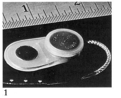
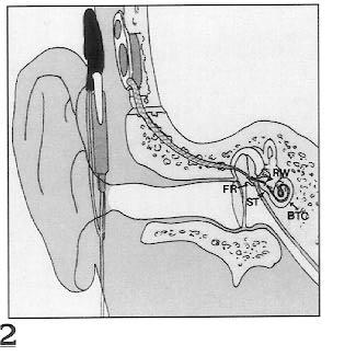
2 104 JOHNSON AJNR: 16, January 1995 Fig 1. The Nucleus 22-channel cochlear implant device. Note the location of the 22- electrode array (arrows). Fig 2. Final surgical placement of the device. RW indicates round window; ST, scala tympani; FR, facial recess; and BTC, basal turn of the cochlea. 1 2 tomic disease using high-resolution CT and attempt to identify the potentially difficult patient receiving a cochlear implant. Materials and Methods Thirteen patients with postmeningitic deafness were evaluated with high-resolution, thin-section CT before implantation of the Nucleus cochlear implant device (Cochlear Corp, East Englewood, Colo). Imaging Imaging was done in both axial and coronal planes with 1.5- to 2.0-mm section thicknesses and a bone algorithm reconstruction technique. One scan from an outside imaging facility included only axial sections with a 3-mm section thickness. All CT scans were assessed for congenital anomalies, cochlear stenosis, cochlear ossification, and osseous hypertrophy of the round window niche (8 13). The CT scans were interpreted by a single neuroradiologist (M.H.J.) without knowledge of the surgical findings. Structural abnormalities on CT were subsequently compared with those found at surgery. Surgical Selection All patients met the criteria for implantation at our medical center, which are: (a) profound bilateral sensorineural hearing loss; (b) age of 2 to 17 years; (c) normal general development; (d) parental and school support systems; and (e) no medical contraindications to surgery and implantation. Implantation Technique The surgical approach to cochlear implantation consists of a limited mastoidectomy. This is followed by opening of the facial recess and entrance into the round window with placement of the electrode array into the scala tympani via the basal turn of the cochlea (Figs 1 and 2) (8). The presence of significant obstruction or narrowing of the basal turn of the cochlea may inhibit the insertion of the full 22-electrode array of the Nucleus device (1, 3, 6, 7, 12, 14). After implantation, the range of hearing frequencies is programmed over the number of electrodes successfully inserted. Insertion of 19 or more electrodes is considered a good insertion, but even a limited number of inserted electrodes generally results in improved hearing (1, 6, 7). Results Meningitic labyrinthitis ossificans (labyrinthine ossification) is a postinflammatory process characterized by fibrous tissue proliferation and ossification within the inner ear (15 17). For the purposes of our analysis, we chose to divide our findings into fibroossific change and cochlear ossification, which are the early and late findings of meningogenic labyrinthitis ossificans, and into ossification of the round window niche and cochlear stenosis, which seem to be more limited manifestations within the spectrum of labyrinthitis ossificans. The results of our CT analyses and the success of cochlear implantation in our thirteen children with postmeningitic deafness are detailed in Table 1. Careful review of CT scans resulted in an interpretation of 3 normal and 10 abnormal examinations. Several patients had more than 1 abnormal finding on CT evaluation. The distribution of the CT findings are detailed in Table 2. The CT scans were analyzed for the following conditions. Normal Anatomy A normal CT scan interpretation indicated normal configuration of the cochlea without narrowing, fibrosis, or bone overgrowth (Fig 3). A normal CT scan did not ensure easy or complete insertion in our patient group (Table 1).
3 AJNR: 16, January 1995 COCHLEAR IMPLANTS 105 TABLE 1: Results of CT analysis and cochlear implantation in 13 patients with postmeningitic deafness Complete Implantation (22 Electrodes) Findings Postimplant Difficulties Incomplete or Difficult Implantation Round Window Ossification Fibroossific Changes Cochlear Ossification Cochlear Stenosis Age at Implantation Age at Meningitis Onset Patient Normal W.C. 9 mo 2 y 6 mo X X 6 electrodes Migrated electrode array, reimplant D.M. 3 wk 12 y 9 mo X 21 electrodes E.S. 19 mo 2 y 3 mo X X X F.R.* 14 mo 3 y 3 mo X X J.L. 3 y 4 mo 6 y 3 mo X 4-mm drilling 22 electrodes J.D. 26 mo 12 y 3 mo X 10 electrodes J.H.* 3 y 3 mo 8 y 4 mo X 6 electrodes High stimulus levels M.L.* 6 mo 6y2mo X 9 electrodes J.S. 5 y 2 mo 6 y 4 mo X X 7 electrodes High stimulus levels R.C.* 4 mo 8 y 4 mo X 7 electrodes High stimulus levels C.W. 5 mo 9 y 8 mo X 8 electrodes Neural adaptation S.C. 3 y 3 mo 5 y 10 mo X 19 electrodes Possible migration T.T. 22 mo 3 y 0 mo X X 7 electrodes Possible migration Note. High stimulus levels are required in cases of limited electrode insertion or decreased neural population. Neural adaptation means the device can be worn only for short periods because of saturation. X indicates CT finding. * Learning disability. Outside CT scan, 3-mm images, suboptimal. TABLE 2: CT findings in patients with postmeningitic diseases CT Findings Number of Patients Percentage Normal examination Cochlear stenosis Fibroossific change Cochlear ossification Osseous hypertrophy at the round window niche Cochlear Stenosis Narrowing of the basal turn of the cochlea was the most commonly observed finding in our series of patients (5 of 13 patients) and may represent the most limited form of labyrinthine ossification (Fig 4). Narrowing may impede or preclude complete insertion of the electrode array into the scala tympani. Cochlear Fibroossific Change (Early Labyrinthine Ossification) The fibrotic cochlea demonstrated hazy increased density within the basal turn of the cochlea on CT (1 of 13 patients) (Fig 5). The pathologic spectrum of fibroossific change ranges from pure fibrosis to fibrosis with significant osseous deposits. This is the most common finding at surgery, and is most frequently misinterpreted as normal on CT (7). Cochlear Ossification (Late Labyrinthine Ossification) Discrete focal cochlear ossifications were demonstrated in 4 of 13 patients (Fig 6). This probably represents an extension of the pathologic spectrum of fibroossific change. In the presence of such ossification, extensive drilling into the scala tympani may be required to achieve even limited electrode insertion. Osseous Hypertrophy of the Round Window Niche This finding was identified in 4 of 13 patients (Fig 7). Ossification of the round window niche requires the surgeon to drill off the obstructing bone before entering the scala tympani. This condition may not cause significant difficulty with insertion of the electrode array if it is not associated with other cochlear region disease. This focal disease may also represent a more isolated (limited) form of labyrinthine ossification.
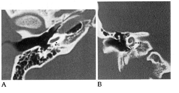
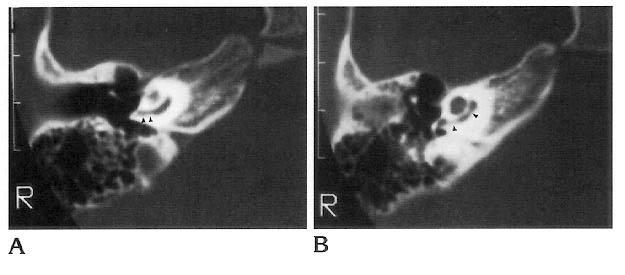
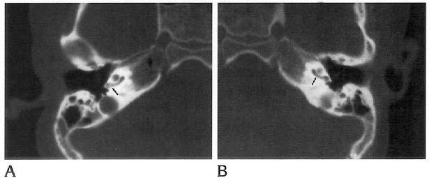
4 106 JOHNSON AJNR: 16, January 1995 Fig 3. A, Axial and B, coronal highresolution CT scans (1.5-mm section thickness) of a normal temporal bone illustrating the round window niche (RW), the scala tympani (ST), and the most proximal basal turn of the cochlea (BTC). Implantation and Postimplantation Difficulties Incomplete and difficult insertions were grouped together for analysis and comparison with preoperative CT interpretations (Tables 1 and 3). Incomplete implantations were defined as those in which, despite drilling at the round window niche or the basal turn of the cochlea, the entire 22-electrode array of the Nucleus device could not be inserted successfully. Difficult insertions were defined as those in which significant drilling at the round window or within the basal turn of the cochlea was required to insert the complete electrode array. Eleven implantations, among our 13 patients, were in the incomplete or difficult group. Postimplantation difficulties encountered included migration of the implanted electrode array in one child, requiring reimplantation. A second patient is currently undergoing evaluation for possible reimplantation. Problems with device programming were also encountered. High stimulus levels were required for adequate device programming, particularly in patients with limited (less than 10) electrode insertion (4 of 13). In one patient (S.C.) with a 19-electrode insertion, high stimulus levels were required, suggesting a decreased neuronal population within the cochlear nerve. One patient s (C.W.) hearing improved only when the device was worn for short periods, because of saturation of the available neural tissue. In total, 7 of the 11 incomplete- or difficult-insertion patients had significant postimplantation problems. Discussion Ossification of the labyrinth (labyrinthitis ossificans or labyrinthine ossification) is a common histologic end point of severe inflammatory disease of the ear and is often associated with profound deafness and loss of vestibular function (15 17). The causes of labyrinthine ossification may be classified as tympanogenic (chronic otitis, cholesteatoma, or postsurgical), meningogenic (bacterial or viral), or hematogenic (mumps or other hematogenous infections) (16, 17). In a patient with meningitis, infection spreads from the meninges to the inner ear via the cochlear aqueduct and the internal auditory canal. In patients who have had meningitis and are deaf, the identification of Fig 4. Axial CT images through the right ear, obtained at 1.5-mm intervals. A, The scan through the inferior aspect of the cochlea demonstrates tapered narrowing of the basal turn (arrowheads). B, More superiorly, a second area of focal stenosis is visible (arrowheads).
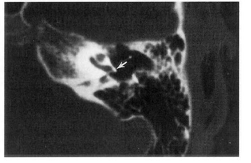
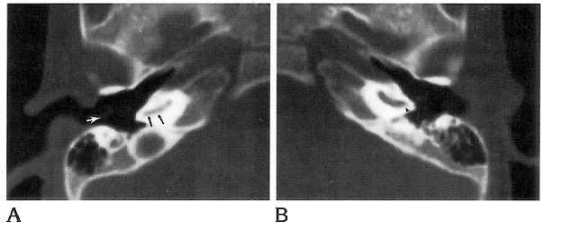
5 AJNR: 16, January 1995 COCHLEAR IMPLANTS 107 Fig 5. Cochlear fibroossific change is demonstrated bilaterally on these 2-mmthick axial CT images. Hazy increased density is present within the basal turn of the cochlea (arrowheads). These changes are most prominent on the right (A). Insertion was limited to only six electrodes because of the dense fibrosis encountered at surgery. new bone formation near the origin of the cochlear aqueduct (in the basilar turn of the cochlea) and near the internal auditory canal (in the apical and middle turns of the cochlea) are evidence to support this mechanism. The presence of bacteria within the perilymphatic spaces incites an acute inflammatory response, characterized by leukocyte infiltration and fibroblast proliferation (initial acute stage). The fibroblasts are derived from undifferentiated mesenchymal cells located in the endosteum, modiolus, and basilar membrane. Fibroblast proliferation leads to fibrosis within the labyrinth (fibrotic stage). Some of these mesenchymal cells and fibroblasts differentiate into osteoblasts, which form ossific deposits within the labyrinth (ossification stage). Green et al studied histologic sections of 24 temporal bones with labyrinthine ossificans (15). In each case, fibrous tissue and new bone were present within the fluid containing spaces of the inner ear after an inflammatory insult (15). Eleven of these patients had meningogenic labyrinthitis. The most common location for fibrosis and new bone deposition in these cochlea was within the basilar turn of the scala tympani. Ossification in this series was found to extend further into the cochlea in patients with meningogenic labyrinthitis ossificans than in those patients with other causative factors. Ossification near the round window membrane was found in all cases of tympanogenic labyrinthitis but was not specifically mentioned in the meningogenic group (15). Swartz et al reported a series of cases of labyrinthine ossification resulting from a variety of causes. Two patients who had previous bacterial meningitis demonstrated marked labyrinthine ossification (16). Becker et al presented a series of 20 children who received cochlear implants as treatment for profound postmeningitic deafness and who were assessed with polytomography after surgery. Fourteen of these patients had round window ossification at surgery. These authors described cochlear and round window ossification as mild forms of labyrinthine ossification (15). Eisenberg et al reported a series of pediatric patients who had meningitis, 80% of whom required drilling of round window or scala tympani ossification at the time of cochlear implantation (18). The number of CT scans with no abnormal findings in our postmeningitic deafness population was small (3 of 13, 23.1%). On axial CT images, volume averaging at the top or bottom of the basal turn of the cochlea occasionally suggested cochlear stenosis. Analysis of coronal images may reduce the incidence of this Fig 6. Axial images of the A, right and B, left temporal bones, obtained at 1.5-mm intervals. There is narrowing of the basal turn of the cochlea on the right with several focal ossifications identified within its lumen (A, arrowheads). On the left, less prominent cochlear stenosis and focal ossifications are demonstrated (B, arrowheads).
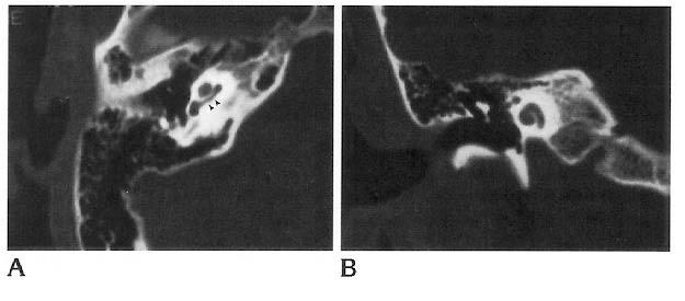
6 108 JOHNSON AJNR: 16, January 1995 TABLE 3: Relation between CT interpretation and quality of implantation Complete implantation Incomplete or difficult implantation Normal CT Abnormal CT Fig 7. CT in the axial plane of the left temporal bones demonstrates a large bone overgrowth overlying the round window niche (arrow). interpretation error (Fig 8). False-negative interpretation may result from failure to diagnose the earliest manifestations of labyrinthine ossification fibroossific change within the basal turn. This diagnosis may be particularly difficult to make when there is little ossification within the fibrous matrix. In our series, only 1 patient with CT findings interpreted as normal received a complete implantation; however, 1 additional patient received more than 19 electrodes (considered a good insertion). The third patient with normal CT findings was scanned outside our institution. The examination was performed at 3-mm intervals, which may have contributed to a suboptimal assessment in that case. Although our series of 13 patients is small, a normal, high-resolution, thin-section CT scan indicated a high probability of a good insertion. In several patients in our series a combination of pathologic findings in the cochlear region contributed to the management of possible incomplete or difficult insertions. The right ear is generally selected for implantation. One patient had bilateral pathologic CT findings that were worse in the right ear, so the left ear (with the lesser disease) was chosen (by the surgeon) for implantation (Fig 9). Note. Two of three were good insertions with more than 19 electrodes. Of the children with abnormal CT findings in our series, 9 of 10 experienced incomplete or difficult insertions, 7 of 10 patients receiving 10 electrodes or less (70%). An abnormal CT scan was predictive of a 90% probability of a difficult or incomplete insertion. Conclusion A high incidence of cochlear region disease was identified by high-resolution CT scans of the temporal bone in a series of pediatric patients with postmeningitic deafness. As demonstrated in our series and others, the changes of meningogenic labyrinthine ossification range from mild fibroossific changes within the labyrinth at the earliest, to profound, near-complete ossification within the cochlea. The ossification of the round window and the cochlear stenosis described in our series seem to be more limited manifestations of labyrinthine ossificans with deposits at the round window or narrowing the cochlea, but without the focal central ossifications demonstrated in more advanced cases. Comparison of the CT analysis with the surgical and postoperative experience suggests that preoperative CT can aide clinicians and families in the anticipation of potential difficulties with cochlear implantation. Preoperative CT review Fig 8. A, Midcochlea. Volume averaging of the basal turn suggests cochlear stenosis (arrowheads). B, The direct coronal image demonstrates a normal basal turn.
7 AJNR: 16, January 1995 COCHLEAR IMPLANTS 109 Fig 9. Axial CT of the right (A) and left (B) temporal bones demonstrate cochlear stenoses (A, arrows; B, arrowhead). In this patient, the observation of more extensive cochlear stenosis on the right led to the selection of the left ear for cochlear implantation. Note the parallel densities representing the myringotomy tube on the right (white arrow). allows the surgeon to anticipate difficulties with cochlear implantation and to plan the surgery accordingly. References 1. Gantz BJ, McCabe BF, Tyler RS. Use of multichannel cochlear implants in obstructed and obliterated cochleas. Otolaryngol Head Neck Surg 1988;98: Brookhouser PE, Worthington DW, Kelly WJ. Severe versus profound sensorineural hearing loss in children: implications for cochlear implantation. Laryngoscope 1990;100: Luxford WM, House WF. Cochlear implants in children: medical and surgical considerations. Ear Hear 1985;6:20S 23S 4. Silverstein H, Smouha E, Morgan N. Multichannel cochlear implantation in a patient with bilateral mondini deformities. Am J Otol 1988;9: Kaplan SL, Catlin FI, Weaver T, Feigin RD. Onset of hearing loss in children with bacterial meningitis. Pediatrics 1984;73: Novak MA, Fifer RC, Barkmeier JC, Firszt JB. Labyrinthine ossification after meningitis: its implications for cochlear. Otolaryngol Head Neck Surg 1990;103: Jackler RK, Luxford WM, Schindler RA, McKerrow WS. Cochlear implant candidates: assessment with CT and MR imaging. Radiology 1987;164: Harnsberger HR, Dart DJ, Parkin JL, Smoker WRK, Osborn AG. Cochlear implant candidates: assessment with CT and MR imaging. Radiology 1987;164: Phelps PD, Annis JAD, Robinson PJ. Imaging for cochlear implants. Br J Radiol 1990;63: Seicshnaydre MA, Johnson M, Hasenstab MS, Williams GH. Cochlear implants in children: the reliability of preoperative computed tomography. Otolaryngol Head Neck Surg 1992;107: Yune HY, Miyamoto RT, Yune ME. Medical imaging in cochlear implantation candidates. Am J Otol 1991;12S: Wiet RJ, Pyle GM, O Connor CA, Russell E, Schramm DR. Computed tomography: how accurate a predictor for cochlear implantation? Laryngoscope 1990;100: Mueller DP, Dolan KD, Gantz BJ. Temporal bone computed tomography in the preoperative evaluation for cochlear implantation. Ann Otol Rhinol Laryngol 1989;98: Balkany T, Dreisbach J. Workshop: surgical anatomy and radiographic imaging of cochlear implant surgery. Am J Otol 1987;8: Swartz JD, Mandell DM, Faerber EN, et al. Labyrinthine ossification: etiologies and CT findings. Radiology 1985;157: Becker TS, Eisenberg LS, Luxford WM, and House WF. Labyrinthine ossification secondary to childhood bacterial meningitis: implications for cochlear implant surgery. AJNR Am J Neuroradiol 1984;5: Green Jr JD, Marion MS, Hinojosa R. Labyrinthitis ossificans: histopathologic consideration for cochlear implantation. Otolaryngol Head Neck Surg 1991;104: Eisenberg LS, Luxford WM, Becker TS, House WF. Electrical stimulation of the auditory system in children deafened by meningitis. Otolaryngol Head Neck Surg 1984;92:
8
9
10
11
12

13
14
15
16
Cochlear Implant Failure: Imaging Evaluation of the Electrode Course
 Clinical Radiology (2003) 58: 288 293 doi:10.1016/s0009-9260(02)00523-8, available online at www.sciencedirect.com Pictorial Review Cochlear Implant Failure: Imaging Evaluation of the Electrode Course
Pediatric Temporal Bone
 Pediatric Temporal Bone Suresh K. Mukherji, MD, FACR Professor and Chief of Neuroradiology Professor of Radiology, Otolaryngology Head Neck Surgery, Radiation Oncology and Periodontics & Oral Medicine
COCHLEAR IMPLANTS Aetiology of Deafness. Bruce Black MD
 COCHLEAR IMPLANTS Aetiology of Deafness Heterochromia iridis. Cases may be healthy or associated with a variety of conditions, e.g. Waardenburg syndrome. Waardenburg syndrome. Note the snowy lock of hair
Modern Imaging & Current Controversies
 Temporal Bone: Modern Imaging & Current Controversies Suresh K. Mukherji, MD, FACR Professor and Chief of Neuroradiology Professor of Radiology, Otolaryngology Head Neck Surgery, Radiation i Oncology,
Difficult Cases: Controversies in Cochlear Implantation
 Difficult Cases: Controversies in Cochlear Implantation David S Haynes, MD FACS Fred F Telischi, MD MEE FACS Lawrence R. Lustig, MD Robert F Labadie, PhD MD Nikolas H Blevins, MD Matthew L. Carlson, MD
Modifying radiology protocols for cochlear implant surgery in a government sponsored scheme: Need of the hour
 Available online at www.ijmrhs.com ISSN No: 2319-5886 International Journal of Medical Research & Health Sciences, 2016, 5, 6:151-157 Modifying radiology protocols for cochlear implant surgery in a government
Congenital Absence of the Oval Window: Radiologic Diagnosis and Associated Anomalies
 AJNR Am J Neuroradiol 21:322 327, February 2000 Congenital Absence of the Oval Window: Radiologic Diagnosis and Associated Anomalies Barbara Zeifer, Paul Sabini, and Jonathan Sonne BACKGROUND AND PURPOSE:
CONFLICTS OF INTEREST
 COCHLEAR IMPLANTATION: A SURGEON S PERSPECTIVE Ravi N. Samy, M.D., F.A.C.S. Ravi.Samy@UC.edu Director, Adult Cochlear Implant Program Program Director, Neurotology Fellowship CONFLICTS OF INTEREST RESEARCH
Clinical Course of Pediatric Congenital Inner Ear Malformations
 The Laryngoscope Lippincott Williams & Wilkins, Inc., Philadelphia 2000 The American Laryngological, Rhinological and Otological Society, Inc. Clinical Course of Pediatric Congenital Inner Ear Malformations
Cochlear implants. Aaron G Benson MD Board Certified Otolaryngologist Board Certified Neurotologist
 Cochlear implants Aaron G Benson MD Board Certified Otolaryngologist Board Certified Neurotologist 1 OBJECTIVES WHAT IS A NEUROTOLOGIST WHAT MAKES AN INDIVIDUAL A COCHLEAR IMPLANT CANDIDATE WHAT IS THE
Electrode Trauma Assessed by Microdissection
 Electrode Trauma Assessed by Microdissection Peter S Roland MD C Gary Wright PhD University of Texas Medical Center Dallas, Texas Why worry about electrode trauma? Bilateral Implants Electro-acoustic implants
ORIGINAL ARTICLE. new implantation method, the combined electric-acoustic stimulation
 ORIGINAL ARTICLE Predicting Basal Cochlear Length for Electric-Acoustic Stimulation Oliver Adunka, MD; Marc H. Unkelbach, MD; Martin G. Mack, MD, PhD; Andreas Radeloff, MD; Wolfgang Gstoettner, MD, PhD
Clinical Workflow. White Paper No. 35 / 2016
 01 The mobile 3D C-arm Ziehm Vision RFD 3D A well-suited tool for intraoperative imaging in cochlear implantation More than 5 % of the world s population 360 million people suffer from hearing loss in
trauma? Bilateral Implants Electro-acoustic implants Preservation of ganglion cells?
 Electrode Trauma Assessed by Microdissection Peter S Roland MD C Gary Wright PhD University of Texas Medical Center Dallas, Texas Why worry about electrode trauma? Bilateral Implants Electro-acoustic implants
MR Evaluation of Vestibulocochlear Anomalies Associated with Large Endolymphatic Duct and Sac
 AJNR Am J Neuroradiol 20:1435 1441, September 1999 MR Evaluation of Vestibulocochlear Anomalies Associated with Large Endolymphatic Duct and Sac H. Christian Davidson, H. Ric Harnsberger, Marc M. Lemmerling,
Abnormal direction of internal auditory canal and vestibulocochlear nerve
 Medicine Otorhinolaryngology fields Okayama University Year 2004 Abnormal direction of internal auditory canal and vestibulocochlear nerve Shin Kariya kazunori Nishizaki Hirofumi Akagi Michael M. Paparella
SCD is an autosomal recessive disease characterized by an
 ORIGINAL RESEARCH N. Saito M. Watanabe J. Liao E.N. Flower R.N. Nadgir M.H. Steinberg O. Sakai Clinical and Radiologic Findings of Inner Ear Involvement in Sickle Cell Disease BACKGROUND AND PURPOSE: SCD
Chapter 180: Cochlear Implants. Richard T. Miyamoto, Amy M. Robbins, Mary Joe Osberger
 Chapter 180: Cochlear Implants Richard T. Miyamoto, Amy M. Robbins, Mary Joe Osberger Cochlear implants are electronic devices that convert mechanical sound energy into electric signals that can be delivered
ORIGINAL ARTICLE. The Cochlear-Carotid Interval: Preoperative Assessment for Cochlear Implant
 ORIGINAL ARTICLE The Cochlear-Carotid Interval: Preoperative Assessment for Cochlear Implant Eshrak Hassanein MD; Eman Geneidi MD; and Mohamed Taha MD From the Departments of Radiology (E.Hassanein), Otorhinolaryngology
STUDY OF MAIS (MEANINGFUL AUDITORY INTEGRATION SCALE) SCORE POST UNILAT- ERAL COCHLEAR IMPLANTATION IN PRELINGUAL DEAF PATIENTS
 SCORE POST UNILAT- ERAL COCHLEAR IMPLANTATION IN PRELINGUAL DEAF PATIENTS") ISSN: 2250-0359 Volume 5 Issue 3 2015 STUDY OF MAIS (MEANINGFUL AUDITORY INTEGRATION SCALE) SCORE POST UNILAT- ERAL COCHLEAR IMPLANTATION IN PRELINGUAL DEAF PATIENTS Neha Lala, Rajesh Vishwakarma, Chandrakant
ISSN: 2250-0359 Volume 5 Issue 3 2015 STUDY OF MAIS (MEANINGFUL AUDITORY INTEGRATION SCALE) SCORE POST UNILAT- ERAL COCHLEAR IMPLANTATION IN PRELINGUAL DEAF PATIENTS Neha Lala, Rajesh Vishwakarma, Chandrakant
The Temporal Bone Anatomy & Pathology
 Department of Radiology University of California San Diego The Temporal Bone Anatomy & Pathology John R. Hesselink, M.D. Temporal Bone Axial View Temporal Bone Coronal View Longitudinal Fracture The Temporal
Incidence and Characteristics of Facial Nerve Stimulation in Children With Cochlear Implants
 The Laryngoscope Lippincott Williams & Wilkins, Inc. 2006 The American Laryngological, Rhinological and Otological Society, Inc. Incidence and Characteristics of Facial Nerve Stimulation in Children With
For Professionals. Electrode Arrays. Designed for Atraumatic Implantation Providing Superior Hearing Performance
 For Professionals Electrode Arrays Designed for Atraumatic Implantation Providing Superior Hearing Performance Electrode Arrays Designed for Atraumatic Implantation Providing Superior Hearing Performance,,,
Paediatric cochlear implantation
 Paediatric cochlear implantation A M U MÜLLER BA (Log), MSc (Sp&H) Senior Lecturer Department of Speech, Language and Hearing Therapy University of Stellenbosch D J H WAGENFELD MB ChB, MMed (L et O), FCS
ALESSANDRA RUSSO MD GRUPPO OTOLOGICO
 Gruppo Otologico Simultaneous Labyrinthectomy with Cochlear Implantation Cenacolo di Audiovestibologia, Chieti, 24-25/06/2016 ALESSANDRA RUSSO MD GRUPPO OTOLOGICO MENIERE DISEASE Therapeutic Steps 1 step
Imaging in patients undergoing cochlear implant: CT and MR technique
 Imaging in patients undergoing cochlear implant: CT and MR technique Poster No.: C-0102 Congress: ECR 2012 Type: Educational Exhibit Authors: G. Posillico Keywords: Ear / Nose / Throat, CT, MR, Comparative
ORIGINAL ARTICLE. Paul W. Bauer, MD; Franz J. Wippold II, MD; Jenifer Goldin, MS, CCC-A; Rodney P. Lusk, MD
 ORIGINAL ARTICLE Cochlear Implantation in Children With CHARGE Association Paul W. Bauer, MD; Franz J. Wippold II, MD; Jenifer Goldin, MS, CCC-A; Rodney P. Lusk, MD Objective: To explore the anomalies
Delayed Endolymphatic Hydrops: Episodic Vertigo of Delayed Onset after Profound Inner Ear Hearing Loss
 Delayed Endolymphatic Hydrops: Episodic Vertigo of Delayed Onset after Profound Inner Ear Hearing Loss Tamio Kamei 1, MD, PhD and Kenji Watanabe 2, MD 1 Professor emeritus at Gunma University, Japan 2
Educational Exhibit Authors: P. Digge, R. K. N. Solanki, S. M. Paruthikunnan, D. S. Shah ; 1
 High-field MRI versus high-resolution CT of temporal bone in inner ear pathologies of children with bilateral profound sensorineural hearing loss: A pictorial essay. Poster No.: C-0867 Congress: ECR 2015
Imaging Profile of the Ear in Hearing Loss Patients in Hospital Universiti Sains Malaysia: 5 year Cross Sectional Analysis at a Tertiary
 Imaging Profile of the Ear in Hearing Loss Patients in Hospital Universiti Sains Malaysia: 5 year Cross Sectional Analysis at a Tertiary Otologic Centre Rohaizam bin Japar 1, Dinsuhaimi bin Sidek 1, Suzina
Imaging of Cochlear and Auditory Brain Stem Implantation
 Review Article Imaging of Cochlear and Auditory Brain Stem Implantation William W. M. Lo The cochlear implant is an electronic auditory prosthesis with a component that is surgically inserted in the ear
Surgeon Oriented Preoperative Radiologic Evaluation in Cochlear Implantation - Our experience with a Proposed Checklist
 THIEME Original Research Surgeon Oriented Preoperative Radiologic Evaluation in Cochlear Implantation - Our experience with a Proposed Checklist Mahmoud Mandour 1 Mohammed Tomoum 1 Saad El Zayat 2 Hisham
Temporal bone imaging in osteogenesis imperfecta patients with hearing loss
 Temporal bone imaging in osteogenesis imperfecta patients with hearing loss F. Swinnen 1, J. Casselman 2, P. Coucke 3, C. Cremers 4, E. De Leenheer 1, I. Dhooge 1 (1) Departement of Otorhinolaryngology,
The close anatomic relationship between the cochlea and
 ORIGINAL RESEARCH R.J. Young D.R. Shatzkes J.S. Babb A.K. Lalwani The Cochlear-Carotid Interval: Anatomic Variation and Potential Clinical Implications BACKGROUND AND PURPOSE: A temporal bone CT study
Visualisation of the Cochlea Implant/Tissue Interface
 Visualisation of the Cochlea Implant/Tissue Interface Christopher Wong 1, 4, Ian S. Curthoys 2, Stephen O Leary 3,4, Allan S. Jones 1 1 Australian Centre for Microscopy and Microanalysis, University of
Surgical intervention in middle-ear cholesterol granuloma
 Medicine Otorhinolaryngology fields Okayama University Year 2003 Surgical intervention in middle-ear cholesterol granuloma Manabu Maeta Ryusuke Saito Fumio Nakagawa Takakazu Miyahara Okayama University
During the past decade, combined electric acoustic stimulation
 ORIGINAL RESEARCH S.E.J. Connor D.J. Bell R. O Gorman A. Fitzgerald- O Connor CT and MR Imaging Cochlear Distance Measurements May Predict Cochlear Implant Length Required for a 360 Insertion BACKGROUND
A surgical approach for a cochlear implant: An anatomical study
 A surgical approach for a cochlear implant: An anatomical study By GRAEME M. CLARK (Melbourne) Introduction THERE is now increased interest in the possibility of restoring brain and nerve function by applying
Original Policy Date
 MP 7.01.66 Auditory Brainstem Implant Medical Policy Section Surgery Issue 12/2013 Original Policy Date 12/2013 Last Review Status/Date Reviewed with literature search/12/2013 Return to Medical Policy
Utility of Preoperative Computed Tomography and Magnetic Resonance Imaging in Adult and Pediatric Cochlear Implant Candidates
 The Laryngoscope VC 2015 The American Laryngological, Rhinological and Otological Society, Inc. Utility of Preoperative Computed Tomography and Magnetic Resonance Imaging in Adult and Pediatric Cochlear
Temporal bone anatomy and imaging features of common conditions causing hearing loss: A pictorial review
 Temporal bone anatomy and imaging features of common conditions causing hearing loss: A pictorial review Poster No.: C-1892 Congress: ECR 2012 Type: Educational Exhibit Authors: A. Masukawa, H. Takeuchi,
Surgical and Non-Surgical Causes of Progressive Hearing Loss in Children: What can be done about it?
 Surgical and Non-Surgical Causes of Progressive Hearing Loss in Children: What can be done about it? Daniela Carvalho, MD, MMM, FAAP Professor, Surgery Department UCSD Pediatric Otolaryngology Rady Children
MEDICAL POLICY SUBJECT: COCHLEAR IMPLANTS AND AUDITORY BRAINSTEM IMPLANTS. POLICY NUMBER: CATEGORY: Technology Assessment
 MEDICAL POLICY PAGE: 1 OF: 5 If the member's subscriber contract excludes coverage for a specific service it is not covered under that contract. In such cases, medical policy criteria are not applied.
Anomalous Facial Nerve Canal with Cochlear Malformations
 AJNR Am J Neuroradiol 22:838 844, May 2001 Anomalous Facial Nerve Canal with Cochlear Malformations Laura Vitale Romo and Hugh D. Curtin BACKGROUND AND PURPOSE: Anteromedial migration of the first segment
Implantable Treatments for Different Types of Hearing Loss. Margaret Dillon, AuD Marcia Adunka, AuD
 Implantable Treatments for Different Types of Hearing Loss Margaret Dillon, AuD Marcia Adunka, AuD Implantable Technologies Types of hearing loss Bone-anchored devices Middle ear implantation Cochlear
Congenital aural atresia (CAA) is a rare disorder of the temporal
 is a rare disorder of the temporal") Published July 3, 2014 as 10.3174/ajnr.A4022 CLINICAL REPORT HEAD & NECK The Boomerang Malleus-Incus Complex in Congenital Aural Atresia S. Mukherjee, B.W. Kesser, and P. Raghavan ABSTRACT SUMMARY: Boomerang
Published July 3, 2014 as 10.3174/ajnr.A4022 CLINICAL REPORT HEAD & NECK The Boomerang Malleus-Incus Complex in Congenital Aural Atresia S. Mukherjee, B.W. Kesser, and P. Raghavan ABSTRACT SUMMARY: Boomerang
Clinical Study Radiological Assessment of the Indian Children with Congenital Sensorineural Hearing Loss
 International Otolaryngology, Article ID 808759, 7 pages http://dx.doi.org/10.1155/2014/808759 Clinical Study Radiological Assessment of the Indian Children with Congenital Sensorineural Hearing Loss Sangeet
Outcomes of cochlear implantation in children with and without inner ear malformations
 Original Article Outcomes of cochlear implantation in children with and without inner ear malformations Mustafa Celik 1, Erkan Karatas 2, Muzaffer Kanlikama 3 ABSTRACT Objective: To evaluate the auditory
Basic Fitting and Evaluation Parameters of a Newly Designed Cochlear Implant Electrode
 Acta Otolaryngol 2003; 00: 1/5 Basic Fitting and Evaluation Parameters of a Newly Designed Cochlear Implant Electrode P.R. DEMAN 1, K. DAEMERS 1,*, M. YPERMAN 1,*, F.F. OFFECIERS 1, A. PLASMANS 2, B. VAN
HEARING AND COCHLEAR IMPLANTS
 HEARING AND COCHLEAR IMPLANTS FRANCIS CREIGHTON, MD NEUROTOLOGY & SKULL BASE SURGERY FELLOW JOHNS HOPKINS SCHOOL OF MEDICINE NOV 9 TH, 2017 THANKS TO: CHARLIE DELLA SANTINA, HEIDI NAKAJIMA AND DOUG MATTOX
Added Value of MDCT in Cochlear Parameters Assessment in Patients with Sensory Neural Hearing Loss Candidates for Cochlear Implantation
 International Journal of Medical Imaging 2018; 6(4): 33-39 http://www.sciencepublishinggroup.com/j/ijmi doi: 10.11648/j.ijmi.20180604.11 ISSN: 2330-8303 (Print); ISSN: 2330-832X (Online) Added Value of
Very far-advanced otosclerosis: stapedotomy or cochlear implantation
 Acta Oto-Laryngologica, 2007; 127: 574 578 ORIGINAL ARTICLE Very far-advanced otosclerosis: stapedotomy or cochlear implantation MARIE-NOËLLE CALMELS 1, CORINTHO VIANA 1,2, GEORGES WANNA 1, MATHIEU MARX
Restudy of malformations of the internal auditory meatus, cochlear nerve canal and cochlear nerve
 Eur Arch Otorhinolaryngol (2015) 272:1587 1596 DOI 10.1007/s00405-014-2951-4 OTOLOGY Restudy of malformations of the internal auditory meatus, cochlear nerve canal and cochlear nerve Youjin Li Jun Yang
Indications and contra-indications of auditory brainstem implants. Systematic review and illustrative cases
 Manuscript: Authors: Journal: Indications and contra-indications of auditory brainstem implants. Systematic review and illustrative cases Merkus P (p.merkus@vumc.nl), Di Lella F, Di Trapani G, Pasanisi
Relationship of the Optic Nerve to the Posterior Paranasal Sinuses: A CT Anatomic Study
 Relationship of the Optic Nerve to the Posterior Paranasal Sinuses: A CT Anatomic Study Mark C. DeLano, F. Y. Fun, and S. James Zinreich PURPOSE: To delineate the relationship between the optic nerves
Investigating the Effect of Cochlear Size in Insertion of Electrode Depth in Patients with Cochlear Implantation Evaluated by CT- Scan
 http:// ijp.mums.ac.ir Original Article (Pages: 9207-9213) Investigating the Effect of Cochlear Size in Insertion of Electrode Depth in Patients with Cochlear Implantation Evaluated by CT- Scan *Mohammad
ORIGINAL ARTICLE. A New Staging System for Tympano-mastoid Cholesteatoma. Aziz Belal, Mahmoud Reda, Ahmed Mehana, Yousef Belal
 Int. Adv. Otol. 2012; 8:(1) 63-68 ORIGINAL ARTICLE A New Staging System for Tympano-mastoid Cholesteatoma Aziz Belal, Mahmoud Reda, Ahmed Mehana, Yousef Belal Alexandria Ear Hospital Alexandria Egypt (AB,
The Value of Computed Tomography Scanning in Assessment of Aditus ad Antrum Patency and Choice of Treatment Line in Revision Myringoplasty
 Med. J. Cairo Univ., Vol. 77, No. 2, September: 53-57, 2009 www.medicaljournalofcairouniversity.com The Value of Computed Tomography Scanning in Assessment of Aditus ad Antrum Patency and Choice of Treatment
Arastoo Vossough, M.D., Ph.D. Associate Professor of Radiology
 Disorders of the Temporal Bone Arastoo Vossough, M.D., Ph.D. Associate Professor of Radiology 1st Branchial Cleft Cyst Remnant of 1 st branchial apparatus Rare Cystic lesions: I- around pinna II- connecting
Cochlear Implants February 2003
 TITLE: Cochlear Implants SOURCE: Grand Rounds Presentation, UTMB, Dept. of Otolaryngology DATE: February 5, 2003 RESIDENT PHYSICIAN: Glen T. Porter, MD FACULTY ADVISOR: Arun K. Gadre, MD SERIES EDITORS:
Scala Tympani Cochleostomy II: Topography and Histology
 The Laryngoscope Lippincott Williams & Wilkins 2007 The American Laryngological, Rhinological and Otological Society, Inc. Scala Tympani Cochleostomy II: Topography and Histology Oliver F. Adunka, MD;
syndrome (LEDS) and its associated anomalies, with clinical features.
 and its associated anomalies, with clinical features.") Relationship of the rea Measurement of the Large Endolymphatic Duct and Sac Syndrome as well as the Clinical Symptoms with CT and MR Imaging Results 1 Ji-Sang Park, M.D., Hyun Sook Hong, M.D., Jong-Sea
Relationship of the rea Measurement of the Large Endolymphatic Duct and Sac Syndrome as well as the Clinical Symptoms with CT and MR Imaging Results 1 Ji-Sang Park, M.D., Hyun Sook Hong, M.D., Jong-Sea
ORIGINAL ARTICLE. Current Techniques in Management of Postmeningitic Deafness in Children. 7-valent pneumococcal conjugate
 ORIGINAL ARTICLE Current Techniques in Management of Postmeningitic Deafness in Children Nancy M. Young, MD; Tina Q. Tan, MD Objectives: To determine pneumococcal vaccination status of children with recent
Cochlear Implant, Bone Anchored Hearing Aids, and Auditory Brainstem Implant
 Origination: 06/23/08 Revised: 10/15/16 Annual Review: 11/10/16 Purpose: To provide cochlear implant, bone anchored hearing aids, and auditory brainstem implant guidelines for the Medical Department staff
DOWNLOAD PDF PRINCIPLES OF COCHLEAR IMPLANT IMAGING
 Chapter 1 : Imaging in cochlear implant patients 7 Principles of Cochlear Implant Imaging. Andrew J. Fishman and Roy A. Holliday. Radiographic imaging plays a major role in cochlear implantation with regard
ORIGINAL ARTICLE. Evaluation of Pediatric Sensorineural Hearing Loss With Magnetic Resonance Imaging
 ORIGINAL ARTICLE Evaluation of Pediatric Sensorineural Hearing Loss With Magnetic Resonance Imaging John E. McClay, MD; Timothy N. Booth, MD; David A. Parry, MD; Romaine Johnson, MD; Peter Roland, MD Objective:
INTRACOCHLEAR FACTORS CONTRIBUTING TO PSYCHOPHYSICAL PERCEPTS FOLLOWING COCHLEAR IMPLANTATION: A CASE STUDY
 INTRACOCHLEAR FACTORS CONTRIBUTING TO PSYCHOPHYSICAL PERCEPTS FOLLOWING COCHLEAR IMPLANTATION: A CASE STUDY A. KAWANO, MD, PHD; H. L. SELDON, MD, PHD; B. PYMAN, MD, FRACS; G. M. CLARK, PHD, FRACS From
ORIGINAL ARTICLE. Revision Cochlear Implantation for Facial Nerve Stimulation in Otosclerosis
 ORIGINAL ARTICLE Revision Cochlear Implantation for Facial Nerve Stimulation in Otosclerosis Marek Polak, PhD; S. Arif Ulubil, MD; Annelle V. Hodges, PhD; Thomas J. Balkany, MD Objective: To find if patients
An optical coherence tomography study for imaging the round window niche and the promontorium tympani
 An optical coherence tomography study for imaging the round window niche and the promontorium tympani T. Just *a, E. Lankenau b, G. Hüttmann b, H.W. Pau a a Department of Otorhinolaryngology, University
Clinical Commissioning Policy: Auditory brainstem implant with congenital abnormalities of the auditory nerves of cochleae
 Clinical Commissioning Policy: Auditory brainstem implant with congenital abnormalities of the auditory nerves of cochleae Reference: NHS England: 16062/P NHS England INFORMATION READER BOX Directorate
CLASSIFICATION OF CONGENITAL MIDDLE AND EXTERNAL EAR MALFORMATIONS: CT STUDY
 VolumeS Medicalloumal of the Islamic Republic of Iran Number 3,4 Payiz & Zemestan 1370 Fall & Winter 1991 CLASSIFICATION OF CONGENITAL MIDDLE AND EXTERNAL EAR MALFORMATIONS: CT STUDY D. SAYIC, MD, DMS:
ESCMID Online Lecture Library. by author
 Neurologische Klinik und Poliklinik Prof. Dr. M. Dieterich Animal models of meningitis-associated hearing loss M Klein, Siena 5/2012 Hearing Loss is common in survivors of Pneumococcal Meningitis oedema
Conductive Hearing Loss in Young Children: Options and Opportunities
 Conductive Hearing Loss in Young Children: Options and Opportunities Donna L. Sorkin, M.A., Vice President, Consumer Affairs Jennifer Lake, Clinical Applications Specialist Cochlear Americas Agenda 1.
External carotid blood supply to acoustic neurinomas
 External carotid blood supply to acoustic neurinomas Report of two cases HARVEY L. LEVINE, M.D., ERNEST J. FERmS, M.D., AND EDWARD L. SPATZ, M.D. Departments of Radiology, Neurology, and Neurosurgery,
Cochlear implantations in Northern Ireland: An overview of the first five years
 The Ulster Medical Journal, Volume 68, No. 1, pp. 12-16, May 1999. Cochlear implantations in Northern Ireland: An overview of the first five years G John, J G Toner Accepted 12 March 1999 SUMMARY During
Cochlear Implant Innovations
 HOSPITAL CHRONICLES 2011, 6(4): 195 199 Special Topic Matilda Chroni, MD, PhD, Spyros Papaspyrou, MD, PhD ENT Department, Evagelismos General Hospital of Athens, Athens, Greece Key words: cochlear implants;
Imaging Findings of Cochlear Nerve Deficiency
 AJNR Am J Neuroradiol 23:635 643, April 2002 Imaging Findings of Cochlear Nerve Deficiency Christine M. Glastonbury, H. Christian Davidson, H. Ric Harnsberger, John Butler, Thomas R. Kertesz, and Clough
New Methods of Deafness and Partial Deafness Treatment
 Biocybernetics and Biomedical Engineering 2006, Volume 26, Number 1, pp. 75 83 New Methods of Deafness and Partial Deafness Treatment ARTUR LORENS*, HENRYK SKAR YÑSKI, ANNA PIOTROWSKA International Center
Clinical Study Imaging of Electrode Position after Cochlear Implantation with Flat Panel CT
 International Scholarly Research Network ISRN Otolaryngology Volume 2012, Article ID 728205, 5 pages doi:10.5402/2012/728205 Clinical Study Imaging of Electrode Position after Cochlear Implantation with
The Significance of a Hypoplastic Bony Canal for the Cochlear Nerve in Patients with Sensorineural Hearing Loss: CT and MRI Findings 1
 The Significance of a Hypoplastic Bony Canal for the Cochlear Nerve in Patients with Sensorineural Hearing Loss: CT and MRI Findings 1 Yoon Jung Choi, M.D., Sang Yoo Park, M.D. 2, Myung Soon Kim, M.D.,
Public Statement: Medical Policy Statement:
 Medical Policy Title: Implantable Bone ARBenefits Approval: 09/28/2011 Conduction Hearing Aids Effective Date: 01/01/2012 Document: ARB0190 Revision Date: Code(s): 69714 Implantation, osseointegrated implant,
Gerard J. Gianoli, MD, FACS The Ear and Balance Institute Baton Rouge, Louisiana
 Gerard J. Gianoli, MD, FACS The Ear and Balance Institute Baton Rouge, Louisiana SSCD is defined anatomically as the absence of bone between the SSC and the middle fossa dura PSCD is a defect of the PSC
MEDICAL POLICY SUBJECT: COCHLEAR IMPLANTS AND AUDITORY BRAINSTEM IMPLANTS
 MEDICAL POLICY. PAGE: 1 OF: 6 If the member's subscriber contract excludes coverage for a specific service it is not covered under that contract. In such cases, medical policy criteria are not applied.
Your ear consists of three parts that play a vital role in hearing-the external ear, middle ear, and inner ear.
 What is a Cochlear Implant? A cochlear implant is an electronic device that restores partial hearing to the deaf. It is surgically implanted in the inner ear and activated by a device worn outside the
Tympanogenic Labyrinthitis (Cochlear Deafness) due to chronic suppurative otitis media (CSOM)
 due to chronic suppurative otitis media (CSOM)") Tympanogenic Labyrinthitis (Cochlear Deafness) due to chronic suppurative otitis media (CSOM) * Dr. Raad A AlObaydi (FICS MBChB) Background: It has been accepted that in patients with CSOM, conductive
Tympanogenic Labyrinthitis (Cochlear Deafness) due to chronic suppurative otitis media (CSOM) * Dr. Raad A AlObaydi (FICS MBChB) Background: It has been accepted that in patients with CSOM, conductive
AUDITORY APPARATUS. Mr. P Mazengenya. Tel 72204
 AUDITORY APPARATUS Mr. P Mazengenya Tel 72204 Describe the anatomical features of the external ear Describe the tympanic membrane (ear drum) Describe the walls of the middle ear Outline the structures
Series Preface. Preface. Acknowledgments. Abbreviations
 Contents Series Preface Preface Acknowledgments Abbreviations vii ix xiii xxv Introduction xxxi Definition...xxxi Normal Hearing...xxxi Deafness...xxxi Overall Concept of the Bionic Ear...xxxii Training
Imaging of Hearing Loss
 Contemporary Imaging of Sensorineural Hearing Loss Imaging of Hearing Loss Discussion Outline (SNHL) Imaging Approaches Anatomic Relationships Lesions: SNHL KL Salzman, MD University of Utah School of
The association between hearing loss and meningitis
 Time course of hearing loss in an animal model of pneumococcal meningitis BRADLEY W. KESSER, MD, GEORGE T. HASHISAKI, MD, JONATHAN H. SPINDEL, PhD, ROGER A. RUTH, PhD, and W. MICHAEL SCHELD, MD, Charlottesville,
Case Report An Unusual Case of Bilateral Agenesis of the Cochlear Nerves
 Case Reports in Neurological Medicine Volume 2012, rticle D 581920, 4 pages doi:10.1155/2012/581920 Case Report n Unusual Case of Bilateral genesis of the Cochlear Nerves Vítor Yamashiro Rocha oares, 1
ORIGINAL ARTICLE. Efficacy of the 2-Staged Procedure in the Management of Cholesteatoma. paradigms have been published
 Efficacy of the 2-Staged Procedure in the Management of Cholesteatoma Steven Y. Ho, MD; John F. Kveton, MD ORIGAL ARTICLE Objective: To demonstrate the efficacy of intact canal wall procedure coupled with
Otosclerosis is an idiopathic disease that can result in spongiosis
 ORIGINAL RESEARCH T.C. Lee R.I. Aviv J.M. Chen J.M. Nedzelski A.J. Fox S.P. Symons CT Grading of Otosclerosis BACKGROUND AND PURPOSE: The CT grading system for otosclerosis was proposed by Symons and Fanning
ORIGINAL ARTICLE. Perimodiolar electrode position: Effects on thresholds, comfort levels, impedance measurements, and neural response telemetry
 The Mediterranean Journal of Otology ORIGINAL ARTICLE Perimodiolar electrode position: Effects on thresholds, comfort levels, impedance measurements, and neural response telemetry Angel Ramos Macias, MD;
UP Bioengineering Our people
 UP Bioengineering Our people Design and application of user-specific models of cochlear implants Tania Hanekom Tiaan K Malherbe, Liezl Gross, Rene Baron, Riaze Asvat, Werner Badenhorst & Johan J Hanekom
COCHLEAR IMPLANTATION IN A CHILD WITH COMPLEX BILATERAL INNER EAR AND COCHLEO-VESTIBULAR NERVE MALFORMATIONS
 COCHLEAR IMPLANTATION IN A CHILD WITH COMPLEX BILATERAL INNER EAR AND COCHLEO-VESTIBULAR NERVE MALFORMATIONS Sebastian Cozma 1,3, Oana Manolache 3, Raluca Olariu 1*, Cristian Mârțu 1,2, Luminița Rădulescu
Meniere s disease and Sudden Sensorineural Hearing Loss
 Meniere s disease and Sudden Sensorineural Hearing Loss Tsutomu Nakashima 1,2 1 Ichinomiya Medical Treatment & Habilitation Center, Ichinomiya, Japan 2 Department of Otorhinolaryngology, Nagoya University,
In CT evaluation of children with sensorineural hearing
 ORIGINAL RESEARCH B. Ozgen M.E. Cunnane P.A. Caruso H.D. Curtin Comparison of 45 Oblique Reformats with Axial Reformats in CT Evaluation of the Vestibular Aqueduct BACKGROUND AND PURPOSE: Measurement of
MICROTIA. The condition is a complex mix of cosmetic, functional, and often psychological difficulties. Microtia: Not only the ear.
 MICROTIA Underdevelopment /deformity of the auricle (pinna) varies from subtle deformities and small pre-auricular rudiments to gross developmental failure, distortion or malpositioned remnants. The external
Peter S Roland M.D. UTSouthwestern Medical Center Dallas, Texas Developments
 Peter S Roland M.D. UTSouthwestern Medical Center Dallas, Texas Developments New electrodes New speech processing strategies Bilateral implants Hybrid implants ABI in Kids MRI vs CT Meningitis Totally
Lamb Temporal Bone as a Surgical Training Model of Round Window Cochlear Implant Electrode Insertion
 Otology & Neurotology 37:52 56 ß 2015, Otology & Neurotology, Inc. Lamb Temporal Bone as a Surgical Training Model of Round Window Cochlear Implant Electrode Insertion Georgios Mantokoudis, Markus E. Huth,