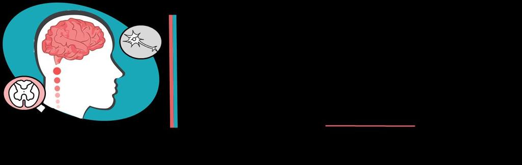
Waseem Abu Obeida. Muhammad Abid. Loai Al-zghoul
|
|
|
- Myrtle Douglas
- 5 years ago
- Views:
Transcription


1 4 Waseem Abu Obeida Muhammad Abid Loai Al-zghoul 1 P a g e
2 We knew that we have two types of deafness; one that resulted from damage to the neuronal part beginning from the hair cells up to any part of the central nervous system, or a type of deafness that is the result that the sound did not reach the neuronal cells due to a problem in conduction of sound waves to the cochlea. Deafness does not mean necessarily a complete loss of hearing, it could mean a decreased sensitivity to the hearing sensation. A patient comes in and he knows that he has deafness and you can easily recognize this simply by inducing any sound and observing if he hears it same as you do or not, assuming that you are the control. If you give any type of sound and he can t hear it and you hear it normally, you know he has a problem. Usually, the first step in the test for the deafness is to know whether it is sensorineural or conductive. Conduction deafness and sensorineural deafness may have different causes, and therefore different tests may be used to diagnose these deficits, the first and the oldest method and still common is Rinne and Weber tests. These tests take the advantage of the difference between conduction of sound through air and conduction of sound through bone. Sound waves conducted through air reach the cochlea in a higher amplitude than through bone; because waves conducted through air have to pass through the middle ear, which means they will be amplified as opposed to waves conducted through bone which surpass middle ear and reach cochlea directly. -To test air conduction, a vibrating tuning fork is held from the opening of the auditory canal. Hearing this sound means that the sound waves generated are passing through the external ear and the middle ear; disease or damage in these areas would result in decreased or lost hearing in this ear. -To test bone conduction, a vibrating tuning fork is placed directly on the skull. Perceiving these vibrations as sounds means that the sound (vibration) is transmitted directly to the cochlea of the inner ear and bypasses the external ear and the middle ear. 2 P a g e
3 Both the Rinne test and the Weber test use these principles to differentiate conduction deafness from sensorineural deafness. - For the Rinne test (bone + air conduction), the tuning fork is placed against the mastoid process. The normal patient perceives the sound in the ear on that side, and after the sound is no longer perceived by bone conduction, the tuning fork is immediately moved to the auditory canal and the sound is again heard (air conduction). If the patient has conductive problem, the sound is perceived by bone conduction better than air conduction because something is blocking sound waves from reaching the cochlea. If the patient has sensorineural deafness, vibrations will be heard through air once bone conduction is over as long as nerve deafness is partial. Seems as a normal person, but down in the example below you will know how to differentiate between the two. For example, a patient comes in, we place the tuning fork on his mastoid to test bone conduction and we ask him, can you hear? He says yes, then you tell him whenever you stop hearing, give me a sign. When he stops hearing because the vibration intensity has decreased with time, you transfer the fork to air near the patient s auricle so that sound waves are now being amplified by the middle ear. A normal person will hear in both situations. If the problem was sensorineural, no conductive problem, so a normal person and a sensorineural deaf patient both will have better air conduction than conduction through bone and will hear sound in both parts. The question now becomes: how to know if this patient has a neural problem or normal like you? The key to this answer is time, a sensorineural patient will stop hearing at a certain moment while you (the doctor) can still hear the sound. So it is not the fork that stopped vibrating, it is his problem that caused early cessation of sound. *when performing the bony part of the test, we flip the auricle to minimize the effect of sound waves conducted through air. Another complimentary test is Weber s test, so still the main one and the one you chose if you have to choose between the two is Rinne. Webber s test also depends on the idea that air conduction is better than bone, but also depends on the background noise. No matter the extent of silence in the room, you still have some background noise which will be compared to the vibrations through the fork. So the tuning 3 P a g e
4 fork is placed on the midline of the skull or forehead thus making sure that vibrations are sent equally to both ears. In a normal patient, the sound (vibrations + background noise) is perceived about equally in both ears. But in conductive deafness, for example in the right ear, you will hear the sound normally by the left ear, but much higher in the diseased ear (right one) because there is no background noise (cannot be conducted) to mask some of the vibrations sent through bone. On the other hand, a sensorineural deafness in one ear will result in hearing the sound louder in the other ear, that is simply because sounds will be lower in all the situations in the ear with neural problem. A good summary from the slides: When technology introduced us the audiogram, Rinne and Webber tests were set aside. -To determine the nature of hearing disabilities we use the audiometer. This instrument is an earphone connected to an electronic oscillator capable of emitting pure tones ranging from low frequencies to high frequencies. In the past, we used different forks with different frequencies in Rinne and Webber but also we had too much overlap, so this feature of the audiogram solved this issue. In performing a hearing test using an audiometer, one tests different frequencies covering the auditory spectrum (20Hz-20kHz), and the hearing loss is determined for each of these frequencies, then the so called audiogram is plotted. 4 P a g e
 and the degree of loudness or intensity (decibels).")
. The audiometer is equipped with an earphone for testing air conduction and with a mechanical vibrator for testing bone conduction.")
5 The audiogram is a graph illustrating a person's hearing capabilities and the amount of hearing loss that an individual has for each ear. The graph represents the relation between the frequency of the voice (Hertz) and the degree of loudness or intensity (decibels). We perform the audiogram test in a silent room, we apply different frequencies to the patient for each ear, for example; we start with 1 khz and increase the frequency gradually, and we plot each frequency with the degree of loudness (decibels). The audiometer is equipped with an earphone for testing air conduction and with a mechanical vibrator for testing bone conduction. -We notice from the figure below that the normal person shouldn t need more than 20 decibels to hear the sound, and other ranges for hearing loss are categorized according to the increase of decibels above 20. Don t memorize categories of severity of loss, just understand the concept. Besides using the audiogram to determine the severity of hearing loss we can also determine the type of hearing loss (conductive or sensory neuronal). 5 P a g e


6 -Now let s examine some possible audiogram charts. 1-In a normal person. As we notice from the figure above, both ears hear with 20 db or less at all frequencies. 2- *brackets represent bone conduction, and (X) and (O) represent air conduction. Each color for each ear. As we can see, the patient exceeded the 20 db limit, so he has a problem. -Sensorineural or conductive? Conductive, because bone conduction is normal. 6 P a g e

7 -Which ear is diseased? Both ears, because both exceeded the 20db limit in air conduction. So, moderate conductive deafness in both ears. 3- Assume (X) for the right ear and (O) for the left ear. Diagnosis: Both ears diseased, the left being more severe. Sensorineural deafness. The problem at high frequencies. Extra: if you are curious and want to be more accurate, he has some mild conductive problem because air conduction was not better than bone, as you can notice that (x) and (o) were below bone conduction symbols. Also, the high amplitude might play a role in this observation. So it is not a simple decision. I asked doctor about it and he said forget about it. Just know and understand the information in the diagnosis above. 7 P a g e

8 The vestibular system: The vestibular system is a sensory system that is essential to normal balanced movement and equilibrium. In the inner ear there are two structures: 1-Cochlea; which is responsible for hearing. 2-The vestibular part which is called *Labyrinth; which is important in the vestibular system which will give information about position and body movement. *the labyrinth actually is of two types; bony and membranous and both types are found in the cochlea and the vestibular part of the inner ear. But go with the doctor and assume that the labyrinth is the second part of the inner ear in addition to the cochlea. The labyrinth -It consist of five parts: 3 semicircular canals situated in three different axes (X, Y, Z), and they are lateral, anterior and posterior semicircular canals. At the end of each semicircular canal there is a dilation called the ampulla. 8 P a g e
9 The other two parts are bulges in the base of the labyrinth called utricle and saccule. The difference is that the Saccule is almost vertical. Inside the labyrinth we have a fluid similar to the one in the cochlea (endolymph and perilymph). Inside the ampulla of the semicircular canals and in the utricle and saccule there is a gelatinous membrane, beneath it there are hair cells similar to the hair cells in the cochlea which are responsible for hearing. The hair cells have hair extensions and have mechanical gated ion channels and have a baseline firing, bending them will open the mechanical doors more and frequency of action potential will increase. If we bend them to the opposite side these doors will close and graded potential will decrease and in this case there will be a decrease in the frequency of action potential. The vestibular system is responsible for detection and calculation of body and head movement and position in all directions. The five parts of the labyrinth is responsible for detection of this movement, each part has its own role. 1-The semicircular canals: The semicircular canals for the rotational movement of the head. So if you turn your face from right to left there is a rotational movement and the fluid will move in one of the semicircular canals and go to the ampulla and push the gelatinous membrane which will bend the hair cells and increase baseline firing, then through neurons, this will be sent to the CNS and deal with it in a certain way as we will see later on. 2-The utricle and saccule: In the utricle and the saccule there is also a gelatinous membrane called otolithic membrane. This membrane was designed to be sensitive to movement against gravity. To achieve this goal, this membrane has to be heavier, so something was added on the top of this membrane, these structures are called otoliths which are deposits of calcium particles. In the utricle, the orientation is almost flat, so if you tilt your head, gravity will bend the gelatinous membrane and hair cells, same in 9 P a g e

10 nodding, driving an accelerating, and decelerating vehicle. All these situations will ignite a neuronal signal. The saccule is more vertical in position than the utricle, so the saccule detects vertical movements such as jumping, sitting, riding an elevator, etc... The vestibular pathway: All of these types of movements detected by the labyrinth will turn into a signal that will be sent to be processed by the CNS. First order neuronal cell bodies are found in the vestibular ganglia, then axons will continue through 8 th nerve until reaching the CNS, there it enters the brain stem and synapses with the vestibular nuclei. We have 2 vestibular systems, one in each ear, each one of them when I rotate my head will have a movement in it. The vestibular system, especially the semicircular canals, are arranged in all three directions so that any type of rotational movement in any direction will cause movement of the fluid. The body uses this system for its advantage and compares between both sides so that it knows what type of movement the head is performing, and the direction too. For example, if I move my head from right to left, in which ear there will be movement? Both (the lateral semicircular canals in this situation). 10 P a g e
11 Which one has a stronger movement? Both will be the same, it is just the opposite direction. So to be more sensitive to the direction of movement, the type of rotation, clock wise or counter clockwise, depends of which canal has the movement, but also the vestibular system arranged itself in both ears opposite direction, so that hair cells are arranged oppositely so that when I rotate my head to the left, cilia of hair cells in the left ear will bend toward big cilia and cause opening of channels and cause firing and excitation, on the right side, cilia of hair cells will bend toward the small cilia and cause close in the channels and cause inhibition, the brain when sees a movement in the lateral canal and there is a difference between the two, the left being more than the right and at higher firing rate, knows I am moving to which direction(left) and that I am rotating counter clock wise. Exactly happens when I rotate to the opposite side. This applies to all types of movement so that the body can interpret and know which direction and type of movement. Another example, tilting to the right side or left side will cause the utricle to bend to the side of firing, although hair cells in utricle and saccule have complex arrangement but still the body needs to compare between the right and the left. This signal will move from the hair cells to the synapsing axons of neurons to neuronal cell bodies in the vestibular ganglia. This signal will be conducted in the 8 th cranial nerve (the vestibulocochlear nerve) to the brain stem. In the brain stem they will synapse with vestibular nuclei. From the vestibular nuclei to the thalamus then to the vestibular cortex (the conscious pathway). The most important vestibular nuclei in the thalamus are the pulvinar and the ventrolateral nucleus. From there the signal will go to different destinations in the vestibular cortex. The most important three of these vestibular cortices: 1. The insular area. 2. The posterior parietal cortex (area 5 and 7). 3. Area 2 and 3 in the somatosensory cortex. This part of vestibular pathway is called the conscious pathway which is responsible for awareness about position and body movement and status. 11 P a g e
12 Besides the vestibular cortical pathway (the conscious pathway), there is the vestibular subcortical pathway which gives the unconscious pathway and the quick reflexes that keep you balanced or other functions as we will see. There are four important destinations in this pathway, starting from the vestibular nuclei to: -The first target is the vestibulospinal tract. From vestibular nuclei to different levels of the spinal cord to control the motor neurons that supply muscles, mainly the trunk or core muscles. This tract is very important to keep you balanced. For example, when you are climbing stairs, a quick reflex orders contraction of muscles to tilt your body towards the leg that is on the ground so you don t fall. - The second target is the accessory nucleus to control the neck muscles through the accessory nerve (the sternocleidomastoid and trapezius muscles). This might help in preventing your head from crashing on the ground when you fall for example. -The third destination is the cerebellum (for coordination and balance). *when we study cerebellum we will see how it receives much input from the vestibular system and see how it will use it as part of its function. -The fourth destination is the nuclei of eye movement. The vestibular system can unconsciously move your eyes opposite to your body movement, that s why you can look at your friend and talk to him while you are walking downstairs. As you notice from the figure below, some neurons of the vestibular nuclei synapse in the oculomotor, trochlear, and abducent motor nuclei that control the extraocular muscles of the eye. The vestibular system gives orders for these nerves to move the eye opposite to the direction of the body. If you lower your head, your eyes will move upward and when you move your head to the left your eyes will go to the right. This compensatory reflex helps in keeping your gaze (visual field) fixed on the object of interest while the head is moving. When you reach the point where your eyes can t move any further to keep its focus on an object, it returns back to the center 12 P a g e


13 Quick recap: when you move your head from right to left, excitation (firing) will increase in the left side and decrease in the right. This difference in firing will reach all destinations; it will reach to the muscles to turn on the reflex that will keep you balanced, it will reach the areas of the cortex (like the posterior insular and parietotemporal regions) that are responsible for informing you about your position in relations to things around you in cooperation with your vision. Also, it will reach the motor nuclei of the eye as we explained before to move the eyes to the right. This mind map illustrates destination of fibers from the vestibular nuclei. Don t memorize the extra info. 13 P a g e
14 Damage of the vestibular system: Normally, when you are not moving, both sides will give the same baseline firing. When you move your head to the left, firing from the left will increase and firing from the right will decrease and so on. If someone has a problem in the right side (damage or cut in the right vestibular branch for example), there will be no firing going from the right side to the CNS (almost zero). Since the left side is still normal, it will still send baseline firing to the CNS. So the CNS is receiving higher firing from the left side compared to the right. The CNS will think that he is moving from right to left. The patient in this case will present to you with the feeling that he is spinning or the room is spinning around him حاس حا ي ل بلف vertigo.. This is what is called بالدنيا أو حاس الدنيا بتلف فيي Signs and symptoms of vestibular system damage: a. Any damage of the vestibular system either on the sensory level, the apparatus, labyrinth or the nerve will cause vertigo. The most common cause is Meniere's disease (described below). b. Here, the CNS is receiving information that you are turning you head to the left for example, as a reflex it will try to move you to the right. That's why the patient is easier to fall and imbalanced. c. Lastly, the eyes will receive orders to move to the right (in our example), when they reach the end of the visual field it will return fast to the center. So, the eyes will first move slowly to the right (or left) till the end then return fast and so on. This movement (slow shifting beating back to center) is called nystagmus. Nystagmus happens at rest or in certain positions. If you would like to see it in real life, you can ask a child to spin a few times then make him stop and look at his eyes. The movement that you will see is what is called nystagmus. Meniere's disease is an inner ear disease characterized by increased fluid in the labyrinth, this means that pressure inside the labyrinth is high, so the movement will be abnormal and hard. 14 P a g e
15 Symptoms include: Vertigo Nystagmus at certain position or movement (positional nystagmus) Nausea Vomiting Tinnitus due to increased pressure inside the cochlea. Treatment is decreasing the fluid by decreasing sodium and salt control and diuretics, but unfortunately, doesn t always resolve and sometimes we have to give alternative types of diuretics. We will take the details in medicine. Vertigo vs dizziness: Dizziness: non-specific term I can feel that I am dizzy. حاس حا ي ل بلف بالدنيا او problem. Vertigo: a specific term for vestibular حاس الدنيا بتلف فيي 15 P a g e
A&P 1. Ear, Hearing & Equilibrium Lab. Basic Concepts. Pre-lab Exercises
 A&P 1 Ear, Hearing & Equilibrium Lab Basic Concepts Pre-lab Exercises In this "Lab Exercise Guide", we will be looking at the basics of hearing and equilibrium. NOTE: these notes do not follow the order
Vestibular Physiology Richard M. Costanzo, Ph.D.
 Vestibular Physiology Richard M. Costanzo, Ph.D. OBJECTIVES After studying the material of this lecture, the student should be able to: 1. Describe the structure and function of the vestibular organs.
Vestibular physiology
 Vestibular physiology 2017 Utricle A flat epithelium: horizontal in the upright head Utricle Hair cells: no axons hair cells Utricle Hair cells synapse onto 8th nerve afferents. 8th nerve afferents Hair
to vibrate the fluid. The ossicles amplify the pressure. The surface area of the oval window is
 Page 1 of 6 Question 1: How is the conduction of sound to the cochlea facilitated by the ossicles of the middle ear? Answer: Sound waves traveling through air move the tympanic membrane, which, in turn,
Ear. Utricle & saccule in the vestibule Connected to each other and to the endolymphatic sac by a utriculosaccular duct
 Rahaf Jreisat *You don t have to go back to the slides. Ear Inner Ear Membranous Labyrinth It is a reflection of bony labyrinth but inside. Membranous labyrinth = set of membranous tubes containing sensory
A&P 1. Ear, Hearing & Equilibrium Lab. Basic Concepts. These notes follow Carl s Talk at the beginning of lab
 A&P 1 Ear, Hearing & Equilibrium Lab Basic Concepts These notes follow Carl s Talk at the beginning of lab In this "Lab Exercise Guide", we will be looking at the basics of hearing and equilibrium. NOTE:
Unit VIII Problem 9 Physiology: Hearing
 Unit VIII Problem 9 Physiology: Hearing - We can hear a limited range of frequency between 20 Hz 20,000 Hz (human hearing acuity is between 1000 Hz 4000 Hz). - The ear is divided into 3 parts. Those are:
The Physiology of the Senses Lecture 10 - Balance
 The Physiology of the Senses Lecture 10 - Balance www.tutis.ca/senses/ Contents Objectives... 1 The sense of balance originates from the labyrinth... 2 The auditory and vestibular systems have a common
Hearing and Balance 1
 Hearing and Balance 1 Slide 3 Sound is produced by vibration of an object which produces alternating waves of pressure and rarefaction, for example this tuning fork. Slide 4 Two characteristics of sound
Before we talk about the auditory system we will talk about the sound and waves
 The Auditory System PHYSIO: #3 DR.LOAI ZAGOUL 24/3/2014 Refer to the slides for some photos. Before we talk about the auditory system we will talk about the sound and waves All waves have basic characteristics:
Hearing. istockphoto/thinkstock
 Hearing istockphoto/thinkstock Audition The sense or act of hearing The Stimulus Input: Sound Waves Sound waves are composed of changes in air pressure unfolding over time. Acoustical transduction: Conversion
Course: PG- Pathshala Paper number: 13 Physiological Biophysics Module number M23: Posture and Movement Regulation by Ear.
 Course: PG- Pathshala Paper number: 13 Physiological Biophysics Module number M23: Posture and Movement Regulation by Ear Principal Investigator: Co-Principal Investigator: Paper Coordinator: Content Writer:
Νευροφυσιολογία και Αισθήσεις
 Biomedical Imaging & Applied Optics University of Cyprus Νευροφυσιολογία και Αισθήσεις Διάλεξη 11 Ακουστικό και Αιθουσιαίο Σύστημα (Auditory and Vestibular Systems) Introduction Sensory Systems Sense of
Gathering information the sensory systems; Vision
 Visual System Gathering information the sensory systems; Vision The retina is the light-sensitive receptor layer at the back of the eye. - Light passes through the cornea, the aqueous chamber, the lens,
Chapter 17, Part 2! The Special Senses! Hearing and Equilibrium!
 Chapter 17, Part 2! The Special Senses! Hearing and Equilibrium! SECTION 17-5! Equilibrium sensations originate within the inner ear, while hearing involves the detection and interpretation of sound waves!
Chapter 17, Part 2! Chapter 17 Part 2 Special Senses! The Special Senses! Hearing and Equilibrium!
 Chapter 17, Part 2! The Special Senses! Hearing and Equilibrium! SECTION 17-5! Equilibrium sensations originate within the inner ear, while hearing involves the detection and interpretation of sound waves!
Hearing. By: Jimmy, Dana, and Karissa
 Hearing By: Jimmy, Dana, and Karissa Anatomy - The ear is divided up into three parts - Sound enters in through the outer ear and passes into the middle where the vibrations are received and sent to the
Otoconia: Calcium carbonate crystals Gelatinous mass. Cilia. Hair cells. Vestibular nerve. Vestibular ganglion
 VESTIBULAR SYSTEM (Balance/Equilibrium) The vestibular stimulus is provided by Earth s, and. Located in the of the inner ear, in two components: 1. Vestibular sacs - gravity & head direction 2. Semicircular
What is the effect on the hair cell if the stereocilia are bent away from the kinocilium?
 CASE 44 A 53-year-old man presents to his primary care physician with complaints of feeling like the room is spinning, dizziness, decreased hearing, ringing in the ears, and fullness in both ears. He states
SPECIAL SENSES: THE AUDITORY SYSTEM
 SPECIAL SENSES: THE AUDITORY SYSTEM REVISION OF PHYSICS: WAVES A wave is an oscillation of power, sound waves have two main characteristics: amplitude, which is the maximum displacement or the power of
Vestibular System. Dian Yu, class of 2016
 Vestibular System Dian Yu, class of 2016 Objectives 1. Describe the functions of the vestibular system: What is it? How do you stimulate it? What are the consequences of stimulation? 2. Describe the vestibular
Hearing Lab. Name. Materials: tuning forks, sterile cotton
 Hearing Lab Name Through the sense of hearing we are placed into direct, intimate contact with t surrounding world. Musical, vocal, and other sonic impressions flood us constantly. We possess a wealth
THE EAR AND HEARING Be sure you have read and understand Chapter 16 before beginning this lab. INTRODUCTION: hair cells outer ear tympanic membrane
 BIOLOGY 211: HUMAN ANATOMY & PHYSIOLOGY ****************************************************************************************************** THE EAR AND HEARING ******************************************************************************************************
COGS 107B Week 2. Hyun Ji Friday 4:00-4:50pm
 COGS 107B Week 2 Hyun Ji Friday 4:00-4:50pm Lecture 3: Proprioception Principles: The Neuron Doctrine and The Law of Dynamic Polarization Proprioception Joint-protecting reflexes (ex. Knee jerk reflex)
Ch. 9 Sensory Systems. Steps of sensation and perception
 Ch. 9 Sensory Systems Sensation = information about environmental conditions (inside or outside of the body) is detected and sent to CNS Vs. perception = consciously aware of sensation (only ~1% of sensations
The Physiology of the Senses Lecture 10 - Balance
 The Physiology of the Senses Lecture 10 - Balance www.tutis.ca/senses/ Contents Objectives... 1 The sense of balance originates in the labyrinth.... 2 The vestibular system has two parts.... 3 The Anatomy
VESTIBULAR SYSTEM. Deficits cause: Vertigo. Falling Tilting Nystagmus Nausea, vomiting
 VESTIBULAR SYSTEM Objectives: Understand the functions of the vestibular system: What is it? How do you stimulate it? What are the consequences of stimulation? Describe the vestibular apparatus, the 2
Sensory Physiology. Sensory Range Varies. Introduction to the Special Senses. How do we sense the world around us?
 Sensory Physiology How do we sense the world around us? We do not see things as they are; we see things as we are. --Anais Nin Anais Nin, French author 1903-1977 Sensory Range Varies Introduction to the
Taste buds Gustatory cells extend taste hairs through a narrow taste pore
 The Special Senses Objectives Describe the sensory organs of smell, and olfaction. Identify the accessory and internal structures of the eye, and explain their function. Explain how light stimulates the
Anatomy of the Ear Region. External ear Middle ear Internal ear
 Ear Lecture Objectives Make a list of structures making the external, middle, and internal ear. Discuss the features of the external auditory meatus and tympanic membrane. Describe the shape, position,
Anatomy of the ear: Lymphatics
 Anatomy of the ear: 1. External ear which consist of auricle and external auditory canal. The auricle has a framework of cartilage except the lobule, the skin is closely adherent to perichonderium at the
Converting Sound Waves into Neural Signals, Part 1. What happens to initiate neural signals for sound?
 The Ear Outer Ear: Pinna. Collects sounds. Middle Ear: Chamber between eardrum and cochlea containing three tiny bones (hammer, anvil, stirrup) that concentrate the vibrations of the eardrum on the cochlea
Pathways of sound conduction
 Pathways of sound conduction [Purpose] 1. To learn how to use a tuning fork to generate sound; 2. To understand the function of the auditory organ; 3. To understand the pathways of sound conduction. [Principle]
Chapter 5 Test Review. Try the practice questions in the Study Guide and on line
 Chapter 5 Test Review Try the practice questions in the Study Guide and on line Printing game plan Put six slides on a page Select pure black and white as the printing option Okay, now wade into the answers>>>>
Assisting in Otolaryngology
 Assisting in Otolaryngology Learning Objectives Identify the structures and explain the functions of the external, middle, and internal ear. Describe the conditions that can lead to hearing loss, including
Chapter 11: Sound, The Auditory System, and Pitch Perception
 Chapter 11: Sound, The Auditory System, and Pitch Perception Overview of Questions What is it that makes sounds high pitched or low pitched? How do sound vibrations inside the ear lead to the perception
THE EAR Dr. Lily V. Hughes, Audiologist
 WHY AM I HERE? HEARING & THE BRAIN THE EAR Dr. Lily V. Hughes, Audiologist Fairbanks Hearing & Balance Center at the ENT Clinic 1 out of every 5 adults has hearing loss. That s more than 48 million people
5. Which word refers to making
 Name: Date: WEEK 6 1 Read the text and then answer the questions. How do people hear? Look in a mirror, and you will see that your ears are shaped a little like a funnel. That shape amplifies sounds you
SPECIAL SENSES PART I: OLFACTION & GUSTATION
 SPECIAL SENSES PART I: OLFACTION & GUSTATION 5 Special Senses Olfaction Gustation Vision Equilibrium Hearing Olfactory Nerves Extend through cribriform plate into nasal cavity on both sides of nasal septum
Sound Waves. Sensation and Perception. Sound Waves. Sound Waves. Sound Waves
 Sensation and Perception Part 3 - Hearing Sound comes from pressure waves in a medium (e.g., solid, liquid, gas). Although we usually hear sounds in air, as long as the medium is there to transmit the
Physiology Unit 2 SENSORY PHYSIOLOGY
 Physiology Unit 2 SENSORY PHYSIOLOGY In Physiology Today Sensory System Sensory information Conscious sensations Unconscious sensations Sensory processing Transferring stimulus energy into a graded potential
University of Connecticut Schools of Medicine and Dental Medicine Systems Neuroscience Meds Vestibular System
 University of Connecticut Schools of Medicine and Dental Medicine Systems Neuroscience Meds 371 2007-08 Vestibular System S. Kuwada Reading: Purves et al. (2008, 4 th edition), Neuroscience, Chapter 14.
THE VESTIBULAR APPRATUS AND PATHWAY
 Dental Neuroanatomy February 23, 2012 Suzanne Stensaas, Ph.D. Reading: Waxman Chapter 17 Also pp 105-108 on control of eye movments Computer Resources: HyperBrain Ch. 8 Vestibulospinal Pathway Quiz http://library.med.utah.edu/kw/animations/hyperbrain/pathways/
VESTIBULAR SYSTEM ANATOMY AND PHYSIOLOGY. Professor.Dr. M.K.Rajasekar MS., DLO.,
 VESTIBULAR SYSTEM ANATOMY AND PHYSIOLOGY Professor.Dr. M.K.Rajasekar MS., DLO., Life is hard for those who don t have a VOR During a walk I found too much motion in my visual picture of the surroundings
Receptors / physiology
 Hearing: physiology Receptors / physiology Energy transduction First goal of a sensory/perceptual system? Transduce environmental energy into neural energy (or energy that can be interpreted by perceptual
AUDITORY APPARATUS. Mr. P Mazengenya. Tel 72204
 AUDITORY APPARATUS Mr. P Mazengenya Tel 72204 Describe the anatomical features of the external ear Describe the tympanic membrane (ear drum) Describe the walls of the middle ear Outline the structures
Cranial Nerve VIII (The Vestibulo-Cochlear Nerve)
") Cranial Nerve VIII (The Vestibulo-Cochlear Nerve) Please view our Editing File before studying this lecture to check for any changes. Color Code Important Doctors Notes Notes/Extra explanation Objectives
Cranial Nerve VIII (The Vestibulo-Cochlear Nerve) Please view our Editing File before studying this lecture to check for any changes. Color Code Important Doctors Notes Notes/Extra explanation Objectives
Human Anatomy and Physiology - ANAT 14 Sensory System Lab Goals Activities
 Sensory System Human Anatomy and Physiology - ANAT 14 Lab Goals Observe many characteristics of our somatic and special senses. Activity descriptions noted in your lab manual are specified. Activities
Hearing. By Jack & Tori
 Hearing By Jack & Tori 3 Main Components of the Human Ear. Outer Ear. Middle Ear. Inner Ear Outer Ear Pinna: >Visible part of ear and ear canal -Acts as a funnel to direct sound Eardrum: >Airtight membrane
Scrub In. What is the function of cerumen? Which part of the ear collects sound waves and directs them into the auditory canal?
 Scrub In What is the function of cerumen? a. Keeps the ear canal from collapsing b. Helps transmit sound waves c. Protection d. Lubrication Which part of the ear collects sound waves and directs them into
4. Which letter in figure 9.1 points to the fovea centralis? Ans: b
 Chapter 9: The Sensory System 1. Proprioceptors are involved in the sense of A) pain. B) temperature. C) pressure. D) movement of limbs. 2. Which are chemoreceptors? A) taste B) olfactory C) proprioceptors
Control of eye movement
 Control of eye movement Third Nerve Palsy Eye down and out Trochlear Nerve Palsy Note: Right eye Instead of intorsion and depression action of superior oblique See extorsion and elevation Observe how
-Ensherah Mokheemer. -Amani Nofal. -Loai Alzghoul
 -1 -Ensherah Mokheemer -Amani Nofal -Loai Alzghoul 1 P a g e Today we will start talking about the physiology of the nervous system and we will mainly focus on the Central Nervous System. Introduction:
The nervous system Ear and Hearing Balance. Aerodoc
 The nervous system Ear and Hearing Balance The nervous system Central Nervous System (CNS) - The Brain (1,4 kg) - The spinal cord Peripheral Nervous System (PNS) - Sensory nerves - Motor nerves Autonomic
Auditory System. Barb Rohrer (SEI )
") Auditory System Barb Rohrer (SEI614 2-5086) Sounds arise from mechanical vibration (creating zones of compression and rarefaction; which ripple outwards) Transmitted through gaseous, aqueous or solid medium
Auditory System Barb Rohrer (SEI614 2-5086) Sounds arise from mechanical vibration (creating zones of compression and rarefaction; which ripple outwards) Transmitted through gaseous, aqueous or solid medium
Auditory and Vestibular Systems
 Auditory and Vestibular Systems Objective To learn the functional organization of the auditory and vestibular systems To understand how one can use changes in auditory function following injury to localize
The Outer and Middle Ear PERIPHERAL AUDITORY SYSTEM HOW WE HEAR. The Ear in Action AUDITORY NEUROPATHY: A CLOSER LOOK. The 3 parts of the ear
 AUDITORY NEUROPATHY: A CLOSER LOOK HOW WE HEAR The 3 parts of the ear The ear consists of three main parts: 1. The outer ear The part you see, which is called the auricle (ohr-a-kal). 2. The middle ear
Auditory Physiology Richard M. Costanzo, Ph.D.
 Auditory Physiology Richard M. Costanzo, Ph.D. OBJECTIVES After studying the material of this lecture, the student should be able to: 1. Describe the morphology and function of the following structures:
The Nervous System: Sensory and Motor Tracts of the Spinal Cord
 15 The Nervous System: Sensory and Motor Tracts of the Spinal Cord PowerPoint Lecture Presentations prepared by Steven Bassett Southeast Community College Lincoln, Nebraska Introduction Millions of sensory
Chapter 15 Hearing & Equilibrium
 Chapter 15 Hearing & Equilibrium ANATOMY OF THE OUTER EAR EAR PINNA is the outer ear it is thin skin covering elastic cartilage. It directs incoming sound waves to the EXTERNAL AUDITORY CANAL, which is
Acquired Deafness Loss of hearing that occurs or develops sometime in the course of a lifetime, but is not present at birth.
 Page 1 of 5 URMC» Audiology Glossary of Terms A Acoustic Neuroma A tumor, usually benign, which develops on the hearing and balance nerves and can cause gradual hearing loss, tinnitus, and dizziness. Acquired
Nervous System. Made of two parts. Central Peripheral
 Nervous System Made of two parts Central Peripheral The Central Nervous System is made of the brain and the spinal cord. The Central Nervous System controls everything in the body. A system that controls
The Vestibular System
 The Vestibular System Vestibular and Auditory Sensory Organs Bill Yates, Ph.D. Depts. Otolaryngology & Neuroscience University of Pittsburgh Organization of Sensory Epithelium Displacement of Stereocilia
Corporate Medical Policy
 Corporate Medical Policy File Name: Origination: Last CAP Review: Next CAP Review: Last Review: vestibular_function_testing 5/2017 N/A 10/2017 5/2017 Description of Procedure or Service Dizziness, vertigo,
COGS 107B. TA: Alexander Johnson Office Hours: Fridays Before Section 10am - 11:50 Mandeville Coffee Cart
 COGS 107B TA: Alexander Johnson abj009@ucsd.edu Office Hours: Fridays Before Section 10am - 11:50 Mandeville Coffee Cart Week 3 Have covered so far (all on midterm): Neuron Doctrine & System Basics Somatosensory
Hearing Aids. Bernycia Askew
 Hearing Aids Bernycia Askew Who they re for Hearing Aids are usually best for people who have a mildmoderate hearing loss. They are often benefit those who have contracted noise induced hearing loss with
THE COCHLEA AND AUDITORY PATHWAY
 Dental Neuroanatomy Suzanne S. Stensaas, PhD February 23, 2012 Reading: Waxman, Chapter 16, Review pictures in a Histology book Computer Resources: http://www.cochlea.org/ - Promenade around the Cochlea
Unit # 10 B Assessment of Ears
 In The Name of God (A PROJECT OF NEW LIFE HEALTH CARE SOCIETY KARACHI) Unit # 10 B Assessment of Ears Shahzad Bashir RN, BScN, DCHN, MScN (Std. DUHS) Instructor New Life College of Nursing Updated, January
UNDERSTANDING HEARING LOSS
 Helping Babies and Toddlers get a Strong Start UNDERSTANDING HEARING LOSS You have recently been told that your child has a hearing loss. You may feel emotional and overwhelmed as you begin to learn more
UNDERSTANDING HEARING LOSS
 Helping Babies and Toddlers get a Strong Start UNDERSTANDING HEARING LOSS You have recently been told that your child has a hearing loss. You may feel emotional and overwhelmed as you begin to learn more
Neural Integration I: Sensory Pathways and the Somatic Nervous System
 15 Neural Integration I: Sensory Pathways and the Somatic Nervous System PowerPoint Lecture Presentations prepared by Jason LaPres Lone Star College North Harris An Introduction to Sensory Pathways and
Required Slide. Session Objectives
 Auditory Physiology Required Slide Session Objectives Auditory System: At the end of this session, students will be able to: 1. Characterize the range of normal human hearing. 2. Understand the components
SENSORY SYSTEM VII THE EAR PART 1
 SENSORY SYSTEM VII THE EAR PART 1 Waves Sound is a compression wave The Ear Ear Outer Ear Pinna Outer ear: - Made up of the pinna and the auditory canal Auditory Canal Outer Ear Pinna (also called the
Learning Targets. Module 20. Hearing Explain how the ear transforms sound energy into neural messages.
 Learning Targets Module 20 Hearing 20-1 Describe the characteristics of air pressure waves that we hear as sound. 20-2 Explain how the ear transforms sound energy into neural messages. 20-3 Discuss how
Vestibular System Dr. Bill Yates Depts. Otolaryngology and Neuroscience 110 Eye and Ear Institute
 Vestibular System Dr. Bill Yates Depts. Otolaryngology and Neuroscience 110 Eye and Ear Institute 412-647-9614 byates@pitt.edu What is the Vestibular System? The vestibular system is the sensory system,
Joel F. Lehrer, MD, FACS
 Joel F. Lehrer, MD, FACS I will provide some background information, some clinical experiences with patients with vestibular disorders and cite some research and literature to accomplish my aim. This is
Benign Paroxysmal Positional Vertigo
 Benign Paroxysmal Positional Vertigo Information for patients and families Read this booklet to learn about: What Benign Paroxysmal Positional Vertigo (BPPV) is Symptoms How your doctor will diagnose it
Learning Modules - Medical Gross Anatomy Nervous System Overview - Page 1 of 14
 Nervous System Overview - Page 1 of 14 Overview of the Nervous System Every minute of every day, your nervous system is sending and receiving countless messages about what is happening both inside and
Stimulus any aspect of or change in the environment to which an organism responds. Sensation what occurs when a stimulus activates a receptor
 Chapter 8 Sensation and Perception Sec 1: Sensation Stimulus any aspect of or change in the environment to which an organism responds Sensation what occurs when a stimulus activates a receptor Perception
Sensory system. Dr. Carmen E. Rexach Anatomy 35 Mt San Antonio College
 Sensory system Dr. Carmen E. Rexach Anatomy 35 Mt San Antonio College Sensory receptors Detect stimuli Classified by structure Origin Distribution Modality Structural Classification naked nerve endings
cortical and brain stem control of motor function
 cortical and brain stem control of motor function cortical and brain stem control of motor function most voluntary movements initiated by the cerebral cortex are achieved when the cortex activates patterns
Chapter 18 Senses SENSORY RECEPTION 10/21/2011. Sensory Receptors and Sensations. Sensory Receptors and Sensations. Sensory Receptors and Sensations
 SENSORY RECEPTION Chapter 18 Senses s convert stimulus energy to action potentials s 1. Are specialized cells, or 2. Specialized endings that detect stimuli All stimuli are forms of energy s in eyes detect
is the clear, transparent part at the front of the eye. It allows light to enter the eye and it also refracts (focuses) the light onto the retina.
 the light onto the retina.") Senses- Vision Light is a small part (1/70th) of the total electromagnetic (EM) spectrum. The EM band extends from radio waves at one extreme to x-rays at the other. The eye detects light and converts
Senses- Vision Light is a small part (1/70th) of the total electromagnetic (EM) spectrum. The EM band extends from radio waves at one extreme to x-rays at the other. The eye detects light and converts
Vestibular Function and Anatomy. UTMB Grand Rounds April 14, 2004 Gordon Shields, MD Arun Gadre, MD
 Vestibular Function and Anatomy UTMB Grand Rounds April 14, 2004 Gordon Shields, MD Arun Gadre, MD System of balance Membranous and bony labyrinth embedded in petrous bone 5 distinct end organs 3 semicircular
NERVOUS SYSTEM & SENSES TEACHER COPY
 NERVOUS SYSTEM & SENSES TEACHER COPY FUNCTIONS OF THE NERVOUS SYSTEM What are the three functions of the Nervous System? 1. Receives information about what is happening inside and outside of your body
Copyright 2009 Pearson Education, Inc.
 Outline Nervous System Sensory Systems I. II. III. IV. V. VI. Biol 105 Lecture 11 Chapter 9 Senses Sensory receptors Touch Vision Hearing and balance Smell Senses Sensory receptor cells Sensory receptors
MECHANISM OF HEARING
 MECHANISM OF HEARING Sound: Sound is a vibration that propagates as an audible wave of pressure, through a transmission medium such as gas, liquid or solid. Sound is produced from alternate compression
Systems Neuroscience Oct. 16, Auditory system. http:
 Systems Neuroscience Oct. 16, 2018 Auditory system http: www.ini.unizh.ch/~kiper/system_neurosci.html The physics of sound Measuring sound intensity We are sensitive to an enormous range of intensities,
! Can hear whistle? ! Where are we on course map? ! What we did in lab last week. ! Psychoacoustics
 2/14/18 Can hear whistle? Lecture 5 Psychoacoustics Based on slides 2009--2018 DeHon, Koditschek Additional Material 2014 Farmer 1 2 There are sounds we cannot hear Depends on frequency Where are we on
Inner Ear Disorders. Information for patients and families
 Inner Ear Disorders Information for patients and families Read this booklet to learn about: What are inner ear disorders Symptoms Tests you may need Treatment options Please visit the UHN Patient Education
Vestibular/Auditory Systems
 Vestibular/Auditory Systems Jay Zenner on February 3, 2012 Dental Neuroanatomy Scott Rogers Office: SOM 2C132 Boney Labyrinth Vestibular Apparatus Two Major Divisions Cochlea (anterior) VIII VII Semicircular
Sensation and Perception. 8.2 The Senses
 Sensation and Perception 8.2 The Senses I. Introduction A. You probably think that you have just five senses: vision, hearing, taste, smell, and touch. In addition, people have two more internal senses:
Deafness and hearing impairment
 Auditory Physiology Deafness and hearing impairment About one in every 10 Americans has some degree of hearing loss. The great majority develop hearing loss as they age. Hearing impairment in very early
The balance organs and dizziness
 The balance organs and dizziness 1 of 12 Contents 3 What is inside your ear? 4 What keeps you balanced? 4 How do the balance organs work? 5 How do the balance organs control your eye movements? 7 Why do
CNS MCQ 2 nd term. Select the best answer:
 Select the best answer: CNS MCQ 2 nd term 1) Vestibular apparatus: a) Represent the auditory part of the labyrinth. b) May help in initiating the voluntary movements. c) Contains receptors concerned with
THE NERVOUS SYSTEM Functions of the Nervous System nervous system stimulus response Neuron structure and function neurons nerve impulses dendrite
 THE NERVOUS SYSTEM Functions of the Nervous System The nervous system is a network of communication used by body parts to maintain homeostasis and bodily functions. The nervous system gathers information
Hearing 101. Presented by: Hearing Neuro Health, Bridgett Wallace, PT, DPT. Brad Melancon, MS, FAAA
 Hearing 101 Brought to you by 360 Balance & Hearing Presented by: Bridgett Wallace, PT, DPT Physical Therapist and Educator Owner of 360 Balance & Hearing 20+ years specializing in dizziness & balance
Sensation and Perception
 Sensation and Perception Sensation & Perception The interplay between the external world, physiological systems, and psychological experience How the external world makes impressions on our nervous system
HEARING. Structure and Function
 HEARING Structure and Function Rory Attwood MBChB,FRCS Division of Otorhinolaryngology Faculty of Health Sciences Tygerberg Campus, University of Stellenbosch Analyse Function of auditory system Discriminate
Ear Exam and Hearing Tests
 Ear Exam and Hearing Tests Test Overview A thorough evaluation of a person's hearing requires an ear exam and hearing tests. In children, normal hearing is important for language to develop correctly.