Dental Implant Trainers Advanced Concepts Course
|
|
|
- Rodger Robbins
- 5 years ago
- Views:
Transcription

1 Dental Implant Trainers Advanced Concepts Course Anterior Implant Dentistry Foundation Variables Immediate Techniques When to Load Patient Assessment Tips Flap Designs Peri-implantitis
2 What is Esthetics? Defined as inspired by nature Esthetics are different for every person Younger Elderly Shade Tooth Shape Tooth visibility

3 Biological Principles Soft tissue around teeth vs around an implant Sulcus, junctional epithelium, connective tissue Perpendicular fibers Sulcus, junctional epithelium, connective tissue Parallel fibers Fewer blood vessels than natural teeth Peri implant tissue is really scar tissue A,B,C-Oral, sulcular and junctional epithelium D- Lack of perpendicular fibers E-no connective tissue attachment F-No PDL
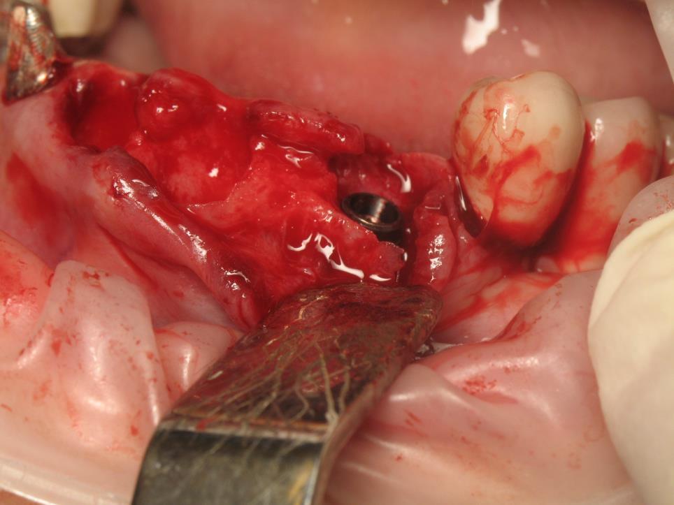
4 Biological Principles Inter-implant distance 3mm is our magic number between implants At 3 mm even though the facial bone goes apically. The bone around the adjacent teeth stays coronal=a papilla If we place implants less than 3 mm apart, then the interproximal level will be apical or equal to the facial bone=no papilla Results in thinner soft tissue


5 Biological Principles Tarnow, 1992 Study Tarnow Study: 0.45mm loss when Implant Distance is >3mm 1.04mm loss when Implant Distance is <3mm
6 Biological Principles Clinical implications of the Tarnow study Peri-implant Bone loss occurs around the junction between the implant and the abutment Results from bacterial colonization in the inside of the implant We can move this interface coronally and solve this bone loss problem, but then it will not be covered by soft tissue Unacceptable in the anterior where we want all implant parts buried


7 Biological Principles Bone remodeling occurs. This is a fact
8 Biological Principles Bone remodeling occurs. This is a fact Minimum distance to preserve bone is 1.5 mm between implant and tooth Bone loss occurs in a vertical direction usually down to the first thread On a x-ray this looks like a crater This loss will not approach the natural tooth next to it if it is 1.5 mm away This will cause a loss in papilla height This makes a case for using a narrower diameter implant in esthetic areas
9 Biological Principles Bone remodeling Most coronal height of bone of a natural tooth determine papilla support Even though an implant is more apical than the adjacent tooth a papilla will still be present If the bone is lower than the adjacent natural teeth then the papilla will be vary in height as we move across the anterior area
10 Biological Principles At inter-implant distances of 5mm, we have the luxury of creating ideal bony architecture much like we do in perio surgery We can sculpt the bone and create depressions in the bone before we place an implant
 Prefer 2mm of labial bone Dentinal gingival fibers provide support for gingival tissues in natural teeth Periodontal healthy anterior tooth not")
11 Biological Principles 7mm diameter 4mm for implant diameter, 1.5mm either side of implant (for single implants) Prefer 2mm of labial bone Dentinal gingival fibers provide support for gingival tissues in natural teeth Periodontal healthy anterior tooth not greater than 5mm papilla height coronal to interproximal alveolar bone crest Implants must be submerged to create a higher interproximal bone height to support papilla Papilla morphology dependent on perio status ridge morphology dependent on quantity/shape of bone


12 Biological Principles At the same time we need to consider the buccal soft tissue thickness Third dimension At least 2 mm of buccal soft tissue is needed. 3-4mm is more ideal This will be outside the area of circumferential loss and ensure long term support of the papilla over a long time. Without the buccal thickness the soft tissue collapses and it looks dark This would appear to dictate buccal bone grafting on all cases or placement of the implant slightly palatal to get the desired thickness of soft tissue
13 Gingival Architecture Gingival biotype is described as the thickness of the gingiva in the facio-palatal/ facio-lingual dimension. Seibert and Lindhe categorized the gingiva into thick-flat and thin scalloped biotypes in 1989


14 Gingival Architecture Patients with thick and flat gingival biotypes exhibit short papillae Thin and scalloped biotypes represent long papillae.
15 Gingival Architecture Thick biotypes include flat soft tissue and bony architecture, denser and more fibrotic soft tissue with large amount of attached masticatory mucosa Thick biotype is more resistant to any acute trauma and respond to disease by pocket formation and infra bony defect This difference may be the cause of more papilla loss in the thin biotype. This affect on the treatment outcome may be because of the difference in the amount of blood supply to the underlying bone and susceptibility to resorption according to Manjunath, Rana, et al, J of Clinical and Diagnostic Research, 2015
16 Gingival Architecture Thick gingival biotype usually depicts broad zone of keratinized tissue. This coupled with flat gingival contour, which indicates thick underlying bony architecture, is more resilient to any inflammation or trauma. Thin gingival biotype is related with a thin band of the keratinized tissue and scalloped gingival contour which suggest thin bony architecture and is more sensitive to any inflammation or trauma. >2.0mm is defined as thick <1.5mm is defined as thin
17 Biotype and Success Thin Decreased blood supply Complicates tissue response to surgical trauma Complicates suturing as it tears easily Show through of metal is an issue Patient needs to be informed of the challenge Thick Increase blood supply and support Esthetic success is predictable Downside is that bad incisions can create scars
18 Gingival Architecture In a study of 336 patients in India, the following was found: Thick biotype was more prevalent in the majority of the population Men 76.9% Women 55.3% Sex was not a difference Age was a difference for women(thin was more prevalent as women aged)
19 Gingival Architecture Drs. Fu and Lee, et all proposed the following in 2011 Create a thick biotype around implants as it is more conducive to implant health. Compared to a thin biotype it is preferred. Suggested a PDP management Implant Position Implant Design Design of Prosthetic
20 PDP Principals Implant position refers to depth and angle Implant design which includes diameter and platform can help prevent crestal bone loss Prosthetic design can provide additional space for soft tissue in growth creating a fuller tissue profile
21 PDP Principals Standard surgical guidelines mandate that an implant be placed with 2mm of buccal bone, 3mm apical to the CEJ of the adjacent tooth and 1.5mm from the tooth root and 3mm from an implant In situations where we need more facial soft tissue growth, placing an implant more apically and more palatal/lingual may be warranted. To deal with appropriate emergence, for every mm of palatal/lingual placement, the implant should be placed apically by an additional mm.
22 PDP Principals How do we accomplish the B-L bone requirement? Ridge expansion, implant position, or narrower implants can accomplish this. Platform switching will require the use of a narrower abutment over a wider body. This shift of the microgap toward the center of the implant, reducing crestal bone loss and fostering more soft tissue growth The use of a concave abutment or crown will create additional space for peri-implant tissue to occupy


23 PDP Principals Ridge expansion during osteotomy Densah auxillary burs which creates more bone on the facial surface of an implant


24 Gingival Architecture Clinical Applications of Biotype Determination Review Thick biotype is more resistant to recession, better at hiding metal and more forgiving to implant positional errors Thick biotype will handle trauma including prosthetic trauma better Utilize platform switching to increase tissue thickness
25 Let s discuss ''Biologic Width'' Sicher coined the term "dentogingival junction" in In 1961,Gargiulo et al., found out that the vertical dimension of the dentogingival junction comprising sulcus depth (SD), junctional epithelium (JE), and connective tissue attachment (CTA), is a physiologically formed and stable dimension, which forms at a level dependent on the location of the crest of the alveolar bone This protects the two most vulnerable structures of a tooth-the periodontal ligament and alveolar bone, which ultimately determine the survival and longevity of the dental elements

26 Biologic Width Gingival sulcus 0.69 mm Junctional epithelium 0.97 mm Connection tissue attachment coronal to bone 1.07 mm Gargiulo A., Wentz F., Orban F. Dimensions and Relations of the Dentogingival Junction in Humans. J. Periodontol :261
27 Where do we end our crowns with respect to the biological width Above the gingiva Best biological practice At crest of the gingiva Subgingival The most critical factor regarding the long-term gingival health is the relationship between the margin location, the location of the base of the sulcus and the supracrestal fiber attachment
28 Above the gingiva Preservation of tooth structure during tooth preparation. Impressions are more predictable, with minimal or no cord packing. Provisional restorations are easier to make, and the soft tissues will be healthier when the patient returns for cementation of the final restoration. Removing excess cement is much easier when the margin is visible.
29 Level with the gingiva Traditionally, not recommended as they were thought to retain more plaque than supragingival or subgingival margins and therefore cause greater gingival inflammation This plaque retention is typically true as patients did not clean as well. Older cements were relatively soluble Easier to take an impression blended with esthetic vigilance
30 Subgingival If the sulcus probes 1.5 mm or less, place the restoration margin 0.5 mm below the gingival tissue crest. This is especially important on the facial aspect. If the sulcus probes more than 1.5 mm, place the margin one half the depth of the sulcus below the tissue crest. This places the margin far enough below the tissue so that it still is covered if the patient is at higher risk of recession. Never place the margin closer than 2.5 mm to the bone level
31 Subgingival If we do place the margin closer than 2.5 mm to the bone level. We have now entered into the zone of the biologic width, and a very important biologic principle is being violated. When this happens an inflammatory response results in alveolar bone resorption, increased pocket depths, increased loss of periodontal support, exacerbation of accumulation of subgingival bacteria, increased chronic inflammation, and further localized periodontal breakdown. So if this is such a crucial area, why would we even consider going subgingival?

32 Reasons to go subgingival. Need to improve the resistance and retention form of a short clinical crown Presence of caries or restorations extending apical to the gingival margin Modification of the emergence profile Esthetics


33 Biological Principles Platform switching accident. Alters the horizontal position of the prosthetic-implant interface No solid guidelines on the exact nature of this technique Do surfaces make a difference Does thread position make a difference Does type of interface make a difference Bone loss was limited to 0.65 mm That being said 25-36% of the implants showed 1mm + bone loss Not a guarantee If 2 implants are close together then platform switching will help with bone loss around the implant, but the narrow space between the implants won t help with buccal papilla loss
34 Biological Principles Micro movement theory Reduce the movement and minimize bone loss around the implant No real studies to support it this At the end of the day, I utilize this concept as there is nothing to lose Morse taper
35 Biological Principles What should I do? Since Platform switching is not a guarantee of no bone loss around the implant, what s the right thing to do? At the same time there is no disadvantage to doing this As long as the larger implant does not reduce the mesio-distal distance As long as the larger implant does not result in a reduction of bone width on the buccal


36 Patient Assessment Lip line High line Low line when I pick up my lip, I see..


37 Patient Assessment Tooth shape Square tooth Triangular tooth
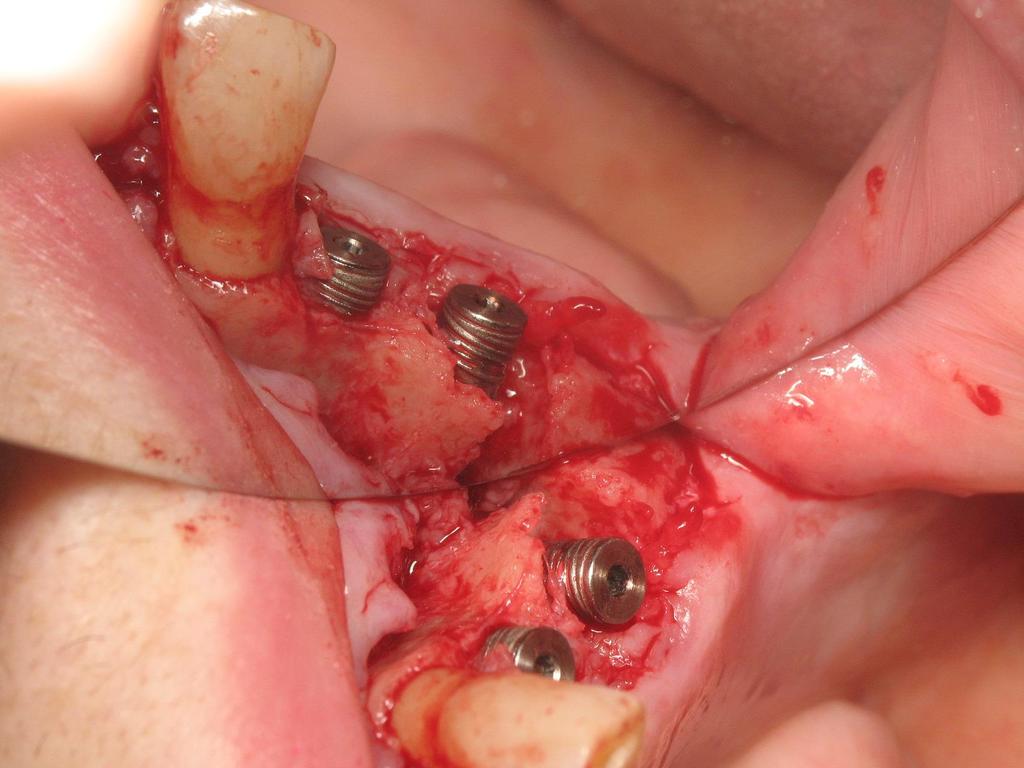
38 Patient Assessment Tooth shape Minimize a large black triangle by changing the tooth shape
39 Patient Assessment Jaw shape Convex jaw Thinner biotype May see a high lip line Lip pressure will cause thinning of buccal tissue Flat jaw Less tissue loss after extraction may favor an immediate implant Thicker biotype


40 Patient Assessment


41 Patient Assessment Diastema Very difficult to get a papilla Can t fake the papilla by lengthening the contact point Possibly suggest closing the diastema


42 Patient Assessment Mesial or Distal Tilt Tooth axis will determine the contact point If both centrals are tilted mesially then we have an incisal point contact If both centrals have are tilted distally then the contact point is shifted gingivally and can be broad Solutions: Ortho Bonding Crown both teeth
43 Patient Assessment Concavity on the adjacent tooth Emphasizes or creates a black triangle Discuss this prior to treatment showing them their issue After the fact it is a rationalization for failure

44 Patient Assessment Large frenum Remove it at the beginning Leaving it will leave mucosa around the implant Increase the possibility of recession

45 Patient Assessment Large Incisive canal Never place the implant in the incisive canal with live tissue Remove all nerves and vessels and graft the area Not a situation for immediate implant placement

46 Patient Assessment Replacing 2 teeth 2 implants=6-8mm Tooth to implants = 1.5x2=3mm Space between implants = 3mm or more Total needed is 12-15mm at least Bonded bridge may be a better esthetic choice


47 Temporary Restoration Fixed-bonded zirconia bridge Removable Essix Fuse abutment
48 Implant position Facio-lingual(palatal) Mesio-distal Apico-coronal
49 Facio-lingual(palatal) A 25% decrease this dimension occurs within the first year of tooth loss and rapidly evolves into a 30% to 40% decrease within 3 years. The bone width loss is primarily from the facial region, because the labial plate is very thin compared with the palatal plate, and facial undercuts are often found over the roots of the teeth. Darkness of the implant shows through the gingiva
50 Facio-lingual(palatal) The crestal bone should be at least 1.5 mm wider on the facial aspect of the implant and 0.5 mm on the palatal aspect. The thickness of bone on the facial aspect of a natural root is usually 0.5 mm thick. As a result, the implant is 1mm or more palatal than the facial emergence of the adjacent crowns at the free gingival margin. Best for screw retention

51 Mesio distal
. The best mesiodistal position for a cuspid is centered in the cuspid position.")
52 Mesio distal The best implant position is under the incisal edge of the final crown, or slightly more palatal (A). The ideal mesio distal implant position for a central incisor is 0.5 to 1.0 mm more distal than the midtooth position. This decreases the risk of encroachment on the incisive canal (B). The best mesiodistal position for a cuspid is centered in the cuspid position.


53 Mesio distal Two-piece implant should be at least 1.5 mm from an adjacent tooth. When the implant is closer than this to an adjacent tooth bone loss related to the microgap, and biological width violation. Use a Mini

54 Mesio distal in Narrow Space Megagen just like standard sizes except narrower Don t need to buy a separate kit
55 Apico coronal Midcrest position of the edentulous site should be 2 mm below the facial CEJ of the adjacent teeth. The interproximal bone should be scalloped 3 mm more incisal than the midcrest position. Becker et al classified the rang of interpoximal bone height above the midfacial scallop from less than 2.1 mm (flat) to scalloped 2.8 mm to pronounced scalloped < 4.1 mm. flat anatomy scalloped-anatomy square-shaped tooth ovoid-shaped tooth
56 Implant Angulation 3 Choices: Facial angulation so that emergence of the final crown will be similar to adjacent teeth. Under the incisal edge of the final restoration. Within the cingulum position of the implant crown.

57 Ideal position A. Position below the incisal edge is best used for a cemented crown in the esthetic zone. A. An implant is in the place of the natural root of the tooth. Although this makes sense, it places the implant too facial, and an angled abutment is usually necessary B. In the cingulum position when a screw-retained crown is the treatment of choice. This position requires a facial ridge lap of porcelain when used for FP-l prostheses

58 Immediate placement


59 Immediate placement
60 Flap Design Incision facts: We need to end up with tension free closure. Incisions will scar. If we add bone, the volume that must be covered by the flap has increased.


61 Flap Design Small minimal incision with or without verticals Makes a tension free incision impossible Makes grafting impossible Papilla preservation technique 1.5-2mm away from adjacent tooth


62 Flap Design Vertical Incisions in the Anterior Avoid in a visible esthetic zone If this must be done, then place them at the distal portion of the adjacent teeth as the highest portion of the gingiva is usually at the distal and your eye is used to going upward

63 Flap Design Ideal Flap Design Wide flap Only use 1 vertical incision Place the vertical outside the esthetic zone Circumferential incision in esthetic sensitive areas


64 Delayed/Immediate placement Incisions Use an Endodontic flap design Semi-trapezoidal Sling suture to close


65 Immediate placement


66 Immediate placement Remember the patient amidst all of you planning


67 Immediate placement


68 Immediate placement


69 Immediate placement


70 Immediate placement

71 Immediate placement


72 Immediate placement

73 Immediate placement


74 Position


75 Position


76 Position

77 Position


78 Position


79 Position

80 Immediate/Grafting


81 Immediate/Grafting


82 Immediate/Grafting


83 Immediate/Grafting


84 Immediate/Grafting
85 Implantitis A New Battleground in Implant Dentistry
86 Implantitis Prevalence Difficult to assess as definitions vary Bone loss Pocket appearance This is important as we try to diagnose a problem early Requires a radiograph to confirm the diagnosis >5mm has been defined as Stage 1 peri-implantitis >6mm is called Stage 2 A 2012 study showed that 22% of implants placed in approx. 28 out of 70 patients after 8 years
87 Implantitis Risk factors Smoking Previous perio Current perio in other areas of the mouth should be a red flag Hard to clean restorations Residual cement Guide our choice of screw vs cement retained Removal of cement fixes the problem
88 Implantitis Incubation period Swiss Study 5-10 years after implantation, 20% of all patients showed peri-implant disease
89 Implantitis Early Diagnosis Perio probe 4 sites Watch for increased depths Deep implant placement could give 5-6mm without inflammation Observe redness or swelling No bleeding is an indication of health Radiographs must be done in conjunction with a visual exam Use the bone level at the time of uncovery or restoration as a baseline Mobility means we have no osseointegration. If no signs or bleeding with probing, or bone loss is an indication of overloading
90 Implantitis Risk Factors General perio yields poorer results Have 3 month recall Smoking Success rate goes down about 30% Encourage smoking cessation Poor Oral Hygiene Demonstrate, provide and document aids Poor access Try to design the restoration with convex sufaces Residual cement Removal will usually resolve the issue Gingiva vs Alveolar mucosa At least 2 mm of gingiva is associated with reduced plaque accumulation
91 Implantitis Microbiology Similar to general perio? Fusobacteria, Prevotella, Porphyromanas, Spirochetes These are same bacteria that we find in perio around teeth These are all anaerobes The difference occurs as we see Staphylococci at times as the predominate bacteria on an implant This is also seen in general orthopedic implants and catheter infections BOTTOM LINE: The infection is populated by a mix of bacteria that are present in the mouth. Poor hygiene, for example, around an implant may allow one bug to increase in number
92 Implantitis Based on what we just said, we can not eradicate a certain type of bacteria to solve this problem as the entire mouth is populated with these types. Consequently, there is no special bacterial test to diagnose peri-implantitis. Use radiographs Use probing depths Visual exam
93 Implantitis If we decide to employ antibiotics, which one? International Journal of Oral Maxillofacial Impl, 2014 Heitz-Mayfield and Mombelli Amoxicillin Metronidazole(Flagyl) The treatment regime for all peri-implant infections is that they should be treated with 500mg Amoxicillin and 375 mg capsules Metronidazole for 7 days. Coumadin patients need to monitor INR with Metronidazole Peri-implantitis cannot be treated solely by meds Requires cleaning the implant surface to totally remove the biofilm which allows the bugs to grow
94 Implantitis Let s get organized Assessment which results in prosthetic fixes Screw loosening Cement Poor fit Poor contours Assessment which involves behaviour fixes Smoking Poor oral hygiene Bruxism Treating adjacent perio
95 Implantitis Let s get organized Non-surgical debridement Anitibiotics Laser Improved home care Re-assess 4 weeks later Resolution results in going to proper maintenance therapy Non-resolution will take us to surgery
96 Implantitis Let s get organized Surgical approach Full thickness flap Remove any cement Remove any granulation tissue Decontaminate the surface Rubbing with saline gauze Rubbing with citric acid gauze Rubbing with hydrogen peroxide gauze Mechanical cleaning with acurrette Mechanical cleaning with a brush Laser treatment
97 Implantitis Let s get organized Surgical approach If bone loss is circumferential and there is an outside wall, aka an infrabony defect, then we could regrow bone If not our goal is to stop inflammation and further loss of attachment In not esthetic areas then bone elevations can be reduced to allow apical flap adaptation Must be associated with implantoplasty to allow oral hygiene following healing Analagous to the long root on natural teeth Regeneration as opposed to resection Fill infrabony defect Membrane use Used very often with early bone loss(ie before loading)
98 Implantitis Let s get organized Post surgical protocol Antibiotics Chlorhexidine Implant removal if no improvement Maintenance for Frequent recalls Proper oral hygiene
99 Implantitis Bone Loss Class 1 Usually found at Second Stage Surgery No exudate Located at the coronal surface Managed by Debridement Decorticate bone Graft Membrane Classification is Based on text from Louie Al-Faraje, DDS
100 Implantitis Bone Loss Class 2 Usually found at Second Stage Surgery No exudate Located at the mid-buccal surface No exposure to the oral environment Cause: Narrow Ridge Managed by Soft tissue graft to prevent showing through thin gingival tissue CT graft or Alloderm Adding a bone Graft Membrane
101 Implantitis Bone Loss Class 3 Usually found at Second Stage Surgery No exudate Located at the coronal surface circumferentially No exposure to the oral environment Cause: Narrow Ridge/Possible pressure from provisional restoration Managed by Raise buccal and lingual/palatal flap Debridement Adding a bone Graft after decorticated Membrane

102 Implantitis Bone Loss Class 4 Usually found at recall visit Exudate present Located at the coronal surface circumferentially No exposure to the oral environment Cause: Infection Managed by Raise buccal and lingual/palatal flap Debridement Adding a bone Graft after decorticated Membrane OR Removal and Grafting

103 Implantitis Bone Loss Class 5 Gross bone and soft tissue loss Infection present Part of the implant is exposed to the oral cavity Cause: Prolonged history of infection Managed by Remove implant Grafting and healing for 6 months Replace the implant


104 Implantitis


105 Implantitis


106 Implantitis R-Brush I-Brush

107 Implantitis
108 Implantitis


109 Implantitis
110 Implantitis Success and predictability of dental implants have been well documented throughout the years with purported success rates of more than 90%. These success rates, however, have been established according to an older criteria. An implant without pain, mobility, radiolucency, or 1 mm of bone loss during the first year and.2 mm thereafter was considered a success. A recent review that considered excessive bone loss as an additional criteria of implant failure suggested that "implant survival and success rates in general dental practices may be lower than those reported in studies conducted in academic or specialty settings" and quoted an 80% success rate.(3) Two systematic reviews and metaanalyses reported that the prevalence of peri-implantitis was present in approximately 10% of implants and 20% of patients eight to 10 years after implant placement
111 Implantitis Early implant complications arise prior to dental implant integration and often are the result of intraoperative or short-term postoperative problems. Early complications during implant placement include overpreparation of the implant osteotomy or underpreparation of the implant osteotomy. Underpreparing an implant site can result in surgically overheating the bone and bone necrosis if the critical threshold of greater than 47 C is reached by the surgical drills or dental implant. Contamination of the surgical site and/or the dental implant surface from bacteria is also possible during fixture placement, which can result in the failure of the implant to integrate. Lack of primary stability, stability of the implant when it is first placed into the alveolus, can result in implant micro-motion above 100 microns and loss of the implant due to fibrous tissue bonding to the implant surface instead of bone.
112 Late implant complications occur after the implant has integrated and the final prosthesis has been placed. Recognition of these complications via radiographic and clinical analysis is extremely important since many of these problems can be corrected if detected early. On the other hand, if allowed to progress, a minor complication can often result in loss of the implant and/or prosthesis. Late complications of the dental implant fall into the category of biologic or mechanical complications. Late biologic complications are those in which the peri-implant soft and hard tissues are affected. Peri-implant mucositis describes a reversible inflammatory reaction in the mucosa adjacent to an implant,(8) a term that has become known as implant gingivitis. Studies show that the prevalence of peri-implant mucositis can be as high as 50% to 80% of implants in function(9) with the etiology of peri-implant mucositis being bacterial plaque. Typical clinical presentation includes erythema, edema, swelling, and redness Although bleeding upon probing and increased probing depths are not always indicative of peri-implant mucositis, the absence of these two factors usually means implant health.
113 Finances: Fee-for-service with no insurance involvement is the preferable method of payment. Insurance reimbursements and involvement are often solutions to help patients have access to peri-implant disease treatment. Insurance submissions and obtaining approvals are often confusing when it comes to peri-implant treatment. The first step in understanding the tangled web of insurance is to understand that insurance companies have created their own definition and standard of treatment for periimplant disease. Insurance companies do not recognize peri-implant mucositis, only peri-implantitis. In addition, most insurance companies will only accept treatment in the form of surgical access, debridement, and flap replacement without the use of regenerative treatment
114 Implantitis Aetna Policy Despite large numbers of publications available regarding peri-implantitis treatments, there are no long term studies which indicate that the uses of osseous contouring, placement of barriers or bone grafts are superior to flap reflection with debridement. Therefore, Aetna considers flap reflection with debridement as the accepted clinical protocol for peri-implantitis treatment. Background Peri-implantitis is defined as an inflammatory process affecting the tissues around an osseointergrated implant in function resulting in loss of supporting bone. Clinical signs are deep probing depth (> 5 mm) bleeding and/or suppuration on probing. Loss of supporting bone usually forms a circumferential crater defect. Large scale studies detected peri-implantitis in 12% of implants in function for at least 5 years and 43% of implants in function for 9-14 years. Biofilms consist predominately of gram negative anaerobes and are similar to those found in chronic periodontits, but bone loss is more rapid in peri-implantitis. Risk factors include poor oral hygiene, smoking, diabetes and a history of chronic periodontitis.
115 Implantitis Codes D Gingival flap procedure, including root planing - one to three contiguous teeth or tooth bounded spaces per quadrant D Debridement of a peri-implant defect and surface cleaning of exposed implant surfaces, including flap entry and closure D Debridement and osseous contouring of a peri-implant defect; includes surface cleaning of exposed implant surfaces and flap entry and closure D Bone graft for repair of peri-implant defect - not including flap entry and closure or, when indicated, placement of a barrier membrane or biologic materials to aid in osseous regeneration D Bone graft at time of implant placement D Scaling and debridement in the presence of inflammation or mucositis of a single implant, including cleaning of the implant surfaces, without flap entry and closure. This procedure is not performed in conjunction with D1110 or D4910.
116 Implantitis Diagnosed with severe peri-implantitis. The patient was in a financial crisis at the time and asked that we submit to his primary insurance and secondary insurance. Treatment approved by primary insurance (Delta Dental): D debridement of peri-implant defect and surface cleaning of exposed implant surfaces, including flap entry and closure Treatment denied by primary insurance (Delta Dental): D bone graft for repair of peri-implant defect - not including flap entry and closure or, when indicated, placement of a barrier membrane or biological material to aid in osseous regeneration Treament approved by secondary insurance (Aetna Dental): D implant-maintenance procedures, including removal of prosthesis, cleansing of prosthesis, and abutments and reinsertion of prosthesis Treatment denied by secondary insurance (Aetna Dental): D debridement of a peri-implant defect and surface cleaning of exposed implant surfaces, including flap entry and closure D bone graft for repair of peri-implant defect - not including flap entry closure or, when indicated, placement of a barrier membrane or biological material to aid in osseous regeneration
117 Implantitis January 2018-Not much has changed Recently, a regenerative surgical protocol that uses both mechanical and chemical means for implant surface detoxification together with a bone graft and/or substitute including coverage with a barrier membrane and a two- to 10-year follow-up period has been shown to have a 98.8% implant survival rate. The results of this study were based on peri-implantitis treatment of 170 consecutive dental implants in 100 patients. (13) In that specific study, an air powder abrasive was utilized as part of the surface decontamination. The authors of two of the more complete reviews could not identify any one protocol as being more predictable or superior than the others in terms of treatment outcomes
Prosthetic Options in Implant Dentistry. Hakimeh Siadat, DDS, MSc Associate Professor
 Prosthetic Options in Dentistry Hakimeh Siadat, DDS, MSc Associate Professor Dental Research Center, Department of Prosthodontics & Dental s Faculty of Dentistry, Tehran University of Medical Sciences
Rehabilitating a Compromised Site for Restoring Form, Function and Esthetics- A Case Report
 Research & Reviews: Journal of Dental Sciences Rehabilitating a Compromised Site for Restoring Form, Function and Esthetics- A Case Report Priyanka Prakash* Division of Periodontology, Department of Dental
Principles of Periodontal flap surgery. Dr.maryam khosravi
 Principles of Periodontal flap surgery Dr.maryam khosravi Goals of periodontal SURGICAL phase 1 - Controlling or eliminating periodontal disease. 2 Correcting anatomic conditions that may a. favor periodontal
Core build-up using post systems
 Core build-up using post systems Dr. Gergely Pataky Department of Conservative Dentistry What to speak about today General considerations Classification of post systems Dowel-core or fibre post? Biologic
HDS PROCEDURE CODE GUIDELINES
 D4000 - D4999 Local anesthesia is usually considered to be part of Periodontal procedures. General Guidelines 1. Periodontal services are only benefited when performed on natural teeth for treatment of
Extraction with Immediate Implant Placement and Ridge Preservation in the Posterior
 Extraction with Immediate Implant Placement and Ridge Preservation in the Posterior by Timothy F. Kosinski, DDS, MAGD The following case presentation illustrates the diagnosis, planning and treatment for
WHAT IS THE PURPOSE OF WHAT WE DO? TEAM PERIODONTICS: WORKING TOGETHER TO IMPROVE PATIENT CARE YOU ARE THE PERIODONTISTS IN YOUR PRACTICE!
 Setter Periodontics 2075 SW 1 st Ave #2L Portland, OR 97201 503-222-9961 michael@setterperio.com WHAT IS THE PURPOSE OF WHAT WE DO? Gum Gardeners Study Club 2.27.17 TEAM PERIODONTICS: WORKING TOGETHER
Periodontal Disease. Radiology of Periodontal Disease. Periodontal Disease. The Role of Radiology in Assessment of Periodontal Disease
 Radiology of Periodontal Disease Steven R. Singer, DDS srs2@columbia.edu 212.305.5674 Periodontal Disease! Includes several disorders of the periodontium! Gingivitis! Marginal Periodontitis! Localized
Advanced Probing Techniques
 Module 21 Advanced Probing Techniques MODULE OVERVIEW The clinical periodontal assessment is one of the most important functions performed by dental hygienists. This module begins with a review of the
Osseointegrated dental implant treatment generally
 Placement of Dental Implants Without Flap Surgery: A Clinical Report Bader H. Al-Ansari, BDS, MScD*/Robert R. Morris, DMD** Traditionally, the procedure of implant placement requires a surgical periosteal
All Dentistry is Cosmetic Betsy Bakeman, DDS Arkansas State Dental Association
 All Dentistry is Cosmetic Betsy Bakeman, DDS Arkansas State Dental Association Patients have traditionally sought treatment when concerned with the way their teeth look, function or feel. Over the past
Practical Advanced Periodontal Surgery
 Practical Advanced Periodontal Surgery Serge Dibart Blackwell Munksgaard Chapter 8 Papillary Construction After Dental Implant Therapy Peyman Shahidi, DOS, MScD, Serge Dibart, DMD, and Yun Po Zhang, PhD,
The International Journal of Periodontics & Restorative Dentistry
 The International Journal of Periodontics & Restorative Dentistry 3 Influence of the 3-D Bone-to-Implant Relationship on Esthetics Ueli Grunder, DMD* Stefano Gracis, DMD** Matteo Capelli, DMD** There are
Case Study. Case # 1 Author: Dr. Suheil Boutros (USA) 2013 Zimmer Dental, Inc. All rights reserved. 6557, Rev. 03/13.
 2013 Zimmer Dental, Inc. All rights reserved. 6557, Rev. 03/13.") Placement of a Zimmer Trabecular Metal Dental Implant with Simultaneous Ridge Augmentation and Immediate Non-Functional Loading Following Tooth Extraction and Orthodontic Treatment for Implant Site Development
Placement of a Zimmer Trabecular Metal Dental Implant with Simultaneous Ridge Augmentation and Immediate Non-Functional Loading Following Tooth Extraction and Orthodontic Treatment for Implant Site Development
What You Should Know About Dental Implants: The Process of Care Applies
 Learn how the process of care model can apply to dental implants and empower decisions for providing quality care for clients. Starting with assessing the need for implants, the process covers documentation,
The 2B-3D rule for implant planning, placement and restoration
 IJOI 27 INTERDISCIPLINARY TREATMENT The 2B-3D rule for implant planning, placement and restoration 1. What is biologic width? Is there a golden rule for implant planning, placement and restoration as the
Surgical Therapy. Tuesday, April 2, 13. Alessan"o Geminiani, DDS, MS
 Surgical Therapy Alessan"o Geminiani, DDS, MS Periodontal Flap: a surgical procedure in which incisions are made in the gingiva or mucosa to allow for separation of the epithelium and connective tissues
Immediate implant placement in the Title central incisor region: a case repo. Journal Journal of prosthodontic research,
 Immediate implant placement in the Title central incisor region: a case repo Author(s) Sekine, H; Taguchi, T; Yamagami, M; Alternative Takanashi, T; Furuya, K Journal Journal of prosthodontic research,
Principles of endodontic surgery
 Principles of endodontic surgery Note: the doctor said that this lecture mainly contain notes, so we should study it from the book for further information (chapter 18) principles of endodontic surgery.
Indications The selection of amalgam as a restorative material for class V cavity should involve the following considerations:
 1 Lec.7 د.عبد املنعم اخلفاجي CLASS V CAVITY PREPARATION FOR AMAGLAM Indications The selection of amalgam as a restorative material for class V cavity should involve the following considerations: 1- Caries:
Biologic width: Understanding and its preservation Malathi K 1, Singh A 2
 Biologic width: Understanding and its preservation Malathi K 1, Singh A 2 Review Article 1 Dr K Malathi Professor & Head, Periodontics Government Dental college and Hospital Tamil Nadu, India 2 Dr Arjun
Esthetic Crown Lengthening for Upper Anterior Teeth: Indications and Surgical Techniques
 I J Pre Clin Dent Res 2014;1(2):49-53 April-June All rights reserved International Journal of Preventive & Clinical Dental Research Esthetic Crown Lengthening for Upper Anterior Teeth: Indications and
Replacement of a congenitally missing lateral incisor in the maxillary anterior aesthetic zone using a narrow diameter implant: A case report
 C A S E R E P O R T Replacement of a congenitally missing lateral incisor in the maxillary anterior aesthetic zone using a narrow diameter implant: A case report Rhoodie Garrana 1 and Govindrau Mohangi
Case Report. RapidSorb Rapid Resorbable Fixation System. Ridge augmentation in a one-step surgical protocol.
 Case Report RapidSorb Rapid Resorbable Fixation System. Ridge augmentation in a one-step surgical protocol. RapidSorb Rapid Resorbable Fixation System. Ridge augmentation in a one-step surgical protocol.
MINI System CASE REPORT. Name: Dr. Achraf Souayah Na<on: Tunisia
 MINI System SE REPORT Name: Dr. chraf Souayah Na
Controlling Tissue Contours with a Prosthetically Driven Approach to Implant Dentistry
 Controlling Tissue Contours with a Prosthetically Driven Approach to Implant Dentistry Go online for in-depth content by Timothy F. Kosinski, DDS, MAGD With continual improvements in the design and production
Masking Buccal Plate Remodeling in the Esthetic Zone with Connective Tissue Grafts: Concepts and Techniques with Immediate Implants
 Peer-Reviewed and Indexed Annual Implant Issue Masking Buccal Plate Remodeling in the Esthetic Zone with Connective Tissue Grafts: Concepts and Techniques with Immediate Implants of Continuing Education
Patient s Presenting Complaint V.C. presented with discomfort and mobility from the crowned maxillary left central incisor tooth. Fig 1.
 Patient s Presenting Complaint V.C. presented with discomfort and mobility from the crowned maxillary left central incisor tooth. Fig 1. A longitudinal root fracture was suspected and confirmed when the
Then and Now. Implant Therapy:
 Implant Therapy: Then and Now by Timothy F. Kosinski, DDS, MAGD Implant dentistry has come a long way since blade and subperiostal implants were widely used. Improvements in implant design and site preparation
Restorative Dentistry and Papilla Reconstruction in Reduced Periodontium
 CLINICAL AND RESEARCH REPORT Restorative Dentistry and Papilla Reconstruction in Reduced Periodontium Robert Azzi, Daniel Etienne, Bernard Schweitz Restoring the loss of periodontal soft and hard tissues
Nicholas Caplanis DMD MS 6/13/2012
 Considerations In The Esthetic Zone Nick Caplanis DMD MS Private Practice Periodontics and Implant Surgery Mission Viejo, California Nick@drcaplanis.com Assistant Professor Loma Linda University Anatomic
Periodontal Maintenance
 Periodontal Maintenance Friday, February 20, 2015 1:06 PM Periodontal disease control always begins with patient education - Plaque control, diet, smoking cessation, impact that systemic health has on
The patient gave a history of hypertension and gastritis for which was taking Lacidipine 4mg, Omeprazole 20mg and Simvastatin 40mg.
 A.S. was referred by her general dental practitioner for assessment for possible implant placement to restore the space where her bridge replacing her maxillary central incisors had recently failed. Fig
LIST OF COVERED DENTAL SERVICES
 LIST OF COVERED DENTAL SERVICES The following is a complete list of those dental Services which will be considered for payment by Constitution Life Insurance Company after the expiration of any applicable
Lecture 2 Maxillary central incisor
 Lecture 2 Maxillary central incisor Generally The deciduous tooth appears in the mouth at 3 18 months of age, with 6 months being the average and is replaced by the permanent tooth around 7 8 years of
Implant Esthetic Failure
 Kevin G. Murphy, DDS, MS Associate Professor of Periodontics Baltimore College of Dentistry University of Maryland Faculty, Pankey Foundation Key Biscayne, FL Private Practice, Baltimore, MD PROVISIONAL
Surgical Procedure in Guided Tissue Regeneration with the. Inion GTR Biodegradable Membrane System
 Surgical Procedure in Guided Tissue Regeneration with the Inion GTR Biodegradable Membrane System 1 Introduction This presentation familiarizes you with the basic steps how to use the Inion GTR membrane
أ.م. هدى عباس عبد اهلل CROWN AND BRIDGE جامعة تكريت كلية. Lec. (2) طب االسنان
 طب االسنان") Lec. (2) CROWN AND BRIDGE أ.م. هدى عباس عبد اهلل Patient selection and examination A thorough diagnosis must first be made of the patient's dental condition, considering both hard and soft tissues. this
Lec. (2) CROWN AND BRIDGE أ.م. هدى عباس عبد اهلل Patient selection and examination A thorough diagnosis must first be made of the patient's dental condition, considering both hard and soft tissues. this
Osseous surgery in periodontal treatment
 SCIENTIFIC SESSION Osseous surgery in periodontal treatment Essayist: Roberto Pontoriero Introduction In periodontally involved patients, periodontal therapy usually results in various degrees of soft-tissue
Working together as a team, the periodontist
 The Team Approach to Esthetic Immediate Implant Placement Bobby L. Butler, DDS; and Greggory Kinzer, DDS Working together as a team, the periodontist and restorative dentist can provide an increased level
Management of a complex case
 2 Soft- and hard-tissue reconstruction of a severely deficient site prior to implant placement: a case report Management of a complex case Younes Khosroshahy, DDS, MFDS RCS (Eng), Dip Imp Dent RCSEd, Blue
Dental Implant Placement in the Maxillary Anterior Region: Guidelines for Aesthetic Success Michael Tischler, DDS
 Page 1 of 10 Issue Date: March 2005, Posted On: 5/2/2005 Dental Implant Placement in the Maxillary Anterior Region: Guidelines for Aesthetic Success Michael Tischler, DDS Figure 1. A 43-year-old female
Purpose: To assess the long term survival of sites treated by GTR.
 Cortellini P, Tonetti M. Long-term tooth survival following regenerative treatment of intrabony defects. J Periodontol 2004; 75:672-8. (28 Refs) Purpose: To assess the long term survival of sites treated
CASE REPORT. CBCT-Assisted Treatment of the Failing Long Span Bridge with Staged and Immediate Load Implant Restoration
 Computer Aided Implantology Academy Newsletter - Newsletter 20 - July 2009 CASE REPORT CBCT-Assisted Treatment of the Failing Long Span Bridge with Staged and Immediate Load Implant Restoration Case Report
Senior Dental Insurance Scheduled Allowance
 Senior Dental Insurance Scheduled Allowance LIST OF COVERED DENTAL SERVICES The following is a complete list of those dental services which will be considered for payment by The American Progressive Life
Evidence-based decision making in periodontal tooth prognosis
 Clin Dent Rev (2017) 1:3 https://doi.org/10.1007/s41894-017-0004-2 TREATMENT Evidence-based decision making in periodontal tooth prognosis Carlos Ernesto Nemcovsky 1 Received: 12 April 2017 / Accepted:
Fundamental & Preventive Curvatures of Teeth and Tooth Development. Lecture Three Chapter 15 Continued; Chapter 6 (parts) Dr. Margaret L.
 Dr. Margaret L.") Fundamental & Preventive Curvatures of Teeth and Tooth Development Lecture Three Chapter 15 Continued; Chapter 6 (parts) Dr. Margaret L. Dennis Proximal contact areas Contact areas are on the mesial and
Fundamental & Preventive Curvatures of Teeth and Tooth Development Lecture Three Chapter 15 Continued; Chapter 6 (parts) Dr. Margaret L. Dennis Proximal contact areas Contact areas are on the mesial and
Socket preservation in the daily practice: A clinical case report
 Clinical Socket preservation in the daily practice: A clinical case report Rabih Abi Nader 1 and Carine Tabarani 2 Abstract Soft tissue contour depends on the underlying bone anatomy. Following tooth extraction,
Dental Implant Treatment Planning and Restorative Considerations
 Dental Implant Treatment Planning and Restorative Considerations Aldo Leopardi, BDS, DDS, MS Practice Limited to Implant, Fixed and Removable Prosthodontics Greenwood Village, Colorado www.knowledgefactoryco.com
Contemporary Implant Dentistry
 Contemporary Implant Dentistry C H A P T ER 1 4 O F C O N T E M P OR A R Y O R A L A N D M A X I L L OFA C IA L S U R G E RY B Y : D R A R A S H K H O J A S T EH Dental implant is suitable for: completely
Treatment Options for the Compromised Tooth
 New Edition Treatment Options for the Compromised Tooth A Decision Guide American Association of Endodontists www.aae.org/treatmentoptions TREATMENT PLANNING CONSIDERATIONS The Treatment Options for the
REGENERATIONTIME. A Case Report by. Ridge Augmentation and Delayed Implant Placement on an Upper Lateral Incisor
 A Case Report by Dr. Daniele Cardaropoli Ridge Augmentation and Delayed Implant Placement on an Upper Lateral Incisor The Situation An adult female patient presented with an endodontic/prosthetic failure
COMBINED PERIODONTAL-ENDODONTIC LESION. By Dr. P.K. Agrawal Sr. Prof and Head Dept. Of Periodontia Govt. Dental College, Jaipur
 COMBINED PERIODONTAL-ENDODONTIC LESION By Dr. P.K. Agrawal Sr. Prof and Head Dept. Of Periodontia Govt. Dental College, Jaipur Differential diagnosis For differential diagnostic purposed the endo-perio
How I use Lasers with Dental Implants for Soft and Hard Tissue Management, making the whole process easier (0052) Dr Robin Horton BDS
 Dr Robin Horton BDS") How I use Lasers with Dental Implants for Soft and Hard Tissue Management, making the whole process easier (0052) Dr Robin Horton BDS Wayside Dental Practice- Harpenden - England Disclosure: I have no
How I use Lasers with Dental Implants for Soft and Hard Tissue Management, making the whole process easier (0052) Dr Robin Horton BDS Wayside Dental Practice- Harpenden - England Disclosure: I have no
Please visit the C.E. Pavilion to validate your course attendance Or If There s a Line Go cdapresents.com
 UCLA Innovations 2016 CDA Presents in Anaheim Tara Aghaloo, DDS, MD, PhD Dean Ho, MS, PhD Jay Jayanetti Eric C. Sung, DDS David T. W. Wong, DMD, DMSc Benjamin M. Wu, DDS, PhD Saturday, May 14, 2016 8:00
MANAGEMENT OF ATROPHIC ANTERIOR MAXILLA USING RIDGE SPLIT TECHNIQUE, IMMEDIATE IMPLANTATION AND TEMPORIZATION
 Case Report International Journal of Dental and Health Sciences Volume 02, Issue 06 MANAGEMENT OF ATROPHIC ANTERIOR MAXILLA USING RIDGE SPLIT TECHNIQUE, IMMEDIATE IMPLANTATION AND TEMPORIZATION Rakshith
Inclusive Tooth Replacement System
 Optimizing Anterior Esthetics with the Inclusive Tooth Replacement System by Timothy F. Kosinski, DDS, MAGD Implant treatment has changed so much over the years. In the past it was acceptable to place
A new approach with an in-situ self-hardening grafting material
 74 Bone grafting with simultaneous early implant placement A new approach with an in-situ self-hardening grafting material MINAS LEVENTIS 1,2, PHD; PETER FAIRBAIRN 1,3, BDS; ORESTIS VASILIADIS 2,4, DDS
The majority of the early research concerning
 Gingival Recession Around Implants: A 1-Year Longitudinal Prospective Study Paula N. Small, DDS, MPH 1 /Dennis P. Tarnow, DDS 2 A longitudinal study was performed, which measured the soft tissue around
Immediate Loading with Flapless Implant Surgery for Rehabilitation of Single Bound Edentulous Space
 Case Report Immediate Loading with Flapless Implant Surgery for Rehabilitation of Single Bound Edentulous Space Nidhi Bhatia 1, Shweta Bali 2, Meenu Taneja Bhasin 3, Priyanka Aggarwal 4, Vaibhav Joshi
Treatment of dental recessions in the esthetic zone by gingival and osseous recontouring. A multidisciplinary perio-prosthodontic case report.
 European International Journal of Science and Technology Vol. 6 No. 5 July 2017 Treatment of dental recessions in the esthetic zone by gingival and osseous recontouring. A multidisciplinary perio-prosthodontic
COURSE CURRICULUM FOR AESTHETIC DENTISTRY
 COURSE CURRICULUM FOR AESTHETIC DENTISTRY Esthetic Dentistry is actually the fourth dimension in clinical dentistry. In addition to biologic, Physiologic, and mechanical factors, all of which must be understood
Persson GR, Salvi GE, Heitz-Mayfield LJA et al. Antimicrobial therapy using a local drug delivery system (Arestin) in the treatment of
 in the treatment of") Persson GR, Salvi GE, Heitz-Mayfield LJA et al. Antimicrobial therapy using a local drug delivery system (Arestin) in the treatment of peri-implantitis I: microbiological outcomes. Clin Oral Imp Res 2006;
Persson GR, Salvi GE, Heitz-Mayfield LJA et al. Antimicrobial therapy using a local drug delivery system (Arestin) in the treatment of peri-implantitis I: microbiological outcomes. Clin Oral Imp Res 2006;
Immediate Implant Placement:
 Immediate Implant Placement: Parameters Influencing Tissue Remodeling Bernard Touati, DDS and Mario Groisman, DDS In esthetic implant therapy, the patient s objective is to obtain an imperceptible, natural-looking
Prosthodonticstown. Immediate Implant Placement in Fresh Extraction Sites. clinical. Table I
 Prosthodonticstown clinical Immediate Implant Placement in Fresh Extraction Sites Charles A. Babbush, DDS, MScD Table I Benefits Of Immediate Implantation Improved prosthesis fabrication and/or design
Townie Guest Editorial. Gingival Attachment Loss: Evaluation and Surgical Options. Daniel J. Melker, DDS. fig. 1
 Gingival Attachment Loss: Evaluation and Surgical Options Daniel J. Melker, DDS Attached connective tissue (a.k.a. attached tissue) in the simplest terms is the body s only barrier between the underlying
EFFECTIVE DATE: 04/24/14 REVISED DATE: 04/23/15, 04/28/16, 06/22/17, 06/28/18 POLICY NUMBER: CATEGORY: Dental
 MEDICAL POLICY SUBJECT: DENTAL IMPLANTS PAGE: 1 OF: 5 If a product excludes coverage for a service, it is not covered, and medical policy criteria do not apply. If a commercial product (including an Essential
Case Presentation. Overall Health. Oral Hygiene. Chief Complaint. I hate my upper denture. I can t taste food. I want an implant solution
 Case Presentation Medical History Age & Gender 62 years old female Peri-implant Osteitis Overall Health Good Hx of Smoking 5 cigarets per day (Peri-implantitis) Oral Hygiene Fair Systemic Disease Osteoarthritis
Creating emergence profiles in immediate implant dentistry
 Creating emergence profiles in immediate implant dentistry AUTHORS Dr. Daniel Capitán Maraver Dr. Manuel Fuentes Ortiz Visiting lecturers in the Master s Degree in Clinical Practice in Implantology and
Flapless, Immediate Implantation & Immediate Loading with Socket Preservation in the Esthetic Area Using the Alpha-Bio Tec's NeO Implants
 Flapless Surgery Case Study 48 Flapless, Immediate Implantation & Immediate Loading with Socket Preservation in the Esthetic Area Using the Alpha-Bio Tec's NeO Implants Dr. Gadi Schneider DMD, Specialist
Contemporary Periodontal Surgery
 Contemporary Periodontal Surgery Chris van Kesteren, D.D.S. CPCC Dental Hygiene Program October 18, 2011 Surgical Management of Periodontitis Periodontal Plastic Surgery Soft tissue and esthetics Dental
Educational Training Document
 Educational Training Document Table of Contents Part 1: Resource Document Disclaimer Page: 2 Part 2: Line Item Grade Sheets Page: 3 Release: 11/2016 Page 1 of 6 Part 1: Resource Document Disclaimer The
Osseointegrated implant-supported
 CLINICAL SCREWLESS FIXED DETACHABLE PARTIAL OVERDENTURE TREATMENT FOR ATROPHIC PARTIAL EDENTULISM OF THE ANTERIOR MAXILLA Dennis Flanagan, DDS This is a case report of the restoration of a partially edentulous
14/09/15. Assessment of Periodontal Disease. Outline. Why is Periodontal assessment needed? The Basics of Periodontal assessment
 Assessment of Periodontal Disease Dr Wendy Turner Outline Why is Periodontal assessment needed? The Basics of Periodontal assessment Probing: Basic Periodontal Examination for adults and children. Detailed
Minimally invasive techniques for periodontal regeneration
 2016; 2(12): 230-234 ISSN Print: 2394-7500 ISSN Online: 2394-5869 Impact Factor: 5.2 IJAR 2016; 2(12): 230-234 www.allresearchjournal.com Received: 04-10-2016 Accepted: 05-11-2016 Dr. Rizwan M Sanadi Professor,
Case report: Replacement of failing 2 stage implants by basal implants and conventional bridgework
 Case report: Replacement of failing 2 stage implants by basal implants and conventional bridgework Authors Dr. Aleksandar Lazarov Solunska Str. 3 BG-1000 Sofia Bulgaria Email: alex.lazarov@yahoo.co.uk
Implant Site Development Part I
 REVIEW ARTICLE Implant Site Development Part I 1 Umang Nayar, 2 Shankar Iyer IJCID Implant Site Development Part I 1 Consultant, Dental Surgeon and Periodontist at Max Health Care, New Delhi, Professor
Esthetic Crown Lengthening
 Esthetic Crown Lengthening Esthetic Crown Lengthening ACCELERATED OSTEOGENIC ORTHODOTNICS (WILKODONTICS) It is a technique developed by Wilko brothers. has roots in orthopedics, back to the early 1900s
Smile Design. Daniel H Ward DDS 1080 Polaris Pkwy Ste 130 Columbus OH
 Smile Design Daniel H Ward DDS 1080 Polaris Pkwy Ste 130 Columbus OH 43240 614-430-8990 dward@columbus.rr.com Incisal Placement Incisal plane parallel to the interpupillary line Vertical midline in the
Revisions for CDT 2016
 Revisions for CDT 2016 This document was developed from preliminary actions of the Code Maintenance Committee (CMC). This document has been compared to the CMC meeting notes and the ASCII file. This document
Esthetic Crown Lengthening for Upper Anterior Teeth: Indications and Surgical Techniques
 Esthetic Crown Lengthening for Upper Anterior Teeth: Indications and Surgical Techniques Mohammad Assaf Assistant Professor, Faculty of Dentistry, Al-Quds University, Jerusalem, Palestine. ABSTRACT Correspondence
Gum Graft? Patient Need a. Does My. 66 JANUARY 2017 // dentaltown.com. by Dr. Brian S. Gurinsky
 by Dr. Brian S. Gurinsky Dr. Brian S. Gurinsky was born in Dallas and attended college at the University of Texas at Austin. He continued his education at Baylor College of Dentistry in Dallas, where he
Clinical crown lengthening a case report
 FOLIA MEDICA CRACOVIENSIA Vol. LV, 3, 2015: 25 35 PL ISSN 0015-5616 Clinical crown lengthening a case report Weronika Lipska 1, Marcin Lipski 2, Małgorzata Lisiewicz 3, Andrzej Gala 3, Krzysztof Gronkiewicz
Case Report - Dr. Arthur Weiss
 High Esthetic Solutions with a Team Approach to Implant Dentistry The placement of hybrid abutments in the highly esthetic anterior zone Introduction Proper planning with a team approach ensures that highly
From planning to surgery: a totally digital working flow for Leone implants placement
 Dr. Giancarlo Romagnuolo Roma, Italy From planning to surgery: a totally digital working flow for Leone implants placement Keywords guided surgery, 3D implant planning, single missing tooth, delayed immediate
6610 NE 181st Street, Suite #1, Kenmore, WA
 660 NE 8st Street, Suite #, Kenmore, WA 9808 www.northshoredentalacademy.com.08.900 READ CHAPTER The Professional Dental Assistant (p.-9) No Key Terms Recall Questions:,,,, and 6 CLASS SYLLABUS DAY READ
Plaque and Occlusion in Periodontal Disease Wednesday, February 25, :54 AM
 Plaque and Occlusion in Periodontal Disease Wednesday, February 25, 2015 9:54 AM 1. The definition of Trauma From Occlusion: Primary TFO, Secondary TFO, and Combined TFO 2. Clinical and Radiographic signs
RAJ M. SAINI, DDS, MSD
 Restoring and Maintaining Periodontal Health with Orthodontic Treatment RAJ M. SAINI, DDS, MSD rajmsaini@yahoo.com Diplomate Of The American Board Of Orthodontics Clinical Professor Of Orthodontics New
Management of millers class III marginal tissue recession associated with endodontic lesion: Report of two cases managed using second stage surgery
 CASE SERIES 1 OPEN ACCESS Management of millers class III marginal tissue recession associated with endodontic lesion: Report of two cases managed using second stage surgery Sangeeta ABSTRACT Introduction:
Failures in implant therapy. Biological and mechanical complications. Their prevention management. Dr. Katalin Csurgay Dr.
 Failures in implant therapy. Biological and mechanical complications. Their prevention management. Dr. Katalin Csurgay Dr. Tatiana Shkolnik Complications could be: Doctor related Patient related Early
When performing conventional crown
 Case of the Month Biologic Shaping Kurtzman Daniel Melker, DDS 1 Abstract When performing conventional crown lengthening, the existing margins of an old restoration or the cementoenamel junction (CEJ)
Maintenance in the Periodontally Compromised Patient. Dr. Van Vagianos January 22, 2009 Charlotte Dental Hygiene Study Club
 Maintenance in the Periodontally Compromised Patient Dr. Van Vagianos January 22, 2009 Charlotte Dental Hygiene Study Club Periodontal Maintenance for Natural Teeth and Implants What is Periodontal Maintenance?
م.م. طارق جاسم حممد REMOVABLE PARTIAL DENTURE INTRODUCTION
 Lec.1 م.م. طارق جاسم حممد REMOVABLE PARTIAL DENTURE INTRODUCTION االسنان طب Prosthodontics is the branch of dentistry pertaining to the restoration and maintenance of oral function, comfort, appearance,
PERIODONTAL CASE PRESENTATION - 1
 PERIODONTAL CASE PRESENTATION - 1 Overview A 32 year-old patient presented with generalized aggressive periodontitis. Treatment included non-surgical therapy with adjunctive antibiotics and surgical treatment.
Full Arch Hygiene Protocol
 Full Arch Hygiene Protocol Patients should be evaluated and categorized into 3 different Recall Frequency Groups (RFG s). An evaluation of homecare should be made during the prosthetic phase of treatment
The Use of Alpha-Bio Tec's Narrow NeO Implants with Cone Connection for Restoration of Limited Width Ridges
 Case Study 48 The Use of Alpha-Bio Tec's Narrow NeO Implants with Cone Connection for Restoration of Limited Width Ridges Dr. Amir Gazmawe DMD, Specialist in Prosthodontics, Israel Dr. Amir Gazmawe graduated
Patients esthetic demands and
 Predictable Periimplant Gingival Esthetics: Use of the Natural Tooth as a Provisional following Implant Placement ROBERT C. MARGEAS, DDS* ABSTRACT Maintaining the interdental papilla and bone height following
Optimizing Lateral Incisor Function and Esthetics with the Hahn Tapered Implant System
 Optimizing Lateral Incisor Function and Esthetics with the Hahn Tapered Implant System by Timothy Kosinski, DDS, MAGD Maxillary lateral incisor agenesis (MLIA) is a condition in which at least one of the
International Journal of Health Sciences and Research ISSN:
 International Journal of Health Sciences and Research www.ijhsr.org ISSN: 2249-9571 Case Report Bridge Flap: A Sine Qua Non For Mucogingival Deformities Debajyoti Mondal, Anju L, Rajul Choradia, Somen