Automatic Segmentation of Digital Orthopantomograms for Forensic Human Identification
|
|
|
- Arabella Shonda Booker
- 5 years ago
- Views:
Transcription
1 Automatic Segmentation of Digital Orthopantomograms for Forensic Human Identification Dariusz Frejlichowski and Robert Wanat West Pomeranian University of Technology, Szczecin, Faculty of Computer Science and Information Technology, Zolnierska 52, , Szczecin, Poland Abstract. Dental radiographic images are one of the most popular biometrics used in the process of forensic human identification. This led to the creation of the Automatic Dental Identification System with the goal of decreasing the time it takes to perform a single search in a large database of dental records. A fully automated system identifying people based on dental X-ray images requires a prior segmentation of the radiogram into sections containing a single tooth. In this paper, a novel method for such segmentation is presented, developed for the dental radiographic images depicting the full dentition pantomograms. The described method utilizes the locations of areas between necks of teeth in order to determine the separating lines and does not depend on the articulation of gaps between adjacent teeth, thus improving the results achieved in the situation of severe occlusions. Keywords: image segmentation, dental pantomography, dental human identification, ADIS, forensic identification. 1 Introduction Human forensic identification is the process of determining the identity of a person using the evidence available in legal investigations. Teeth and bite are particularly popular for this application ([1]), as they are both robust to decomposition and highly discriminant ([2]). In the wake of ever growing use of digital radiology, the Federal Bureau of Investigation (FBI) created the Dental Task Force (DTF) to improve the use of digital dental information in legal proceedings ([3]). One of its main tasks was the creation of an Automated Dental Identification System (ADIS) in order to preliminarily browse through a large database of dental X-ray images in the search for radiograms with the most similar characteristics to present in an input image. The features used for comparison in ADIS are morphologic, e.g. the shapes of individual teeth or their dental restorations. The model and functionality of ADIS were described in [4]. In the simplified model of ADIS ([5]), there are three preliminary steps before two radiograms are compared: image enhancement, image segmentation (separation of the image into regions containing at most one tooth) and feature G. Maino and G.L. Foresti (Eds.): ICIAP 2011, Part II, LNCS 6979, pp , c Springer-Verlag Berlin Heidelberg 2011
 showing the full dentition on a single image.")
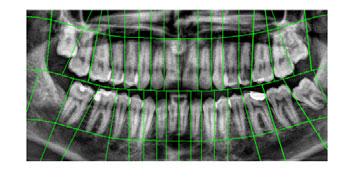
2 Automatic Segmentation of Orthopantomograms 295 extraction (finding the contour of the tooth). In this paper, a novel method for segmentation of panoramic dental X-rays is proposed and presented. Panoramic X-rays, or pantomograms, are a type of extraoral radiograms (i.e. radiograms, where the film is located outside of the patient s mouth) showing the full dentition on a single image. A sample pantomogram (all pantomograms in this paper are presented courtesy of Pomeranian University of Medicine in Szczecin) can be seen in Fig. 1. To our best knowledge, few segmentation algorithms were developed solely for the use with pantomograms (see Section 2). Panoramic images contain the largest amount of information about dentition, but due to the rendering 3-dimensional, semi-circular geometry of the jaw onto a 2-dimensional image, the teeth presented on pantomograms have a tendency of occluding with each other. Fig. 1. A sample pantomogram after image enhancement and cropping 2 Existing Segmentation Methods As has been mentioned in the previous section, there are several existing methods for dental radiogram segmentation. Most of these methods have been created with intraoral images in mind. The first method was presented by Jain and Chen in [6]. It is focused on the application of the integral projections of pixels for the detection of gaps between teeth. The algorithm is separated into two parts: the first is the detection of the gap between lower and upper jaw, and the second is the detection of gaps separating individual teeth. The former step needs user input, so it can be considered semi-automatic. After the initial point of the gap has been selected by the user, moving in both directions, the algorithm chooses short horizontal lines with the highest probability of belonging to the gap. The probability is calculated using the equation ([6]): p vi (D i,y i )=p vi (D i )p vi (y i ), (1)
3 296 D. Frejlichowski and R. Wanat where p vi (D i ) is the normalized integral projection of a given horizontal line subtracted from 1 and p vi (y i ) is a Gaussian with expected value equal to the position of the last chosen line (or the user selection in the first iteration). This probability function has its maximum for the horizontal line that is vertically close to the last selected line and that is composed of pixels with low values. After the maxima have been found for the whole image, a spline function is applied to form a smooth line that becomes the separating line between upper and lower jaw. Once the spline has been calculated, for every point on the curve a new integral projection is calculated in the direction perpendicular to its local curvature. These projections obtain low values in areas between teeth, thus the search for gaps can be reduced to searching for valleys in the plot of the integrals. The areas between these three lines (gap between upper/lower jaw, two successive gaps between teeth) and the horizontal borders of the image become the segments used later in the process of feature extraction. An improved version of this method was proposed in [7]. In this case, the images are processed through wavelet kernels before calculating integral projections to further accentuate the gaps between the necks of teeth. Another method, presented in [8], consists of the use of active contour models, also known as snakes. Initially described in [9], snakes are a model of parametrized splines driven towards edges and lines on the image by external forces, i.e. forces derived from the image on which they operate, as well as internal forces, i.e. user imposed control over the elasticity and rigidity of the contour. In [8], the external driving force along the contour of the snake E ext is given as: E ext (x, y) =G σ (x, y) I(x, y), (2) where G σ is a 2-dimensional Gaussian and I(x, y) is the original image. Thus defined external force amounts to the intensities Gaussian-filtered original image, in which case it takes the lowest values in the dark areas of the radiogram, such as the gap between upper and lower jaw or in spaces between teeth. When using properly selected initial approximations of these curves it achieves very good segmentation of the image. 3 Description of the Proposed Method 3.1 Preliminary Steps For the proper work of the proposed method it is assumed that before an image is segmented using the method, it had been enhanced using the algorithm presented in [10]. That method is based on the decomposition of the image into a Laplacian pyramid, separating the radiogram into smaller images containing progressively lower frequencies of the signal present in the original image. Then, a range of simple filters is applied to selected layers of the pyramid, including sharpening filter and contrast enhancement methods, before the image is recomposed again. It is also necessary to locate the gap between the frontal teeth before the segmentation. In this paper, a nose position detection is used and then a vertical line on the same position is considered the center.
4 3.2 Separating the Upper and Lower Jaw Automatic Segmentation of Orthopantomograms 297 The first step is the determination of the line separating the upper and lower jaw. The same method as presented in [6] and briefly described in the previous section is used. In order to automatize the process of segmentation, instead of requiring that the user inputs the initial separating point used by the algorithm, it is selected by choosing the horizontal integral projection around the center of the image with the lowest value, usually between 40% and 60% of the height of the image. Since the teeth on the image create an arc, instead of using the full horizontal line that would pass through teeth further from the incissors, only a small number of pixels (equal to 20% of the width of the image) closest to the previously selected frontal teeth gap is used to calculate the projections. The mentioned paramaters were established experimentally. Afterwards the algorithm proceeds as described in [6]. 3.3 Localization of the Areas between the Necks of Teeth The obtained curve is then used to estimate the position of the neck of every tooth that is the part of the tooth where roots end and the formation of crown and enamel begins. While the crowns of separate teeth tend to occlude with each other and roots are difficult to separate from the underlying bone, the area between the necks of two adjacent teeth is distinguishable enough to be easily found on a pantomogram. Since the necks of teeth are on the same height as dental pulp, which is darker than the surrounding teeth, the simplest method for finding a line going through dental necks is to translate the line separating upper and lower jaw vertically, sum the intensities of pixels the line passes through for every translation and select the ones for which there is a distinctive drop of values, indicating the line passes through darker areas of the pulp. In order to guarantee that neither the original gap between jaws nor a gap between roots of teeth and the edge of the image are selected in lieu of the desired dental pulp line, the vertical translation scope should be chosen to conduct the search for a limited range, automatically discarding translations too close and too far from the line separating jaws. The result of this step are two values, one negative and one positive, that indicate the amount of pixels that the vertical position of every point belonging to the spline separating jaws should be moved for in order to receive the spline that passes through dental pulps of teeth in each jaw. Thenextstepistheselectionofpointsoneachsplinerepresentingagap between the necks of two adjacent teeth. To refine the results of this stage of the algorithm, a new image is created by multiplying the value-inverted original image with local range filtered version of the original image. That image has high values for darker pixels that lie in areas with neighboring points of low and high intensity. For both upper and lower jaw, an array of values is saved containing the intensities of points belonging to the splines passing through their respective dental pulps. Sharp spikes on the plot of these values indicate

5 298 D. Frejlichowski and R. Wanat dark spots surrounded by light regions, indicating a gap between necks of teeth. In order to remove false spikes, the values of the function are smoothed using the Gaussian filter. Then, starting from the previously selected line separating frontal teeth, small subsets of the values in the array are chosen for comparison. To determine the size of these subsets, average widths of teeth on every position were calculated based on twenty sample pantomograms. Both jaws are fairly symmetrical considering the size of teeth on a given position, thus only 8 values need to be calculated for each jaw, one for every tooth from first incissor to last molar. To select the proper spike value indicating the gap between the necks of teeth for a currently searched tooth on the position p c, a Bayesian probability P (x i,p c ) is calculated for each point on the curve (x i ) according to the equation: P (x i,p c )=I(x i )G(x i,p c )D(x i,p c ), (3) where I is the intensity of the range filtered and inverted original image in the point x i and G is a discrete Gaussian function with the expected value equal to the horizontal position of the last detected gap displaced left or right (depending on the current search direction) by the amount of pixels equal to the average widthofatoothonpositionp c. Thus, if the last detected gap lied on position 100 and the average width of the currently searched tooth is 50 then the expected value should lie on position 50, if the search direction is left, or 150, if the search direction is right. D is a function introduced to narrow the amount of pixels considered during every iteration of the algorithm, equal to 1 for points in the horizontal distances from the previous detected gap between 75% and 175% of the expected width of a tooth on position p c, with the regard to the current search direction, and equal to 0 elsewhere. During every iteration the current argument maximum of the equation 3 is added to the list of gap positions and becomes the starting point of the next iteration of the algorithm. If the amount Fig. 2. Located gaps between necks of teeth for the upper jaw of the sample pantomogram (top) and the corresponding values of the pixels on the range filtered and inverted original image through which the dental pulp spline passes (bottom)

6 Automatic Segmentation of Orthopantomograms 299 of gaps in a given search direction for either upper or lower jaw equals 8 or the vertical edge of the image was reached, the algorithm stops. The result of this step of algorithm for the sample pantomogram is presented in Fig. 2. Thus calculated gap locations provide a good estimation of the position of areas between teeth. However, in some cases a simple vertical line is not sufficient for the separation of two adjacent teeth. Molars and, in some cases, premolars require an additional step of the algorithm to determine the angle of the segmenting line. In order to find a straight line seperating these teeth, an additional point needs to be found between them. A greedy algorithm was used, iteratively moving 1 pixel towards the top or bottom of the radiogram, choosing the pixel in horizontal vicinity with the highest intensity on the inverted and range-filtered image and using it as the basis for the next iteration. After the number of iterations equal to half of the length of an average tooth on a pantomogram, the position of the last result becomes the second separating point. The line passing through the first and second separating point becomes the segmentation line. 3.4 Removing the Areas Below the Roots of Teeth The last step is to remove the areas below the roots of teeth. It is similar to the detection of dental pulp line, i.e. the curve separating both jaws is translated vertically in search of an alignment where the sum of pixels it passes through is lower than the surrounding results, indicating that the area between the teeth line and the cheekbone line was achieved. The only difference between this part of the algorithm and the search for the line of necks of teeth is the range of translations considered during the search. After finding separating lines, every segment of the image lying between the jaws gap line, two consecutive lines separating adjacent teeth and the line below the dental roots is considered an area possibly containing a tooth and is later used in the process of feature extraction. 4 Results and Discussion The described method was tested on a database containing 218 orthopantomograms and some exemplary test results are presented in this section. Firstly, (a) (b) Fig. 3. A comparison of the results abtained for the method proposed in the paper (3(a)) and the integral projections method described in [6] (3(b))


7 300 D. Frejlichowski and R. Wanat (a) (b) (c) (d) (e) (f) (g) (h) (i) Fig. 4. Exemplary test results obtained using the presented method
8 Automatic Segmentation of Orthopantomograms 301 Fig. 3 presents a comparison between the results of the proposed method and the most popular approach applied so far the integral projections ([6]). The results are comparable, with a slightly better result achieved for the proposed method smaller number of lines passing through dental roots and the removal of the area below the roots of teeth make the segments more compact and improve the possibility of a correct shape recognition in the next step of the system. It should also be noted that during the tests the calculation of the function seen in the bottom of Fig. 2, used to detect spikes in the values indicating the gaps between teeth, took less than 1 second, whereas the calculation of the analogous function for the integral projections, i.e. the p vi (D i ) in equation 1 took around 212 seconds. After these calculations, both methods use the same algorithm to detect the spikes in the resulting plot, as described in equation 3. Further results were presented in Fig. 4. It can be seen that the segmentation algorithm provides satisfactory results. If the teeth can be separated using a single line, the algorithm is usually able to find the optimal line of separation. The described method is also able, in some cases, to separate unerupted teeth, as can be seen in Fig. 4(c). The obvious bad results can be found in cases of occlusions so severe that it is impossible to find a straight line to separate both teeth, such as in the case of the incissor and the first premolar (respectively third and fourth tooth from the center of the image) of Fig. 4(a) and Fig. 4(e). Malaligned teeth can also be separated to a degree, but in the case of severe problems in alignment the separating line can not capture the whole tooth within a segment, for example the furthermost bottom left molar in Fig. 3(a). Detection of the ends of dental roots helps in removing bright areas of the underlying bone that would be otherwise attributed to the tooth, but in some cases a fragment of the tooth is removed as well, e.g. in Fig. 4(e). The last problem stems from the fact that dental pulp can easily be mistaken for the edge of the tooth, resulting in a mis-segmentation. This happened in the case of the lower left canine in Fig. 4(b), where the separating line passes through the middle of the tooth, but because average teeth widths are used, the final 8th tooth is separated correctly. 5 Summary and Conclusions In this paper a novel method for segmenting dental panoramic radiograms into regions containing single teeth was described. It uses a different approach than the existing algorithms developed for intraoral images, focusing on detecting gaps between necks of teeth and roots of teeth, that are both easy to find on a pantomogram and allow to separate teeth even in the presence of occlusions. The method provides satisfactory results, however some changes can be introduced in the future. A considerable improvement of its results could be achieved if a more sophisticated method was used instead of the greedy algorithm to determine the second point through which the separating line between two teeth is traced. The method could also be used as an initial step for further segmentation using for example active contours, what can improve the obtained results.
9 302 D. Frejlichowski and R. Wanat References 1. Bowers, M.C.: Forensic Dental Evidence. Elsevier, Amsterdam (2004) 2. Lee, S., et al.: The Diversity of Dental Patterns in Orthopantomography and its Significance in Human Identification. Journal of Forensic Science 49(4), (2004) 3. Nassar, D., Ammar, H.H.: A Prototype Automated Dental Identification System (ADIS). In: Proceedings of the 2003 Annual National Conference on Digital Government Research, pp. 1 4 (2003) 4. Abdel-Mottaleb, M., et al.: Challenges of Developing an Automated Dental Identification System. In: IEEE Mid-West Symposium for Circuits and Systems, Cairo, Egypt, pp (2003) 5. Fahmy, G., Nassar, D.E., Haj-Said, E., Chen, H., Nomir, O., Zhou, J., Howell, R., Ammar, H.H., Abdel-Mottaleb, M., Jain, A.K.: Towards an Automated Dental Identification System (ADIS). In: Zhang, D., Jain, A.K. (eds.) ICBA LNCS, vol. 3072, pp Springer, Heidelberg (2004) 6. Jain, A.K., Chen, H.: Matching of Dental X-ray Images for Human Identification. Pattern Recognition 37(7), (2004) 7. Said, A., et al.: Dental X-ray Image Segmentation. In: SPIE Technologies for Homeland Security and Law Enforcement Conference (2001) 8. Zhou, J., Abdel-Mottaleb, M.: A Content-based System for Human Identification Based on Bitewing Dental X-ray Images. Pattern Recognition 38(11), (2005) 9. Kass, M., Witkin, A., Terzopoulos, D.: Snakes: Active Contour Models. International Journal of Computer Vision 1(4), (1988) 10. Frejlichowski, D., Wanat, R.: Application of the Laplacian Pyramid Decomposition to the Enhancement of Digital Dental Radiographic Images for the Automatic Person Identification. In: Campilho, A., Kamel, M. (eds.) ICIAR 2010, Part II. LNCS, vol. 6112, pp Springer, Heidelberg (2010)
AUTOMATIC TOOTH SEGMENTATION USING ACTIVE CONTOUR WITHOUT EDGES. Samir Shah, Ayman Abaza, Arun Ross and Hany Ammar
 AUTOMATIC TOOTH SEGMENTATION USING ACTIVE CONTOUR WITHOUT EDGES Samir Shah, Ayman Abaza, Arun Ross and Hany Ammar West Virginia University, Morgantown, WV 26506 USA {sshah, ayabaza, ross, ammar}@csee.wvu.edu
Segmentation of Periapical Dental X-Ray Images by applying Morphological Operations
 Segmentation of Periapical Dental X-Ray Images by applying Morphological Operations [1] Anuj kumar, [2] H.S.Bhadauria, [3] Nitin Kumar [1] Research scholar, [2][3] Assistant Professor, [1][2][3] Department
Preprocessing, Segmentation and Matching of Dental Radiographs used in Dental Biometrics
 ISSN No. 2278-3083 Volume 1, No.2, May June 2012 International Journal of Science and Applied Information Technology Available Online at www.warse.org/ijsait/info.html Shubhangi C. Dighe et al., International
Towards an Automated Dental Identification System (ADIS) Abstract Introduction Background
 Abstract Introduction Background") Towards an Automated Dental Identification System (ADIS) Gamal Fahmy*, Diaa Nassar*, Eyad Haj-Said*, Hong Chen**, Omaima Nomir***, Jindan Zhou***, Robert Howell*, Hany H. Ammar*, Mohamed Abdel-Mottaleb***
Towards an Automated Dental Identification System (ADIS) Gamal Fahmy*, Diaa Nassar*, Eyad Haj-Said*, Hong Chen**, Omaima Nomir***, Jindan Zhou***, Robert Howell*, Hany H. Ammar*, Mohamed Abdel-Mottaleb***
Automated Dental Identification with Lowest Cost Path- Based Teeth and Jaw Separation
 Automated Dental Identification with Lowest Cost Path- Based Teeth and Jaw Separation Jan-Vidar Ølberg, Morten Goodwin* University of Agder, Kristiansand, Norway *E-mail: morten.goodwin@uia.no, Abstract:
Dental Image Matching By Canny Algorithm for Human Identification
 International Journal of Advanced Computer Research (ISSN (Print): 22-7277 ISSN (Online): 2277-770) Volume- Number- Issue-17 December-201 Dental Image Matching By Canny Algorithm for Human Identification
Abstract. 1. Introduction. 1 Dental X-ray Image Segmentation
 1 Dental X-ray Image Segmentation EyadHaj Said, Gamal Fahmy, Diaa Nassar, and Hany Ammar Lane Department of Computer Science and Electrical Engineering West Virginia University, Morgantown, WV 26506 Contact
Automated Dental Identification System: An Aid to Forensic Odontology
 10.5005/jp-journals-10011-1169 Parvathi Devi et al REVIEW ARTICLE Automated Dental Identification System: An Aid to Forensic Odontology 1 Parvathi Devi, 2 Thimmarasa VB, 3 Vishal Mehrotra, 4 Vikas Singla
Dental Data Checklist. UNIDENTIFIED PERSON FILE Data Collection Entry Guide. City, State, and ZIP. Street Address. FAX Number.
 Investigating Agency Agency Case Number Street Address City, State and Zip Telephone Number FAX Number Medical Examiner/Coroner Medical Examiner/Coroner Case Number Street Address City, State, and ZIP
Dental X-Ray Based Human Identification System for Forensic
 Engineering and Technology Journal Vol. 35, Part A, No. 1, 2017 S. Dh. Khudhur Computer Engineering Department University of Technology Baghdad, Iraq saja_alzubaidy@yahoo.com M.S. Croock Computer Engineering
Gabor Wavelet Approach for Automatic Brain Tumor Detection
 Gabor Wavelet Approach for Automatic Brain Tumor Detection Akshay M. Malviya 1, Prof. Atul S. Joshi 2 1 M.E. Student, 2 Associate Professor, Department of Electronics and Tele-communication, Sipna college
Computerized Approach for Dental Identification Using Radiographs
 International Journal of Scientific and Research Publications, Volume 3, Issue 5, May 2013 1 Computerized Approach for Dental Identification Using Radiographs Meenal Maurya 1, Sneha Narvekar 2, Sachin
Full Mouth Survey. Delta Dental of Massachusetts DeltaDentalMA.com
 X-Rays Having X-rays taken is a painless procedure that uses small amounts of radiation to capture images of your teeth and bones. Because your dentist takes precautions and the amount of radiation used
European Veterinary Dental College
 European Veterinary Dental College EVDC Training Support Document Preparation of Radiograph Sets (Cat and Dog) Document version : evdc-tsd-radiograph_positioning_(dog_and_cat)-20120121.docx page 1 of 13
A Multi-Stage Approach for 3D Teeth Segmentation from Dentition Surfaces
 A Multi-Stage Approach for 3D Teeth Segmentation from Dentition Surfaces Marcin Grzegorzek 1, Marina Trierscheid 0,1,2, Dimitri Papoutsis 2, and Dietrich Paulus 1 1 Research Group for Active Vision, University
A System for Simulating the Orthodontic Treatment Plan
 A System for Simulating the Orthodontic Treatment Plan Lamia N. Omran*, Ahmed M. El-Bialy**, Ahmed H. Kandil*** & Sahar Ali Fawzy**** *Assistant Lecturer, System and Biomedical Engineering Department,
PH-04A: Clinical Photography Production Checklist With A Small Camera
 PH-04A: Clinical Photography Production Checklist With A Small Camera Operator Name Total 0-49, Passing 39 Your Score Patient Name Date of Series Instructions: Evaluate your Series of photographs first.
Microcalcifications Segmentation using Three Edge Detection Techniques on Mammogram Images
 Microcalcifications Segmentation using Three Edge Detection Techniques on Mammogram Images Siti Salmah Yasiran, Abdul Kadir Jumaat, Aminah Abdul Malek, Fatin Hanani Hashim, Nordhaniah Nasrir, Syarifah
Arrangement of the artificial teeth:
 Lecture Prosthodontic Dr. Osama Arrangement of the artificial teeth: It s the placement of the teeth on a denture with definite objective in mind or it s the setting of teeth on temporary bases. Rules
Texas A&M College of Dentistry Caruth School of Dental Hygiene
 Texas A&M College of Dentistry Caruth School of Dental Hygiene Course Number and Name: 3120 Dental Anatomy Course Type: Lecture Laboratory Seminar Academic Year/Semester Offered: 2016/Fall Semester Course
Segmentation of Color Fundus Images of the Human Retina: Detection of the Optic Disc and the Vascular Tree Using Morphological Techniques
 Segmentation of Color Fundus Images of the Human Retina: Detection of the Optic Disc and the Vascular Tree Using Morphological Techniques Thomas Walter and Jean-Claude Klein Centre de Morphologie Mathématique,
A Review on Dental Image Processing for Human Recognizetion
 A Review on Dental Image Processing for Human Recognizetion Payal P. Tankaria 1, Dharmesh B. Prajapati 2 1 E.C. Dept, SAL institute of Tech. & Engg. Research, Ahmedabad,India, payal_tankaria@yahoo.com
Periodontal Disease. Radiology of Periodontal Disease. Periodontal Disease. The Role of Radiology in Assessment of Periodontal Disease
 Radiology of Periodontal Disease Steven R. Singer, DDS srs2@columbia.edu 212.305.5674 Periodontal Disease! Includes several disorders of the periodontium! Gingivitis! Marginal Periodontitis! Localized
Mammogram Analysis: Tumor Classification
 Mammogram Analysis: Tumor Classification Term Project Report Geethapriya Raghavan geeragh@mail.utexas.edu EE 381K - Multidimensional Digital Signal Processing Spring 2005 Abstract Breast cancer is the
Automated Assessment of Diabetic Retinal Image Quality Based on Blood Vessel Detection
 Y.-H. Wen, A. Bainbridge-Smith, A. B. Morris, Automated Assessment of Diabetic Retinal Image Quality Based on Blood Vessel Detection, Proceedings of Image and Vision Computing New Zealand 2007, pp. 132
Unsupervised Caries Detection in Non-Standardized Bitewing Dental X-Rays
 UNIVERSITY OF KWAZULU-NATAL College of Agriculture, Engineering and Science Unsupervised Caries Detection in Non-Standardized Bitewing Dental X-Rays by Darren John Osterloh Supervisor: Prof. Serestina
Forensic Archaeology & Forensic Anthropology. ADJ14 Advanced Criminal Investigations
 Forensic Archaeology & Forensic Anthropology ADJ14 Advanced Criminal Investigations Anthropology & Archaeology Anthropology is the study of the biological and cultural aspects of all humans in all places
Reading Assignments: Lecture 18: Visual Pre-Processing. Chapters TMB Brain Theory and Artificial Intelligence
 Brain Theory and Artificial Intelligence Lecture 18: Visual Pre-Processing. Reading Assignments: Chapters TMB2 3.3. 1 Low-Level Processing Remember: Vision as a change in representation. At the low-level,
AN ALGORITHM FOR EARLY BREAST CANCER DETECTION IN MAMMOGRAMS
 AN ALGORITHM FOR EARLY BREAST CANCER DETECTION IN MAMMOGRAMS Isaac N. Bankman', William A. Christens-Barryl, Irving N. Weinberg2, Dong W. Kim3, Ralph D. Semmell, and William R. Brody2 The Johns Hopkins
A Comparative Analysis on the Dental X-ray Image Segmentation Approaches
 A Comparative Analysis on the Dental X-ray Image Segmentation Approaches Ritu Bala 1, Divjot Kaur 2 1, 2 Department of Computer Science and Engineering, Rayat Institute of Engineering & Information Technology,
Intraoral Imaging. Chapter 41. Copyright 2018, Elsevier Inc. All Rights Reserved. 1
 Intraoral Imaging Chapter 41 Copyright 2018, Elsevier Inc. All Rights Reserved. 1 Learning Objectives Lesson 41.1: Projection Types and the Paralleling Technique 1. Pronounce, define, and spell the key
Evaluation of proximity of the floor of the maxillary. Zanco J. Med. Sci., Vol. 16, No. (2), 2012
, 2012") Evaluation of proximity of the floor of the maxillary sinus to the alveolar bone crest, using digital panoramic imaging system, in Erbil city Received: 10/2/2011 Accepted: 13/9/2011 Sarkawt H. Ali* Saeed
Evaluation of proximity of the floor of the maxillary sinus to the alveolar bone crest, using digital panoramic imaging system, in Erbil city Received: 10/2/2011 Accepted: 13/9/2011 Sarkawt H. Ali* Saeed
A New Method to Secondary Caries Detection in Restored Teeth
 International Journal of Scientific & Engineering Research Volume 2, Issue 11, November-2011 1 A New Method to Secondary Caries Detection in Restored Teeth A.Sadeghi Qaramaleki, H.Hassanpour Abstract Dental
Automatic cardiac contour propagation in short axis cardiac MR images
 International Congress Series 1281 (2005) 351 356 www.ics-elsevier.com Automatic cardiac contour propagation in short axis cardiac MR images G.L.T.F. Hautvast a,b, T, M. Breeuwer a, S. Lobregt a, A. Vilanova
Mammogram Analysis: Tumor Classification
 Mammogram Analysis: Tumor Classification Literature Survey Report Geethapriya Raghavan geeragh@mail.utexas.edu EE 381K - Multidimensional Digital Signal Processing Spring 2005 Abstract Breast cancer is
Automated analysis of whole skeletal muscle for muscular atrophy detection of ALS in whole-body CT images: Preliminary study
 Automated analysis of whole skeletal muscle for muscular atrophy detection of ALS in whole-body CT images: Preliminary study Naoki Kamiya *a, Kosuke Ieda b, Xiangrong Zhou b, Megumi Yamada c, Hiroki Kato
NAILFOLD CAPILLAROSCOPY USING USB DIGITAL MICROSCOPE IN THE ASSESSMENT OF MICROCIRCULATION IN DIABETES MELLITUS
 NAILFOLD CAPILLAROSCOPY USING USB DIGITAL MICROSCOPE IN THE ASSESSMENT OF MICROCIRCULATION IN DIABETES MELLITUS PROJECT REFERENCE NO. : 37S0841 COLLEGE BRANCH GUIDE : DR.AMBEDKAR INSTITUTE OF TECHNOLOGY,
#45 Ortho-Tain, Inc PREVENTIVE ERUPTION GUIDANCE -- PREVENTIVE OCCLUSAL DEVELOPMENT
 #45 Ortho-Tain, Inc. 1-800-541-6612 PREVENTIVE ERUPTION GUIDANCE -- PREVENTIVE OCCLUSAL DEVELOPMENT Analysis and Diagnosis of Occlusion: The ideal child of 5 y ears of age that probably has the best chance
Particle Swarm Optimization based K-mediods Clustering on Dental X-ray Images
 Volume 8, No. 4, May 2017 (Special Issue) International Journal of Advanced Research in Computer Science REVIEW ARTICLE Available Online at www.ijarcs.info Particle Swarm Optimization based K-mediods Clustering
Tooth/Teeth Segmentation and modeling from X-ray/CT images: A Survey
 I J C T A, 10(8), 2017, pp. 423-428 International Science Press ISSN: 0974-5572 Tooth/Teeth Segmentation and modeling from X-ray/CT images: A Survey Niharika Thakur 1, Mamta Juneja 2 and Prashant Jindal
Edge Detection Techniques Using Fuzzy Logic
 Edge Detection Techniques Using Fuzzy Logic Essa Anas Digital Signal & Image Processing University Of Central Lancashire UCLAN Lancashire, UK eanas@uclan.a.uk Abstract This article reviews and discusses
The M Ruler (Figure 6) Figure 6 M Ruler (Figure 7)
 Figure 6 M Ruler (Figure 7)") The M Ruler Dr. Alain Méthot, D.M.D. M.Sc. It has been shown that the Golden Rule cannot be universally applied to all patients; it therefore became necessary to find a formula adaptable for each patient.
The M Ruler Dr. Alain Méthot, D.M.D. M.Sc. It has been shown that the Golden Rule cannot be universally applied to all patients; it therefore became necessary to find a formula adaptable for each patient.
The wonderful world of dental radiology
 The wonderful world of dental radiology Dr Christine Hawke BSc(Vet)(Hons) BVSc(Hons) PhD MACVSc (Veterinary Dentistry) PO Box 3001 Willoughby North 2068 Phone: 0408 782 611 Email: christine@sydneypetdentistry.com.au
EDGE DETECTION OF THE SCOLIOTIC VERTEBRAE USING X-RAY IMAGES
 Journal of Engineering Science and Technology 4 th EURECA 2015 Special Issue February (2016) 166-175 School of Engineering, Taylor s University EDGE DETECTION OF THE SCOLIOTIC VERTEBRAE USING X-RAY IMAGES
Computing Neck-Shaft Angle of Femur for X-Ray Fracture Detection
 Computing Neck-Shaft Angle of Femur for X-Ray Fracture Detection Tai Peng Tian 1, Ying Chen 1, Wee Kheng Leow 1, Wynne Hsu 1, Tet Sen Howe 2, and Meng Ai Png 3 1 Dept. of Computer Science, National University
Implementation of Automatic Retina Exudates Segmentation Algorithm for Early Detection with Low Computational Time
 www.ijecs.in International Journal Of Engineering And Computer Science ISSN: 2319-7242 Volume 5 Issue 10 Oct. 2016, Page No. 18584-18588 Implementation of Automatic Retina Exudates Segmentation Algorithm
Towards an Automatic Classification of Spinal Curves from X-Ray Images
 Towards an Automatic Classification of Spinal Curves from X-Ray Images Luc DUONG a,b,1, Farida CHERIET a,b and Hubert LABELLE a a Research Center, Sainte-Justine Hospital, 3175 Côte-Sainte-Catherine, Montreal,
Research Article. University, Mangalore Corresponding author Dr. Junaid Ahmed
 Scholars Journal of Applied Medical Sciences (SJAMS) ISSN 2320-6691 Sch. J. App. Med. Sci., 2013; 1(5):427-431 Scholars Academic and Scientific Publisher (An International Publisher for Academic and Scientific
SAPOG Edge Detection Technique GUI using MATLAB
 SAPOG Edge Detection Technique GUI using MATLAB Poonam Kumari 1, Sanjeev Kumar Gupta 2 Software Engineer, Devansh Softech Consultancy Services Pvt. Ltd., Agra, India 1 Director, Devansh Softech Consultancy
Conclusions and future directions
 Chapter 9 Conclusions and future directions This chapter summarises the findings within this thesis, examines its main contributions, and provides suggestions for further directions. 9.1 Thesis summary
Extraoral Imaging. Chapter 42. Copyright 2018, Elsevier Inc. All Rights Reserved. 1
 Extraoral Imaging Chapter 42 Copyright 2018, Elsevier Inc. All Rights Reserved. 1 Learning Objectives Lesson 42.1: Panoramic Imaging 1. Pronounce, define, and spell the key terms. 2. Discuss panoramic
Cancer Cells Detection using OTSU Threshold Algorithm
 Cancer Cells Detection using OTSU Threshold Algorithm Nalluri Sunny 1 Velagapudi Ramakrishna Siddhartha Engineering College Mithinti Srikanth 2 Velagapudi Ramakrishna Siddhartha Engineering College Kodali
AD2 MEASURES CONDYLE DISPLACEMENT (MCD) MANUAL
 MANUAL") AD2 MEASURES CONDYLE DISPLACEMENT (MCD) MANUAL Dr. Jorge Ayala Puente, DDS* Dr. Gonzalo Gutiérrez Álvarez, DDS* Dr. José Miguel Obach M., DDS Translation: Dr. Barbara Fernández Lübbert, DDS Edited: Dr.
AD2 MEASURES CONDYLE DISPLACEMENT (MCD) MANUAL Dr. Jorge Ayala Puente, DDS* Dr. Gonzalo Gutiérrez Álvarez, DDS* Dr. José Miguel Obach M., DDS Translation: Dr. Barbara Fernández Lübbert, DDS Edited: Dr.
Validating the Visual Saliency Model
 Validating the Visual Saliency Model Ali Alsam and Puneet Sharma Department of Informatics & e-learning (AITeL), Sør-Trøndelag University College (HiST), Trondheim, Norway er.puneetsharma@gmail.com Abstract.
VistaCam ix HD for diagnostics support in HD quality
 VistaCam ix HD for diagnostics support in HD quality The innovative intraoral camera with interchangeable head system COMPRESSED AIR SUCTION IMAGING DENTAL CARE HYGIENE Vista Cam ix HD real HD resolution
Fundamental & Preventive Curvatures of Teeth and Tooth Development. Lecture Three Chapter 15 Continued; Chapter 6 (parts) Dr. Margaret L.
 Dr. Margaret L.") Fundamental & Preventive Curvatures of Teeth and Tooth Development Lecture Three Chapter 15 Continued; Chapter 6 (parts) Dr. Margaret L. Dennis Proximal contact areas Contact areas are on the mesial and
Fundamental & Preventive Curvatures of Teeth and Tooth Development Lecture Three Chapter 15 Continued; Chapter 6 (parts) Dr. Margaret L. Dennis Proximal contact areas Contact areas are on the mesial and
Feasibility Evaluation of a Novel Ultrasonic Method for Prosthetic Control ECE-492/3 Senior Design Project Fall 2011
 Feasibility Evaluation of a Novel Ultrasonic Method for Prosthetic Control ECE-492/3 Senior Design Project Fall 2011 Electrical and Computer Engineering Department Volgenau School of Engineering George
Impacted teeth including surgery for canine teeth
 Impacted teeth including surgery for canine teeth What are impacted teeth? When one or more teeth fails to grow in the correct position and is therefore held below the normal gum line, it is called an
I. INTRODUCTION III. OVERALL DESIGN
 Inherent Selection Of Tuberculosis Using Graph Cut Segmentation V.N.Ilakkiya 1, Dr.P.Raviraj 2 1 PG Scholar, Department of computer science, Kalaignar Karunanidhi Institute of Technology, Coimbatore, Tamil
Development of novel algorithm by combining Wavelet based Enhanced Canny edge Detection and Adaptive Filtering Method for Human Emotion Recognition
 International Journal of Engineering Research and Development e-issn: 2278-067X, p-issn: 2278-800X, www.ijerd.com Volume 12, Issue 9 (September 2016), PP.67-72 Development of novel algorithm by combining
Grading of Vertebral Rotation
 Chapter 5 Grading of Vertebral Rotation The measurement of vertebral rotation has become increasingly prominent in the study of scoliosis. Apical vertebral deformity demonstrates significance in both preoperative
ADHESIVE RECONSTRUCTION IN HELP OF THE ORTHODONTIC TREATMENT
 ISSN: 1312-773X (Online) Journal of IMAB - Annual Proceeding (Scientific Papers) 2006, vol. 12, issue 2 ADHESIVE RECONSTRUCTION IN HELP OF THE ORTHODONTIC TREATMENT Snezhanka Topalova-Pirinska 1, R. Pirinska
Diagnostics and treatment planning. Dr. Attila Szűcs DDS
 Diagnostics and treatment planning. Dr. Attila Szűcs DDS Considering both surgical Aim and prosthetic aspects in the planning of implant prosthetics Arrangements for implant therapy Preliminary examinations
arxiv: v1 [cs.cv] 9 Feb 2018
![arxiv: v1 [cs.cv] 9 Feb 2018](/thumbs/87/95688976.jpg "arxiv: v1 [cs.cv] 9 Feb 2018") Automatic segmenting teeth in X-ray images: Trends, a novel data set, benchmarking and future perspectives Gil Silva 1, Luciano Oliveira 2 Ivision Lab, Federal University of Bahia, Brazil arxiv:1802.03086v1
Automatic segmenting teeth in X-ray images: Trends, a novel data set, benchmarking and future perspectives Gil Silva 1, Luciano Oliveira 2 Ivision Lab, Federal University of Bahia, Brazil arxiv:1802.03086v1
MBT System as the 3rd Generation Programmed and Preadjusted Appliance System (PPAS) by Masatada Koga, D.D.S., Ph.D
 by Masatada Koga, D.D.S., Ph.D") MBT System as the 3rd Generation Programmed and Preadjusted Appliance System (PPAS) by Masatada Koga, D.D.S., Ph.D Dr. Masatada Koga, D.D.S., Ph.D, is an assistant professor in the Department of Orthodontics
MBT System as the 3rd Generation Programmed and Preadjusted Appliance System (PPAS) by Masatada Koga, D.D.S., Ph.D Dr. Masatada Koga, D.D.S., Ph.D, is an assistant professor in the Department of Orthodontics
A Pattern Classification Approach to Aorta Calcium Scoring in Radiographs
 A Pattern Classification Approach to Aorta Calcium Scoring in Radiographs Marleen de Bruijne IT University of Copenhagen, Denmark marleen@itu.dk Abstract. A method for automated detection of calcifications
Large Dentigerous Cyst
 Volume 16.2.1 Feb 2016 This Lecture Series qualifies for 0.5 Informal CPD Learning Hours Large Dentigerous Cyst By Dr Hassem Geha A 55 year-old male presented with a painless swelling in the right mandible.
Q: Human v. Animal? When the bite occurs postmortem. Typical bite has a double horseshoe pattern. The area bruises and swells
 Q: Human v. Animal? Bite marks look different in soft and stretchy substances like skin versus hard substances like cheese or a pencil When the bite occurs antemortem 1 The area bruises and swells When
Dental Biometrics: Alignment and Matching of Dental Radiographs
 IEEE TRANSACTIONS ON PATTERN ANALYSIS AND MACHINE INTELLIGENCE, VOL. 27, NO. 8, AUGUST 2005 1319 Dental Biometrics: Alignment and Matching of Dental Radiographs Hong Chen, Student Member, IEEE, and Anil
(51) Int Cl.: A61C 9/00 ( )
 Int Cl.: A61C 9/00 ( )") (19) TEPZZ 7 6448B_T (11) EP 2 736 448 B1 (12) EUROPEAN PATENT SPECIFICATION (4) Date of publication and mention of the grant of the patent: 23.09.1 Bulletin 1/39 (21) Application number: 12747898.0 (22)
(19) TEPZZ 7 6448B_T (11) EP 2 736 448 B1 (12) EUROPEAN PATENT SPECIFICATION (4) Date of publication and mention of the grant of the patent: 23.09.1 Bulletin 1/39 (21) Application number: 12747898.0 (22)
Image Enhancement and Compression using Edge Detection Technique
 Image Enhancement and Compression using Edge Detection Technique Sanjana C.Shekar 1, D.J.Ravi 2 1M.Tech in Signal Processing, Dept. Of ECE, Vidyavardhaka College of Engineering, Mysuru 2Professor, Dept.
3D Morphological Tumor Analysis Based on Magnetic Resonance Images
 3D Morphological Tumor Analysis Based on Magnetic Resonance Images Sirwoo Kim Georgia Institute of Technology The Wallace H. Coulter Department of Biomedical Engineering, Georgia. Abstract In this paper,
Katsunari Shibata and Tomohiko Kawano
 Learning of Action Generation from Raw Camera Images in a Real-World-Like Environment by Simple Coupling of Reinforcement Learning and a Neural Network Katsunari Shibata and Tomohiko Kawano Oita University,
The width of the MCXL step bur is 1.4 mm wide and has a blunt end. As the bur approaches the inside of
 As I teach first year dental students how to prep a tooth for a full gold crown, get an impression, pour and mount models, wax-up, cast and polish, they are almost always amazed at all the required steps
Periapical Radiography
 Periapical Radiography BARBARA E. DIXON B.D.S., M.Sc., D.P.D.S. Main Indications Detection of Apical infection/inflammation Assessment of the periodontal status After trauma Assessment of Unerupted teeth
Neural Networks-Based Tool for Diagnosis of Paranasal Sinuses Conditions
 SIP-9 856 CSNDSP 2010 Neural Networks-Based Tool for Diagnosis of Paranasal Sinuses Conditions Abdel Razzak Natsheh, Prasad VS Ponnapalli, Nader Anani, Dalil Benchebra and Atef El-kholy* Manchester Metropolitan
EARLY STAGE DIAGNOSIS OF LUNG CANCER USING CT-SCAN IMAGES BASED ON CELLULAR LEARNING AUTOMATE
 EARLY STAGE DIAGNOSIS OF LUNG CANCER USING CT-SCAN IMAGES BASED ON CELLULAR LEARNING AUTOMATE SAKTHI NEELA.P.K Department of M.E (Medical electronics) Sengunthar College of engineering Namakkal, Tamilnadu,
 1 of 9 7/11/2012 11:29 AM For many years, producers, veterinarians, and exhibitors (at cattle shows) have used cattle dentition to make general age determinations. Dentition will vary from herd-to-herd
Keywords Fuzzy Logic, Fuzzy Rule, Fuzzy Membership Function, Fuzzy Inference System, Edge Detection, Regression Analysis.
 Volume 6, Issue 3, March 2016 ISSN: 2277 128X International Journal of Advanced Research in Computer Science and Software Engineering Research Paper Available online at: www.ijarcsse.com Modified Fuzzy
Detection Of Red Lesion In Diabetic Retinopathy Using Adaptive Thresholding Method
 Detection Of Red Lesion In Diabetic Retinopathy Using Adaptive Thresholding Method Deepashree Devaraj, Assistant Professor, Instrumentation Department RVCE Bangalore. Nagaveena M.Tech Student, BMSP&I,
Development of an Image-Based Modelling Tool for the Spinal Deformity Detection Using Curve Fitting Technique
 Development of an Image-Based Modelling Tool for the Spinal Deformity Detection Using Curve Fitting Technique [1] Tabitha Janumala, [2] Dr.K.B.Ramesh, [3] Harish A Haralachar [1] Research Scholor, R.V.College
AUTOMATIC DIABETIC RETINOPATHY DETECTION USING GABOR FILTER WITH LOCAL ENTROPY THRESHOLDING
 AUTOMATIC DIABETIC RETINOPATHY DETECTION USING GABOR FILTER WITH LOCAL ENTROPY THRESHOLDING MAHABOOB.SHAIK, Research scholar, Dept of ECE, JJT University, Jhunjhunu, Rajasthan, India Abstract: The major
The wonderful world of dental radiology
 The wonderful world of dental radiology Dr Christine Hawke BSc(Vet)(Hons) BVSc(Hons) PhD MACVSc (Veterinary Dentistry) PO Box 3001 Willoughby North 2068 Phone: 0408 782 611 Email: christine@sydneypetdentistry.com.au
Guided immediate loading implant surgery planned with Implant Studio D.D.S. Jae-min, Lee
 Guided immediate loading implant surgery planned with Implant Studio D.D.S. Jae-min, Lee Jung-plant Dental office 1 PROLOGUE How can we deal with the immediate loading implant cases easier and more accurate
Dental Morphology and Vocabulary
 Dental Morphology and Vocabulary Palate Palate Palate 1 2 Hard Palate Rugae Hard Palate Palate Palate Soft Palate Palate Palate Soft Palate 4 Palate Hard Palate Soft Palate Maxillary Arch (Maxilla) (Uppers)
IJBPAS, September, 2015, 4(9), Special Issue: ISSN: AGE ESTIMATION USING DIGITAL PANORAMIC RADIOGRAPHY
, Special Issue: ISSN: AGE ESTIMATION USING DIGITAL PANORAMIC RADIOGRAPHY") : 124-129 ISSN: 2277 4998 AGE ESTIMATION USING DIGITAL PANORAMIC RADIOGRAPHY AREZOU TORKIAN E Mail: Torkian_ar@yahoo.com ABSTRACT One of the interesting applications of forensic deontology is age estimation
: 124-129 ISSN: 2277 4998 AGE ESTIMATION USING DIGITAL PANORAMIC RADIOGRAPHY AREZOU TORKIAN E Mail: Torkian_ar@yahoo.com ABSTRACT One of the interesting applications of forensic deontology is age estimation
EUROPEAN SOCIETY OF LINGUAL ORTHODONTICS
 EUROPEAN SOCIETY OF LINGUAL ORTHODONTICS CANDIDATE NUMBER: Dr. Stefan Blasius Year: 2010 WBLO 01 EUROPEAN SOCIETY OF LINGUAL ORTHODONTICS CANDIDATE NUMBER: Dr. Stefan Blasius Year: 2010 WBLO 01 RÉSUMÉ
Noise-Robust Speech Recognition Technologies in Mobile Environments
 Noise-Robust Speech Recognition echnologies in Mobile Environments Mobile environments are highly influenced by ambient noise, which may cause a significant deterioration of speech recognition performance.
South Asian Journal of Engineering and Technology Vol.3, No.9 (2017) 17 22
 17 22") South Asian Journal of Engineering and Technology Vol.3, No.9 (07) 7 Detection of malignant and benign Tumors by ANN Classification Method K. Gandhimathi, Abirami.K, Nandhini.B Idhaya Engineering College
South Asian Journal of Engineering and Technology Vol.3, No.9 (07) 7 Detection of malignant and benign Tumors by ANN Classification Method K. Gandhimathi, Abirami.K, Nandhini.B Idhaya Engineering College
Investigating the performance of a CAD x scheme for mammography in specific BIRADS categories
 Investigating the performance of a CAD x scheme for mammography in specific BIRADS categories Andreadis I., Nikita K. Department of Electrical and Computer Engineering National Technical University of
Computerized image analysis: Estimation of breast density on mammograms
 Computerized image analysis: Estimation of breast density on mammograms Chuan Zhou, Heang-Ping Chan, a) Nicholas Petrick, Mark A. Helvie, Mitchell M. Goodsitt, Berkman Sahiner, and Lubomir M. Hadjiiski
Performance Assessment of Various Molar Crown Designs
 2012 2nd International Conference on Biomedical Engineering and Technology IPCBEE vol. 34 (2012) (2012) IACSIT Press, Singapore Performance Assessment of Various Molar Crown Designs Rashmi Uddanwadiker
Treatment of a Rare Bilateral Severe Ectopic Eruption of the Maxillary First Permanent Molar: A Case Report
 Case Report Treatment of a Rare Bilateral Severe Ectopic Eruption of the Maxillary First Permanent Molar: A Case Report MS. Ahmad Akhoundi 1, 2, AH. Sadrhaghighi 3 1 Associate Professor, Dental Research
SYLLABUS: CERTIFICATE IN ORTHODONTIC DENTAL NURSING
 SYLLABUS: CERTIFITE IN ORTHODONTIC DENTAL NURSING 1. GENERAL PROFESSIONAL CONTENT 1.1 MAINTAINING GOOD CLINIL PRACTICE Each learning outcome should be prefaced by: On completion of training the orthodontic
A CONVENTIONAL STUDY OF EDGE DETECTION TECHNIQUE IN DIGITAL IMAGE PROCESSING
 Available Online at www.ijcsmc.com International Journal of Computer Science and Mobile Computing A Monthly Journal of Computer Science and Information Technology IJCSMC, Vol. 3, Issue. 4, April 2014,
SURVEYING OF REMOVABLE PARITAL DENTURES FEB, 11, 2015
 SURVEYING OF REMOVABLE PARITAL DENTURES FEB, 11, 2015 Dental Surveyor: It is a mechanical device used to determine the relative parallelism of the teeth surfaces and the undercuts areas in relation to
Intraoral radiographic techniques Introduction There are three main types of intraoral radiographs: Periapical radiograph Bitewing radiograph
 Intraoral radiographic techniques Introduction There are three main types of intraoral radiographs: Periapical radiograph Bitewing radiograph Occlusal radiograph The anatomic area of interest and type
3Shape TRIOS Implant Scanning
 Dr. Anthony Mak W Dental 3Shape TRIOS Implant Scanning Simplifying digital implant prosthetics with implant libraries and 3Shape scan bodies Solutions featured: 3Shape TRIOS 3Shape scan bodies 3Shape Dental
Replacing Missing or Debonded Brackets
 CFAST Help Sheet Replacing Missing or Debonded Brackets Brackets becoming deboned are not uncommon you will almost certainly experience this. The important thing to remember is - DON T PANIC! And tell
Tumor cut segmentation for Blemish Cells Detection in Human Brain Based on Cellular Automata
 Tumor cut segmentation for Blemish Cells Detection in Human Brain Based on Cellular Automata D.Mohanapriya 1 Department of Electronics and Communication Engineering, EBET Group of Institutions, Kangayam,