sagittal split ramus osteotomy
|
|
|
- Sabrina Gardner
- 5 years ago
- Views:
Transcription
1 Title Author(s) Citation Position of mandibular canal and ra sagittal split ramus osteotomy Ueki, Koichiro; Okabe, Katsuhiko; M Marukawa, Kohei; Nakagawa, Kiyomasa Journal of Oral and Maxillofacial S Issue Date 2010 Type Journal Article Text version author URL Right KURA に登録されているコンテンツの著作権は, 執筆者, 出版社 ( 学協会 ) などが有します KURA に登録されているコンテンツの利用については, 著作権法に規定されている私的使用や引用などの範囲内で行ってください 著作権法に規定されている私的使用や引用などの範囲を超える利用を行う場合には, 著作権者の許諾を得てください ただし, 著作権者から著作権等管理事業者 ( 学術著作権協会, 日本著作出版権管理システムなど ) に権利委託されているコンテンツの利用手続については, 各著作権等管理事業者に確認してください
2 Position of mandibular canal and ramus morphology before and after sagittal split ramus osteotomy KOICHIRO UEKI, DDS, PhD,# KATSUHIKO OKABE, DDS, MAO MIYAZAKI, DDS, AYA MUKOZAWA, DDS, KOHEI MARUKAWA, DDS, PhD, KIYOMASA NAKAGAWA, DDS, PhD, and ETSUHIDE YAMAMOTO, DDS, PhD, Department of Oral and Maxillofacial Surgery, Graduate School of Medicine, Kanazawa University, 13-1 Takaramachi, Kanazawa , Japan. # Assistant Professor Graduate Student Clinical Instructor Associate Professor Professor and Head Address correspondence to: Koichiro Ueki, DDS, PhD. Department of Oral and Maxillofacial Surgery, Graduate School of Medicine, Kanazawa University, 13-1 Takaramachi, Kanazawa , Japan. Tel: ; Fax: kueki@med.kanazawa-u.ac.jp
3 Abstract Purpose. The purpose of this study was to evaluate changes in the mandibular canal and ramus morphology before and after a sagittal split ramus osteotomy (SSRO). Subjects and Methods. The subjects were 30 patients (60 sides) with mandibular prognathism who had undergone bilateral SSRO setback surgery. The mandibular canal position and ramus morphology were measured at the three horizontal planes under the mandibular foramen level (level A), 1cm lower (level B) of level A and 2cm lower (level C) of level A preoperatively and 1 year postoperatively by computed tomography (CT). Results. Postoperative ramus width, lateral distance, lateral marrow distance and canal length were significantly larger than the pre-operative values, at the foramen, 1cm lower and 2 cm lower levels. The mandibular canal completely contacted the lateral cortex without lateral bone marrow in 6 sides (10%) in level A and B, and 4 sides (6.7%) in level C pre-operatively and 6 sides (10%) in level C post-operatively. Conclusion. This study suggested that postoperative mandibular canal position was located more posteriorly and the postoperative lateral bone marrow became thicker compared to the pre-operative state. Key words: Sagittal split ramus osteotomy Mandibular canal Ramus morphology Computed tomography
4 Introduction Sagittal split ramus osteotomy (SSRO) is the most common surgical method for correcting jaw deformities. However, osteoperative hypoesthesia after mandibular orthognathic surgery is a known complication, caused by direct or indirect intraoperative damage to the inferior alveolar nerve. 1 The incidence of this postoperative trigeminal nerve hypoesthesia is reported to be highest among patients undergoing sagittal splitting ramus osteotomy (SSRO). 2 The induction of neural impairment is thought to be influenced by multiple causal factors, including fixation methods, 3-5 patient s age, 6, 7 postoperative swelling, 8 9, 10 and surgical procedures, particularly a bad split. With regards to the effect of a fixation method, Lemke at al. 11 reported that rigid fixation resulted in more anesthesia in the mental nerve distribution than wire fixation when tested with brush stroke direction. Fujioka at al. 12 also reported that mono-cortical osteosynthesis caused less damage to the inferior alveolar nerve. Some surgeons have suggested that compressive forces can occur when fixing the 2 mandibular segments together, resulting in the nerve being sandwiched. Takeuchi at al. 13 reported that in SSRO setback cases, the distance between the mental foramen and the mandibular ramus always decreased, and that this change may cause trigeminal nerve hypoesthesia by compression of the nerve trunk due to posterior shifting of the proximal segments. There is evidence that wire fixation and mono-cortical fixation is less likely to cause direct trauma and has no risk of compressing the segments. 11 However, the results of our previous study suggested that mono-cortical or bi-cortical fixation methods did not influence the recovery period from hypoesthesia. 14 Although it still unclear what factors affect the incidence of lower lip hypoesthesia after SSRO, it is very important to know the relationship between the mandibular bone and inferior alveolar canal to avoid direct damage to the inferior alveolar nerve preoperatively. The post-operative change in the location of the inferior mandibular canal and bone healing and regeneration is also important. There are some studies regarding the location of the mandibular canal in mandibular prognathism before SSRO, however no report has described any postoperative change in the relationship between the mandibular canal and
5 ramus morphology. The purpose of this study was to evaluate the change in the mandibular canal and ramus morphology before and after sagittal split ramus osteotomy (SSRO). Patients and Methods Patients The 30 Japanese adults (men: 4, women: 26) in this study presented with jaw deformities diagnosed as mandibular prognathism. At the time of orthognathic surgery, the patients ranged in age from 16 to 42 years, with a mean age of 25.8 years (standard deviation, 7.6 years). Informed consent was obtained from the patients and the study was approved by Kanazawa University Hospital. 26 of 30 patients were diagnosed as mandibular prognathism and the remaining 4 patients were diagnosed as mandibular prognathism with maxillary retrognathia. Surgery Of the 30 patients in this study, 26 underwent bilateral SSRO. The other 4 patients underwent SSRO and a Le Fort I osteotomy; rigid fixation was achieved with min-plates and monocortical screws. The plates were bent to prevent the proximal segments from rotating internally. Therefore the gap was created between the osteotomy surfaces on both sides (Fig.1). 15 Elastic was placed to maintain the ideal occlusion without inter-maxillary fixation. The patients did not receive any physical therapy after surgery. All patients received orthodontic treatment before and after surgery. CT measurement CT was taken for all patients preoperatively and one year after surgery. The patients were placed in the gantry with the tragacanthal line perpendicular to the ground for CT scanning.
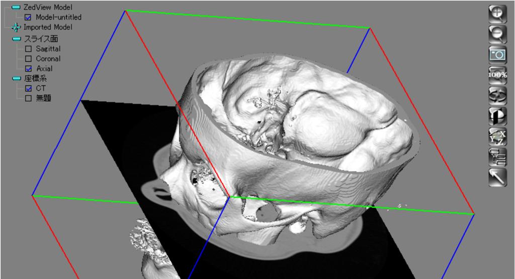
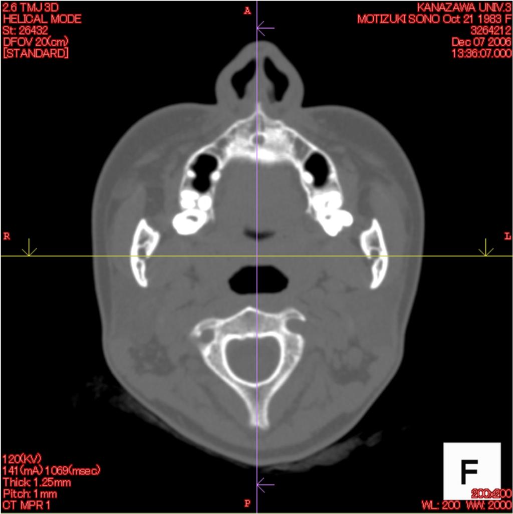
6 They were instructed to breathe normally and to avoid swallowing during the scanning process. CT scans were obtained in the radiology department by skilled radiology technicians using a high-speed, advantage-type CT generator (Light Speed Plus; GE Healthcare, Milwaukee, WI, USA) with each sequence taken 1.25 mm apart for 3D reconstruction (120 kv, average 150 ma, 0.7 sec/rotation, helical pitch 0.75). The resulting images were stored in the attached workstation computer (Advantage workstation version 4.2; GE Healthcare, Milwaukee, WI, USA) and the 3D reconstruction was performed using the volume rendering method. ExaVision LITE version 1.10 medical imaging software (Ziosoft, Inc, Tokyo, Japan) was used for 3D morphologic measurements. The RL line was determined as the line between the most anterior points of the bilateral auricles. Multi planner reconstruction can be established with the software, so that the arbitrary plane can be moved parallel to the plane that the RL line was determined (Fig. 2). Three horizontal planes at the mandibular foramen level (Level A)(Fig. 3), a 1 cm level under the mandibular foramen (Level B)(Fig. 4) and a 2 cm level under the mandibular foramen (Level C)(Fig. 5) parallel to the FH plane was identified in the right and left sides, and ramus area was measured pre- and postoperatively and bilaterally as follows (Fig. 6). 1) Ramus length: the distance between the most anterior point and most posterior point of ramus. 2) Ramus width: the distance between the most medial point and the cross point between the lateral outline of the ramus and the line through the most medial point parallel to the RL line. 3) Anterior length: the distance between the most anterior point of the ramus and the most anterior point of the mandibular canal. 4) Posterior length: the distance between the most posterior point of the ramus and the most posterior point of the mandibular canal. 5) Medial distance: the distance between the most medial point of the mandibular canal and the medial outline of the ramus on the parallel line to the RL line. 6) Lateral distance: the distance between the most lateral point of the mandibular canal and
7 the lateral outline of the ramus on the parallel to the RL line. 7) Medial marrow distance: the distance between the most medial point on the outer cortex of the mandibular canal and the most lateral point of the medial cortex of the ramus on the parallel line to the RL line. 8) Lateral marrow distance: the distance between the most lateral point on the outer cortex of the mandibular canal and the most medial point of the lateral cortex of the ramus on the parallel line to the RL line. 9) Canal length: the antero-posterior length of the mandibular canal 10) Canal width: the medio-lateral length of the mandibular canal All CT images were measured by an author (K.U.). Fifteen patients were selected calculated using Dahlberg s formula 16 : ME= d 2 /2n where d is the difference between 2 registrations of a pair, and n is the number of double registrations. The random errors did not exceed 0.21 mm for the linear measurements. Statistical analysis Data were statistically analyzed with StatView software, version 4.5 (ABACUS Concepts, Inc., Berkeley, CA, USA) and Dr. SPSSII (SPSS Japan Inc., Tokyo, Japan). The statistical significance of a difference between pre- and postoperative values was analyzed by paired t-test. The statistical significance of differences among three levels was analyzed by Bonferroni/Dunn (Dunn s procedure as a multiple comparison procedure) test. Results No patient had post-surgical wound infection or dehiscence, bone instability or non-union, or long-term malocclusion. The mean setback amount was 6.5 ± 3.2 mm on the
8 right side and 6.7 ± 3.2 mm on the left side. These differences were not significant. Comparison among three levels Preoperative ramus width in the lowest level (level C) significantly showed the largest value than those in the upper levels (level A versus B; P=0.0001, level B versus C; P=0.0106, level A versus C; P<0.0001). Preoperative anterior length in level A was a significantly larger value than that in level B (P<0.0001), however preoperative posterior length in Level A was significantly smaller value than that in level B (P<0.0001) and C (P<0.0001). Preoperative medial distance in the lowest level (level C) was the largest value than those in the upper levels (level A versus. B; P<0.0001, level B versus C; P=0.0072, level A versus C; P<0.0001). Preoperative lateral distance in the lowest level (level C) was significantly larger value than that in the upper level A (P=0.0003) and level B (P<0.0001). Preoperative medial marrow distance in level A was significantly smaller value than that in level B (P< ) and level C (P=0.0001). With regard to lateral marrow distance, canal length and canal width, there were no significant differences among the three levels. Postoperative ramus width in the lowest level (level C) was the largest value compared to those in the upper levels (level A versus B; P=0.0076, level A versus C; P=0.0006). Postoperative anterior length in level A was significantly larger value than that in level B (P<0.0001), however postoperative posterior length in Level A was significantly smaller value than that in levels B (P<0.0001) and C (P<0.0001). In postoperative lateral distance, there were no significant differences among the three levels. Postoperative medial marrow distance in level A was significantly smaller value than that in level B (P= ) and level C (P=0.0009). Postoperative lateral marrow distance in level B was significantly larger value than that in level C (P=0.0101). In postoperative canal length, there was no significant difference among the three levels. Postoperative canal width in level C was significantly smaller than that in level A (P<0.0001) and Level B (P=0.0079). Comparisons between pre and post-operative findings
9 Postoperative ramus length was significantly smaller than the preoperative one in level B (P<0.0001). Postoperative ramus width was significantly larger than the preoperative value in levels A (P<0.0001), B (P<0.0001) and C (P=0.0005). Postoperative anterior length was significantly larger than the preoperative value in level A (P<0.0001). Postoperative posterior length was significantly smaller than the preoperative value in level A (P<0.0001), B (P<0.0001) and C (P<0.0001). In medial distance, there were no significant differences in all the levels. Postoperative lateral distance was significantly larger than the preoperative value in levels A (P<0.0001), B (P<0.0001) and C (P=0.0001). In medial marrow distance, there were no significant differences in all the levels. Postoperative lateral marrow distance was significantly larger than the preoperative value in levels A (P<0.0001), B (P<0.0001) and C (P=0.0064). Postoperative canal length was larger than the preoperative value in levels A (P=0.0071), B (P=0.0331) and C (P=0.0122). Postoperative canal width was larger than the preoperative value in level A (P=0.0001) and B (P=0.0211). Six sides (10%) in levels A and B, and 4 sides (6.7%) in level C showed a lateral marrow distance of 0, pre-operatively. Six sides (10%) in level C showed a lateral marrow distance of 0, post-operatively. Discussion SSRO is one of the preferred orthognathic surgical procedures. The disadvantages associated with this procedure, such as causing alveolar nerve damage during the operation, are generally understood and accepted. However, it was difficult to determine how much these factors were related to hypoesthesia of the lower lip after SSRO. With the use of CT, the cross-sectional area has been used frequently as a parameter of ramus and the mandibular canal. 17 Our previous study using horizontal images of CT
10 showed that the distance between the mandibular canal and the split surface correlated with TSEP latency recovery. 18 In a previous study 19, the sagittal split area in the Obwegeser-Dal Pont group was more prominently displayed than that in the Obwegeser group, and the distance between the plate (the most medial positioned screw) and the mental foramen in the Obwegeser-Dal Pont group was more prominently displayed than that in the Obwegeser group. Both the sagittal split area of osteotomy and the distance between the plate (the most medial point of screw) and the mental foramen were strongly associated with the recovery period of lower lip hypoesthesia. However, based on the results of our statistical analysis, the recovery period of lower lip hypoesthesia was affected by the distance between the plate screw and the mental foramen more strongly than the sagittal split area. 18 Anyway, special attention should be given to the exact location of the mandibular canal. The anatomic features (i.e., the width and thickness) of the ascending rami as well as the relationship between the positions of the canals have been studies earlier. 20,21 In the case of thin ramus, the sagittal splitting technique involves a risk for a bad split or neurologic injury. It has also been shown, however, that vascular and nerve bundles may be extremely close to the buccal cortex of the mandible in both broad and thick ramus. In the study by Tamas et al, 21 this was observed in only 6 % (10/164) of the mandible. In the study by Ylikontiola et al., 22 the mandibular canal was in direct contact with the buccal cortex of the mandible in 7 % (3/40) of the mandibular sides. Yamamoto et al. 23 found that the mandibular canal came into contact with the external cortical bone in 10 out of 40 rami (25 %), however, that study did not clarify the entire course of the mandibular canal from the mandibular foramen to the mandibular body. Tsuji et al. 17 found that 16 out of 70 rami (22.9%) had this contact or fusion type of mandibular canal, and in many cases, it was observed from the mandibular foramen to the mandibular angle. In this study, 6 sides (10%) in the mandibular foramen level and 1cm lower level, and 4 sides (6.7%) in the 2 cm lower level showed the contact between the lateral cortex and mandibular canal pre-operatively. However, 6 sides (10%) in the 2cm lower level showed a contact post-operatively. This suggested that the horizontal distance between the mandibular canal and lateral cortex in the mandibular foramen level was made by SSRO with the bent plate fixation.
11 In this study, postoperative decrease in ramus length in level B suggested that the absorption might occur by set back surgery. In contrast, ramus width in all the levels increased. This indicated that the space between the proximal and distal segments was filled with new bone. This fixation method could not induce the compressive force between the proximal and distal segment. Although the anterior length in level A was increased by set back of the distal segment, post-operative posterior length was significantly shorter than the pre-operative value in all levels. This suggested that the posterior portion of the ramus could be absorbed after one year. Epker 24 presented the short lingual osteotomy technique that limited the vertical cutting range to the area just posterior to the lingual to reduce post-operative relapse. Kim et al. 25 stated that applying the distal ostectomy following conventional SSRO could prevent post-operative relapse. The purpose of these methods was to reduce tension in the pterygomasseter sling in the posterior mandible after setback surgery. However, the results of this study suggest that natural resorption in the posterior part of the ramus also could enable dynamic stability even after conventional SSRO. However, when re-operation is necessary in the mandibular ramus, intra-oral vertical ramus osteotomy (IVRO) can not be selected because of post-operative changes such as shorter posterior length and posterior location of the mandibular canal. Tsuji et al. suggested that a vertical cut of the buccal side of the mandible performed just anterior to the mandibular angle may be advantageous on the basis of the result that buccal thickness of the ramus in the mandibular angle level was larger than that in upper level. In this study, in both of pre-operative medial distance and lateral distance, values in the lower level were larger than those in the upper level. A similar tendency was shown in both the pre-operative medial marrow distance and lateral marrow distance. Pre-operative mean lateral distances in level B and C were 5.1 and 6.1 mm, and the respective post-operative values were 7.0 mm and 7.0 mm. Pre-operative lateral marrow distances in level B and C were 1.6 mm and 2.1 mm and the respective post-operative values were 3.6 mm and 2.7mm. Data obtained when vertical cut and internal fixation with plate system mono-cortically is performed in level B or C could be useful to determine the depth of the vertical cut and choosing the screw length. Even if the screw that is shorter than the lateral distance is
12 inserted into the buccal cortex beside the mandibular canal, direct damage to the inferior alveolar nerve can be avoided. In both canal length and width, there were no differences among the three levels pre-operatively. But, a post-operative increase was found in canal length in all the levels and in canal width in levels A and B. Post-operative length and width might be due to position and angle changes in the distal segment. Furthermore, after the split line overlapped the inner line of the mandibular canal, the fixation was performed with the space between the proximal and distal segments. This might cause a spread in the canal width one year after surgery, although measurement errors should also be considered. In conclusion, this study suggested that postoperative mandibular canal position was changed more posteriorly because the posterior distance of the ramus decreased post-operatively. Post-operative lateral bone marrow became thicker than the pre-operative one, due to bent plate fixation. Therefore, it was suggested that post-operative mandibular canal position was changed more medially.
13 References 1. Donoff RB, Colin WC: Complication, poor results, and treatment failures: Diagnosis, prevention, and management. Oral Maxillofac Surg Clin North Am 2:453, Coghlan KM, Irvine GH: Neurological damage after sagittal split osteotomy. Int J Oral Maxillofac Surg 15:369, Westermark A, Bystedt H, Konow L: Inferior alveolar nerve function after mandibular osteotomies. Br J Oral Maxillofac Surg 36:425, Pratt CA, Tippett H, Barnard JD, Birnie DJ: Labial sensory function following sagittal split osteotomy. Br J Oral Maxillofac Surg 34:75, Lemke RR, Rugh JD, Van Sickels J, Bays RA, Clark GM. Neurosensory differenced after wire and rigid fixation on patients with mandibular advancement. J Oral Maxillofac Surg 58:1354, Yikontiola L, Kinnunen J, Oikarinen K: Factors affecting neurosensory disturbance after mandibular bilateral sagittal split osteotomy. J Oral Maxillofac Surg 58:1234, Joseph EVS, John PH: Effects of age, amount of advancement, and genioplasty on neurosensory disturbance after a bilateral sagittal split osteotomy. J Oral Maxillofac Surg 60:1012, Jones DL, Wolford LM: Intraoperative recording of trigeminal evoked potentials during orthognathic surgery. Int J Adult Orthod Orthognath Surg 5:167, Brusati R, Fiamminghi L, Sesenna E, Gazzotti A: Functional disturbances of the inferior alveolar nerve after sagittal osteotomy of the mandibular ramus: operating technique for prevention. J Maxillofac Surg 9:123, August M, Marchena J, Cinady J, Kaban L: Neurosensory deficit and functional impairment after sagittal ramus osteotomy: a long-term follow-up study. J Oral Maxillofac Surg 56:1231, 1998
14 11. Lemke RR, Rugh JD, Van Sickels J, Bays RA, Clark GM: Neurosensory differenced after wire and rigid fixation in patients with mandibular advancement. J Oral Maxillofac Surg 58: 1354, Fujioka M, Akiyoshi H, Fuji: Comparative study of inferior alveolar distribution restoration after sagittal split osteotomy by means of bicortical versus monocortical osteosynthesis. Plast Reconstr Surg 102: 37, Takeuchi T, Furusawa K, Hirose I: Mechanism of transient mental nerve paraesthesia in sagittal split mandibular ramus osteotomy. Br J Oral Maxillofac Surg 32: 105, Hashiba Y, Ueki K, Marukawa K, et al: comparison of lower lip hypoesthesia measured by trigeminal somatosensory-evoked potential between different types of mandibular osteotomies and fixation. Oral Surg Oral Med Oral Pathol Oral Radiol Endod 104: 17, Ueki K, Degerliyurt K, Hashinba Y, et al: Horizontal changes in the condylar head after sagittal split ramus osteotomy with bent plate fixation. Oral Surg Oral Med Oral Pathol Oral Radiol Endod 106: 656, Dahlberg G: Statistical methods for medical and biological students. George Allen and Unwin, London, pp Tsuji Y, Muto T, Kawakami J, Takeda S: Computed tomographic analysis of the position and course of the mandibular canal: relevance to the sagittal split ramus osteotomy. Int J Oral Maxillofac Surg. 34:243, Hashiba Y, Ueki K, Marukawa K, et al: Matsubara K. Relationship between recovery period of lower lip hypoesthesia and sagittal split area or plate screw position after sagittal split ramus osteotomy. Oral Surg Oral Med Oral Pathol Oral Radiol Endod 105: 11, Takazakura D, Ueki K, Nakagawa K, et al: A comparison of postoperative hypoesthesia between two types of sagittal split ramus osteotomy and intraoral vertical ramus osteotomy, using the trigeminal somatosensory-evoked potential method. Int J Oral Maxillofac Surg 36: 11, 2006.
15 20. Raveh J, Vuillemin T, Ladrach K, Sutter F: New techniques for reproduction of the condyle relation and reduction of complications after sagittal ramus split osteotomy of the mandible. J Oral Maxillofac Surg 46: 751, Tamas F: Position of the mandibular canal. Int J Oral Maxillofac Surg 16: 65, Ylikontiola L, Moberg K, Huumonen S, et al: Comparison of three radiographic methods used to locate the mandibular canal in the buccolingual direction before bilateral sagittal split osteotomy. Oral Surg Oral Med Oral Pathol Oral Radiol Endod 93: 736, Yamamoto R, Nakamura A, Ohno K, Michi K: Relationship of the mandibular canal to the lateral cortex of the mandibular ramus as a factor in the development of neurosensory disturbance after bilateral sagittal split osteotomy. J Oral Maxillofac Surg 60: 490, Epker BN: Modification in the sagittal osteotomy of the mandible. J Oral Surg 35: 157, Kim MJ, Kim SG, Park YW: Positional stability following internal posterior ostectomy of the distal segment in bilateral sagittal split ramus osteotomy for correction of mandibular prognathism. J Cranio-Maxillofac Surg 30: 35, 2002.
16 Legends Fig 1. Simulation of plate bending. The plates were bent to prevent the proximal segments from rotating internally. Note the gap between the osteotomy surfaces on both sides. Fig 2. 3DCT image. Multi planner reconstruction can be established with the software, so that the arbitrary plane can be moved parallel to the plane that the RL line was determined. Fig 3. Horizontal CT image at the mandibular foramen level (level A). Fig 4. Horizontal CT image of the 1 cm level under the mandibular foramen (level B). Fig 5. Horizontal CT image of the 2 cm level under the mandibular foramen (level C). Fig 6. Measurement of ramus and mandibular canal. 1) Ramus length, 2) Ramus width, 3) Anterior length, 4) Posterior length, 5) Medial distance, 6) Lateral distance, 7) Medial marrow distance, 8) Lateral marrow distance, 9) Canal length: the antero-posterior length of mandibular canal, 10) Canal width Table 1. Results of measurements. SD indicates standard deviation. Same alphabet letters (a, b, and s): significant difference between pre and post-operation at P<0.05. : significant difference between levels at P<0.05.
17 pre post Gap Gap Fig.1
18 Fig.2
19 Fig.3
20 Fig.4
21 Fig.5
22 Anterior Lateral Medial 4 Posterior Fig.6
23 Pre-operation Ramus length Ramus width Anterior length Posterior length Medial distance Lateral distance Medial marrow distance Lateral marrow distance Canal length Canal width Foramen level Mean b 13.8 e 14.5 f i l 2.6 o 2.1 r (Level A) SD cm lower level Mean 32.9 a 10.6 c g j m 2.5 p 2.0 s (Level B) SD cm lower level Mean 12.0 d 17.8 h k n 2.3 q 1.9 (Level C) SD Post-operation Ramus length Ramus width Anterior length Posterior length Medial distance Lateral distance Medial marrow distance Lateral marrow distance Canal length Canal width Foramen level Mean b 15.2 e 13.0 f i l 2.9 o 2.5 r (Level A) SD cm lower level Mean 30.0 a 12.5 c g j m 2.7 p 2.3 s (level B) SD cm lower level Mean 12.8 d 14.9 h k n 2.5 q 1.8 (Level C) SD Table.1
intraoral vertical ramus osteotomy
 Title Author(s) Citation The effects of changing position an intraoral vertical ramus osteotomy Ueki, Koichiro; Hashiba, Yukari; Ma Kiyomasa; Alam, Shamiul; Okabe, Kat International Journal of Oral and
Title. Citation Journal of Anatomy, 191(3): Issue Date Journal Article. Text version author.
: Issue Date Journal Article. Text version author.") Title Author(s) Absence of the musculocutaneous ner coracobrachialis, biceps brachii, b forearm by branches from the latera Nakatani, Toshio; Tanaka, Shigenori Citation Journal of Anatomy, 191(3): 459-460
Title Author(s) Absence of the musculocutaneous ner coracobrachialis, biceps brachii, b forearm by branches from the latera Nakatani, Toshio; Tanaka, Shigenori Citation Journal of Anatomy, 191(3): 459-460
Title editor. Citation Clinical Nuclear Medicine, 40(7): 6. Issue Date Journal Article. Text version author
: 6. Issue Date Journal Article. Text version author") Title Author(s) Improved detection of sentinel lymp using a low- to medium-energy gener editor Yoneyama, Hiroto Citation Clinical Nuclear Medicine, 40(7): 6 Issue Date 2015-07-20 Type Journal Article Text
Title Author(s) Improved detection of sentinel lymp using a low- to medium-energy gener editor Yoneyama, Hiroto Citation Clinical Nuclear Medicine, 40(7): 6 Issue Date 2015-07-20 Type Journal Article Text
Title. Study of Diabetes. Citation Journal Diabetologia, 50(1): Issue Date Journal Article. Text version author
: Issue Date Journal Article. Text version author") Title Author(s) Comment on: Nathan DM, Buse JB, Dav Management of hyperglycaemia in typ for the initiation and adjustment o the American Diabetes Association a Study of Diabetes. Takamura, Toshinari; Shimizu,
Title Author(s) Comment on: Nathan DM, Buse JB, Dav Management of hyperglycaemia in typ for the initiation and adjustment o the American Diabetes Association a Study of Diabetes. Takamura, Toshinari; Shimizu,
Title. Class III Patients. Author(s) Issue Date Journal Article. Text version author.
 Issue Date Journal Article. Text version author.") Title Author(s) Citation Assessment of Pterygomaxillary Sepa Class III Patients Ueki, Koichiro; Hashiba, Yukari; Ma Alam, Shamiul; Nakagawa, Kiyomasa; Journal of Oral and Maxillofacial S Issue Date 2009-04
Title Author(s) Citation Assessment of Pterygomaxillary Sepa Class III Patients Ueki, Koichiro; Hashiba, Yukari; Ma Alam, Shamiul; Nakagawa, Kiyomasa; Journal of Oral and Maxillofacial S Issue Date 2009-04
inflammatory peeling skin syndrome
 Title Author(s) Increased expression of epidermal t inflammatory peeling skin syndrome Wada, Taizo; Toma, Tomoko; Muraoka, Yachie, Akihiro Citation Journal of Dermatology, 41(5): 448- Issue Date 2014-05
Title Muscular Dystrophy. Issue Date Journal Article. Text version author. Right
 Title Author(s) Bite Force and Maxillofacial Morpho Muscular Dystrophy Ueki, Koichiro; Nakagawa, Kiyomasa; Citation Journal of Oral and Maxillofacial S Issue Date 2007-01 Type Journal Article Text version
endproducts and the receptor for th
 Title Author(s) Nurture vs. nature in diabetic vasc endproducts and the receptor for th Yamamoto, Yasuhiko; Yonekura, Hidet Nobushige; Hui, Li; Khin-Mar, Myint Osawa, Mari; Takeuchi, Masayoshi; W Citation
Title frequencies. Citation Vision Research, 38: Issue Date Journal Article. Text version.
 Title Author(s) Figure/ground segregation from temp frequencies Kojima, Haruyuki Citation Vision Research, 38: 3729-3734 Issue Date 1998 Type Journal Article Text version URL http://hdl.handle.net/2297/1250
application to medical care Hidetsugu; Yamakoshi, KenIchi
 Title Author(s) Citation Development of a ubiquitous healthc non-conscious and ambulatory physio application to medical care Motoi, Kosuke; Taniguchi, Sayaka; Y Tanaka, Naoto; Hata, Kazuhiro; Baek Morikuni;
Citation Journal of Orthopaedic Science, 16(
 Title Author(s) Transverse incision advantages for Ojima, Tomohiro; Yoshimura, Mitsuo; Katsunori; Yamakado, Kotaro; Hayash Citation Journal of Orthopaedic Science, 16( Issue Date 2011-09 Type Journal Article
Research report for MSc Dent. University of Witwatersrand. Faculty of health science. Dr J Beukes. Student number: h
 Research report for MSc Dent University of Witwatersrand Faculty of health science Dr J Beukes Student number: 9507510h Supervisor: Prof JP Reyneke October 2011 1 1. Title 2. Aim 3. Introduction 4. Objectives
Author(s) Fujimura, Kazuma; Bessho, Kazuhisa.
 Fujimura, Kazuma; Bessho, Kazuhisa.") Title Rigid fixation of intraoral mandibular prognathism. vertico Author(s) Fujimura, Kazuma; Bessho, Kazuhisa Citation Journal of oral and maxillofacial s 1173 Issue Date 2012-05 URL http://hdl.handle.net/2433/155855
Title Rigid fixation of intraoral mandibular prognathism. vertico Author(s) Fujimura, Kazuma; Bessho, Kazuhisa Citation Journal of oral and maxillofacial s 1173 Issue Date 2012-05 URL http://hdl.handle.net/2433/155855
Title Serotypes 1 and 8, Isolated from Gi. ITO, Hiroko; OHTA, Hideyuki; MURAKA TAHARAGUCHI, Satoshi; TAKASE, Kozo.
 Title A Survey of Chicken Sera for Antibo Serotypes 1 and 8, Isolated from Gi Author(s) ITO, Hiroko; OHTA, Hideyuki; MURAKA TAHARAGUCHI, Satoshi; TAKASE, Kozo Citation Memoirs of the Faculty of Agricultu
Postoperative Evaluation on SSRO performed by Short Lingual Osteotomy and IVRO
 140 J Meikai Dent Med 43 2, 140 147, 2014 Short Lingual Osteotomy SSRO IVRO 1 1 1 1 1 1 2 2 1 2 1 1 2 SSRO SSRO IVRO SSRO short lingual osteotomy SL SL IVRO SL 4 6 IVRO SL IVRO SL 1 IVRO SL short lingual
Citation International Journal of Urology, 1
 Title Author(s) Novel HER2 selective tyrosine kinas kidney and androgen-independent pro Nagasawa, Joji; Mizokami, Atsushi; Naito, Kenjiro; Namiki, Mikio Citation International Journal of Urology, 1 Issue
Title cycle and eccentric maximum strengt. Miyaguchi, Kazuyoshi; Demura, Shini. Issue Date Journal Article. Text version publisher
 Title Author(s) Relationships between muscle power cycle and eccentric maximum strengt Miyaguchi, Kazuyoshi; Demura, Shini Citation Journal of Strength and Conditionin Issue Date 2008-11 Type Journal Article
We are IntechOpen, the world s leading publisher of Open Access books Built by scientists, for scientists. International authors and editors
 We are IntechOpen, the world s leading publisher of Open Access books Built by scientists, for scientists 4,000 116,000 120M Open access books available International authors and editors Downloads Our
Variations in the anatomical dimensions of the mandibular ramus and the presence of third molars: its effect on the sagittal split ramus osteotomy
 1 Variations in the anatomical dimensions of the mandibular ramus and the presence of third molars: its effect on the sagittal split ramus osteotomy J. Beukes 1,, J. P. Reyneke 1,2,3,4, P. J. Becker 5,6
assessments in schizophrenia
 Title Author(s) Citation The relationship between auditory E assessments in schizophrenia Nagasawa, Tatsuya; Kamiya, Takahiro Masato; Urata, Katsumi; Sakaki, Nao International Journal of Psychophys Issue
general population of Japan
 Title Author(s) Urinary excretion of 3-phenoxybenzo general population of Japan Ueyama, Jun; Kimata, Akiko; Kamijim Ito, Yoshinori; Suzuki, Koji; Inoue Kenji; Saito, Isao; Miyamoto, Kenic Takaaki Citation
Relationship of the Mandibular Canal and Fixation Placement to Sensory Alteration following Orthognathic Surgery.
 Relationship of the Mandibular Canal and Fixation Placement to Sensory Alteration following Orthognathic Surgery. Gary R Tucker Jr., DDS A thesis submitted to the faculty of the University of North Carolina
Assessment of Relapse Following Intraoral Vertical Ramus Osteotomy Mandibular Setback and Short-term Immobilization
 Assessment of Relapse Following Intraoral Vertical Ramus Osteotomy Mandibular Setback and Short-term Immobilization Koroush Taheri Talesh, DDS, a Mohammad Hosein Kalantar Motamedi, DDS, b Mahdi Sazavar,
Title providing home care in Japan: An ob
 Title Author(s) The impact of sleep on ambulatory b providing home care in Japan: An ob Tsukasaki, Keiko; Makimoto, Kiyoko; Citation International Journal of Nursing St Issue Date 2008-12 Type Journal
Post-operative stability of the maxilla treated with Le Fort I and horseshoe osteotomies in bimaxillary surgery
 European Journal of Orthodontics 24 (2002) 471 476 2002 European Orthodontic Society Post-operative stability of the maxilla treated with Le Fort I and horseshoe osteotomies in bimaxillary surgery Kiyoshi
Case Report. Orthognathic Correction of Class II Open Bite. Using the Piezoelectric System and MatrixORTHOGNATHIC Plating System.
 Case Report Orthognathic Correction of Class II Open Bite. Using the Piezoelectric System and MatrixORTHOGNATHIC Plating System. Orthognathic Correction of Class II Open Bite. Using the Piezoelectric System
Correspondence should be addressed to Ahmet Ercan Sekerci;
 iomed Research nternational, Article D 945671, 11 pages http://dx.doi.org/10.1155/2014/945671 Clinical tudy Cone eam Computed Tomographic Analyses of the Position and Course of the Mandibular Canal: Relevance
Evaluation of Maximum Mouth Opening after Bilateral Sagittal Split Osteotomy in Patients with Mandibular Prognathism
 43 1 / 35 / 1390 / **** *** ** #* * * ** *** **** 89/9/22 : 89/3/12 : Evaluation of Maximum Mouth Opening after Bilateral Sagittal Split Osteotomy in Patients with Mandibular Prognathism Baratoallah Shaban*,
Intramembranous autogenous bone graft is the gold
 CASE LETTER CBCT Morphologic Analysis of Edentulous Posterior Mandible for Mandibular Body Bone Graft Jae-Min Song, DDS, MSD, PhD 1 Jae-Yeol Lee, DDS, MSD, PhD 1,2 Yong-Deok Kim, DDS, MSD, PhD 2,3 * INTRODUCTION
Condylar positioning changes following unilateral sagittal split ramus osteotomy in patients with mandibular prognathism
 Kim et al. Maxillofacial Plastic and Reconstructive Surgery (2015) 37:36 DOI 10.1186/s40902-015-0036-y CASE REPORT Open Access Condylar positioning changes following unilateral sagittal split ramus osteotomy
Correlation between Gonial Angle and Different Variables after Bilateral Sagittal Split Ramus Osteotomy
 Original Article Correlation between Gonial Angle and Different Variables after Bilateral Sagittal Split Ramus Osteotomy M. Bayat 1,2, M. Ja'farian 3, O. Ghassemi Habashi 4 1 Assistant Professor, Department
The Skeletal Stability of Maxillary Advancement in Combination with Bilateral Sagittal Split Ramus Osteotomy. Mohamed Diaa Z.
 The Skeletal Stability of Maxillary Advancement in Combination with Bilateral Sagittal Split Ramus Osteotomy Mohamed Diaa Z. Ismail Associate Professor of Oral and Maxillofacial Surgery, Faculty of Dentistry,
Unilateral intraoral vertical ramus osteotomy based on preoperative three-dimensional simulation surgery in a patient with facial asymmetry
 CASE REPORT http://dx.doi.org/10.5125/jkaoms.2014.40.1.32 pissn 2234-7550 eissn 2234-5930 Unilateral intraoral vertical ramus osteotomy based on preoperative three-dimensional simulation surgery in a patient
Maxillofacial Plastic and Reconstructive Surgery. Sung-Ho Shin, Yei-Jin Kang and Seong-Gon Kim *
 Shin et al. Maxillofacial Plastic and Reconstructive Surgery (2018) 40:36 https://doi.org/10.1186/s40902-018-0174-0 Maxillofacial Plastic and Reconstructive Surgery RESEARCH Open Access The effect of botulinum
Cover Page. The handle holds various files of this Leiden University dissertation.
 Cover Page The handle http://hdl.handle.net/1887/31632 holds various files of this Leiden University dissertation. Author: Mensink, Gertjan Title: Bilateral sagittal split osteotomy by the splitter-separator
Assessment of bone healing after Le Fort I osteotomy with 3-dimensional computed tomography
 Assessment of bone healing after Le Fort I osteotomy with 3-dimensional computed tomography Koichiro UEKI, DDS PhD 1, Mao MIYAZAKI, DDS 1, Katsuhiko OKABE, DDS 1, Aya MUKOZAWA, DDS 1, Kohei MARUKAWA, DDS
Skeletal Relapse after Correction of Mandibular Prognathism by Bilateral Sagittal Split Ramus Osteotomy
 Original Article Skeletal Relapse after Correction of Mandibular Prognathism by Bilateral Sagittal Split Ramus Osteotomy H. Mohajerani 1, M. Mehdizadeh 2, A. Khalighi Sigaroodi 3 1 Assistant Professor,
The Application of Cone Beam CT Image Analysis for the Mandibular Ramus Bone Harvesting
 44 The Application of Cone Beam CT Image Analysis for the Mandibular Ramus Bone Harvesting LivingWell Institute of Dental Research Lee, Jang-yeol, Youn, Pil-sang, Kim, Hyoun-chull, Lee Sang-chull Ⅰ. Introduction
Research Article Intraoperative Hemorrhage and Postoperative Sequelae after Intraoral Vertical Ramus Osteotomy to Treat Mandibular Prognathism
 BioMed Research International Volume 2015, Article ID 318270, 6 pages http://dx.doi.org/10.1155/2015/318270 Research Article Intraoperative Hemorrhage and Postoperative Sequelae after Intraoral Vertical
Cover Page. The handle holds various files of this Leiden University dissertation.
 Cover Page The handle http://hdl.handle.net/1887/31632 holds various files of this Leiden University dissertation. Author: Mensink, Gertjan Title: Bilateral sagittal split osteotomy by the splitter-separator
2017/9/
 3 01 6 1 5 6 1 7 8 ( ) 弁解は, 否定的な結果の原因帰属を自己の中核的な部分から逸らせようと動機づけられた過程である Snyder & Higgins (1988) Fig. 1 ) Tedeschi & Norman (1985) (199) 9 11 1 13 1 15 あなたと同じ授業を受講する A 君はある授業の課題として, あなたを含む数人とグループを作り協力して課題の発表をすることになりました
Techniques of local anesthesia in the mandible
 Techniques of local anesthesia in the mandible The technique of choice for anesthesia of the mandible is the block injection and this is attributed to the absence of the advantages which are present in
(BSSO). The most common manifestation is numbness of the lower lip (hypoaesthesia). several months after surgery. 1
. The most common manifestation is numbness of the lower lip (hypoaesthesia). several months after surgery. 1") Int. J. Oral Maxillofac. Surg. 2016; xxx: xxx xxx http://dx.doi.org/10.1016/j.ijom.2016.01.011, available online at http://www.sciencedirect.com Clinical Paper Orthognathic Surgery Incidence and recovery
Int. J. Oral Maxillofac. Surg. 2016; xxx: xxx xxx http://dx.doi.org/10.1016/j.ijom.2016.01.011, available online at http://www.sciencedirect.com Clinical Paper Orthognathic Surgery Incidence and recovery
9th Annual Global Implantology Week At NYU
 9th Annual Global Implantology Week At NYU March 19-22, 2018 New York City, NY 1.5 Days Auxiliary Training 4.5 Days Clinical Topics 9 Speakers 21 CE Credits 2,200+ Attendees Since 2009 Build the framework
NIH Public Access Author Manuscript Int J Oral Maxillofac Surg. Author manuscript; available in PMC 2014 June 01.
 NIH Public Access Author Manuscript Published in final edited form as: Int J Oral Maxillofac Surg. 2013 June ; 42(6): 780 789. doi:10.1016/j.ijom.2013.01.002. One-year assessment of surgical outcomes in
Clinical evaluation of temporomandibular joint disorder after orthognathic surgery in skeletal class II malocclusion patients
 ORIGINAL ARTICLE http://dx.doi.org/10.5125/jkaoms.2012.38.3.139 pissn 22347550 eissn 22345930 Clinical evaluation of temporomandibular joint disorder after orthognathic surgery in skeletal class II malocclusion
Intraoral mandibular distraction osteogenesis in facial asymmetry patients with unilateral temporomandibular joint bony ankylosis
 Int. J. Oral Maxillofac. Surg. 2002; 31: 544 548 doi:10.1054/ijom.2002.0297, available online at http://www.idealibrary.com on Intraoral mandibular distraction osteogenesis in facial asymmetry patients
Clinical Study Open Reduction of Subcondylar Fractures Using a New Retractor
 Plastic Surgery International Volume 2011, Article ID 421245, 5 pages doi:10.1155/2011/421245 Clinical Study Open Reduction of Subcondylar Fractures Using a New Retractor Akira Sugamata, 1 Naoki Yoshizawa,
放射性物質テロへの公衆衛生対応. Public health preparedness and response to a radiological terrorist attack
 2016Vol.65No.6p.569575 特集 :CBRN( 化学剤, 生物剤, 核 放射性物質 ) テロに対する公衆衛生対策の進展 < 総説 > 放射性物質テロへの公衆衛生対応 Public health preparedness and response to a radiological terrorist attack Ichiro Yamaguchi Department of Environmental
Anatomical study of the location of the antilingula, lingula, and mandibular foramen for vertical ramus osteotomy
 Park et al. Maxillofacial Plastic and Reconstructive Surgery (2018) 40:15 https://doi.org/10.1186/s40902-018-0155-3 Maxillofacial Plastic and Reconstructive Surgery RESEARCH Open Access Anatomical study
White Paper. Study to Demonstrate Efficiency of Sharpness Recovery. No Revision A 作成 : 2016 年 8 月 EIZO 株式会社企画部商品技術課
 White Paper Study to Demonstrate Efficiency of Sharpness Recovery No.16-001 Revision A 作成 : 2016 年 8 月 EIZO 株式会社企画部商品技術課 White Paper (Q16B014-AS-10001A) 1/3 医用モニターには高輝度タイプの液晶パネルが広く使われているが 液晶パネルの開口率を大きくすることが高輝度を実現する手段の
King's College Hospital Dental School, London, S.E. 5.
 OSTECTOMY AT THE MANDIBULAR SYMPHYSIS J. H. SOWRAY, B.D.S., F.D.S.R.C.S. (Eng.), L.R.C.P., M.R.C.S. and R. HASKELL, M.B., B.S., F.D.S.R.C.S. (Eng.). King's College Hospital Dental School, London, S.E.
Post-graduate Student, Department of Oral and Maxillofacial Radiology, School of Dentistry, Isfahan University of Medical Sciences, Isfahan, Iran
 Journal section: Oral Surgery Publication Types: Research doi:10.4317/jced.53824 http://dx.doi.org/10.4317/jced.53824 Evaluation of orthognathic surgery on articular disc position and temporomandibular
Three-dimensional analysis of pharyngeal airway change of skeletal class III patients in cone beam computed tomography after bimaxillary surgery
 ORIGINAL ARTICLE http://dx.doi.org/10.5125/jkaoms.2012.38.1.9 Three-dimensional analysis of pharyngeal airway change of skeletal class III patients in cone beam computed tomography after bimaxillary surgery
Intermediate Conversation Material #30
 Intermediate Conversation Material #30 I NEED TO CATCH SOME Zs Sleeping Habits Exercise 1: Picture Conversation A. Read the dialogue below. 次の会話を読んでみましょう Going already? Yeah. I need to catch some Zs. Page
EBV-positive nasopharyngeal carcino. Citation International Journal of Cancer, 11
 Title Author(s) Ribonucleotide reductase inhibitors EBV-positive nasopharyngeal carcino Wakisaka, Naohiro; Yoshizaki, Tomok Joseph S. Citation International Journal of Cancer, 11 Issue Date 2005-07 Type
Objective: The antilingular prominence (AP) is a well-known landmark used during planning
 is a well-known landmark used during planning") *Revised Manuscript without title page Abstract Objective: The antilingular prominence (AP) is a well-known landmark used during planning of intraoral vertical ramus osteotomy (IVRO) in order to prevent
*Revised Manuscript without title page Abstract Objective: The antilingular prominence (AP) is a well-known landmark used during planning of intraoral vertical ramus osteotomy (IVRO) in order to prevent
Intermediate Conversation Material #30
 Intermediate Conversation Material #30 I NEED TO CATCH SOME Zs Sleeping Habits Sections in red are for the tutor s reference only. This material aims to teach intermediate students new words and expressions
Orthognathic surgery for a patient with trichorhinophalangeal syndrome type I: A case report
 Orthognathic surgery for a patient with trichorhinophalangeal syndrome type I: A case report Kentaro Kunimori, DDS, a Kiyoshi Harada, DDS, PhD, b Yutaka Maruoka, DDS, PhD, c and Ken Omura, DDS, PhD, d
トラムセット :tramadol( トラマドール ) の効果 (110721)
 の効果 (110721)") トラムセット :tramadol( トラマドール ) の効果 (110721) メーカーの置いて行ったパンフレットの中に トラムセット があった トラムセットはトラマドール塩酸塩とアセトアミノフェンを配合した新しい鎮痛剤 トラマドールのμオピオイド受容体作動作用 セロトニン ノルアドレナリン再取り込み阻害作用とアセトアミノフェンの疼痛域値の上昇がトラムセットの作用とされる 非オピオイド鎮痛剤で治療困難な
トラムセット :tramadol( トラマドール ) の効果 (110721) メーカーの置いて行ったパンフレットの中に トラムセット があった トラムセットはトラマドール塩酸塩とアセトアミノフェンを配合した新しい鎮痛剤 トラマドールのμオピオイド受容体作動作用 セロトニン ノルアドレナリン再取り込み阻害作用とアセトアミノフェンの疼痛域値の上昇がトラムセットの作用とされる 非オピオイド鎮痛剤で治療困難な
Title Spirodela polyrhiza L. (duckweed) Issue Date Journal Article. Text version author.
 Issue Date Journal Article. Text version author.") Title Author(s) Influence of EDTA and chemical spec Spirodela polyrhiza L. (duckweed) Rahman, M.A.; Hasegawa, Hiroshi; Ue Citation Ecotoxicology and Environmental Saf Issue Date 2008-06 Type Journal Article
Title Author(s) Influence of EDTA and chemical spec Spirodela polyrhiza L. (duckweed) Rahman, M.A.; Hasegawa, Hiroshi; Ue Citation Ecotoxicology and Environmental Saf Issue Date 2008-06 Type Journal Article
Professor, Department of Craniofacial Orthodontics, Chang Gung Memorial Hospital,
 Dr. Ellen Wen-Ching Ko, DDS, MS Professor, Department of Craniofacial Orthodontics, Chang Gung Memorial Hospital, Taipei, Taiwan Professor, Graduate Institute of Craniofacial and Dental Science, Chang
Triple antithrombotic therapy:dapt+ 抗凝固療法のリスク (161012)
") Triple antithrombotic therapy:dapt+ 抗凝固療法のリスク (161012) AF の患者が PCI を受けることだって多い でも DAPT+ 抗凝固薬という組み合わせは 出血と いう点から考えるとかなりハイリスクのようにも見える もちろん こういう治療は効果と副作用の バランスを正味の利益で考えなければならない 3 剤併用のリスクについての論文をいくつか読んでみた
Triple antithrombotic therapy:dapt+ 抗凝固療法のリスク (161012) AF の患者が PCI を受けることだって多い でも DAPT+ 抗凝固薬という組み合わせは 出血と いう点から考えるとかなりハイリスクのようにも見える もちろん こういう治療は効果と副作用の バランスを正味の利益で考えなければならない 3 剤併用のリスクについての論文をいくつか読んでみた
Japanese 2019 v1.3. IA2 sample assessment instrument. Examination combination response (30%) Assessment objectives. September 2018
 Assessment objectives. September 2018") Examination combination response (30%) This sample has been compiled by the QCAA to assist and support teachers in planning and developing assessment instruments for individual school settings. Assessment
Examination combination response (30%) This sample has been compiled by the QCAA to assist and support teachers in planning and developing assessment instruments for individual school settings. Assessment
Design choices and issues in Likert-item questionnaires
 国際関係研究 日本大学 第 巻 号 平成 年 月 研究ノート Design choices and issues in Likert-item questionnaires Marcus Grandon マーカス グランドン リッカート尺度を用いた質問用紙におけるデザインの選択性と問題点 Studies in International Relations Vol., No.. February 0.
Effects of different surgical procedures on the pharyngeal space with mandibular prognathism
 J Osaka Dent Univ 2015 (October) ; 49 (2) : 143 148. Effects of different surgical procedures on the pharyngeal space with mandibular prognathism Yutaka Yamada and Naoyuki Matsumoto Department of Orthodontics,
未承認薬 適応外薬の要望 学会 ( 学会名 ; 日本眼科学会 ) 患者団体 ( 患者団体名 ; ) 個人. ベバシズバブアバスチン点滴静注用 100mg 中外製薬株式会社日本眼炎症学会
 患者団体 ( 患者団体名 ; ) 個人. ベバシズバブアバスチン点滴静注用 100mg 中外製薬株式会社日本眼炎症学会") 未承認薬 適応外薬の要望 ( 別添様式 1. 要望内容に関連する事項 要望者 ( 該当するものにチェックする ) 学会 ( 学会名 ; 日本眼科学会 ) 患者団体 ( 患者団体名 ; ) 個人 ( 氏名 ; ) 優先順位 9 位 ( 全 14 要望中 ) 要望する医薬品 成 分 名 ( 一般名 ) 販 売 名 会 社 名 国内関連学会 ベバシズバブアバスチン点滴静注用 100mg 中外製薬株式会社日本眼炎症学会
未承認薬 適応外薬の要望 ( 別添様式 1. 要望内容に関連する事項 要望者 ( 該当するものにチェックする ) 学会 ( 学会名 ; 日本眼科学会 ) 患者団体 ( 患者団体名 ; ) 個人 ( 氏名 ; ) 優先順位 9 位 ( 全 14 要望中 ) 要望する医薬品 成 分 名 ( 一般名 ) 販 売 名 会 社 名 国内関連学会 ベバシズバブアバスチン点滴静注用 100mg 中外製薬株式会社日本眼炎症学会
Postoperative mandibular stability after orthognathic surgery in patients with mandibular protrusion and mandibular deviation
 Wenli Lai, DDS, PhD Lecturer Department of Orthodontics West China College of Stomatology Sichuan University Chengdu, China Kazuhiro Yamada, DDS, PhD Lecturer Division of Orthodontics Department of Oral
Correction of Dentofacial Deformities (Orthognathic Surgery)
") Correction of Dentofacial Deformities (Orthognathic Surgery) BDS, MSc, German board of Oral and Maxillofacial Surgery ( Berlin-Germany), Doctoral degree by LBMS Definition Orthognathic surgery is a combination
Correction of Dentofacial Deformities (Orthognathic Surgery) BDS, MSc, German board of Oral and Maxillofacial Surgery ( Berlin-Germany), Doctoral degree by LBMS Definition Orthognathic surgery is a combination
Case Report Decompression of the inferior alveolar nerve to treat the pain of the mandible caused by fibrous dysplasia-case report
 Int J Clin Exp Med 2015;8(10):19535-19539 www.ijcem.com /ISSN:1940-5901/IJCEM0014642 Case Report Decompression of the inferior alveolar nerve to treat the pain of the mandible caused by fibrous dysplasia-case
新型インフルエンザの感染等の防止の為の措置について ( 国内感染確認に伴う措置の変更 )
") 教職員及び学生の皆様へ ( 平成 21 年 5 月 18 日 ) 新型インフルエンザの感染等の防止の為の措置について ( 国内感染確認に伴う措置の変更 ) このことについて 5 月 15 日付けで本学としての措置を通知していま したが 現在の状況を鑑みて 本日以降は下記のとおりに変更致しますので 厳守願います 記 5 月 16 日 神戸や大阪の複数の高校生から新型インフルエンザが検出され その数は次第に増加していることから
教職員及び学生の皆様へ ( 平成 21 年 5 月 18 日 ) 新型インフルエンザの感染等の防止の為の措置について ( 国内感染確認に伴う措置の変更 ) このことについて 5 月 15 日付けで本学としての措置を通知していま したが 現在の状況を鑑みて 本日以降は下記のとおりに変更致しますので 厳守願います 記 5 月 16 日 神戸や大阪の複数の高校生から新型インフルエンザが検出され その数は次第に増加していることから
平成 23 年度特別研究報告 感性情報に着目したデザイン支援 ( 平成 24 年 10 月 16 日受理 ) Report for Special Research 2011 An Approach to Design by Kansei Information
 Report for Special Research 2011 An Approach to Design by Kansei Information") 広島国際学院大学研究報告, 第 45 巻 (2012),33~37 33 平成 23 年度特別研究報告 感性情報に着目したデザイン支援 趙 領逸 ( 平成 24 年 10 月 16 日受理 ) Report for Special Research 2011 An Approach to Design by Kansei Information Youngil CHO (Received October
広島国際学院大学研究報告, 第 45 巻 (2012),33~37 33 平成 23 年度特別研究報告 感性情報に着目したデザイン支援 趙 領逸 ( 平成 24 年 10 月 16 日受理 ) Report for Special Research 2011 An Approach to Design by Kansei Information Youngil CHO (Received October
Soft and Hard Tissue Changes after Bimaxillary Surgery in Chinese Class III Patients
 Original Article Soft and Hard Tissue Changes after Bimaxillary Surgery in Chinese Class III Patients Ming Tak Chew a Abstract: Cephalometric studies have shown that the Chinese race tends to have a greater
Prosthetic Options in Implant Dentistry. Hakimeh Siadat, DDS, MSc Associate Professor
 Prosthetic Options in Dentistry Hakimeh Siadat, DDS, MSc Associate Professor Dental Research Center, Department of Prosthodontics & Dental s Faculty of Dentistry, Tehran University of Medical Sciences
OIST Graduate University X-ray Instrument Management Rules 沖縄科学技術大学院大学エックス線装置管理規程
 OIST Graduate University X-ray Instrument Management Rules 沖縄科学技術大学院大学エックス線装置管理規程 (Version 1.01, August 1, 2015) ( 平成 27 年 8 月 1 日第 1.01 版 ) Chapter I General Provisions 第 1 章総則 (Purpose 目的 ) Article 1
Mixed-reality simulation for orthognathic surgery
 Fushima and Kobayashi Maxillofacial Plastic and Reconstructive Surgery (2016) 38:13 DOI 10.1186/s40902-016-0059-z METHODOLOGY Mixed-reality simulation for orthognathic surgery Kenji Fushima 1* and Masaru
Reconstruction of a Mandibular Osteoradionecrotic Defect with a Fibula Osteocutaneous Flap.
 Case Report Reconstruction of a Mandibular Osteoradionecrotic Defect with a Fibula Osteocutaneous Flap. Using Synthes ProPlan CMF, Patient Specific Plate Contouring (PSPC) and the MatrixMANDIBLE Plating
Midline Mandibular Osteotomy in an Asymmetric Patient
 Case Report Midline Mandibular Osteotomy in an Asymmetric Patient M. L. Anghinoni a ; A. S. Magri b ; A. Di Blasio c ; L. Toma d ; E. Sesenna e ABSTRACT This case report shows the possibility of the application
Stability and Relapse in Orthognathic Surgery
 Stability and Relapse in Orthognathic Surgery Neeraj Panchal, DDS, MD, MA Christine Ellis, DDS, MSD Paul Tiwana, DDS, MD, MS, FACS INTRODUCTION The long-term success of orthognathic reconstructive surgery
Stability of Soft Tissue Profile After Mandibular Setback in Sagittal Split Osteotomies: A Longitudinal and Long-Term Follow-Up Study
 J Oral Maxillofac Surg 66:1610-1616, 2008 Stability of Soft Tissue Profile After Mandibular Setback in Sagittal Split Osteotomies: A Longitudinal and Long-Term Follow-Up Study Christof Urs Joss, DMD,*
MAHP Orthognathic Surgery Guidelines. Medical Policy Statement. Criteria
 Introduction The word orthognathic comes from the Greek words for straighten and jaw. Orthognathic surgery is the surgical correction of abnormalities of the mandible and/or maxilla. 1 It involves the
Conventional radiograph verses CT for evaluation of sagittal fracture of mandibular condyle
 Case Report: Conventional radiograph verses CT for evaluation of sagittal fracture of mandibular condyle Dr Anjali Wadhwa, Dr Gaurav Shah, Dr Shweta Sharma, Dr Anand Bhatnagar, Dr Pallavi Malaviya NIMS
A lingual orthodontic case with 3M Incognito Appliance System combined with orthognathic surgery.
 SM 3M Health Care Academy A lingual orthodontic case with 3M Incognito Appliance System combined with orthognathic surgery. Dr. B. Iglesias-Sánchez Dr. F. Hernandez-Alfaro Dr. J.C. Pérez-Varela DDS, MS.
Elsevier B.V.; この論文は出版社版でありま Right 引用の際には出版社版をご確認ご利用ください This is
 Title Refractory cutaneous lichenoid sarc tranilast. Author(s) Nakahigashi, Kyoko; Kabashima, Kenj Utani, Atsushi; Miyachi, Yoshiki Citation Journal of the American Academy of 63(1): 171-172 Issue Date
Radiographic assessment of lower third molar prior to surgery: A report of four cases
 Radiographic assessment of lower third molar prior to surgery: A report of four cases V Sreenivas Prasad Department of Oral and Maxillofacial Surgery, College of Dentistry, Gulf Medical University, Ajman,
学位論文の内容の要旨 YILIYAER AIMAIJIANG 論文提出者氏名 論文審査担当者
 学位論文の内容の要旨 論文提出者氏名 YILIYAER AIMAIJIANG 論文審査担当者 主査副査 水口俊介泰羅雅登, 笛木賢治 Relationships between perceived chewing ability, objective masticatory function and oral health-related quality of life in 論文題目 mandibulectomy
Surgically assisted rapid palatal expansion (SARPE) prior to combined Le Fort I and sagittal osteotomies: A case report
 prior to combined Le Fort I and sagittal osteotomies: A case report") 200 Carlos Alberto E. Tavares, DDS, MS, DOrth Professor Department of Orthodontics Associação Brasileira de Odontologia - RS Porto Alegre, Brazil Miguel Scheffer, DDS, MS Chairman Department of Oral and
200 Carlos Alberto E. Tavares, DDS, MS, DOrth Professor Department of Orthodontics Associação Brasileira de Odontologia - RS Porto Alegre, Brazil Miguel Scheffer, DDS, MS Chairman Department of Oral and
Novel three-dimensional position analysis of the mandibular foramen in patients with skeletal class III mandibular prognathism
 Imaging Science in Dentistry 2016; 46: 77-85 http://dx.doi.org/10.5624/isd.2016.46.2.77 Novel three-dimensional position analysis of the mandibular foramen in patients with skeletal class III mandibular
がんの統計 15 公益財団法人がん研究振興財団 CANCER STATISTICS IN JAPAN Foundation for Promotion of Cancer Research
 がんの統計 15 CANCER STATISTICS IN JAPAN 2015 公益財団法人がん研究振興財団 Foundation for Promotion of Cancer Research 序 がんの統計 15 年版が関係各位にご協力を頂き 発刊できましたことに感謝申し上げます 本書は わかりやすいがんの統計本 として1974 年から発刊しており 多くの方々に親しまれてきました 今日まで発刊された
Yokoi, Tsuyoshi; Nakajima, Miki
 Title Author(s) Citation Cigarette smoking substantially alt healthy subjects Takahashi, Kei; Yokota, Shin-ichi; Yokoi, Tsuyoshi; Nakajima, Miki Toxicology and Applied Pharmacology Issue Date 203-0-0 Type
DOWNLOAD OR READ : RIGID FIXATION FOR MAXILLOFACIAL SURGERY PDF EBOOK EPUB MOBI
 DOWNLOAD OR READ : RIGID FIXATION FOR MAXILLOFACIAL SURGERY PDF EBOOK EPUB MOBI Page 1 Page 2 rigid fixation for maxillofacial surgery rigid fixation for maxillofacial pdf rigid fixation for maxillofacial
Lower face reduction with full-thickness marginal ostectomy of mandibular corpus-angle followed by corticectomy
 Journal of Plastic, Reconstructive & Aesthetic Surgery (2010) 63, 1251e1259 Lower face reduction with full-thickness marginal ostectomy of mandibular corpus-angle followed by corticectomy Toshitsugu Hirohi
Characterizing scaphoid nonunion deformity using 2-D and 3-D imaging techniques ten Berg, P.W.L.
 UvA-DARE (Digital Academic Repository) Characterizing scaphoid nonunion deformity using 2-D and 3-D imaging techniques ten Berg, P.W.L. Link to publication Citation for published version (APA): ten Berg,
PREDICTING LOWER LIP AND CHIN RESPONSE TO MANDIBULAR ADVANCEMENT WITH GENIOPLASTY A CEPHALOMETRIC STUDY
 PREDICTING LOWER LIP AND CHIN RESPONSE TO MANDIBULAR ADVANCEMENT WITH GENIOPLASTY A CEPHALOMETRIC STUDY Dr. Deepthi T. Amanna Authors : Dr. Deepthi T. Amanna Assistant Professor Dr. E.T. Roy Professor
Three Dimensional Titanium Mini Plates in Management of Mandibular Fractures
 Biomedical & Pharmacology Journal Vol. 7(1), 241-246 (2014) Three Dimensional Titanium Mini Plates in Management of Mandibular Fractures R. BALAKRISHNAN, VIJAY EBENEZER and ABU DAKIR Department of Oral
Contemporary Implant Dentistry
 Contemporary Implant Dentistry C H A P T ER 1 4 O F C O N T E M P OR A R Y O R A L A N D M A X I L L OFA C IA L S U R G E RY B Y : D R A R A S H K H O J A S T EH Dental implant is suitable for: completely
Original Article Three-dimensional printing automatic registration navigational template for mandibular angle osteotomy surgery
 Int J Clin Exp Med 2016;9(7):13065-13069 www.ijcem.com /ISSN:1940-5901/IJCEM0021585 Original Article Three-dimensional printing automatic registration navigational template for mandibular angle osteotomy
An Adult Case of Skeletal Open Bite with a Severely Narrowed Maxillary Dental Arch
 Case Report An Adult Case of Skeletal Open Bite with a Severely Narrowed Maxillary Dental Arch Michiru Takeuchi, DDS a ; Eiji Tanaka, DDS, PhD b ; Daisuke Nonoyama, DDS c ; Junko Aoyama, DDS d ; Kazuo
SURGICAL - ORTHODONTIC TREATMENT OF CLASS II DIVISION 1 MALOCCLUSION IN AN ADULT PATIENT: A CASE REPORT
 Case Report International Journal of Dental and Health Sciences Volume 02, Issue 02 SURGICAL - ORTHODONTIC TREATMENT OF CLASS II DIVISION 1 MALOCCLUSION IN AN ADULT PATIENT: A CASE REPORT Amit Dahiya 1,Minakshi
THE USE OF TEMPORARY ANCHORAGE DEVICES FOR MOLAR INTRUSION & TREATMENT OF ANTERIOR OPEN BITE By Eduardo Nicolaievsky D.D.S.
 THE USE OF TEMPORARY ANCHORAGE DEVICES FOR MOLAR INTRUSION & TREATMENT OF ANTERIOR OPEN BITE By Eduardo Nicolaievsky D.D.S. Skeletal anchorage, the concept of using the facial skeleton to control tooth