Impact of different acid etching time on microtensile bond strength to vital dentin
|
|
|
- Maurice Garrett
- 5 years ago
- Views:
Transcription

 thesis, University of Iowa, 2009. https://ir.uiowa.edu/etd/291. Follow this and additional works at: https://ir.uiowa.edu/etd Part of the Other Dentistry Commons")
1 University of Iowa Iowa Research Online Theses and Dissertations Summer 2009 Impact of different acid etching time on microtensile bond strength to vital dentin Aadarsh Gopalakrishna University of Iowa Copyright 2009 Aadarsh Gopalakrishna This thesis is available at Iowa Research Online: Recommended Citation Gopalakrishna, Aadarsh. "Impact of different acid etching time on microtensile bond strength to vital dentin." MS (Master of Science) thesis, University of Iowa, Follow this and additional works at: Part of the Other Dentistry Commons
2 IMPACT OF DIFFERENT ACID ETCHING TIMES ON MICROTENSILE BOND STRENGTH TO VITAL DENTIN by Aadarsh Gopalakrishna A thesis submitted in partial fulfillment of the requirements for the Master of Science degree in Operative Dentistry in the Graduate College of The University of Iowa July 2009 Thesis Supervisor: Assistant Professor Saulo Geraldeli
3 Graduate College The University of Iowa Iowa City, Iowa CERTIFICATE OF APPROVAL MASTER S THESIS This is to certify that the Master s thesis of Aadarsh Gopalakrishna has been approved by the Examining Committee for the thesis requirement for the Master of Science degree in Operative Dentistry at the July 2009 graduation. Thesis Committee: Saulo Geraldeli, Thesis Supervisor Steve Armstrong Deborah Cobb Fang Qian
4 To my parents, for their motivation, constant support and love. To my wife, who is always there for me. To all my family and friends, who are my well wishers. To my mentors, for their willingness to teach. ii
5 ACKNOWLEDGMENTS I would like to express my gratitude to Dr. Saulo Geraldeli, my thesis advisor, who has constantly supported me throughout this thesis project. I greatly appreciate his passion in teaching and encouragement. His continued support helped me finish this project on time. I would like to acknowledge Dr. Steve Armstrong for being a part of my research committee member and sharing his research experience in my thesis project. I would like to thank him for letting me use his biomaterial lab for this thesis. I would also like to thank him for all the extended support in literature development. It is my great honor to have Dr. Deborah Cobb as my graduate program director and my committee member. I thank her for her encouragement during my program and my thesis project. She made me feel home away from home. I would like to acknowledge Dr. Fang Qian for her support for analyzing the statistics of this thesis and her valuable suggestions. I would like to thank Dr. Ricardo Atui from Guarulhos University, Brazil, for all his clinical support in this project with patient selection and placement of the restorations for the in vivo aspect of this research. I would like to thank John Laffon for his help with the Scanning Electron Microscopy procedures. I would like to acknowledge and thank Dr. Gerald Denehy for all his support and help to make me a better clinician and for his motivation to teach. I would also like to thank all the friends and faculty members of the Operative Dentistry department. iii
6 TABLE OF CONTENTS LIST OF TABLES vi LIST OF FIGURE vii CHAPTER 1. INTRODUCTION LITERATURE REVIEW Statement of problem Background of tooth structures and adhesion Enamel Dentin- pulp complex Resin based composite (RBC) Adhesion/Bonding Definitions and characteristics Acid etch and acids used in dentistry Acid etching and its effects on enamel Acid etching and its effects on dentin-pulp complex Smear layer Effect of acid on dentin Effect of etching time on enamel Effect of etching time on dentin Microtensile bond strength as a method for evaluation of resin- dentin interface Scanning electron microscopy Studies supporting extended etching times on dentin MATERIALS AND METHODS Overview Research question Hypotheses Outcome of interest Operational definitions Variables IRB approval Teeth samples Scanning electron microscopy Statistical methods RESULTS Microtenslie bond strength evaluation iv
7 4.2 Statistical analysis Statistical results without the pretest failure data Statistical results with the pretest failure data Results from Scanning electron microscopy DISCUSSION CONCULSIONS APPENDIX BIBILOGRAPHY v
8 LIST OF TABLES Table 1: Materials, compositions and manufacturer Table 2: Data of microtensile bond strength in MPa from for each group Table 3: Mean microtensile bond strength by surfaces and etching times without including the failure mode Table 4: Mean microtensile bond strength by surfaces and etching times with the pretest failure as 1 MPa Table 5: Comparison of mean tensile bond strength with and without pre test failure Table 6: Failure mode results vi
9 LIST OF FIGURES Figure 1: Procedure of tooth preparation Figure 2: Procedure of restoration Figure 3: Tooth sectioning Figure 4: Dumbell sample on Dirks device Figure 5: Graph showing data comparison of mean microtensile bond strength with and without including pretest failures Figure 6: Graph showing data comparison of mean microtensile bond strength Figure 7: Graph showing the fracture modes in 5 seconds group Figure 8: Graph showing the fracture modes in 20 seconds group Figure 9: Graph showing the failure modes in 80 seconds group Figure 10: Graph showing a comparison of the fracture modes from each group Figure11: Cohesive failure in dentin Figure 12: Cohesive failure in composite resin Figure 13: Joint failures Figure 14: Mixed failure Figure 15: Adhesive failure in the hybrid layer vii
10 1 CHAPTER 1: INTRODUCTION Currently there is an increase in demand for tooth colored restorations in dentistry. With this growing demand for esthetic dentistry and decreasing demand for amalgam restorations, these restorations are very popular as they resemble the natural tooth in terms of color and translucency. Resins based composites (RBC) are the most widely used tooth colored restorative materials. This material not only mimics tooth structure in color, translucency and texture, but also exhibits adequate strength, durability, good marginal adaptation and sealing and excellent biocompatibility compared to other tooth colored materials. The evolution in both the physical and esthetic properties of resin restorative materials has led to greater longevity and more esthetic restorations. Another advantage of RBC is that these restorations can be bonded to the tooth structure instead of the traditional mechanical way of obtaining retention at the cost of removing healthy tooth structure. This bonding to tooth structure can be obtained by dental adhesives. These adhesives, decrease the microleakage in the tooth-restoration interface, decrease sensitivity and improve marginal sealing. Adhesives have allowed for esthetically restoring the teeth with minimal or no tooth preparation. RBC adheres to enamel and dentin by adhesives. However a major shortcoming of today s adhesive restoration is their limited durability in the mouth (Van Meerbeek et al. 1998, 1-20) as bonding to dentin has been shown to be less reliable than bonding to enamel and is considered to be a major cause for adhesive failure. Bonding to dentin is a less reliable technique due to the intrinsic characteristics and composition of this substrate, especially when compared to enamel bonding. Whenever dentin is cut for placement of restorations, considerable quantities of debris cover the surface of the dentin forming a smear layer. This layer is advantageous to protect the pulp-dentin complex when non adhesive restorative materials are indicated. However, adhesive materials such as resins require a more porous enamel and dentin before its application in order to increase material retention.
11 2 Resin adhesives classified as etch-and-rinse adhesives require the removal of the smear layer. To achieve that, the bur prepared dentin surface should be treated with an inorganic acid such as ortho phosphoric acid. When dentin is etched, the acid removes the smear layer and modifies the outer most surface of the dentin morphologically by dissolution of mineral content, which are predominately hydroxyapatite crystals. This leads to opening of the dentinal tubules of dentin and the exposure of non mineral layer of dentin made of collagen fibrils and a depleted mineral content. This is desirable with the use of etch-and-rinse adhesives as they bridge the restoration to the tooth by forming resin tags in the micropores created by etching and they micromechanically interlock with the exposed collagen of the dentin to form the hybrid layer, thereby providing retention. Most of the adhesive systems presently on the market use an acidic conditioner, generally 30 to 40% phosphoric acid, to prepare the dentin surface to receive the bonding components. Although the interaction of the etching agents with dentin is limited by the mineral and non-mineral phases, there is often a discrepancy between the depths of dentin demineralization versus monomer penetration of the adhesive, which is greatly influenced by the etching time. This remaining unprotected mineral depleted layer at the interface permits leakage, degradation and alters the integrity of the bond that may lead to bonding failure. It has been suggested that the degree of resin infiltration of the exposed collagen fibrils within the demineralized dentin has a profound influence on bond integrity. Many in vitro studies (Abu-Hanna and Gordan 2004, ; Abu-Hanna, Gordan, and Mjor 2004, 28-33; Bolanos-Carmona et al. 2006, ;Hashimoto et al. 2002, ; Jacques and Hebling 2005, ; Sardella et al. 2005, ) have been carried out to evaluate the effect of different etching times on the dentin prior to bonding and placing restorations, the relationship between tensile bond strength in these restorations and the effect of etching times influence on the demineralized dentin. Based on previous studies that excessive etching creates a deep demineralized zone, this research will evaluate the effect of different etch times on microtensile bond
12 3 strength of teeth which were treated in vivo. All teeth used for this research were on patients who were scheduled for orthodontic treatment and who needed those teeth to be extracted solely for orthodontic treatment. These teeth were treated with three different etch times, bonded and restored with RBC. After extraction, these teeth were evaluated for their microtensile bond strength. The bond strengths were compared between groups. Therefore, the current study evaluates the effect of etch times on tensile bond strength in a clinical situation versus evaluating in vitro where an ideal clinical environment is hard to mimic. In addition to comparing bond strengths, the predominant failure mode was determined for the different etching times. The results of this study may provide information as to the clinical importance of avoiding extended phosphoric acid application. On the other hand, 5 seconds application might result in equal values for bond strength which will point out that shorter times could be used. This study could be a valuable source of information in terms of improving the clinical longevity of the restoration and better success of treatment with tooth colored restorations.
13 4 CHAPTER 2: LITERATURE REVIEW 2.1 Statement of problem The durability of the bond between adhesive materials and tooth structure is most desired in restorative dentistry. The integrity of this interface between tooth structure and adhesive dental materials is considered as the key to the longevity of the restoration. However there is a broad spectrum of factors affecting the adhesion of the materials to the tooth structure. This includes; the tooth as a substrate and its intrinsic qualities, the type of adhesive systems, the properties of the adhesive systems, types of application procedures, pretreatment of tooth structures and much more. The focus of this research is to understand and evaluate the effect of acid conditioning time on dentin bond strength using microtensile bond strength as a measure and to analyze the fracture modes with scanning electron microscopy. Many in vitro studies (Hashimoto et al. 2002, ) (Abu-Hanna, Gordan, and Mjor 2004, 28-33) have demonstrated that increase in etching times can reduce the bond strength; however this research project focuses on comparing the microtensile bond strength on the vital dentin when treated with different etching times. 2.2 Background of tooth structures and adhesion Understanding different tooth structures is important for a successful adhesive restorative dentistry as different tooth structures as substrate behave differently to dental adhesive bonding systems. It would be important to understand the adhesion principal as well.
14 Enamel Tooth enamel is the hardest substance in the human body. It is of ectodermal origin and chemically composed of a highly mineralized crystalline structure with 95% to 98% inorganic matter by weight and 86% inorganic matter by volume, 2% organic matter and 12% water by volume. Enamel is formed by ameloblasts. There is formation of organic matrix and then calcium and phosphate in the form of hydroxyapitite are crystallized and these crystals enlarge. The majority of enamel is in the form of Ca 10 (Po 4 ) 6 (OH) 2, although there are other minerals present as trace elements. Structurally, enamel is composed of millions of enamel rods or prisms. They are closely packed crystal forms of enamel made from small elongated apatite crystals arranged in a distinctive pattern which gives strength and structural identity to enamel rods. These enamel rods can be described as a keyhole with a circular core and are about 5µm in diameter. Enamel is homogenous in structure except for the outer surface where the crystals are prismless and run parallel to each other and perpendicular to the surface. The hardness and density of enamel vary on different locations of the tooth. Enamel is very brittle structure with low tensile strength and high modulus of elasticity making it a rigid structure but dentin below the enamel acts as a cushion and withstands the masticatory forces (Sturdevant 1995, 18-24) Dentin-pulp complex: Dentin and pulp tissues, in spite of the differences in structure and composition, they are related in many physiologic and pathologic reactions. They have the same embryonic origin and are formed from the dental papilla and maintain this relationship throughout the life of a vital tooth. The cells of the dentin-pulp complex are the
15 6 odontoblasts. These cells are considered as part of dentin and pulp tissues as their cell bodies are in the peripheral part of the pulp but the cytoplasmic process called the tomes process are in the dentinal tubules of dentin. Dentin is considered as a living tissue as it lodges the odontoblastic cell process and can react to physiologic and pathologic stimuli. The odontoblastic process plays a primary role in the formation of dentin and continues to slowly form dentin even after the tooth has erupted in what is termed secondary dentin (Sturdevant 1995, 24). After eruption localized stimuli such as caries, wear process or restorative procedures affect the formation of dentin and this type of dentin is called the tertiary dentin. Tertiary dentin varies in structure and components and represents the defense mechanism of the dentin-pulp complex (Mjor, Sveen, and Heyeraas 2001, ). The composition of human dentin is 70% inorganic, 18% organic material and 12% water by weight. Dentin is less mineralized than enamel but more than cementum or bone. The mineral content is hydroxyapatite arranged in a less systemic manner than enamel. The hardness of dentin is less compared to enamel and even within the dentin the hardness decreases from superficial dentin to circumpulpal dentin. The morphologic characteristic of dentin is the dentinal tubules which extend from pulp to dentin enamel junction (DEJ). The dentinal tubules are filled with odontoblastic process and dentinal fluid which is a transudate of plasma (Sturdevant 1995, 28-29). The odontoblastic processes are extensions of odontoblasts which are present the peripheral layer of the pulp which is responsible of dentin formation. The tubules have a highly mineralized lining along the tubular wall termed as peritubular dentin. Dentinal tubules are separated by hydroxyapatite embedded collagen matrix called intertubular dentin. However,
16 7 anastamosis between tubules have been described (Mjor and Nordahl 1996, ). The number and diameter of the dentinal tubules decreases towards the dentinoenamel junction. Superficial dentin contains about 20,000/mm 2 of dentinal tubules, which are each about 0.8µm in diameter and deep dentin contains about 76,000/mm 2 of dentinal tubules which are about 2.5-3µm in diameter (Fosse, Saele, and Eide 1992, ); (Pashley 1996, ). This translates to more dentinal tubules close to the pulp where are greater in diameter than the superficial dentin close to the DEJ Resin based composite (RBC) A major problem in restorative dentistry is that dental materials do not adhere efficiently to natural tooth structure. Classic restorative materials such as amalgam do not bond to the tooth structure and provide little or no reinforcement of the weakened tooth structure (Swift, Perdigao, and Heymann 1995a, ). Conversely, resin bonded composites can be adhered to the tooth surface. The adhesion of RBC to tooth structure also has been shown to increase resistance to caries (Grogono and Mayo 1994, 89-90). Adhesion can also reduce marginal leakage of bacterial and salivary components at the tooth/restoration interface (Asmussen 1985, 61-73). Research suggests that bonded resin composite restorations provide substantial reinforcement (McCullock and Smith 1986, ). RBC has a wide variety of use such as direct anterior and posterior restorations, composite veneers, and pit and fissure sealants Adhesion/Bonding Adhesion or bonding can be described as attachment or intimate contact of two materials. RBC can be bonded to the tooth structure by dental adhesives. Adhesion of restorative materials to the hard components of the tooth structure has been a goal
17 8 pursued by many researchers ever since Buonocore pioneered adhesive dentistry in 1955 (Buonocore 1955, ). The basic mechanism of bonding to enamel and dentin is essentially an exchange process involving replacement of minerals removed from the hard dental tissue by resin monomers which, upon setting become micro mechanically interlocked in the porosities created. This was first described by Nakabayashi, in 1992 (Nakabayashi and Takarada 1992, ) and commonly referred to as hybridization or the formation of hybrid layer. Based upon the adhesion strategy, three mechanisms of adhesion are currently in use. (Van Meerbeek et al. 2003a, ). A review by De Munck described the different types of adhesives (De Munck et al. 2005, ). First type is the etch-andrinse adhesives which involve separate etch and rinse phases where acid is applied and rinsed off followed by a application of primer and application of adhesive step or a simplified procedure where in prime and adhesive are combined in one application preceded by etch and rinse. Second type is the self etch adhesives which are based on the use of non-rinse acidic monomers that simultaneously condition and prime dentin. Regarding userfriendliness and technique-sensitivity, this approach seems clinically most promising. This approach eliminates the rinsing phase, which not only lessens the clinical application time, but also significantly reduces the technique-sensitivity or the risk of making errors during application. There are two types of self-etch adhesives: mild and strong (Van Meerbeek et al. 2003b, ; Van Meerbeek et al. 2003b, ). Strong self-etch adhesives have a very low ph of 1 and exhibit a bonding mechanism and interfacial ultra-morphology in dentin resembling that produced by etch-and-rinse
18 9 adhesives. Mild self-etch adhesives have a ph of around 2 and dissolve the dentin surface only partially, so that a substantial number of hydroxyapatite crystals remain within the hybrid layer. Specific carboxyl or phosphate groups of functional monomers can then chemically interact with this residual hydroxyapatite (Yoshida et al. 2004, ). This two-fold bonding mechanism of mild self etch i.e., micro-mechanical and chemical bonding is believed to be advantageous in terms of restoration durability. It has a micro-mechanical bonding component that may provide particular resistance to debonding stress. The chemical interaction may result in bonds that better resist hydrolytic break-down and thus keep the restoration margins sealed for a longer period. The third type is the Glass ionomers and glass ionomer adhesives which are considered to self-adhere to tooth tissue (Yoshida et al. 2000, ). A short polyalkenoic acid pre-treatment cleans the tooth surface; it removes the smear layer and exposes collagen fibrils up to about µm deep (Inoue et al. 2001, ); therein, glass-ionomer components inter-diffuse and establish a micro-mechanical bond following the principle of hybridization (Lin, McIntyre, and Davidson 1992, ); Van Meerbeek et al., 2001). In addition to this, chemical bonding is obtained by ionic interaction of the carboxyl groups of the polyalkenoic acid with calcium ions of hydroxyapatite that remained attached to the collagen fibrils (Yoshida et al. 2000, ). This additional chemical adhesion may be beneficial in terms of resistance to hydrolytic degradation. Consequently, a two-fold bonding mechanism is established, similar to that mentioned above for mild self-etch adhesives. The basic difference with the resin based self-etch approach is that glass ionomers are self-etching through the use of a relatively high-molecular-weight polycarboxyl-base polymer. This limits their
19 10 infiltration capacity, so that only shallow hybrid layers are formed. In addition, because of this high molecular weight, they cannot infiltrate phosphoric acid decalcified dentin. Consequently, such aggressive conditioners should not be used with glass ionomers (De Munck et al. 2004, 73-83). 2.3 Definitions/Characteristics Acid etch and acids used in dentistry Buonocore in 1955 proposed the use of acids in dentistry. He stated industries used acids like phosphoric acids to obtain better adhesion of paints and resin coating to metal surface, so he proposed that acids could be used to change the surface of the enamel and render it more receptive to adhesion. (Buonocore 1955, ). He started the use of acids on enamel. Further work suggested that a tag like extension of resins into enamel after the acid use (Gwinnett and Matsui 1967, ). Later it was actually known as acid etch technique (Swift, Perdigao, and Heymann 1995b, ). Ever since then different acids have been tried, they include polyacrylic acid, citric acid, nitric acid, with phosphoric acid most commonly used Acid etching and its effects on enamel Acidic solutions are normally used to etch enamel and dentin in commercial dentin adhesive systems. Bonding to enamel is a reliable technique due to the composition of enamel. The goal of enamel etching is to increase the surface free energy for better monomer infiltration (Nakabayashi et al., 1998) and form resin tags. Peumans described two types of resin tags. Macro tags are formed between prism periphery in a circular manner and micro tags which is much finer network at the core of the prisms where hydroxyapitite crystals are been removed by the effect of the acid. Micro tags are
20 11 responsible for most of the bond strength because of their large surface area. Generally acid etchants remove about 10µm of the enamel surface and dissolve the rod core and periphery to form microporisities from 5 to 50 µm in depth. The effect of acid etching on enamel depends on many factors like type of acid, acid concentration, the time of etching, rinsing time etc. There are three enamel etching patterns described. Type 1 is predominantly dissolution of prism cores, type 2 is predominantly dissolution of prism periphery and Type 3 in which no prism structure remain evident. Enamel surface treated with acid has a high surface energy that allows resin monomer flow by capillary attraction before polymerization to form resin tags. Enamel consists of mainly inorganic hydroxyapatite which has high surface energy. Thus, bonding to enamel is easier than to other tooth tissues and has been proven to be successful and predictable (De Munck et al. 2003, ). However the solubility of enamel when exposed to acid many vary from enamel surface to DEJ or the presence of fluoride (Sturdevant 1995, 23) Acid etching and its effects on dentin-pulp complex The composition of dentin differs markedly from that of enamel. Dentin is a dynamic and a heterogeneous substrate which makes it more difficult to bond. Most of the commercially available dentin bonding system use acid conditioners which remove the smear layer and partially demineralize the intertubular and peritubular dentin. Dentin has a higher amount of organic content than enamel and when acid demineralizes dentin, protein rich collagen is exposed. This process changes the surface free energy of dentin. The amount of demineralization depends on many factors like application time, concentration and ph of acid, modifiers, surfactants and thickeners in acid. The depth of dentin also plays a role in acid demineralization. The deeper dentin where the dentinal tubules are closer show more demineralization than the superficial dentin as the distance between the tubules are less with less intertubular dentin. The deeper dentin show more dentinal tubules and the diameter of the dentinal tubules increase with lesser intertubular dentin (Sturdenant 1995, 24). The degree of
21 12 mineralization also decreases with deeper dentin thus resulting in a more demineralization when compared to superficial dentin when acid is applied. Dentin is a dynamic structure which undergoes continuous physiologic and pathologic changes. With age, dentin undergoes physiologic dentin sclerosis or reactive sclerosis in response to mild irritation like abrasion and erosion. The result of dental sclerosis is the formation of a precipitation of mineral crystals into the peritubular dentin which leads to obstruction of the dentinal tubules. This process leads to less receptiveness to adhesive treatments. With response to caries or attrition, there is formation of hyperminerization leading to obstruction of dentinal tubules by crystalline deposits and these dentin would respond to longer etching time for adhesive treatments and have shown better bond strengths with extended duration of acid exposure in caries affected dentin (Arrais et al. 2004, ). Numerous dentinal tubules are the present in the dentin which are filled with dentinal fluid. The fuild in the dentinal tubules are under positive pressure. (Ciucchi et al. 1995, ). There is no outward fluid movement from these tubules when they are sealed with enamel and cementum but the fluid can show an outward movement when this external seal is lost due to dental caries, tooth preparation, or tooth wear which could interfere with adhesive procedures. This fluid due to dentin permeability caused by tooth preparation makes dentin more challenging for bonding compared to enamel since the fluid in these tubules can interfere with monomer infiltration of the adhesive system (Pashley 1991, ) Smear Layer The smear layer has been described as any surface debris, produced by grinding or instrumentation of enamel, dentin, cementum or as contaminant that precludes interaction with the underlying substrate (Ishioka and Caputo 1989, ). The thickness of the smear layer varies from µm (Pashley 1992, ). A smear
22 13 plug is debris that occludes dentinal tubules. The surface smear layer and smear plugs are porous with submicron channels but are reported to reduce dentin permeability by 86% (Pashley 1992, ). Removal of the smear layer or plug, as well as the mineral content of intertubular dentin will increase dentin permeability. An in vitro study by Spencer closely examined the smear layer, created by carbide and diamond burs and the effect of acid etch on this smear layer (Spencer et al. 2001, ). In this study, use of acid showed incomplete removal of the smear layer Effect of acid on dentin Smear layer is not a stable layer and during its formation, the smear layer gets incorporated into the underlying tissue which makes it impossible to remove by scrubbing. On the contrary these particles are small giving them a high surface area to mass ratio facilitating its rapid demineralization in acids (Pashley et al. 1988, ). So application of acids such as phosphoric, maleic, nitric or citric acid to dentin surface results in removal of smear layer and demineralizing the underlying dentin (Eliades 1994, 73-81;Eliades 1994, 73-81). This acid demineralizes intertubular and peritubular dentin and exposes the collagen along with increasing the microporosity of intertubular dentin (Pashley 1992, ; Perdigao et al. 1995, ). Dentin is demineralized up to 7.5µm depending on the type of acid used and its concentration (Chiba, Itoh, and Wakumoto 1989, 76-85; Van Meerbeek et al. 1992, ). The changes produced in the mineral content of the substrate also change the surface free energy of dentin making it more receptive to adhesive (Erickson 1992, 81-94) Effect of etching time on Enamel With the use of acids, there is microporosities created within the enamel which promote micromechanical retention of adhesive resin material into this porosity, however, this depends on several parameters of which etching time is one. Traditionally
23 14 enamel was etched up to 60 seconds to produce an optimum surface for bonding but etching times with lower etching times of 30 seconds is considered as an ideal time for the peak quality of the etched enamel (Gardner and Hobson 2001, 64-67) Effect of etching time on dentin Acids, such as phosphoric acid, demineralize dentin, but the depth of demineralization depends on the concentration of the acid and the duration (Chiba, Itoh, and Wakumoto 1989, 76-85). Excessive acid conditioning for dentin pretreatment would form a deep demineralized dentin zone within the bonded structure (Hashimoto et al. 2002, ;Hashimoto et al. 2000, ); (Nakabayashi, Watanabe, and Arao 1998, ). There is deeper demineralization of both intertubular and peritubular dentin, which would lead to incomplete infiltration by resin monomers.(hashimoto et al. 2002, ; Hashimoto et al. 2000, ) These studies suggested that when failure was initiated, this weaker zone created decreased bond strength. There was a direct correlation between etching time and depth of demineralized zone. The hybrid layer thickness is correlated directly to the etching time. Increased etching time demineralizes the dentin surface to a depth greater than resin monomers could penetrate, producing a thick, poorly infiltrated hybrid layer. Reducing etching time reduces the depth of the demineralized zone and may be effective for achieving complete penetration and for sealing the dentin surface (Abu-Hanna, Gordan, and Mjor 2004, 28-33);(Abu-Hanna and Gordan 2004, ). On the contrary, the shorter etching times less than 15 seconds did not affect the shear bond strengths of dentin as much as the longer etching times (Abu-Hanna and Gordan 2004, ) Microtensile bond strength as a method for evaluation of resin-dentin interface Microtensile bond strength is considered as a suitable measure for the dentin adhesive restoration joint evaluation (Pashley and Carvalho 1997, ; Strang et al. 1998, ; Tam and Pilliar 1993, ; Eick et al. 1997, ). More over
24 15 many investigators have suggested that shear bond strength is less used as they do not measure the adhesive properties of dentin-resin interface (Nakabayashi, Watanabe, and Arao 1998, ; Pashley and Carvalho 1997a, ; Strang et al. 1998, ;Eick et al. 1997, ). The dentin adhesive restoration joint is made of dissimilar materials and when force is applied there is non-uniform distribution of stress with adhesive showing higher strain than the adherends (Armstrong, Keller, and Boyer 2001, ). This would lead to joint failure that can be measured. Microtensile bond testing is being known to produce distinctive debonding in the joint but if the samples are tested after a very short storage it may not produce joint failures consistently Scanning electron microscopy Scanning electron microscopy (SEM) can demonstrate the different layers of the dentin-adhesive-resin joint and the dentinal tubules showing resin tags formed by the bonding systems after demineralization. SEM could evaluate the depth of dentin demineralization and adhesive penetration into them.(van Meerbeek et al. 1993, ) The specimens for SEM should be air-dried or vacuum dried prior to fixation and examination with the SEM. Sometimes artifacts can develop generally during this process which may provide distortion in the images (Perdigao et al. 1995, ; Perdigao et al. 1995, ). 2.4 Studies supporting extended etching times on dentin There are many reported studies comparing the effects and alterations in etch times to microtensile bond strengths of dentin-adhesive-resin interface. Many parameters are looked into using scanning electron microscopy, transmission electron microscopy, optical microscopy and dye staining to better understand the interface itself. An in vitro study done by Hashimoto et al (Hashimoto et al. 2002, ); compared the over etching effects on tensile bond strength in two dentin bonding systems. Their aim was to determine the weakest zone of resin-dentin bonds and the relation between bond strength and failure mode to clarify the effect of demineralized
25 16 dentin. They used human premolars which were sectioned to expose the dentin surfaces, and the dentin surfaces were conditioned with phosphoric acid for 15, 60, 120 or 180 seconds. Resin-dentin bonded specimens were produced using two adhesives, One-Step and OptiBond Solo and tested for microtensile bond tests. Mean bond strengths were statistically compared using two-way ANOVA and Duncan's multiple-range test and p value was set to The fractured surfaces of all specimens were examined using SEM, and the areas of failure were measured using an image analyzer. They found that for One- Step, the bond strength decreased with increase in acid conditioning time; 15 seconds showed bond strengths measuring 50.7+/-9.7 MPa, 60 seconds = 40.8+/-11.0 MPa, 120s = 23.6+/-4.9 MPa and 180 seconds = 12.1+/-4.6MPa. For OptiBond Solo, the bond strength in the case of 15 seconds acid conditioning time = 42.6+/-7.9MPa which was significantly greater than that for the other times of 60 seconds = 31.9+/-10.3 MPa, 120 seconds = 31.8+/-14.4 MPa and 180 seconds had 31.8+/-7.4MPa. The rationale for their study was excess etching of dentin has been shown to reduce in vitro bond strength as adhesive may fail to penetrate the over etched demineralized collagen network. The purpose of the present research is similar, but evaluates over etching with vital dentin using the micro tensile bond strength as a measure. The hypothesis for their study was that increasing acid etching time in dentin will reduce the microtensile bond strength and a reduction in etching time may produce more functional hybrid layers. The hypothesis in the above study was tested in vitro whereas the current research may provide evidence of clinical relevance regarding the relationship of bond strength to vital dentin. Extending etching times has shown similar results on primary dentin as well. In contrast to the permanent teeth, primary teeth have less mineral content (Sanchez- Quevedo et al. 2001, ; Sanchez-Quevedo et al. 2001, ). An in vitro study (Bolanos-Carmona et al. 2006b, ) compared the tensile bond strength of primary teeth etched at 3 different intervals and compared with a case control study. The results revealed that 5 seconds etching time of primary dentin with 37% orthophosphoric
26 17 acid produced a visibly demineralized layer when specimens were stained with Masson s technique using optical microscopy. Tensile bond strength was significantly lower in primary dentin etched for 5 s compared to 15 or 30 seconds of etching. They concluded that 15 seconds and 30 seconds etch times produced better tensile bond strength nevertheless 5 seconds still produced some demineralization. Although their primary aim was to evaluate etch times and bond strength, they also evaluated the demineralized zone thickness in the dentin after etching by using a dye which could stain the demineralized dentin and compare their thickness to etch times. They used interface morphology along with tensile bond strength determination. The interface morphology was used to determine the associated interdiffusion zones by scanning electron microscopy and optical microscopy. They treated the fractured /failure areas after testing for tensile bond strength with Masson s trichromic acid staining technique which stains the mineralized type I collagen, resulting in staining collagen green. Etching of dentin with orthophosphoric acid removes collagen, resulting in generally red stain. Specimens were then examined in an optical microscope for presence or absence of a red band. They used image analysis software. In each slide, three measurements of the depth of the demineralized dentin layer were taken and 5 seconds etching time of primary dentin with 37% orthophosphoric acid produced a visibly demineralized layer when specimens were stained but the thickness of demineralization increased with increase in etching time. Most of the studies carried out are based on the in vitro hypothesis testing which is specific to sound dentin. A few studies comparing etch time with caries affected dentin were done. A study (Arrais et al. 2004, ) concluded that extended etching can improve bonding to caries affected dentin; however the adhesives applied on sclerotic showed the best results for bonding. The authors evaluated the effects of additional and extended acid etching on microtensile bond strength (µtbs) of two adhesive systems to sound and caries-affected dentin. Additional samples were prepared for scanning electron microscopy observations. They found that extended etching significantly increased
27 18 microtensile bond strength in caries affected dentin but they concluded that longevity with these results were questionable and recommended further studies. In relation to microtensile bond strength, (Paul et al. 1999, ) tested the microtensile bond strength on extracted teeth which was stored in dry storage, wet storage and in a dye and the results showed no significance statistically in terms of storage but showed a significant difference with etch time. With the increase in etch time, the bond strength decreased. The etch times used were 15s, 30s and 60s. The aim with the study was to evaluate the influence of etch times with microtensile bond strength of Single Bond and to verify the leakage of silver ions within the hybrid layer. After etch, bond and resin application, the teeth were sectioned and alternate slices were either dried for 30 minutes in air, kept wet, or they were coated with fingernail varnish except for 0.5 mm around the bonded area. Only the varnished samples were then stained with 50% AgNO3. Microtensile bond strength was tested using a Vitrodyne V universal tester. The samples of the stained group were embedded in self-curing PMMA and polished. All samples were observed with an SEM. Nanoleakage of silver ions was measured by exposure to laser ablation with an inductively connected plasma mass spectrometer and by electron dispersive elemental analysis. Increasing etching times seemed to have a negligible effect on bond strength of Single Bond, producing an average value of ca 38 MPa. However, the silver uptake increased upon prolonged etching times. Short-term results suggested that overetching has no detrimental effect on bond strength values of Single Bond. Increased silver uptake, depending on the etching time, raises concern about the long-term stability of the bond. In another study (Spencer and Swafford 1999, ), they stained the exposed collagen in various adhesive group interfaces, found that exposed protein stained red/orange in color using a light microscope which were indentified with all the adhesives and were obscure with transmission electron microscopcy. Simple techniques to evaluate the hybrid layers would lead to their improvements. Here the author
28 19 concluded that microtensile bond strength studies with One-Step as the adhesive invariably showed most failures in the hybrid layer with the increase in etching time of 15 to 180 seconds. Their aim was to determine acid conditioning times associated with various dentin bonding agents, that resulted in incomplete penetration of bonding areas leaving a weak interface and to demonstrate this interface by a staining technique. Their rationale was that other studies done to demonstrate the weak interface in bonding created by the acid conditioning were not being effective, so developing a non destructive staining technique to expose the collagen at this interface. Incomplete resin infiltration of demineralized dentin can leave exposed collagen which may be penetrated and degraded by some exogenous substances leading to weak interface. They used third molars with various adhesive systems and the interface junction was cut for microscopic sectioning and stained with Goldner s trichrome. The exposed protein stained red/orange in color using a light microscope was indentified with all the adhesives and were obscure with transmission electron microscopy. They concluded that evaluation of adhesive penetration in the decalcified dentin should be the first step in determining the tooth resin-composite interface and simple techniques to evaluate the hybrid layers would lead to their improvements. On the other hand, a in vitro study (Abu-Hanna and Gordan 2004, ) compared etch times with lower and higher etching times than the recommended 15 seconds with 3 different 2 step etch and rinse adhesive system with shear bond strength as a measure. The main aim of their study was to evaluate the effect of etching time by lowering it to 5 seconds and increasing it to 30 seconds from the recommended etching time of 15 seconds and evaluate their dentin bond strengths. 108 molars where distributed equally among the 3 bonding agent groups which used a 2 step total etch system and each group was further divided into 3 groups based on the etching times. Acetone based One- Step (OS), ethonal based Single Bond (SB) and water based Syntac Single Component (SSC) were the three different bonding agents. After etching the exposed flattened dentin
29 20 based on the etching times of the groups, restoration was done with resin based composite (Z100) using one of the three adhesive systems based on the bonding agent groups following the manufacturer s instructions. After 300 cycles of thermocycling between 5 o C and 55 o C, the teeth were tested for the shear bond strength and the fracture mode was analyzed using SEM. The results analyzed using two way ANOVA showed no statistical differences between the etching groups in OS and SB but the SSC groups showed higher bond strengths with 5 seconds when compared to 15 and 30 seconds. This results may indicate that lower etch times would create an area of demineralization enough to facilitate complete infiltration of the adhesive monomer, which produces better bond strengths. A study evaluated the effect of acid concentration on dentin (Perdigao et al. 1996, ). Six types of phosphoric acid etching agents were evaluated and the independent variables were two acid concentrations of 10% and 32%-37% and three thickener conditions. The hypothesis was that the use of different etchants with similar concentrations of phosphoric acid would result in similar depths of dentin demineralization. They obtained thirty dentin disks from extracted human teeth by sectioning. The dentin surfaces were etched with one of the etching agents, fixed, dehydrated and dried. The specimens were observed with SEM. The mean deepest demineralization of intertubular dentin was measured from the fracture surfaces of the disks. These values were analyzed by ANOVA and Duncan s test. The morphological appearance of the dentin surfaces was compared using the presence of a cuff of peritubular dentin; relative thickness of the layer containing residual collagen or smear layer particles and formation of a submicron hiatus at the bottom of the exposed collagen network. The ph of each of the etching agents was measured. A correlation analysis was made of the ph vs. the depth of dentin demineralization. The results indicated that silicathickened etchants did not demineralize dentin as deeply as the polymer-thickened etchants and unthickened etchants. High magnifications revealed three distinct zones
30 21 within the demineralized dentin layer: An upper porous zone of residual smear layer or denatured collagen and residual silica particles in silica thickened etchants, an intermediate area with randomly oriented collagen fibers, and a lower zone with a submicron hiatus, few collagen fibers, and scattered hydroxyapatite inclusions. The results obtained suggested that similar concentrations of phosphoric acid etchants containing distinct thickeners result in different demineralization depths as well as different morphology of etched dentin.
31 22 CHAPTER 3: MATERIALS AND METHODS 3.1 Overview The aim of this study was to evaluate the influence of the different etching times on microtensile bond strength in vital dentin associated to a simplified two-step etch-andrinse adhesive. Twenty-six adults in the age range of years and who needed extractions of premolars for orthodontic reasons were selected in this study. The subjects were randomly divided into three groups of 5, 20 and 80 seconds. Class I cavity preparation were made in these teeth and restored with resin-based composite. After extraction, the teeth were sectioned to obtain two beams from each tooth and they were subjected to microtensile bond testing and the data was recorded. Statistical analysis was made to evaluate any difference between the three etching groups. Once the microtensile bond strength test was complete, the type of failure mode in each sample was analyzed with the help of scanning electron microscope (SEM). 3.2 Research Question Based on the previous in vitro studies (Hashimoto et al. 2002, ) which found that excessive pretreatment of dentin with phosphoric acid would create a deep demineralized poor resin infiltrated zone decreasing dentin bond strength, and considering the lack of in vivo systematic observations on bond strength of over-etched dentin, the following question is posed: Does increase of acid etching on vital dentin has an impact on microtensile bond strength? 3.3 Hypotheses It is hypothesized that due to the vital and fresh condition of dentin in vivo after cavity preparation, the variation on phosphoric acid time application will translate into similar microtensile bond strength. The specific aims of this research are: A. Investigate the influence of different phosphoric acid application on vital dentin microtensile bond strength.
32 23 B. Investigate any association between different acid etching time and fracture mode. Null Hypothesis There is no difference in microtensile bond strengths with different etch times on vital dentin. Alternate Hypothesis With different etch times on vital dentin, there is a difference in the mean microtensile bond strengths, specifically as the etching times increases, a decrease in microtensile bond strength will be observed. 3.4 Outcome of interest Based on the hypothesis that excessive etching of dentin will significantly reduce the microtensile bond strength of resin dentin bond, the 80 seconds group was expected to see lower bond strengths than 5 and 20 seconds. On the other hand, 5 seconds may represent enough time to achieve bond strength similar to 20 seconds. 3.5 Operational Definition Specimen or Beam: A rectangular 2x2 mm sample of the resin-dentin interface obtained from the extracted tooth. 3.6 Variables Etching time is the independent variable and microtensile bond strength measured in Megapascals (MPa) is the dependent variable. 3.7 IRB approval This study involved three phases in order to be completed. The first phase was the placement of the restorations in vivo; the second was the mechanical testing evaluation of resin-dentin interfaces created in vital dentin after different acid etching times, and the third was the processing of the samples for scanning electron microscope evaluation of debonded interfaces.
33 24 The in vivo phase was conducted at Guarulhos University (Brazil) where teeth were restored and extracted. The human subject s office at Guarulhos University (UNG CEPPE) reviewed and approved the project. The second and third phases, namely mechanical testing and fracture mode evaluations, were conducted in the University of Iowa, College of Dentistry after the approval by Iowa Human Subjects Office. The IRB approval number for this project is Teeth Sample The aim of the research was to evaluate the influence of different acid etching times on microtensile bond strength of a simplified two-step etch-and-rinse adhesive applied on vital dentin. All in vivo procedures were conducted at Guarulhos University, Sao Paul, Brazil. A total of 26 subjects in the age group of years were selected to participate in this study. The inclusion criteria used for subject selection were: a. Having premolars scheduled to be extracted for orthodontic reasons. b. No caries or hypoplasia in these premolars selected. c. Absence of history of spontaneous pain. d. Absence of apical pathology. e. Willingness to participate in the study. All premolars (at least one tooth per subject) selected in this study were diagnosed for extraction as part of the orthodontic treatment plan proposed by the department of Orthodontics in the same university. After receiving the informed consent signed from the patient or parent, a standard class I occlusal cavity preparation was performed on each tooth preceded by local anesthesia and rubber dam placement for complete isolation. Class I cavities were designed with an occlusal depth of ±1mm below the dentin-enamel junction (DEJ). The occlusal preparation was centered at the middle of the occlusal surface with a smooth
34 25 even pulpal floor in the dentin using a pear shaped Brassler diamond bur. The buccolingual dimension was between 2.5-3mm and the total depth of about mm. The premolars were randomly divided based on the acid etching times of 5 (n=8), 20 (n=9) and 80 seconds (n=9). A 34% Ortho Phosphoric acid (CAULK 34% Tooth conditioner gel, Dentsply/Caulk, Table 1) was used to etch the enamel and dentin. The gel was applied first on the enamel cavosurface and then extended to the dentin pulpal floor. Once in dentin, a dental assistant with a digital watch precisely checked the time. The acid was water rinsed for 15 seconds and the excess of water removed with high suction from the surrounding areas of the tooth followed by blot dry of the cavity. In the end, dentin was considered ready to receive the adhesive if superficial moisture was present. A generous amount of simplified two-step etch-and-rinse adhesive (Prime Bond NT, Dentsply, Caulk, Table 1) was applied to the cavity preparation with a disposable microbrush. The acid etched dentin was kept fully wet by the adhesive for 20 seconds. Excess of solvent could be removed by applying a gently dry, clean air form the syringe for at least 5 seconds. A uniform glossy appearance indicated the removal of the solvent. The adhesive was light cured for 20 seconds. A thin layer of microhybrid resin-based composite (Esthet-X, Dentsply, Caulk, Table 1) shade A1 was first placed in the pulpal floor of the cavity. This increment was then light cured for 40 seconds as per manufacturer s recommendations using a lightcuring unit. To obtain consistency and to minimize inter operator discrepancies in the procedure; a single well-trained clinician performed all restorative procedures. Shortly after the completion of the restoration, the tooth was extracted following conventional techniques. Patient and/or parents were given post-operatory recommendations as per Oral Surgery Department of the same university. Existing soft tissues were removed and
35 26 the teeth were stored in 100% humidity wrapped in water moist gauze and stored in refrigerator at 32º F until shipping to the Unviversity of Iowa, College of Dentistry.
36 27 Table 1 - Materials, composition and manufacturer MATERIAL COMPOSITION MANUFACTURER Acid etchant Phosphoric acid 34% Phosphoric Acid Highly dispensed silicon dioxide Colorant Water Prime & Bond NT TM Di- and Trimethacrylate resins PENTA (dipentaerythritol penta acrylate monophosphate) simplified twostep etch-and- DENTSPLY Caulk Nanofillers-Amorphous Silicon Dioxide rinse adhesive Photoinitiators Stabilizers Cetylamine hydrofluoride Acetone Resin-Based Composite Esthet- X T M Microhybrid resin based composite
37 28 Figure 1 - Procedure of tooth preparation Sequence of images depicting initial prophylaxis, rubber dam placement, cavity preparation and measurement of pulpal depth of the cavity preparation with a periodontal probe.
38 29 Figure 2 - Procedure of restoration Sequence of images showing application of the phosphoric acid into the cavity preparation, application of the simplified two-step etch-and-rinse adhesive and insertion of the resin-based composite layers
39 30 Upon receiving the teeth, a well-trained operator (AG) proceeded with the microtensile sample preparation and mechanical testing. Specimen sample preparation for microtensile bond strength analysis Teeth were flattened horizontally at the occlusal surface by slightly grinding the tip of the cusps. The roots were cut flat to about 2 mm below the CEJ using a saw machine and a diamond disc (Isomet 1000, Buehler). This was done to facilitate mounting of the teeth on 2 2 inch Plexy glass support (Figure 3). The Isomet trimmer uses rotary diamond blades at varying speed to allow sectioning of the tooth to any desired thickness by varying the distance between the cutting discs. Using a sticky wax, each tooth was attached to the center of the Plexy-glass square support followed by sectioning in a saw machine. The goal of the sectioning was to obtain two beams out of each tooth, one from the mesial and one from the distal surface. In order to achieve this, the mesial beam was distinguished from the distal by marking it with a blue highlight marker. Each beam was further trimmed to an dumbbell shape at the interface of resin and dentin to about 0.8 mm diameter using the CNC specimen former (University of Iowa, IA, USA), which is a computerized system used to obtain specimens for microtensile bond strength (Figure 3).
40 31 Figure 3 - Tooth sectioning Sequence of images showing a grinded premolar prior to its stabilization with a stick wax over the Plexy-glass square support; sectioned premolar with two beam identified with different water resistant shades; beam attached to attached to a Plexy by aid of a sticky wax; beam attached to a special device prior trimming of the resin-dentin interface; final dumbbell sample obtained and ready to be mechanically tested
41 32 Out of the 26 teeth, 52 beams were obtained and trimmed to a dumbbell shape. During mechanical testing of the samples 8 failures were observed before testing and were considered pre-testt failures. These failures were from the following groups: two failures in the 5 seconds group, one failure in 20 seconds group and five failures in the 80 seconds group. Before mechanical testing, each dumbbell was examined under a stereomicroscope for any preparation defects and their diameter measured using a digital caliper. Tensile testing was performed at a crosshead speed of 1 mm/min in a calibrated materials testing machine (Zwick, GmbH & Co, Germany, Figure 4) and subjected to tensile strength. Data were collected and transferred to an appropriate sheet. Figure 4 - Dumbbell sample on Dirks device Image of a dumbbell sample inserted on Dirks device and set in the testing machine prior microtensile bond strength testing
42 Scanning electron microscope (SEM) After submitting the dumbbell to the testing microtensile bond strength, the fractured specimens were mounted on round aluminum stubs using cyanoacrylate glue (Zapit, Dental Ventures of America, CA). A gold sputter coater (SCD-040) was used to sputter a layer of about 15µm thickness on the samples. The failure mode was observed under SEM (Amray 1820-D, Boston, MA, USA). The fracture surface of each specimen was recorded as one of the four failure types: cohesive failure in the dentin, cohesive failure in the resin composite, joint failure at the adhesive interface and mixed failure involving both cohesive failure in dentin or resin composite and adhesive interface Statistical Methods Descriptive statistics were computed using the tooth as a statistical unit. One-way ANOVA with post-hoc Tukey-Kramer s test was used to determine whether there was significant difference in microtensile bond strength between the three etching times. Possible association between failure type and group of etching times was assessed using Fisher s exact test. In addition, the Shapiro-Wilks test was conducted to test normality. All tests employed a 0.05 level of statistical significance. SAS for Windows (v9.1, SAS Institute Inc, Cary, NC, USA) was used for the data analysis.
43 34 CHAPTER 4: RESULTS A total of 52 beams were obtained from 26 teeth samples among the 3 groups. There were 8 beams which failed before testing for the microtensile bond strength. These beams were considered as pre test failures. There were two failures in the 5 seconds group, one failure in 20 seconds group and five failures in the 80 seconds group. The remaining intact 44 beams were used for testing the microtensile bond strength and fracture mode analysis. 4.1 Microtensile bond strength evaluation Descriptive statistics were computed from the recorded data using one-way ANOVA with post-hoc Tukey-Kramer s test to determine any significant differences in microtensile bond strength among the three etching times. Statistical analyses were applied to test the hypothesis in two types of data collected. All data collected are included in Table 2. There were 8 specimen samples which failed before the tensile loading and were treated as left-censored data and assigned a bond strength of 1MPa based on half the preload value of 1 Newton applied in material testing machine i.e. 0.5newton/0.5mm 2 = 1MPa (Vachiramon et al. 2008, ) The pretest failure data were included the data as 1MPa and used in the statistical analysis. The descriptive statistics are summaries in Tables 3 and 4 by the surface and the etching time, including the mean and standard deviation values, and by the two conditions with and without considering the failure mode as 1 MPa. Table 5 includes mean bond strengths for all groups with and without the pretest failure. For 5 seconds group the bond strengths were / MPa with the pretest failure data included at 1MPa and / without including the pretest failure. Mean bond strengths for 20 seconds groups was / MPa with the pretest failure considered at 1MPa and / without including the pretest failure. Mean bond strengths for 80 seconds groups was / MPa with the pretest failure
44 35 included at 1MPa and / without including the pretest failure. Figure 5 and 6 shows the comparison in the mean tensile bond strength with and without including the pretest failure.
45 36 Table 2: Data for microtensile bond strength in MPa for each group Tooth Group TBS_M TBS_D AVG_TBS
46 37 Table 2 continued TBS_M = mesial microtensile bond strength in MPa of mesial beam TBS_D = distal microtensile bond strength in MPa of distal beam Avg_TBS = Average tensile bond Strength of mesial and distal beams Groups =Based on etching times of 5, 20and 80 seconds Note: Pre test failures are recorded as 1 MPa
47 38 Table3 - Mean microtensile bond strength by surfaces and etching times without including the failure mode. Group Mean_TBS Mean_TBS/M Mean_TBS/D Std_Dev Std_Dev/M Std_Dev/D 5 sec sec sec Analysis variable: Microtensilebond strength Groups = Based on etch times of 5, 20 and 80 seconds Mean_TBS = Average mean microtensile bond strength in MPa Mean_TBS/M = Mean microtensile bond strength in MPa of mesial beams Mean_TBS/D = Mean microtensile bond strength in MPa of distal beams Std_Dev = Standard deviation Std_Dev/M= Standard deviation of mesial beams Std_Dev/D= Standard deviation of distal beams
48 39 Table 4 - Mean microtensile bond strength by surfaces and etching times with the pre test failure as 1MPa Group Mean_TBS Mean_TBS/M Mean_TBS/D Std_Dev Std_Dev/M Std_Dev/D 5 sec sec sec Analysis Variable: Microtensile bond strength Groups = Based on etch times of 5, 20 and 80 seconds Mean_TBS = Average Mean microtensile bond strength in MPa Mean_TBS/M = Mean microtensile bond strength in MPa of mesial beams Mean_TBS/D = Mean microtensile bond strength in MPa of distal beams Std_Dev = Standard deviation Std_Dev/M Standard deviation of mesial beams Std_Dev/D Standard deviation of distal beams
49 40 Table5 Comparison of mean microtensile bond strength (MPa) with and without pretest failures Groups Mean_TBS PTF [1MPa] Mean_TBS without PTF 5 sec A A 20 sec A A 80 sec B A Groups Mean_TBS PTF[1MPa] = Based on etch times of 5, 20 and 80 seconds = Mean microtensile bond strength in MPa including pretest failure as 1 MPa in data Mean_TBS without PTF = Mean microtensile bond strength in MPa without including pre test failure Letters A and B = Represents group comparison of the mean tensile bond strength. Means with same letters represent no significant statistical difference.
50 41 Figure 5 - Graph showing data comparison of mean microtensile bond strength with and without including the pretest failure seconds 20 seconds 80 seconds PTF WITHOUT PTF PTF: Pre test failure Y axis: Mean microtensile bond strength in MPa X axis: 5, 20 and 80 seconds groups with and without pretest failure

51 42 Figure 6 - Graph showing comparison of mean microtensile bond strength PTF: Pre test failure Y axis: Mean microtensile bond strength in MPa X axis: Groups based on etching time of 5, 20 and 80 seconds
52 Statistical analysis Statistical results without the pretest failures data Comparisons of microtensile bond strength for mesial surfaces The results from one-way ANOVA procedure revealed that there was no statistically significant effect for the etching time on the micro-tensile bond strength for mesial surfaces, F (2, 18)=1.84, p= that is, the data showed that there was no significant difference in microtensile bond strength among three etching times. Comparisons of microtensile bond strength for distal surfaces The results from one-way ANOVA procedure revealed that there was no statistically significant effect for the etching time on the micro-tensile bond strength for distal surfaces, F (2, 18) =1.18, p= That is, the data showed that there was no significant difference in micro-tensile bond strength among three etching times. Comparisons of microtensile bond strength at tooth level The results from one-way ANOVA procedure revealed that there was no statistically significant effect for the etching time on the micro-tensile bond strength at tooth level, F(2, 22)=2.76, p= That is, the data showed that there was no significant difference in micro-tensile bond strength among three etching times (Table 5). If there was a missing value at either distal or mesial surface, the average value was equal to the one value left Statistical results with the pretest failure data Comparisons of microtensile bond strength at mesial surface The results from one-way ANOVA procedure revealed that there was no statistically significant effect for the etching time on the micro-tensile bond strength at mesial surface, F (2, 23) =2.33, p= That is, the data showed that there was no
53 44 significant difference in micro-tensile bond strength among three etching times at mesial surface. Comparisons of micro-tensile bond strength at distal surface The results from one-way ANOVA procedure revealed that there was no statistically significant effect for the etching time on the micro-tensile bond strength at tooth level, F(2, 22)=2.30, p= That is, the data showed that there was no significant difference in micro-tensile bond strength among three etching times on distal surfaces Comparisons of microtensile bond strength at tooth level The results from one-way ANOVA procedure revealed that there was statistically significant effect for the etching time on the micro-tensile bond strength at tooth level, F (2, 23) =6.91, p= The post-hoc Tukey-Kramer s test indicated that the mean micro-tensile bond strengths at distal surface observed in 20 seconds and 5 seconds groups were significantly greater than that observed in 80 seconds group, while there was no significant difference between 20 and 5 etching times. Table 5 shows the results from Tukey-Kramer s tests. 4.3 Results from scanning electronic microscopy Once the bond strength was evaluated in each sample, the fracture mode was observed under SEM (Amray 1820-D) to evaluate the predominant type of failure. The list of fracture mode is represented in Table 6. All fractures are grouped in any of the 4 categories which include cohesive failure in dentin, cohesive failure in resin composite, adhesive failure or joint failure and mixed failure if the fracture was in the adhesive layer and dentin and/or resin composite. The failure mode was examined at 90 times magnification and analyzed using Fisher s exact test. The results revealed no association between groups of etching times and failure association. Table 6 shows the failure mode in each group based on etching time. 5 seconds (Figure 7) and 20 seconds group (Figure 8) showed majority of their
54 45 failures as cohesive failures in resin composite and dentin followed by mixed failures and a few failures as joint failure. 80 seconds group (Figure 9) showed about 40% of the failure as mixed failures with about 40 % of joint failures with only about 20% of failures as cohesive failure in dentin with no cohesive failures in resin composite.
55 46 Table 6 - Failure mode results Group N Type 1 Type 2 Type 3 Type Groups = Based on etch times of 5, 20and 80 seconds N = Sample size Type 1 = Cohesive failure at dentin Type 2 = Cohesive failure at resin based composite Type 3 = Adhesive failure Type 4 = Mixed (adhesive failure + dentin/ resin based composite or both)
56 47 Figure 7 - Graph showing the fracture modes in 5 secondss group Cohesive/Dentin 5 Secs Cohesive/RBC Mixed= Adhesive+Dentin/RBC Adhesive 14% 29% 43% 14% A chart representing a 5 seconds group and their percentage of failure mode
57 48 Figure 8: Graph showing the fracture modes in 20 seconds group 20 Secs Cohesive/Dentin Mixed=Adhesive+Dentin/RBC 18% Cohesive/RBC Adhesive 35% 35% 12% A chart representing a 20 seconds group and their percentage of failure mode

58 49 Figure 9: Graph showing the failure modes in 80 secondss group 80 Secs Cohesive/Dentin Cohesive/RBC Mixed=Adhesive+Dentin/RBC Adhesive 38% 23% 0% 39% A chart representing an 80 second group and their percentage of failure mode
59 50 Figure 10 - Graph showing a comparison of the fracture modes from each group Secs 20 Secs 80 Secs Cohesive/Dentin Cohesive/RBC Mixed-Adhesive + Dentin/RBC Adhesivee A graph comparing the failure mode among the 3 etching groups

")
60 51 Figure 11 Cohesive failure in dentin Picture 1 Picture 2 Cohesive fractures in dentin entin in 5 seconds (picture1) and 20 seconds (picture picture 2) groups
61 52 Figure 12 - Cohesive failure in composite resin Picture 3 Cohesive fracture in resin based composite omposite from 20 seconds group


62 53 Figure 13 - Joint failures Picture 4 Picture 5 Picture 4 and 5 showing jjoint oint failure in the adhesive


63 54 Figure 14: Mixed failures Picture 6 Picture 7 Picture 6 and picture 7 showing mixed failure


64 55 Figure 15: Adhesive failure in the hybrid layer Picture 8 Picture 8 Higher magnification View of dentin side of specimen of 80 second group in low magnification showing failure in the hybrid layer (Picture 8). Higher magnification of picture 8 of specimen from 80 seconds groups showing failure in the bottom of the hybrid layer.
65 56 CHAPTER 5: DISCUSSION When performing an adhesive restoration, clinicians are daily challenged with several steps involved that might be difficult to control, of which conditioning of enamel and dentin with acid to a specific time is one of the variables among the many factors that influence this adhesion process. For instance, there is a thought that phosphoric acid should be on enamel and dentin within periods of 30 and 15 seconds, respectively as recommended by various manufactures. However some cavity preparations for restoring direct or indirect restorations may involve large areas of exposed dentin to which need to be etched with acid uniformly. Conceivably, the dentin located on some surfaces of those preparations may undergo uneven or longer periods of acid conditioning than anticipated depending on the manner it is applied. So in this study the effect of extended etching time on vital dentin was evaluated in comparison to some in vitro studies that have been carried out to evaluate the same effect on the dentin (Abu-Hanna and Gordan 2004, ;Abu-Hanna, Gordan, and Mjor 2004, 28-33;Hashimoto et al. 2002, ;Jacques and Hebling 2005, ) and primary dentin (Bolanos-Carmona et al. 2006a, ;Sardella et al. 2005, ) by measuring bond strengths with either tensile bond strength or shear bond strength tests. Consistent with previous in vitro studies, the results of this study showed a reduction in the bond strengths with increase in etching time on vital dentin and the null hypothesis was rejected within the limits of the study and when pretest failure were included in the data. In all these studies the results have been consistent that excessive etching of dentin has an inverse effect on the bond strengths. Most of these tests were microtensile bond testing which is a suitable approach in evaluating the joint interface.
66 57 The groups with excessive etching time of dentin showed a drop in the bond strength irrespective of some variation with the type of adhesive system, or methods and these results were comparable to the results in this study. Major limitation of this study was the sample size. When a sample and power analysis was applied to the existing data, the total sample size needed to obtain a detectable difference between three groups with different etching times including the pretest failure were 375 premolars at 80% power, effect size and standard deviation as 11. The total sample size increased to 492 premolars when the power was increased to 90% to detect any contrast with the same effect size and standard deviation. It is not uncommon to have some failure of samples during trimming or sectioning of beams before testing them for the microtensile bond strength testing and in these cases the bond strength of these samples is considered as 1 MPa and included in the data as left censored data for statistical analysis. On the contrary, when the pretest failures were excluded from the data, the total sample size needed to obtain a detectable difference between those groups were 54 at 80% power, effect size and 10 as standard deviations. The total sample size increased to 69 when the power was increased to 90% to detect any contract with same effect size and standard deviation. There was a limitation to obtain these many subjects to fulfill this statistical requirement. Bond strengths with altered etching time produced a distinguished difference in the microtensile bond strength in the overetched group which were comparable to other in vitro studies, justifying that a deeper depth of demineralization and incomplete penetration of monomers of the adhesive to the base of the demineralized dentin. It is reported that monomer diffusion and polymerization is competitive in the deminerialized dentin in that the polymerization process will increase in the molecular weight which reduces the diffusion rate in this dentin and demonstrated non resin impregnated dentin in the groups with prolonged etching time (Nakabayashi, Watanabe, and Arao 1998, 379-
67 58 385). On the other hand 5 seconds group showed no significant difference in bond strength from the 20 seconds group. This could be due to the fact that the sample sizes in each of these groups were small for analyzing any statistical significant difference. At the same time when sample size was estimated using sample and power analysis to detect any possible difference between the groups showed that at 80% or 90% power, the sample size needed where very high yet there was a difference between the 80 second group and the other two groups even with a limitation of samples when the pretest failures were included. This raises a question and further research areas of lower etching times on vital dentin and its affect on the durability and bond strengths at the interface of the joint. Previous in vitro study (Abu-Hanna and Gordan 2004, ;Abu-Hanna and Gordan 2004, ) comparing the shear bond strength to different etching times had no significant adverse effect on bond integrity with etching times less than 15 seconds. This relates to optimum depth of demineralization and complete infiltration of the adhesive monomer to the base of the hybrid layer leaving no unprotected mineral depleted collagen.(eick et al. 1997, ) A comparison was made in this study with the different etch times with one group having excessive etching time of 80 seconds on vital dentin. Putting this into clinical perspective, there may be various clinical situations while etching a large area of tooth structure where extensive dentin is being exposed. In spite of being aware of the manufacturer s recommendations of etching time with the particular etching agents, controlling the time on dentin to the specific time could be a challenge combined with the coordination of etching of enamel to be optimally prepared with longer etch times with up to 60 seconds (Gardner and Hobson 2001, 64-67) compared to dentin for optimum bonding surface and dentin not being over etched at the same time. Control of etching time is important; particularly over etching can be detrimental to adhesive strength, durability and among the many variables which affect the bond integrity, this is a variable which is completely determined by the operator.
68 59 Besides etching time, there are many other variables which determine the integrity of the joint and durability of the restoration in the oral environment. Some variables like the choice of adhesive systems, their application methods or the type of restorative material can be controlled by the operator but some variables like age of the patient, the type or location of dentin as a substrate can still have significant effect on the joint integrity. Vital dentin is a diverse structure for bonding. The dentin pulp complex demonstrates permeability that is characteristic of dentin which induces diffusion of the substrates responsible for the intrinsic wetness of exposed dentin (Pashley and Carvalho 1997, ) and most monomers in the adhesive systems being hydrophobic can result in incomplete infiltration due to outward movement of fluid from the dentinal tubules and thus interfere with the adhesive mechanism. Extrinsic wetness of the dentin with moisture or saliva contamination can also affect the bond strength (Barghi, Knight, and Berry 1991, ); however, in this study the restorations were performed under optimal moisture control with the use of rubber dam. The subjects included in this study were in the age group of years and at this age the dentin is considered as young dentin. This type of dentin can behave differently as a substrate when compared to dentin with increasing age as there is clinical evidence that with increasing age, there is dentinal sclerosis which can affect bonding and related restoration loss rate. (Van Meerbeek et al. 1993, ; Van Meerbeek et al. 1993, ). However some studies show no difference in bond strengths with young and aged dentin (Brackett et al. 2008, ) An etch-and-rinse system with acetone based solvent in which primer and adhesive are combined in one bottle was implemented. The solvent in this adhesive drives the water away and facilitate a better affinity of the adhesive to penetrate into the demineralized collagen rich dentin (Hashimoto et al. 2002, ;Hashimoto et al. 2000, ) and many in vitro studies have shown good bonding results with this system however, due the technique sensitivity of this adhesive the in vivo results are lower in
69 60 long term (Van Meerbeek et al. 2003a, ) due to intrinsic wetness of the dentin. Acetone acts as an excellent monomer solvent but at the same time it evaporates quickly after being applied and leaves a thick viscous adhesive fluid that does not penetrate completely in the exposed collagen. The amount and concentration of acetone as solvent in a important and when applied in thin multiple layers it resulted in increased monomer conversion (Holmes et al. 2007, ). This makes the use of acetone based adhesive system more challenging due to technique sensitivity. Adding to this when dentin is over etched, it produces deeper areas of demineralized collagen and makes viscous adhesive fluid harder to penetrate to the complete depth as acetone evaporates quickly. Every specimen sample tested for the microtensile bond strength was analyzed for failure modes using a SEM and the percentage failures were calculated. Majority of the failures were in the mixed failures in all the groups with the large percentage of mixed failures in the adhesive interface and dentin. With the etching time, there was an increase in adhesive failures and the failures occurred at the base of the adhesive hybrid layer in the 80 second groups. This is suggestive that there could have been discrepancies between depth of demineralization and adhesive penetration and when stress occurred in these areas, the failures occurred in these weaker areas. On the contrary, the 5 and 20 seconds groups showed more cohesive failures than the adhesive failures. However there was no statistical significant association with the type of failure mode and the etching times. The study was performed on short term duration which would generate various questions about long term durability in vital dentin treated with extended etching time. This could bring the better insight and fate of the unprotected collagen especially when left for a long time period in association with acetone as solvent. The limitations of such long term in vivo studies could be the recruiting of subjects for the desired time frame since most of the subjects who are commonly recruited are subjects who are undergoing
70 61 orthodontic treatment and need their teeth extracted for orthodontic reasons and waiting long term in these cases who interfere with the orthodontic treatment. Further studies are suggested in comparing vital dentin with different types of adhesives systems response to different etching times. Nonoleakege analysis can also play a vital role in analyzing vital dentin treated with the extended etching times to evaluate the fate of the unprotected collagen and its degradation over a time due to incomplete monomer infiltration. It would be valuable to assess the areas of deeper dentin that has lesser intertubular dentin than superficial dentin to which the adhesive monomers penetrates. Further studies should be continued on vital dentin to evaluate any differences by increasing the sample size and power of the statistical procedures as this was a limitation of this research and the area of great consideration should be etching under 15 seconds which did not produce any statistically significant difference which could provide valuable information if any.
71 62 CHAPTER 6: CONCLUSIONS Etch-and-rinse adhesive system requires etching with phosphoric acid as a separate step when bonding RBC to enamel and dentin. However, differences on etching time, particularly on dentin has an influence on the resin-dentin interface. This study evaluated the effect of three different etching times (5, 20 and 80 seconds) on microtensile bond strength of RBC bonded to vital dentin with 2 steps etch and rinse adhesive system. The results inferred that the group with overextended etch time of 80 seconds, inversely effected the microtensile bond strength by lowering it and thus rejecting the null hypothesis. However there was no difference with the other two groups of 5 and 20 seconds. These results were achieved when pretest failures as 1MPa were included in the data but when they were excluded from data, there was no statistically significant difference among the groups. On the other hand, the 5 seconds group and the 20 seconds groups had no statistical difference with the available sample size. The failure mode analysis provided evidence that the majority of the fractures occurred in the overextended etch group where the joint failures and mixed failures representing weakened areas when compared to cohesive failures in dentin or RBC but these failures had no statistical significance with the duration of etching time. In conclusion, within the limitations of this study, over extended etching times on vital dentin associated with 2-step total etch bonding system showed lower bond strengths There was no association with the percentage of failure and the etching time in spite of the majority of the failures in the over etched groups being adhesive failures. Further research is needed to evaluate the impact of shorter acid etching time on longevity of in vitro and in vivo resin-dentin interfaces.


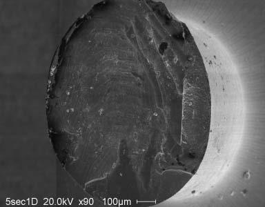
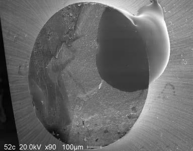
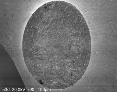
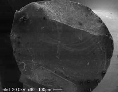
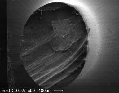
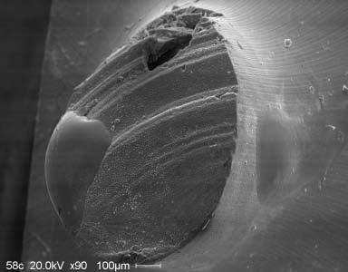
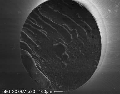
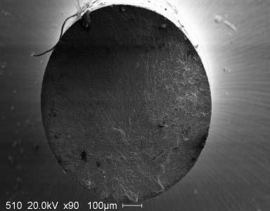
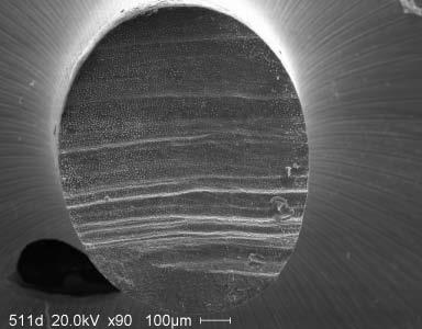
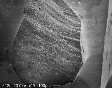
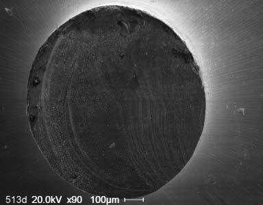
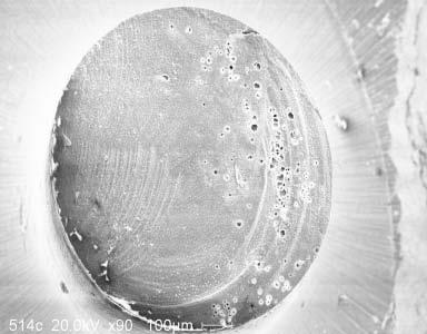
72 63 APPENDIX Failure mode 5 seconds group 1 Composite side Dentin side 1 Composite side Dentin side


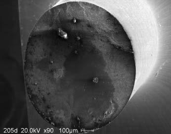
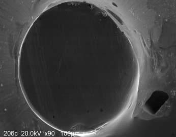
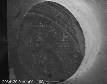
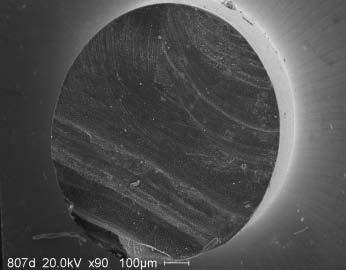
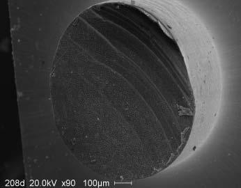
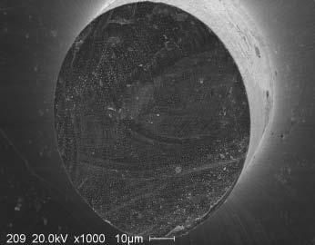
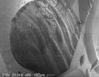
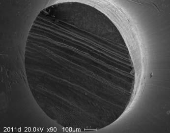
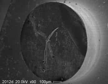
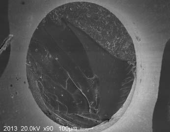
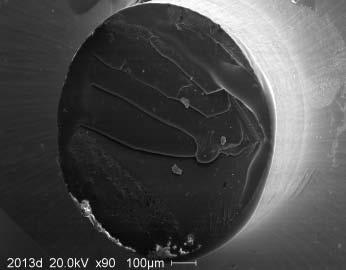

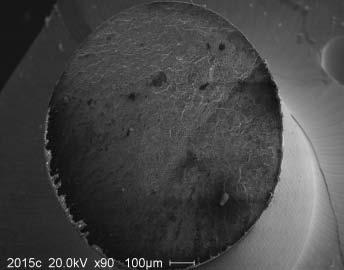
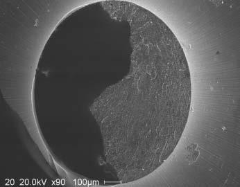
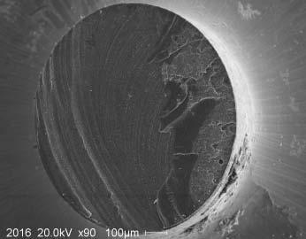
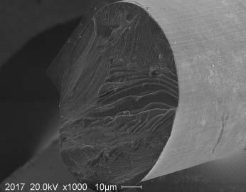
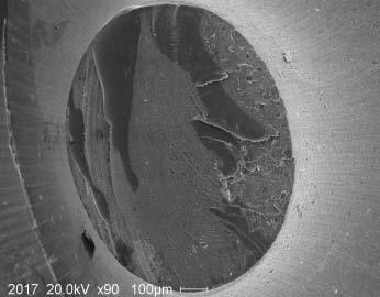
73 64 Failure mode 20 seconds group Composite side Dentin side Composite side Dentin side


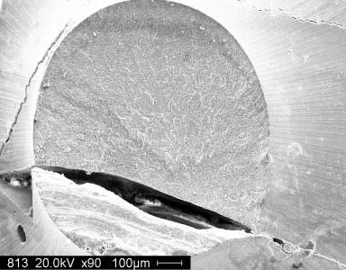
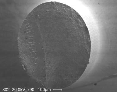
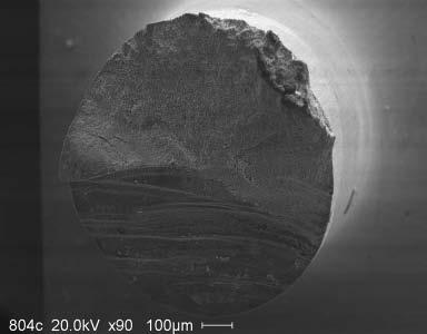

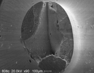
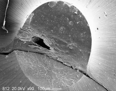
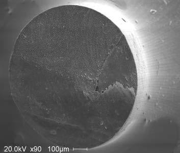
74 65 Failure mode 80 seconds group 1 Composite side Dentin side 2 Composite side Dentin side
Adper Easy Bond. Self-Etch Adhesive. Technical Product Profile
 Adper Easy Bond Self-Etch Adhesive Technical Product Profile Table of Contents Table of Contents Introduction... 4 Product Description... 4 Composition...5-8 Background... 5 Mechanism of Adhesion to Enamel
DH220 Dental Materials
 DH220 Dental Materials Lecture #5 Prof. Lamanna RDH, MS Restorative Dentistry: Glass Ionomer Bird & Robinson p.740-741 I. Use Liner Base Luting agent Restorative material: Class III, V, & eroded/abraded
Lecture. Caries of dentin
 Lecture Caries of dentin Dentin structure The characteristic feature of dentin structure is the dentinal tubules. The dentinal tubules have a hollow structure and they are responsible for dentin permeability.
The effect of ph of etchant on the bond strength of a two-step total-etching adhesive
 The effect of ph of etchant on the bond strength of a two-step total-etching adhesive by Mythili Rajesh Dr John O Burgess, CHAIR Dr Firoz Rahemtulla Dr Lillian Mitchell Dr Mark Stephen Litaker Dr Mona
Pulpal Protection: bases, liners, sealers, caries control Module C: Clinical applications
 Readings: Fundamentals of Operative Dentistry, 3 nd Edition; Summitt, et al Chapters 5, 6 and 8 Pulpal Protection: bases, liners, sealers, caries control Module C: Clinical applications REST 528A Operative
Is there any clinical evidence?
 Current treatment objectives Anticariogenic capacity of restorative materials in paediatric dentistry: in vitro evidence vs. clinical efficiency Prof. Lisa Papagianoulis Restoration with minimal intervention
Fuji II LC. A Perfect Choice
 A Perfect Choice is a remarkable restorative material The world s first resin-reinforced glass ionomer has remained the benchmark for light cured glass ionomer cements, delivering more than 15 years of
Continually Fluoride Releasing Aesthetic Dental Restorative Material
 Continually Fluoride Releasing Aesthetic Dental Restorative Material Research is our best product Image provided by Dr. Sushil Koirala BEAUTIFIL II More than just filling BEAUTIFIL II stands out for its
DENTIN-PULP COMPLEX. Erlina Sih Mahanani. School of Dental sciences Universiti Sains Malaysia. Erlina Sih Mahanani
 DENTIN-PULP COMPLEX School of Dental sciences Universiti Sains Malaysia Introduction Overview anatomy & histology of dentin and pulp. Development of dentin and pulp Structure of dentin and pulp Dentin
Operative dentistry. Lec: 10. Zinc oxide eugenol (ZOE):
:") Operative dentistry Lec: 10 د.عبذالمنعم الخفاجي Zinc oxide eugenol (ZOE): There are 2 types: 1) Unreinforced ZOE (ordinary type): supplied as powder (zinc oxide + some additives like zinc acetate, white
Operative dentistry Lec: 10 د.عبذالمنعم الخفاجي Zinc oxide eugenol (ZOE): There are 2 types: 1) Unreinforced ZOE (ordinary type): supplied as powder (zinc oxide + some additives like zinc acetate, white
Bonding to dentine: How it works. The future of restorative dentistry
 Bonding to dentine: How it works The future of restorative dentistry There are 82 adhesive systems on the market today! Are dentists confused about bonding? Michael Buonocore Buonocore MG. A simple method
Etching with EDTA- An in vitro study
 ISSN 0970-4388 Etching with EDTA- An in vitro study BOGRA P a, KASWAN S b ABSTRACT: In the present study, 25% EDTA, in gel form, was used to analyse its micromorphological effects on tooth surfaces with
Anisotropy of Tensile Strengths of Bovine Dentin Regarding Dentinal Tubule Orientation and Location
 Original paper Dental Materials Journal 21 (1): 32-43, 2002 Anisotropy of Tensile Strengths of Bovine Dentin Regarding Dentinal Tubule Orientation and Location Toshiko INOUE, Hidekazu TAKAHASHI and Fumio
Forgives Nothing. Forgives Almost Anything. Science Update
 Forgives Nothing Forgives Almost Anything Science Update Shear bond strength on wet and dry dentin (Ultradent) Latta M, Omaha Since etching of dentin with phosphoric acid was introduced, one focus of research
The Future of Dentistry Now in Your Hands Changes everything you know about traditional Composites, Glass Ionomers and RMGIs
 The Future of Dentistry Now in Your Hands Changes everything you know about traditional Composites, Glass Ionomers and RMGIs PULPDENT BioACTIVE Products for ProACTIVE Dentistry Advances in dental materials
values is of great interest.
 User Report Dr. Ralph Schönemann, May 21, 2008 There are numerous self-etchbonding systems on the market that sometimes differ very much from one another with regards to their chemical composition. It
Pulpal Protection: bases, liners, sealers, caries control Module A: Basic Concepts
 Readings: Fundamentals of Operative Dentistry, 3 nd Edition; Summitt, et al Chapters 5, 6 and 8 Pulpal Protection: bases, liners, sealers, caries control Module A: Basic Concepts REST 528A Operative #3A
MDJ Evaluation the effect of eugenol containing temporary Vol.:9 No.:2 2012
 MDJ Evaluation the effect of eugenol containing temporary fillings on shear bond strength of composite restoration Dr. Farid G. Numan B.D.S., M.Sc Abstract The purpose of this in-vitro study was to evaluate
General dentists in private practice place numerous
 PROACTIVE INTERVENTION DENTISTRY Incorporating Glass Ionomers into Everyday Dental Practice Todd Snyder, DDS, FAACD, FIADFE Introduction General dentists in private practice place numerous direct tooth
Near-UV light detection
 C L I N I C A L Near-UV light detection Javier Tapia Guadix 1 Near-UV light induced fluorescence has already proven to be very useful as an alternative to classic caries-detector dyes. However its potential
JBR Journal of Interdisciplinary Medicine and Dental Science
 ISSN: 2376-032X JBR Journal of Interdisciplinary Medicine and Dental Science Yashpal and Monika, 2014, 2:1 DOI: 104172/2376-032X1000111 JBR Journal of Interdisciplinary Medicine and Dental Science Research
Adhesive Solutions. Scotchbond Universal Adhesive. SEM pictures of Scotchbond Universal Adhesive. One bottle for all cases! Total-Etch and Self-Etch
 Adhesive Solutions Adhesive SEM pictures of. One bottle for all cases! Total-Etch and Self-Etch One adhesive for Total-Etch and Self-Etch Discover the Universal Bonding Solution. Unleash the power of the
DENTIN It a hard vital tissue, surrounds the pulp & underlies the enamel on the crown & the cementum on the roots of the teeth.
 Lec. 7 Dr. Ali H.Murad DENTIN It a hard vital tissue, surrounds the pulp & underlies the enamel on the crown & the cementum on the roots of the teeth. Physical properties: 1-Dentin is pale yellow in color,
Adhese Universal. The universal adhesive. Direct Indirect Total-Etch Selective-Etch Self-Etch Wet & Dry. All in. one click
 Adhese Universal The universal adhesive Direct Indirect Total-Etch Selective-Etch Self-Etch Wet & Dry All in one click 1 Universal Adhesion with advanced delivery Adhese Universal is a singlecomponent,
***Handout*** Adhesive Dentistry Harald O. Heymann, DDS MEd Dentin Bonding Rewetting/Desensitization
 ***Handout*** Adhesive Dentistry Harald O. Heymann, DDS MEd Dentin Bonding * Regardless of the type dentinal adhesive used, the primary mechanism for dentin adhesion is still establishment of the hybrid
Howard E. Strassler, DMD University of Maryland School of Dentistry
 Part II National Board Review Operative Dentistry-Biomaterials Structures, chemical properties, physical properties BASIC CONCEPTS ON HOW DENTAL MATERIALS WORK! Howard E. Strassler, DMD University of Maryland
Filling materials are used to replace missing parts of the tooth.
 Filling materials are used to replace missing parts of the tooth. 1- Dental caries. 2- Trauma. 3- Abrasion. Parts of teeth which require replacement by restorative materials vary in size of cavity, shape,
The Histology of Dentin
 The Histology of Dentin Pauline Hayes Garrett, D.D.S. Department of Endodontics, Prosthodontics, and Operative Dentistry University of Maryland, Baltimore This material was taken from: Essentials of Oral
riva helping you help your patients
 riva helping you help your patients what is a glass ionomer? how will a dentist benefit from using glass ionomers? how will a patient benefit from their glass ionomer? Glass ionomer is the generic name
Preparation and making fillings Class V., III., IV.
 Preparation and making fillings Class V., III., IV. Class V. Cervical defects - Dental caries - Non carious lesions (erosion, abrasion, V shaped defects) Types of defects Caries Erosion Abrasion V shaped
BioCoat Featuring SmartCap Technology
 R R Breakthrough! BioCoat Featuring SmartCap Technology The only bioactive pit and fissure sealant that provides relentless protection through daily release of fluoride, calcium and phosphate ions. Bioactive
ORIGINAL STUDY BRACKET BONDING TO ENAMEL AND DENTIN - ESEM STUDIES -
 MEDICINĂ, no 1, 2013 ORIGINAL STUDY BRACKET BONDING TO ENAMEL AND DENTIN - ESEM STUDIES - Delia Ioana Ciocan 1, Florin Miculescu 2, Lucian Toma Ciocan 3, Dragos Stanciu 1 1 Orthodontics and Dento-Facial
stabilisation and surface protection
 Guiding the way to caries stabilisation and surface protection Fissure sealing MI restorations Pulp capping Hypersensitivity Protection Caries stabilisation Fuji Triage from GC. Temporary restorations
Contactless Dentistry. Cleans, Cuts, Prepares - Gently
 Contactless Dentistry Cleans, Cuts, Prepares - Gently Contactless Dentistry Unlike conventional rotary cutting instruments, the AquaCare handpiece is not in direct contact with the tooth structure, removing
Heraeus Kulzer Adhesives. Adhesive Guide
 Heraeus Kulzer Adhesives Adhesive Guide Contents Principles of adhesion Enamel Preparation and conditioning Dentin Conditioning Wettability and surface tension Classification of adhesive systems Classification
Effect of Self-etchant ph on Shear Bond Strength of Orthodontic Brackets: An in vitro Study
 10.5005/jp-journals-10015-1126 ORIGINAL RESEARCH Effect of Self-etchant ph on Shear Bond Strength of Orthodontic Brackets: An in vitro Study Amit Goyal, H Jyothikiran, BM Shivalinga WJD ABSTRACT Aims:
Chapter 5. Investigation of in vitro Dental Erosion by Optical Techniques
 Chapter 5 Investigation of in vitro Dental Erosion by Optical Techniques 5.1 INTRODUCTION 5 Dental erosion is becoming an increasing predicament in dentistry and can be attributed to either exogenous
Change the Way You Think About Bulk Fill Composites
 Change the Way You Think About Bulk Fill Composites Are you ready for bulk fill composites? Because the updated edition will blow you away! 1 Foreword by Dr. Joshua Austin, DDS As a general practitioner,
EVALUATION OF COLOR CHANGE IN WHITE SPOT LESIONS OF ENAMEL FLUOROSIS USING A RESIN INFILTRATE
 EVALUATION OF COLOR CHANGE IN WHITE SPOT LESIONS OF ENAMEL FLUOROSIS USING A RESIN INFILTRATE Monisha Singhal PG STUDENT DEPARTMENT OF PEDODONTICS & PREVENTIVE DENTISTRY CHANDRA DENTAL COLLEGE & HOSPITAL
Study of Shear Bond Strength of Two Adhesive Resin Systems
 Cronicon OPEN ACCESS EC DENTAL SCIENCE Research Article Mohamed Atta Gowida 1 *, Ahmed Yehia Ashour 2, Seham Ahmed Hanafy 3, Waleed Abdel-Maguid El-mahy 4 and Wegdan M Abdel-Fattah 5 1 Senior Registrar
BioCoat Featuring SmartCap Technology
 new! BioCoat Featuring SmartCap Technology Premier s new bioactive pit and fissure sealant provides relentless protection, outstanding adhesion and extended fluoride release. Bioactive Release - Uptake
UNIVERSAL ADHESIVE SYSTEM. PEAK UNIVERSAL BOND Light-Cured Adhesive with Chlorhexidine
 UNIVERSAL ADHESIVE SYSTEM PEAK UNIVERSAL BOND Light-Cured Adhesive with Chlorhexidine 1 TOTAL-ETCH PEAK UNIVERSAL BOND SELF-ETCH UNIVERSAL BONDING The Peak Universal Adhesive System features a universal
lec: Dental material dr. Aseel Mohammed Filling material
 Filling material Filling material: the material that is used to replace a missing part of the tooth which may result from dental caries, trauma or abrasion. It can be divided in to: 1. Direct filling materials:
EQUIA. Self-Adhesive, Bulk Fill, Rapid Restorative System
 EQUIA EQUIA Fil EQUIA Coat + Self-Adhesive, Bulk Fill, Rapid Restorative System From the World Leader in Glass Ionomer Technology - A Complete Glass Ionomer Based Bulk Fill Rapid Restorative System Class
DEFENSE MECHANISM OF DENTINE AND PULP AGAINST INJURY
 DEFENSE MECHANISM OF DENTINE AND PULP AGAINST INJURY DR.AHMED IBRAHIM AL-JOBORY B.D.S. M.SC. CONSERVATIVE DEPARTMENT/ BAGHDAD UNIVERSITY MEMBER OF IRAQI ENDODONTICS SOCIETY The reaction in dentine are
New Nano-Hybrid Technology for your everyday Use
 New Nano-Hybrid Technology for your everyday Use NEW New NANO-HYBRID TECHNOLOGY For Your every Day USE You need a composite that allows you to easily achieve high quality restorations? Polofil NHT, the
Clinical report. Drs Paul and Alexandre MIARA and F. CONNOLLY COMPOSITE POSTERIOR FILLINGS. How to control. layering? 8 - Dentoscope n 124
 COMPOSITE POSTERIOR FILLINGS How to control layering? 8 - Dentoscope n 124 CV FLASH Dr Paul MIARA Dental surgeon Dr Alexandre MIARA Dental surgeon Dr F. CONNOLLY Dental surgeon Thanks to continuous improvements
Acknowledgments Introduction p. 1 Objectives p. 1 Goals p. 2 History of Dental Materials p. 3 The Oral Environment p. 4 Characteristics of the Ideal
 Preface p. v Acknowledgments p. vii Introduction p. 1 Objectives p. 1 Goals p. 2 History of Dental Materials p. 3 The Oral Environment p. 4 Characteristics of the Ideal Dental Material p. 5 Quality Assurance
Remaining dentin thickness Shallow cavity depth Preparation 0.5 mm into dentin (ideal depth) Moderate cavity depth Remaining dentin over pulp of at le
 Moderate cavity depth Remaining dentin over pulp of at le") Deep carious lesions management Remaining dentin thickness Shallow cavity depth Preparation 0.5 mm into dentin (ideal depth) Moderate cavity depth Remaining dentin over pulp of at least 1-2 mm Deep cavity
Deep carious lesions management Remaining dentin thickness Shallow cavity depth Preparation 0.5 mm into dentin (ideal depth) Moderate cavity depth Remaining dentin over pulp of at least 1-2 mm Deep cavity
One cement that can take you just about anywhere! RelyX. Unicem. Self-Adhesive Universal Resin Cement
 One cement that can take you just about anywhere! TM Self-Adhesive Universal Resin Cement Indications Inlays, Onlays, Crowns, Bridges Metal/PFM, Porcelain/Ceramics, Composites. Pins and Posts Metal, Zirconia
Ketac Universal Aplicap
 Ketac Universal Aplicap Technical Data Sheet 2 Introduction Ketac Universal Aplicap is the latest development in a long history of proven glass ionomer technology from 3M. It s designed to save steps for
Part II National Board Review Operative Dentistry. Module 3D General Questions Answers in BOLD (usually the first answer)
") Part II National Board Review Operative Dentistry Module 3D General Questions Answers in BOLD (usually the first answer) Howard E. Strassler, DMD University of Maryland Dental School With special acknowledgements
Part II National Board Review Operative Dentistry Module 3D General Questions Answers in BOLD (usually the first answer) Howard E. Strassler, DMD University of Maryland Dental School With special acknowledgements
Anterior Esthetic Techniques & Materials
 Anterior Esthetic Techniques & Materials Lee Ann Brady DMD www.pinterest.com Lee Ann Brady www.leeannbrady.com www.restorativenation.com Planning Information Tooth Position Arrangement Contour Color Material
3M Scotchbond Universal Adhesive. Technical Product Profile
 3M Scotchbond Universal Adhesive 2 3M Scotchbond Universal Adhesive Table of Contents Product Description.... 3 Indications... 4 Composition....5 6 3M Scotchbond Universal DCA Dual Cure Activator....7
CONSERVATIVE OPERATIVE DENTISTRY Page 1 Lecture: Bonding Systems (Enamel, Dentin, Compomer, Amalgam, Resin Cement)
") CONSERVATIVE OPERATIVE DENTISTRY Page 1 Lecture: Bonding Systems (Enamel, Dentin, Compomer, Amalgam, Resin Cement) OVERVIEW OF BONDING SYSTEMS: A. Goals of Bonding (and Adhesion): 1. Sealing 2. Retention
CONSERVATIVE OPERATIVE DENTISTRY Page 1 Lecture: Bonding Systems (Enamel, Dentin, Compomer, Amalgam, Resin Cement) OVERVIEW OF BONDING SYSTEMS: A. Goals of Bonding (and Adhesion): 1. Sealing 2. Retention
Original Article. in depth, 4 mm in mesiodistal width and 3 mm occlusogingival
 Original Article A Comparative Evaluation of Microleakage in Class V Composite Restorations using a Fifth Generation Adhesive and a Glass Ionomer Bonding Agent - An In Vitro Dye Leakage Study Dipali Shah
Correlation between the Strength of Glass Ionomer Cements and Their Bond Strength to Bovine Teeth
 Dental Materials Journal 23(4): 656-660, 2004 Correlation between the Strength of Glass Ionomer Cements and Their Bond Strength to Bovine Teeth Yasushi HIBINO, Ken-ichi KURAMOCHI, Atsushi HARASHIMA, Muneaki
Relationship between caries-affected dentin mineral density and microtensile bond strength
 University of Iowa Iowa Research Online Theses and Dissertations Spring 2011 Relationship between caries-affected dentin mineral density and microtensile bond strength Savitri Vaseenon University of Iowa
Scotchbond Universal Adhesive. Technical Product Profi le
 Adhesive Technical Product Profi le Table of Contents Product Description... 3 Indications... 4 Composition... 5 6 DCA Dual Cure Activator... 7 Etchant... 7 Summary of Adhesive Unique Chemistry Powered
Get in front of the 8 ball with the new Fuji VIII GP. The first auto-cure, resin reinforced glass ionomer restorative
 Get in front of the 8 ball with the new Fuji VIII GP The first auto-cure, resin reinforced glass ionomer restorative 8 reasons to choose Fuji VIII GP Auto-cure, resin reinforced glass ionomer restorative
G-COAT PLUS G-COAT PLUS GET THE BEST OF BOTH WORLDS WITH THE STROKE OF A BRUSH
 G-COAT PLUS BEAUTY BRAWN GET THE BEST OF BOTH WORLDS WITH THE STROKE OF A BRUSH 1 Introducing a Revolutionary new Coating G-COAT PLUS gives your restorations the Best of Both Worlds: A beautiful new finish
G-CEM LinkAce. The new strength in self-adhesive cement
 The new strength in self-adhesive cement is stronger Now offering comparative physical properties to current adhesive resin cements, is the next evolution in easy-to-use, versatile, self-adhesive. Designed
Direct composite restorations for large posterior cavities extended range of applications for high-performance materials
 Direct composite restorations for large posterior cavities extended range of applications for high-performance materials A case study by Ann-Christin Meier, Dr. med. dent., Stapelfeld, Germany When large
CERASMART. The new leader in hybrid ceramic blocks
 The new leader in hybrid ceramic blocks Introducing, a new force absorbing hybrid ceramic block 20 µm Contains high density of ultra fine homogeneously-dispersed fillers in a highly cross-linked resin
The Dental Board of California - Dental Materials Fact Sheet Adopted by the Board on October 17, 2001
 The Dental Board of California - Dental Materials Fact Sheet Adopted by the Board on October 17, 2001 As required by Chapter 801, Statutes of 1992, the Dental Board of California has prepared this fact
Comparison of the microstructure of crown and root dentin by a scanning electron microscopic study
 J Dent Sci 2010;5(1):14 20 ORIGINAL ARTICLE Comparison of the microstructure of crown and root dentin by a scanning electron microscopic study Chia-Yi Chu, 1 Tien-Chun Kuo, 2,3 Shu-Fang Chang, 1 Yow-Chyun
Structure and mechanical properties of teeth. Advisors: M.A. Meyers & E. A. Olevsky Student :Yen-Shan Lin Date: 6/23/2008
 Structure and mechanical properties of teeth Advisors: M.A. Meyers & E. A. Olevsky Student :Yen-Shan Lin Date: 6/23/2008 Outline Basic structure and components of teeth Mechanical properties 1. tubule
G-Premio BOND. One component light cured universal adhesive. BOND with the BEST
 One component light cured universal adhesive BOND with the BEST The world s most clinically successful adhesive is now even better! Introducing. G-BOND has proven to be the world s most clinically successful
Effect of Different Bur Grinding on the Bond Strength of Self etching Adhesives
 Operative Dentistry, 2006, 31-3, 317-323 Laboratory Research Effect of Different Bur Grinding on the Bond Strength of Self etching Adhesives S Semeraro D Mezzanzanica D Spreafico M Gagliani D Re T Tanaka
Adper Scotchbond SE Self-Etch Adhesive. technical product profile. Adper
 Adper Scotchbond SE Self-Etch Adhesive technical product profile Adper 2 Table of Contents Introduction................................................................5 Product Description.......................................................5
SCIENTIFIC. Effects of various acids on the buccal surface of human permanent teeth: a study using scanning electron microscopy*
 PEDIATRIC DENTISTRY/Copyright 1979 by The American Academy o! Pedodontics/VoL 1, No. 3/Printed in U.S.A. SCIENTIFIC Effects of various acids on the buccal surface of human permanent teeth: a study using
Technical Product Profile
 Adhesive www.3mespe.com Technical Product Profile 3M Deutschland GmbH Location Seefeld 3M ESPE ESPE Platz 82229 Seefeld Germany info3mespe@mmm.com www.3mespe.com 3M, ESPE, Adper, Aplicap, Clinpro, CoJet,
For the Perfect Class V and All Cervical Area Gingival Margins when Placing Direct Composites, Create an Injection Molding Matrix
 Cronicon OPEN ACCESS EC DENTAL SCIENCE Case Report For the Perfect Class V and All Cervical Area Gingival Margins when Placing Direct Composites, Create an Injection Molding Paul C Belvedere* Adjunct Professor,
Dowel restorations Treatment with a post and core
 Dowel restorations Treatment with a post and core A post and core is a dental restoration used to sufficiently buildup tooth structure for future restoration with a crown when there is not enough tooth
Ceramic Based Aesthetic CAD/CAM Restorative
 Ceramic Based Aesthetic CAD/CAM Restorative Create Natural Aesthetics with High-Strength Materials SHOFU Block HC and Disk HC are the latest generation of hybrid-ceramic restorative materials for use with
The Dental Board of California Dental Materials Fact Sheet Adopted by the Board on October 17, 2001
 The following document is the Dental Board of California s Dental Materials Fact Sheet. The Department of Consumer Affairs has no position with respect to the language of the Dental Materials Fact Sheet;
CONTINUING EDUCATION 2 INDIRECT COMPOSITE RESIN RESTORATIONS. Adhesive Cementation of Indirect Composite Inlays and Onlays: A Literature Review
 CONTINUING EDUCATION 2 INDIRECT COMPOSITE RESIN RESTORATIONS Adhesive Cementation of Indirect Composite Inlays and Onlays: A Literature Review Camillo D Arcangelo, DDS; Lorenzo Vanini, MD, DDS; Matteo
DPI Luting Cement. DPI Restorative Cement
 DPI Luting Cement DPI Luting Cement is Type I glass ionomer white luting cement. DPI glass ionomers are remarkably biocompatible and can be safely used in deep cavities provided the pulp is not exposed,
CARIES STABILIZATION AND TEMPORARY RESTORATION
 CARIES STABILIZATION AND TEMPORARY RESTORATION LEARNING OUTCOMES Justify the importance of caries stabilisation procedure in operative care. List and discuss the methods to stabilise caries ( include preventive
Chapter 14 Outline. Chapter 14: Hygiene-Related Oral Disorders. Dental Caries. Dental Caries. Prevention. Hygiene-Related Oral Disorders
 Chapter 14 Outline Chapter 14: Hygiene-Related Oral Disorders Hygiene-Related Oral Disorders Dental caries Prevention Gingivitis Prevention Tooth hypersensitivity Pathophysiology Treatment 2 Hygiene-Related
Glass ionomer cements in complex oral rehabilitations. A case report.
 Università degli Studi di Milano - Department of Technologies for the Health I.R.C.C.S Istituto Ortopedico Galeazzi Department of Odontology Director: Prof. Roberto Weinstein Glass ionomer cements in complex
Pelagia Research Library. Comparison of microleakage in bonded amalgam restrorations using different adhesive materials: An invitro study
 Available online at www.pelagiaresearchlibrary.com European Journal of Experimental Biology, 2011, 1 (2):92-96 ISSN: 2248 9215 Comparison of microleakage in bonded amalgam restrorations using different
Comparison of shear bond strength of aesthetic restorative materials
 Comparison of shear bond strength of aesthetic restorative materials Suryakumari B. P. Nujella, Manisha T. Choudary 1, Satyanarayana P. Reddy 2, Kiran kumar M. 3, Gopal T. 4 Abstract Aim: The present study
Posterior Adhesive Dentistry
 Posterior Adhesive Dentistry Stephen D. Poss DDS Michigan Dental The following pages contain a summary of information designed to assist you with the armamentarium, preparation, and cementation of anterior
The Effects of the Acid Etch and Direct Bonding Technique in Orthodontics on Enamel Surface Topography
 J. Dent. Ass. S. Afr. Vol. 31, No 10 pp 509-513 The Effects of the Acid Etch and Direct Bonding Technique in Orthodontics on Enamel Surface Topography P L Sadowsky and D H Retief Department of Orthodontics,
the new dna of high strength glass ceramics
 the new dna of high strength glass ceramics 1 the new dna of high strength glass ceramics Together with the Fraunhofer Institute and VITA, we have developed a new class of materials for high-strength glassceramics
SEM study of a self-etching primer adhesive system used for dentin bonding in primary and permanent teeth
 Scientic Article SEM study of a self-etching primer adhesive system used for dentin bonding in primary and permanent teeth Paloma Dias da Silva Telles DDS, MS Maria Aparecida A. Moreira Machado DDS, MS,
SDR has proven reliability in high C-factor cavities 2
 R R SDR has proven reliability in high C-factor cavities 2 SDR was introduced by DENTSPLY DETREY, Konstanz, Germany in 200 and has since been used in over 20 million restorations across the globe. SDR
Lec. 3-4 Dr. Saif Alarab Clinical Technique for Class I Amalgam Restorations The outline form
 Lec. 3-4 Dr. Saif Alarab Clinical Technique for Class I Amalgam Restorations Class I refers to -Restorations on the occlusal surfaces of posterior teeth, - The occlusal two thirds of facial and lingual
Original Research. Contributors: 1
 Received: 21 st February 2015 Accepted: 15th May 2015 Conflicts of Interest: None Source of Support: Nil Original Research Comparative Evaluation of Bond Strength of Dual-Cured Resin Cements: An In-Vitro
RelyX Unicem Self-Adhesive Universal Resin Cement Frequently Asked Questions
 RelyX Unicem Self-Adhesive Universal Resin Cement Frequently Asked Questions Q1. What about the clinical history of The first restorations were cemented with RelyX Unicem cement in 2001. The excellent
Enamel Bond Strength of New Universal Adhesive Bonding Agents
 Ó Operative Dentistry, 2015, 40-4, 410-417 Enamel Bond Strength of New Universal Adhesive Bonding Agents DE McLean EJ Meyers VL Guillory KS Vandewalle Clinical Relevance This laboratory study will facilitate
Calibra. Cements. The Simple Choice for Easy Cleanup
 Calibra Cements The Simple Choice for Easy Cleanup www.calibracement.com The Calibra family of definitive cements was designed to make it easier than ever to achieve consistent, successful results in your
THE ONE AND ONLY. CLEARFIL Universal Bond
 THE ONE AND ONLY THIS ADHESIVE LETS YOU BOND ALMOST ALL DENTAL MATERIALS Universal. Easy. Reliable. NEW! With the new you can bond various dental materials and select the etch mode of your choice. You
SHEAR BOND STRENGTH OF SOME SEALANTS UNDER SALIVA CONTAMINATION
 SHEAR BOND STRENGTH OF SOME SEALANTS UNDER SALIVA CONTAMINATION Praphasri Rirattanapong 1, Kadkao Vongsavan 1 and Rudee Surarit 2 1 Department of Pediatric Dentistry, 2 Department of Physiology and Biochemistry,
Scanning Electron Microscopy Evaluation of the Bonding Mechanism of a Self-etching Primer on Enamel
 Original Article Scanning Electron Microscopy Evaluation of the Bonding Mechanism of a Self-etching Primer on Enamel Julio P. Cal-Neto a ; José Augusto M. Miguel b Abstract: The aim of this study was to
EQUIA Forte Glass Hybrid Restorative System. For long term posterior restorations
 EQUIA Forte Glass Hybrid Restorative System For long term posterior restorations The original EQUIA system EQUIA is a self-adhesive posterior restorative system that combines the chemical-bonding, bulk-fill
SmartCrown. The Cavity Fighting SmartCrown. Patient Education Booklet. SmartCrown.com Toll Free Local
 The Cavity Fighting SmartCrown SmartCrown Ca2+ SmartCrown.com Patient Education Booklet www.smartcrown.com support@smartcrown.com 800.255.2839 Toll Free 801.281.4012 Local 801.281.4022 Fax F- PO43- Origin
Immediate Dentin Sealing and Cerec How to avoid sensivities 100 pc and how to have lots of practical advantages
 Immediate Dentin Sealing and Cerec How to avoid sensivities 100 pc and how to have lots of practical advantages Some abbreviations in this ppt. IDS: Immediate dentin sealing means the application of the