Understanding. Retinitis pigmentosa. and other inherited retinal dystrophies
|
|
|
- Melvyn Moore
- 6 years ago
- Views:
Transcription
1 Understanding Retinitis pigmentosa and other inherited retinal dystrophies
2 Contact us We re here to answer any questions you have about your eye condition or treatment. If you need further information about retinal conditions or on coping with changes in your vision, then our Helpline is there for you. Just give us a call on or us at helpline@rnib.org.uk and we ll be happy to speak with you. RNIB s Understanding series The Understanding series is designed to help you, your friends and family understand a little bit more about your eye condition. The series covers a range of eye conditions, and is available in audio, print and braille formats. 2
3 Contents 4 What is retinitis pigmentosa? 6 How your eye works 8 What are the symptoms of RP? 12 What caused my RP? 14 How is RP inherited? 18 How does RP affect my family? 20 Can other parts of my body be affected? 21 What tests are used to detect RP? 30 What other eye conditions might I get? 32 Is there any treatment for RP and what research is being carried out? 36 Coping 38 Further help and support 3
4 What is retinitis pigmentosa? Retinitis pigmentosa (RP) is the name given to a group of inherited eye conditions called retinal dystrophies. A retinal dystrophy such as RP affects the retina at the back of your eye and, over time, stops it from working. This means that RP causes gradual but permanent changes that reduce your vision. How much of your vision is lost, how quickly this happens and your age when it begins depends on the type of RP that you have. If you have RP, the changes to your retina can affect your peripheral vision (also known as your side vision) and make it difficult for you to see in dim light or in the dark. Your central vision can also become affected and this will make it difficult for you to see colour and do things such as reading or watching television. The changes in your vision happen over years rather than months, and some people lose more sight than others. 4

5 5
6 How your eye works When light enters your eye, it is focused onto your retina at the back of your eye. The retina has a number of layers, but the most important for vision is a layer made up of cells called photoreceptors. Photoreceptors are cells which are sensitive to light. The macula, which is the central part of the retina, contains a few million specialised photoreceptor cells called cone cells. These cone cells work best in bright light and allow you to see fine detail for activities like reading and writing and recognising colours. The peripheral retina is further away from the central macula. It is mostly made up of the other type of photoreceptors called rod cells. They allow us to see when light is dim and provide peripheral vision outside of the main line of sight. RP causes damage to the rod cells in particular. Other types of retinal dystrophies may affect the cone cells which are concentrated in your macula. 6
7 Retina Iris Cornea Macula Pupil Lens Optic nerve 7
8 What are the symptoms of RP? Early symptoms When you have a retinal dystrophy like RP, your rod and cone cells gradually stop working. Depending on the type of RP you have, you may notice your first symptoms in your early childhood or later, between the ages of 10 to 30. Some people don t have symptoms until later in life. In RP, the first symptom you ll notice is not seeing as well as people without a sight condition in dim light, such as outside at dusk, or at night. This is often called night blindness. People without a sight condition can fully adapt to dim light in 15 to 30 minutes, but if you have RP it will either take you much longer or it won t happen at all. You may start having problems with seeing things in your peripheral vision. You may miss things to either side of you and you might trip over or bump into things that you would have seen in the past. Difficulty seeing in dim light and loss of peripheral vision are signs that the rod cells in your retina are being affected by your RP. 8
9 In the more common forms of RP, your cones are not affected in the early stages so your central vision will still be good enough to recognise faces and to continue reading. If your peripheral vision has started to change, it may mean it is no longer safe for you to drive. If you have a condition such as RP which can affect your sight in both eyes, you are required by law to report it to the Driver and Vehicle Licensing Authority (DVLA). They will assess your vision regularly to find out if your sight meets their standards. Early symptoms of other retinal dystrophies In some other retinal dystrophies, such as cone-rod dystrophy, central vision is affected before peripheral vision. This is because the cone cells are affected first or affected more severely than the rod cells. You ll find reading and seeing detail difficult and your colour vision will be affected. 9
10 Later symptoms RP is a progressive condition, which means that your sight will continue to get worse over the years. Often, changes in sight can happen suddenly over a short period of time. You may then have a certain level of vision for quite some time. However, there may be further changes to your vision in the future. This may mean that you have to keep re-adapting to lower levels of sight. The type of RP that you have can affect how quickly these changes develop. As your RP progresses, you ll gradually lose your peripheral sight, leaving a central narrow field of vision, often referred to as having tunnel vision. You may still have central vision until you re in your 50s, 60s or older. However, advanced RP will often affect your central vision and you may find reading or recognising faces difficult. You may find that bright lights and sunlight give you problems with glare and moving between a light and a dark room can be difficult. This is because, as your RP progresses, your retinal cells become less able to adapt to changing light levels. 10

11 Your family or the people you live with can help you as your vision deteriorates by keeping your home environment free of obstacles and by putting things away in the same place so they are easy to find. 11
12 What caused my RP? RP is a hereditary condition caused by a fault in one of the genes involved in maintaining the health of the retina. You inherit genes from your parents. Your genes give the cells in your body the instructions they need to work well and stay healthy. When a gene is faulty, it is because there is a fault in their instructions and the cells using those instructions don t work as they should. In RP, the faulty gene causes your retinal cells to stop working and to eventually die over time. Researchers have identified many of the genes that cause RP and the faults within them, but there are still other genes to discover. There have always been different types of RP, but in the past, they ve all been given the same name retinitis pigmentosa. This happened because many of the conditions looked the same when ophthalmologists looked inside someone s eyes. However, as research developed, giving a better understanding of how our genes cause eye conditions, it became possible to tell the difference between conditions which would have all been called RP in the past. This means that some people may have their RP more accurately renamed as a rod-cone dystrophy or cone-rod dystrophy because of the genes involved. 12

13 Other retinal dystrophies include Leber s congenital amaurosis, cone dystrophy, cone-rod dystrophies, rod-cone dystrophies, choroideremia and macular dystrophies. 13
14 How is RP inherited? About half of people with RP have another family member with the condition. The way RP is passed from generation to generation can tell you who in your family has had the condition, how severely your vision could be affected and the chances of your children being affected. Genes usually come in pairs. You inherit one gene from each of your parents to make each pair. When you have children, you only pass on one gene to them. You can inherit RP in three different ways autosomal dominant, autosomal recessive and X-linked. Autosomal dominant inheritance To have autosomal dominant RP, you only need one faulty gene to have the condition. This can be inherited from either your mother or your father. Usually this parent will also have RP. Autosomal dominant RP affects men and women equally and there tends to be a known history of the condition in your family. If you have autosomal dominant RP, there is a one in two risk of passing on the condition to each of your children. 14
15 Autosomal recessive inheritance Autosomal recessive RP requires two faulty genes, one inherited from your mother and one inherited from your father. If both your parents have one normal gene and one faulty gene, they are carriers of RP and their vision is unaffected by the condition. If both your parents pass on their faulty gene to you, you will inherit autosomal recessive RP. If you have autosomal recessive RP, you will pass on a faulty gene to all your children. If they inherit a normal copy of the gene from their other parent, they will be carriers of RP. Because you need two copies of the faulty gene to have this type of RP, it usually appears in families without any history of the condition in other generations. This type of RP affects men and women equally. 15
16 X-linked inheritance This is a type of RP that affects men. It can be severe and result in poor sight by the age of 30 to 40. X-linked RP relates to our sex chromosomes. Men have one X and one Y chromosome. Women have two X chromosomes. The gene relating to X-linked RP is found on an X chromosome. This means men with a faulty gene on their X chromosome will have the condition. If a woman has a faulty gene on one X chromosome but a normal gene on the other, she is usually more mildly affected or does not have any symptoms from the condition at all. This means she is a carrier of the condition. A man with X-linked RP will pass on the faulty gene to all his daughters but not to his sons. His daughters will be carriers of the condition but his sons won t be affected. 16

17 No known relative About half of people with RP don t know of any members of their family with the condition. This may be because their relatives were carriers of RP and haven t shown signs of the condition themselves. If there is no known relative in your family, it may not be possible to find out how your RP has been inherited without the help of genetic testing to find out which genes are faulty. 17
18 How does RP affect my family? Genetic testing Genetic testing can be carried out to try to find out if you have a faulty gene that causes RP. This can either identify the faulty gene that is causing your RP or enable you to find out if you re carrying a faulty gene that your children may inherit. There are several genetic centres around the country that carry out genetic tests and your ophthalmologist (also known as a hospital eye doctor) or your GP can refer you to one. Genetic testing uses a blood test to look at your genes to see if they re faulty. Testing for RP and other inherited retinal dystrophies is complicated. It doesn t identify all forms of these conditions as new faulty genes are still being discovered. Ask your ophthalmologist or GP to discuss genetic testing with you. 18
19 Genetic counselling Genetic counselling can help you to understand the type of RP you have, how it s likely to affect you in the future and the risks of passing on the condition to any children you may have. Genetic counselling is usually advised when you have genetic testing. A genetic counsellor asks about your family tree in detail to try and understand how RP has been inherited in your family. Genetic counselling is a free NHS service. It may be provided by a specialist RP clinic or a medical genetics department. You can ask your GP or ophthalmologist to refer you to your local genetic counselling service. Having a genetic condition in your family may cause emotional concerns. Talking to a genetic counsellor may help you and your family to discuss the eye condition in your family. Knowing the chances of passing on any condition you have can help if you are thinking about starting a family. 19
20 Can other parts of my body be affected? In most cases, the inherited faulty gene only affects the eyes. Sometimes, other parts of the body are also affected. One example of this is Usher syndrome, where people develop both hearing loss and sight loss. Others include Refsum disease, and Alström and Bardet-Biedl (BBS) syndromes, all of which cause RP-like sight loss along with other health conditions. 20
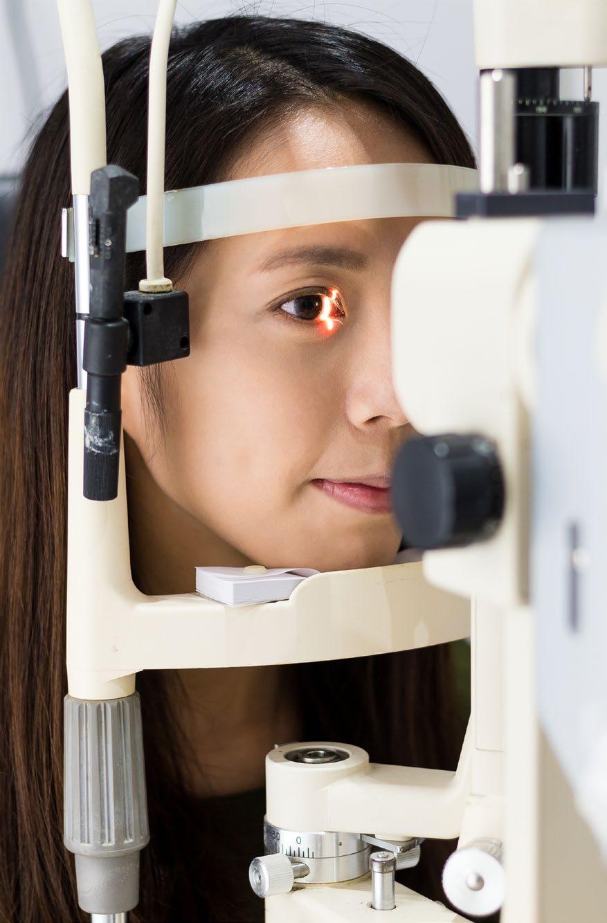
21 What tests are used to detect RP? If you ve noticed that you re having problems seeing in dim light or at night, you should see your optometrist (also known as an optician) or your GP. Early symptoms can vary from person to person so your RP might be diagnosed at an early stage or after many years of having the condition. An optometrist will examine your retina to detect RP. To do this, they will look into your eyes using either a special microscope called a slit lamp or an instrument called an ophthalmoscope. If you have the early signs of classic RP, there will be tiny but distinctive clumps of dark pigment around your retina. Any changes to your peripheral vision can only be detected by a field of vision test, which your optometrist can also carry out. This test may not be offered to you routinely so if you re worried about your peripheral vision, you should ask your optometrist to check your field of vision for you. 21
22 22
23 If you ve got a family history of RP and you have problems with your vision in dim light or problems when moving from light to dark, you should tell the person examining your eyes. This will help them to carry out the most appropriate tests. If your optometrist is concerned after your eye examination, they can refer you to an ophthalmologist for more tests. What tests will the hospital do? When you re referred to hospital, you ll be examined by an ophthalmologist. There are various tests that can diagnose RP, but it s unlikely that they ll do all of them at your first visit. These tests can also monitor how your RP changes over time. Your ophthalmologist may be able to say that you have RP when they ve got the results of these tests, but it may not be possible to know exactly what type of RP you have and what the long term effects on your vision will be without genetic testing. It s important to ask your ophthalmologist about these tests and about what the results mean for you. None of the tests are painful or cause you any harm but they may take a long time and be repetitive. Here are some of the tests you may need to undergo: 23
24 Examining the retina at the back of your eye Your retina will be examined each time you visit the hospital. You ll be given drops to dilate (widen) your pupils to allow the ophthalmologist to see your retina clearly. These drops take about 30 minutes to work. They ll make you sensitive to light and make your vision blurry. The effects of the drops usually wear off in about six hours, though sometimes it can take overnight. It isn t safe to drive until the effects have worn off. Retinal photographs, fluorescein angiograms and autofluorescence imaging Your retina may be photographed using a special camera. By comparing the photographs taken on different visits, your ophthalmologist might be able to monitor how your RP is changing over time. Your ophthalmologist may ask for a more specialised set of photographs to be taken after a yellow dye called fluorescein has been injected into a vein in your arm. As the dye travels into the tiny blood vessels in your retina, a series of photographs are taken. 24
25 The dye in the blood vessels will reveal the changes in your retina that can t be seen with normal photography. The fluorescein dye can make your skin look yellow for up to 24 hours. It leaves your body through your urine, which will be a deep yellow colour for about 24 hours too. Autofluorescence imaging involves taking more pictures of the back of your eye that show another retinal change that can t be seen with normal photography. These pictures show the ophthalmologist how the layer under your retina, called the retinal pigment epithelium, is functioning. This layer helps the retina to work and if it s not working properly itself, the health of the retina will be affected. 25
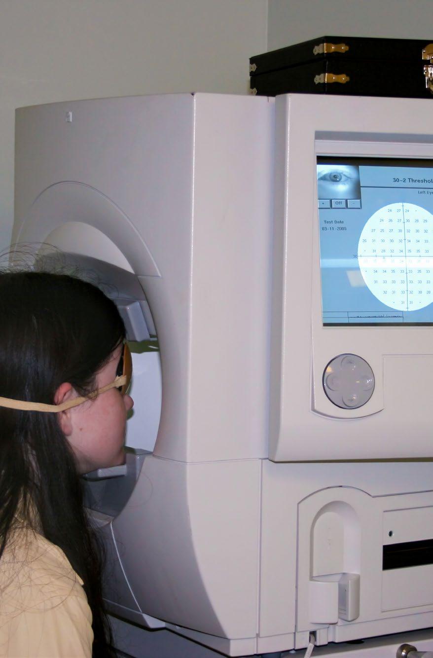
26 Visual field test A visual field test uses a machine which checks how much of your peripheral vision has been affected by an eye condition. One of your eyes is covered with a patch and your chin rests on the machine, which is in a darkened room. You ll be given a button to hold in your hand and asked to look straight ahead at a central point on the machine s bowl-shaped screen. It s important to keep looking at this central point and not to move while the test is being carried out. You ll notice spots of light flash on the screen and each time you see one, you press the button you re holding. The test takes about 10 minutes for each eye and shows how much vision you have above, below and to the sides of looking straight ahead. Colour vision To test your colour vision, you ll be asked to pick out numbers or patterns that you can see on a background of coloured dots. This test takes less than five minutes to do and shows what colours you re able to see. 26
27 27
28 Electro-diagnostic tests Electro-diagnostic tests can tell your ophthalmologist how well your retina is working. They check how your retina responds electrically to patterns and different lighting conditions. Different tests can be carried out to show the results of your retina s electrical activity. These test results will indicate which layers of your retina have been affected. The tests you may be offered include the electroretinogram (ERG), the pattern electroretinogram (PERG) and the electrooculogram (EOG). The ERG shows how your retinal cells are working and you ll usually have to sit in a darkened room or wear dark goggles for about 20 to 30 minutes to start with. Then you ll be given anaesthetic eye drops before being asked to wear a special contact lens on your eye. During this test, you ll be shown flashing lights and the response of your retina will be recorded on an electrical trace or plot. The ERG tests the whole of your retina but the PERG uses a checkerboard pattern to check how your macula, at the centre of your retina, is working. 28
29 The EOG shows how the rods and cones and the retinal pigment epithelium behind them are working. These tests are usually carried out by the electrodiagnostics department of the eye clinic and you should ask the staff to explain exactly what will happen for each test before they re carried out. The tests are painless and straightforward, but may involve having your eyes dilated and/or numbed, a tiny electrode being placed on your eye and a sensor on your skin. 29
30 What other eye conditions might I get? Some people with RP also develop cataracts. A cataract is a clouding of the lens in your eye. Your lens sits just behind your iris, the coloured part of your eye. When you have RP, you may develop a cataract as young as your 20s, but it s more often picked up in your 40s. Your ophthalmologist may recommend that you have your cataract removed, particularly if it s making your remaining useful vision misty. Your cataract can be removed and replaced with an artificial lens made of plastic or silicone. For more information about cataracts or to order a copy of our publication called Understanding Cataracts, call our Helpline on Many people find that although they still have sight loss due to their RP, their useful remaining vision is of a better quality after their cataract has been removed. 30

31 Some people with RP develop macular swelling, known as oedema. The macula is at the centre of your retina and you use it to see fine detail and colour. If the blood vessels near your macula leak, they can make your macula swollen. This can blur and distort your central vision. Macular oedema can happen occasionally after cataract surgery. It can be diagnosed with a scan of the macula using a technique called optical coherence tomography (OCT). 31
32 Is there any treatment for RP and what research is being carried out? While much progress has been made in the past few years in the understanding of the genes involved in RP, there is currently no cure or treatment which can slow down or stop RP from getting worse. Ongoing research may lead to a treatment in the future, but it may be a number of years before a tried and tested treatment for RP is produced. The types of treatment which are being researched at the moment include: Gene therapy Once a faulty gene causing RP has been identified, gene therapy aims to replace the faulty gene within the affected retinal cells with a new gene that works properly. Normal genes are injected into the affected retina using a harmless virus to carry the genetic material. The hope is that the affected cells then begin to work correctly and the damage is either stopped or reversed. Gene therapy relies on knowing which gene is faulty. In many cases of RP, the faulty genes are still to be discovered. 32
33 However, in recent clinical trials, there s already been some success in using gene therapy to improve vision for people with conditions similar to RP. It is hoped that gene therapy will be successful in treating RP in the future. Stem cell therapy The body contains many different types of cells, and some are more specialised than others. The retinal cells affected by RP are specialised cells that the body cannot easily replace. Stem cells are cells that can divide many times and can replace damaged or missing cells in different organs and tissues of the body. If stem cells can be turned into the specialised retinal cells, it may be possible to replace the cells that have been damaged in RP. Growth factors Growth factors are chemicals that support cells to grow and repair. Research groups are working on using growth factors to treat retinal disease. 33
34 Artificial vision When RP has caused severe visual loss, it s possible that damaged retinal cells could be replaced by electronic implants. These implants are placed on or beneath the retina to stimulate the remaining retinal cells. When the remaining retinal cells are stimulated and the optic nerve is healthy, a signal may be passed along the optic nerve to the brain allowing the person to see patterns of light or outlines of objects. These implants don t bring your vision back or stop your vision from getting worse. You d still need the other aids you have, such as your cane or your guide dog. Artificial vision systems are still being investigated and there isn t a system which is able to be easily implanted which returns high levels of vision. No artificial systems are currently available on the NHS. 34
35 Nutrition There s no evidence that suggests taking vitamin supplements or having a particular diet will help you avoid sight loss if you have RP. In the past there has been some debate about vitamin A and whether people with RP should take vitamin A supplements. However, research has found that vitamin A supplements do not protect sight for people with RP. Taking large doses of vitamin A can be bad for your health and should be discussed with your GP and ophthalmologist. People with a condition called Refsum s syndrome, which includes RP-like changes to the retina, should have a special diet which avoids something called phytanic acid which is found in dairy, beef, lamb and fatty fish such as tuna, cod and haddock. If you have been diagnosed with Refsum s syndrome, you should see a dietician for help with your diet. 35
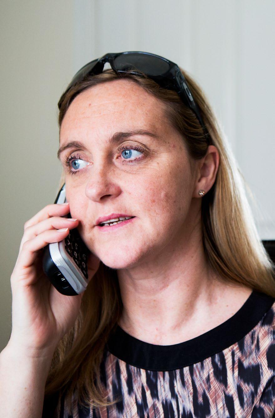
36 Coping It s completely natural to be upset when you ve been diagnosed with a retinal dystrophy like RP and it s normal to find yourself worrying about the future and how you ll manage with the change in your vision. It can sometimes be helpful to talk about these feelings with someone outside your circle of friends or family. At RNIB, we can help with our telephone Helpline and our Sight Loss Counselling team. Your GP or social worker may also find a counsellor for you if you feel this might help. Your eye clinic may also have a sight loss adviser (also known as an Eye Clinic Liaison Officer or ECLO), who can be on hand to provide you with further practical and emotional support about your eye condition. RP Fighting Blindness is a UK charity that gives advice and support to anyone affected by RP and other retinal dystrophies. They re able to provide up to date information about the latest research being carried out. Their national Helpline number is
37 37
38 Further help and support Having RP means that you ll eventually lose sight, but there are things that you can do to make the most of your remaining vision. This may mean making things bigger or smaller, using brighter lighting or using colour to make things easier to see. We have a lot of helpful information about living with sight loss as well as making the most of the sight that you have. You can find out more by calling our Helpline. Ask your ophthalmologist, optometrist or GP about low vision aids, such as a magnifier, and ask for a referral to your local low vision service. You should also ask your ophthalmologist whether you re eligible to register as sight impaired (partially sighted) or severely sight impaired (blind). Registration can act as your passport to expert help and sometimes to financial concessions. Even if you aren t registered, a lot of this support is still available to you. Local social services should be able to give you information on staying safe in your home and getting out and about safely. They should also be able to offer you some practical mobility training to give you more confidence when you are out. 38
39 If you have questions about anything you ve read in this publication, please get in touch with us. Our Helpline is your direct line to the support, advice and services you need. Whether you want to know more about your eye condition, buy a product from our shop, join our library, find out about possible benefit entitlements, or be put in touch with a trained counsellor, we re only a call away. It s also a way for you to join RNIB Connect, our community for anyone affected by sight loss. RNIB Connect is free to join and you ll have the chance to meet other people with similar experiences in our helpful, welcoming and supportive community. Give us a call today to find out how we can help you. RNIB Helpline helpline@rnib.org.uk We re ready to answer your call Monday to Friday 8am to 8pm and Saturday 9am to 1pm. 39
40 You can also get in touch by post or by visiting our website: RNIB 105 Judd Street London WC1H 9NE rnib.org.uk Other useful contacts RP Fighting Blindness PO Box 350 Buckingham MK18 1GZ RP Helpline Office telephone: rpfightingblindness.org.uk Sense 101 Pentonville Road London N1 9LG Telephone Text sense.org.uk 40

41 Driver and Vehicle Licensing Authority (DVLA) Drivers Medical Enquiries Swansea SA99 1TU
42 We value your feedback You can help us improve this publication by letting us know what you think about it. Please complete and return the form opposite to: RNIB Eye Health Information 105 Judd Street London WC1H 9NE You can also us at Please include your contact details if you re requesting information. 42
43 1. Where did you receive your copy of this publication? 2. Did you find the information easy to read and understand? Please give details of anything you feel could be improved. continued next page
44 3. Is there any information you would have found helpful, or were expecting to find, that was missing? 4. Do you have any other comments about this publication or any aspect of your contact with RNIB? URP 10879/10/2017
45 Information sources RNIB and The Royal College of Ophthalmologists do all we can to ensure that the information we supply is accurate, up to date and in line with the latest research and expertise. This publication uses information from: The Royal College of Ophthalmologists guidelines for treatment clinical research and studies obtained through literature reviews specific support groups for individual conditions medical text books RNIB publications and research. For a full list of references and information sources used in the compilation of this publication, eyehealth@rnib.org.uk. 45
46 About The Royal College of Ophthalmologists The Royal College of Ophthalmologists champions excellence in the practice of ophthalmology and is the only professional membership body for medically qualified ophthalmologists. The College is unable to offer direct advice to patients. If you re concerned about the health of your eyes, you should seek medical advice from your GP or ophthalmologist. rcophth.ac.uk 46
47
48 If you or someone you know is living with sight loss, we re here to help. RNIB Helpline Ask RNIB is the simple and easy way to find the answers to your questions online try it today at rnib.org.uk/ask This publication has been produced jointly by RNIB and The Royal College of Ophthalmologists, with kind support from The Lord Leonard and Lady Estelle Wolfson Foundation. RNIB and The Royal College of Ophthalmologists have full editorial control over the content of this publication RNIB and RCOphth RNIB reg charity nos , SC RCOphth reg charity no Produced date October 2017 Review date October 2020 PR10879 ISBN Ed 1
How the eye works. Causes of retinitis pigmentosa
 Retinitis pigmentosa Retinitis pigmentosa (RP) is the name given to a diverse group of inherited eye disorders which affect a part of the eye called the retina. RP causes permanent changes to your vision
Understanding. Posterior vitreous detachment
 Understanding Posterior vitreous detachment Contact us We re here to answer any questions you have about your eye condition or treatment. If you need further information about posterior vitreous detachment
Understanding. Glaucoma
 Understanding Glaucoma Contact us We re here to answer any questions you have about your eye condition or treatment. If you need further information about glaucoma or on coping with changes in your vision,
Understanding. Cataracts
 Understanding Cataracts Contact us We re here to answer any questions you have about your eye condition or treatment. If you need further information about cataracts or on coping with changes in your vision,
Understanding. Eye conditions related to diabetes
 Understanding Eye conditions related to diabetes Contact us We re here to answer any questions you have about your eye condition or treatment. If you need further information about eye conditions related
Understanding. Charles Bonnet syndrome
 Understanding Charles Bonnet syndrome Contact us We re here to answer any questions you have about your eye condition or treatment. If you need further information about Charles Bonnet syndrome or on coping
Understanding. cataracts. RCOphth
 Understanding cataracts RCOphth RNIB s Understanding series The Understanding series is designed to help you, your friends and family understand a little bit more about your eye condition. Other titles
What is Coloboma? How do we see with our eyes? Which parts of the eye can coloboma affect? Choroid
 Coloboma Factsheet Contents 3 What is Coloboma? 3 How do we see with our eyes? 3 Which parts of the eye can coloboma affect? 3 Iris 4 Lens zonules 4 Retina and choroid (chorioretinal) 4 Optic disc 4 Eyelids
RNIB UNDERSTANDING GLAUCOMA
 RNIB UNDERSTANDING GLAUCOMA Eye Info Understanding glaucoma Summary: Designed to help you understand more about your eye condition, this guide has been written by our experienced eye health team. What
Understanding Cataracts
 Understanding Cataracts For more information, please contact T 0116 249 8839 E info@vistablind.org.uk W www.vistablind.org.uk 2 Contents 3 About cataracts 4 How your eye works 5 Causes 7 Symptoms 9 Treatment
eye conditions related to diabetes
 Understanding eye conditions related to diabetes RCOphth RNIB s Understanding series The Understanding series is designed to help you, your friends and family understand a little bit more about your eye
Eye screening for patients taking hydroxychloroquine (Plaquenil )
") Eye screening for patients taking hydroxychloroquine (Plaquenil ) This leaflet contains important information for people taking hydroxychloroquine. This leaflet is available on audio CD. No one need face
Eye screening for patients taking hydroxychloroquine (Plaquenil ) This leaflet contains important information for people taking hydroxychloroquine. This leaflet is available on audio CD. No one need face
Eye screening for patients taking hydroxychloroquine (Plaquenil )
") Eye screening for patients taking hydroxychloroquine (Plaquenil ) Support throughout central vision loss This leaflet contains important information about eye health checks for people taking hydroxychloroquine.
Eye screening for patients taking hydroxychloroquine (Plaquenil ) Support throughout central vision loss This leaflet contains important information about eye health checks for people taking hydroxychloroquine.
Myopia and pathological myopia. Fundus Photo, Right Eye (OD)
") Myopia and pathological myopia Fundus Photo, Right Eye (OD) Contents 3 What is myopia and pathological myopia 3 How does the eye focus light? 3 What is myopia? 4 Development of myopia 4 What causes myopia?
Myopia and pathological myopia Fundus Photo, Right Eye (OD) Contents 3 What is myopia and pathological myopia 3 How does the eye focus light? 3 What is myopia? 4 Development of myopia 4 What causes myopia?
Understanding Glaucoma
 Understanding Glaucoma What is glaucoma? Glaucoma is the name for a group of eye conditions in which the optic nerve is damaged at the point at which it leaves the eye. As the diagram below shows, this
Age-Related. macular degeneration.
 Age-Related Macular Degeneration This pamphlet is designed to help people with age-related macular degeneration and their families better understand the disease. It describes the causes, symptoms, diagnosis,
Charles Bonnet syndrome
 Understanding Charles Bonnet syndrome RCOphth RNIB s Understanding series The Understanding series is designed to help you, your friends and family understand a little bit more about your eye condition.
GENERAL INFORMATION DIABETIC EYE DISEASE
 GENERAL INFORMATION DIABETIC EYE DISEASE WHAT IS DIABETIC EYE DISEASE? Diabetic eye disease is a term used to describe the common eye complications seen in people with diabetes. It includes: Diabetic retinopathy
Diabetes & Your Eyes
 Diabetes & Your Eyes Diabetes is a disease that occurs when the pancreas does not secrete enough insulin or the body is unable to process it properly. Insulin is the hormone that regulates the level of
RETINAL CONDITIONS RETINAL CONDITIONS
 GENERAL INFORMATION RETINAL CONDITIONS RETINAL CONDITIONS WHAT ARE RETINAL CONDITIONS? Retinal conditions affect the light-sensitive tissue at the back of eye known as the retina. They include diseases
Macular hole. Information for patients Ophthalmology (Vitreal Retina) Large Print
 Large Print") Macular hole Information for patients Ophthalmology (Vitreal Retina) Large Print page 2 of 16 What is the macula? The back of the eye has a light-sensitive lining called the retina, similar to the film
Macular hole Information for patients Ophthalmology (Vitreal Retina) Large Print page 2 of 16 What is the macula? The back of the eye has a light-sensitive lining called the retina, similar to the film
Understanding. Charles Bonnet syndrome
 Understanding Charles Bonnet syndrome 1 Further information Our Understanding series is designed to help you, your friends and family understand a little bit more about your eye condition. Other titles
Diabetic Retinopathy Information
 http://www.midwestretina.com Phone: (614)-339-8500 Toll Free: (866)-373-8462 Sugat S. Patel, M.D. Louis J. Chorich III, M.D. Dino D. Klisovic, M.D. Lisa M. Borkowski, M.D. Dominic M. Buzzacco, M.D. Johnstone
Brampton Hurontario Street Brampton, ON L6Y 0P6
 Diabetic Retinopathy What is Diabetic Retinopathy Diabetic retinopathy is one of the leading causes of blindness world-wide. Diabetes damages blood vessels in many organs of the body including the eyes.
Having an eye examination Eye examinations are important for everyone, they check; Factsheet
 Factsheet Vision 2020 UK The right to sight Dementia and Sight Loss Interest Group Eye examinations for people with dementia When you have dementia it is important to make the most of your sight. Wearing
Patient Information Cataract Surgery
 Patient Information Cataract Surgery Introduction This leaflet has been written to help you understand more about surgery for a cataract. It explains what the operation involves, the benefits and risks
Introduction How the eye works
 1 Introduction Diabetic retinopathy is a condition that can cause permanent loss of eyesight and even blindness. It is a major cause of loss of vision. But if a person with diabetes receives proper eye
Age-Related Macular Degeneration (AMD)
") Age-Related Macular Degeneration (AMD) What is the Macula? What is Dry AMD (Age-related Macular Degeneration)? Dry AMD is an aging process that causes accumulation of waste product under the macula leading
Age-Related Macular Degeneration (AMD) What is the Macula? What is Dry AMD (Age-related Macular Degeneration)? Dry AMD is an aging process that causes accumulation of waste product under the macula leading
Factsheet. Glaucoma. Are there different types of glaucoma? Yes. There are four main types.
 What is glaucoma? Glaucoma is the name for a group of eye conditions in which the optic nerve is damaged at the point where it leaves the eye. This nerve carries information from the light sensitive layer
Cataract Surgery Patient Information
 Cataract Surgery Patient Information www.ihg.org.uk Independent Health Group Patient Referral Centre T: 0330 3801362 E: ihg.referralsandenquiries@nhs.net Independent Health Group Limited is registered
Specialist Referral Service Willows Information Sheets. Cataract surgery
 Specialist Referral Service Willows Information Sheets Cataract surgery An operating microscope in use A total cataract - the normally black pupil is bluish white Cataract surgery These notes do not cover
About Charles Bonnet syndrome (CBS)
") Charles Bonnet syndrome (CBS) is a common condition among people who have lost their sight. It causes people who have lost a lot of vision to see things that aren't really there, known as visual hallucinations.
Charles Bonnet syndrome (CBS) is a common condition among people who have lost their sight. It causes people who have lost a lot of vision to see things that aren't really there, known as visual hallucinations.
UVEITIS IN GENERAL. Information for patients UVEITIS CLINIC WHAT IS UVEITIS? MAIN CATEGORIES OF UVEITIS
 Information for patients UVEITIS CLINIC UVEITIS IN GENERAL WHAT IS UVEITIS? The uvea is a name given to the pigmented layer of tissue inside the eye. When all or part of the uvea becomes inflamed, the
Your guide to juvenile macular dystrophies
 Support throughout Your guide to juvenile macular dystrophies No one need face macular degeneration alone. For information and support call 0300 3030 111. Macular Society QR codes Throughout this leaflet
Don t lose sight of the ones you love
 Don t lose sight of the ones you love Every day 100 people in the UK start to lose their sight. Only an eye examination can pick up the early signs of eye disease. Book your eye examination today. Your
OAKLEIGH EYE CENTRE. THE EYE Before looking at diabetic retinopathy it is important to understand what the healthy eye looks like and how it works.
 ABN: 80 836 359 971 Dr Mark Steiner 345 799X 135 Warrigal Road Dr Helen Steiner 292 419A OAKLEIGH VIC 3166 Tel: 03 9568 7706 Fax: 03 9568 4498 E-Mail: oakeye13@bigpond.com DIABETIC RETINOPATHY DIABETES
AgePage. Aging And Your Eyes. Steps To Protect Your Eyesight
 National Institute on Aging AgePage Aging And Your Eyes Are you holding the newspaper farther away from your eyes than you used to? Join the crowd age can bring changes that affect your eyesight. Some
Diabetic Retinopathy. What should know
 Diabetic Retinopathy What should know Diabetic Retinopathy: What you should know This booklet is for people with diabetic retinopathy and their families and friends. It provides information about diabetic
Diabetic retinopathy damage to the blood vessels in the retina. Cataract clouding of the eye s lens. Cataracts develop at an earlier age in people
 Diabetic Retinopathy What is diabetic eye disease? Diabetic eye disease refers to a group of eye problems that people with diabetes may face as a complication of diabetes. All can cause severe vision loss
Cataract Surgery. Patient Information. How your care will be organised. Introduction
 Patient Information Cataract Surgery If you have any questions regarding your operation please contact Parkerswell Day Case Unit on 01392 406013. They are available between 09:00-17:30, Monday to Friday.
Macular holes. What is a macular hole?
 Patient information Macular holes We hope this information will answer some of your questions about macular holes. If there is anything you do not understand, or if you have any concerns, please tell us
PATIENT INFORMATION LEAFLET MACULAR HOLE. What is the macula?
 What is the macula? The back of the eye has a light-sensitive lining called the retina, similar to the film in a camera. Light is focused through the eye onto the retina, allowing us to see. The centre
Ophthamology Directorate. Eye Injection for Macular Disorders Information for Patients
 Ophthamology Directorate Eye Injection for Macular Disorders Information for Patients As discussed at your appointment today, please call the Medical Retinal Services Coordinator as soon as possible (within
Glaucoma. College of Optometrists
 Glaucoma Overview Glaucoma is a group of eye diseases in which the optic nerve, which connects your eye to your brain, is damaged by the pressure of the fluid inside your eye. This may be because the pressure
Preparing for laser treatment for diabetic retinopathy and maculopathy
 Preparing for laser treatment for diabetic retinopathy and maculopathy Information for patients Preparing for laser treatment for diabetic retinopathy and maculopathy. This leaflet sets out to answer the
Diabetic Eye Disease
 Manchester Royal Eye Hospital Medical Retinal Services Information for Patients Diabetic Eye Disease This leaflet sets out to answer some of your questions about diabetic eye disease. You may wish to discuss
DIABETIC RETINOPATHY
 THE UK GUIDE DIABETIC RETINOPATHY Everything you need to know about diabetic retinopathy Jaheed Khan BSc (Hons) MBBS MD FRCOphth Fellow of the Royal College of Ophthalmologists Association for Research
Diabetic Retinopathy WHAT IS DIABETIC RETINOPATHY? WHAT CAUSES DIABETIC RETINOPATHY? WHAT ARE THE STAGES OF DIABETIC RETINOPATHY?
 Diabetic Retinopathy WHAT IS DIABETIC RETINOPATHY? Diabetic retinopathy affects 8 million Americans with diabetes. A leading cause of blindness in American adults, it is caused by damage to the small blood
Let s Learn About. Visual Impairment
 Let s Learn About Visual Impairment Vision impairment and Blindness have many causes. Birth defects, eye disorders or injuries, and age-related diseases such as glaucoma and cataracts can lead to loss
Patient information. Retinal Detachment Surgery St. Paul s Eye Unit PIF 024 V7
 Patient information Retinal Detachment Surgery St. Paul s Eye Unit PIF 024 V7 Your eye specialist has advised you to have retinal detachment surgery. This leaflet gives you information that will help you
Toxoplasmosis Of The Eye
 Medical Information Document On Toxoplasmosis Of The Eye What we see is made in the brain from signals given to it by the eyes. What we see is in fact made in the brain. The brain makes sight from signals
Screening for Uveitis in Children
 Information for patients and parents Manchester Royal Eye Hospital Paediatric Uveitis Service Screening for Uveitis in Children What is uveitis? Uveitis is inflammation of a layer of the eye, called the
Taking care of hearing and sight: a quality of life issue for residents and staff of care homes
 R&RA Information paper Eyes and Ears Taking care of hearing and sight: a quality of life issue for residents and staff of care homes A paper produced by R&RA for professionals working in UK care homes
Flashers and Floaters
 Flashers and Floaters Introduction Sometimes people see small, moving spots or specks in their field of vision. These sensations are called floaters. About 7 out of 10 people experience floaters at some
Asymptomatic retinal detachment
 Patient information Asymptomatic retinal detachment We hope this information will answer some of your questions about asymptomatic retinal detachment. If there is anything you do not understand, or if
Epiretinal membrane. Information for patients Ophthalmology (Vitreal Retina) Large Print
 Large Print") Epiretinal membrane Information for patients Ophthalmology (Vitreal Retina) Large Print What is epiretinal membrane? An epiretinal membrane is a condition where a very thin layer of scar tissue forms on
Epiretinal membrane Information for patients Ophthalmology (Vitreal Retina) Large Print What is epiretinal membrane? An epiretinal membrane is a condition where a very thin layer of scar tissue forms on
Patient Information Argon macular laser treatment Introduction
 Patient Information Argon macular laser treatment Introduction The eye doctor has found that your diabetic eye disease (retinopathy) has now progressed to a stage where there has been some water logging
GHPI0100_06_10 Contact: Ophthalmology Review due: June What is a Cataract?
 GHPI0100_06_10 Contact: Ophthalmology Review due: June 2013 What is a Cataract? Further information If you or a relative have access to the internet, you can use the following websites for further information:
Diabetic Eye Screening
 Diabetic Eye Screening Lorraine Lockwood Engagement Manager Health Intelligence Classification: Internal / Public 26/10/16 Who we are (EDESP) formed on 1 st April 2016 Who should be screened? All patients
Information for patients. Epiretinal Membrane. Royal Hallamshire Hospital
 Information for patients Epiretinal Membrane Royal Hallamshire Hospital 2 What is an Epiretinal Membrane? An Epiretinal Membrane is a condition where a very thin layer of scar tissue forms on the surface
If you or one of your relatives has Parkinson s, you may want to know
 Information and support Does Parkinson s run in families? If you or one of your relatives has Parkinson s, you may want to know if the condition can be passed down through families. This information sheet
Emotional impact of sight loss
 Emotional impact of sight loss The biggest cause of sight loss in this country is macular disease. Coping with your emotions after being diagnosed can be difficult. No one need face macular degeneration
NHS Diabetic Eye Screening Programme
 NHS Diabetic Eye Screening Programme Slit lamp examination Public Health England leads the NHS Screening Programmes Who is this leaflet for? This leaflet is for people who need to be examined for diabetic
Understanding Diabetic Retinopathy
 Understanding Diabetic Retinopathy What Is Diabetic Retinopathy? Diabetes damages blood vessels in the rear of the eye. This condition is called diabetic retinopathy. It can lead to vision loss or blindness.
Introduction. Diagnosis
 Introduction Life and Change with Usher is a research study about the lives of people with Usher syndrome. Over two years we studied the lives of people with Usher, first in books, articles and blogs,
Frequently Asked Questions about General Ophthalmology:
 1. Normal Eye Structure The eye is a slightly asymmetrical globe, about an inch in diameter. The parts of the eye include: Cornea (a clear dome over the iris), Iris (the pigmented part); Pupil (the black
Caregiver s guide to wet AMD
 Caregiver s guide to wet AMD Learn more about wet AMD (wet age-related macular degeneration) and how to support someone with wet AMD. Who is LUCENTIS for? LUCENTIS (ranibizumab injection) is a prescription
Having a vitrectomy surgery to repair your retinal detachment
 Having a vitrectomy surgery to repair your retinal detachment If you need information on audiotape about having a vitrectomy or your hospital visit, please call 020 7188 8815. You have been given this
Epiretinal membrane. What is an epiretinal membrane?
 Patient information Epiretinal membrane We hope this information will answer some of your questions about epiretinal membrane. If there is anything you do not understand, or if you have any concerns, please
Treatment of Retinal Vein Occlusion (RVO)
") Manchester Royal Eye Hospital Medical Retina Services Information for Patients Treatment of Retinal Vein Occlusion (RVO) What is a Retinal Vein Occlusion (RVO)? The retina is the light sensitive layer
Manchester Royal Eye Hospital Medical Retina Services Information for Patients Treatment of Retinal Vein Occlusion (RVO) What is a Retinal Vein Occlusion (RVO)? The retina is the light sensitive layer
Cataract. What is a Cataract?
 Cataract What is a Cataract? We all have a lens in our eye. This is positioned just behind the iris, which is the coloured ring in the eye that gives your eye its colour. The lens function is to focus
Dementia and Sight Loss
 Dementia and Sight Loss About this leaflet This leaflet has useful information for anyone with dementia. You may also find this leaflet helpful if you're caring for someone with dementia. As well as learning
Cataract. What is a Cataract?
 Cataract What is a Cataract? We all have a lens in our eye. This is positioned just behind the iris, which is the coloured ring in the eye that gives your eye its colour. The lens s function is to focus
JUST DIAGNOSED WITH DIABETES?
 JUST DIAGNOSED WITH DIABETES? Being told that you have diabetes can be a real shock. And learning to live with it can be a challenge. You might be going through all sorts of emotions. It s perfectly normal
Cataract Surgery: Information for patients. Back of eye. Vitreous. Retina. Lens
 Patient information Cataract Surgery: Information for patients Front of eye Cornea Pupil Iris Back of eye Vitreous Retina Lens The anatomy of the eye is illustrated above. Your cataract is a clouding of
Information for Patients. Retinal Detachment
 Information for Patients Retinal Detachment Manchester Royal Eye Hospital Retinal services Your eye doctor has told you that you have a retinal detachment. This leaflet will help you understand your condition
Treatment of Age Related Macular Degeneration (AMD) by Intravitreal Injection
 by Intravitreal Injection") Information for Patients Manchester Royal Eye Hospital Medical Retina Services Treatment of Age Related Macular Degeneration (AMD) by Intravitreal Injection What is age related macular degeneration (AMD)?
Information for Patients Manchester Royal Eye Hospital Medical Retina Services Treatment of Age Related Macular Degeneration (AMD) by Intravitreal Injection What is age related macular degeneration (AMD)?
Retinopathy Of Prematurity (or) Retrolental Fibroplasia )
 Retrolental Fibroplasia )") Medical Information Document On Retinopathy Of Prematurity (or) Retrolental Fibroplasia ) What we see is made in the brain from signals given to it by the eyes. What we see is in fact made in the brain.
Medical Information Document On Retinopathy Of Prematurity (or) Retrolental Fibroplasia ) What we see is made in the brain from signals given to it by the eyes. What we see is in fact made in the brain.
Patient Information Brochure. Cataract
 Patient Information Brochure Cataract Q: What is cataract? A: A cataract is an opacity (or cloudiness) in the lens of the eye. This cloudiness develops inside the lens and restricts light passing through
A helping hand when you need it most
 A helping hand when you need it most Priority Services Register Promise The Priority Services Register (PSR) is free to join. It helps energy companies * like us to look after customers who have extra
Advances in assessing and managing vision impairment
 Advances in assessing and managing vision impairment John Grigg Associate Professor and Head Discipline of Ophthalmology Consultant Ophthalmologist Sydney Eye Hospital and The Children s Hospital at Westmead
Ophthalmology. Ophthalmology Services
 Ophthalmology Ophthalmology Services The Ophthalmology service offers the latest and most comprehensive eye care for patients. With a dedicated team of eye surgeons and consultants, we treat vision problems
What is posterior cortical atrophy?
 What is posterior cortical atrophy? Introduction This booklet is about posterior cortical atrophy (PCA). It is for people who have PCA, their family, friends and carers or anyone else with an interest
Information for patients
 Information for patients Intravitreal Anti-VEGF Treatment (vascular endothelial growth factor) This leaflet gives you information that will help you decide whether to have intravitreal treatment. It also
Moving Forward. Support for you after a diagnosis of breast cancer. The breast cancer support charity
 Moving Forward Support for you after a diagnosis of breast cancer The breast cancer support charity We understand breast cancer changes everything. If you ve had treatment for breast cancer and are looking
VISIONCARE S IMPLANTABLE MINIATURE TELESCOPE (by Dr. Isaac Lipshitz)
") PATIENT INFORMATION BOOKLET PAGE 1 OF 32 VISIONCARE S IMPLANTABLE MINIATURE TELESCOPE (by Dr. Isaac Lipshitz) AN INTRAOCULAR TELESCOPE FOR TREATING SEVERE TO PROFOUND VISION IMPAIRMENT DUE TO BILATERAL
PATIENT INFORMATION BOOKLET PAGE 1 OF 32 VISIONCARE S IMPLANTABLE MINIATURE TELESCOPE (by Dr. Isaac Lipshitz) AN INTRAOCULAR TELESCOPE FOR TREATING SEVERE TO PROFOUND VISION IMPAIRMENT DUE TO BILATERAL
Uveitis. Pt Info Brochure. Q: What is Uvea?
 Pt Info Brochure Uveitis Q: What is Uvea? A: Uvea is the middle layer of the eye. It is the most vascular structure of the eye. It provides nutrition to the other parts of the eye. The uvea is made up
Helping you decide 2014 edition Easy Read
 Breast Screening Helping you decide 2014 edition Easy Read We have made this leaflet as accessible as possible. There are still some difficult ideas in it. You might need to ask a friend, family member,
Retinal Tear and Detachment
 Retinal Tear and Detachment Introduction The retina is the layer of tissue in the back of the eye that is responsible for vision. It is attached to the choroid tissue, which supplies the retina with blood.
Autosomal Dominant Polycystic Kidney Disease
 I want to know more about... Autosomal Dominant Polycystic Kidney Disease together the answer This booklet is about autosomal dominant polycystic kidney disease (ADPKD). It is intended as a general guide
Seeking treatment for an eating disorder?
 Seeking treatment for an eating disorder? The first step is a GP appointment. This leaflet has guidance for the person who has or may have an eating disorder, anyone supporting them, and their GP. It s
Cataract and Cataract Surgery
 What is a cataract? A cataract is a cloudy area in the lens of your eye. The lens is located just behind the iris (coloured part of the eye). The lens helps to focus light entering the eye to give a clear
Open Patient Meeting
 What s new in the treatment of glaucoma Andrew Tatham MBChB FRCOphth FRCSEd FEBO Consultant Eye Surgeon, Princess Alexandra Eye Pavilion, Edinburgh Good afternoon everyone, it s very nice to be here and
Vitrectomy for diabetic vitreous haemorrhage
 Vitrectomy for diabetic vitreous haemorrhage Why have I been given this leaflet? If you have been given this leaflet it is likely that you have been asked to think about whether you want to go ahead with
What is Fundus Fluorescein Angiography and Indocyanine Green Angiography?
 What is Fundus Fluorescein Angiography and Indocyanine Green Angiography? Information for patients The Leeds Teaching Hospitals NHS Trust n Introduction Fundus Fluorescein Angiography (FFA) is a routine
Important: Please read before your appointment
 Cataract Surgery Important: Please read before your appointment Consent for cataract surgery Prior to you having cataract surgery, you will be asked to sign a consent form. It is important that you understand
If you would like further information, please ask any of the staff at the clinic. They will be happy to help.
 Photodynamic Therapy Patient Information Ninewells Hospital The aim of this leaflet is to give you some information about Photodynamic Therapy. It contains some useful information on what you can expect
Glaucoma. Cornea. Iris
 Glaucoma Introduction Glaucoma is a group of eye diseases that can lead to blindness if not treated. Openangle glaucoma, the most common form of glaucoma, affects about 3 million Americans. Half of those
Driving. Living with dementia series
 Driving Living with dementia series B For more information visit alzheimers.org.uk Driving If you drive, you may need to make some changes after getting a diagnosis of dementia. You may still be able to