Cardiac FDG Patterns Seen In Oncologic PET Studies: What s Normal and What s Not?
|
|
|
- Norma Nichols
- 6 years ago
- Views:
Transcription
1 Cardiac FDG Patterns Seen In Oncologic PET Studies: What s Normal and What s Not? Alan H. Maurer, M.D. Director of Nuclear Medicine Temple University Hospital And School of Medicine Philadelphia, PA
2 Objectives Review PET myocardial metabolism Role of fatty acids, glucose, oxidative metabolism Fasting vs glucose loaded conditions Classic patterns» Acute ischemia» Chronic stunning/hibernation Review differential diagnosis of abnormal cardiac FDG uptake seen in fasted oncologic patients Explore range of normal patterns of cardiac FDG uptake seen in oncologic PET studies Personal observations Literature
3 Cardiac PET Agents Blood flow NH-13 (cyclotron) Rubidium -82 (generator) Viability F18 FDG Other Oxygen utilization» O-15 Fatty Acids» C-11 Palmitate Acetate» C-11 Acetate Apoptosis» F-18 annexin V
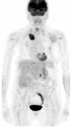
4 Normal Distribution of FDG Brain: high uptake in the gray matter Head/Neck: Salivary and lymphoid tissues variable Myocardium: very variable uptake Lungs: very low/no uptake Mediastinum: low uptake (nodes vessles) Liver: low uptake Spleen: no/very low uptake GI tract: variable activity (esophagus, stomach, colon) Urinary tract: excretes FDG Ovarian/uterine: varible with menstrual cycle Testicular: variable Muscular system: low uptake at rest Musculoskeletal Promininet periarticular muscle, joints
 Liver: low uptake Spleen: no/very low uptake GI tract: variable activity (esophagus, stomach, colon) Urinary tract: excretes FDG Ovarian/uterine: varible with menstrual cycle")
5 Normal Distribution of FDG Brain: high uptake in the gray matter Head/Neck: Salivary and lymphoid tissues variable Myocardium: very variable uptake Lungs: very low/no uptake Mediastinum: low uptake (nodes vessles) Liver: low uptake Spleen: no/very low uptake GI tract: variable activity (esophagus, stomach, colon) Urinary tract: excretes FDG Ovarian/uterine: varible with menstrual cycle Testicular: variable Muscular system: low uptake at rest Musculoskeletal Promininet periarticular muscle, joints
6 What is Normal Cardiac Appearance on Oncologic FDG PET? Answer? Depends!
7 Normal Myocardial Metabolism Complex interplay between: Myocardial blood flow Energy demand/work load Substrate availability» Fasted» Fed state Hormonal control
8 Simplified Cardiac Metabolism Glucose Fatty Acids Glycolysis ß Oxidation Pyruvate Acetyl CoA Krebs cycle ATP Contraction
9 Myocardial Substrait Metabolism Plasticity Normal Conditions 50-70% energy from oxidation of fatty acids 50-30% energy from carbohydrate (glucose & lactate) Fasting > 90% from Fatty Acids» Increased lipolysis in peripheral adipose tissue... increases plasma fatty acids» Decrease in transport of glucose into myocyte

10 PET Tracers Myocardial Metabolism Herrero and Gropler: J Nucl Cardiol without 2005:12: permission of author.
11 Factors Which Can Change Myocardial Metabolism Herrero and Gropler: J Nucl without Cardiol permission 2005:12: of author.
12 FDG Fasted/Non Fasting Patient Preparation Myocardial Viability Studies Oral glucose loading (one hour before injection) Euglycemic-hyperinsulinemic clamp Niacin (nicotinic acid derivative) Oncologic Studies ( Glucose < 200 ) NPO after midnight Minimum 4-6 hr fast Diabetics (no recent insulin)

13 Dietary Effect Myocardial Uptake Fasting /Regular Diet Low Carbohydrate/High Protein Diet
14 What is Normal Cardiac Appearance on Oncologic FDG PET? Depends - Patient Prep! & Depends Technique» Window/Leveling» Gating

15 Window/Levels for Cardiac Detail

16 Window/Levels - Cardiac Heterogeneity


17 Normal Patterns 01-03

18 Normal Cardiac Appearance Fasted vs Glucose Loading
19 First Report - Increased Posterolateral Cardiac FDG(1990) Gropler et al - Nonuniformity in Myocardial Accumulation of Fl -18- Fluorodeoxyglucose in Normal Fasted Humans J Nucl Med 1990;31: Fasting Fed FDG C11-acetate FDG C11-acetate Septum Anterior Lateral Posterior Fasted 100 Fed


20 Iozzo et al
21 Intra-individual Variability Is fasted FDG cardiac activity consistent on serial studies? Answer = YES Visual grading of low(less than lung) or high (greater than lung) cardiac FDG activity 47 patients with 218 scans Good reproducibility Diabetics less likely to have high cardiac activity Pts with lymphoma more likely to have high uptake No association with age, sex or weight Khandani et al - Intra-individual variability of cardiac uptake on serial whole-body 18F-FDG PET. Nuc Med Com without 26(9):787-91, permission of 2005 author.
22 PET FDG - Viability Potentially Reversible Cardiac Dysfunction Concepts Non viable tissue = infarction with no recovery of function Hibernation = Perfusion and contractile function decreases to reduce demand, new supplydemand balance Repetitive stunning = Transient ischemic episode, prompt normalization of perfusion but delayed recovery of contractile function
23 Stress Nl Stress Rest - FDG Rest FDG Stress Rest FDG Stress Rest FDG H. Schelbert: SNM Learning Center


24 Classic Ischemia Rest MBF Stress MBF FDG without H permission Schelbert, of UCLA author. - PLC
25 Perfusion 13 NH 3 Metabolism 18 FDG Perfusion-Metabolism Mismatch Reversible Contractile Dysfunction without H permission Schelbert, of UCLA author. - PLC
26 Perfusion 13 NH 3 Metabolism 18 FDG Perfusion-Metabolism Match Irreversible Contractile Dysfunction H Schelbert, UCLA - PLC
27 Abnormal Cardiac FDG Activity Differential Diagnoses Diffuse LVH, RVH Atrial Fibrillation Post Cardiac Transplantation Myocarditis» Radiation induced Focal Ischemia/Hibernation Fat Sarcoidosis Pericardial disease (mets) Valve infection» Native and prosthetic Muscular Dystrophy Technical» Misalignment artifact (attenuation correction)
28 Diffuse Chamber Enlargement LV Hypertension Other myopathies RV COPD Shunts Atria A Fib» LA, RA, Bi-atrial Normal Variant» Right
29 4 Chamber Enlargement


30 Atrial Enlargement Generalized cardiac enlargement: chronic atrial fibrillation

31 LA Enlargement (Atrial Fibrillation)

32 Case: 54 yo male, lymphoma (No Cardiac History)


33 Increased R Atrial Activity? Common Finding No! - Observed in 10 patients of 2,367 screenings
34 RA Uptake vs LA in A Fib (RA > LA inc glucose utilization?)
35 Focal FDG Abnormalities Ischemia/Hibernation (short term and chronic) Fat Sarcoidosis Pericardial disease (mets) Endocarditis» Native and prosthetic valve infection Muscular Dystrophy Technical» Misalignment artifact (attenuation correction)

36 Papillary Muscle Ischemia - Case Report


37 Case: 71 yo male, s/p resection LUL NSCLC (Prominent Fat AV Groove)


38 LHIS (Chest 2005; 128: )
39 Cardiac Sarcoidosis Cardiac involvement 20-80% patients Bundle branch or AV block 25% Cardiac death up to 75%

40 FDG Sarcoid Detection

41 Metastatic Disease

42 Infection 74 yo female with history of melanoma, breast ca and new pulmonary nodule Diagnosis: Infected mitral valve prosthetic ring Abidov et al: Clin Nucl Med: 2004;29:848
43 Personal Observations on Normal Oncologic FDG Cardiac Patterns Predominant anterolateral and inferolateral wall FDG activity Basal ring FDG activity Variants: partial and full Combinations of A and B Uniform FDG pattern Physiologic variants Prominent papillary muscle Fat:» Epicardial, pericardial, interatrial septum RVH, LVH Atrial variants» Normal junction with SVC» Atrial fibrillation Myocardial heterogeneity

44 Normal Diffuse - Min Cardiac 43 yo female, cervical cancer (not blood pool )

45 Nl Ant-Lat, Posterior with Basal Ring

46 Normal - Basal Ring Patterns

47 Normal - Half Base

48 Focal Base + Pap Muscle
49 FDG Uptake Vascular Inflammation/Atherosclerosis FDG activity commonly seen in atherosclerotic areas in vessles on CT Animal studies correlate FDG activity with Macrophage rich experimental atherosclerotic lesions Macrophage activity is increased in: Acute coronary syndrome Ruptured plaques

50 Aortitis B. Siegel, Mallinckrodt without permission Radiology of author.

51 FDG Correlation with Macrophages


52 FDG Uptake in Symptomatic Plaque Rudd et al: Circulation 2002;105:

53 CS - AORTIC FINDINGS
54 Normal AO/ Abn Node vs Calc AO and Pre Carinal Node


55 FDG + Calcified AO Plaque vs FDG + Neg Plaque
56 Conclusions Cardiac FDG patterns are very heterogeneous in the fasted state and any interpretations should be cautious without clinical history or other evidence of significance Both diffuse and focal patterns however can have clinical relevance and should be reported when appropriate Focal FDG activity in atherosclerotic plaque may also have clinical significance and can be confused with nodal pathology
57 Question Under fasting conditions regional myocardial glucose activity is normally highest in which of the following left ventricular myocardial segments? A. Septum and anterior walls B. Septum and posterior walls C. Lateral and posterior walls D. Lateral and anterior walls
58 Answers A is incorrect - * B is incorrect - * C is correct. While regional glucose activity is very heterogeneous in the fasted state several articles document higher uptake in the lateral and posterior LV walls.* D is incorrect - * Reference: Gropler RJ et al. Nonuniformity in myocardial accumulation of F-18 FDG in normal fasted humans. J Nucl Med 1990;31:

59 Case: 38 yo male with LUL nodule without permission of Hartigan author.

60 Nl Pattern 05
Radiologic Assessment of Myocardial Viability
 November 2001 Radiologic Assessment of Myocardial Viability Joshua Moss, Harvard Medical School Year III Patient EF 66yo female with a 3-year history of intermittent chest pain previously relieved by sublingual
PET/CT and PET/MR of Cardiac Tumors
 PET/CT and PET/MR of Cardiac Tumors Andrew W. Bowman, MD PhD Assistant Professor of Radiology Mayo Clinic Florida NASCI Annual Meeting San Antonio, TX Oct 9, 2017 2017 MFMER slide-1 Disclosures No financial
1. LV function and remodeling. 2. Contribution of myocardial ischemia due to CAD, and
 1 The clinical syndrome of heart failure in adults is commonly associated with the etiologies of ischemic and non-ischemic dilated cardiomyopathy, hypertrophic cardiomyopathy, hypertensive heart disease,
Cardiac Imaging. Kimberly Delcour, DO, FACC. Mahi Ashwath, MD, FACC, FASE. Director, Cardiac CT. Director, Cardiac MRI
 Cardiac Imaging Kimberly Delcour, DO, FACC Director, Cardiac CT Mahi Ashwath, MD, FACC, FASE Director, Cardiac MRI Cardiac Imaging Discuss the clinical applications of and indications for: Cardiac CT Nuclear
Pearls & Pitfalls in nuclear cardiology
 Pearls & Pitfalls in nuclear cardiology Maythinee Chantadisai, MD., NM physician Division of Nuclear Medicine, Department of radiology, KCMH Principle of myocardial perfusion imaging (MPI) Radiotracer
@02-126_Coronary_calcification.ppt. Professor Molecular and Medical Pharmacology
 Assessment of Myocardial Viability Jamshid Maddahi, M.D., FACC, FASNC Professor Molecular and Medical Pharmacology (Nuclear Medicine) and Medicine (Cardiology) David Geffen School of Medicine at UCLA Director,
Cardiac Viability Testing A Clinical Perspective Annual Cardiac Imaging Symposium. Lisa M Mielniczuk MD FRCPC University of Ottawa Heart Institute
 Cardiac Viability Testing A Clinical Perspective Annual Cardiac Imaging Symposium Lisa M Mielniczuk MD FRCPC University of Ottawa Heart Institute 62 year old male Anterior STEMI late presentation, occluded
The Heart. The Heart A muscular double pump. The Pulmonary and Systemic Circuits
 C H A P T E R 19 The Heart The Heart A muscular double pump circuit takes blood to and from the lungs Systemic circuit vessels transport blood to and from body tissues Atria receive blood from the pulmonary
Cardiac Imaging Tests
 Cardiac Imaging Tests http://www.medpagetoday.com/upload/2010/11/15/23347.jpg Standard imaging tests include echocardiography, chest x-ray, CT, MRI, and various radionuclide techniques. Standard CT and
the Cardiovascular System I
 the Cardiovascular System I By: Dr. Nabil A Khouri MD, MsC, Ph.D MEDIASTINUM 1. Superior Mediastinum 2. inferior Mediastinum Anterior mediastinum. Middle mediastinum. Posterior mediastinum Anatomy of
Gated blood pool ventriculography: Is there still a role in myocardial viability?
 Gated blood pool ventriculography: Is there still a role in myocardial viability? Oliver C. Alix, MD Adult Clinical and Nuclear Cardiology St. Luke s Medical Centre - Global City Case Presentation A 62-year-old
12 Lead EKG Chapter 4 Worksheet
 Match the following using the word bank. 1. A form of arteriosclerosis in which the thickening and hardening of the vessels walls are caused by an accumulation of fatty deposits in the innermost lining
CARDIAC PET PERFUSION IMAGING with RUBIDIUM-82
 CARDIAC PET PERFUSION IMAGING with RUBIDIUM-82 Pr Denis AGOSTINI Président du Groupe de Cardiologie Nucléaire et IRM CHU Caen Bordeaux 2006 Cardiac Perfusion-Metabolism Mismatch with PET Cumulative Survival
POSITRON EMISSION TOMOGRAPHY (PET)
") Status Active Medical and Behavioral Health Policy Section: Radiology Policy Number: V-27 Effective Date: 08/27/2014 Blue Cross and Blue Shield of Minnesota medical policies do not imply that members should
Status Active Medical and Behavioral Health Policy Section: Radiology Policy Number: V-27 Effective Date: 08/27/2014 Blue Cross and Blue Shield of Minnesota medical policies do not imply that members should
Cardiac MRI: Cardiomyopathy
 Cardiac MRI: Cardiomyopathy Laura E. Heyneman, MD I do not have any relevant financial relationships with any commercial interests Cardiac MRI: Cardiomyopathy Laura E. Heyneman, MD Duke University Medical
THE CARDIOVASCULAR SYSTEM. Part 1
 THE CARDIOVASCULAR SYSTEM Part 1 CARDIOVASCULAR SYSTEM Blood Heart Blood vessels What is the function of this system? What other systems does it affect? CARDIOVASCULAR SYSTEM Functions Transport gases,
NUCLEAR CARDIOLOGY UPDATE
 Nuclear Cardiology David K. Shelton, Jr., MD NUCLEAR CARDIOLOGY UPDATE No Conflicts. No Disclosures. No Smoking. David K. Shelton UCDMC Nuclear Cardiology Nuclear Cardiology Radionuclide Ventriculography
2. right heart = pulmonary pump takes blood to lungs to pick up oxygen and get rid of carbon dioxide
 A. location in thorax, in inferior mediastinum posterior to sternum medial to lungs superior to diaphragm anterior to vertebrae orientation - oblique apex points down and to the left 2/3 of mass on left
New Visions in PET: Surgical Decision Making and PET/CT
 New Visions in PET: Surgical Decision Making and PET/CT Stanley J. Goldsmith, MD Director, Nuclear Medicine Professor, Radiology & Medicine New York Presbyterian Hospital- Weill Cornell Medical Center
The Heart. Happy Friday! #takeoutyournotes #testnotgradedyet
 The Heart Happy Friday! #takeoutyournotes #testnotgradedyet Introduction Cardiovascular system distributes blood Pump (heart) Distribution areas (capillaries) Heart has 4 compartments 2 receive blood (atria)
Le FDG dans l évaluation de la viabilité : Quelle place par rapport aux autres traceurs?
 Le FDG dans l évaluation de la viabilité : Quelle place par rapport aux autres traceurs? Pierre-Yves MARIE, Médecine Nucléaire, CHU de Nancy PET Gated-SPECT MRI FDG imaging: effect of dietary status fasting
Ch.15 Cardiovascular System Pgs {15-12} {15-13}
 Ch.15 Cardiovascular System Pgs {15-12} {15-13} E. Skeleton of the Heart 1. The skeleton of the heart is composed of rings of dense connective tissue and other masses of connective tissue in the interventricular
Why Cardiac MRI? Presented by:
 Why Cardiac MRI? Presented by: Lisa G. Carkner, MD, FACC 1 Disclosures I have no financial disclosures Objectives Review basic principles of Cardiac MRI. What patient characteristics do I need to consider
Emerging role of PET in nuclear cardiology. Dr. Erick Alexánderson. Rosas
 Emerging role of PET in nuclear cardiology Dr. Erick Alexánderson Rosas PET principles e + e - 180 PET/CT Cyclotron Unit UNAM Cyclotron Radiopharmacy PET Camera CT PET PET CT Perfusion Anatomic evaluation
ADVANCED CARDIOVASCULAR IMAGING. Medical Knowledge. Goals and Objectives PF EF MF LF Aspirational
 Medical Knowledge Goals and Objectives PF EF MF LF Aspirational Know the basic principles of magnetic resonance imaging (MRI) including the role of the magnetic fields and gradient coil systems, generation
411.1 INTERMED CORONARY SYNDROME 412 OLD MYOCARDIAL INFARCT ANGINA PECTORIS OT/UNSPEC CORONARY ATHRSCL UNS VESSEL
 78451 Myocardial perfusion imaging, tomographic (SPECT) (including attenuation correction, qualitative NUC 1073 410.91 AMI UNSP INITIAL EPISODE 410.92 AMI UNSP SUBSEQUENT EPISODE 414.8 CHR ISCHEMIC HRT
HEALTHFIRST 2011 RADIOLOGY PROGRAM CODE LIST
 HEALTHFIRST 2011 RADIOLOGY PROGRAM CODE LIST Outpatient Radiology utilization call Carecore at 1-877-773-6964 Modality CPT CODE Description CT SCANS 70450 CT HEAD/BRAIN W/O CONTRAST CT SCANS 70460 CT HEAD/BRAIN
Conflict Disclosures. Vermont Cardiac Network. Outline. Series Learning Objectives 4/27/2016. Scott E. Friedman April 28, 2016
 Conflict Disclosures Vermont Cardiac Network The Speaker has reported no significant financial relationship with any companies whose product may be germane to the content of their presentations or who
Update in Nuclear Imaging of Amyloidosis and Sarcoidosis
 Update in Nuclear Imaging of Amyloidosis and Sarcoidosis Balaji Tamarappoo MD, PhD, Cedars-Sinai Heart Institute and Biomedical Imaging Research Institute Cedars-Sinai Medical Center Los Angeles, CA, USA.
CV Anatomy Quiz. Dr Ella Kim Dr Pip Green
 CV Anatomy Quiz Dr Ella Kim Dr Pip Green Q1 The location of the heart is correctly described as A) lateral to the lungs. B) medial to the sternum. C) superior to the diaphragm. D) posterior to the spinal
The Value of Stress MRI in Evaluation of Myocardial Ischemia
 The Value of Stress MRI in Evaluation of Myocardial Ischemia Dr. Saeed Al Sayari, MBBS, EBCR, MBA Department of Radiology and Nuclear Medicine Mafraq Hospital, Abu Dhabi United Arab Emirates Introduction
Cardiology/Cardiothoracic
 Cardiology/Cardiothoracic ICD-9-CM to ICD-10-CM Code Mapper 800-334-5724 www.contexomedia.com 2013 ICD-9-CM 272.0 Pure hypercholesterolemia 272.2 Mixed hyperlipidemia 272.4 Other and hyperlipidemia 278.00
I have no financial disclosures
 Manpreet Singh MD I have no financial disclosures Exercise Treadmill Bicycle Functional capacity assessment Well validated prognostic value Ischemic assessment ECG changes ST segments Arrhythmias Hemodynamic
PET myocard perfusion & viability Riemer Slart
 PET myocard perfusion & viability Riemer Slart Nuclear Medicine Physician Dept. of Nuclear Medicine and Molecular Imaging University Medical Center Groningen, the Netherlands Professor in Molecular Imaging,
Appendix D Output Code and Interpretation of Analysis
 Appendix D Output Code and Interpretation of Analysis 8 Arrhythmia Code No. Description 8002 Marked rhythm irregularity 8110 Sinus rhythm 8102 Sinus arrhythmia 8108 Marked sinus arrhythmia 8120 Sinus tachycardia
Heart disease remains the leading cause of morbidity and mortality in industrialized nations. It accounts for nearly 40% of all deaths in the United
 Heart disease remains the leading cause of morbidity and mortality in industrialized nations. It accounts for nearly 40% of all deaths in the United States, totaling about 750,000 individuals annually
12 Lead ECG Interpretation
 12 Lead ECG Interpretation Julie Zimmerman, MSN, RN, CNS, CCRN Significant increase in mortality for every 15 minutes of delay! N Engl J Med 2007;357:1631-1638 Who should get a 12-lead ECG? Also include
Malignant Cardiac Tumors Rad-Path Correlation
 Malignant Cardiac Tumors Rad-Path Correlation Vincent B. Ho, M.D., M.B.A. 1 Jean Jeudy, M.D. 2 Aletta Ann Frazier, M.D. 2 1 Uniformed Services University of the Health Sciences 2 University of Maryland
Section 5.1 The heart and heart disease
 Section 5.1 The heart and heart disease Mammals are too large to rely on diffusion. They need a circulatory system to move substances around the body. Blood moves down pressure gradients, from high to
SPECT-CT: Τι πρέπει να γνωρίζει ο Καρδιολόγος
 SPECT-CT: Τι πρέπει να γνωρίζει ο Καρδιολόγος Δρ Αναστασία Κίτσιου Διευθύντρια, Καρδιολογική Κλινική, Σισμανόγλειο ΓΝΑ Chair, Education Committee, Section on Nuclear Cardiology & Cardiac CT, EACVI, ESC
The Cardiovascular System
 The Cardiovascular System The Manila Times College of Subic Prepared by: Stevens B. Badar, RN, MANc THE HEART Anatomy of the Heart Location and Size approx. the size of a person s fist, hollow and cone-shaped,
Common Codes for ICD-10
 Common Codes for ICD-10 Specialty: Cardiology *Always utilize more specific codes first. ABNORMALITIES OF HEART RHYTHM ICD-9-CM Codes: 427.81, 427.89, 785.0, 785.1, 785.3 R00.0 Tachycardia, unspecified
Ve V rmont rmon Card Car iac d Netw Ne ork tw Scott E. Friedman April 28, 2016
 Vermont Cardiac Network Scott E. Friedman April 28, 2016 Conflict Disclosures Th S k h d i ifi fi i l l i hi ih The Speaker has reported no significant financial relationship with any companies whose product
Cardiovascular Disorders Lecture 3 Coronar Artery Diseases
 Cardiovascular Disorders Lecture 3 Coronar Artery Diseases By Prof. El Sayed Abdel Fattah Eid Lecturer of Internal Medicine Delta University Coronary Heart Diseases It is the leading cause of death in
Myocardial Infarction
 Myocardial Infarction MI = heart attack Defined as necrosis of heart muscle resulting from ischemia. A very significant cause of death worldwide. of these deaths, 33% -50% die before they can reach the
Lab Activity 23. Cardiac Anatomy. Portland Community College BI 232
 Lab Activity 23 Cardiac Anatomy Portland Community College BI 232 Cardiac Muscle Histology Branching cells Intercalated disc: contains many gap junctions connecting the adjacent cell cytoplasm, creates
Section V. Objectives
 Section V Landscape of an MI Objectives At the conclusion of this presentation the participant will be able to Outline a systematic approach to 12 lead ECG interpretation Demonstrate the process for determining
FDG-PET/CT for cancer management
 195 REVIEW FDG-PET/CT for cancer management Hideki Otsuka, Naomi Morita, Kyo Yamashita, and Hiromu Nishitani Department of Radiology, Institute of Health Biosciences, The University of Tokushima, Graduate
My Patient Needs a Stress Test
 My Patient Needs a Stress Test Amy S. Burhanna,, MD, FACC Coastal Cardiology Cape May Court House, New Jersey Absolute and relative contraindications to exercise testing Absolute Acute myocardial infarction
BUSINESS. Articles? Grades Midterm Review session
 BUSINESS Articles? Grades Midterm Review session REVIEW Cardiac cells Myogenic cells Properties of contractile cells CONDUCTION SYSTEM OF THE HEART Conduction pathway SA node (pacemaker) atrial depolarization
Blood supply of the Heart & Conduction System. Dr. Nabil Khouri
 Blood supply of the Heart & Conduction System Dr. Nabil Khouri Arterial supply of Heart Right coronary artery Left coronary artery 3 Introduction: Coronary arteries - VASAVASORUM arising from aortic sinuses
A DAYS CARDIOVASCULAR UNIT GUIDE DUE WEDNESDAY 4/12
 A DAYS CARDIOVASCULAR UNIT GUIDE DUE WEDNESDAY 4/12 MONDAY TUESDAY WEDNESDAY THURSDAY FRIDAY 3/20 - B 3/21 - A 3/22 - B 3/23 - A 3/24 - B 3/27 - A Dissection Ethics Debate 3/28 - B 3/29 - A Intro to Cardiovascular
MPI Overview. Artifacts and Pitfalls in MPI. Acquisition and Processing. Peeyush Bhargava MD, MBA
 Artifacts and Pitfalls in MPI MPI Overview Peeyush Bhargava MD, MBA www.nuclearmd.com ~ 40% of all Nuclear Medicine Procedures ~ 9 million procedures every year in US >95% are SPECT,
Review of Cardiac Imaging Modalities in the Renal Patient. George Youssef
 Review of Cardiac Imaging Modalities in the Renal Patient George Youssef ECHO Left ventricular hypertrophy (LVH) assessment Diastolic dysfunction Stress ECHO Cardiac CT angiography Echocardiography - positives
Cardiac Sarcoidosis. Millee Singh DO Non Invasive Cardiology First Coast Heart and Vascluar
 Cardiac Sarcoidosis Millee Singh DO Non Invasive Cardiology First Coast Heart and Vascluar Introduction Multisystem granulomatous disease of unknown etiology characterized by noncaseating granulomas in
SPECT CT Current Status and Future Direction Nuclear Cardiology
 SPECT CT Current Status and Future Direction Nuclear Cardiology BIR London 25 th February 2013 Nik Sabharwal Consultant Cardiologist Oxford Heart Centre nikant.sabharwal@ouh.nhs.uk No conflict of interest
Title. CitationJournal of Nuclear Cardiology, 23(3): Issue Date Doc URL. Rights. Type. File Information
: Issue Date Doc URL. Rights. Type. File Information") Title Incidental focal myocardial 18F-FDG uptake indicatin Aikawa, Tadao; Naya, Masanao; Manabe, Osamu; Obara, Author(s) Hiroyuki CitationJournal of Nuclear Cardiology, 23(3): 596-598 Issue Date 2016-06
Title Incidental focal myocardial 18F-FDG uptake indicatin Aikawa, Tadao; Naya, Masanao; Manabe, Osamu; Obara, Author(s) Hiroyuki CitationJournal of Nuclear Cardiology, 23(3): 596-598 Issue Date 2016-06
Middle mediastinum---- heart & pericardium. Dep. of Human Anatomy Zhou Hongying
 Middle mediastinum---- heart & pericardium Dep. of Human Anatomy Zhou Hongying eaglezhyxzy@163.com Subdivisions of the mediastinum Contents of Middle mediastinum Heart Pericardium: a serous sac enclosing
Ch 19: Cardiovascular System - The Heart -
 Ch 19: Cardiovascular System - The Heart - Give a detailed description of the superficial and internal anatomy of the heart, including the pericardium, the myocardium, and the cardiac muscle. Trace the
An Introduction to PET Imaging in Oncology
 January 2002 An Introduction to PET Imaging in Oncology Janet McLaren, Harvard Medical School Year III Basics of PET Principle of Physiologic Imaging: Allows in vivo visualization of structures by their
PET IMAGING (POSITRON EMISSION TOMOGRAPY) FACT SHEET
 FACT SHEET") Positron Emission Tomography (PET) When calling Anthem (1-800-533-1120) or using the Point of Care authorization system for a Health Service Review, the following clinical information may be needed to
Positron Emission Tomography (PET) When calling Anthem (1-800-533-1120) or using the Point of Care authorization system for a Health Service Review, the following clinical information may be needed to
AP2 Lab 3 Coronary Vessels, Valves, Sounds, and Dissection
 AP2 Lab 3 Coronary Vessels, Valves, Sounds, and Dissection Project 1 - BLOOD Supply to the Myocardium (Figs. 18.5 &18.10) The myocardium is not nourished by the blood while it is being pumped through the
Cardiovascular System Notes: Heart Disease & Disorders
 Cardiovascular System Notes: Heart Disease & Disorders Interesting Heart Facts The Electrocardiograph (ECG) was invented in 1902 by Willem Einthoven Dutch Physiologist. This test is still used to evaluate
Chapter 20 (1) The Heart
 The Heart") Chapter 20 (1) The Heart Learning Objectives Describe the location and structure of the heart Describe the path of a drop of blood from the superior vena cava or inferior vena cava through the heart out
Chapter 20 (1) The Heart Learning Objectives Describe the location and structure of the heart Describe the path of a drop of blood from the superior vena cava or inferior vena cava through the heart out
Radiology of the respiratory/cardiac diseases (part 2)
") Cardiology Cycle - Lecture 6 436 Teams Radiology of the respiratory/cardiac diseases (part 2) Objectives Done By Team Leaders: Khalid Alshehri Hanin Bashaikh Team Members: Leena Alwakeel Aroob Alhuthail
Cardiology Cycle - Lecture 6 436 Teams Radiology of the respiratory/cardiac diseases (part 2) Objectives Done By Team Leaders: Khalid Alshehri Hanin Bashaikh Team Members: Leena Alwakeel Aroob Alhuthail
THE HEART. A. The Pericardium - a double sac of serous membrane surrounding the heart
 THE HEART I. Size and Location: A. Fist-size weighing less than a pound (250 to 350 grams). B. Located in the mediastinum between the 2 nd rib and the 5 th intercostal space. 1. Tipped to the left, resting
10/8/2018. Lecture 9. Cardiovascular Health. Lecture Heart 2. Cardiovascular Health 3. Stroke 4. Contributing Factor
 Lecture 9 Cardiovascular Health 1 Lecture 9 1. Heart 2. Cardiovascular Health 3. Stroke 4. Contributing Factor 1 The Heart Muscular Pump The Heart Receives blood low pressure then increases the pressure
Mapping and Ablation of Challenging Outflow Tract VTs: Pulmonary Artery, LVOT, Epicardial
 Mapping and Ablation of Challenging Outflow Tract VTs: Pulmonary Artery, LVOT, Epicardial Samuel J. Asirvatham, MD Mayo Clinic Rochester California Heart Rhythm Symposium San Francisco, CA September 8,
Ischemic heart disease
 Ischemic heart disease Introduction In > 90% of cases: the cause is: reduced coronary blood flow secondary to: obstructive atherosclerotic vascular disease so most of the time it is called: coronary artery
Disclosures. GETTING TO THE HEART OF THE MATTER WITH MULTIMODALITY CARDIAC IMAGING Organ Review Meeting 25 September. Overview
 GETTING TO THE HEART OF THE MATTER WITH MULTIMODALITY CARDIAC IMAGING Organ Review Meeting 25 September Disclosures None relevant to this presentation Mini Pakkal Assistant Professor of Radiology University
PET in Prostate Cancer
 PET in Prostate Cancer Tom R. Miller, M.D., Ph.D. Mallinckrodt Institute of Radiology Washington University School of Medicine St. Louis, Missouri, USA Prostate Imaging Bone Scintigraphy primarily for
objectives Pitfalls and Pearls in PET/CT imaging Kevin Robinson, DO Assistant Professor Department of Radiology Michigan State University
 objectives Pitfalls and Pearls in PET/CT imaging Kevin Robinson, DO Assistant Professor Department of Radiology Michigan State University To determine the regions of physiologic activity To understand
HEART CONDITIONS IN SPORT
 HEART CONDITIONS IN SPORT Dr. Anita Green CHD Risk Factors Smoking Hyperlipidaemia Hypertension Obesity Physical Inactivity Diabetes Risks are cumulative (multiplicative) Lifestyles predispose to RF One
1) Severe, crushing substernal chest pain 2) radiate to the neck, jaw, epigastrium, or left arm. 3- rapid and weak pulse 4- nausea (posterior MI).
 Severe, crushing substernal chest pain 2) radiate to the neck, jaw, epigastrium, or left arm. 3- rapid and weak pulse 4- nausea (posterior MI).") 1) Severe, crushing substernal chest pain 2) radiate to the neck, jaw, epigastrium, or left arm. 3- rapid and weak pulse 4- nausea (posterior MI). 5- cardiogenic shock (massive MIs >40% of the left ventricle)
1) Severe, crushing substernal chest pain 2) radiate to the neck, jaw, epigastrium, or left arm. 3- rapid and weak pulse 4- nausea (posterior MI). 5- cardiogenic shock (massive MIs >40% of the left ventricle)
Heart Anatomy. 7/5/02 Stephen G Davenport 1
 Heart Anatomy Copyright 1999, Stephen G. Davenport, No part of this publication may be reproduced, stored in a retrieval system, or transmitted, in any form without prior written permission. 7/5/02 Stephen
CT for Myocardial Characterization of Cardiomyopathy. Byoung Wook Choi, Yonsei University Severance Hospital, Seoul, Korea
 CT for Myocardial Characterization of Cardiomyopathy Byoung Wook Choi, Yonsei University Severance Hospital, Seoul, Korea Cardiomyopathy Elliott P et al. Eur Heart J 2008;29:270-276 The European Society
EKG Competency for Agency
 EKG Competency for Agency Name: Date: Agency: 1. The upper chambers of the heart are known as the: a. Atria b. Ventricles c. Mitral Valve d. Aortic Valve 2. The lower chambers of the heart are known as
ISUOG Basic Training. Obtaining & Interpreting Heart Views Correctly Alfred Abuhamad, USA. Basic training. Editable text here
 ISUOG Basic Training Obtaining & Interpreting Heart Views Correctly Alfred Abuhamad, USA Learning Objectives 6, 7 & 8 At the end of the lecture you will be able to: describe how to assess cardiac situs
11/10/2014. Muscular pump Two atria Two ventricles. In mediastinum of thoracic cavity 2/3 of heart's mass lies left of midline of sternum
 It beats over 100,000 times a day to pump over 1,800 gallons of blood per day through over 60,000 miles of blood vessels. During the average lifetime, the heart pumps nearly 3 billion times, delivering
Echocardiography as a diagnostic and management tool in medical emergencies
 Echocardiography as a diagnostic and management tool in medical emergencies Frank van der Heusen MD Department of Anesthesia and perioperative Care UCSF Medical Center Objective of this presentation Indications
Cardiogenic Shock. Carlos Cafri,, MD
 Cardiogenic Shock Carlos Cafri,, MD SHOCK= Inadequate Tissue Mechanisms: Perfusion Inadequate oxygen delivery Release of inflammatory mediators Further microvascular changes, compromised blood flow and
Value of Assessment of Viable and Ischemic Myocardium and Techniques Such as MRI, Radionuclide Imaging
 Chapter 2 Imaging for Viable and Ischemic Myocardium Value of Assessment of Viable and Ischemic Myocardium and Techniques Such as MRI, Radionuclide Imaging Catalin Loghin and K. Lance Gould Introduction
Case # 1. Page: 8. DUKE: Adams
 Case # 1 Page: 8 1. The cardiac output in this patient is reduced because of: O a) tamponade physiology O b) restrictive physiology O c) coronary artery disease O d) left bundle branch block Page: 8 1.
Anatomy of the Heart. Figure 20 2c
 Anatomy of the Heart Figure 20 2c Pericardium & Myocardium Remember, the heart sits in it s own cavity, known as the mediastinum. The heart is surrounded by the Pericardium, a double lining of the pericardial
Myocardial viability testing. What we knew and what is new
 Myocardial viability testing. What we knew and what is new Dr B K S Sastry, MD, DM. CARE Hospitals, Hyderabad What is Viability Viability Dysfunctional myocardium subtended by diseased coronary arteries
Cardiovascular Nursing Practice: A Comprehensive Resource Manual and Study Guide for Clinical Nurses 2 nd Edition
 Cardiovascular Nursing Practice: A Comprehensive Resource Manual and Study Guide for Clinical Nurses 2 nd Edition Table of Contents Volume 1 Chapter 1: Cardiovascular Anatomy and Physiology Basic Cardiac
General Introduction to ECG. Reading Assignment (p2-16 in PDF Outline )
") General Introduction to ECG Reading Assignment (p2-16 in PDF Outline ) Objectives 1. Practice the 5-step Method 2. Differential Diagnosis: R & L axis deviation 3. Differential Diagnosis: Poor R-wave progression
General Introduction to ECG Reading Assignment (p2-16 in PDF Outline ) Objectives 1. Practice the 5-step Method 2. Differential Diagnosis: R & L axis deviation 3. Differential Diagnosis: Poor R-wave progression
The Cardiovascular System Part I: Heart Outline of class lecture After studying part I of this chapter you should be able to:
 The Cardiovascular System Part I: Heart Outline of class lecture After studying part I of this chapter you should be able to: 1. Describe the functions of the heart 2. Describe the location of the heart,
Ejection across stenotic aortic valve requires a systolic pressure gradient between the LV and aorta. This places a pressure load on the LV.
 Valvular Heart Disease Etiology General Principles Cellular and molecular mechanism of valve damage Structural pathology Functional pathology - stenosis/regurgitation Loading conditions - pressure/volume
Ryan Niederkohr, M.D. Slides are not to be reproduced without permission of author
 Ryan Niederkohr, M.D. CMS: PET/CT CPT CODES 78814 Limited Area (e.g., head/neck only; chest only) 78815 78816 Regional (skull base to mid-thighs) True Whole Body (skull vertex to feet) SELECTING FIELD
Conflict of Interest Disclosure
 Challenges and Opportunities for SPECT & PET in 2013: Implementing Latest Acquisition and Processing Protocols Timothy M. Bateman M.D. Co-Director, Cardiovascular Radiologic Imaging Mid America Heart Institute
Imaging of the Heart Todd Tessendorf MD FACC
 Imaging of the Heart Todd Tessendorf MD FACC Outline Imaging Modalities for Structural Heart Disease ECHO, MRI Imaging Modalities for Ischemic Heart Disease SPECT, PET, CCTA Show lots of pretty pictures
Cardiac PET. John Buscombe
 Cardiac PET John Buscombe Why PET? Improved resolution-not really required in cardiology Improved sensitivity this may be important-financially as reduced acquisition time Improved attenuation correction-good
Positron Emission Tomography in Lung Cancer
 May 19, 2003 Positron Emission Tomography in Lung Cancer Andrew Wang, HMS III Patient DD 53 y/o gentleman presented with worsening dyspnea on exertion for the past two months 30 pack-year smoking Hx and
Molecular Imaging of Coronary Plaques
 Molecular Imaging of Coronary Plaques Daniel S. Berman, MD Director, Cardiac Imaging Cedars-Sinai Heart Institute CSMC 20113 Professor of Medicine David Geffen School of Medicine at UCLA Zahi A. Fayad,
Positron emission tomography and molecular imaging
 Positron emission tomography and molecular imaging EHH, May, 2010 Juhani Knuuti Turku PET Centre University of Turku Turku, Finland Juhani.knuuti@utu.fi PET Imaging in Medicine MRI CT MRI PET US SPET MEG
The HEART. What is it???? Pericardium. Heart Facts. This muscle never stops working It works when you are asleep
 This muscle never stops working It works when you are asleep The HEART It works when you eat It really works when you exercise. What is it???? Located between the lungs in the mid thoracic region Apex
Previous MI with no intervention
 Previous MI with no intervention F. Mut, M. Beretta Nuclear Medicine Service, Asociacion Española Montevideo, Uruguay Clinical history Woman 68 y.o. Recent acute MI (3 weeks) with no intervention. Discharged
4. The two inferior chambers of the heart are known as the atria. the superior and inferior vena cava, which empty into the left atrium.
 Answer each statement true or false. If the statement is false, change the underlined word to make it true. 1. The heart is located approximately between the second and fifth ribs and posterior to the