Two major types of arterial calcification have been observed
|
|
|
- Moris Nichols
- 6 years ago
- Views:
Transcription
1 The Elastic Lamellae of Devitalized Arteries Calcify When Incubated in Serum Evidence for a Serum Calcification Factor Paul A. Price, Wai Si Chan, Dawn M. Jolson, Matthew K. Williamson Downloaded from by guest on April 21, 2018 Objective To determine whether serum contains an activity that induces artery calcification. Methods and Results The elastic lamellae of devitalized rat aortas calcify rapidly in rat or bovine serum, or in human serum provided [Pi] 2 mmol/l. This calcification is attributable to a potent serum calcification factor (SCF), one that causes devitalized aortas to calcify when incubated in DMEM containing as little as 1.5% serum but not in DMEM alone. The SCF that initiates medial elastin calcification has the same 50- to 150-kDa size and protease sensitivity as the SCF shown previously to initiate calcification of type I collagen. Our working hypothesis is that the same SCF initiates calcification of collagen and elastin, and that this SCF arises from sites of normal bone mineralization and, like alkaline phosphatase, is released into general circulation. The SCF does not initiate medial elastin calcification in living arteries, which suggests that vascular cells may prevent this calcification. This hypothesis is supported by the observations that living arteries secrete the calcification inhibitor matrix Gla protein (MGP); that inactivation of MGP with warfarin causes living arteries to calcify; and that addition of MGP to medium containing warfarin prevents this calcification. Conclusion The elastic lamellae of devitalized aortas calcify rapidly in serum. (Arterioscler Thromb Vasc Biol. 2006; 26: ) Key Words: medial artery calcification elastic lamellae serum calcification factor matrix Gla protein devitalized and living arteries Two major types of arterial calcification have been observed in human patients. 1,2 One affects the intimal layer of arteries and occurs within atherosclerotic plaques. The other involves the artery media and initially occurs within the elastic lamellae. This second type of vascular calcification is common in patients with chronic kidney disease and in patients with diabetes mellitus. Each type of arterial calcification has different physiological consequences, with clearcut evidence for adverse hemodynamic changes attributable to medial wall calcification but not to atherosclerotic plaque calcification, and the possible contribution of atherosclerotic plaque calcification to plaque rupture and subsequent thrombosis, an issue that does not apply to medial wall calcification. Our long-term goal is to understand the mechanisms that initiate calcification of the elastic lamellae of the artery media and the mechanisms that inhibit this calcification. In the course of our investigations, we became intrigued with the evidence for an association between bone metabolism and artery calcification, 3 an association that led us to propose that medial artery calcification is linked to bone resorption. One prediction of this hypothesis is that inhibitors of bone resorption should inhibit artery calcification. 3 In previous studies, we tested this prediction using 3 different types of bone resorption inhibitors, each with an entirely different mode of action on the osteoclast, the amino bisphosphonates alendronate and ibandronate, 3 5 the cytokine osteoprotegerin, 6 and the V-H -ATPase inhibitor SB Each bone resorption inhibitor proved to potently inhibit medial artery calcification in both of the rat models tested, rats in which the calcification inhibitory activity of matrix Gla protein (MGP) was removed by treatment with warfarin and rats treated with high doses of vitamin D. Ibandronate, osteoprotegerin, and SB are all highly specific inhibitors of the osteoclast at the concentrations used in these studies and have no known actions on vascular cells. Their ability to potently inhibit artery calcification therefore strongly supports the hypothesis that medial artery calcification is linked to bone resorption. Although the nature of the link between bone metabolism and artery calcification has not yet been established, we recently proposed the bloodborne theory for artery calcification to account for this link, the theory that a causative agent (or agents) for artery calcification arises in bone metabolism, travels in blood, and then Original received August 15, 2005; final version accepted February 24, From the Division of Biological Sciences, University of California, San Diego, La Jolla, Calif. Correspondence to Dr Paul A. Price, Division of Biological Sciences, 0368, University of California, San Diego, La Jolla, CA pprice@ucsd.edu 2006 American Heart Association, Inc. Arterioscler Thromb Vasc Biol. is available at DOI: /01.ATV c 1079

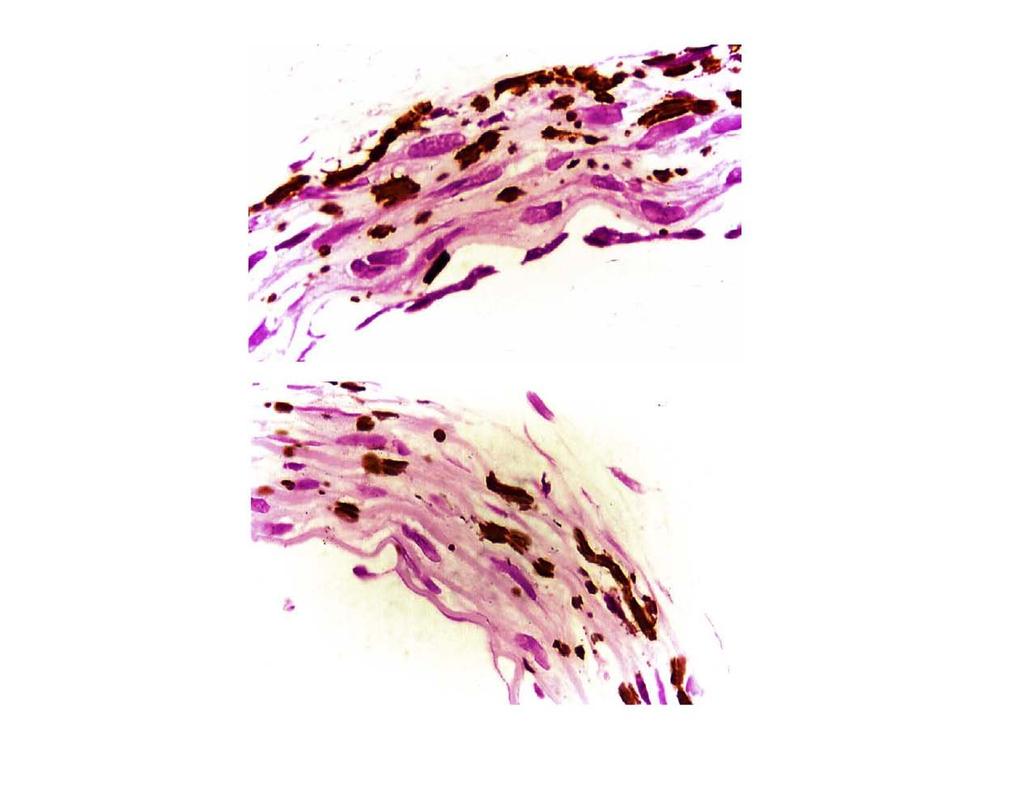
2 1080 Arterioscler Thromb Vasc Biol. May 2006 Downloaded from by guest on April 21, 2018 induces calcification in the elastic lamellae of the artery media. 3 This theory predicts that serum contains a causative agent for medial artery calcification, and that the elastic lamellae of the artery media should therefore calcify when devitalized arteries are incubated in serum. It has been known for 40 years that devitalized rat aortas do indeed calcify when incubated in rat serum at 37 C. 8,9 This calcification is confined to the elastic lamellae of the aorta media, and examination of the calcified tissue by electron diffraction and electron microscopy has shown that the calcification consists of apatite-like crystals localized almost exclusively within elastin fibers. The objectives of the present experiments were to confirm that incubation of devitalized rat aortas in rat serum does indeed cause calcification within the elastic lamellae of the aorta media, and to determine whether a similar activity is also found in bovine and human serum. Arteries did calcify during incubation in rat, bovine, and human serum, and additional experiments were accordingly performed to compare the biochemical characteristics of the serum calcification factor responsible for medial artery calcification with the recently described serum calcification factor that initiates the calcification of the type I collagen, 10 and to assess the ability of living arteries in organ culture to resist serum-initiated medial calcification. Materials and Methods The methods used in these studies are available in the online supplement at and only a brief synopsis of these methods is presented here. The devitalized artery calcification assay used here was initially developed to investigate the recalcification of demineralized bone. 10,11 In brief, EDTAdevitalized rat aortas are placed in rat or human serum, or in DMEM ([Pi] 2 mmol/l) containing different amounts of serum, and incubated at 37 C and 5% CO 2. Tissue calcification was evaluated by staining of whole arteries with Alizarin red, staining of histological sections by von Kossa, and quantification of calcium and phosphate in acid extracts of the arteries using procedures described previously. 10,11 Procedures for characterizing the serum factor that induces artery calcification have also been published previously. 10,11 To determine the effect of living cells on medial calcification, thoracic aortas were removed from rats within 15 minutes of death and separately incubated for 6 days in 10 ml of DMEM containing 15% FBS. To determine the effect of warfarin and MGP on medial calcification in arteries containing living cells, carotid arteries were removed from rats within 15 minutes of death, cut into 3- to 4-mm segments, and placed separately in culture dishes containing 2 ml of DMEM or 2 ml DMEM plus 15% FBS. Some dishes were treated with sodium warfarin at a dose of 10 g/ml, whereas other dishes were treated with this warfarin dose together with 30 or 100 g/ml purified bovine MGP. MGP was added as an aliquot of 5 mg/ml MGP in 50 mmol/l HCl, with continuous swirling to rapidly disperse the protein in the culture medium. Results Incubation of EDTA-Devitalized Rat Aortas in Rat Serum Causes Medial Artery Calcification In Vitro The initial test to determine whether serum contains a causative agent for artery calcification was performed using rat thoracic aortas that had been first freed of any possible endogenous mineral by extraction for 72 hours with 0.5 mol/l EDTA, ph 7.5, a procedure that also killed all cells Figure 1. Time course of calcium uptake by devitalized aortas during incubation in rat or human serum. Rat thoracic aortas were treated with 0.5 mol/l EDTA, ph 7.5, and then incubated for 6 days at 37 C in 2 ml rat serum (ion product: 1.2 mmol/l ionic Ca 3.2 mmol/l Pi 3.8 mmol 2 /L 2 ), 2 ml adult human serum (ion product: 1.2 mmol/l ionic Ca 1.2 mmol/l Pi 1.4 mmol 2 /L 2 ), or 2 ml of adult human serum, in which the phosphate concentration had been increased to 3.2 mmol/l by the addition of 0.5 mol/l sodium phosphate buffer, ph 7.4 (ion product: 3.8 mmol 2 /L 2 ). Calcium uptake was determined from the decrease in serum calcium attributable to the presence of the devitalized aorta. This experiment has been repeated twice with comparable results. originally found in the artery. Each devitalized aorta was placed in 2-mL rat serum and incubated for 6 days at 37 C. As seen in Figure 1, 3.5 mol of serum calcium was taken up by the aorta over the 6-day incubation, with half of the uptake occurring in the first day of incubation. After incubation in rat serum for 6 days, Alizarin red staining of the thoracic aortas revealed the presence of extensive calcification throughout most of the thoracic aorta and the smaller branch arteries, and von Kossa staining of aorta sections showed that this calcification is associated with the elastic lamellae of the artery media (Figure 2). Chemical analysis showed that incubation of devitalized aortas for 6 days in rat serum also increased aorta levels of calcium and phosphate (supplemental Table I, available online at the amount of calcium recovered from the aorta accounted for the 3.5 mol of serum calcium taken up by the aorta during the 6-day incubation (Figure 1). Comparable artery calcification was also seen after incubation for 6 days Figure 2. Calcification of devitalized rat aortas by incubation for 6 days in rat serum. After incubation for 6 days in rat serum (Figure 1 legend), the devitalized thoracic aortas were either stained for calcification with Alizarin red (stains calcification red) or fixed in formalin, cut in 5- m sections, and stained for calcification with von Kossa (stains calcification brown to black) and counterstained with nuclear fast red (stains elastin and cytoplasm pink and nuclei red).

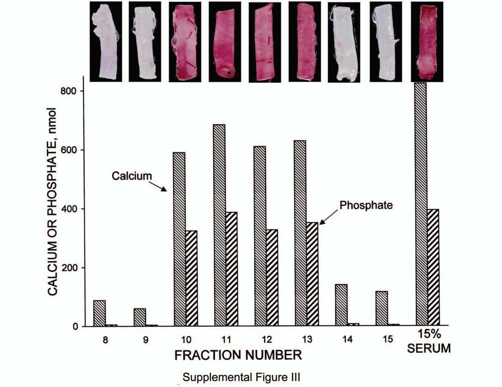
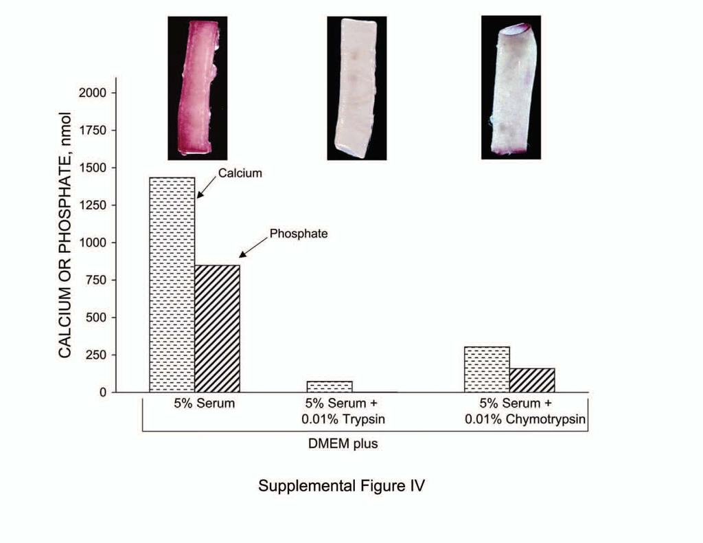
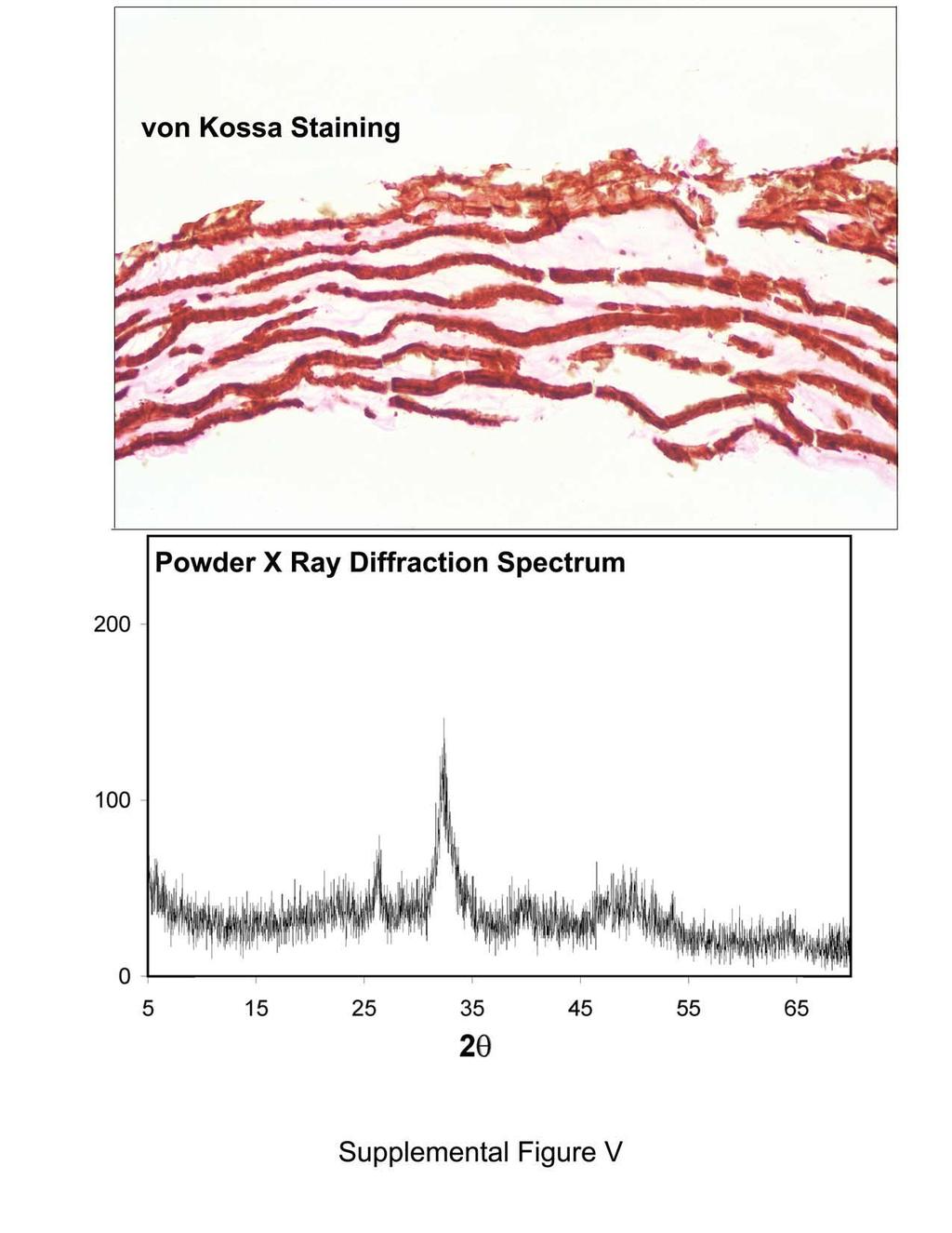
3 Price et al Devitalized Arteries Incubated in Serum Calcify 1081 in heparinized rat plasma, FBS, or newborn calf serum (data not shown). Downloaded from by guest on April 21, 2018 Evidence That the Elastic Lamellae of the Artery Media, Not Cell Debris, Is the Location of Calcification in Arteries That Have Been Incubated in Serum To evaluate the possible effect of vascular cell debris on serum-initiated medial artery calcification, the EDTA-treated aortas were subsequently extracted with acetone for 2 hours to remove lipid and with 6 mol/l guanidine HCl for 24 hours to solubilize protein. Histological examination of aorta crosssections showed that these additional extraction steps did indeed remove all detectable cellular debris. However, in spite of the complete absence of cellular debris in the artery media, the aortas still stained extensively with Alizarin red after incubation for 6 days in serum, and calcium and phosphate were still taken up by the aortas (supplemental Table II); the location of calcification was again restricted to the elastic lamellae of the aorta media (data not shown). In another test, elastin fibers were purified from the media of bovine aortas using procedures that remove all vascular cell debris as well as collagen; 12,13 as can be seen in supplemental Table II, this purified aortic elastin is also extensively calcified by incubation for 6 days in rat serum. As a final test, devitalization procedures were used that intentionally preserved some or most of the cellular debris and did not involve EDTA extraction. As seen in supplemental Table II, devitalization by fixation in formalin or by lyophilization had no significant impact on the quantitative extent of calcium and phosphate uptake by the aortas during incubation in serum for 6 days. Despite the presence of the formalin-fixed vascular cells in the aorta wall, von Kossa staining for calcification was again restricted to the elastic lamellae of the aorta media, with no evidence of calcification associated with vascular cells or other structures between the lamellae (Figure 3; a hematoxylin/eosin stained section from the same sample is shown in supplemental Figure I). This lamellar calcification appears as discrete brown calcification foci that range from 0.5 to 5 m in size, each probably representing separate nucleation events. Biochemical Characterization of the Serum Activity Responsible for Calcifying Elastic Lamellae in the Artery Media Additional experiments were performed to further characterize the biochemical activity that is responsible for the calcification of the elastic lamellae of devitalized arteries during incubation in serum. The initial experiments were conducted to determine whether the elastic lamellae of devitalized aortas calcify when incubated in DMEM containing a calcium phosphate ion product (1.8 mmol/l [Ca] 2 mmol/l [Pi] 3.6 mmol 2 /L 2 ) comparable to the ion product of rat serum (1.2 mmol/l [Ca] 3.2 mmol/l [Pi] 3.8 mmol 2 /L 2 ). Devitalized arteries did not calcify when incubated in this modified DMEM solution alone but did consistently calcify if the DMEM contained 1.5% serum (Table). Further experiments showed that DMEM must contain a physiological calcium phosphate ion product Figure 3. Effect of formalin fixation on the histological location of serum-induced artery calcification. Rat thoracic aortas were placed in formalin within 15 minutes of death and fixed for 3 days. A 2-cm segment of each aorta was then washed with water and incubated in 2 ml of rat serum for 6 days at 37 C. Five-micron sections were stained by von Kossa and counterstained by nuclear fast red magnification; Bar 10 m. VSMC indicates vascular smooth muscle cells; M, calcification foci. Note that calcification is restricted to the 8 elastic lamellae (E1 to E8 in image). (3.6 mmol 2 /L 2, achieved by increasing phosphate in DMEM to 2 mmol/l) for artery calcification to occur in the presence of added serum; no calcification occurs in basal DMEM (1.8 mmol/l [Ca] 0.9 mmol/l [Pi] 1.6 mmol 2 /L 2 ) even in the presence of 15% serum. This phosphate-boosted DMEM ([Pi] 2 mmol/l) was used in biochemical characterization experiments, the results of which are summarized in the Table. These experiments show that the serum activity responsible for calcification of medial elastin fibers in devitalized arteries has an apparent gel filtration molecular weight of 50 to 150 kda (supplemental Figures II and III), is inactivated by digestion with trypsin or chymotrypsin (supplemental Figure IV), and introduces a mineral phase into the elastin fiber that has an apatite-like X-ray diffraction pattern (supplemental Figure V). The biochemical properties of the serum activity responsible for Comparison of the Serum Activities That Initiate the Calcification of Demineralized Bone and Devitalized Arteries Molecular weight (Gel filtration) Demineralized Bone Devitalized Artery kda kda Protease sensitivity Trypsin inactivates Trypsin inactivates Chymotrypsin inactivates Chymotrypsin inactivates Dose dilution of activity: DMEM with 1.5% Calcification Calcification serum DMEM with 0.15% No calcification No calcification serum Mineral type Apatite-like Apatite-like (X-ray diffraction) Location of mineral Collagen fibers Elastin fibers Demineralized bone data are from Price et al, 10 and devitalized artery data are from experiments presented in the online data supplement.


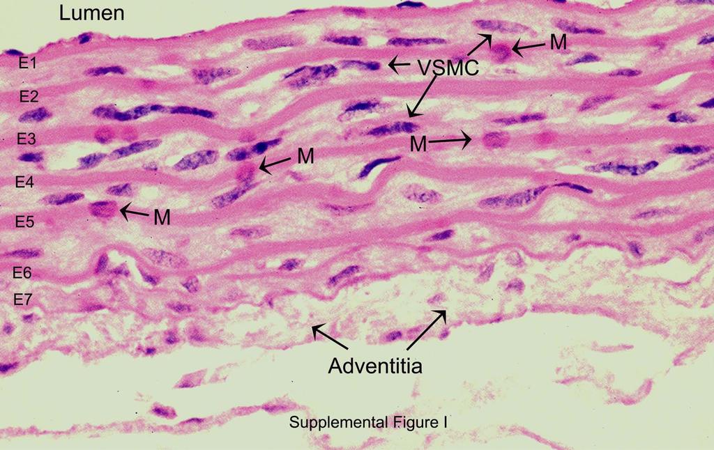
4 1082 Arterioscler Thromb Vasc Biol. May 2006 Downloaded from by guest on April 21, 2018 Figure 4. Calcification of devitalized aorta by incubation for 6 days in human serum. After incubation for 6 days in human serum or in human serum containing 2 mmol/l added Pi (Figure 1 legend), the devitalized thoracic aortas were either stained for calcification with Alizarin red or fixed in formalin, cut in 5- m thick sections, and stained for calcification with von Kossa and counterstained with nuclear fast red. calcification of medial elastin in serum is compared in the Table to the previously identified serum activity that initiates calcification in type I collagen of tendon and demineralized bone in serum. 10 A consistent feature of the calcification of tendon, demineralized bone, or devitalized arteries during incubation in neat serum is the apparent absence of calcification associated with cellular debris, even when this debris is intentionally preserved by formalin fixation. Calcification is instead always within the fibers of collagen 10 or elastin (Figure 3). Calcification of Devitalized Rat Aortas by Incubation in Human Serum Experiments were performed to determine whether devitalized aortas will also calcify after incubation in human serum. As seen in Figure 4, no aorta calcification could be detected by Alizarin red staining after incubation for 6 days in adult human serum, and analysis of calcium levels in serum during incubation failed to provide evidence for calcium uptake by the aorta (Figure 1). Because the level of phosphate in this human serum sample was 1.2 mmol/l, whereas the level of phosphate in the rat serum used in the above experiments was 3.2 mmol/l, a possible explanation for the failure of aortas to calcify in adult human serum could be that the level of phosphate is too low. To test this possibility, phosphate was added to adult human serum to achieve a net increase in phosphate concentration of 2 mmol/l. Incubation of aortas in this phosphate-boosted human serum did cause uptake of serum calcium by the aorta over the 6-day incubation period (Figure 1) as well as extensive Alizarin red staining for calcification (Figure 4) and accumulation of calcium and phosphate in the artery (supplemental Table I). Similar results were seen on histological examination of von Kossa stained aorta sections, with extensive focal staining of the elastic lamellae in the media of aortas incubated for 6 days in adult human serum in which the phosphate concentration had been increased by 2 mmol/l (Figure 4) but not in aortas incubated in adult human serum alone (data not shown). To better understand the role of serum phosphate concentration in artery calcification, devitalized thoracic aortas were incubated for 6 days in adult human serum containing Figure 5. Living rat arteries secrete the calcification inhibitor MGP and are resistant to serum-induced medial artery calcification. Thoracic aortas were removed from rats within 15 minutes of death and placed in organ culture with 10 ml of DMEM (2 mmol/l Pi) containing 15% FBS. EDTA-devitalized rat thoracic aortas were placed in the same medium as a positive calcification control. Aortas were then incubated for 6 days at 5% CO 2 and 37 C. Top, Alizarin red staining for calcification in representative living and EDTA-devitalized thoracic aortas. Bottom, Cumulative MGP production by living thoracic aortas as determined by radioimmunoassay of the cell culture medium. 24 Each point shows the average MGP production in the 6 aortas examined, and the error bars show the SDs. different amounts of added phosphate and calcification was assessed by quantitative analysis of the amount of calcium and phosphate incorporated into the aorta. As seen in supplemental Figure VII, there is a sigmoid dependence of artery calcification on the concentration of phosphate in adult human serum, with a threshold for artery calcification between 1.5 and 2 mmol/l phosphate. Serum-Induced Medial Artery Calcification Does Not Occur if Viable Cells Remain in the Artery Wall To evaluate the possible role of vascular cells in regulating serum-induced medial calcification, 3-cm segments of rat thoracic aorta were removed immediately after death and separately placed into 10 ml of DMEM supplemented with 15% FBS and incubated at 37 C for 6 days. There was no detectable Alizarin red staining in any of the 6 living thoracic aortas examined after incubation for 6 days in this organ culture medium, whereas there was extensive staining in each of the 6 devitalized aortas; examples of the Alizarin red staining seen in this experiment are shown in Figure 5. Examination of hematoxylin/eosin stained sections of the living aortas after the 6-day incubation showed that vascular cell morphology was indistinguishable from that seen in thoracic aortas examined immediately after death, a result that is in agreement with observations made in previous studies of rat arteries in organ culture. 14,15 These results demonstrate that the elastic lamellae of the artery media fail to calcify if living cells remain in the vascular wall.

5 Price et al Devitalized Arteries Incubated in Serum Calcify 1083 the serum requirement for artery calcification in this system. Assay of the conditioned medium from these experiments showed that each 3 to 4 mm segment of carotid artery secreted 0.05 g MGP per day; this rate is 50-fold lower than that observed for thoracic aorta, a difference that is probably attributable to the size difference between the tissues. A final experiment was performed to determine the effect of adding purified bovine MGP on warfarin-induced calcification in living arteries. As shown in Figure 6, addition of 100 g MGP/mL completely prevented warfarin-induced carotid artery calcification; similar inhibition of calcification was also seen at 30 g MGP/mL (data not shown). As expected, purified bovine MGP also prevented calcification of devitalized arteries in the same medium (data not shown). Downloaded from by guest on April 21, 2018 Figure 6. Living rat arteries calcify when incubated in culture medium containing serum and warfarin, and this calcification is prevented by the addition of purified MGP. Carotid arteries were removed from rats within 15 minutes of death and cut into 3- to 4-mm segments; each segment was immediately placed in culture dishes containing 2 ml of DMEM (2 mmol/l Pi) or 2 ml DMEM plus 15% FBS. Some dishes were treated with 10 g/ml sodium warfarin, whereas other dishes were treated with 10 g/ml warfarin together with 100 g/ml purified bovine MGP. Arteries were incubated for 4 days at 5% CO 2 and 37 C, and then stained for calcification with Alizarin red and analyzed for calcium and phosphate. There were 24 carotid artery segments in all groups except the MGP-treated group, which had 12. The figure shows representative examples of Alizarin red staining and the mean and SD of artery calcium and phosphate. The increase in artery calcium and phosphate attributable to warfarin treatment was highly significant (group 3 vs 4; P 0.001), as was the decrease in calcium and phosphate attributable to addition of MGP (group 4 vs 5; P ). The conditioned culture medium from this experiment was examined to determine whether living thoracic aortas secrete MGP, a known inhibitor of the calcification of elastic lamellae in the artery media in vivo. 16,17 As seen in Figure 5, MGP is secreted at a constant rate from 24 to 144 hours in organ culture. The final concentration of MGP in culture medium was 1.5 g/ml, which is far higher than the 200 ng/ml level of MGP found in the serum of rats of this age. These results show that vascular cells may prevent medial calcification by secretion of calcification inhibitors such as MGP. Effect of Warfarin and MGP on Serum-Induced Medial Artery Calcification in Living Arteries Additional experiments were performed to assess the effect of inhibiting the vitamin K dependent -carboxylation of MGP with warfarin, using a dose of warfarin shown previously to inhibit -carboxylation in cell culture. 18 As seen in Figure 6, the addition of warfarin to culture medium caused extensive Alizarin red staining for calcification in the living carotid artery segment, whereas no staining could be detected in living carotid arteries incubated in the same medium without warfarin. Supplemental Figure VI shows that this warfarininduced calcification is largely associated with the elastic lamellae of the artery media. No calcification could be detected if the living arteries were incubated in culture medium containing warfarin but not serum, which confirms Discussion Evidence That Blood Contains a Causative Agent for Medial Artery Calcification The present studies demonstrate that devitalized rat arteries calcify rapidly when incubated in serum or heparin plasma at the temperature, carbonate concentration, and ph of blood. Calcification is confined to the elastic lamellae of the artery media regardless of the devitalization procedure used and even when cellular material is intentionally fixed in the vascular wall. These observations confirm and extend the results of previous investigations. 8,9 The present studies further provide the first biochemical characterization of the molecular basis for the calcification of devitalized arteries in serum. The initial characterization experiments showed that devitalized arteries also calcified rapidly when incubated in DMEM containing as little as 1.5% rat serum but not when incubated in DMEM alone. This result demonstrates that blood contains a potent calcification factor, one sufficiently potent that it can still promote calcification in the elastic lamella of the artery when it is present in DMEM at a 70-fold lower concentration than in serum itself. The subsequent experiments showed that this calcification factor consists of 1 protein of 50- to 150-kDa molecular weight. The microscopic pattern of serum-induced calcification consists of numerous calcification foci that are scattered within the elastic lamella of the aorta media. These foci are distributed more or less evenly among the different elastic lamellae in sections near the aorta ends (Figure 3) but are typically more numerous in the elastic lamellae nearest the exterior surface of sections in the middle of the 3-cm-long aorta (Figures 2 and 4). This pattern of calcification suggests that diffusion of the nucleating agent from serum into the medial wall may determine the order with which elastic lamellae calcify. All elastic lamellae eventually calcify when devitalized aortas are incubated in serum-containing medium for a prolonged period (supplemental Figure V). If devitalized arteries calcify so rapidly when incubated in serum, why don t all animals have hardened arteries? The answer to this important question probably lies in the ability of the cells in the artery wall to prevent the serum-initiated calcification of the adjacent elastic lamella by secretion of calcification inhibitors. This hypothesis is supported by the
6 1084 Arterioscler Thromb Vasc Biol. May 2006 Downloaded from by guest on April 21, 2018 present observation that thoracic aortas in which cells remain viable during incubation in DMEM containing 15% FCS do not calcify, whereas devitalized aortas calcify rapidly in the same medium (Figure 5). This hypothesis is also supported by the observations that living arteries in organ culture secrete 2 well-established inhibitors of calcification, pyrophosphate, 19 and MGP (Figure 5). Previous studies have shown that impaired MGP activity causes calcification of the elastic lamellae of the artery media in vivo, 16,17,20 a pattern of calcification essentially identical to that observed in devitalized arteries that have been incubated in serum. It is therefore possible that the MGP secreted by vascular cells in the artery wall contributes to the ability of living arteries to resist serum-initiated calcification in cell culture. Several experiments were performed to test this hypothesis. Inactivation of MGP -carboxylation by treatment with warfarin, a treatment known to inactivate the calcification inhibitory activity of MGP in vivo, 17 caused extensive calcification of living arteries in organ culture (Figure 6), and this calcification is restricted to the elastic lamellae of the artery media (supplemental Figure VI). Addition of purified bovine MGP to culture medium completely prevented this warfarin-induced calcification of living arteries in organ culture. These experiments show that MGP secreted by cells in the vascular wall plays an important role in preventing serum-initiated calcification of the elastic lamellae during the organ culture of living arteries and therefore support the hypothesis that the unique susceptibility of the elastic lamellae to serum-initiated calcification in vivo is actively opposed by the secretion of MGP and other calcification inhibitors. Is Serum Always Required for Calcification Under Cell Culture Conditions? A discussion of this important question is available in the online supplement. Possible Relevance of the Calcification of Devitalized Arteries in Serum to Artery Calcification in Chronic Kidney Disease The present studies show that human serum also has the ability to rapidly calcify devitalized rat aortas, and that the microscopic pattern of this calcification is identical to that seen in aortas incubated in rat serum. However, the calcification of aortas during incubation in human serum appears to be dependent on the phosphate content of the serum sample, and normal adult human serum ([Pi] 1 to 1.5 mmol/l) does not induce artery calcification unless phosphate levels are boosted to 2 mmol/l (supplemental Figure VII). A similar dependence of calcification on medium phosphate concentration has also been noted in vascular smooth muscle cells cultured in DMEM with 15% FBS, with no calcification when [Pi] 1.4 mmol/l and significant calcification when [Pi] 2 mmol/l. 21 It is of interest to note that newborn human serum, with [Pi] 3 to 4 mmol/l, induces rapid and specific calcification of the elastic lamella of devitalized aortas without need for phosphate supplementation (personal observations). A discussion of the possible relevance of these observations to the well-established correlation of serum phosphate and artery calcification in chronic kidney disease is available in the online supplement. Relationship Between the Serum-Initiated Calcification of Arteries and the Serum-Initiated Recalcification of Demineralized Bone The serum-initiated calcification of arteries is similar to the previously demonstrated serum-initiated recalcification of demineralized bone. 10 These similarities include: (1) a similar time course of calcification when the respective matrices are incubated in neat rat serum at 37 C; (2) a similar dose dilution of the serum activity, with calcification of both matrices occurring in DMEM containing as little as 1.5% serum; (3) a similar molecular weight range of the serum activity, estimated to be 50 to 150 kda in each case; (4) a similar sensitivity of the serum activity to trypsin and chymotrypsin digestion; (5) a similar requirement for the phosphate level needed for calcification in human serum, with no calcification at the 1.2 mmol/l phosphate level found in adult human serum and extensive calcification when the phosphate level is boosted to 2 mmol/l; (6) a similar histological appearance of the initial calcification, with numerous calcification foci that could represent discrete nucleation events; and (7) a similar apatite-like mineral phase. Together, these observations strongly suggest that the same calcification factor may be responsible for calcification of collagen and elastin fibers during incubation in serum. One could well ask why serum might contain a calcification factor. Our working hypothesis is that this calcification factor arises from sites of mineralization in the skeleton, where it is involved in the normal mineralization of the collagenous bone matrix. Because of the vascular nature of bone mineralization sites such as the basic multicellular unit 22 and the bone remodeling compartment, 23 any macromolecule secreted by osteoblasts for the purposes of local bone mineralization will unavoidably escape to blood, just as any serum protein with affinity for mineralizing sites will accumulate in bone. Bone-derived alkaline phosphatase is accordingly released from osteoblasts and appears in serum, and the serum protein fetuin is secreted by hepatocytes and accumulates in bone to become one of the most abundant noncollagenous bone proteins. In our view, the escape of a calcification factor to serum may therefore be just the unavoidable consequence of bone anatomy. Acknowledgments This work was supported in part by grant HL58090 from the National Heart, Lung, and Blood Institute of the National Institutes of Health. References 1. Goodman WG, London G. Vascular calcification in chronic kidney disease. Am J Kidney Dis. 2004;43: Salusky IB, Goodman WG. Cardiovascular calcification in end-stage renal disease. Nephrol Dial Transplant. 2002;17: Price PA, Faus SA, Williamson MK. The bisphosphonates alendronate and ibandronate inhibit artery calcification at doses comparable to those which inhibit bone resorption. Arterioscler Thromb Vasc Biol. 2001;21: Price PA, Buckley JR, Williamson MK. The amino bisphosphonate ibandronate prevents vitamin D toxicity and inhibits vitamin D-induced calcification of arteries, cartilage, lungs, and kidneys in rats. J Nutr. 2001; 131:
7 Price et al Devitalized Arteries Incubated in Serum Calcify 1085 Downloaded from by guest on April 21, Price PA, Omid N, Than TN, Williamson MK. The amino bisphosphonate ibandronate prevents calciphylaxis in the rat at doses that inhibit bone resorption. Calcif Tissue Int. 2002;71: Price PA, June HH, Buckley JR, Williamson MK. Osteoprotegerin inhibits artery calcification induced by warfarin and by vitamin D. Arterioscler Thromb Vasc Biol. 2001;21: Price PA, June HH, Buckley JR, Williamson MK. SB , a selective inhibitor of the osteoclastic V-H -ATPase, inhibits artery calcification at doses that inhibit bone resorption. Circ Res. 2002;91: Martin GR, Schiffmann E, Bladen HA, Nylen M. Chemical and morphological studies on the in vitro calcification of the aorta. J Cell Biol. 1963;16: Schiffmann E, Martin GR. In vitro calcification of rat aorta in serum. Nature. 1962;194: Price PA, June HH, Hamlin NJ, Williamson MK. Evidence for a serum factor that initiates the re-calcification of demineralized bone. J Biol Chem. 2004;279: Hamlin NJ, Price PA. Mineralization of decalcified bone occurs under cell culture conditions and requires bovine serum but not cells. Calcif Tissue Int. 2004;75: Song SH, Roach MR. Quantitative changes in the size of fenestrations of the elastic laminae of sheep thoracic aorta studies with SEM. Blood Vessels. 1983;20: Steven FS, Minns RJ, Thomas H. The isolation of chemically pure elastin in a form suitable for mechanical testing. Connect Tissue Res. 1974;2: Merrilees MJ, Scott L. Organ culture of rat carotid artery: maintenance of morphological characteristics and of pattern of matrix synthesis. In Vitro. 1982;18: Merrilees MJ, Scott L. Antisense S-oligonucleotide against transforming growth factor beta1 inhibits proteoglycan synthesis in arterial wall. J Vasc Res. 1994;31: Luo G, Ducy P, McKee MD, Pinero GJ, Loyer E, Behringer RR, Karsenty G. Spontaneous calcification of arteries and cartilage in mice lacking matrix Gla protein. Nature. 1997;386: Price PA, Faus SA, Williamson MK. Warfarin causes rapid calcification of the elastic lamellae in rat arteries and heart valves. Arterioscler Thromb Vasc Biol. 1998;18: Pan LC, Williamson MK, Price PA. Sequence of the precursor to rat bone Gla protein that accumulates in warfarin-treated osteosarcoma cells. J Biol Chem. 1985;260: Lomashvili KA, Cobbs S, Hennigar RA, Hardcastle KI, O Neill WC. Phosphate-induced vascular calcification: role of pyrophosphate and osteopontin. J Am Soc Nephrol. 2004;15: Price PA, Faus SA, Williamson MK. Warfarin induced artery calcification is accelerated by growth and by vitamin D. Arterioscler Thromb Vasc Biol. 2000;20: Jono S, McKee MD, Murry CE, Shioi A, Nishizawa Y, Mori K, Morii H, Giachelli CM. Phosphate regulation of vascular smooth muscle cell calcification. Circ Res. 2000;87:E10 E Parfitt AM. The mechanism of coupling: a role for the vasculature. Bone. 2000;26: Parfitt AM. The bone remodeling compartment: a circulatory function for bone lining cells. J Bone Miner Res. 2001;16: Otawara Y, Price PA. Developmental appearance of matrix Gla protein during calcification in the rat. J Biol Chem. 1986;261:


8 Downloaded from by guest on April 21, 2018 The Elastic Lamellae of Devitalized Arteries Calcify When Incubated in Serum: Evidence for a Serum Calcification Factor Paul A. Price, Wai Si Chan, Dawn M. Jolson and Matthew K. Williamson Arterioscler Thromb Vasc Biol. 2006;26: ; originally published online March 9, 2006; doi: /01.ATV c Arteriosclerosis, Thrombosis, and Vascular Biology is published by the American Heart Association, 7272 Greenville Avenue, Dallas, TX Copyright 2006 American Heart Association, Inc. All rights reserved. Print ISSN: Online ISSN: The online version of this article, along with updated information and services, is located on the World Wide Web at: Data Supplement (unedited) at: Permissions: Requests for permissions to reproduce figures, tables, or portions of articles originally published in Arteriosclerosis, Thrombosis, and Vascular Biology can be obtained via RightsLink, a service of the Copyright Clearance Center, not the Editorial Office. Once the online version of the published article for which permission is being requested is located, click Request Permissions in the middle column of the Web page under Services. Further information about this process is available in the Permissions and Rights Question and Answer document. Reprints: Information about reprints can be found online at: Subscriptions: Information about subscribing to Arteriosclerosis, Thrombosis, and Vascular Biology is online at:
9 METHODS Materials. Forty-day-old male albino rats (Sprague-Dawley derived) were purchased from Charles River Labs (Wilmington, MD). Each 500ml volume of Dulbecco's modified eagle medium (DMEM; Gibco) was supplemented with 5ml of penicillinstreptomycin (Gibco) and 1ml of 10% sodium azide to prevent bacterial growth. Unless otherwise stated, the concentration of phosphate in DMEM was increased from the basal 0.9mM to a final 2mM by the addition of 0.5 M sodium phosphate buffer ph 7.4. Human serum was obtained from healthy males 47 and 59 years of age. Alizarin red S was purchased from Sigma. Matrix Gla protein (MGP) was purified from bovine cortical bone as described 1. Rats were killed by exsanguination while under isoflurane anesthetic; the UCSD Animal Subjects Committee approved all animal experiments. For serum preparation, blood was allowed to clot for 30 minutes at room temperature and centrifuged at 1,400 x g for 10 minutes in a clinical centrifuge. Thoracic aorta segments measuring 3cm from the heart and including the aortic arch were dissected from 40-day-old rats immediately after death. Unless otherwise stated, aortas were extracted with 0.5 M EDTA ph 7.5 for 72h at room temperature, washed exhaustively with ultra pure water to remove all traces of EDTA, and stored at 20ºC until use. In one experiment, the EDTA-extracted aortas were subsequently extracted for 2h with acetone and for 24h with 6M guanidine HCl in 0.1M Tris ph 9. In another experiment, thoracic aortas were removed within 15min of death and immediately fixed for 3 days in formalin, or freeze-dried. Elastin was purified from the media of bovine aorta by extraction with 0.1M NaOH for 3h at 70 C 2, 3, dried,
10 and briefly powdered in a blender; the purified elastin powder was fractionated by size to obtain elastin particles between 0.25 and 0.43mm in average diameter. Calcification procedures. Experiments to examine aorta calcification in rat serum were carried out with 35 mm culture dishes (Falcon 3001) in a humidified incubator at 37ºC and 7.5% CO 2 using 2 ml rat serum containing 0.02% sodium azide to prevent bacterial growth. After equilibration with 7.5% CO 2 at 37ºC, serum maintained a stable ph value of 7.4 throughout the duration of the 6 d incubation, and the physiological buffer maintained a ph of A single devitalized thoracic aorta segment was incubated for 6 days in each of the test solutions. To assess the progress of aorta calcification, 50 µl aliquots were removed at the start of the incubation and at each 24 h thereafter and stored frozen until analysis to determine medium calcium levels. The same procedures were followed to examine aorta calcification in human serum and in human serum in which the phosphate concentration had been increased by 2mM by the addition of 0.5 M sodium phosphate buffer ph 7.4. To determine the effect of adding different amounts of phosphate on the calcification of arteries in adult human serum, a 1cm section of rat thoracic aorta was placed in 1 ml of adult human serum ([P i ] = 1.0mM) or in 1ml of adult human serum in which the phosphate concentration had been increased to the desired levels by the addition of 0.5 M sodium phosphate buffer ph 7.4. Culture medium calcification experiments were carried out with 100mm culture dishes (Falcon 3803) in a humidified incubator at 37ºC and 5% CO 2 using a 10ml volume of DMEM alone or of DMEM containing 15% or 1.5% rat serum by volume. A 3cm devitalized thoracic aorta segment was incubated for 6 days in each of the DMEM test solutions. The DMEM used in these and all other experiments was routinely
11 supplemented with phosphate to a final concentration of 2mM, as DMEM containing the basal 0.9mM phosphate does not support serum-initiated calcification 4. This supplementation was achieved by adding the appropriate volume of 75 mm sodium phosphate ph 7.4 to ice cold DMEM using constant mixing in order to avoid apatite crystal nucleation at the phosphate/dmem mixing interface. DMEM containing 2mM phosphate is stable for at least 3 weeks at 37 C, with no evidence for loss of calcium or phosphate from the medium or formation of a mineral phase. (In contrast, DMEM containing phosphate levels of 3mM or higher is unstable, with loss of over 50% of the ionic calcium due to mineral precipitation within 3 days at 37 C.) To determine the effect of living cells on medial calcification, thoracic aortas were removed from rats within 15 min of death and separately incubated for 6 days in 10ml of DMEM containing 15% fetal bovine serum. Cumulative MGP production by living thoracic aortas was determined by radioimmunoassay of the cell culture medium 5. To determine the effect of warfarin and matrix Gla protein on medial calcification in arteries containing living cells, carotid arteries were removed from rats within 15 min of death, cut into 3 to 4 mm segments, and placed separately in culture dishes containing 2 ml of DMEM or 2 ml DMEM plus 15% fetal bovine serum. Some dishes were treated with sodium warfarin at a dose of 10 µg/ml, while other dishes were treated with this warfarin dose together with 30 or 100 µg/ml purified bovine MGP. MGP was added as an aliquot of 5mg/ml MGP in 50 mm HCl, with continuous swirling to rapidly disperse the protein in the culture medium; radioimmunoassay of this culture medium, using an antibody that recognizes bovine MGP but not rat MGP, showed that the concentration of bovine MGP remained at the initial value throughout the experiment.
12 Biochemical analyses. The procedures used for Alizarin red staining have been described 6. For histological analyses, aortas were fixed in 10% buffered formalin for at least 1d at room temperature; San Diego Pathology Inc. (San Diego, CA) sectioned and von Kossa stained the aortas. For quantitative assessment of the extent of calcification, aortas were rinsed for 10 minutes with 10 mm NH 4 OH, patted dry with a paper towel, placed in 2ml epitubes, and extracted for 24h at room temperature with 1 ml of 0.15 M HCl. Calcium levels in culture media and in the acid extracts of tissues were determined colorimetrically using cresolphthalein complexone (JAS Diagnostics, Miami FL) and phosphate levels were determined colorimetrically as described 7. In order to characterize the molecular mass of the serum agent that initiates medial artery calcification, 1 ml of serum from a 40-day-old rat was fractionated over a 25 ml column of Sephacryl S-100HR equilibrated at room temperature with DMEM using a buffer reservoir that was continuously flushed with 5% CO 2 in order to maintain a ph of The absorbance at 280 nm was determined for each 1 ml fraction, small aliquots of each fraction were set aside for electrophoresis, and each fraction was then placed into separate wells of a 24-well tissue culture plate. A 1 cm section of EDTAdevitalized rat thoracic aorta was then added to each well and incubated for 6 days at 37 C and 5% CO 2. To determine the apparent molecular mass of serum proteins that coeluted with the serum calcifying activity in this experiment, 2µl aliquots of fractions 9-14 were electrophoresed on a 4-12% SDS Bis-Tris polyacrylamide gel (Invitrogen Inc.) and stained with Coomassie Brilliant Blue. To determine whether the serum agent that initiates medial artery calcification is inactivated by proteases, four 10 ml samples of DMEM were prepared that contained: no
13 serum, 5% rat serum, 5% serum and 1mg trypsin, and 5% serum and 1 mg chymotrypsin. Each solution was pre-incubated for 24 h at 37ºC, a time found in previous experiments to cause extensive proteolysis of serum proteins 8. A single devitalized aorta segment was then added and the solutions were incubated for 6 days at 37º C, stained with Alizarin red, and analyzed for calcium and phosphate (see Figure IV). To control for possible effects of the proteases on the devitalized aorta, devitalized aortas were incubated for 6 days in DMEM containing 100 µg/ml trypsin or in DMEM alone. Aortas were then washed with fresh medium (to remove trypsin) and incubated for another 6 days in DMEM containing 5% rat serum and no added trypsin. This experiment showed that there was comparable Alizarin red staining for calcification in aortas pre-incubated for 6 days in DMEM plus trypsin and in aortas pre-incubated for 6 days in DMEM alone. To characterize the nature of the mineral phase found in calcified arteries, 3 cm segments of thoracic aortas from fifteen rats were devitalized with EDTA and separately incubated for 7 weeks in 10 ml volumes of DMEM containing 15% fetal bovine serum, with weekly medium change. 14 aortas were then dried and weighed; this measurement showed that incubation increased the dry weight of the aortas from 8.7±0.2 mg/aorta before incubation to 16.8±1.4mg/aorta after. Five of these aortas were extracted with acid and the acid extracts were analyzed for calcium and phosphate; the amount of calcium was 51.0±2.3 µmole/aorta, the amount of phosphate was 33.1±1.5 µmole/aorta, and the Ca/PO4 ratio was 1.56±0.12. Nine aortas and the midshaft segments of 5 weanling rat tibias were separately ground to a fine powder with an agate mortar, and the powders were analyzed using a Scintag SDF 2000 X ray diffractometer. The remaining
14 aorta was not dried, but was instead fixed in formalin, cut in 5 micron thick sections and stained with von Kossa. DISCUSSION Is serum always required for calcification under cell culture conditions? We have employed a commonly used cell culture medium, DMEM, in tests to determine the potency of the serum activity that is responsible for medial calcification and to determine the biochemical characteristics of this activity. It is therefore appropriate that we place this portion of our studies in the broader context of other calcification studies that have used this and similar cell culture media. In the present studies we have observed that the calcification of devitalized arteries in DMEM containing serum requires that the ion product of DMEM be increased from the basal 1.6 mm 2 to about 3.6 mm 2, an ion product comparable to that found in rat and bovine serum. DMEM containing this physiological ion product but no serum is stable, with no evidence for spontaneous mineral formation even after incubation for 4 weeks at 37 C. Devitalized arteries incubated in DMEM containing this physiological calcium phosphate ion product never calcify unless at least 1.5% serum is added. The spontaneous formation of a calcium phosphate mineral phase is known to be highly dependent on the solution calcium phosphate ion product, and DMEM solutions containing ion products of 6 mm 2 and above invariably form a mineral phase within 3 days at ph 7.4 and 37 C. The instability associated with preparing DMEM containing high levels of phosphate is sometimes circumvented by using 10 mm β glycerophosphate, but the problem with this approach is that phosphate is rapidly
15 released from this substrate by alkaline phosphatase (present in the serum typically added in cell culture experiments), and can exceed the threshold for spontaneous formation of a mineral phase within 3 days of culture even in the absence of cells 4. Cell culture studies carried out in solutions containing such supra physiological ion products do not exclusively test for the role of a nucleator produced by vascular or bone cells, and may merely show that cellular material produced by the cells provides a nidus for the growth of crystals which originated spontaneously within the bulk solution 4 and references therein. Under these supra physiological ion product conditions, serum is not required for calcification in cell culture, or for calcification of elastin or collagen fibers added to culture medium, and indeed there is often a massive, spontaneous precipitation of mineral on a variety of matrices, including cartilage, that do not normally mineralize 4. If DMEM contains a physiological calcium phosphate ion product, however, the calcification of elastin fibers (present study) or of collagen fibers 4 only occurs if a nucleating agent is present. Serum contains one type of nucleating agent; some bone and vascular cells in culture almost certainly express the same and/or different nucleating agents and also secrete a suitable collagenous matrix to support calcification How is it possible for elastin and collagen to calcify in serum, when serum clearly contains potent calcification inhibitors such as the 48 kda protein fetuin? Fetuin and other abundant serum inhibitors have been shown to prevent the precipitation of a calcium phosphate mineral phase in solution, not to prevent the initiation and growth of a mineral phase within a collagen fiber, and indeed it could be argued that the ability of bone to mineralize collagen in a vascular compartment would strongly select against the ability of serum proteins such as fetuin to inhibit calcification within collagen fibers. We
16 believe that fetuin and other serum calcification inhibitors are similarly unable to inhibit calcification within elastin fibers, and that calcium phosphate mineral nuclei introduced by a serum calcification factor are able to grow in collagen and elastin fibers precisely because both matrices provide an environment in which serum calcification inhibitors can t act. Possible relevance of the calcification of devitalized arteries in serum to artery calcification in chronic kidney disease. The present studies show that human serum also has the ability to rapidly calcify devitalized rat aortas, and that the microscopic pattern of this calcification is identical to that seen in aortas incubated in rat serum. The calcification of aortas during incubation in human serum appears to be dependent on the phosphate content of the serum sample, however, and normal adult human serum ([Pi] = 1 to 1.5mM) does not induce artery calcification unless phosphate levels are boosted to at least 2mM (supplemental Figure VII). A similar dependence of calcification on medium phosphate concentration has also been noted in vascular smooth muscle cells cultured in DMEM with 15% fetal bovine serum, with no calcification when [Pi] = 1.4 mm and significant calcification when [Pi] = 2 mm 12. It is of interest to note that newborn human serum, with [Pi] = 3 to 4 mm, induces rapid and specific calcification of the elastic lamella of devitalized aortas without need for phosphate supplementation (personal observations). While future studies will be needed to determine whether the phosphate dependence of artery calcification during incubation in human serum in vitro has any bearing on artery calcification in human disease, it is intriguing to note that elevated serum phosphate (and elevated Ca X Pi product) is associated with increased morbidity
17 and mortality in patients with end stage renal disease. In a study of 6,407 uremic patients, 30% were found to have serum phosphate levels of greater than 2.3 mm, and 10% had serum phosphate levels above 2.9 mm 13. In this study group, the risk of death for patients with a serum phosphate greater than 2.1 mm was 1.27 relative to those with a serum phosphate of 0.8 to 2.1 mm. A subsequent study showed that the increased risk of death in uremic patients with elevated serum phosphate is largely due to increases in coronary artery disease and sudden death 14. While the mechanisms responsible for the association between increased serum phosphate levels and cardiac causes of death in uremic patients are unknown, a number of studies have found an association between uremia and vascular calcification 15. Since the threshold level of phosphate associated with increased risk of death in uremic patients, 2.1 mm 13, 14, is also the minimum level of phosphate needed in adult human serum in order to observe spontaneous calcification of arteries in vitro ( supplemental Figure VII), it is tempting to speculate that increased artery calcification may contribute significantly to the increased cardiac causes of death in uremic patients with high serum phosphate levels.
18 SUPPLEMENTAL REFERENCES 1. Hale JE, Williamson MK, Price PA. Carboxyl-terminal proteolytic processing of matrix Gla protein. J. Biol. Chem. 1991;266: Song SH, Roach MR. Quantitative changes in the size of fenestrations of the elastic laminae of sheep thoracic aorta studies with SEM. Blood Vessels. 1983;20: Steven FS, Minns RJ, Thomas H. The isolation of chemically pure elastin in a form suitable for mechanical testing. Connective Tissue Research. 1974;2: Hamlin NJ, Price PA. Mineralization of decalcified bone occurs under cell culture conditions and requires bovine serum but not cells. Calcif.Tiss. Internat. 2004;75: Otawara Y, Price PA. Developmental appearance of matrix Gla protein during calcification in the rat. J. Biol. Chem. 1986;261: Price PA, June HH, Buckley JR, Williamson MK. Osteoprotegerin inhibits artery calcification induced by warfarin and by vitamin D. Arterioscler. Thromb. Vasc. Biol. 2001;21: Chen PS, Toribara TY, Warner H. Microdetermination of phosphorus. Anal. Chem. 1956;28: Price PA, June HH, Hamlin NJ, Williamson MK. Evidence for a serum factor that initiates the re-calcification of demineralized bone. J. Biol. Chem. 2004;279: Abedin M, Tintut Y, Demer LL. Vascular calcification: mechanisms and clinical ramifications. Arterioscler. Thromb. Vasc. Biol. 2004;24: Giachelli CM. Inducers and inhibitors of biomineralization: lessons from pathological calcification. Orthod. Craniofacial Res. 2005;8: Moe SM, Chen NX. Pathophysiology of vascular calcification in chronic kidney disease. Circ. Res. 2004;95: Jono S, McKee MD, Murry CE, Shioi A, Nishizawa Y, Mori K, Morii H, Giachelli CM. Phosphate regulation of vascular smooth muscle cell calcification. Circ. Res. 2000;87:E Block GA, Hulbert-Shearon TE, Levin NW, Port FK. Association of serum phosphorus and calcium X phosphate product with mortality risk in chronic hemodialysis patients: a national study. Am. J. Kidney Diseases. 1998;31: Ganesh SK, Stack AG, Levin NW, Hulbert-Shearon T, Port FK. Association of elevated serum PO4, Ca X PO4 product, and parathyroid hormone with cardiac mortality risk in chronic hemodialysis patients. J. Am. Soc. Nephrol. 2001;12: Goldsmith DJA, Covic A, Sambrook PA, Ackrill P. Vascular calcification in long-term haemodialysis patients in a single unit: a retrospective analysis. Nephron. 1997;77:37-43.
19 SUPPLEMENTAL FIGURES Figure I. Effect of formalin fixation on the histological location of serum-induced artery calcification: H&E staining. Rat thoracic aortas were placed in formalin within 15min of death and fixed for 3 days. A 2 cm segment of each aorta was then washed with water and incubated in 2 ml of rat serum for 6 days at 37 C. Five micron sections were stained by H & E. 1000X magnification. (The von Kossa stained section cut from the same tissue block is shown in Figure 3 of the print text.) VSMC, vascular smooth muscle cells; M, calcification foci. Note that calcification is restricted to the seven elastic lamellae (E1 to E7 in image). Figure II. Gel filtration evidence that the serum nucleator that initiates medial artery calcification is a macromolecule: Protein analyses. A 1ml sample of serum from a 40-day-old rat was applied to a 25 ml column of Sephacryl S-100 HR equilibrated with DMEM (2 mm Pi) at room temperature. The graph shows the absorbance at 280nm for each 1ml effluent fraction. Inset, 2µl aliquots of fractions 9-14 were electrophoresed on a 4-12% SDS polyacrylamide gel and stained with Coomassie Brilliant Blue. Figure III. Gel filtration evidence that the serum nucleator that initiates medial artery calcification is a macromolecule: Assays for calcification activity. Each 1ml fraction from the Sephacryl S-100HR fractionation described in Figure II was placed into a well of a 24-well tissue culture plate. A 1cm section of devitalized rat thoracic aorta was added to each fraction and incubated for 6 days at 37ºC and 5% CO 2. Aortas were stained with Alizarin red, photographed, and then analyzed for calcium and phosphate (see Methods). The data show the Alizarin red staining and the amount of calcium and phosphate recovered from each aorta. A 1cm section of devitalized rat thoracic aorta was
20 incubated for 6 days in 1ml of DMEM containing 15% rat serum as a positive control. Devitalized aortas incubated in DMEM column buffer alone showed no evidence for calcification (not shown). Figure IV. Effect of trypsin or chymotrypsin on the serum-induced calcification of devitalized aorta. Ten ml of DMEM (2 mm Pi) containing 5% rat serum by volume was pre-incubated for 24h at 37ºC and 5% CO 2 with 100µg/ml trypsin, 100µg/ml chymotrypsin, or no added protease. A fourth sample of DMEM alone was also prepared. After the 24h pre-incubation, a 1cm section of devitalized rat thoracic aorta was added to each sample and incubated for 6 days at 37ºC and 5% CO 2. Aortas were stained with Alizarin red, photographed, and then analyzed for calcium and phosphate. The data show the Alizarin red staining and the amount of calcium and phosphate recovered from each aorta (See Methods). Figure V. Powder X ray diffraction spectrum of devitalized thoracic aortas that have been calcified by incubation for 7 weeks in DMEM containing 15% fetal bovine serum. Ten EDTA-devitalized rat thoracic aortas were incubated for 7 weeks at 37 C in DMEM (2 mm Pi) containing 15% fetal bovine serum (with weekly medium change). One aorta was cut in 5 micron thick sections, and stained for calcification with von Kossa. The remaining 9 aortas were ground in an agate mortar, and the X ray diffraction spectrum of the powder was determined with a Scintag SDF 2000 X ray diffractometer (see Methods). Top, von Kossa stained section; note that all elastic lamellae are calcified, and that fragmentation of the lamellae occurred during sectioning due to extensive calcification. Bottom, X ray diffraction spectrum; note that this spectrum is essentially identical to the published X ray spectrum of rat bone 4, 8.
21 Figure VI. The elastic lamellae of living rat arteries calcify when incubated in culture medium containing serum and warfarin. Carotid arteries were removed from rats within 15min of death and cut into 3 to 4 mm segments; each segment was immediately placed in a culture dish containing 2ml of DMEM (2 mm Pi) plus 15% fetal bovine serum and 10µg/ml sodium warfarin. Arteries were incubated for 4 days at 5 % CO 2 and 37 C, and then cut in 5 micron thick sections and stained for calcification with von Kossa (see Legend to text Figure 6). Figure VII. The calcification of devitalized aorta in adult human serum containing different total levels of phosphate. A 1 cm section of devitalized rat thoracic aorta was placed in 1 ml of adult human serum ([P i ] = 1.0mM) or in 1 ml of adult human serum in which the phosphate concentration had been increased to the indicated levels by the addition of 0.5 M sodium phosphate buffer ph 7.4. Samples were then placed for 6 days in a humidified incubator at 7.5 % CO 2 and 37 C. Aortas were analyzed for calcium and phosphate as described in Methods.
22 Table I. Analysis for calcium and phosphate in devitalized aorta after incubation for 6d in rat serum, human serum, or in DMEM containing rat serum. A 3cm segment of EDTA-devitalized rat thoracic aorta was incubated for 6 d at 37ºC in 2 ml of: rat serum; DMEM containing the indicated amount of rat serum; adult human serum; or adult human serum containing 2mM added phosphate. The results show the total calcium and phosphate recovered in single thoracic aortas after the 6 d incubation; prior to incubation aortas did not have detectable levels of calcium or phosphate. Incubation Medium Calcium, µmol Phosphate, µmol Alizarin Red Staining Rat Serum DMEM with 15% Rat Serum DMEM with 1.5% Rat Serum DMEM with 0.15% Rat Serum < 0.1 < DMEM Alone < 0.1 < Human Serum < 0.1 < Human Serum with 2 mm added phosphate
23 Table II. Effect of Devitalization Procedure on Serum-induced Medial Artery Calcification. A 3cm segment of devitalized rat thoracic aorta or 4mg of purified bovine aortic elastin were incubated for 6 d at 37ºC in 2 ml of rat serum. The aorta devitalization procedures were: lyophilization; fixation in formalin for 3d; extraction with 0.5M EDTA, ph7.4 for 3d; and extraction with 0.5M EDTA for 3d, acetone for 2h, and 6M guanidine for 24h. Bovine aortic elastin was purified as described 2, 3 (Methods). Devitalization Procedure Calcium, µmol Phosphate, µmol Lyophilization only Formalin fixation only Extraction with EDTA Extraction with EDTA acetone 6M guanidine Purified aortic elastin
24
25
26
27
28
29
Evidence for a Serum Factor That Initiates the Re-calcification of Demineralized Bone* S
 THE JOURNAL OF BIOLOGICAL CHEMISTRY Vol. 279, No. 18, Issue of April 30, pp. 19169 19180, 2004 2004 by The American Society for Biochemistry and Molecular Biology, Inc. Printed in U.S.A. Evidence for a
The Inhibition of Calcium Phosphate Precipitation by Fetuin Is Accompanied by the Formation of a Fetuin-Mineral Complex*
 THE JOURNAL OF BIOLOGICAL CHEMISTRY Vol. 278, No. 24, Issue of June 13, pp. 22144 22152, 2003 2003 by The American Society for Biochemistry and Molecular Biology, Inc. Printed in U.S.A. The Inhibition
Bone Markers and Vascular Calcification in CKD-MBD
 Bone Markers and Vascular Calcification in CKD-MBD Pierre Delanaye, MD, PhD Department of Nephrology, Dialysis, Transplantation CHU Sart Tilman University of Liège BELGIUM Bone Markers and Vascular Calcification
SUPPLEMENTARY MATERIAL
 SUPPLEMENTARY MATERIAL Purification and biochemical properties of SDS-stable low molecular weight alkaline serine protease from Citrullus Colocynthis Muhammad Bashir Khan, 1,3 Hidayatullah khan, 2 Muhammad
We recently proposed the hypothesis that arterial calcification
 SB 242784, a Selective Inhibitor of the Osteoclastic V-H -ATPase, Inhibits Arterial Calcification in the Rat Paul A. Price, Helen H. June, Jessica R. Buckley, Matthew K. Williamson Abstract The present
Calcification of Porcine Aortic Valvular Interstitial Cells
 Calcification of Porcine Aortic Valvular Interstitial Cells Liwen Gu 1,2* Supervisor: Craig A. Simmons 1 Department of Engineering Science, 2 Institute of Biomaterials and Biomedical Engineering, University
Stefanos K. Roumeliotis. Department of Nephrology, Medical School Democritus University of Thrace, Alexandroupolis, Greece. Stefanos K.
 Department of Nephrology, Medical School Democritus University of Thrace, Alexandroupolis, Greece Passive, degenerative accumulation process of Ca ++ /P +++ without treatment options Active, complex, condition:
Elastic Skeleton of Intracranial Cerebral Aneurysms in Rats
 1722 Elastic Skeleton of Intracranial Cerebral Aneurysms in Rats Naohiro Yamazoe, MD, Nobuo Hashimoto, MD, Haruhiko Kikuchi, MD, and Fumitada Hazama, MD In an attempt to clarify the developmental mechanism
We have recently proposed the hypothesis that arterial
 Osteoprotegerin Inhibits Artery Calcification Induced by Warfarin and by Vitamin D Paul A. Price, Helen H. June, Jessica R. Buckley, Matthew K. Williamson Abstract The present experiments were carried
Supplementary material: Materials and suppliers
 Supplementary material: Materials and suppliers Electrophoresis consumables including tris-glycine, acrylamide, SDS buffer and Coomassie Brilliant Blue G-2 dye (CBB) were purchased from Ameresco (Solon,
TRACP & ALP Assay Kit
 Cat. # MK301 For Research Use TRACP & ALP Assay Kit Product Manual Table of Contents I. Description...3 II. III. IV. Introduction...3 Components...4 Materials Required but not Provided...4 V. Storage...4
Phosphate-Induced Rat Vascular Smooth Muscle Cell Calcification and the Implication of Zinc Deficiency in A7r5 Cell Viability
 Prev. Nutr. Food Sci. 2013;18(2):92-97 http://dx.doi.org/10.3746/pnf.2013.18.2.092 pissn 2287-1098 ㆍ eissn 2287-8602 Phosphate-Induced Rat Vascular Smooth Muscle Cell Calcification and the Implication
Work-flow: protein sample preparation Precipitation methods Removal of interfering substances Specific examples:
 Dr. Sanjeeva Srivastava IIT Bombay Work-flow: protein sample preparation Precipitation methods Removal of interfering substances Specific examples: Sample preparation for serum proteome analysis Sample
TRACP & ALP double-stain Kit
 Table of Content I. Description... 2 II. Introduction... 2 III. Principles... 2 IV. Kit components... 3 V. Storage... 3 VI. Preparation of reagents... 3 VII. Methods... 4-7 Cell fixation... 4 Activity
Molecular Mechanisms of Vascular Calcification
 Molecular Mechanisms of Vascular Calcification Catherine Shanahan, PhD Cardiovascular Division, King s College London, UK ESC, Munich, August 2012 CONFLICTS OF INTEREST: NONE TO DECLARE Vascular smooth
CHAPTER EFFECT OF CHRONIC ADMINISTRATION OF GLUCAGON ON THE CONCENTRATIO~ OF COLLAGEN AND ELASTIN
 CHAPTER VI EFFECT OF CHRONIC ADMINISTRATION OF GLUCAGON ON THE CONCENTRATIO~ OF COLLAGEN AND ELASTIN EFFECT OF CHRONIC ADMIN1STRATION OF GLUCAGON ON THE CONCENTRATION OF COLLAGEN AND ELASTIN In the previous
BONE TISSUE. Dr. Heba Kalbouneh Associate Professor of Anatomy and Histology
 BONE TISSUE Dr. Heba Kalbouneh Associate Professor of Anatomy and Histology BONE FUNCTION Support Protection (protect internal organs) Movement (provide leverage system for skeletal muscles, tendons, ligaments
Declaration of conflict of interest
 Declaration of conflict of interest Inhibitors of vascular calcification what have we learned from animal models Ralf Westenfeld Department of Cardiology Heinrich-Heine-University Düsseldorf Do you know
Note: During 30 minute incubation; proceed thru appropriate sections below (e.g. sections II, III and V).
.") LEGEND MAX β Amyloid x 40 LEGEND MAX β Amyloid x 40 ELISA Kit Components and Protocol Kit Components Capture Antibody Coated Plate 1 stripwell plate 1 40 Standard (2) 20μg vial 5X Wash Buffer 125mL Standard
LEGEND MAX β Amyloid x 40 LEGEND MAX β Amyloid x 40 ELISA Kit Components and Protocol Kit Components Capture Antibody Coated Plate 1 stripwell plate 1 40 Standard (2) 20μg vial 5X Wash Buffer 125mL Standard
B16-F10 (Mus musculus skin melanoma), NCI-H460 (human non-small cell lung cancer
, NCI-H460 (human non-small cell lung cancer") Electronic Supplementary Material (ESI) for ChemComm. This journal is The Royal Society of Chemistry 2017 Experimental Methods Cell culture B16-F10 (Mus musculus skin melanoma), NCI-H460 (human non-small
Electronic Supplementary Material (ESI) for ChemComm. This journal is The Royal Society of Chemistry 2017 Experimental Methods Cell culture B16-F10 (Mus musculus skin melanoma), NCI-H460 (human non-small
Target Protein Antibody name Product number Manufacturer Species Epitope Dilution Aggrecan Anti-aggrecan AB1031 EMD Millipore Corp Rabbit
 Family history Hypertension/ Maximum Degree of aortic Bicuspid Disease Age/Sex Diagnosed CTD of aortic disease Treated aortic insufficiency/stenosis aortic Aneurysm 63/M No Yes Yes/Yes diameter 59mm 1-2+/None
Module 2:! Functional Musculoskeletal Anatomy A! Semester 1! !!! !!!! Hard Tissues, Distal Upper Limb & Neurovascular Supply of Upper Limb!
 Functional Musculoskeletal Anatomy A Module 2: Hard Tissues, Distal Upper Limb & Neurovascular Supply of Upper Limb Semester 1 1 18. Bone Tissue & Growth of Bones 18.1 Describe the structure of bone tissue
Proteomic profiling of small-molecule inhibitors reveals dispensability of MTH1 for cancer cell survival
 Supplementary Information for Proteomic profiling of small-molecule inhibitors reveals dispensability of MTH1 for cancer cell survival Tatsuro Kawamura 1, Makoto Kawatani 1, Makoto Muroi, Yasumitsu Kondoh,
Europium Labeling Kit
 Europium Labeling Kit Catalog Number KA2096 100ug *1 Version: 03 Intended for research use only www.abnova.com Table of Contents Introduction... 3 Intended Use... 3 Background... 3 Principle of the Assay...
Deposition of Bone by the Osteoblasts. Bone is continually being deposited by osteoblasts, and it is continually being resorbed where osteoclasts are
 Bone remodeling Deposition of Bone by the Osteoblasts. Bone is continually being deposited by osteoblasts, and it is continually being resorbed where osteoclasts are active. This mechanism is always is
(From the Laboratories of The Rockefeller Institute for Medical Research, Princeton, New Jersey)
") CRYSTALLIZATION OF SALT-FREE CHYMOTRYPSINOGEN AND CHYMOTRYPSIN FROM SOLUTION IN DILUTE ETHYL ALCOHOL BY M. KUNITZ (From the Laboratories of The Rockefeller Institute for Medical Research, Princeton, New
CRYSTALLIZATION OF SALT-FREE CHYMOTRYPSINOGEN AND CHYMOTRYPSIN FROM SOLUTION IN DILUTE ETHYL ALCOHOL BY M. KUNITZ (From the Laboratories of The Rockefeller Institute for Medical Research, Princeton, New
Histopathology: Vascular pathology
 Histopathology: Vascular pathology These presentations are to help you identify basic histopathological features. They do not contain the additional factual information that you need to learn about these
Trypsin Mass Spectrometry Grade
 058PR-03 G-Biosciences 1-800-628-7730 1-314-991-6034 technical@gbiosciences.com A Geno Technology, Inc. (USA) brand name Trypsin Mass Spectrometry Grade A Chemically Modified, TPCK treated, Affinity Purified
Human IL-2. Pre-Coated ELISA Kit
 Human IL-2 (Interleukin 2) Pre-Coated ELISA Kit Catalog No: 90-2083 1 96 well Format (96 tests) Detection Range: 31.2 2000 pg/ml Sensitivity: < 18.75 pg/ml This immunoassay kit allows for the in vitro
Total Phosphatidic Acid Assay Kit
 Product Manual Total Phosphatidic Acid Assay Kit Catalog Number MET- 5019 100 assays FOR RESEARCH USE ONLY Not for use in diagnostic procedures Introduction Phosphatidic Acid (PA) is a critical precursor
Recombinant Trypsin, Animal Origin Free
 Recombinant Trypsin, Animal Origin Free PRODUCT INFORMATION: BioGenomics r-trypsin powder is ready to use, animal origin free optimized for cell culture applications. It is derived by r-dna technology.
Protocol for Gene Transfection & Western Blotting
 The schedule and the manual of basic techniques for cell culture Advanced Protocol for Gene Transfection & Western Blotting Schedule Day 1 26/07/2008 Transfection Day 3 28/07/2008 Cell lysis Immunoprecipitation
Supplemental Experimental Procedures
 Cell Stem Cell, Volume 2 Supplemental Data A Temporal Switch from Notch to Wnt Signaling in Muscle Stem Cells Is Necessary for Normal Adult Myogenesis Andrew S. Brack, Irina M. Conboy, Michael J. Conboy,
SUPPLEMENTAL INFORMATION
 SUPPLEMENTAL INFORMATION EXPERIMENTAL PROCEDURES Tryptic digestion protection experiments - PCSK9 with Ab-3D5 (1:1 molar ratio) in 50 mm Tris, ph 8.0, 150 mm NaCl was incubated overnight at 4 o C. The
The Skeletal System:Bone Tissue
 The Skeletal System:Bone Tissue Dynamic and ever-changing throughout life Skeleton composed of many different tissues cartilage, bone tissue, epithelium, nerve, blood forming tissue, adipose, and dense
Regulation of the skeletal mass through the life span
 Regulation of the skeletal mass through the life span Functions of the skeletal system Mechanical protection skull Movement leverage for muscles Mineral metabolism calcium store Erythropoiesis red blood
Pathology of Coronary Artery Disease
 Pathology of Coronary Artery Disease Seth J. Kligerman, MD Pathology of Coronary Artery Disease Seth Kligerman, MD Assistant Professor Medical Director of MRI University of Maryland Department of Radiology
Urinary 8-Isoprostane ELISA kit
 Urinary 8-Isoprostane ELISA kit Catalog Number: 8isoU Store at -20 C. FOR RESEARCH USE ONLY V.020420 Introduction This competitive ELISA kit is for determination of free and glucronidated 8-isoprostane
INSTRUCTIONS Pierce Primary Cardiomyocyte Isolation Kit
 INSTRUCTIONS Pierce Primary Cardiomyocyte Isolation Kit 88281 Number Description 88281 Pierce Primary Cardiomyocyte Isolation Kit, contains sufficient reagents to isolate cardiomyocytes from 50 neonatal
Cardiovascular Mortality: General Population vs ESRD Dialysis Patients
 Cardiovascular Mortality: General Population vs ESRD Dialysis Patients Annual CVD Mortality (%) 100 10 1 0.1 0.01 0.001 25-34 35-44 45-54 55-64 66-74 75-84 >85 Age (years) GP Male GP Female GP Black GP
JBC Papers in Press. Published on June 11, 2007 as Manuscript M
 JBC Papers in Press. Published on June 11, 2007 as Manuscript M700591200 The latest version is at http://www.jbc.org/cgi/doi/10.1074/jbc.m700591200 THE SIZE EXCLUSION CHARACTERISTICS OF TYPE I COLLAGEN:
SensoLyte pnpp Alkaline Phosphatase Assay Kit *Colorimetric*
 SensoLyte pnpp Alkaline Phosphatase Assay Kit *Colorimetric* Catalog # 72146 Kit Size 500 Assays (96-well plate) Optimized Performance: This kit is optimized to detect alkaline phosphatase activity Enhanced
Caution: For Laboratory Use. A product for research purposes only. Eu-W1024 ITC Chelate & Europium Standard. Product Number: AD0013
 TECHNICAL DATA SHEET Lance Caution: For Laboratory Use. A product for research purposes only. Eu-W1024 ITC Chelate & Europium Standard Product Number: AD0013 INTRODUCTION: Fluorescent isothiocyanato-activated
Protease Assay. (Cat. # ) think proteins! think G-Biosciences
 think proteins! think G-Biosciences") 389PR 01 G-Biosciences 1-800-628-7730 1-314-991-6034 technical@gbiosciences.com A Geno Technology, Inc. (USA) brand name Protease Assay (Cat. # 786 028) think proteins! think G-Biosciences www.gbiosciences.com
389PR 01 G-Biosciences 1-800-628-7730 1-314-991-6034 technical@gbiosciences.com A Geno Technology, Inc. (USA) brand name Protease Assay (Cat. # 786 028) think proteins! think G-Biosciences www.gbiosciences.com
Chapter 6: Skeletal System: Bones and Bone Tissue
 Chapter 6: Skeletal System: Bones and Bone Tissue I. Functions A. List and describe the five major functions of the skeletal system: 1. 2. 3.. 4. 5.. II. Cartilage A. What do chondroblasts do? B. When
Development of a near-infrared fluorescent probe for monitoring hydrazine in serum and living cells
 Supporting Information for Development of a near-infrared fluorescent probe for monitoring hydrazine in serum and living cells Sasa Zhu, Weiying Lin,* Lin Yuan State Key Laboratory of Chemo/Biosensing
Supplementary data Supplementary Figure 1 Supplementary Figure 2
 Supplementary data Supplementary Figure 1 SPHK1 sirna increases RANKL-induced osteoclastogenesis in RAW264.7 cell culture. (A) RAW264.7 cells were transfected with oligocassettes containing SPHK1 sirna
Developmental Appearance of Matrix GLA Protein during Calcification in the Rat*
 THE JOURNAL OF BIOLOGICAL CHEMISTRY 1986 by The American Society of Biological Chemists, Inc. Vol. 261, No. 23, Issue of August 15, pp. 1828-1832,1986 Printed in U.S.A. Developmental Appearance of Matrix
LANCE Eu-W1024 ITC Chelate & Europium Standard AD0013 Development grade
 AD0017P-4 (en) 1 LANCE Eu-W1024 ITC Chelate & Europium Standard AD0013 Development grade INTRODUCTION Fluorescent isothiocyanato-activated (ITC-activated) Eu-W1024 chelate is optimized for labelling proteins
Lecithin Cholesterol Acyltransferase (LCAT) ELISA Kit
 ELISA Kit") Product Manual Lecithin Cholesterol Acyltransferase (LCAT) ELISA Kit Catalog Number STA-616 96 assays FOR RESEARCH USE ONLY Not for use in diagnostic procedures Introduction Cholesterol is a lipid sterol
Product Manual Lecithin Cholesterol Acyltransferase (LCAT) ELISA Kit Catalog Number STA-616 96 assays FOR RESEARCH USE ONLY Not for use in diagnostic procedures Introduction Cholesterol is a lipid sterol
Matrix Gla protein is associated with coronary artery calcification as assessed by electron-beam computed tomography
 2004 Schattauer GmbH, Stuttgart Cell Signaling and Vessel Remodeling Matrix Gla protein is associated with coronary artery calcification as assessed by electron-beam computed tomography Shuichi Jono 1,Yuji
DELFIA Tb-N1 DTA Chelate & Terbium Standard
 AD0029P-1 (en) 1 DELFIA Tb-N1 DTA Chelate & AD0012 Terbium Standard For Research Use Only INTRODUCTION DELFIA Tb-N1 DTA Chelate is optimized for the terbium labeling of proteins and peptides for use in
SUPPLEMENTARY INFORMATION. Bacterial strains and growth conditions. Streptococcus pneumoniae strain R36A was
 SUPPLEMENTARY INFORMATION Bacterial strains and growth conditions. Streptococcus pneumoniae strain R36A was grown in a casein-based semisynthetic medium (C+Y) supplemented with yeast extract (1 mg/ml of
HISTOLOGY Lecture TWO DR. ASHRAF SAID
 HISTOLOGY Lecture TWO DR. ASHRAF SAID Start Of this lecture TISSUES TISSUE: A DEFINITION A group of connected and interdependent cells that cooperate to perform a specific function CONNECTIVE TISSUE The
DIDS INHIBITION OF SARCOPLASMIC RETICULUM ANION EFFLUX AND CALCIUM TRANSPORT
 DIDS INHIBITION OF SARCOPLASMIC RETICULUM ANION EFFLUX AND CALCIUM TRANSPORT Kevin P. Campbell and David H. MacLennan Reprinted from ANNALS OF THE NEW YORK ACADEMY OF SCIENCES Volume 358 Pages 328-331
DELFIA Eu-DTPA ITC Chelate & Europium Standard
 AD0026P-3 (en) 1 DELFIA Eu-DTPA ITC Chelate & AD0021 Europium Standard For Research Use Only INTRODUCTION DELFIA Eu-DTPA ITC Chelate is optimized for the europium labelling of proteins and peptides for
Assessment of the in vitro skin irritation of chemicals using the Vitrolife-Skin human skin model
 AATEX 14, Special Issue, 417-423 Proc. 6th World Congress on Alternatives & Animal Use in the Life Sciences August 21-25, 2007, Tokyo, Japan Assessment of the in vitro skin irritation of chemicals using
Nano-analytical electron microscopy reveals fundamental insights into human cardiovascular tissue calcification
 Nano-analytical electron microscopy reveals fundamental insights into human cardiovascular tissue calcification Supplementary table 1: Aortic valve donor information (n=32). Patient age (yr) 57±11 (range:
조현재 서울대학교병원순환기내과, 심혈관연구실,
 Molecular Mechanism of Vascular Calcification 2010. 4. 17. 조현재 서울대학교병원순환기내과, 심혈관연구실, 서울대학교병원심혈관센터 Vascular calcification : clinical hurdles Vascular calcification : clinical significance! Clinical consequences
OSSEOUS TISSUE & BONE STRUCTURE PART I: OVERVIEW & COMPONENTS
 OSSEOUS TISSUE & BONE STRUCTURE PART I: OVERVIEW & COMPONENTS The Skeletal System Skeletal system includes: bones of the skeleton, cartilages, ligaments, and connective tissues What are the functions of
Defining Severe Familial Hypercholesterolemia. Raul D. Santos MD, PhD Brazil
 Defining Severe Familial Hypercholesterolemia Raul D. Santos MD, PhD Brazil 1 Disclosure Honoraria received for consulting, speaker and or researcher activities : Astra Zeneca, Akcea, Amgen, Biolab, Esperion,
DELFIA Tb-DTPA ITC Chelate & Terbium Standard
 AD0035P-2 (en) 1 DELFIA Tb-DTPA ITC Chelate & AD0029 Terbium Standard For Research Use Only INTRODUCTION DELFIA Tb-DTPA ITC Chelate is optimized for the terbium labelling of proteins and peptides for use
Protocol for A-549 VIM RFP (ATCC CCL-185EMT) TGFβ1 EMT Induction and Drug Screening
 TGFβ1 EMT Induction and Drug Screening") Protocol for A-549 VIM RFP (ATCC CCL-185EMT) TGFβ1 EMT Induction and Drug Screening Introduction: Vimentin (VIM) intermediate filament (IF) proteins are associated with EMT in lung cancer and its metastatic
Protocol for A-549 VIM RFP (ATCC CCL-185EMT) TGFβ1 EMT Induction and Drug Screening Introduction: Vimentin (VIM) intermediate filament (IF) proteins are associated with EMT in lung cancer and its metastatic
Human Leptin ELISA Kit
 Product Manual Human Leptin ELISA Kit Catalog Numbers MET-5057 MET-5057-5 96 assays 5 x 96 assays FOR RESEARCH USE ONLY Not for use in diagnostic procedures Introduction Leptin is a polypeptide hormone
Improve Protein Analysis with the New, Mass Spectrometry- Compatible ProteasMAX Surfactant
 Improve Protein Analysis with the New, Mass Spectrometry- Compatible Surfactant ABSTRACT Incomplete solubilization and digestion and poor peptide recovery are frequent limitations in protein sample preparation
SUPPLEMENTAL MATERIAL. Supplementary Methods
 SUPPLEMENTAL MATERIAL Supplementary Methods Culture of cardiomyocytes, fibroblasts and cardiac microvascular endothelial cells The isolation and culturing of neonatal rat ventricular cardiomyocytes was
Santulli G. et al. A microrna-based strategy to suppress restenosis while preserving endothelial function
 ONLINE DATA SUPPLEMENTS Santulli G. et al. A microrna-based strategy to suppress restenosis while preserving endothelial function Supplementary Figures Figure S1 Effect of Ad-p27-126TS on the expression
In-Solution Digestion for proteomics
 In-Solution Digestion for proteomics Guidelines for sample preparation (How to protect your samples from contamination with keratin) 1. Try to avoid any contact of samples and solutions with dust, skin
Effects of Kidney Disease on Cardiovascular Morbidity and Mortality
 Effects of Kidney Disease on Cardiovascular Morbidity and Mortality Joachim H. Ix, MD, MAS Assistant Professor in Residence Division of Nephrology University of California San Diego, and Veterans Affairs
ab Membrane Fractionation Kit Instructions for Use For the rapid and simple separation of membrane, cytosolic and nuclear cellular fractions.
 ab139409 Membrane Fractionation Kit Instructions for Use For the rapid and simple separation of membrane, cytosolic and nuclear cellular fractions. This product is for research use only and is not intended
Supporting Information. Maximizing the Supported Bilayer Phenomenon: LCP Liposomes Comprised Exclusively
 S-1 Supporting Information Maximizing the Supported Bilayer Phenomenon: LCP Liposomes Comprised Exclusively of PEGylated Phospholipids for Enhanced Systemic and Lymphatic Delivery Matthew T. Haynes and
Human ipsc-derived Ventricular Cardiomyocytes. Protocol version 3.1
 Human ipsc-derived Ventricular Cardiomyocytes Protocol version 3.1 Protocol version 3.1 Table of Contents Product Information 2 Recommendations 2 Preparing Cardiomyocyte Maintenance Medium 3 Cardiomyocyte
Biology. Dr. Khalida Ibrahim
 Biology Dr. Khalida Ibrahim BONE TISSUE Bone tissue is a specialized form of connective tissue and is the main element of the skeletal tissues. It is composed of cells and an extracellular matrix in which
Choline Assay Kit (Fluorometric)
") Product Manual Choline Assay Kit (Fluorometric) Catalog Number MET- 5042 96 assays FOR RESEARCH USE ONLY Not for use in diagnostic procedures Introduction Choline is a water soluble amine that is an essential
Product Manual Choline Assay Kit (Fluorometric) Catalog Number MET- 5042 96 assays FOR RESEARCH USE ONLY Not for use in diagnostic procedures Introduction Choline is a water soluble amine that is an essential
FOCUS SubCell. For the Enrichment of Subcellular Fractions. (Cat. # ) think proteins! think G-Biosciences
 think proteins! think G-Biosciences") 169PR 01 G-Biosciences 1-800-628-7730 1-314-991-6034 technical@gbiosciences.com A Geno Technology, Inc. (USA) brand name FOCUS SubCell For the Enrichment of Subcellular Fractions (Cat. # 786 260) think
169PR 01 G-Biosciences 1-800-628-7730 1-314-991-6034 technical@gbiosciences.com A Geno Technology, Inc. (USA) brand name FOCUS SubCell For the Enrichment of Subcellular Fractions (Cat. # 786 260) think
Yara Saddam. Amr Alkhatib. Ihsan
 1 Yara Saddam Amr Alkhatib Ihsan NOTE: Yellow highlighting=correction/addition to the previous version of the sheet. Histology (micro anatomy) :- the study of tissues and how they are arranged into organs.
NEW MK-7 SELECT INTRODUCTION
 NEW MK-7 SELECT INTRODUCTION As we have all known for several years now, the presence of optimal amounts of vitamin D is essential for maintaining optimal bone health and bone calcification, as noted by
Cell Walls, the Extracellular Matrix, and Cell Interactions (part 1)
") 14 Cell Walls, the Extracellular Matrix, and Cell Interactions (part 1) Introduction Many cells are embedded in an extracellular matrix which is consist of insoluble secreted macromolecules. Cells of bacteria,
14 Cell Walls, the Extracellular Matrix, and Cell Interactions (part 1) Introduction Many cells are embedded in an extracellular matrix which is consist of insoluble secreted macromolecules. Cells of bacteria,
QS S Assist KINASE_ADP-Glo TM Kit
 QS S Assist KINASE_ADP-Glo TM Kit Description KINASE ADP-Glo TM kit is designed for use in biochemical kinase assays based on a Luminescent ADP Detection Assay (ADP-Glo TM ). The kit includes human kinase,
ab Adipogenesis Assay Kit (Cell-Based)
") ab133102 Adipogenesis Assay Kit (Cell-Based) Instructions for Use For the study of induction and inhibition of adipogenesis in adherent cells. This product is for research use only and is not intended
ab133102 Adipogenesis Assay Kit (Cell-Based) Instructions for Use For the study of induction and inhibition of adipogenesis in adherent cells. This product is for research use only and is not intended
Mouse Cathepsin B ELISA Kit
 GenWay Biotech, Inc. 6777 Nancy Ridge Drive San Diego, CA 92121 Phone: 858.458.0866 Fax: 858.458.0833 Email: techline@genwaybio.com http://www.genwaybio.com Mouse Cathepsin B ELISA Kit Catalog No. GWB-ZZD154
Serum Phosphorus level: A marker of myocardial infarction
 Neha Sheth and H.B Sirajwala / International Journal of Biomedical Research 217; 8(11): 618-622. 618 International Journal of Biomedical Research ISSN: 976-9633 (Online); 2455-566 (Print) Journal DOI:
Osteoclast Culture Kit
 K-ASSAY KAMIYA BIOMEDICAL COMPANY Osteoclast Culture Kit For the culture of Osteoclasts from precursor cells. Cat. No.: CC-107 Rat Osteoclast Precursor Cells, V-1 CC-109 Mouse Osteoclast Precursor Cells,
Construction of Nephron by Fusion of Adult Glomeruli to Ureteric Buds with Type V Collagen. Yusuke Murasawa, Pi-chao Wang
 Construction of Nephron by Fusion of Adult Glomeruli to Ureteric Buds with Type V Collagen Yusuke Murasawa, Pi-chao Wang Abstract Although tissue engineering of artificial organs such as skin or cartilage
ab CytoPainter Golgi/ER Staining Kit
 ab139485 CytoPainter Golgi/ER Staining Kit Instructions for Use Designed to detect Golgi bodies and endoplasmic reticulum by microscopy This product is for research use only and is not intended for diagnostic
Lipid (Oil Red O) staining Kit
 staining Kit") Lipid (Oil Red O) staining Kit Catalog Number KA4541 1 Kit Version: 02 Intended for research use only www.abnova.com Table of Contents Introduction... 3 Background... 3 General Information... 4 Materials
Lipid (Oil Red O) staining Kit Catalog Number KA4541 1 Kit Version: 02 Intended for research use only www.abnova.com Table of Contents Introduction... 3 Background... 3 General Information... 4 Materials
Anti-Aging Activity Of Cucurbita moschata Ethanolic Extract Towards NIH3T3 Fibroblast Cells Induced By Doxorubicin
 Indonesian Journal of Cancer Chemoprevention, 2016, 7(2): 49-53 ISSN: 2088 0197 Anti-Aging Activity Of Cucurbita moschata Ethanolic Extract Towards NIH3T3 Fibroblast Cells Induced By Doxorubicin Laeli
SUPPLEMENTARY INFORMATION
 SUPPLEMENTARY INFORMATION DOI: 10.1038/NNANO.2012.80 Protein-Inorganic Hybrid Nanoflowers Jun Ge, Jiandu Lei, and Richard N. Zare Supporting Online Material Materials Proteins including albumin from bovine
Declaration of conflict of interest. Nothing to disclose
 Declaration of conflict of interest Nothing to disclose Strategies for prevention and regression of vascular calcification: new treatment options? Leon J Schurgers, PhD Department of Biochemistry Maastricht
Functions of the Skeletal System. Chapter 6: Osseous Tissue and Bone Structure. Classification of Bones. Bone Shapes
 Chapter 6: Osseous Tissue and Bone Structure Functions of the Skeletal System 1. Support 2. Storage of minerals (calcium) 3. Storage of lipids (yellow marrow) 4. Blood cell production (red marrow) 5. Protection
Caution: For Laboratory Use. A product for research purposes only. Eu-W1284 Iodoacetamido Chelate & Europium Standard. Product Number: AD0014
 TECHNICAL DATA SHEET Lance Caution: For Laboratory Use. A product for research purposes only. Eu-W1284 Iodoacetamido Chelate & Europium Standard Product Number: AD0014 INTRODUCTION: Iodoacetamido-activated
HOLE S ANATOMY CHAPTER 5, PART II Lecture notes
 HOLE S ANATOMY CHAPTER 5, PART II Lecture notes I. Connective Tissue A. Structure 1. have few cells that are spaced apart and can divide; two categories: a. fixed cells cells that are present in tissue
Arteriosclerosis & Atherosclerosis
 Arteriosclerosis & Atherosclerosis Arteriosclerosis = hardening of arteries = arterial wall thickening + loss of elasticity 3 types: -Arteriolosclerosis -Monckeberg medial sclerosis -Atherosclerosis Arteriosclerosis,
Chapter 6: SKELETAL SYSTEM
 Chapter 6: SKELETAL SYSTEM I. FUNCTIONS A. Support B. Protection C. Movement D. Mineral storage E. Lipid storage (Fig. 6.8b) F. Blood cell production (Fig. 6.4) II. COMPONENTS A. Cartilage 1. Hyaline 2.
Organic Molecule Composition of Milk: Lab Investigation
 Name: Organic Molecule Composition of Milk: Lab Investigation Introduction & Background Milk & milk products have been a major food source from earliest recorded history. Milk is a natural, nutritionally
SUPPLEMENTARY INFORMATION. CXCR4 inhibitors could benefit to HER2 but not to Triple-Negative. breast cancer patients
 SUPPLEMENTARY INFORMATION CXCR4 inhibitors could benefit to HER2 but not to Triple-Negative breast cancer patients Lefort S. 1,2, Thuleau A. 3, Kieffer Y. 1,2, Sirven P. 1,2, Bieche I. 4, Marangoni E.
APPLICATION SPECIFIC PROTOCOL ANGIOGENESIS... 1 TABLE OF CONTENTS... 1 MONOLAYER FORMATION... 2 OPTION 1: APPLICATION OF ANGIOGENIC STIMULI...
 APPLICATION SPECIFIC PROTOCOL ANGIOGENESIS AIM 3D Cell Culture Chips offer a new perspective in studying angiogenesis by allowing the growth of new vascular sprouts in a 3D matrix from a pre-existing endothelial
Phosphatase Activity of Drosophila Salivary Glands
 Phosphatase Activity of Drosophila Salivary Glands BY W. L. DOYLE (From the Department of Anatomy, University of Chicago) THE presence of alkaline phosphatase in chromosomes has been demonstrated by means
Total Bile Acid Assay Kit (Fluorometric)
") Product Manual Total Bile Acid Assay Kit (Fluorometric) Catalog Number MET-5005 100 assays FOR RESEARCH USE ONLY Not for use in diagnostic procedures Introduction Bile is a complex mixture of lipids, protein,
Product Manual Total Bile Acid Assay Kit (Fluorometric) Catalog Number MET-5005 100 assays FOR RESEARCH USE ONLY Not for use in diagnostic procedures Introduction Bile is a complex mixture of lipids, protein,
Title: Smooth muscle cell-specific Tgfbr1 deficiency promotes aortic aneurysm formation by stimulating multiple signaling events
 Title: Smooth muscle cell-specific Tgfbr1 deficiency promotes aortic aneurysm formation by stimulating multiple signaling events Pu Yang 1, 3, radley M. Schmit 1, Chunhua Fu 1, Kenneth DeSart 1, S. Paul