Looking for changes in testicular echotexture: review of pathology with diffuse alteration of testicular echogenicity.
|
|
|
- Pauline Edwards
- 5 years ago
- Views:
Transcription
1 Looking for changes in testicular echotexture: review of pathology with diffuse alteration of testicular echogenicity. Poster No.: C-1884 Congress: ECR 2015 Type: Educational Exhibit Authors: M. Atencia Ballesteros, A. Rodriguez Molina, A. Tapia Guerrero, J. A. Andrades Delgado, S. Moreno Manzano; Málaga/ES Keywords: Genital / Reproductive system male, Ultrasound, elearning, Education and training DOI: /ecr2015/C-1884 Any information contained in this pdf file is automatically generated from digital material submitted to EPOS by third parties in the form of scientific presentations. References to any names, marks, products, or services of third parties or hypertext links to thirdparty sites or information are provided solely as a convenience to you and do not in any way constitute or imply ECR's endorsement, sponsorship or recommendation of the third party, information, product or service. ECR is not responsible for the content of these pages and does not make any representations regarding the content or accuracy of material in this file. As per copyright regulations, any unauthorised use of the material or parts thereof as well as commercial reproduction or multiple distribution by any traditional or electronically based reproduction/publication method ist strictly prohibited. You agree to defend, indemnify, and hold ECR harmless from and against any and all claims, damages, costs, and expenses, including attorneys' fees, arising from or related to your use of these pages. Please note: Links to movies, ppt slideshows and any other multimedia files are not available in the pdf version of presentations. Page 1 of 25
2 Learning objectives -Know the scrotal anatomy and normal testicular echotexture. -Review of the most frequent pathological disorders of the testicles with diffuse alteration in echogenicity. -Determine ultrasound findings that orient the differential diagnosis. Background Ultrasound of the scrotum is the first image test we do to evaluate the testicular disease. US is a comfortable image modality and highly accurate in differentiation extratesticular and intratesticular, and between solid and cystic lesions. Thanks to US we can do a differential diagnosis in testicular patologhy and orient the treatment. Findings and procedure details ANATOMY The two testes are ovoid glandular organs of approximately 5 x 3 x 2.5 cm in adults, smooth, separated by a partition and housed inside the scrotum. The epididymis, tubular structure that takes the form of coma, is located in the posterior and lateral surface of the testis and consists of head, body and tail. The testis and epididymis are contained in a fibrous, tough and adhered capsule, the tunica albuginea and partially surrounded by the visceral tunica vaginalis that joins the parietal vaginal tunic in the posterolateral portion of the testis, which are anchored to the scrotal wall. Between the both tunics there is usually small amount of fluid. Testis, epididymis and vas deferens are enveloped completely by the internal spermatic fascia, the cremasteric fascia and the cremaster muscle or external spermatic fascia and are housed in the scrotal sac whose wall consists of skin and dartos. Both testes are surrounded by a tough fibrous tunic attached, the tunica albuginea. The back surface of the tunica projecting inwards to form the mediastinum testis. Page 2 of 25
3 From the mediastinum, numerous fibrous septae dividing the teste extend in lobes Each lobe contains: seminiferous tubules (containing cilia and smooth muscle that surrounds), Sertoli cells and spermatocytes.the seminiferous tubules drain through straight tubes in a dilated spaces within the mediastinum testis, rete testis. This epididymal drains through the efferent ducts (12 testis). At the head of the epididymis efferent ducts converge into one body and tail leading to the ductus deferens, located in the spermatic cord. The scrotal appendages are embryologic remnants intraescrotales. There are four: hydatid of Morgagni or testicular, epididymal, paradidymis or body Girard and aberrant duct of the epididymis. SCROTAL ULTRASOUND (Image 1) (Image 2) Scrotal US is performed with the patient in the supine position. Optimal results are obtained with a 7-10-MHz high-frequency linear array transducer. The testes are examined in at least two planes, along the long and transverse axes. The size and echogenicity of each testis and the epididymis are compared with those on the opposite side. Scrotal skin thickness is evaluated. Color Doppler and pulsed Doppler parameters are optimized to display low-flow velocities, to demonstrate blood flow in the testes and surrounding scrotal structures. Power Doppler US may also be used to demonstrate intratesticular flow in patients with an acute scrotum. In patients being evaluated for an acute scrotum, the asymptomatic side should be scanned initially in order to set the gray-scale and color Doppler gain settings to allow comparison with the affected side. Additional techniques such as use of the Valsalva maneuver or upright positioning can be used as needed for venous evaluation. The scrotum is divided by the raphe into two halves, each half are contained in a testis, epididymis and spermatic cord. Page 3 of 25
4 The TESTICLE in neonates and children are sonographically shown as hypoechoic oval structure corresponding to the tunica albuginea hyperechoic halo. The echogenicity increases progressively with age presenting the testicular parenchyma granular and homogeneous echogenic typical pattern. Testicular longitudinal axis is between 40 and 50 mm ( adult ). The posterior surface of the tunica albuginea is projected into the testis forming the MEDIASTINUM Testicular that ultrasound appears as a thick echogenic band partially through the testicle. The EPIDIDYMIS is best viewed in the axial plane, consists of three parts, the head resting on the upper pole of the testis, pyramidal morphology, the body located on the back surface of the testis and the tail. The echogenicity of the epididymis altough homogeneous and virtually isoechogenic regarding the testis shows a more granular pattern. Its size is variable but it seems that the maximum diameter of the head of the epididymis should not exceed 12 mm. (Image 3) SPERMATIC CORD appears as a linear band echogenic with vessels inside the longitudinal section. In the transverse view it is seen as ovoid echogenic structure with glasses inside. (Image 4) DIFFUSE TESTICULAR INVOLVEMENT Ultrasound can differentiate those processes that require urgent surgical treatment (testicular torsion, testicular trauma and malignancy) of those who can be managed conservatively. The color Doppler and power Doppler ultrasound is essential for differential diagnosis between processes such as epididymitis and testicular torsion, although may present with similar clinical they have a completely different management (conservative in orchyoepididymitis and surgical in torsion). Also, in the case of diffuse testicular involvement, there are sonographic findings that will allow us to orient the diagnosis towards neoplasic infiltration or inflammatory infectious process based on aspects such as: - The increase in testicular size. - Morphology and testicular contour. - Alteration of testicular echogenicity. -Afectation of the epididymis and scrotal covered. Page 4 of 25
5 A. INFLAMMATORY Testicular INVOLVEMENT: increased mild/ moderate testicular size, oval shape and smooth contours preserved echostructure homogeneous and regular secondary thickening of the epididymis and scrotal covered. B. Testicular NEOPLASIC INVOLVEMENT: increased moderate/ severe testicular size, teste of globular morphology and lobed outline, heterogeneous echostructure with epididymis and scrotal usually covered respected. Epididymo-orchitis Epididymo-orchitis and epididymitis are common causes of acute scrotal pain in adolescent boys and adults. Clinically, scrotal pain associated with epididymitis is usually relieved when the testes are elevated over the symphysis pubis (the Prehn sign). Gray-scale US findings of acute epididymitis include an enlarged hypoechoic or hyperechoic epididymis (presumably secondary to hemorrhage). (Image 5) Indirect signs of inflammation, such as reactive hydrocele or pyocele with scrotal wall thickening, are present in most cases. The epididymis is the organ primarily involved in epididymo-orchitis, with orchitis developing in 20%-40% of cases due to direct spread of infection. Diffuse testicular involvement is confirmed by the presence of testicular enlargement and an inhomogeneous testicular echotexture. Gray-scale US findings are nonspecific, but acute epididymo-orchitis is the most common disorder with this combination of findings. At color and power Doppler US, the hallmark of scrotal infection is hyperemia of the epididymis, testis, or both. (Image 6) (Image 7) (Image 8) Complications: -Chronic pain -Infarct (Image 9) -Abscess (Image 10) -Necrosis Page 5 of 25
6 -Infertility -Atrophy -Pyocele Testicular Torsion It represents a surgical emergency where the elapsed time is critical to preserve the testicular viability. Establishing a maximum of 6 hours for the establishment of infarction with subsequent loss of the organ. It is crucial not to delay the ultrasound examination in suspected testicular torsion. Two types of torsion have been described: extravaginal and intravaginal. INTRAVAGINAL TORSION It is the most common (95%) and it can occur at any age but it is most common in adolescence. Intravaginal torsion occurs within the tunica vaginalis. The predisposing factors include a long and narrow mesentery or a bell-clapper deformity, in which the tunica vaginalis completely encircles the epididymis, distal spermatic cord, and testis rather than attaches to the posterolateral aspect of the testis. The deformity leaves the testis free to swing and rotate within the tunica vaginalis much like a clapper inside a bell. The sonographic findings depend on the time course and degree of torsion of the spermatic cord : - ACUTE PHASE (< 6h): enlargement of the testicle that may appear diffusely hypoechoic or normal echogenicity. No flow. The scrotal echogenicity preserved with slight testicular enlargement suggest viability, whereas a marked increase in testicular size with heterogeneous parenchyma and hypervascularization of the scrotum with edema and reactive hydrocele are signs of infarction and necrosis. (Image 11) - SUBACUTE PHASE(1-10 days): teste enlarged, preserving oval morphology and smooth contours, diffusely heterogeneous echogenicity by vascular congestion and/ or hemorrhage and infarction. - CHRONIC PHASE: hydrocele and atrophic testis. Page 6 of 25
7 EXTRAVAGINAL TORSION (5%) Extravaginal testicular torsion occurs exclusively in newborns. Torsion occurs outside the tunica vaginalis when the testes and gubernacula are not fixed and are free to rotate. The affected neonate presents with swelling, discoloration of the scrotum on the affected side, and a firm painless mass in the scrotum. The testis is typically infracted and necrotic at birth. US findings include an enlarged heterogeneous testis, ipsilateral hydrocele, skin thickening, and no color Doppler flow signal in the testis or spermatic cord. The treatment is orchiectomy. Trauma testicular There is trauma history. The degree of testicular involvement will depend on the degree of intensity of impact. Contusion, hematoma or testicular rupture can be appreciated. Testicular rupture is a urological emergency and over 80 % of the testicle can be saved if it is in the first 72 hours post- trauma. Ultrasonography findings: - Patchy areas of altered echogenicity secondary to hematoma and / or infarction. (Image 12) - Thickening of the scrotal wall and hematocele. (Image 13) - It is important to visualize the integrity of the tunica albuginea as its absence indicates testicular outbreak of the teste that has to be surgically corrected. (Image 14) TESTICULAR TUMORS Testicular tumors can be circumscribed and focal or diffusely infiltrative. The rigid and fibrous nature of the tunica albuginea prevents neoplasic capsular invasion. Page 7 of 25
8 Both history and clinical patient and ultrasound findings guide the etiologic diagnosis of diffuse infiltration allowing differentiation between testicular neoplasia, diffuse orchitis and testicular infarct. Testicular cancer accounts for 1% of all malignancies in men. However in the age group among men aged years is the most frequent neoplasia. The testicular tumors are classified into primary, germ or no germ lineage primary, and metastasis. Germ cell tumors constitute 95% of testicular tumors. Of those between 40 and 50% are seminomas and 40% are mixed germ cell tumors. (Image 15) Among the non-germ derivatives Leydig cells are the most common. His way of common presentation is as complex solid nodule. The sonographic signs are nonspecific and can not predict the histological type. Testicular metastases are diagnosed in patients over 50 years. Testicular lesions manifest pattern of diffuse testicular involvement most often are: lymphomas and leukemia. (Image 16) Diffuse involvement both lymphoma and leukemia may manifest as diffuse hypoechoic heterogeneous or striated pattern, with hyperechoic and hypoechoic alternatively nonspecific bands. This presentation is rare in germinal neoplasms. The involvement by leukemia or lymphoma of the testis is an indication of orchiectomy. TESTICULAR MICROLITHIASIS (MT) It consists of deposits in the lumen of the seminiferous tubules, resulting in hyperechoic points 1-3 mm without further shadow on the ultrasound. (Image 17) The concomitant with neoplasia intra - or extratesticular is particularly striking, occurring with a frequency ranging between 6 and 45%.. This suggests that coexistence is not a coincidence. Therefore in patients with testicular microlithiasis attention should be paid to discard focal testicular lesions by ultrasound. Monitoring of patients with MT depends on the existence of testicular neoplasia. To patients with MT annual physical examination and palpation is recommended. The effectiveness of monitoring is questioned today by annual ultrasound of patients with testicular microlithiasis in the absence of a history of malignancy. Page 8 of 25
9 In a patient with unilateral testicular neoplasia and the incidence of contralateral microlithiasis intratubular germ cell neoplasia is very high. Intratubular neoplasia is equivalent to carcinoma "in situ" testicular. If neoplasia is not demonstrated in this group of patients with microlithiasis follow up is indicated. At 5 years, 50% of patients with intratubular neoplasia develop a germ cell tumor. BURN OUT TUMOR It is rarely see. In most cases the scrotal ultrasound can explain extrascrotal injury. It consists of metastatic germ cell tumors without evidence of gonadal neoplasia or intratubular neoplasia. It represents a spontaneous and complete regression of a testicular tumor. (Image 18) Images for this section: Page 9 of 25

10 Fig. 1 Page 10 of 25

11 Fig. 2: Mediastinum testis Page 11 of 25

12 Fig. 3: Epididymis Page 12 of 25

13 Fig. 4: Spermatic cord Page 13 of 25

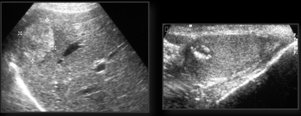
14 Fig. 5: Enlargement and diffuse and homogeneous hypoeconecity of the right testis with oval morphology and smooth contours. Page 14 of 25

15 Fig. 6: Increased vascularity of the right testis compared to the contralateral Page 15 of 25

16 Fig. 7: Comparative study both testes with inflammatory changes in the right one with normal left testicle. Page 16 of 25

17 Fig. 8: Right epididymis thickened, diffusely hypoechoic and hypervascularized Page 17 of 25

18 Fig. 9 Page 18 of 25


19 Fig. 10 Fig. 11: Torsion left testicle: left teste hypoechoic, with internal serration and absence of vascularization. Comparative study with contralateral teste. Page 19 of 25

20 Fig. 12 Page 20 of 25


21 Fig. 13 Fig. 14: Patient with right testicular trauma. Ultrasound diffuse and patched hypoechoic testicular involvement with preserved tunica and vascular paths with regard to testicular contusion. Thickening associated scrotal covered. Page 21 of 25


22 Fig. 15: Testicular diffuse seminoma. Testicular diffuse neoplasic infiltration of the right teste appreciate enlarged relative to the contralateral testicle, heterogeneous, with loss of oval morphology and lobulated contours. Fig. 16: Lymphoma Page 22 of 25

23 Fig. 17 Page 23 of 25
24 Fig. 18: 40 year old patient with hyperechoic liver metastases of choriocarcinoma. The scrotal ultrasound shows atrophic testis with peripherally calcified nodule in relation to testicular choriocarcinoma with gonadal level regression but with liver metastases Page 24 of 25
25 Conclusion Ultrasound is the imaging modality of choice in the evaluation and screening of testicular pathology (inflammatory, traumatic, ischemic and neoplasic testicular involvement may primarily cause focal or diffuse alteration in the testicular parenchyma). Personal information References -Bala R.Subramanyam, Steven C.Horii, Susan Hilton. Diffuse Testicular Disease: Sonographic Features and significance. AJR 145: , Diciembre Celestino Aso, MD. Goya Enriquez, MD, et.al. Gray-Scale and Color Doppler Sonography of Scrotal Disorders in Children: An Update. Radiographics 2005; 25: Anil T. Ahuja, MD. Et al. Diagnostic imaging ultrasound Fernández GC, Tardáguila FM, Rivas C, et al. Papel de la resonancia magnética en la patología testicular y paratesticular. Radiología 2001;43(7): De Luis Pastor E et al./ Actas Urol Esp. 2007;31(8): Julve E, Quiñonero A, etal. Actualización en Medicina de Urgencias. Primera Parte. Edición Amorós F.J, Cerezo F. et al. Utilidad de la ecografía en el estudio del escroto. Medicina General Nº Extraordinario Martín C, Rodríguez G, Rengifo D. Escroto agudo. En: Urgencias en Urología: Manual para residentes.jarpyo editores. Madrid p: Galens LE,Kas EJ. Diagnosis and treatment of the acute scrotum. En: American Academy of Family Physician. National Guideline Clearinghouse. U.S.A Deborah J. Rubens, MD. Testicular Torsion. Radiologic Pathology. Fith Edition Volume 1.Genitourinary Radiology Page 25 of 25
Testicular ultrasound in acute scrotal pain - beyond testicular torsion
 Testicular ultrasound in acute scrotal pain - beyond testicular torsion Poster No.: C-1284 Congress: ECR 2015 Type: Educational Exhibit Authors: I. Rolla, M. Nogueira, M. J. Aguiar, D. S. Garrido, J. A.
The Acute Scrotum: Sonographic Findings
 The Acute Scrotum: Sonographic Findings 가천의대길병원방사선과 양달모 Gachon Medical School Introduction Many diseases presenting as acute scrotal pain DDx is important for determining the appropriate treatment US with
Sonography of the scrotum: still the best!
 Sonography of the scrotum: still the best! Poster No.: C-1110 Congress: ECR 2013 Type: Educational Exhibit Authors: W. Mnari, A. Zrig, M. Maatouk, B. Hmida, R. Salem, W. HarzallahHizem, M. Golli; Monastir/TN
Let s see beyond the testicle: paratesticular pathology (anatomy, ultrasound findings and clinical-radiologic interpretation).
.") Let s see beyond the testicle: paratesticular pathology (anatomy, ultrasound findings and clinical-radiologic interpretation). Poster No.: C-1896 Congress: ECR 2015 Type: Educational Exhibit Authors: M.
Let s see beyond the testicle: paratesticular pathology (anatomy, ultrasound findings and clinical-radiologic interpretation). Poster No.: C-1896 Congress: ECR 2015 Type: Educational Exhibit Authors: M.
The role of ultrasonography with colour Doppler in the acute scrotum
 The role of ultrasonography with colour Doppler in the acute scrotum Poster No.: C-1509 Congress: ECR 2016 Type: Educational Exhibit Authors: M. S. R. O. Faustino, A. L. Amado Costa, J. J. B. Leitão, I.
Ultrasonography of Acute Scrotum
 Ultrasonography of Acute Scrotum Poster No.: C-0331 Congress: ECR 2012 Type: Educational Exhibit Authors: O. Nikolic, T. Mrdanin, M. Basta Nikolic, S. Stojanovic, K. 1 1 1 1 1 1 2 1 1 Petrovic, V. Vucaj
Whirlpool sign of testis, a sonographic sign of incomplete torsion
 Whirlpool sign of testis, a sonographic sign of incomplete torsion Poster No.: C-0965 Congress: ECR 2011 Type: Educational Exhibit Authors: F. Esposito, P. Sgambati, M. Di Serafino, D. Noviello, G. 1 3
Scrotum Kacey Morrison Amanda Baxter Sabrina Tucker July 18, 2006 SCROTUM
 Scrotum Kacey Morrison Amanda Baxter Sabrina Tucker July 18, 2006 SCROTUM 1) Other Names: Scrotum None Testicles Testes (Curry Tempkin, p. 236, 2/3/2) Ductus deferens spermatic cord (Tempkin, p. 279, Anatomy
Hyperechoic breast lesions can be malignant.
 Hyperechoic breast lesions can be malignant. Poster No.: C-0041 Congress: ECR 2015 Type: Educational Exhibit Authors: G. Babu, R. bradley; Edinburgh/UK Keywords: Breast, Ultrasound, Biopsy, Cancer DOI:
Intracystic papillary carcinoma of the breast
 Intracystic papillary carcinoma of the breast Poster No.: C-1932 Congress: ECR 2011 Type: Educational Exhibit Authors: V. Dimarelos, F. TZIKOS, N. Kotziamani, G. Rodokalakis, 1 2 3 1 1 1 2 T. MALKOTSI
Evaluation of thyroid nodules: prediction and selection of malignant nodules for FNA (cytology)
") Evaluation of thyroid nodules: prediction and selection of malignant nodules for FNA (cytology) Poster No.: C-0221 Congress: ECR 2014 Type: Authors: Keywords: DOI: Scientific Exhibit E. Papadaki, I. Tritou,
Evaluation of thyroid nodules: prediction and selection of malignant nodules for FNA (cytology) Poster No.: C-0221 Congress: ECR 2014 Type: Authors: Keywords: DOI: Scientific Exhibit E. Papadaki, I. Tritou,
Characterisation of cervical lymph nodes by US and PET-CT
 Characterisation of cervical lymph nodes by US and PET-CT Poster No.: C-1807 Congress: ECR 2010 Type: Educational Exhibit Topic: Head and Neck Authors: J. I. Garcia Gomez; Mexico City/MX Keywords: cervical
Testicular tumors; Ultrasonographic and Pathologic correlation
 Testicular tumors; Ultrasonographic and Pathologic correlation Poster No.: C-0106 Congress: ECR 2014 Type: Educational Exhibit Authors: Y. Kim, S. W. Shin, E. T. Kim, M. Y. Kim ; Kuri City/KR, 1 1 2 1
Vikram Dogra, M.D. Professor of Radiology, Urology & BME Department of Imaging Sciences University Of Rochester Medical Center
 Ultrasound of the Scrotum Vikram Dogra, M.D. Professor of Radiology, Urology & BME Department of Imaging Sciences University Of Rochester Medical Center Etiologies of Acute Scrotal Pain Epididymitis/Orchitis
Imaging characterization of renal clear cell carcinoma
 Imaging characterization of renal clear cell carcinoma Poster No.: C-0327 Congress: ECR 2011 Type: Educational Exhibit Authors: S. Ballester 1, A. Gaser 2, M. Dotta 1, M. F. CAPPA 1, F. Hammar 1 ; 1 2
"Burned out" testicular tumor: Clinical and radiological features
 "Burned out" testicular tumor: Clinical and radiological features Poster No.: C-0799 Congress: ECR 2012 Type: Educational Exhibit Authors: G. Viteri, I. Simon Yarza, R. Saiz-Mendiguren, J. Arias, A. Villanueva
Scientific Exhibit Authors: V. Moustakas, E. Karallas, K. Koutsopoulos ; Rodos/GR, 2
 Diagnosis of Acute Appendicitis: the role of Color Doppler Ultrasound as first-line imaging method and evaluation of the higher diagnostic performances of CT against its disadvantages. Poster No.: C-0708
Differential Diagnosis of The Acute Scrotum in The Pediatric Population.
 Differential Diagnosis of The Acute Scrotum in The Pediatric Population. Poster No.: C-1736 Congress: ECR 2013 Type: Educational Exhibit Authors: D. M. Castaño Palacios, J. C. Rayón-Aledo, I. Solis Muniz,
US features of scrotal disorders: A pictorial essay
 US features of scrotal disorders: A pictorial essay Poster No.: C-0463 Congress: ECR 2014 Type: Educational Exhibit Authors: J. SAAD, F. Marrakchi ; nejran, sa/sa, Monastir/TN Keywords: Genital / Reproductive
Superior Labrum Anterior Posterior lesions: ultrasound evaluation
 Superior Labrum Anterior Posterior lesions: ultrasound evaluation Poster No.: C-0472 Congress: ECR 2017 Type: Scientific Exhibit Authors: D. Belyaev; Yaroslavl/RU Keywords: Trauma, Arthrography, Ultrasound,
Lung sonography in the diagnosis of pneumothorax.
 Lung sonography in the diagnosis of pneumothorax. Poster No.: C-0526 Congress: ECR 2011 Type: Educational Exhibit Authors: K. Stefanidis, K. Vintzilaios, D. D. Cokkinos, E. Antypa, S. Dimopoulos, S. Nanas,
Sonographic and Mammographic Features of Phyllodes Tumours of the Breast: Correlation with Histological Grade
 Sonographic and Mammographic Features of Phyllodes Tumours of the Breast: Correlation with Histological Grade Poster No.: C-0046 Congress: ECR 2014 Type: Authors: Keywords: DOI: Scientific Exhibit C. Y.
Slowly growing malignant nodules and rapidly growing benign nodules: Evaluation of the value of volume doubling time
 Slowly growing malignant nodules and rapidly growing benign nodules: Evaluation of the value of volume doubling time Poster No.: C-208 Congress: ECR 2009 Type: Educational Exhibit Topic: Chest Authors:
Shear Wave Elastography in diagnostics of supraspinatus tendon.
 Shear Wave Elastography in diagnostics of supraspinatus tendon. Poster No.: C-2168 Congress: ECR 2013 Type: Authors: Keywords: DOI: Scientific Exhibit V. Saltykova; Moscow/RU Musculoskeletal joint, Musculoskeletal
Triple-negative breast cancer: which typical features can we identify on conventional and MRI imaging?
 Triple-negative breast cancer: which typical features can we identify on conventional and MRI imaging? Poster No.: C-1862 Congress: ECR 2013 Type: Educational Exhibit Authors: V. Bertani 1, A. Gualano
COLOR DOPPLER ULTRASOUND IN EVALUATION OF SCROTAL LESIONS
 COLOR DOPPLER ULTRASOUND IN EVALUATION OF SCROTAL LESIONS Desai Sanjay D Associate Professor, Department of Radiology, RCSM Govt. Medical College, Kolhapur. ABSTRACT: Color Doppler ultrasound is a non-invasive,
Psoriatic arthritis: early ultrasound findings
 Psoriatic arthritis: early ultrasound findings Poster No.: C-0399 Congress: ECR 2014 Type: Educational Exhibit Authors: R. Persechino 1, L. Cristiano 1, A. Bartoloni 1, C. Cantone 2, A. Keywords: DOI:
Radiologic and pathologic correlation of non-mass like breast lesions on US and MRI: Benign, high risk, versus malignant
 Radiologic and pathologic correlation of non-mass like breast lesions on US and MRI: Benign, high risk, versus malignant Poster No.: C-1161 Congress: ECR 2013 Type: Educational Exhibit Authors: J. Kwak,
Ultrasonic evaluation of superior mesenteric vein in cancer of the pancreatic head
 Ultrasonic evaluation of superior mesenteric vein in cancer of the pancreatic head Poster No.: C-1430 Congress: ECR 2012 Type: Authors: Keywords: DOI: Scientific Exhibit E. Fisenko, N. Vetsheva, E. Pershina;
Pleomorphic adenoma head and neck
 Pleomorphic adenoma head and neck Poster No.: C-1042 Congress: ECR 2015 Type: Educational Exhibit Authors: M. E. Pérez Montilla, I. Bravo Rey, E. Roldán Romero, F. BravoRodríguez; Cordoba/ES Keywords:
MRI BI-RADS: How to make it out?
 MRI BI-RADS: How to make it out? Poster No.: C-1850 Congress: ECR 2016 Type: Educational Exhibit Authors: M. Ben Ammar, A. Ben Miled, O. Ghdes, S. Harguem, A. Gaja, N. Mnif; Tunis/TN Keywords: Breast,
Valsalva-manoeuvre or prone belly position for computed tomography (CT) scan when an orbita varix is suspected: a single-case study.
 scan when an orbita varix is suspected: a single-case study.") Valsalva-manoeuvre or prone belly position for computed tomography (CT) scan when an orbita varix is suspected: a single-case study. Poster No.: C-0512 Congress: ECR 2012 Type: Authors: Keywords: DOI:
Valsalva-manoeuvre or prone belly position for computed tomography (CT) scan when an orbita varix is suspected: a single-case study. Poster No.: C-0512 Congress: ECR 2012 Type: Authors: Keywords: DOI:
Ethanol ablation of benign thyroid cysts and predominantly cystic thyroid nodules: factors that predict outcome.
 Ethanol ablation of benign thyroid cysts and predominantly cystic thyroid nodules: factors that predict outcome. Poster No.: C-0322 Congress: ECR 2014 Type: Authors: Keywords: DOI: Scientific Exhibit J.
Thyroid pathology: What radiologists need to know.
 Thyroid pathology: What radiologists need to know. Poster No.: C-1310 Congress: ECR 2014 Type: Educational Exhibit Authors: C. de la Torre, A. Sánchez Tovar, M. Molinero Pérez, R. Rodriguez Ortega, M.
MRI in staging of rectal carcinoma
 MRI in staging of rectal carcinoma Poster No.: C-0152 Congress: ECR 2015 Type: Scientific Exhibit Authors: J. R. Ramos Rodriguez, M. Atencia Ballesteros, M. D. M. Muñoz Ruiz, A. J. Márquez Moreno, M. D.
The "whirl sign". Diagnostic accuracy for intestinal volvulus.
 The "whirl sign". Diagnostic accuracy for intestinal volvulus. Poster No.: C-0670 Congress: ECR 2014 Type: Scientific Exhibit Authors: M. Pire, M. Marti, A. Borobia, A. Verón; Madrid/ES Keywords: Abdomen,
CT evaluation of small bowel carcinoid tumors
 CT evaluation of small bowel carcinoid tumors Poster No.: C-0060 Congress: ECR 2015 Type: Educational Exhibit Authors: N. V. V. P. Costa, L. Nascimento, T. Bilhim ; Estoril/PT, PT, 1 2 3 1 2 3 Lisbon/PT
Alex Lam, HMS III. September The Acute Scrotum. Alex Lam, Harvard Medical School Year III Gillian Lieberman, MD. Gillian Lieberman, MD
 September 2002 The Acute Scrotum Alex Lam, Harvard Medical School Year III DDx: : Acute Scrotal Pain & Enlargement PAIN Inflammatory disorder Testicular torsion Testicular infarction Testicular abscess
Intratendinous tears of the Achilles tendon - a new pathology? Analysis of a large 4 year cohort.
 Intratendinous tears of the Achilles tendon - a new pathology? Analysis of a large 4 year cohort. Poster No.: C-1680 Congress: ECR 2014 Type: Scientific Exhibit Authors: S. Morton, T. Parkes, O. Chan,
Digital breast tomosynthesis (DBT) occult breast cancers: clinical, radiological and histopathological features.
 occult breast cancers: clinical, radiological and histopathological features.") Digital breast tomosynthesis (DBT) occult breast cancers: clinical, radiological and histopathological features. Poster No.: C-1707 Congress: ECR 2015 Type: Scientific Exhibit Authors: V. Vinci 1, A. Iqbal
Digital breast tomosynthesis (DBT) occult breast cancers: clinical, radiological and histopathological features. Poster No.: C-1707 Congress: ECR 2015 Type: Scientific Exhibit Authors: V. Vinci 1, A. Iqbal
Breast Pathology in Men: Radiologic-Pathologic Correlation
 Breast Pathology in Men: Radiologic-Pathologic Correlation Poster No.: C-0243 Congress: ECR 2012 Type: Scientific Exhibit Authors: G. Garrido; Málaga/ES Keywords: Breast, Ultrasound, Mammography, Biopsy,
Role of ultrasound in the evaluation of the ileocecal valve
 Role of ultrasound in the evaluation of the ileocecal valve Poster No.: C-1581 Congress: ECR 2010 Type: Scientific Exhibit Topic: GI Tract Authors: M. Mohammed, M. Hussain, U. Momin, S. Lakhtakia, N. D.
The Radiologic Features of Xanthogranulomatous Cholecystitis: An Important Mimic of Gallbladder Carcinoma
 The Radiologic Features of Xanthogranulomatous Cholecystitis: An Important Mimic of Gallbladder Carcinoma Poster No.: C-0691 Congress: ECR 2014 Type: Authors: Keywords: DOI: Educational Exhibit H. L. khosa
Single cold nodule in Graves' disease: benign vs malignant
 Single cold nodule in Graves' disease: benign vs malignant Poster No.: C-0073 Congress: ECR 2011 Type: Authors: Keywords: DOI: Scientific Paper L. I. Sonoda, M. Halim, K. Balan; Cambridge/UK Head and neck,
US-guided steroid and hyaluronic acid infiltration for the treatment of hand and wrist tenosynovitis: Preliminary experience
 US-guided steroid and hyaluronic acid infiltration for the treatment of hand and wrist tenosynovitis: Preliminary experience Poster No.: C-2342 Congress: ECR 2010 Type: Scientific Exhibit Topic: Musculoskeletal
Purpose. Methods and Materials. Results
 Prevalence and significance of hypoattenuating hepatic lesions deemed too small to characterise: How are we following up these lesions and what are the outcomes? Poster No.: C-014 Congress: ECR 2009 Type:
Applied scrotal ultrasound anatomy and pathology:a pictorial review
 Applied scrotal ultrasound anatomy and pathology:a pictorial review Poster No.: C-1166 Congress: ECR 2014 Type: Educational Exhibit Authors: S. Santos Magadán, D. Gómez-Santos, J. García-Yavar, 1 1 2 1
Diffuse pseudo angiomatous stromal hyperplasia of breast - A case report
 Diffuse pseudo angiomatous stromal hyperplasia of breast - A case report Poster No.: C-0011 Congress: ECR 2011 Type: Scientific Exhibit Authors: S. L. Penukonda, B. Dev; CHENNAI, TN/IN Keywords: Breast,
64-MDCT imaging of the pancreas: Scan protocol optimisation by different scan delay regimes
 64-MDCT imaging of the pancreas: Scan protocol optimisation by different scan delay regimes Poster No.: C-051 Congress: ECR 2009 Type: Scientific Exhibit Topic: Abdominal and Gastrointestinal Authors:
ARDS - a must know. Page 1 of 14
 ARDS - a must know Poster No.: C-1683 Congress: ECR 2016 Type: Authors: Keywords: DOI: Educational Exhibit M. Cristian; Turda/RO Education and training, Edema, Acute, Localisation, Education, Digital radiography,
BI-RADS 3, 4 and 5 lesions on US: Five categories and their diagnostic efficacy and pitfalls in interpretation
 BI-RADS 3, 4 and 5 lesions on US: Five categories and their diagnostic efficacy and pitfalls in interpretation e-poster: C-118 Congress: ECR 2008 Type: Educational Exhibit Topic: Breast / Ultrasound Authors:
Excavated pulmonary nodule: steps to diagnosis?
 Excavated pulmonary nodule: steps to diagnosis? Poster No.: C-1044 Congress: ECR 2014 Type: Authors: Keywords: DOI: Educational Exhibit W. Mnari, M. MAATOUK, A. Zrig, B. Hmida, M. GOLLI; Monastir/ TN Metastases,
Long bones manifestations of congenital syphilis
 Long bones manifestations of congenital syphilis Poster No.: C-0139 Congress: ECR 2011 Type: Educational Exhibit Authors: T. F. de Souza 1, P. P. Collier 1, E. J. M. Bronzatto 1, G. L. P. Keywords: DOI:
Atypical ductal hyperplasia diagnosed at ultrasound guided biopsy of breast mass
 Atypical ductal hyperplasia diagnosed at ultrasound guided biopsy of breast mass Poster No.: C-1483 Congress: ECR 2014 Type: Authors: Keywords: DOI: Scientific Exhibit J. Cho, J. Chung, E. S. Cha, J. E.
Parathyroid Glands: location, condition and value of imaging tests.
 Parathyroid Glands: location, condition and value of imaging tests. Poster No.: C-2283 Congress: ECR 2015 Type: Educational Exhibit Authors: E. Elías Cabot, P. Segui, G. D. Tobar Murgueitio; Cordoba/ES
Imaging spectrum of angiosarcoma of breast
 Imaging spectrum of angiosarcoma of breast Poster No.: C-1097 Congress: ECR 2015 Type: Educational Exhibit Authors: T. Sehgal, S. K. Ramani, N. Nair, A. Patil, M. H. Thakur; Mumbai/ IN Keywords: Neoplasia,
Diffuse Alveolar Hemorrhage: Initial and Follow-up HRCT Features
 Diffuse Alveolar Hemorrhage: Initial and Follow-up HRCT Features Poster No.: E-0037 Congress: ESTI 2012 Type: Authors: Keywords: Scientific Exhibit M. Y. Kim; Seoul/KR Lung, CT-High Resolution, CT, Computer
ESUR SCROTAL AND PENILE IMAGING WORKING GROUP MULTIMODALITY IMAGING APPROACH TO SCROTAL AND PENILE PATHOLOGIES 2ND ESUR TEACHING COURSE
 ESUR SCROTAL AND PENILE IMAGING WORKING GROUP MULTIMODALITY IMAGING APPROACH TO SCROTAL AND PENILE PATHOLOGIES 2ND ESUR TEACHING COURSE NORMAL ANATOMY OF THE SCROTUM MICHAEL NOMIKOS M.D. F.E.B.U. UROLOGICAL
Malignant Transformation of Endometriosis: Magnetic Resonance Imaging Aspects
 Malignant Transformation of Endometriosis: Magnetic Resonance Imaging Aspects Poster No.: C-0084 Congress: ECR 2014 Type: Scientific Exhibit Authors: E. A. Yukhno, I. Trofimenko, G. Trufanov; St. Petersburg/RU
Malignant Transformation of Endometriosis: Magnetic Resonance Imaging Aspects
 Malignant Transformation of Endometriosis: Magnetic Resonance Imaging Aspects Poster No.: C-0084 Congress: ECR 2014 Type: Scientific Exhibit Authors: E. A. Yukhno, I. Trofimenko, G. Trufanov; St. Petersburg/RU
Ultrasound assessment of most frequent shoulder disorders
 Ultrasound assessment of most frequent shoulder disorders Poster No.: C-2026 Congress: ECR 2014 Type: Educational Exhibit Authors: S. P. Ivanoski; Ohrid/MK Keywords: Trauma, Athletic injuries, Arthritides,
The follow-up of uterine fibroids treated with HIFU: role of DWI and Dynamic contrast-study MRI
 The follow-up of uterine fibroids treated with HIFU: role of DWI and Dynamic contrast-study MRI Poster No.: C-1137 Congress: ECR 2011 Type: Authors: Keywords: DOI: Scientific Exhibit V. Zampa, V. Vallini,
Post-catheterization pseudoaneurysms treatment with ultrasound-guided thrombin injection
 Post-catheterization pseudoaneurysms treatment with ultrasound-guided thrombin injection Poster No.: C-2107 Congress: ECR 2010 Type: Topic: Scientific Exhibit Interventional Radiology Authors: A. Ladas,
Biliary tree dilation - and now what?
 Biliary tree dilation - and now what? Poster No.: C-1767 Congress: ECR 2012 Type: Educational Exhibit Authors: I. Ferreira, A. B. Ramos, S. Magalhães, M. Certo; Porto/PT Keywords: Pathology, Diagnostic
Ultrasound assessment of T1 Squamous Cell Carcinomas of the Tongue.
 Ultrasound assessment of T1 Squamous Cell Carcinomas of the Tongue. Poster No.: C-2014 Congress: ECR 2015 Type: Educational Exhibit Authors: S. R. Rice, G. Price, L. Firmin, S. Morley, T. Beale; London/UK
Prostate biopsy: MR imaging to the rescue
 Prostate biopsy: MR imaging to the rescue Poster No.: C-1855 Congress: ECR 2014 Type: Educational Exhibit Authors: N. V. V. B. Marques 1, J. Ip 1, A. Loureiro 2, J. Niza 1, M. Palmeiro 2, Keywords: DOI:
THI-RADS. US differentiation of thyroid lesions.
 THI-RADS. US differentiation of thyroid lesions. Poster No.: C-0864 Congress: ECR 2015 Type: Scientific Exhibit Authors: A. N. Sencha, Y. Patrunov, M. S. Mogutov, E. Penyaeva, A. 1 1 1 2 1 1 1 2 Gruzdev,
THI-RADS. US differentiation of thyroid lesions.
 THI-RADS. US differentiation of thyroid lesions. Poster No.: C-0864 Congress: ECR 2015 Type: Scientific Exhibit Authors: A. N. Sencha, Y. Patrunov, M. S. Mogutov, E. Penyaeva, A. 1 1 1 2 1 1 1 2 Gruzdev,
Acute scrotum. Acute Epididymo-orchitis. Phyllis Yan, APDR (QEH)
") Acute scrotum Acute Epididymo-orchitis Phyllis Yan, APDR (QEH) Conditions leading to acute pain Torsion Acute Epididymitis / Epididymoorchitis Scrotal trauma Inguinal hernias Testicular tumors Epididymitis/epididymo
Acute scrotum Acute Epididymo-orchitis Phyllis Yan, APDR (QEH) Conditions leading to acute pain Torsion Acute Epididymitis / Epididymoorchitis Scrotal trauma Inguinal hernias Testicular tumors Epididymitis/epididymo
"Ultrasound measurements of the lateral ventricles in neonates: A comparison of multiple measurements methods."
 "Ultrasound measurements of the lateral ventricles in neonates: A comparison of multiple measurements methods." Poster No.: C-1557 Congress: ECR 2014 Type: Authors: Keywords: DOI: Educational Exhibit I.
Curious case of Misty Mesentery
 Curious case of Misty Mesentery Poster No.: C-1385 Congress: ECR 2015 Type: Authors: Keywords: DOI: Educational Exhibit T. Simelane 1, H. Khosa 2, N. Ramesh 2 ; 1 Dublin/IE, 2 Portlaoise/IE Abdomen, Anatomy,
Popliteal pterygium syndrome
 Popliteal pterygium syndrome Poster No.: C-1816 Congress: ECR 2011 Type: Educational Exhibit Authors: L. B. S. Santos, J. L. D. O. Schiavon, O. O. Guimaraes Neto, 1 1 2 3 1 1 C. A. P. Braga, R. S. LEMOS,
Radiological features of Legionella Pneumophila Pneumonia
 Radiological features of Legionella Pneumophila Pneumonia Poster No.: E-0048 Congress: ESTI 2012 Type: Scientific Exhibit Authors: M. Vinciguerra, L. Stefanetti, E. Teti, G. Argentieri, L. G. 1 1 1 1 1
High frequency US of the temporomandibualar joint (TMJ) - practical guide
 - practical guide") High frequency US of the temporomandibualar joint (TMJ) - practical guide Poster No.: C-2352 Congress: ECR 2014 Type: Educational Exhibit Authors: M. Lasecki, C. M. Olchowy, K. Kaczorowski, J. S#onina,
High frequency US of the temporomandibualar joint (TMJ) - practical guide Poster No.: C-2352 Congress: ECR 2014 Type: Educational Exhibit Authors: M. Lasecki, C. M. Olchowy, K. Kaczorowski, J. S#onina,
Extrapulmonary Manifestations of Tuberculosis: A Radiologic Review
 Extrapulmonary Manifestations of Tuberculosis: A Radiologic Review Poster No.: C-1958 Congress: ECR 2014 Type: Authors: Educational Exhibit J. Isern 1, S. Llaverias Borrell 1, A. Olarte 1, E. Grive 1,
Renal masses - the role of diagnostic imaging
 Renal masses - the role of diagnostic imaging Poster No.: C-2471 Congress: ECR 2015 Type: Educational Exhibit Authors: V. Rai#; Bjelovar/HR Keywords: Cysts, Cancer, Structured reporting, Ultrasound, MR,
Acute Sialadenitis in Childhood: CT Findings and Clinical Manifestation according to the Gland Involvements
 Acute Sialadenitis in Childhood: CT Findings and Clinical Manifestation according to the Gland Involvements Poster No.: C-0669 Congress: ECR 2012 Type: Scientific Exhibit Authors: A. Lee; Bucheon/KR Keywords:
Contrast-enhanced ultrasound (CEUS) in the evaluation and characterization of complex renal cysts
 in the evaluation and characterization of complex renal cysts") Contrast-enhanced ultrasound (CEUS) in the evaluation and characterization of complex renal cysts Poster No.: C-2812 Congress: ECR 2018 Type: Educational Exhibit Authors: J. A. Torres de Abreu Macedo,
Contrast-enhanced ultrasound (CEUS) in the evaluation and characterization of complex renal cysts Poster No.: C-2812 Congress: ECR 2018 Type: Educational Exhibit Authors: J. A. Torres de Abreu Macedo,
Imaging findings in neonates with hypoxic-ischaemic encephalopathy and terapeutic hypothermia.
 Imaging findings in neonates with hypoxic-ischaemic encephalopathy and terapeutic hypothermia. Poster No.: C-1577 Congress: ECR 2014 Type: Scientific Exhibit Authors: S. Manso Garcia, M. J. Velasco Marcos,
When to suspect Wegener Granulomatosis: A radiologic review
 When to suspect Wegener Granulomatosis: A radiologic review Poster No.: P-0038 Congress: ESTI 2015 Type: Educational Poster Authors: A. Tilve Gómez, R. Díez Bandera, P. Rodríguez Fernández, M. Garcia Vazquez-Noguerol,
Role of positron emission mammography (PEM) for assessment of axillary lymph node status in patients with breast cancer
 for assessment of axillary lymph node status in patients with breast cancer") Role of positron emission mammography (PEM) for assessment of axillary lymph node status in patients with breast cancer Poster No.: C-1260 Congress: ECR 2011 Type: Scientific Paper Authors: K. M. Kulkarni,
Role of positron emission mammography (PEM) for assessment of axillary lymph node status in patients with breast cancer Poster No.: C-1260 Congress: ECR 2011 Type: Scientific Paper Authors: K. M. Kulkarni,
Diffuse high-attenuation within mediastinal lymph nodes on non-enhanced CT scan: Usefulness in the prediction of benignancy
 Diffuse high-attenuation within mediastinal lymph nodes on non-enhanced CT scan: Usefulness in the prediction of benignancy Poster No.: C-1785 Congress: ECR 2012 Type: Authors: Keywords: DOI: Scientific
Ultrasound evaluation of patients with acute abdominal pain in the emergency department
 Ultrasound evaluation of patients with acute abdominal pain in the emergency department Poster No.: C-2584 Congress: ECR 2012 Type: Authors: Keywords: DOI: Educational Exhibit A. A. Falticeanu, A.-M. Alecsa-Lupu,
Imaging findings in neonates with hypoxic-ischaemic encephalopathy and terapeutic hypothermia.
 Imaging findings in neonates with hypoxic-ischaemic encephalopathy and terapeutic hypothermia. Poster No.: C-1577 Congress: ECR 2014 Type: Scientific Exhibit Authors: S. Manso Garcia, M. J. Velasco Marcos,
Differentiation of osteoporosis from metastasis in the vertebral fracture using chemical shift and diffusion weighted imaging
 Differentiation of osteoporosis from metastasis in the vertebral fracture using chemical shift and diffusion weighted imaging Poster No.: C-0444 Congress: ECR 2012 Type: Educational Exhibit Authors: H.
B-330 Cervical lymph node ultrasonography in HIV infected children
 B-330 Cervical lymph node ultrasonography in HIV infected children Scientific Paper B-330 Cervical lymph node ultrasonography in HIV infected children A. Butnaru (Cluj Napoca/RO) S. D. Bolboaca (Cluj Napoca/RO)
Lesions of the pancreaticoduodenal groove, a pictorial review
 Lesions of the pancreaticoduodenal groove, a pictorial review Poster No.: C-2131 Congress: ECR 2013 Type: Educational Exhibit Authors: E. Ni Mhurchu, L. Lavelle, I. Murphy, S. Skehan ; IE, Dublin/ IE Keywords:
Department of Medical Imaging, The Ottawa Hospital. Satheesh Krishna Sabarish Narayanasamy Wael Shabana Adnan Sheikh
 Department of Medical Imaging, The Ottawa Hospital. Satheesh Krishna Sabarish Narayanasamy Wael Shabana Adnan Sheikh Nothing to disclose Common and unusual presentations and manifestations of testicular
Synovial hemangioma of the suprapatellar bursa
 Synovial hemangioma of the suprapatellar bursa Poster No.: P-0040 Congress: ESSR 2013 Type: Authors: Keywords: DOI: Scientific Exhibit A. YESILDAG, S. Keskin, H. Kalkan, S. Kucuksen, U. Kerimoglu; Konya/TR
Lung cancer in patients with chronic empyema
 Lung cancer in patients with chronic empyema Poster No.: P-0025 Congress: ESTI 2015 Type: Scientific Poster Authors: Y. Lee, C.-K. Park; Guri/KR Keywords: Neoplasia, Biopsy, PET-CT, CT, Thorax, Lung DOI:
ShearWave elastography in lymph nodes
 ShearWave elastography in lymph nodes Poster No.: B-0158 Congress: ECR 2015 Type: Authors: Keywords: DOI: Scientific Paper F. Houari, O. Lucidarme, J. Gabarre, F. Charlotte, C. Pellot- Barakat, M. Lefort,
A pictorial review of normal anatomical appearences of Pericardial recesses on multislice Computed Tomography.
 A pictorial review of normal anatomical appearences of Pericardial recesses on multislice Computed Tomography. Poster No.: C-1787 Congress: ECR 2012 Type: Educational Exhibit Authors: N. Ahmed 1, G. Avery
Ultrasound (US) evaluation of peritoneal thickness in children and young patients on peritoneal dialysis (PD): A single centre experience
 evaluation of peritoneal thickness in children and young patients on peritoneal dialysis (PD): A single centre experience") Ultrasound (US) evaluation of peritoneal thickness in children and young patients on peritoneal dialysis (PD): A single centre experience Poster No.: C-2812 Congress: ECR 2010 Type: Scientific Exhibit
Ultrasound (US) evaluation of peritoneal thickness in children and young patients on peritoneal dialysis (PD): A single centre experience Poster No.: C-2812 Congress: ECR 2010 Type: Scientific Exhibit
CT Fluoroscopy-Guided vs Multislice CT Biopsy ModeGuided Lung Biopies:a preliminary experience
 CT Fluoroscopy-Guided vs Multislice CT Biopsy ModeGuided Lung Biopies:a preliminary experience Poster No.: C-0097 Congress: ECR 2016 Type: Scientific Exhibit Authors: A. Casarin, G. Rech, C. Cicero, A.
Breast calcification: Management and Pictorial Review
 Breast calcification: Management and Pictorial Review Poster No.: C-0692 Congress: ECR 2014 Type: Educational Exhibit Authors: V. de Lara Bendahan, M. F. Ramos Solis, A. Amador Gil, C. 1 2 3 2 4 4 Gómez
HRUS in the evaluation of the nails in patients with Psoriasis.
 HRUS in the evaluation of the nails in patients with Psoriasis. Poster No.: C-0512 Congress: ECR 2017 Type: Scientific Exhibit Authors: I. Mussetto, N. Romano, A. Fischetti, M. Burlando, A. Muda; Genova/IT
Pneumo-esophageal 64-MDCT technique for gastric cancer evaluation
 Pneumo-esophageal 64-MDCT technique for gastric cancer evaluation Poster No.: C-1627 Congress: ECR 2010 Type: Scientific Exhibit Topic: GI Tract Authors: M. Ulla, E. Gentile, E. Levy, D. Cavadas, J. Ithurralde
Computed tomography and Modified RECIST criteria for assessment of response in malignant pleural mesothelioma
 Computed tomography and Modified RECIST criteria for assessment of response in malignant pleural mesothelioma Poster No.: C-0729 Congress: ECR 2013 Type: Scientific Exhibit Authors: A. Marin, I. Pozek,
Pathologic outcomes of coarse heterogeneous calcifications detected on mammography
 Pathologic outcomes of coarse heterogeneous calcifications detected on mammography Poster No.: C-1957 Congress: ECR 2011 Type: Scientific Paper Authors: H. J. Lim, K. R. Cho, K. W. Hwang, B. K. Seo, O.