Squamous carcinoma of the penis: a case report
|
|
|
- Philomena Ferguson
- 5 years ago
- Views:
Transcription
1 Squamous carcinoma of the penis: a case report De Fiores A., Diosi D., Bella G., De Marco V. Introdution Penile cancer is a relatively rare neoplasm in the developed world and 95% of the cases histologically correspond to squamous cell carcinoma (SCC)(1). Although it accounts for 10% 20% of all malignancies in males in Asia, Africa, and South America, it has a prevalence of only 1% in Western countries (2). In the United States and Europe, SCC of the penis accounts for 0.4 to 0.7% of all malignancies found in men; the incidence varies from 0.1 to 0.7 per 100,000 population (1). Several etiologic risk factors have been recognized in the development of this malignancy. Infection with oncogenic types of Human Papilloma Virus (HPV) and lichen sclerosus seem to be the main risk factors for this neoplasm, but also, lack of neonatal circumcision (especially when associated with phimosis), and exposure to tobacco, among other causes, have been implicated (3). We describe an interesting case of a squamous cell carcinoma of the penis in a 67-year-old patient with a 9- months history of tumor growth associated with phimosis, for which total penectomy, perineal urethrostomy and bilateral inguinal lymphadenectomy were carried out.
2 Case report A 67-year-old man with a 9-months history of penile tumor growth associated with surface ulceration attended the our Hospital. The presence of phimosis was observed in the previous month during urological examination. The patient denied any hematuria and urinary symptoms. Physical examination indicated the presence of a solid swelling at the level of the distal portion of the penis associated with foul smelling purulent ulceration involving the skin nearby (Fig.1). Examination of the urethra, scrotum and testes were normal. Several movable inguinal lymph nodes of cm in size with moderate consistency could be palpated on both sides. The results of laboratory examinations were normal except for leucocytosis. He had full genital screening but the tests were negative for gonorrhoea, chlamydia, mycoplasma, ureaplasma and Trichomonas vaginalis. Subpreputial swab taken for candida was also negative. Antibiotics had been administered for 2 weeks, without improvements. In agreement with the urologist, the patient underwent to ultrasound exam of the penis that showed a solid inhomogeneus mass involving the corpora cavernosa associated to thickening of the tunica albuginea (Fig. 2). The Color-Doppler US reveled an incresed arterial vascularization of the solid lesion (Fig.3a) and, partly, of the corpora cavernosa (Fig.3b).
3 Under continuous epidural anesthesia, total penectomy with perineal urethrostomy and bilateral inguinal lymphadenectomy were carried out. Postoperative pathological investigations confirmed that it was a moderate-differentiated squamous cell carcinoma of the penis involving the Buck fascia, the corpora cavernosa and the skin nearby, whereas the surgical margin was negative. Inguinal lymph nodes showed lymphoid inflammatory hyperplasia without metastatic disease. Neither recurrence nor distant metastasis has been observed during a 10- month follow-up visit. Normal Penile Anatomy and B-mode ultrasound appearances The penis is composed of three cylindric structures consisting of endothelium-lined cavernous spaces: two dorsal corpora cavernosa and the ventral corpus spongiosum (Fig 4). The corpora cavernosa are the main erectile bodies, and the corpus spongiosum contains the urethra. A layer of fibrous tissue, the tunica albuginea, surrounds each corpus cavernosum and the corpus spongiosum. All three corpora are sur- rounded by two outer fascial layers, one deep (Buck fascia) and one superficial (Colles fascia). The arterial supply to the penis is from the internal pudenal artery, which divides into terminal branches, the dorsal penile artery, the cavernosal artery and bulbar artery. Emissary veins pierce the tunica albuginea, drain into the deep dorsal vein via the spongiosal, circumflex and cavernosal veins.(4)
4 The corpora cavernosa and the corpus spongiosum manifest at US as homogeneous cylindric structures. The urethra can be evaluated at US after distending it with fluid. The tunica albuginea and the Buck fascia are stuck together and appear as a thin echogenic line surrounding the three corpora. The Colles fascia is barely visible in healthy patients. On transverse US images, the cavernosal arteries appear as a pair of dots, whereas on longitudinal images they manifest as linear or narrow tubular structures. The dorsal vessels are visible as anechoic structures in the dorsal aspect of the penile shaft. Color Doppler US clearly depicts the penile vasculature (5). Discussion Penile cancer is seen in men in the 6th and 7th decades of life; less than one-quarter of patients are under 40 years of age. The most important etiologic factor in penile cancer is the presence of foreskin, which results in the accumulation of smegma. Therefore, the risk of this disease is three times higher in uncircumcised men than in circumcised men. Poor hygiene also contributes to the development of penile cancer through the accumulation of smegma and other irritants. The presence of phimosis has a strong association with penile cancer and is seen in 25% of penile cancer patients. Other important risk factors include chronic inflammatory conditions (eg, balanoposthitis, lichen sclerosus et atrophicus), smoking, human papilloma virus 16, and human papilloma virus 18 (2).
5 Primary neoplasms of the penis can be classified into the following histologic types: squamous cell carcinoma, sarcoma, melanoma, basal cell carcinoma, and lymphoma. Squamous cell carcinoma accounts for more than 95% of all primary neoplasms of the penis. Squamous cell carcinoma of the penis is most commonly located in the glans penis (48% of cases). In decreasing order of frequency, other locations include the prepuce (21% of cases), glans penis and prepuce (9%), coronal sulcus (6%), and shaft (2%) (2). The spread of penile cancer usually occurs via lymphatic vessels, with the Buck fascia acting as a barrier to corporal invasion and hematogenous spread. Invariably, the lymphatic vessels of the penis first drain into the inguinal nodes before reaching the pelvic nodes (2). Distant metastases are uncommon in patients who present with penile cancer (<3%-5% of cases). Generally, hematogenous metastases occur late in the disease course and are associated with a dismal prognosis (6).Appropriate management and treatment outcomes in men with penile cancer depend critically on the correct diagnosis, grading and staging of the malignancy. Grading of penile SCC is usually determinated by the degree of cell anaplasia. A common approach is to grade penile cancer as grade 1, well differentiated (no evidence of anaplasia); grade 2, moderately differentiated (< 50% anaplasia); grade 3, poorly differentiated (> 50% anaplastic cell).
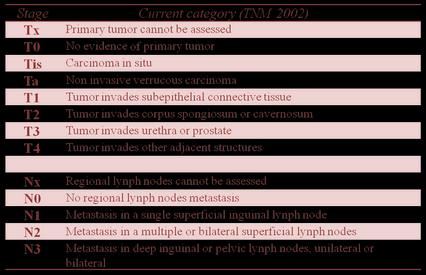
6 A more sophisticated system with 4 grades has been proposed accrding to the degree of keratinization, cell atypia, mitotic activity, and the amount of inflammatory cell infiltrate (7). Historically, several staging systems have been used for carcinoma of the penis. The Jackson system (Table 1) was introduced in 1966, and the TNM classification was introduced in 1968 and revised in 1978, 1987, and 2002 (Table 2) (6). In this article we want to underline the importance of early diagnosis of penile cancer through clinical examination and ultrasonography. Clinical examination and palpation of the primary tumor and the inguinal nodes has been the traditional approach for assessment of local invasion of the corpora and skin and evaluation for inguinal nodal metastases (2). Infact, clinical examination of the primary penile lesion should evaluate and document the number of lesions, tumor dimensions (size), sites involved (foreskin, glans, shaft), color, morfology (flat, papillary, nodular, ulcerating, fungating), relationship with other structures (corpus spongiosum, corpora cavernosa, urethra), and boundaries (edges) (6). Although physical examination can reliably help predict primary tumor size and the extent of cavernosal infiltration with a high positive predictive value, primary tumor staging by means of palpation can sometimes result in understaging of the disease or overstaging from edema and infection. Tumors with minimal or no infiltration but presenting as large exophytic growths may also be overstaged clinically (8, 9).
7 Also, nodal assessment based on the palpation of inguinal lymph nodes can produce both false-positive and false-negative results (10). For these reasons, US has a very important role in the management of penile cancer. In our experience, we found that the penile anatomy was easily demonstrated by sonography and that any encroachment by a mass was clearly evident. Any breach in the tunica albuginea was seen, and urethral involvement even when clinically silent was disclosed. US can also demonstrate the tumor invasion of the tunica or corpora cavernosa and accurately assess regional lynph nodes. In conclusion, although clinical examination remains the first approach for the diagnosis and staging of penile cancer, US can complement clinical examination and can be used to evaluate the primary lesion for local invasion, assess the status of the regional lymph nodes, and look for evidence of distant metastases.

8 Table 1. Jackson Classification of Penile Carcinoma


9 Figure 1. The image shows an increase in size of the distal portion of the penis associated with phimosis and ulceration of the glans Figure 2. Transverse US image of the penis showed a solid inhomogeneus mass involving the corpora cavernosa associated to thickening of the tunica albuginea.


10 Figure 3 a. Figure 3b. The Color-Doppler US reveled an incresed arterial vascularization of the solid lesion (Fig.3 a) and, partly, of the corpora cavernosa (Fig.3 b).

11 Figure 3b. The Color-Doppler US reveled an incresed arterial vascularization of the solid lesion (Fig.3 a) and, partly, of the corpora cavernosa (Fig.3 b).
12 References 1. Wanick FB, Teichner TC, Silva R, Magnanini MM, Azevedo LM. Squamous cell carcinoma of the penis: clinicopathologic study of 34 cases. An Bras Dermatol Nov-Dec;86(6): Singh AK, Saokar A, Hahn PF, Harisinghani MG. Imaging of penile neoplasms. Radiographics Nov-Dec;25(6): Caso JR, Rodriguez AR, Correa J, Spiess PE. Update in the management of penile cancer. Int Braz J Urol Jul-Aug;35(4): Wilkins CJ, Sriprasad S, Sidhu PS. Colour Doppler ultrasound of the penis. Clin Radiol Jul;58(7): Bertolotto M, Serafini G, Savoca G, Liguori G, Calderan L, Gasparini C, Mucelli RP. Color Doppler US of the postoperative penis: anatomy and surgical complications. Radiographics May-Jun;25(3): Heyns CF, Mendoza-Valdés A, Pompeo AC. Diagnosis and staging of penile cancer. Urology Aug;76(2 Suppl 1):S Maiche AG, Pyrhönen S, Karkinen M. Histological grading of squamous cell carcinoma of the penis: a new scoring system. Br J Urol May;67(5): Agrawal A, Pai D, Ananthakrishnan N, Smile SR, Ratnakar C. Clinical and sonographic findings in carcinoma of the penis. J Clin Ultrasound Oct;28(8): Lont AP, Besnard AP, Gallee MP, Van Tinteren H, Horenblas S. A comparison of physical examination and imaging in determining the extent of primary penile carcinoma. BJU Int 2003;91(6): Horenblas S. Lymphadenectomy for squamous cell carcinoma of the penis. I. Diagnosis of lymph node metastasis. BJU Int 2001;88:
Penis Cancer. What is penis cancer? Symptoms. Patient Information. Pagina 1 / 9. Patient Information - Penis Cancer
 Patient Information English 31 Penis Cancer The underlined terms are listed in the glossary. What is penis cancer? Cancer is abnormal cell growth in the skin or organ tissue. When this cell growth starts
NORMAL ANATOMY OF THE PENIS
 NORMAL ANATOMY OF THE PENIS IOANNIS VARKARAKIS ASOSCIATE PROFESSOR OF UROLOGY 2 ND DEPT OF UROLOGY NATIONAL & KAPODISTRIAN UNIVERSITY OF ATHENS PENILE GROSS ANATOMY 3 ERECTILE COLUMNS TWO CORPORA CAVERNOSA
Penis Cancer. What is penis cancer? Symptoms. Patient Information. Pagina 1 / 9. Patient Information - Penis Cancer
 Patient Information English 31 Penis Cancer The underlined terms are listed in the glossary. What is penis cancer? Cancer is abnormal cell growth in the skin or organ tissue. When this cell growth starts
GUIDELINES ON PENILE CANCER
 GUIDELINES ON PENILE CANCER (Text update April 2010) G. Pizzocaro, F. Algaba, S. Horenblas, E. Solsona, S. Tana, H. Van Der Poel, N. Watkin 78 Penile Cancer Eur Urol 2010 Jun;57(6):1002-12 Introduction
GUIDELINES ON PENILE CANCER
 GUIDELINES ON PENILE CANCER (Text updated March 2005) G. Pizzocaro (chairman), F. Algaba, S. Horenblas, H. van der Poel, E. Solsona, S. Tana, N. Watkin 58 Penile Cancer Eur Urol 2004;46(1);1-8 Introduction
GUIDELINEs ON PENILE CANCER
 GUIDELINEs ON PENILE CANCER (update April 2010) G. Pizzocaro, F. Algaba, S. Horenblas, E. Solsona, S. Tana, H. Van Der Poel, N. Watkin Eur Urol 2010, doi:10.1016/j.eururo.2010.01.039 Introduction Over
GUIDELINES ON PENILE CANCER
 46 E. Solsona (chairman), F. Algaba, S. Horenblas, G. Pizzocaro, T. Windahl Eur Urol 2002;42(3):199-203 Introduction Penile carcinoma is an uncommon malignant disease with an incidence ranging from 0.1
EAU GUIDELINES ON PENILE CANCER
 EAU GUIDELINES ON PENILE CANCER (Text update April 2014) O.W. Hakenberg (Chair), E. Compérat, S. Minhas, A. Necchi, C. Protzel, N. Watkin Guidelines Associate: R. Robinson Introduction and epidemiology
Handling and Pathology Reporting of Circumcision and Penectomy Specimens $
 European Urology European Urology 46 (2004) 434 439 Review Handling and Pathology Reporting of Circumcision and Penectomy Specimens $ Gregor Mikuz a,*, Alison M. Winstanley b, Claude C. Schulman c, Frans
ACCME/Disclosures. 52 year old man who consulted for a long-standing mass on the distal penis 4/13/2016
 ACCME/Disclosures United States and Canadian Academy of Pathology Seattle, WA 2016 Elsa F Velazquez, MD Director of Dermatopathology, V.P. Clinical Assistant Professor of Dermatology Tufts University,
EAU GUIDELINES ON PENILE CANCER
 EAU GUIDELINES ON PENILE CANCER (Text update April 2014) O.W. Hakenberg (Chair), N. Watkin, E. Compérat, S. Minhas, A. Necchi, C. Protzel Introduction and epidemiology The incidence of penile cancer increases
CUT-UP PROTOCOL Foreskin SCC, Glansectomy, and Penectomy BASIC ANATOMY
 BASIC ANATOMY Orientating the specimen is essential. The urethra runs within the corpus spongiosum which expands to form the glans. The frenulum of the foreskin is on the ventral aspect. DORSAL Proximal
Image study of penile pathology
 Image study of penile pathology Poster No.: C-1200 Congress: ECR 2015 Type: Educational Exhibit Authors: P. Rodríguez de la Fuente, C. Sánchez Rodríguez, D. I. Rodriguez Cerezo, L. Y. Ortega Molina, P.
Lecture name: Urethra and peniile diseases. By Dr.Salam almosawi (F.I.B.M.S)
") Lecture name: Urethra and peniile diseases. By Dr.Salam almosawi (F.I.B.M.S) The male urethera: Anatomy: The male urethra is about 20 cm. in length and classify into 2 parts by the urogenital diaphragm
Lecture name: Urethra and peniile diseases. By Dr.Salam almosawi (F.I.B.M.S) The male urethera: Anatomy: The male urethra is about 20 cm. in length and classify into 2 parts by the urogenital diaphragm
Carcinoma of the penis. Report of three cases and review of literature.
 ISPUB.COM The Internet Journal of Surgery Volume 20 Number 2 Carcinoma of the penis. Report of three cases and review of literature. V Yagnik Citation V Yagnik. Carcinoma of the penis. Report of three
Penile cancer teams in UK. Common variants. Penile cancer teams. Basaloid squamous carcinoma. The Pathology of Penile Tumours
 The Pathology of Penile Tumours Dr Jonathan H Shanks The Christie NHS Foundation Trust, Manchester, UK Penile cancer teams in UK 12 centres for penile cancer work (10 in England and Wales, 2 in Scotland)
Penile Tumors: Their Management by Mohs Micrographic Surgery
 ' Penile Tumors: Their Management by Mohs Micrographic Surgery MARC D. BROWN, M.D. CHRISTOPHER B. ZACHARY, M.D. ROY C. GREKIN, M.D. NEIL A. SWANSON, M.D. FIRST PRIZE Abstract. Penile tumors represent a
A five year study on differential diagnosis of verruciform penile lesions
 Original Research Article A five year study on differential diagnosis of verruciform penile lesions S. Sujatha 1, V. Srinivas Kumar 2*, K. Durga 3 1 Associate Professor, 2 Assistant Professor, 3 Professor
Case Report Pannus Is the New Prepuce? Penile Cancer in a Buried Phallus
 Case Reports in Urology Volume 2015, Article ID 403545, 4 pages http://dx.doi.org/10.1155/2015/403545 Case Report Pannus Is the New Prepuce? Penile Cancer in a Buried Phallus Jared Manwaring, 1 Srinivas
Penis ultrasound: What can we expect?
 Penis ultrasound: What can we expect? Poster No.: C-0126 Congress: ECR 2014 Type: Educational Exhibit Authors: M. Onate Miranda, S. de Agueda Martín, A. Verón Sánchez, M. D. Montero Rey, A. Santiago Hernando,
PRINCESS MARGARET CANCER CENTRE CLINICAL PRACTICE GUIDELINES GYNECOLOGIC CANCER VULVAR
 PRINCESS MARGARET CANCER CENTRE CLINICAL PRACTICE GUIDELINES GYNECOLOGIC CANCER VULVAR Last Revision Date July 2015 1 Site Group: Gynecologic Cancer Vulvar Author: Dr. Stephane Laframboise 1. INTRODUCTION
The Role of Magnetic Resonance Imaging in the Local Staging of Penile Cancer
 european urology 51 (2007) 1313 1319 available at www.sciencedirect.com journal homepage: www.europeanurology.com Penile Cancer The Role of Magnetic Resonance Imaging in the Local Staging of Penile Cancer
Penis and Prostate. Holly White Jennifer Zang September 7, Penis and Prostate. 1) Other Names None
 Other Names None") Penis and Prostate Penis and Prostate Holly White Jennifer Zang September 7, 2006 1) Other Names None 2) Definition/ Location The prostate is a doughnut-like gland that lies inferior to the urinary bladder
Penis and Prostate Penis and Prostate Holly White Jennifer Zang September 7, 2006 1) Other Names None 2) Definition/ Location The prostate is a doughnut-like gland that lies inferior to the urinary bladder
Information for Patients. Priapism. English
 Information for Patients Priapism English Table of contents What is priapism?... 3 What causes priapism?... 3 Diagnosing priapism... 3 Treating priapism... 4 Conservative, first- and second-line treatments...
Diseases of the vulva
 Diseases of the vulva 1. Bartholin Cyst - Infection of the Bartholin gland produces an acute inflammation within the gland (adenitis) and may result in an abscess. Bartholin duct cysts - Are relatively
Repair of Bulbar Urethra Using the Barbagli Technique
 22 Repair of Bulbar Urethra Using the Barbagli Technique G. Barbagli, M. Lazzeri 22.1 Introduction and Historical Background 182 22.2 Anatomical Remarks 182 22.3 Step-by-Step Surgical Details 183 22.3.1
Our Experience in Chordee without Hypospadias: Results
 PEDIATRIC UROLOGY Our Experience in Chordee without Hypospadias: Results of 102 Cases Emre Can Polat, 1 Mehmet Remzi Erdem, 2 Ramazan Topaktas, 3 Cevper Ersoz, 4 Sinasi Yavuz Onol 5 1 Department of Urology,
Protocol for the Examination of Specimens From Patients With Carcinoma of the Penis
 Protocol for the Examination of Specimens From Patients With Carcinoma of the Penis Version: Protocol Posting Date: June 2017 Includes ptnm requirements from the 8 th Edition, AJCC Staging Manual For accreditation
Anatomy & Physiology: Testis Penis. Hosam Serag ST7 Urology University Hospital of Wales
 Anatomy & Physiology: Testis Penis Hosam Serag ST7 Urology University Hospital of Wales Testis Dimensions: 4 to 5 cm long 3 cm wide 2.5 cm deep volume of 30 ml Prader Orchidometer Layers Visceral tunica
Biomechanics. and Functional Anatomy. of Human Male Genitalia. For designers and creators of biomimetic androids, dolls and robots
 Biomechanics and Functional Anatomy of Human Male Genitalia For designers and creators of biomimetic androids, dolls and robots The Penis The shaft or body of the penis is formed principally by a fused
A RARE CASE OF VERRUCOUS CARCINOMA IN MIDDLE AGED MALE
 A RARE CASE OF VERRUCOUS CARCINOMA IN MIDDLE AGED MALE *Alagar Samy R. and Elankumar S. ESIC Medical College and Hospital, Coimbatore, Tamilnadu, India *Author for Correspondence ABSTRACT Penile verrucous
Protocol for the Examination of Specimens from Patients with Carcinoma of the Penis
 Protocol for the Examination of Specimens from Patients with Carcinoma of the Penis Protocol applies to primary carcinoma of the penis. Primary urethral carcinomas and melanomas are not included. Version:
EAU Guidelines on Penile Cancer
 EAU Guidelines on Penile Cancer O.W. Hakenberg (Chair), E. Compérat, S. Minhas, A. Necchi, C. Protzel, N. Watkin Guidelines Associate: R. Robinson European Association of Urology 2017 TABLE OF CONTENTS
Male Reproductive System. Dr Maan Al-Abbasi PhD, MSc, MBChB, MD
 Male Reproductive System Dr Maan Al-Abbasi PhD, MSc, MBChB, MD Learning Objectives 1. Describe the General Anatomy of the Male Reproductive System 2. Identify the structures that are related to the prostate.
Choe s Urology Oral Board Self-Assessment, 4th ed. Anadem Publishing, Inc
 Choe s Urology Oral Board Self-Assessment, 4th ed. Anadem Publishing, Inc. anadem@anadem.com 800-633-0055 CASE (1 of 95 new or revised cases/450+ pages) HPI: PMH: PSH: 65-year-old African-American male
2 to 3% of All New Visceral Cancers Peak Incidence is 6th Decade M:F = 2:1 Grossly is a Bright Yellow, Necrotic Mass with a Pseudocapsule
 GENITOURINARY PATHOLOGY Kathleen M. O Toole, M.D. Renal Cell Carcinoma 2 to 3% of All New Visceral Cancers Peak Incidence is 6th Decade M:F = 2:1 Grossly is a Bright Yellow Necrotic Mass Grossly is a Bright
Glossary of Terms Primary Urethral Cancer
 Patient Information English Glossary of Terms Primary Urethral Cancer Advanced cancer A tumour that grows into deeper layers of tissue, adjacent organs, or surrounding muscles. Anaesthesia (general, spinal,
Human Anatomy Unit 3 REPRODUCTIVE SYSTEM
 Human Anatomy Unit 3 REPRODUCTIVE SYSTEM In Anatomy Today Male Reproductive System Gonads = testes primary organ responsible for sperm production development/maintenan ce of secondary sex characteristics
Dermatopathology: The tumor is composed of keratinocytes which show atypia, increase mitoses and abnormal mitoses.
 Squamous cell carcinoma (SCC): A common malignant tumor of keratinocytes arising in the epidermis, usually from a precancerous condition: 1- UV induced actinic keratosis, usually of low grade malignancy.
UICC TNM 8 th Edition Errata
 UICC TNM 8 th Edition Errata ions are in italics Page 28 Oropharynx p16 positive Pathological Stage II,T2 N2 M0 T3 N0,N1 M0 Stage II,T2 N2 M0 T3,T4 N0,N1 M0 Page 61 Oesophagus Adenocarcinoma Pathological
Comparative Study Between Video Endoscopic Inguinal Lymphadenectomy and Open Inguinal Lymphadenectomy Of Carcinoma of Penis
 IOSR Journal of Dental and Medical Sciences (IOSR-JDMS) e-issn: 2279-0853, p-issn: 2279-0861.Volume 15, Issue 6 Ver. VIII (June. 2016), PP 23-28 www.iosrjournals.org Comparative Study Between Video Endoscopic
1/3/2008. Karen Burke Priscilla LeMone Elaine Mohn-Brown. Medical-Surgical Nursing Care, 2e Karen Burke, Priscilla LeMone, and Elaine Mohn-Brown
 Medical-Surgical Nursing Care Second Edition Karen Burke Priscilla LeMone Elaine Mohn-Brown Chapter 34 Caring for Male Clients with Reproductive System Disorders Benign Prostatic Hyperplasia (BPH) Testosterone
Guidelines, Policies and Statements. Guidelines for Penile Colour Duplex Ultrasound Examination
 Guidelines, Policies and Statements Guidelines for Penile Colour Duplex Ultrasound Examination Disclaimer and Copyright The ASUM Standards of Practice Board have made every effort to ensure that this Guideline/Policy/Statement
Vulvar Carcinoma. Definition: Cases should be classified as carsinoma of the vulva when the primary site growth is in the vulva Malignant melanoma sho
 Carcinoma Vulva & Vagina Subdivisi Onkologi Ginekologi Bagian Obgin FK USU Vulvar Carcinoma. Definition: Cases should be classified as carsinoma of the vulva when the primary site growth is in the vulva
Penile sonography: technique, utilities and radiological findings
 Penile sonography: technique, utilities and radiological findings Poster No.: C-0494 Congress: ECR 2013 Type: Educational Exhibit Authors: U. Sobrino Castro, A. Fuentes Morán, V. Martinez 1 1 2 1 1 Valderrabano,
1: : Lifetime risk for prostate cancer 1:27. Lifetime risk for. testicular cancer. Lifetime risk for. penile cancer
 *Based on the National Cancer Registry of 2010 Lifetime risk for prostate cancer 1:27 Lifetime risk for testicular cancer Lifetime risk for penile cancer 1:2 040 1:1 114 Prostate cancer 1 in 27 South African
Penile fracture: role of ultrasound
 Case Report Penile fracture: role of ultrasound Nishant Gupta 1, Pradeep Goyal 1, Komal Sharma 1, Itisha ansal 2, Sonali Gupta 3, Shuo Li 4, Kenneth Zinn 1, Yogesh Kumar 4 1 Department of Radiology, St.
Melanoma Case Scenario 1
 Melanoma Case Scenario 1 History and physical 11/5/16 Patient is a single, 48-year-old male in good health who presented to his primary physician for a yearly physical exam during which a 3.4 x 2.8 x 1.5
Guido Barbagli. Center for Reconstructive ti Urethral lsurgery
 Guido Barbagli Center for Reconstructive ti Urethral lsurgery Arezzo - Italy E-mail: guido@rdn.it Website: www.urethralcenter.it Portuguese Andrological Association National Meeting June 21-23, 2008 Oporto
Center for Reconstructive Urethral Surgery Guido Barbagli Center for Reconstructive Urethral Surgery Arezzo - Italy
 Guido Barbagli Arezzo - Italy E-mail: info@urethralcenter.it Website: www.urethralcenter.it SHANGHAI February 6 8, 2009 Prof. Qiang FU Professor FU day Professor FU and night Anterior urethroplasty using
How to ensure clitoral bud survival in a sexual reassignment surgery for transsexualism
 How We Do It J Cosmet Med 2018;2(1):57-62 https://doi.org/10.25056/jcm.2018.2.1.57 pissn 2508-8831, eissn 2586-0585 How to ensure clitoral bud survival in a sexual reassignment surgery for transsexualism
Case Report Warty Carcinoma Penis: An Uncommon Variant
 Hindawi Case Reports in Pathology Volume 2017, Article ID 2937592, 4 pages https://doi.org/10.1155/2017/2937592 Case Report Warty Carcinoma Penis: An Uncommon Variant Sushma Thapa, 1 Arnab Ghosh, 1 Santosh
Prognostic factors of genitourinary tumors: Do we have to care?
 Prognostic factors of genitourinary tumors: Do we have to care? Jae Y. Ro, MD, PhD Professor and Director of Surgical Pathology The Methodist Hospital, Weill Medical College of Cornell University, Houston,
A study of incidence and management of carcinoma of penis
 Research Article A study of incidence and management of carcinoma of penis K C T Naik 1*, A Setu Madhavi 2 1 Associate Professor, 2 Assistant Professor, Department of Surgery, RIMS Medical College, Ongole
MUSCLE - INVASIVE AND METASTATIC BLADDER CANCER
 10 MUSCLE - INVASIVE AND METASTATIC BLADDER CANCER Recommendations from the EAU Working Party on Muscle Invasive and Metastatic Bladder Cancer G. Jakse (chairman), F. Algaba, S. Fossa, A. Stenzl, C. Sternberg
Case Report Sarcomatoid Renal Cell Carcinoma Metastasis to the Penis
 Case Reports in Urology Volume 2015, Article ID 467974, 4 pages http://dx.doi.org/10.1155/2015/467974 Case Report Sarcomatoid Renal Cell Carcinoma Metastasis to the Penis Victor D. Liou, 1 Oussama M. Darwish,
UICC TNM 8 th Edition Errata
 UICC TNM 8 th Edition Errata ions are in italics Head and Neck Tumours Pages 20, p27, p34, p38, p41, and p49 ly pn2a Metastasis in a single ipsilateral lymph node, less than 3cm in greatest dimension with
Melanoma Case Scenario 1
 Melanoma Case Scenario 1 History and physical 11/5/16 Patient is a single, 48-year-old male in good health who presented to his primary physician for a yearly physical exam during which a 3.4 x 2.8 x 1.5
The pathology of bladder cancer
 1 The pathology of bladder cancer Charles Jameson Introduction Carcinoma of the bladder is the seventh most common cancer worldwide [1]. It comprises 3.2% of all cancers, with an estimated 260 000 new
TUMOR,NEOPLASM. Pathology Department, Zhejiang University School of Medicine,
 TUMOR,NEOPLASM Pathology Department, Zhejiang University School of Medicine, 马丽琴,maliqin198@zju.edu.cn The points in this chapter What is a neoplasm (conception) Morphology of neoplasm Macroscopy of Neoplasm
BLADDER CANCER: PATIENT INFORMATION
 BLADDER CANCER: PATIENT INFORMATION The bladder is the balloon like organ located in the pelvis that stores and empties urine. Urine is produced by the kidneys, is conducted to the bladder by the ureters,
!! 2 to 3% of All New Visceral Cancers.!! Peak Incidence is 6th Decade!! M:F = 2:1
 !! Kathleen M. O Toole, M.D.!! 2 to 3% of All New Visceral Cancers!! Peak Incidence is 6th Decade!! M:F = 2:1!! Grossly is a Bright Yellow, Necrotic Mass with a Pseudocapsule 1 !!Conventional RCC! Clear
The Female and Male External Genitalia. Prof Oluwadiya KS
 The Female and Male External Genitalia Prof Oluwadiya KS www.oluwadiya.com Anatomy of the female external genitalia This consists of : The vulva which is made up of: o The clitoris o Vestibular apparatus
Topical Therapy for non-invasive penile cancer (Tis) updated results and toxicity
 updated results and toxicity") Review Article Topical Therapy for non-invasive penile cancer (Tis) updated results and toxicity Aditya Manjunath 1, Thomas Brenton 1, Sarah Wylie 1, Catherine M. Corbishley 2, Nick A. Watkin 1 1 Department
Review Article Topical Therapy for non-invasive penile cancer (Tis) updated results and toxicity Aditya Manjunath 1, Thomas Brenton 1, Sarah Wylie 1, Catherine M. Corbishley 2, Nick A. Watkin 1 1 Department
DISSECTION 8: URINARY AND REPRODUCTIVE SYSTEMS
 8546d_c01_1-42 6/25/02 4:32 PM Page 38 mac48 Mac 48: 420_kec: 38 Cat Dissection DISSECTION 8: URINARY AND REPRODUCTIVE SYSTEMS Typically, the urinary and reproductive systems are studied together, because
MALE REPRODUCTIVE SYSTEM
 MALE REPRODUCTIVE SYSTEM The male reproductive system consists of primary sex organs (testes) and secondary or accessory sex organs. The secondary organs consist of a series of genital ducts (ductules
Da Costa was the first to coin the term. Marjolin s Ulcer: A Case Report and Literature Review. Case Report. Introduction
 E-Da Medical Journal 2016;3(2):24-28 Case Report Marjolin s Ulcer: A Case Report and Literature Review Yue-Chiu Su 1, Li-Ren Chang 2 Marjolin s ulcer is an aggressive cutaneous malignancy, which is common
RENAL CELL CARCINOMA 2 to 3% of All New Visceral Cancers Peak Incidence is 6th Decade M:F = 2:1 Grossly is a Bright Yellow, Necrotic Mass with a Pseud
 GENITOURINARY PATHOLOGY Kathleen M. O Toole Toole, M.D. RENAL CELL CARCINOMA 2 to 3% of All New Visceral Cancers Peak Incidence is 6th Decade M:F = 2:1 Grossly is a Bright Yellow, Necrotic Mass with a
CARCINOMA OF THE PENIS STRUCTURED REPORTING PROTOCOL. (1st Edition 2018)
") CARCINOMA OF THE PENIS STRUCTURED REPORTING PROTOCOL (1st Edition 2018) Incorporating the: International Collaboration on Cancer Reporting (ICCR) Dataset for the reporting of Carcinoma of the Penis and
CARCINOMA OF THE PENIS STRUCTURED REPORTING PROTOCOL (1st Edition 2018) Incorporating the: International Collaboration on Cancer Reporting (ICCR) Dataset for the reporting of Carcinoma of the Penis and
Reproductive System. Where it all begins
 Reproductive System Where it all begins When it comes the reproductive anatomy of my gender, I would rate my knowledge (1 very poor, 10 excellent) When it comes the reproductive anatomy of the opposite
Colour Doppler and duplex ultrasound assessment of Peyronie's disease in impotent men
 1993, The British Journal of Radiology, 66, 398-402 Colour Doppler and duplex ultrasound assessment of Peyronie's disease in impotent men 1 Z AMIN, MRCP, 1 U PATEL, MRCP, FRCR, 1 E P FRIEDMAN, MRCP, 2
Understanding Your Pathology Report
 Understanding Your Pathology Report Because every person s breast cancer is unique, it s important to understand the underlying biology of your tumor to personalize your treatment plan. Your physicians
Radiology- Pathology Conference 4/29/2012. Lymph Nodes. John McGrath
 Radiology- Pathology Conference 4/29/2012 Lymph Nodes John McGrath 1 Presentation material is for education purposes only. All rights reserved. 2012 URMC Radiology Page 1 of 24 Case 1: 51 year-old male
Foreskin Problems, Paraphimosis & Phimosis & Circumcision
 Foreskin Problems, Paraphimosis & Phimosis & Circumcision Male patient/carers of patient requesting circumcision Link to guidance: http://www.enhertsccg.nhs.uk/ bedfordshire-and-hertfordshire-priorities-forum
GUIDELINES ON NON-MUSCLE- INVASIVE BLADDER CANCER
 GUIDELINES ON NON-MUSCLE- INVASIVE BLADDER CANCER (Limited text update December 21) M. Babjuk, W. Oosterlinck, R. Sylvester, E. Kaasinen, A. Böhle, J. Palou, M. Rouprêt Eur Urol 211 Apr;59(4):584-94 Introduction
Imaging in gastric cancer
 Imaging in gastric cancer Gastric cancer remains a deadly disease because of late diagnosis. Adenocarcinoma represents 90% of malignant tumors. Diagnosis is based on endoscopic examination with biopsies.
Penile Constrictive Band Injury
 Penile Constrictive Band Injury Pages with reference to book, From 137 To 139 Zafar Nazir, Khalid Rasheed, Farhat Moazam ( Department of Surgery, The Aga Khan University Hospital, Karachi. ) Abstract Penile
Primary sex organs (gonads): testes and ovaries. Accessory reproductive organs: ducts, glands, and external genitalia
: testes and ovaries. Accessory reproductive organs: ducts, glands, and external genitalia") Male Reproductive System Primary sex organs (gonads): testes and ovaries Produce sex cells (gametes) Secrete steroid sex hormones Androgens (males) Estrogens and progesterone (females) Accessory reproductive
Male Reproductive System Primary sex organs (gonads): testes and ovaries Produce sex cells (gametes) Secrete steroid sex hormones Androgens (males) Estrogens and progesterone (females) Accessory reproductive
Fundamentals and Principles of Tissue Transfer
 4 Fundamentals and Principles of Tissue Transfer G.H. Jordan, K. Rourke 4.1 Tissue Composition and Physical Characteristics 20 4.1.1 Tissue Composition 20 4.1.2 Vascularity 21 4.1.3 Tissue Characteristics
Guidelines on Penile Cancer
 Guidelines on Penile Cancer G. Pizzocaro (past-chair), F. Algaba, E. Solsona, S. Tana, H. Van Der Poel, N. Watkin, S. Horenblas (chair) European Association of Urology 2013 TABLE OF CONTENTS PAGE 1. INTRODUCTION
Neoplasia literally means "new growth.
 NEOPLASIA Neoplasia literally means "new growth. A neoplasm, defined as "an abnormal mass of tissue the growth of which exceeds and is uncoordinated with that of the normal tissues and persists in the
Maram Abdaljaleel, MD Dermatopathologist and Neuropathologist University of Jordan, School of Medicine
 Maram Abdaljaleel, MD Dermatopathologist and Neuropathologist University of Jordan, School of Medicine The most common non-skin malignancy of women 2 nd most common cause of cancer deaths in women, following
Male Reproductive System
 Male Reproductive System Please view our Editing File before studying this lecture to check for any changes. Color Code Important Doctors Notes Notes/Extra explanation Objectives At the end of the lecture,
Vascular Related Torsion Venous compression Hemorrhagic infarct Young men At night Very painful Can be reduced Scrotal Masses Testicular Tumors (solid
 Pathology of the Male Reproductive System Testis and Epididymis Failure of Testis to Descend Testis are not always in scrotum at birth. Testes from in abdomen with kidneys Migrate to scrotum May get stuck
SURGERY FOR PEYRONIE S DISEASE. PEYRONIE S DISEASE WITHOUT IMPOTENCE Exposure and Mobilization of Dorsal Nerves and Vessels
 SURGERY FOR 25 PEYRONIE S DISEASE PEYRONIE S DISEASE WITHOUT Exposure and Mobilization of Dorsal Nerves and Vessels FIG. 25-1. Most surgeons use a degloving procedure via a circumferential skin incision
Early View Article: Online published version of an accepted article before publication in the final form.
 : Online published version of an accepted article before publication in the final form. Journal Name: International Journal of Case Reports and Images (IJCRI) Type of Article: Case Series Title: Ultrasound
Guidelines on Penile Cancer
 Guidelines on Penile Cancer G. Pizzocaro (past Chairman), F. Algaba, E. Solsona, S. Tana, H. Van Der Poel, N. Watkin, S. Horenblas (chairman) European Association of Urology 2012 TABLE OF CONTENTS PAGE
Carcinoma of the Urinary Bladder Histopathology
 Carcinoma of the Urinary Bladder Histopathology Reporting Proforma (Radical & Partial Cystectomy, Cystoprostatectomy) Includes the International Collaboration on Cancer reporting dataset denoted by * Family
Resurfacing and Reconstruction of the Glans Penis
 european urology 52 (2007) 893 900 available at www.sciencedirect.com journal homepage: www.europeanurology.com Reconstructive Urology Resurfacing and Reconstruction of the Glans Penis Enzo Palminteri
Guido Barbagli Sava Perovic Salvatore Sansalone
 Guido Barbagli Sava Perovic Salvatore Sansalone European Center for Failed Hypospadias Repair Arezzo Italy Belgrade Serbia Rome - Italy www.failedhypospadias.com Hypospadias: Problems in the adult patient
04/09/2018. Squamous Cell Neoplasia and Precursor Lesions. Agenda. Squamous Dysplasia. Squamo-proliferative lesions. Architectural features
 Squamous Cell Neoplasia and Precursor Lesions Jennifer L. Hunt, MD, MEd Aubrey J. Hough Jr, MD, Endowed Professor of Pathology Chair of Pathology and Laboratory Medicine University of Arkansas for Medical
5/21/2018. Prostate Adenocarcinoma vs. Urothelial Carcinoma. Common Differential Diagnoses in Urological Pathology. Jonathan I.
 Common Differential Diagnoses in Urological Pathology Jonathan I. Epstein Prostate Adenocarcinoma vs. Urothelial Carcinoma 1 2 NKX3.1 NKX3.1 3 4 5 6 Proposed ISUP Recommendations Option to use PSA as a
Although SCCP is a rare disease in the Brazilian male
 Prognostic Factors in Invasive Squamous Cell Carcinoma of the Penis: Analysis of 196 Patients Treated at the Brazilian National Cancer Institute Antonio Augusto Ornellas,* Bernardo Lindenberg Braga Nóbrega,
MUSCLE-INVASIVE AND METASTATIC BLADDER CANCER
 MUSCLE-INVASIVE AND METASTATIC BLADDER CANCER (Text update March 2008) A. Stenzl (chairman), N.C. Cowan, M. De Santis, G. Jakse, M. Kuczyk, A.S. Merseburger, M.J. Ribal, A. Sherif, J.A. Witjes Introduction
Epidemiologic Study on Penile Cancer in Brazil
 Clinical Urology Epidemiologic Study on Penile Cancer in Brazil International Braz J Urol Vol. 34 (5): 587-593, September - October, 2008 Epidemiologic Study on Penile Cancer in Brazil Luciano A. Favorito,
Case Report A Rare Cutaneous Adnexal Tumor: Malignant Proliferating Trichilemmal Tumor
 Case Reports in Medicine Volume 2015, Article ID 742920, 4 pages http://dx.doi.org/10.1155/2015/742920 Case Report A Rare Cutaneous Adnexal Tumor: Malignant Proliferating Trichilemmal Tumor Omer Alici,
8. The polyp in the illustration can be described as (circle all that apply) a. Exophytic b. Pedunculated c. Sessile d. Frank
 a. Exophytic b. Pedunculated c. Sessile d. Frank") Quiz 1 Overview 1. Beginning with the cecum, which is the correct sequence of colon subsites? a. Cecum, ascending, splenic flexure, transverse, hepatic flexure, descending, sigmoid. b. Cecum, ascending,
Quiz 1 Overview 1. Beginning with the cecum, which is the correct sequence of colon subsites? a. Cecum, ascending, splenic flexure, transverse, hepatic flexure, descending, sigmoid. b. Cecum, ascending,
Dr. Syah Mirsya Warli, SpU Dr. Bungaran Sihombing,SpU Div. of Urology, Surgery Dept. Medical Faculty, University of Sumatera Utara
 Emergency Room Urology Dr. Syah Mirsya Warli, SpU Dr. Bungaran Sihombing,SpU Div. of Urology, Surgery Dept. Medical Faculty, University of Sumatera Utara Ref : Clinical Manual of Urology, (Philip M. Hanno