CHAPTER 8 SYNTHESIS, STRUCTURAL, OPTICAL AND ELECTRICAL PROPERTIES OF. TRANSITION METAL (TM) DOPED ZnO NANORODS. (TM=Mn, Co, Ni AND Fe).
|
|
|
- Jeffrey Merritt
- 6 years ago
- Views:
Transcription
1 190 CHAPTER 8 SYNTHESIS, STRUCTURAL, OPTICAL AND ELECTRICAL PROPERTIES OF TRANSITION METAL (TM) DOPED ZnO NANORODS (TM=Mn, Co, Ni AND Fe). 8.1 Introduction The important and fundamental work for developing novel 1D nanostructured materials is the synthesis of proper nanostructured array with a controllable approach, including morphology, size and amount of dopant. Considerable effort has been devoted to developing various 1D semiconductor nanostructures. Thus, the doping with proper metal elements via introducing acceptor or donor in host nanocrystal is an effective approach to tune material s properties [1-3]. Transition metal (TM) doping of ZnO is of great interest for potential spintronic applications [1-3]. Doping of transition metals is an effective method to adjust the energy levels and surface states of ZnO, which can lead to make further changes in the optical and electrical properties of 1D nanostructured ZnO [1-4]. Recent theoretical predictions proposed TM doped ZnO as one of the most promising candidates for room-temperature ferromagnetism (RTFM) [5]. The excellent optical transparency of ZnO and the possibility of band gap engineering through TM doping strongly encourages the exploration of the magneto-optical properties of the TMdoped ZnO system which might lead to the development of novel magneto-optoelectronic devices [1-7]. The TM-doped ZnO nanostructures not only generate novel magnetic properties, but also some novel optical properties such as absorption and photoluminescence [8-12]. Though there have been numerous studies on the magnetic properties of transition metal-doped ZnO films and single crystals, the studies on
2 191 fabrication, optical and electrical characterisation of transition metal-doped ZnO nanorods have been scarce. As far as we know, doping of ZnO with transition elements such as Mn, Co, Fe and Ni offers a viable means of tuning the band gap and offers an effective approach to adjust the electrical, optical and magnetic properties, which is crucial for practical applications such as sensors, field-emitters, p-n diodes, violet-blue LEDs and the diluted magnetic semiconductors (DMS) for spintronics [1-13]. Zn 1-x Mn x O has been regarded as an ideal material for the short wave magneto-optical devices because of its wide band gap of ZnO and the high thermal solubility of Mn in ZnO [14]. Cobalt is one of the most effective dopant to tune optical, electrical and magnetic properties due to its abundant electron states as well as its very small influences on ZnO lattice structure. According to Sedky et. al. [15] doping with Fe improved the non-linear properties of ZnO varistor. Many studies also show that the addition of Fe ions have enhanced and controlled the magnetic and optical performance of ZnO [12, 16-18]. Nickel is an important dopant to achieve Curie-temperature above room temperature [19]. Only a few papers have reported on the preparation and properties of Ni doped ZnO nanorods [19, 20]. This chapter presents the investigation of structural, optical and electrical properties of TM (Mn, Co, Ni and Fe) doped ZnO nanorods, which is very important for both fundamental and applied point of view and explores the possibility of fabricating transition metaldoped ZnO nanorods by hydrothermal synthesis.
3 Zn (1-x) Mn x O Synthesis The Mn-doped ZnO nanorods of composition, Zn 1-x Mn x O (dopant concentration, x = 0.02, 0.05, 0.08) were prepared by hydrothermal method. All starting materials were analytical grade and were used without further purification. Zinc chloride and manganese chloride were used as Zn and Mn sources, respectively. The reaction solution was prepared by adding appropriate quantity of ammonia into zinc chloride and manganese chloride solution with keeping molarity 0.1M and by adjusting the ph value to 10. The as-prepared solution was poured into a bottle with autoclavable screw caps. Cleaned copper strips were immersed into the reaction solution as substrate. The bottle was heated at C using an oven and taken out after 2 h. The samples were thoroughly washed with deionised water and dried in air for further characterisation Morphology and Structure Fig 8.3 EDS pattern of 8mol% Mn doped ZnO nanorods
4
5 193 Atom% O Mn Zn Fig 8.4 Atom% of Zn, O and Mn in Mn-doped ZnO nanorods with concentration 8 mol% The morphology of the Mn-doped ZnO nanorods with 8 mol% is first analysed using TEM. Detailed TEM analysis reveals that the as-doped ZnO nanorods have diameter in the range of nm as shown in figure 8.1. SAED pattern in figure 8.2 exhibits the crystalline nature of Mn-doped nanorods. The chemical analysis of Mn-doped ZnO nanorods by energy disperse X-ray analysis (EDS) shows the doped nanorods are composed of Zn, O and Mn elements only as shown in figure 8.3. The Atom% is very nearly equal to the nominal value of Mn in ZnO as shown in figure 8.4. Figure 8.5 (a-c) shows the XRD pattern of Mn doped ZnO nanorods with concentration 2, 5 and 8 mol%, respectively. The obtained XRD patterns are in good agreement with the hexagonal wrutzite structure of ZnO (JCPDS Code No ). No secondary phases have been observed indicating that there are no additional crystalline structures present in these samples. The absence of the diffraction peaks of manganese or manganese oxide in the XRD pattern implies that Mn has been incorporated within the ZnO nanorods by means of substitution for Zn. The obtained diffraction peaks appeared
6 194 very strong and sharp which demonstrate the as-prepared Mn-doped ZnO nanorods are well crystalline (101) Intensity mol% (100) (002) (110) 2000 (102) θ Fig 8.5(a) XRD pattern of ZnO nanorod with Mn concentration 2 mol%
7 mol% (101) 5000 (100) Intensity (002) (102) (110) θ Fig 8.5 (b) XRD pattern of ZnO nanorod with Mn concentration 5 mol% mol% (101) 5000 Intensity (100) (002) (102) (110) θ Fig 8.5(c) XRD pattern of ZnO nanorod with Mn concentration 8 mol%
8 196 Table 8.1 Peak position (2θ) of Mn-doped ZnO nanorods with dopant concentration 2 mol%, 5 mol% and 8 mol% ZnO (h k l) 2θ d values Lattice parameters nanorod ( 0 ) (Å) (Å) (100) (002) a = mol% (101) c = (102) (100) (002) a = mol% (101) c = (102) (100) mol% (002) a = (101) c = (102) Table 8.1 shows the peak positions as having shifted to lower 2θ values with increasing concentration of Mn. This indicates the substitution of Mn ions at Zn sites, as the ionic radius of Mn 2+ (0.66Å) is slightly larger than that of Zn 2+ (0.60Å) [6]. Thus, the Mn incorporation has lead to an expansion of the ZnO lattice and doping has caused significant change in lattice constant. The lattice constants are calculated from (100) and (002) planes. The lattice constants increase with increasing Mn concentration. The
9 197 average crystallite size of Mn-doped ZnO nanorods with concentration 2, 5 and 8 mol%, calculated from Scherrer s formula is 19.74±0.002, 21.77±0.001 and 25.15±0.001nm, respectively Optical band gap Figure 8.6 shows the absorption spectrum of Mn-doped ZnO nanorods. Absorption edges of ZnO nanorods with Mn concentration 2, 5 and 8 mol% are observed at 371, 373 and 375 nm, respectively and all the absorption edges red shifted compared to the undoped ZnO nanorods (366nm). The band gap energy, E g, is calculated from the reflectance spectra using the Tauc relation which is represented in figure 8.7. The intercept of the linear portion of the curve gives the corresponding bandgap energy. The band gap obtained to be 3.43 ev for undoped ZnO nanorods and it starts decreasing for 2, 5 and 8 mol% of Mn-doped ZnO nanorods as 3.17±0.002, 3.15±0.001 and 3.13±0.003 ev, respectively. When the transition metal Mn ions are substituted for the cations of the host semiconductor ZnO, the electronic structure of the substituted Mn impurities in ZnO is influenced by two competing factors. First is the strong 3d-host hybridization and the second is the strong coloumbic interaction between 3d-3d electrons which is responsible for the multiplet structures observed in d-d optical absorption spectra. The hybridization between the 3d orbital of Mn and valence band of ZnO give rise to the magnetic interaction between the localized 3d spin and the carriers in the ZnO valence band. For wide band gap semiconductors like ZnO, the conduction band consist of cation s-orbitals, where as the valence band consist of anion p-orbitals. The decrease in band gap energy E g for increasing Mn content is attributed to the s d and p d interactions giving rise to band
10 198 gap bowing and it has been theoretically explained using the second-order perturbation theory [21] and first-principles study of carrier-spin exchange interaction [22] nm Absorbance nm 2 mol% 5 mol% nm 8 mol% Wavelength (nm) Fig. 8.6 Absorption Spectrum of Mn-doped ZnO nanorods with different Mn concentration [hνln{(r max - R min )/(R-R min )}] mol% 5 mol% 2 mol% Bandgap Energy (ev) Fig. 8.7 Band gap energy of ZnO nanorods with 2, 5 and 8 mol% Mn doping concentration
11 Photoluminescence emission Intensity mol% 458nm 485 nm 5 mol% nm nm 598 nm Wavelength(nm) 399 nm Intensity mol% 528 nm 485 nm 458 nm nm nm nm Wavelength (nm) Fig. 8.8 PL spectra of ZnO nanorods with 2, 5 and 8 mol% Mn Photoluminescence studies of Mn-doped ZnO nanorods at room temperature exhibit interesting features as shown in figure 8.8. The characteristics of undoped ZnO nanorods at room temperature are explained in detail in chapter 3. Figure 8.7 shows that the Mn-doped ZnO nanorods exhibit a very weak UV emission and strong defect emission in blue-green region. The UV emission peaks of Mn-doped ZnO nanorods are centered at 396, 399 and 402 nm for 2, 5 and 8 mol% of Mn, which is red shifted to 6, 9 and 12 nm, respectively compared to UV emission of pure ZnO nanorods. It is reported that UV emission can be attributed to the near band gap excitonic transition [23, 24]. In the present study, the UV emission peaks of Mn-doped ZnO nanorods are very difficult to detect due to the strong quencher of Mn [24]. Theoretically the doping of Mn into ZnO will lead to the shift of the absorption edge of ZnO to shorter wavelength because of the larger band gap of MnO (4.2 ev) relative to that of ZnO (3.37 ev) [17]. However,
12 200 contrary phenomenon has also been observed in some previous reports [25, 26]. Here the shift of the UV emission to the longer wavelength side has been attributed to the strong exchange interactions between the d electrons of Mn and the s and p electrons of the host band [21, 22]. The ZnO nanorods when doped with Mn, the d orbit of Mn has strong exchange interactions with the s and p orbits of ZnO which can be considered as a short range disorder spin system [21, 22]. The first interaction decreases the energy of conduction band bottom, and the second one increases the energy of valence band top, so that the band gap of the product become narrower than before, resulting in the red-shift of UV emission. In the defect emission of Mn-doped ZnO nanorods, the blue emission centered at 458 and 485 nm and the green emission centered at 528 nm are same as observed in the undoped ZnO nanorods. In addition to these two emission peaks one more peak is observed at 598 nm, which is absent in an undoped ZnO nanorods that may have arisen due to the incorporation of Mn ions in to the ZnO lattice. The yellow-orange band seen at 598nm is induced by Mn 2+ ions. Similar result is obtained in some previous reports [17, 26, 27]. This is due to the spin forbidden transition of Mn 2+ ions in a tetrahedral environment and is explained in terms of the lattice distortion due to the incorporation of the larger Mn 2+ ions inside the ZnO lattice [28]. The intensity of emission peaks in Mn doped nanorods is very low compared to the undoped nanorods. The reduction in emission intensity is attributed to the increased nonradiative recombination process due to Mn doping. Blue emission is found to be superior to green emission in the ZnO nanorods with Mn concentration at 2 mol%. As increasing the Mn concentration, the intensity of blue emission has been found to have decreased. The luminescence peaks
13 201 responsible for the blue emission originated from the electron transition from the shallow donor level (ionized oxygen vacancies/ zinc interstitials) to the top of the valence band. According to Z.Wang et.al.[29] the enhanced blue emission is mainly attributed to the increase of Mn impurities on the surface of ZnO nanorods. So when the concentration of Mn is increased up to 2 mol%, the intensity of the blue emission gets greatly enhanced, which comes from surface defect levels associated with zinc interstitials/oxygen vacancies, Mn 3 O 4 etc. The possibility of existence of Mn relevance compounds such as Mn 3 O 4, MnO etc. in Mn-doped ZnO nanorods are not excluded, even though no secondary phases are detected by XRD. The weak green emission is attributed to the singly ionized oxygen vacancy in the ZnO and the emission results from the radiative recombination of electrons in singly occupied oxygen vacancies with photo excited holes in the valence band. The intensity of the green emission can be used to qualitatively evaluate the oxygen vacancy concentration in ZnO. According to H.L Yan et. al. [24] the increment in green emission is ascribed as arising from the surface defects in the ZnO nanorods. As the concentration of Mn is increased, some interstitial doping happens in addition to substitutional doping. Therefore the Mn impurity on the surface of nanorod combines with oxygen to form a more stable structure of Mn 3 O 4, resulting in more oxygen vacancies and enhancing the green emission. The lower ratio of UV and defect emission indicate that the prepared Zn (1-x) Mn x O nanorods has less optical quality when compared with undoped ZnO nanorods Raman Spectra The room temperature Raman spectra of Mn-doped ZnO nanorods with Mn concentration 2, 5 and 8 mol%, respectively are shown in figure 8.9. Raman peaks
14 202 positioned at 431, 432 and 435 cm -1 for Mn concentration 2, 5 and 8 mol%, respectively are assigned to the E 2 (high) mode of ZnO wurtzite structure, which confirmed that the as prepared nanorods are Zn (1-x) Mn x O crystal and the crystal structure have not changed by the adding of Mn, consistent with the XRD results. intensity 1.2x x x x x mol% 3x10 4 Intensity 2x mol% 8 mol% Raman Shift (cm -1 ) Raman Shift (cm -1 ) Fig. 8.9 Raman spectra of ZnO nanorods with 2, 5 and 8 mol% Mn The peaks at and cm -1 can be assigned to 2E 2 (M) and acoustic overtone with A 1 symmetry, respectively which are observed in the Raman spectrum of undoped ZnO nanorods (Chapter 4). In addition to these peaks, doping with Mn results in an extra broad peak located at cm -1, tentatively attributed to a local vibrational mode of Mn substituting Zn in the lattice sites [30]. With the increment of dopant content, the lattice defects in ZnO are activated and amplified, which is attributed to the large size of Mn 2+ ions when compared to Zn 2+ ionic radius. The intensity of A 1 (TO) and E 1 (TO) modes of undoped ZnO nanorods at 375 and 416 cm -1 are found to be very weak in Mn doped ZnO nanorods. Compared with undoped ZnO nanorods, the Raman peaks of
15 203 Zn (1-x)Mn x O nanorods exhibit a red shift in its wavelength due to the quantum size effect as a result of their small crystallite size. As the Mn content is increased to 8 mol%, the Raman lines becomes broad and weak, which means that the wurtzite crystalline structure of ZnO has been weakened by higher Mn concentration. 8.3 Zn (1-x) Co x O Synthesis The Co-doped ZnO nanorods of composition, Zn 1-x Co x O (x = 0.02, 0.05, 0.08) were prepared by hydrothermal method. All starting materials were analytical grade and were used without further purification. Zinc chloride and cobalt chloride were used as Zn and Co sources, respectively. The reaction solution was prepared by adding appropriate quantity of ammonia into zinc chloride and cobalt chloride solution with keeping molarity 0.1M and by adjusting the ph value to 10. The as prepared solution was poured into a bottle with autoclavable screw caps. Cleaned copper strips were immersed into the reaction solution as substrate. The bottle was heated at C using an oven and taken out after 2 h. The samples were thoroughly washed with deionised water and dried in air for further characterisation Morphology and Structure Figure 8.10 shows the TEM image of single Co-doped ZnO nanorod with 8 mol% of Co. The TEM image reveals the rod-like morphology with diameter nm. SAED pattern in figure 8.11 exhibits the crystalline nature of Co-doped nanorods. The chemical analysis of Co-doped ZnO nanorods by EDS has been shown in figure 8.12 and It confirms the presence of Co contents about 8 mol% in ZnO nanorods.
16 204 Fig 8.12 EDS pattern of 8 mol% Co-doped ZnO nanorods Atom% O Co Zn Fig 8.13 Atom% of Zn, O and Co in Co-doped ZnO nanorods with concentration 8 mol%
17
18 mol% (101) Intensity (100) (002) 1000 (102) (110) θ Fig 8.14 (a) XRD pattern of ZnO nanorod with Co concentration 2 mol% mol % (101) Intensity (100) (002) 1000 (102) (110) θ Fig 8.14 (b) XRD pattern of ZnO nanorod with Co concentration 5 mol%
19 mol% (101) 4000 Intensity (100) (002) 1000 (102) (110) θ Fig 8.14 (c) XRD pattern of ZnO nanorod with Co concentration 8 mol% Figure 8.14 (a-c) shows the XRD patterns of ZnO nanorods doped with cobalt (Co). All diffraction peaks from doped nanorods are indexed to wurtzite hexagonal structure of ZnO (JCPDS Code No ). There is no detectable secondary phase of cobalt or its oxides in the Co-doped ZnO nanorods. The positions of the entire diffraction peaks are slightly shifted towards the larger 2θ angle in comparison with undoped ZnO nanorods. As the Co doping level increases, there is a slight shift observed in the peak position towards the higher 2θ value, which is correlated to the decrease of lattice constants. When ZnO nanorods are doped with cobalt, Co 2+ ions systematically substitute for Zn 2+ ions without changing the wurtzite structure. The effective ionic radius of Co 2+ in the tetrahedral configuration is 0.58Å which is slightly smaller than that of Zn 2+ (0.60Å). That is why, when Zn 2+ ions are substituted by Co 2+ ions, the lattice is shrunk so
20 207 that the lattice constants are decreased. Table 8.2 shows the lattice parameters of Codoped ZnO nanorods with 2, 5 and 8 mol%. Table 8.2 Peak position (2θ) of Co doped ZnO nanorods with dopant concentration at 2 mol%, 5 mol% and 8 mol% ZnO (h k l) 2θ d values Lattice parameters nanorod ( 0 ) (Å) (Å) (100) (002) a = mol% (101) c = (102) (100) (002) a = mol% (101) c = (102) (100) mol% (002) a = (101) c = (102) The average crystallite size of Co-doped ZnO nanorods with concentration 2, 5 and 8 mol%, calculated from Scherrer s formula is 25.67±.001, 28.47±0.003 and 30.27±0.005 nm, respectively. The average crystallite sizes of Co-doped ZnO nanorods are found to be
21 208 less than that of the undoped nanorods and it increases with increasing dopant concentration Optical band gap Absorbance mol% 5 mol% 8 mol% 361 nm 369 nm 365 nm Wavelength (nm) Fig Absorption Spectrum of Co-doped ZnO nanorods with different dopant concentration Figure 8.15 shows the UV-visible absorption spectrum of Co-doped ZnO nanorods. The absorption edges of Co-doped ZnO nanorods with Co concentration 2, 5 and 8 mol% are found to be 361, 365 and 369 nm, respectively. The optical band gap energy calculated from Tauc relation is given in figure The band gap values for the Co-doped nanorods are 3.475±0.004, 3.439±0.003 and 3.404±0.001 ev for 2, 5 and 8 mol% of Co, respectively. The observed blue shift in Co-doped nanorods up to 5 mol% may be due to high quantum confinement effect result by its small crystallite size compared to undoped ZnO nanorods. The red shift observed in the band gap of 8 mol% of Co is attributed to the sp-d interaction [21, 22].
22 [hνln{(r max - R min )/(R-R min )}] mol% 5 mol% 2 mol% Bandgap energy (ev) Fig Band gap energy of ZnO nanorods with 2, 5 and 8 mol% Co The sp-d exchange interaction between the band electrons and the localized d electrons of Co substituting Zn, resulting in a narrowing of band gap [32]. The similar trend decrease of band edge in Co-doped ZnO has been reported by many workers [33-36]. The blue shift of the band edge compared to undoped ZnO nanorods and the observed red shift with increasing dopant concentration confirm the incorporation of Co 2+ ions inside the ZnO lattice Photoluminescence emission Figure 8.17 represents the photoluminescence spectrum of Zn (1-x)Co x O nanorods with x = 2, 5 and 8 mol%, measured at room temperature. A dominant emission peak appears at 382, 384 and 393 nm for 2, 5 and 8 mol% of Co, respectively, which is the excitonic emission associated to the band gap of ZnO nanorods. The UV emission blue shifted to 8 and 6 nm for 2 and 5 mol% of Co, respectively and red shifted to 2 nm for 8 mol% of Co compared to UV emission of pure ZnO nanorods.
23 210 Intensity nm 384 nm 392 nm 420 nm nm 485 nm 2 mol% 5 mol% nm 8 mol% Wavelength (nm) Fig PL spectra of ZnO nanorods with 2, 5 and 8 mol% Co The blue shift decreases with increasing Co concentration inassociation with change in energy band structure. This is consistent with the result obtained from the UV-absorption spectroscopy. The decrease of the band gap energy is explained by the down shift of conduction band position in Zn (1-x)Co x O nanorods due to sp-d coupling interactions between the band electrons and the localized electrons contributed by Co ions. The defect emission which is related to the intrinsic defects in ZnO nanorods, is observed at violetblue region as same as obtained in the pure ZnO nanorods. The intensity of blue emission is found to be decrease when the doping concentration reaches at 8 mol%. This indicates that the optical quality of ZnO nanorods is reduced by increasing the Co doping concentration. For x 8 mol%, a weak emission at 527 nm in the green region is observed. As increasing the dopant concentration, there is a possibility of interstitial doping with substitutional doping. Interstitial Co 2+ incorporation would require additional oxygen ions and / or Zn vacancies for charge neutrality [37]. Such excess oxygen ions present on the surface of Zn (1-x) Co x O nanorods may responsible for the green emission.
24 Raman Spectra Intensity 3.6x x x x x x mol% Intensity mol% 1.2x x x x mol% x Raman Shift (cm -1 ) 1.2x Raman Shift (cm -1 ) Fig Raman spectra of ZnO nanorods with 2, 5 and 8 mol% of Co The Raman spectra of Zn (1-x)Co x O nanorods with x =2, 5 and 8 mol% are shown in figure This figure helps to observe how Co dopant influences the Raman scattering of ZnO nanorods. There are four obvious peaks observed at 331, 380, 435 and 645 cm -1 analogous to the undoped ZnO nanorods. The peaks at 435 and 380 cm -1 are ascribed to the E 2 (high) and A 1 (TO) mode of ZnO, respectively. Two Raman peaks occur near to 331 and 645 cm -1 and are attributed to the multiphonon process as described in chapter 4. Besides these peaks, an additional peak has been observed at 530 cm -1 for 2 and 5 mol% of Co and 532 cm -1 for 8 mol% of Co, respectively. This would be due to the defects in ZnO nanorods lattice resulting from the doping of Co 2+ [38]. Compared with those of undoped ZnO nanorods, the Raman peaks of Co-doped ZnO nanorods are shifted to lower frequency (red shift in wavelength), where the red shift decrease with increase in
25 212 the dopant concentration, which may mainly be ascribed to the strains, quantum size effect and defects caused by Co doping. 8.4 Zn (1-x) Ni x O Synthesis The Ni-doped ZnO nanorods of composition, Zn 1-x Ni x O (x = 0.02, 0.05, 0.08) were prepared by hydrothermal method. All starting materials were analytical grade and were used without further purification. Zinc chloride and nickel chloride were used as Zn and Ni sources, respectively. The reaction solution was prepared by adding appropriate quantity of ammonia into zinc chloride and nickel chloride solution with keeping molarity 0.1M and by adjusting the ph value to 10. The as-prepared solution was poured into a bottle with autoclavable screw caps. Cleaned copper strips were immersed into the reaction solution as substrate. The bottle was heated at C using an oven and taken out after 2 h. The samples were thoroughly washed with deionised water and dried in air for further characterisation Morphology and Structure Figure 8.19 shows the TEM image of Ni-doped ZnO nanorods with Ni 8 mol%. This image reveals the rod-like morphology and diameter ranges from 23-26nm. SAED pattern in figure 8.20 discloses the crystalline nature of as-prepared Ni-doped ZnO nanorods. The amount of Ni doping is examined by EDS. Figures 8.21 and 8.22 display the EDS spectrum of Ni-doped ZnO nanorods at 8 mol%. It confirms the presence of Zinc, Oxygen and Nickel. The quantitative EDS analysis reveals that the molar ratio of Ni is about 8 mol% in each Ni-doped ZnO nanorods.
26
27 213 Fig EDS of Zn 0.92 Ni 0.08 O nanorods Atom% O Ni Zn Fig 8.22 Atom% of Zn, O and Ni in Ni-doped ZnO nanorods with concentration 8 mol%
28 mol% (101) Intensity (100) (002) (102) (110) θ Fig 8.23 (a) XRD pattern of ZnO nanorod with Ni concentration 2mol% mol% (101) Intensity (100) (002) (102) (110) θ Fig 8.23 (b) XRD pattern of ZnO nanorod with Ni concentration 5mol%
29 mol% (101) Intensity (100) (002) (102) (110) θ Fig 8.23 (c) XRD pattern of ZnO nanorod with Ni concentration 8mol% The XRD spectra of the Ni-doped ZnO nanorods are shown in figure 8.23 (a-c). All the diffraction peaks can be indexed to ZnO wurtzite structure. There are no peaks corresponding to Ni or its oxides, indicating that the Ni ions are successfully occupied in the ZnO lattice. All diffraction peaks are highly intense and demonstrate the preferential orientation along (101) direction which is same as in the case of undoped ZnO nanorods. When ZnO is doped with Ni 2+ the peak is moved towards higher angle side and lattice constants are slightly decreased in comparison to undoped ZnO. The observed decrease in lattice constant is therefore attributed to the small ionic radius of Ni 2+ (0.55Å) which can be compared to Zn 2+ (0.60Å) [6]. The average crystallite size of Ni doped ZnO nanorods with concentration 2, 5 and 8 mol%, calculated from Scherrer s formula is 28.20±0.003, 21.67±0.002 and 17.14±0.001 nm, respectively. The average crystallite size of Ni-doped ZnO nanorods is found to be less than that of the undoped nanorods and the crystallite size decreases with increasing Ni concentration. The reduction of grain size may be due to the development of stress or strain in the ZnO lattice with the
30 216 incorporation of Ni atoms. Table 8.3 shows the lattice parameters of Ni-doped ZnO nanorods with 2, 5 and 8 mol%. Table 8.3 Peak position (2θ) of Ni-doped ZnO nanorods with dopant concentration at 2mol%, 5mol% and 8mol% ZnO (h k l) 2θ d values Lattice parameters nanorod ( 0 ) (Å) (Å) (100) (002) a = mol% (101) c = (102) (100) (002) a = mol% (101) c = (102) (100) mol% (002) a = (101) c = (102) Optical band gap. The UV-Visible absorption spectrum of Ni-doped ZnO nanorods are shown in figure , 358 and 354 nm are the absorption edges of Ni-doped ZnO nanorods with Ni concentration 2, 5 and 8 mol%, respectively.
31 217 Absorbance mol% 5 mol% 8 mol% 360 nm 358 nm 354 nm Wavelength (nm) Fig 8.24 Absorption Spectrum of Ni-doped ZnO nanorods with various Ni concentrations [hν{ln[(r max - R min )/(R-R min )}] mol% 5 mol% 8 mol% Bandgap Energy (ev) Fig Band gap energy of Ni-doped ZnO nanorods with 2, 5 and 8 mol%
32 218 Compared with the absorption of undoped ZnO nanorods, the absorption edge of Nidoped ZnO nanorods exhibit an obvious blue shift, which shows that the optical band gap of the nanorod is broadened after doping. Band gap energies of Ni-doped ZnO nanorods at various dopant concentrations are calculated using Tauc relation and are shown in figure The calculated band gap for Ni-doped ZnO nanorods with dopant concentration 2, 5 and 8 mol% are 3.484±0.001, 3.502±0.003 and 3.539±0.001 ev, respectively. According to Venkataprasad Bhat et.al.[39] nickel (Ni 2+ ) doped ZnO shows a progressive decrease in the band gap with dopant concentration. However, the observed blue shift in the absorption edge (increase in the band gap energy) can be attributed to the quantum confinement effect in the Ni-doped nanorods due to their small crystallite size Photoluminescence spectra Figure 8.26 depicts the PL spectra of Ni-doped ZnO nanorods at room temperature with different Ni concentrations. The PL spectrum of Zn (1-x) Ni x O nanorods shows a strong UV emission at 390, 383 and 363 nm for 2, 5 and 8 mol% of Ni, respectively. The ultraviolet peak is the intrinsic peak of ZnO, originating from the transition emission of electrons from the conduction band to the valence band. The shift in the peak position illustrates that Ni doping can slightly tune the structure of energy level and band gap of ZnO nanorods. Therefore, the blue shift in the UV emission of Zn (1-x) Ni x O nanorods results from the quantum confined band edge emission and the quantum size effects. The near band edge emission of ZnO is more pronounced for the
33 219 Zn (1-x) Ni x O nanorods with lower Ni concentration. As exceeding the concentration of Ni in the Zn (1-x) Ni x O nanorods should increase the density of defects which act as nonradiative centers and thus reduce the intensity of the emitted UV light. 900 Intensity nm 420 nm nm 383 nm 390 nm 485 nm 8 mol% 5 mol% Wavwlength (nm) 2 mol% Fig 8.26 PL spectra of ZnO nanorods with 2-8 mol% Ni doping concentration In Zn (1-x) Ni x O nanorods, the visible emissions are observed mainly in violet-blue region and very weak emission in green region similar to the undoped ZnO nanorods. Fei Gao et.al.[40] have reported the same result of blue emission enhanced with Ni doping. According to them, when doped with Ni the concentration of interstitial zinc (Zn i ) increases which is responsible for the blue photoluminescence. The intensity ratio of UV emission peak to visible emission peak is regarded as an indicator of the optical quality of the material. The increased ratio suggests the good optical quality of Ni-doped ZnO nanorods especially at low concentration of Ni. This result shows a great promise for the Ni-doped ZnO nanorods with applications in optoelectronic devices.
34 Raman spectra Figure 8.27 shows the Raman spectra of Ni-doped ZnO nanorods with Ni concentration of 2, 5 and 8 mol% at room temperature. Intensity 2.0x x x mol% Intensity 4x10 3 3x Ni 2 mol% 573 2x Raman Shift (cm -1 ) 8.0x mol% Raman Shift (cm -1 ) Fig 8.27 Raman spectra of Ni-doped ZnO nanorods at 2, 5 and 8 mol% Ni-doped nanorods with 2 mol% shows an obvious band at 441cm -1 which can be assigned to E 2 (high) phonon scattering mode, however Ni with 5 and 8 mol% shows the same band at 437 cm -1. This shift may be attributed to the stress in the lattice occurred due to contraction of the ZnO lattice with Ni substitution. E 2 (high) mode is predominately associated with vibrations of O sublattice which is typical for ZnO hexagonal structure. Besides, A 1 (TO) peak has been observed at 390 cm -1 for Ni with 2 mol% and at 396 cm -1 for Ni with 5 and 8 mol %. The broad peak observed at cm -1 is related to the second order Raman spectrum arising from the zone boundary phonons [41]. The weak peak observed at 261 cm -1 is assigned to laser plasma lines. In addition to these modes, one extra mode has been observed at 571 cm -1 for Ni-doped ZnO
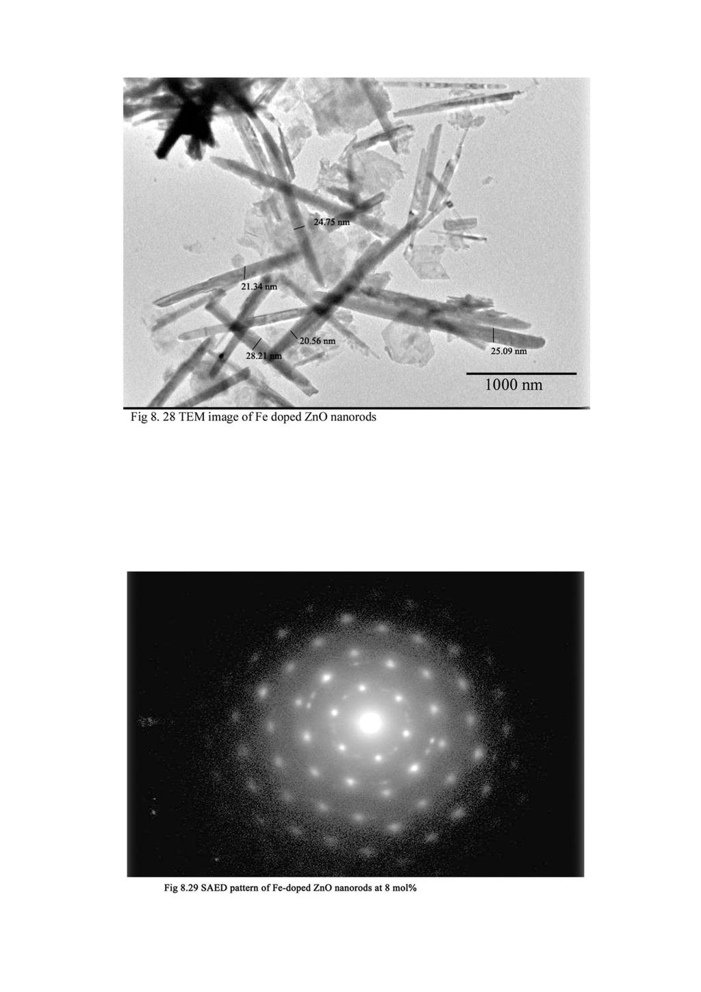
35 221 nanorods at 5 and 8 mol% and 573 cm -1 for 2 mol% and whose intensity increases by increasing the concentration of Ni. This peak is assigned to an impurity related vibration associated with defects in the host lattice induced by the Ni doping [42]. The intensity of the first order modes are found to be decrease with Ni concentration while the second order modes are more vivid by increasing the dopant concentration. This confirm that the Ni incorporation induce slight disorder in the ZnO crystal lattice due to the difference in ionic radius of dopant Ni 2+ and host Zn 2+ ions. 8.5 Zn (1-x) Fe x O Synthesis The Fe-doped ZnO nanorods of composition, Zn 1-x Fe x O (x = 0.05, 0.08) were prepared by hydrothermal method. All starting materials were analytical grade and were used without further purification. Zinc chloride and ferric chloride were used as Zn and Fe sources, respectively. The reaction solution was prepared by adding appropriate quantity of ammonia into zinc chloride and ferric chloride solution with keeping molarity 0.1M and by adjusting the ph value to 10. The as-prepared solution was poured into a bottle with autoclavable screw caps. Cleaned copper strips were immersed into the reaction solution as substrate. The bottle was heated at C using an oven and taken out after 2h. The samples were thoroughly washed with deionised water and dried in air for further characterisation Morphology and Structure Figure 8.28 shows the TEM image of Fe-doped ZnO nanorods with Fe 8 mol%. This image reveals the rod-like morphology and diameter ranges from nm. SAED pattern in figure 8.29 reveals the crystalline nature of as-prepared Fe-doped ZnO
36 222 nanorods. Figure 8.30 and 8.31 shows the representative EDS spectra of 8 mol% of Fedoped ZnO nanorods. It is clear from the EDS that the Fe 2+ ions have successfully been substituted into the crystal structure of ZnO nanorods. Fig 8.30 EDS spectrum of Fe-doped ZnO nanorods at 8 mol% Atom% O Fe Zn Fig 8.31 Atom% of Zn, O and Fe in Fe-doped ZnO nanorods with concentration 8 mol%
37
38 223 The XRD pattern of Zn (1-x)Fe x O nanorods with x = 5 and 8 mol% is shown in figure 8.32 (a-b). All diffraction peaks can be indexed to hexagonal phase of ZnO wurtzite structure (JCPDS Code No ). The very sharp diffraction peaks indicate the crystallinity of the doped ZnO nanorods and is not deteriorated by the incorporation of Fe. The Fe-doping does not alter the crystal structure and growth direction, while it causes the lattice constant to change slightly as evidenced by shift in all peak position. It is clear from the table 8.4 that the peak positions of the planes of Fe-doped ZnO nanorods are shifted to lower angle side and the lattice constants increases with increasing Fe concentration. This shift is mainly due to the larger radius of Fe 2+ ions (0.078 nm) than that of Zn 2+ ions (0.060 nm). No secondary phases of impurities have been observed in the XRD spectrum which reflects the substitution of Fe 2+ ions at Zn 2+ sites. The average crystallite size of Zn (1-x)Fe x O nanorods with x = 5 and 8 mol% are calculated using Scherrer s formula is 28.48±0.002 and 22.25±0.003 nm, respectively. The average crystallite size of the Fe-doped nanorods is found to be reduced in comparison with undoped ZnO nanorods. Table 8.4 shows the lattice parameters of Fe-doped ZnO nanorods with 2, 5 and 8 mol%.
39 mol% (101) Intensity (100) (002) (102) (110) θ Fig 8.32(a) XRD pattern of Fe-doped ZnO nanorods with 5 mol% mol% (101) 2000 Intensity (100) (002) (110) 500 (102) θ Fig 8.32(b) XRD pattern of Fe-doped ZnO nanorods with 8 mol%
40 225 Table 8.4 Peak position (2θ) of Fe-doped ZnO nanorods with dopant concentration at 5 and 8mol% ZnO (h k l) 2θ d values Lattice parameters nanorods ( 0 ) (Å) (Å) (100) (002) a = mol% (101) c = (102) (110) (100) (002) a = mol% (101) c = (102) (110) Optical band gap The absorption spectrum of Zn (1-x)Fe x O nanorods with x = 5 and 8 mol% is shown in figure The absorption peaks are obtained at 372 and 368 nm for 5 and 8 mol% of Fe, respectively. The absorption edges are red shifted in comparison with undoped ZnO nanorods. Eventhough the Fe concentration reached 8 mol%, the absorption edge shifted towards the lower wavelength region (blue shifted). The band gap energy of Fe-doped ZnO nanorods have been calculated using Tauc relation is shown in figure 8.34.
41 226 Absorbance mol% 8 mol% Wavelength (nm) Fig 8.33 Absorption spectra of Fe-doped ZnO nanorods [hν{ln[(r max - R min )/(R-R min )}] Energy (ev) Fe 2 mol% Fe 8 mol% Fig 8.34 Band gap energies of Fe-doped ZnO nanorods The calculated band gap energy of Fe-doped ZnO nanorods are ±0.002 and 3.433±0.001 ev for 5 and 8 mol% of Fe, respectively. When the ZnO nanorods doped
42 227 with Fe at 5 mol%, the band gap energy shrink from 3.43 ev (chapter 3) to 3.358eV. This red shift of band gap energy is attributed to the sp-d spin-exchange interactions between the band electrons and the localized d electrons of Fe 2+ ion substituting the host Zn 2+ cation [42]. This s-d and p-d interactions could give rise to a negative and positive correction to the conduction and the valence band edges, respectively, leading to a band gap narrowing. By increasing the concentration of Fe from 5 to 8 mol%, the band gap energy is found to have increase by ev. This blue shift can be attributed to the quantum confinement effect in Zn 0.92 Fe 0.08 O nanorods due to its small crystallite size. This observation is in good agreement with Para-Palomina s result [43] that band gap energy increase with the increment of Fe dopant due to quantum confinement effect Photoluminescence emission nm Intensity nm 420 nm 398 nm 5 mol% 395 nm 8 mol% 526 nm 605 nm Wavelength (nm) Fig 8.35 PL spectra of Fe-doped ZnO nanorods Figure 8.35 represent the PL spectrum of Fe-doped ZnO nanorods with Fe concentration 5 and 8 mol%, respectively. PL spectrum depicts the weak UV emission at
43 and 395 nm for 5 and 8 mol% of Fe, respectively, which results from the recombination of free exciton. The UV emission of 5 mol% of Fe-doped ZnO nanorods is red shifted by 8 nm in comparison with undoped ZnO nanorods which can be ascribed to the combined effect of size and Fe doping. The quenching of UV emission or near band edge emission implies that some non-radiative recombination process occur in the nanorods [44]. Therefore the doped Fe atoms act as quencher of near band-edge emission in ZnO nanorods. Furthermore, Fe doping has caused defect emission in violet-blue, green and yellow region. The defect emission is very strong in Fe-doped ZnO nanorods. The reason behind the violet, blue and green emission is discussed in the former sections. Besides undoped nanorods, a strong orange emission at 605 nm is observed which can be attributed to the Fe 2+ ions [44, 45]. The orange emission originates from the doubly ionized oxygen vacancies which acts as a trap for photogenerated holes [46]. The rate of hole trapping is faster and efficient when the surface to volume ratio of ZnO nanorods become high. The XRD result shows that the surface to volume ratio of Fe-doped ZnO nanorods at 8 mol% is high in comparison with undoped ZnO nanorods. The enhanced defect intensity indicates that the optical quality of Fe-doped ZnO nanorods decreases with increasing dopant concentration Raman Spectra The Raman spectrum of Fe-doped ZnO nanorods at 5 and 8 mol% is shown in figure The phonon modes at 436, 324 and 546 cm -1 are assigned to E 2 (high), 2E 2 (M) and [E 2 (high) +E 2 (low)] modes, respectively and are similar as reported for undoped ZnO nanorods [41]. The peak at 666 cm -1 which is absent in undoped ZnO nanorods may be attributed to the substitution of Fe 2+ ions into Zn 2+ ions located on the
44 229 tetrahedral sites of wurtzite structure. The incorporation of Fe 2+ ions generates new crystalline lattice defects [38]. It can be seen that all the phonon modes are shifted to lower frequencies with increasing the Fe concentration. The possible reasons can be considered as (1) mass effect due to the difference in the mass of dopant and host atoms (2) size effect due to the difference in the ionic radius of the dopant and host atoms and (3) change in bond strength due to the difference in the chemical nature of dopant and host atoms [38]. According to electron covalency model, zinc d-electron strongly hybridizes with the oxygen p-electron with the substitution of magnetic Fe 2+ ions that decrease the binding energy of Zn-O bond. Intensity mol% Intensity mol% Raman Shift (cm -1 ) Raman Shift (cm -1 ) Fig 8.36 Raman spectra of Fe-doped ZnO nanorods
45 Electrical properties of TM (Mn, Co, Ni and Fe) doped ZnO nanorods The conductivity of ZnO is primarily associated with intrinsic electron producers, V o (oxygen vacancies) and Zn i (cation interstitials) both act as positive charge centers. The formation energy of zinc interstials is at least as high as that of oxygen vacancies, but it act as shallow donors because the 2+/0 transition level is near or above the conduction band minimum. Therefore Zn interstitial (Zn i ) can be considered as the origin of conductivity in undoped ZnO [47]. In ZnO nanorods, hopping conduction is the predominant conduction mechanism and it is associated with electron jumping from occupied donors to empty ones. The surface defects present in the ZnO nanorods have satisfied the condition of hopping conduction [48] Mn-doped ZnO nanorods Variation of ac conductivity of Mn-doped ZnO nanorods with various Mn concentration and frequency is shown in figure It shows that the ac conductivity of Mn-doped ZnO nanorods increase by increasing the Mn concentration up to 2 mol%, beyond that limit it is found to decrease. Hopping conductivity is governed by the hopping probability between the impurity sites or the defect sites. The main shallow donors in the ZnO nanorods are zinc interstitials and oxygen vacancies. The Mn incorporation induces more surface defects into the ZnO nanorods. This increases the defect density and the ac conductivity of Mn-doped ZnO nanorods. However, an increase in the concentration of Mn beyond certain limit depresses the concentration of shallow donors in the nanorod. The lattice of ZnO nanorods is slightly distorted by the incorporation of Mn and this lattice distortion increases by increasing the concentration of Mn, which can be attributed to the scattering effect. Therefore the higher concentration
46 231 of Mn reduces the carrier concentration and increases the defect scattering effect. Hence the ac conductivity of Mn-doped ZnO nanorods is decreased beyond 2 mol% of Mn. This observation is confirmed by the PL data of Mn-doped ZnO nanorods. 1.6x mol% Mn σ ac (Ωcm) x x x mol% Mn 8 mol% Mn 0 mol% Mn x x x x x10 6 Frequency (Hz) Fig 8.37 Variation of ac conductivity of Mn-doped ZnO nanorods with dopant concentration and frequency Co-doped ZnO nanorods Figure 8.38 reveals the variation of ac conductivity of Co-doped ZnO nanorods with 2, 5 and 8 mol% of Co with increase in frequency. It shows that the ac conductivity of Co-doped ZnO nanorods increases with increasing frequency of all dopant concentration. The ac conductivity of Co-doped ZnO nanorods is found to be higher than that of the undoped nanorods. Generally the addition of Co ions decreases the conductivity due to the effect of defect scattering [49, 50]. It is clear from the PL spectra of Co-doped ZnO nanorods that the lattice distortion due to the addition of Co ions is very low, hence the scattering effect is a bare minimum up to 5 mol% of Co. When the
47 232 Co ions are doped with ZnO nanorods there is a possibility of releasing the oxygen vacancies. According to Alaria et.al. [51] the concentration of oxygen related defects in Co-doped ZnO increases with increasing Co content. In Co-doped ZnO nanorods, the bonding of Co ions with oxygen in tetrahedral symmetry is weaker than that of oxygen with Zn ions. It is clear from the XRD data that the Co dopant even at substitutional site makes diminutive distortion for the lattice due to the difference of ionic radius of dopant and host atoms. Consequently, the bonding strength of oxygen becomes weaker near Co ions when compared to the Zn ions in ZnO lattice. 2.0x mol% Co σ ac (Ω cm) x x x mol% Co 2 mol% Co 0 mol% Co x x x x x10 6 Frequency (Hz) Fig 8.38 Variation of ac conductivity of Co-doped ZnO nanorods with dopant concentration and frequency As a result the oxygen vacancies are enhanced with Co doping and this observation is consistent with the PL data. The increased oxygen vacancy augments the hopping conduction in Co-doped ZnO nanorods, hence its ac conductivity increases with
48 233 increasing Co concentration. However when the Co concentration reaches at 8 mol% scattering effect is predominant which reduces the values of ac conductivity Ni-doped ZnO nanorods Figure 8.39 shows the variation of ac conductivity of Ni-doped ZnO nanorods with various Ni concentration and frequency. The ac conductivity of ZnO nanorods is found to increase with increasing Ni concentration. The enhanced ac conductivity of ZnO nanorods with increasing concentration of Ni can be explained in accordance to the hopping conduction mechanism. The hopping probability increases with increasing dopant concentration which induces more surface defects in the ZnO nanorods. 2.0x10⁻⁴ σ ac (Ω cm) x x x x mol%ni 2 mol%ni 8 mol%ni 0 mol%ni x x x x x10 6 Frequency (Hz) Fig 8.39 Variation of ac conductivity of Ni-doped ZnO nanorods with dopant concentration and frequency The presence of oxygen vacancies/zinc interstitials increases with Ni doping that is confirmed by PL data of Ni-doped ZnO nanorods. Thus the increased surface defect
49 234 density by Ni-doping increases the hopping conduction. Hence the ac conductivity of Nidoped ZnO nanorods increases with increasing Ni concentration. According to Jr H. He et.al. [52] doping of cations of higher valence state (such as Ga, In and Ni) than Zn in to ZnO, leads to an increase in the electrical conductivity. The experimental results are in good agreement with this literature [52]. However the ac conductivity of Ni-doped ZnO nanorods at 8 mol% of Ni is found to be decrease which may be due to the scattering effect. Scattering effect is considerably small up to 5 mol% of Ni due to their good crystalline quality. Photoluminescence and Raman spectrum of Ni-doped ZnO nanorods agrees with this observation Fe-doped ZnO nanorods The variation of ac conductivity of Fe-doped ZnO nanorods with dopant concentration and frequency is shown in figure It shows that the ac conductivity of ZnO nanorods increases sharply by doping with Fe 2+ ions. When the dopant concentrations exceed to 5 mol%, the conductivity of ZnO nanorods begins to decrease. At all dopant concentration, the ac conductivity is found to increase with increasing frequency. When Fe is doped with ZnO, large number of surface defects are produced in the ZnO nanorods. PL spectrum of Fe-doped ZnO nanorods shows that the defect emission is enhanced by Fe doping. The increased surface defect density due to Fe doping enhances the probability of hopping and hence the ac conductivity of ZnO nanorods increases precipitously with Fe doping. However, Fe in ZnO acts as a deep donor that decreases the concentration of intrinsic shallow donors such as oxygen vacancies /zinc interstitials [53]. This reduction in intrinsic donor concentration increases with increase of Fe content. PL spectrum shows that the defect peak intensity in Fe-doped
CHAPTER 6. BLUE GREEN AND UV EMITTING ZnO NANOPARTICLES SYNTHESIZED THROUGH A NON AQUEOUS ROUTE
 71 CHAPTER 6 BLUE GREEN AND UV EMITTING ZnO NANOPARTICLES SYNTHESIZED THROUGH A NON AQUEOUS ROUTE 6.1 INTRODUCTION Several techniques such as chemical vapour deposition, electrochemical deposition, thermal
CHAPTER 8 SUMMARY AND FUTURE SCOPE
 CHAPTER 8 SUMMARY AND FUTURE SCOPE The potential of room temperature ferromagnetism in many diluted magnetic semiconductors has opened up a new route for realization of spintronic devices. Based on the
CHAPTER 3. EFFECT OF PRASEODYMIUM DOPING ON THE STRUCTURAL AND OPTICAL PROPERTIES OF ZnO NANORODS
 46 CHAPTER 3 EFFECT OF PRASEODYMIUM DOPING ON THE STRUCTURAL AND OPTICAL PROPERTIES OF ZnO NANORODS 3.1 INTRODUCTION Zinc oxide, one of the most promising materials, has been demonstrated to be applicable
Zinc Oxide. & Springer. Jean Geurts. Claus R Klingshirn. Andreas Waag Axel Hoffmann. Bruno K. Meyer. Towards Novel Applications
 Claus R Klingshirn Bruno K. Meyer Axel Hoffmann Jean Geurts Zinc Oxide From Fundamental Properties Towards Novel Applications With 226 Figures & Springer Contents 1 Introduction 1 I. I History of ZnO Research
Abstract. Keywords: Zinc Oxide, Eu doped ZnO, Dy doped ZnO, Thin film INTERNATIONAL JOURNAL OF INFORMATION AND COMPUTING SCIENCE ISSN NO:
 Synthesis and Structural study of Rare Earth activated ZnO Thin film Pawan Kumar Department of Physics, University Institute of Sciences, Chandigarh University, Gharuan (Mohali), Punjab (India) e-mail-pawan.uis@cumail.in
Influence of Indium doping on Zinc oxide thin film prepared by. Sol-gel Dip coating technique.
 Influence of Indium doping on Zinc oxide thin film prepared by Sol-gel Dip coating technique. Shazia Umar & Mahendra Kumar Department of Physics, University of Lucknow, Lucknow 226007 Abstract Dip coating
Keywords: Thin films, Zinc Oxide, Sol-gel, XRD, Optical properties
 Advanced Materials Research Vol. 895 (2014) pp 250-253 Online available since 2014/Feb/13 at www.scientific.net (2014) Trans Tech Publications, Switzerland doi:10.4028/www.scientific.net/amr.895.250 Structural
ISSN International Journal of Luminescence and Applications Vol.1 (II)
") Influence of rare-earth doping on the photoluminescence of Zinc Oxide nanophosphors Partha P. Pal* and J. Manam Deptt. of Applied Physics Indian School of Mines, Dhanbad-826004 * Corresponding author email:
Influence of rare-earth doping on the photoluminescence of Zinc Oxide nanophosphors Partha P. Pal* and J. Manam Deptt. of Applied Physics Indian School of Mines, Dhanbad-826004 * Corresponding author email:
Exploring Physical And Optical Behavior Of Co:Zno Nanostructures
 Exploring Physical And Optical Behavior Of Co:Zno Nanostructures Durga Prasad Gogoi 1 1 Associate Professor, Dept. of Physics, Namrup college, Dist: Dibrugarh, Assam: 786623, India Abstract- Zinc oxide
Structural, Optical & Surface Morphology of Zinc Oxide (ZnO) Nanorods in Molten Solution
 Nanorods in Molten Solution") Journal of Materials Science and Engineering B 6 (3-4) (2016) 68-73 doi: 10.17265/2161-6221/2016.3-4.002 D DAVID PUBLISHING Structural, Optical & Surface Morphology of Zinc Oxide (ZnO) Nanorods in Molten
Journal of Materials Science and Engineering B 6 (3-4) (2016) 68-73 doi: 10.17265/2161-6221/2016.3-4.002 D DAVID PUBLISHING Structural, Optical & Surface Morphology of Zinc Oxide (ZnO) Nanorods in Molten
EFFECT OF SOLVENTS ON PARTICLE STRUCTURE, MORPHOLOGY AND OPTICAL PROPERTIES OF ZINC OXIDE NANOPARTICLES
 EFFECT OF SOLVENTS ON PARTICLE STRUCTURE, MORPHOLOGY AND OPTICAL PROPERTIES OF ZINC OXIDE NANOPARTICLES A.Vanaja 1 and K.Srinivasa Rao 2 1 Department of Physics, Lingayya s University, Old Faridabad, Haryana,
Characterization of ZnO:Cu Nanoparticles by Photoluminescence Technique
 Characterization of ZnO:Cu Nanoparticles by Photoluminescence Technique Binildev R 1, Hareesh P S 2, Shilpa Prasad 3, Saravana Kumar 4 1,2,3 Department of Physics, Sree Narayana College Chengannur 4 Department
Tungston Doped ZnO Thin film Prepared by Spray Pyrolysis for enhanced Hydrogen Sensing
 International Journal of ChemTech Research CODEN (USA): IJCRGG, ISSN: 0974-4290, ISSN(Online):2455-9555 Vol.11 No.05, pp 467-471, 2018 Tungston Doped ZnO Thin film Prepared by Spray Pyrolysis for enhanced
PREPARATION AND CHARACTERIZATION OF METAL OXIDE NANOPOWDERS BY MICROWAVE- ASSISTED COMBUSTION METHOD FOR GAS SENSING DEVICES
 i PREPARATION AND CHARACTERIZATION OF METAL OXIDE NANOPOWDERS BY MICROWAVE- ASSISTED COMBUSTION METHOD FOR GAS SENSING DEVICES THESIS SUBMITTED TO ALAGAPPA UNIVERSITY IN PARTIAL FULFILMENT FOR THE AWARD
Ceramic Processing Research
 Journal of Ceramic Processing Research. Vol. 18, No. 6, pp. 435~439 (2017) J O U R N A L O F Ceramic Processing Research Enhancement of visible light emission from Tb-doped ZnO nanorods grown on silicon
Evidence of intrinsic ferromagnetism in individual dilute magnetic semiconducting nanostructures O-K. (a) Zn-L Zn-L 2,3
 Zn-L Zn-L 2,3") SUPPLEMENTARY INFORMATION Evidence of intrinsic ferromagnetism in individual dilute magnetic semiconducting nanostructures O-K (a) O-K Fe-L Co-L 2,3 2,3 Zn-L Zn-L 2,3 2,3 (b) Intensity (a. u.) 500 750
SUPPLEMENTARY INFORMATION Evidence of intrinsic ferromagnetism in individual dilute magnetic semiconducting nanostructures O-K (a) O-K Fe-L Co-L 2,3 2,3 Zn-L Zn-L 2,3 2,3 (b) Intensity (a. u.) 500 750
CHAPTER 4. CHARACTERIZATION OF ZINC DOPED TIN OXIDE (Zn:SnO 2 ) THIN FILMS
 THIN FILMS") 84 CHAPTER 4 CHARACTERIZATION OF ZINC DOPED TIN OXIDE (Zn:SnO 2 ) THIN FILMS 4.1 INTRODUCTION Tin oxide (SnO 2 ) is one of the most important Transparent Conductive Oxide (TCOs) materials, which finds
84 CHAPTER 4 CHARACTERIZATION OF ZINC DOPED TIN OXIDE (Zn:SnO 2 ) THIN FILMS 4.1 INTRODUCTION Tin oxide (SnO 2 ) is one of the most important Transparent Conductive Oxide (TCOs) materials, which finds
Structural and luminescent properties of ZnO flower-like microstructures synthesized using the chemical bath deposition method
 Structural and luminescent properties of ZnO flower-like microstructures synthesized using the chemical bath deposition method LF Koao 1, FB Dejene 1* and HC Swart 2 1 Department of Physics, University
Influence of Growth Time on Zinc Oxide Nano Rods Prepared By Dip Coating Method
 Influence of Growth Time on Zinc Oxide Nano Rods Prepared By Dip Coating Method P.Thamarai selvan 1, M.Venkatachalam 2, M.Saroja 2, P.Gowthaman 2, S.Ravikumar 3, S.Shankar 2 Department of Electronics &
UV Photoluminescence of ZnO Nanostructures Based Thin films synthesized by Sol Gel method
 UV Photoluminescence of ZnO Nanostructures Based Thin films synthesized by Sol Gel method S Sajjad Hussain 1), Hadia Noor 2), Saira Riaz 3), Asghar Hashmi 4) and *Shahzad Naseem 5) 1), 2), 3), 5) Centre
Reagent-Free Electrophoretic Synthesis of Few-Atom- Thick Metal Oxide Nanosheets
 Supporting Information Reagent-Free Electrophoretic Synthesis of Few-Atom- Thick Metal Oxide Nanosheets Chengyi Hou,*,, Minwei Zhang, Lili Zhang, Yingying Tang, Hongzhi Wang, and Qijin Chi*, State Key
Influence of Lead Substitution in Zinc Oxide Thin Films
 Chemical Science Transactions DOI:10.7598/cst2013.33 ISSN/E-ISSN: 2278-3458/2278-3318 RESEARCH ARTICLE Influence of Lead Substitution in Zinc Oxide Thin Films I. INIGO VALAN a, S. RAJA b, K. RAMAMURTHI
Structural, morphological and luminescence properties of hexagonal ZnO particles synthesized using wet chemical process
 Structural, morphological and luminescence properties of hexagonal ZnO particles synthesized using wet chemical process FB Dejene 1*, L. Koao 1, JJ Dolo 1 and HC Swart 2 1 Department of Physics, University
Supplementary Information
 Supplementary Information for Chemical Synthesis of Blue-emitting Metallic Zinc Nano-hexagons Nguyen T. Mai, Trinh T. Thuy, Derrick M. Mott and Shinya Maenosono* School of Materials Science, Japan Advanced
Supporting Information
 This journal is The Royal Society of Chemistry 011 Supporting Information Vertically-Aligned ZnO Nanorods Doped with Lithium for Polymer Solar Cells: Defect Related Photovoltaic Properties Pipat Ruankham,
INFLUENCE OF POINT DEFECTS' CONCENTRATION ON THE ZnO MATRIX A SIMULATION STUDY
 Ife Journal of Science vol. 16, no. 3 (2014) INFLUENCE OF POINT DEFECTS' CONCENTRATION ON THE ZnO MATRIX A SIMULATION STUDY 335 Akinnifesi, J.O. Department of Physics and Engineering Physics, Obafemi Awolowo
CHAPTER 3. PHOTOCATALYTIC DEGRADATION OF METHYLENE BLUE AND ACID RED 18 DYES BY Bi-Au-ZnO
 CHAPTER 3 PHOTOCATALYTIC DEGRADATION OF METHYLENE BLUE AND ACID RED 18 DYES BY Bi-Au-ZnO In this chapter, characterization of Bi-Au-ZnO and its photocatalytic activity on the degradation of Methylene Blue
Mechanochemical Doping of a Non-Metal Element into Zinc Oxide
 Chemistry for Sustainable Development 15 (2007) 249 253 249 Mechanochemical Doping of a Non-Metal Element into Zinc Oxide J. WANG, J. F. LU, Q. W. ZHANG, S. YIN, T. SATO and F. SAITO Institute of Multidisciplinary
Fe-doped ZnO synthesized by parallel flow precipitation process for improving photocatalytic activity
 IOP Conference Series: Materials Science and Engineering PAPER OPEN ACCESS Fe-doped ZnO synthesized by parallel flow precipitation process for improving photocatalytic activity To cite this article: Q
The structural and optical properties of ZnO thin films prepared at different RF sputtering power
 Journal of King Saud University Science (2013) 25, 209 215 King Saud University Journal of King Saud University Science www.ksu.edu.sa www.sciencedirect.com ORIGINAL ARTICLE The structural and optical
The electrical properties of ZnO MSM Photodetector with Pt Contact Electrodes on PPC Plastic
 Journal of Electron Devices, Vol. 7, 21, pp. 225-229 JED [ISSN: 1682-3427 ] Journal of Electron Devices www.jeldev.org The electrical properties of ZnO MSM Photodetector with Pt Contact Electrodes on PPC
Synthesis of nickel doped ZnO nanoparticles by hydrothermal decomposition of zinc hydroxide nitrate and its antimicrobial assay
 Available online at www.ijpab.com ISSN: 232 751 Int. J. Pure App. Biosci. 3 (1): 186-19 (215) INTERNATIONAL JOURNAL OF PURE & APPLIED BIOSCIENCE Research Article Synthesis of nickel doped ZnO nanoparticles
Investigation of Structure, Morphology, Optical And Luminescent Properties of Hydrothermally Grown Zno Nanorods for Photocatalytic Applications
 Investigation of Structure, Morphology, Optical And Luminescent Properties of Hydrothermally Grown Zno Nanorods for Photocatalytic Applications S.Kumar 1, J.Deenathayalan 2, M.Baskaran 3, D.D.Saravanan
COBALT DOPED ZINC OXIDE NANOPARTICLES FOR PHOTOCATALYTIC APPLICATIONS
 Journal of Ovonic Research Vol. 13, No. 5, September - October 217, p. 263-269 COBALT DOPED ZINC OXIDE NANOPARTICLES FOR PHOTOCATALYTIC APPLICATIONS S. KALPANA a*, S. S. KRISHNAN a, T. S. SENTHIL b, S.V.
STRUCTURAL CHARACTERIZATIONAND OPTICAL PROPERTIESOF Fe & Ni-DOPEDZINC OXIDE NANOPORED PARTICLES, SYNTHESIZEDUSING MICROWAVE METHOD
 STRUCTURAL CHARACTERIZATIONAND OPTICAL PROPERTIESOF Fe & Ni-DOPEDZINC OXIDE NANOPORED PARTICLES, SYNTHESIZEDUSING MICROWAVE METHOD Sabpreet Bhatti 1, Sachin Surve 2, V. N. Shukla 3 1 Centre For converging
International Journal of ChemTech Research CODEN (USA): IJCRGG ISSN: Vol.8, No.6, pp , 2015
: IJCRGG ISSN: Vol.8, No.6, pp , 2015") International Journal of ChemTech Research CODEN (USA): IJCRGG ISSN: 0974-4290 Vol.8, No.6, pp 297-302, 2015 Effect on Annealing Temperature on Zno Nanoparticles Sugapriya S* 1, Lakshmi S 1, Senthilkumaran
International Journal of ChemTech Research CODEN (USA): IJCRGG ISSN: 0974-4290 Vol.8, No.6, pp 297-302, 2015 Effect on Annealing Temperature on Zno Nanoparticles Sugapriya S* 1, Lakshmi S 1, Senthilkumaran
Chapter CHAPTER 7. ELECTRICAL PROPERTIES OF ZnO DOPED MAGESIUM ALUMIUM SILICATE GLASS-CERAMICS
 Chapter 7 102 CHAPTER 7 ELECTRICAL PROPERTIES OF ZnO DOPED MAGESIUM ALUMIUM SILICATE GLASS-CERAMICS Chapter 7 103 CHAPTER 7 ELECTRICAL PROPERTIES OF ZnO DOPED MAGNESIUM ALUMINUM SILICATE GLASS-CERAMICS
Hydrogen-Sensing Characteristics of Palladium-Doped Zinc-Oxide Nanostructures
 Hydrogen-Sensing Characteristics of Palladium-Doped Zinc-Oxide Nanostructures Undergraduate Researcher Saranya Sathananthan University of Tennessee, Knoxville Faculty Mentor Vinayak P. Dravid Department
Synthesis and Photoluminescence Property of Bentonite Doped Zinc Oxide Nanoparticles
 Synthesis and Photoluminescence Property of Bentonite Doped Zinc Oxide Nanoparticles R. Parimaladevi*, I. Suganya Department of Physics, Mother Teresa Women s University, Kodaikanal *Corresponding author:
SYNTHESIS AND CHARACTERIZATION OF Al DOPED ZnO NANOPARTICLES
 International Conference on Ceramics, Bikaner, India International Journal of Modern Physics: Conference Series Vol. 22 (2013) 630 636 World Scientific Publishing Company DOI: 10.1142/S2010194513010775
Annealing Influence on the Optical Properties of Nano ZnO
 Available online www.ejaet.com European Journal of Advances in Engineering and Technology, 2014, 1(1): 69-73 Research Article ISSN: 2394-658X Annealing Influence on the Optical Properties of Nano ZnO Saad
Epitaxial Growth of ZnO Nanowires on Graphene-Au
 Epitaxial Growth of ZnO Nanowires on Graphene-Au 1 Schematic of Growth Process Nanorod Nanowire Nanoribbon Giri et al.. ACS Appl. Mater. Interf. 6, 377 (2014). 2 1 FESEM image of ZnO NWs/NRBs Grown on
Outline of the talk. FIB fabrication of ZnO nanodevices. Properties of ZnO 4/19/2011. Crystal structure of ZnO. Collaborators. Wurtzite structure
 FIB fabrication of ZnO nanodevices Crystal structure of ZnO Wurtzite structure Lee Chow Department of Physics University of Central Florida 1 4 Collaborators X-ray diffraction pattern of ZnO nanorods Synthesis,
A low magnification SEM image of the fabricated 2 2 ZnO based triode array is
 Chapter 6 Characteristics of Field Emission Triode 6.1 Planar Gated Field Emission Triode 6.1.1 Structural and Electrical Analysis A low magnification SEM image of the fabricated 2 2 ZnO based triode array
Room-temperature ferromagnetic properties of Cu-doped ZnO rod arrays
 Bull. Mater. Sci., Vol. 34, No. 5, August 2011, pp. 1083 1087. c Indian Academy of Sciences. Room-temperature ferromagnetic properties of Cu-doped ZnO rod arrays CHXIA,CGHU, C H HU, Z PING and F WANG Science
Deposition of aluminum-doped zinc oxide films by RF magnetron sputtering and study of their surface characteristics
 Surface and Coatings Technology 174 175 (2003) 187 192 Deposition of aluminum-doped zinc oxide films by RF magnetron sputtering and study of their surface characteristics a b b a a, S.H. Jeong, S. Kho,
ZnO nanostructures epitaxially grown on ZnO seeded Si (100) substrates by chemical vapor deposition
 substrates by chemical vapor deposition") ZnO nanostructures epitaxially grown on ZnO seeded Si (100) substrates by chemical vapor deposition Zhuo Chen 1, T. Salagaj 2, C. Jensen 2, K. Strobl 2, Mim Nakarmi 1, and Kai Shum 1, a 1 Physics Department,
ZnO nanostructures epitaxially grown on ZnO seeded Si (100) substrates by chemical vapor deposition Zhuo Chen 1, T. Salagaj 2, C. Jensen 2, K. Strobl 2, Mim Nakarmi 1, and Kai Shum 1, a 1 Physics Department,
Structural and Photoluminescence Study of Zinc Oxide Thin Films Grown by Laser Induced Plasma
 Structural and Photoluminescence Study of Zinc Oxide Thin Films Grown by Laser Induced Plasma Usman Ilyas 1,2, R. S. Rawat 1, G. Roshan 1, T.L. Tan 1, P. Lee 1, S.V.Springham 1, R. Chen 3, H. D. Sun 3,
Optical phonon confinement in ZnO nanorods and nanotubes
 Indian Journal of Pure & Applied Physics Vol. 48, October 010, pp. 703-708 Optical phonon confinement in ZnO nanorods and nanotubes Soosen Samuel M, Jiji Koshy, Anoop Chandran & K C George* Department
ZnO Thin Films Generated by Ex-Situ Thermal Oxidation of Metallic Zn for Photovoltaic Applications
 Macalester Journal of Physics and Astronomy Volume 4 Issue 1 Spring 2016 Article 12 May 2016 ZnO Thin Films Generated by Ex-Situ Thermal Oxidation of Metallic Zn for Photovoltaic Applications Kovas Zygas
ISSN: [Koteeswari * et al., 7(4): April, 2018] Impact Factor: 5.164
![ISSN: [Koteeswari * et al., 7(4): April, 2018] Impact Factor: 5.164](/thumbs/91/107668145.jpg "ISSN: [Koteeswari * et al., 7(4): April, 2018] Impact Factor: 5.164") IJESRT INTERNATIONAL JOURNAL OF ENGINEERING SCIENCES & RESEARCH TECHNOLOGY INVESTIGATIONS ON STRUCTURAL, DIELECTRIC AND OPTICAL PROPERTIES OF Cu- DOPED ZnO NANOPARTICLES P.Koteeswari*, T.Kavitha 1, S.Vanitha
IJESRT INTERNATIONAL JOURNAL OF ENGINEERING SCIENCES & RESEARCH TECHNOLOGY INVESTIGATIONS ON STRUCTURAL, DIELECTRIC AND OPTICAL PROPERTIES OF Cu- DOPED ZnO NANOPARTICLES P.Koteeswari*, T.Kavitha 1, S.Vanitha
X-RAY PHOTOELECTRON EMISSION, PHOTOLUMINESCENCE AND RAMAN ANALYSIS OF SOLID SOLUTIONS OF ALUMINIUM ZINC OXIDE
 International Journal of Physics and Research (IJPR) Vol.1, Issue 1 Dec 2011 59-69 TJPRC Pvt. Ltd., X-RAY PHOTOELECTRON EMISSION, PHOTOLUMINESCENCE AND RAMAN ANALYSIS OF SOLID SOLUTIONS OF ALUMINIUM ZINC
ULTRA THIN INDIUM TIN OXIDE FILMS ON VARIOUS SUBSTRATES BY PULSED LASER DEPOSITION
 ULTRA THIN INDIUM TIN OXIDE FILMS ON VARIOUS SUBSTRATES BY PULSED LASER DEPOSITION X. W. Sun 1, D. H. Kim 2, and H. S. Kwok 1 1 Department of Electrical & Electronic Engineering, Hong Kong University of
Zinc Oxide Nanoparticles Prepared by the Reaction of Zinc Metal with Ethanol
 JKAU: Sci., Vol. 21 No. 1, pp: 61-67 (2009 A.D. / 1430 A.H.) Zinc Oxide Nanoparticles Prepared by the Reaction of Zinc Metal with Ethanol M. A. Shah and M. Al-Shahry 1 Department of Physics, and 1 Department
Sunil P. Chavan, Vaishali A. Bambole Department of Physics, University of Mumbai, Mumbai, Maharashtra, India
 2018 IJSRST Volume 4 Issue 5 Print ISSN: 2395-6011 Online ISSN: 2395-602X Themed Section: Science and Technology Effect of carbon doping on electronic structure and optical properties of ZnO clusters ABSTRACT
Structural and luminescence properties of sol-gel derived Cu doped ZnO films
 Indian Journal of Pure & Applied Physics Vol. 47, May 2009, pp. 377-382 Structural and luminescence properties of sol-gel derived Cu doped ZnO films K Das a, S Ray a, S Chaudhuri a & A B Maity b a Department
SPONTANEOUS AND STIMULATED EMISSION OF ZnO NANORODS OF DIFFERENT SHAPE
 SPONTANEOUS AND STIMULATED EMISSION OF ZnO NANORODS OF DIFFERENT SHAPE A.N. Gruzintsev, A.N. Redkin,**G.A. Emelchenko, *C. Barthou Institute of Microelectronics Technology, Russian Academy of Sciences,
Supporting Information
 Supporting Information Intense visible emission from ZnO/PAAX (X = H or Na) nanocomposite synthesized via a simple and scalable sol-gel method Yao Zhu, Aleksandra Apostoluk, Pierrick Gautier, Audrey Valette,
CHAPTER 1 GENERAL INTRODUCTION OF ZINC OXIDE AND IT S PROPERTIES. In recent years, scientists have made rapid and significant advances in the field of
 CHAPTER 1 GENERAL INTRODUCTION OF ZINC OXIDE AND IT S PROPERTIES 1.1 Introduction In recent years, scientists have made rapid and significant advances in the field of materials science, especially in semiconductor
Study of ZnO:Zn Phosphors Prepared by Sol-gel and Ionimplantation
 Available online at www.sciencedirect.com Physics Procedia 25 (212 ) 35 354 212 International Conference on Solid State Devices and Materials Science Study of ZnO:Zn Phosphors Prepared by Sol-gel and Ionimplantation
GaN/ZnO and AlGaN/ZnO heterostructure LEDs: growth, fabrication, optical and electrical characterization
 Mater. Res. Soc. Symp. Proc. Vol. 1201 2010 Materials Research Society 1201-H01-08 GaN/ZnO and AlGaN/ZnO heterostructure LEDs: growth, fabrication, optical and electrical characterization J. Benz1, S.
A Correlation between Optical and Structural Property of. ZnO Nanocrystalline Films
 Journal Homepage: www.katwacollegejournal.com A Correlation between Optical and Structural Property of ZnO Nanocrystalline Films Surajit Mandal, Physics, Burdwan Raj College, India Article Record: Received
PHOTOLUMINESCENCE STUDY OF ZnO DOPED WITH NITROGEN AND ARSENIC JULIEN KOUADIO DANGBEGNON PHILOSOPHIAE DOCTOR
 PHOTOLUMINESCENCE STUDY OF ZnO DOPED WITH NITROGEN AND ARSENIC JULIEN KOUADIO DANGBEGNON Submitted in fulfilment of the requirements for the degree of PHILOSOPHIAE DOCTOR In the Faculty of Science at the
Supplementary Figure S1. Statistical measurements on particle size and aspect ratio of
 Supplementary Figure S1. Statistical measurements on particle size and aspect ratio of as-prepared Cd 0.5 Zn 0.5 S nanocrystals. a,b,c, Histograms of the particle size distribution of the three-type of
DISTRIBUTION A: Distribution approved for public release.
 AFRL-OSR-VA-TR-2015-0044 Review of Zinc Oxide Thin Films Tom Otiti COLLEGE OF COMPUTING AND INFORMATION SCIENCE MAKERERE U 12/23/2014 Final Report DISTRIBUTION A: Distribution approved for public release.
Analysis of Li-related defects in ZnO thin films influenced by annealing ambient
 Bull. Mater. Sci., Vol. 37, No. 1, February 2014, pp. 35 39. c Indian Academy of Sciences. Analysis of Li-related defects in ZnO thin films influenced by annealing ambient BING WANG and LIDAN TANG Department
Institute of Physics and Chemistry, Chinese Academy of Sciences, Beijing, , P. R.
 Electrochemical synthesis of p-type Zn-Doped α-fe 2 O 3 nanotube arrays for photoelectrochemical water splitting Xiaopeng Qi, a,b Guangwei She,* a Meng Wang, a,b Lixuan Mu, a and Wensheng Shi* a a Key
Theerapong Santhaveesuk, * Duangmanee Wongratanaphisan and Supab Choopun
 NU Science Journal 2009; 6(S1): 43-50 Ethanol Sensing Property of Tetrapods Prepared by Thermal Oxidation of Zn and TiO 2 Mixture Theerapong Santhaveesuk, * Duangmanee Wongratanaphisan and Supab Choopun
The study of external electric field effect on the growth of ZnO crystal
 ISBN 978-979-18962-0-7 The study of external electric field effect on the growth of ZnO crystal Evi Maryanti 1, B. Prijamboedi 2 *, Ismunandar 2 1 Chemistry Departement, University of Bengkulu 2 Inorganic
Material properties that control the cytotoxicity of ZnO nanoparticles
 Material properties that control the cytotoxicity of ZnO nanoparticles Alex Punnoose, Jordan Chess, Catherine Anders, John Rassmussen, Denise Wingett, Kelsey Dodge and Katie Rainey, Boise State University,
Effect of doping and annealing on the physical properties of ZnO:Mg nanoparticles
 PRAMANA c Indian Academy of Sciences Vol. 81, No. 2 journal of August 2013 physics pp. 319 330 Effect of doping and annealing on the physical properties of ZnO:Mg nanoparticles SABER FARJAMI SHAYESTEH
A Solution Processed ZnO Thin Film
 Applied Mechanics and Materials Vols. 239-240 (2013) pp 1585-1588 Online available since 2012/Dec/13 at www.scientific.net (2013) Trans Tech Publications, Switzerland doi:10.4028/www.scientific.net/amm.239-240.1585
Fabrication, structural and optical properties of Ni and Cr doped ZnO nanocomposites for photocatalyst under UV light
 Fabrication, structural and optical properties of Ni and Cr doped ZnO nanocomposites for photocatalyst under UV light Shanmugam Vignesh and Jeyaperumal Kalyana Sundar* Materials Science Laboratory, Department
Research Article Optical and Magnetic Properties of Fe Doped ZnO Nanoparticles Obtained by Hydrothermal Synthesis
 Nanomaterials, Article ID 792102, 6 pages http://dx.doi.org/10.1155/2014/792102 Research Article Optical and Magnetic Properties of Fe Doped ZnO Nanoparticles Obtained by Hydrothermal Synthesis Xiaojuan
Zoltán Szabó. Synthesis and characterisation of zinc-oxide thin films and nanostructures for optoelectronical purposes
 PHD theses Synthesis and characterisation of zinc-oxide thin films and nanostructures for optoelectronical purposes Zoltán Szabó Supervison: Dr. János Volk Consultant: Dr. György Hárs HAS Centre for Energy
Supplementary Fig. 1.
 Supplementary Fig. 1. (a,b,e,f) SEM and (c,d,g,h) TEM images of (a-d) TiO 2 mesocrystals and (e-h) NiO mesocrystals. The scale bars in the panel c, d, g, and h are 500, 2, 50, and 5 nm, respectively. SAED
Research Article Stoichiometry Control of ZnO Thin Film by Adjusting Working Gas Ratio during Radio Frequency Magnetron Sputtering
 Materials Volume 2013, Article ID 547271, 6 pages http://dx.doi.org/10.1155/2013/547271 Research Article Stoichiometry Control of ZnO Thin Film by Adjusting Working Gas Ratio during Radio Frequency Magnetron
-V P. C.W.M. Castleton, S.Mirbt Theory of Condensed Matter, Department of Physics, Uppsala University, Box 530, Uppsala, Sweden.
 The Structure of the Zn In -V doped InP. P defect complex in Zn C.W.M. Castleton, S.Mirbt Theory of Condensed Matter, Department of Physics, Uppsala University, Box 53, 75 Uppsala, Sweden. Abstract. We
ABSTRACT. GROWTH AND TRANSPORT PROPERTIES OF Sb-DOPED ZnO NANO/MICROWIRES. by Nada Ali Masmali
 ABSTRACT GROWTH AND TRANSPORT PROPERTIES OF Sb-DOPED ZnO NANO/MICROWIRES by Nada Ali Masmali In this research, the transport properties of Sb-doped ZnO wires were investigated. ZnO:Sb wires were grown
First-principle study on the effect of high Ag 2N co-doping on the conductivity of ZnO
 Bull. Mater. Sci., Vol. 38, No. 3, June 2015, pp. 747 751. c Indian Academy of Sciences. First-principle study on the effect of high Ag 2N co-doping on the conductivity of ZnO WENXUE ZHANG 1, YUXING BAI
Comparative study of zinc oxide and aluminum doped zinc oxide transparent thin films grown by direct current magnetron sputtering
 Thin Solid Films 515 (2007) 6562 6566 www.elsevier.com/locate/tsf Comparative study of zinc oxide and aluminum doped zinc oxide transparent thin films grown by direct current magnetron sputtering M. Suchea
Identifying the Sources of Ferromagnetism in Sol- Gel Synthesized Zn 1-x Co x O (0 x 0.10) Nanoparticles
 Nanoparticles") Boise State University ScholarWorks Physics Faculty Publications and Presentations Department of Physics 8-1-2016 Identifying the Sources of Ferromagnetism in Sol- Gel Synthesized Zn 1-x Co x O (0 x 0.10)
Boise State University ScholarWorks Physics Faculty Publications and Presentations Department of Physics 8-1-2016 Identifying the Sources of Ferromagnetism in Sol- Gel Synthesized Zn 1-x Co x O (0 x 0.10)
Integrated Sci-Tech : The Interdisciplinary Research Approach
 Chapter 32 Influence of the Concentration of Ga-doped on the Structural and Optical Properties of ZnO Thin Films Putut Marwoto 1,a, Dwi Suprayogi 1, Edy Wibowo 2, Didik Aryanto 3, Sulhadi 1, Sugiyanto
Dependence of the Optical Anisotropy of ZnO Thin Films on the Structural Properties
 Journal of the Korean Physical Society, Vol. 57, No. 2, August 2010, pp. 389 394 Dependence of the Optical Anisotropy of ZnO Thin Films on the Structural Properties Man-Il Kang and Sok Won Kim Department
Preparation of ZnO Nanowire Arrays Growth on Sol-Gel ZnO-Seed-Coated Substrates and Studying Its Structure and Optical Properties
 Advances in Nanomaterials 2017; 1(1): 1-5 http://www.sciencepublishinggroup.com/j/an doi: 10.11648/j.an.20170101.11 Preparation of ZnO Nanowire Arrays Growth on Sol-Gel ZnO-Seed-Coated Substrates and Studying
SYNTHESIS OF ZnO NANOROD ARRAYS ON ZnO NANOPARTICLES- COATED ITO SUBSTRATE. Universiti Kebangsaan Malaysia UKM Bangi, Selangor, Malaysia
 SYNTHESIS OF ZnO NANOROD ARRAYS ON ZnO NANOPARTICLES- COATED ITO SUBSTRATE C.C. Yap 1, M. Yahaya 1, M.H. Jumali 1 and M.M. Salleh 2 1 School of Applied Physics, Faculty of Science and Technology, Universiti
ZnO Thin Films Synthesized by Chemical Vapor Deposition
 ZnO Thin Films Synthesized by Chemical Vapor Deposition Zhuo Chen *1, Kai Shum *2, T. Salagaj #3, Wei Zhang #4, and K. Strobl #5 * Physics Department, Brooklyn College of the City University of New York
THE EFFECTS OF DOPING CONCENTRATION ON THE ELECTRICAL PERFORMANCE OF DC-SPUTTERED p-zno/n-si HETEROJUNCTION
 THE EFFECTS OF DOPING CONCENTRATION ON THE ELECTRICAL PERFORMANCE OF DC-SPUTTERED p-zno/n-si HETEROJUNCTION Dao Anh Tuan, Bui Khac Hoang, Nguyen Van Hieu, Le Vu Tuan Hung Department of Applied Physics,
Characterization of ZnO/TiO 2 Nanocomposites Prepared via the Sol-Gel Method
 Journal of the Korean Ceramic Society Vol. 55, No., pp. 140~144, 018. https://doi.org/10.4191/kcers.018.55..10 Communication Characterization of ZnO/TiO Nanocomposites Prepared via the Sol-Gel Method Nalumaga
Substrate Temperature Effect on Optical property of ZnO Thin Films
 Article Substrate Temperature Effect on Optical property of ZnO Thin Films Achour Rahal 1, Said Benramache 2, *, and Boubaker Benhaoua 1 1 VTRS Laboratory, Institute of Technology, University of El-oued,
Nanostructured ZnO as a solution-processable transparent electrode material for low-cost photovoltaics
 Nanostructured ZnO as a solution-processable transparent electrode material for low-cost photovoltaics Investigators P.I: Alberto Salleo, Assistant Professor, Materials Science and Engineering Dr. Ludwig
Effect of Mn Doping on Optical Properties of ZnO Nanoparticles
 Effect of Mn Doping on Optical Properties of ZnO Nanoparticles Binapani Goswami 1, Ranjit Singha 2 Research Scholar, Department of Physics, Assam University, Diphu Campus, Karbi Anglong, Assam, India 1
Comparitive Study Of Pure ZnO and Copper Doped Zno Nanoparticles Synthesized via Coprecipitation Method
 Comparitive Study Of Pure ZnO and Copper Doped Zno Nanoparticles Synthesized via Coprecipitation Method V.Kalaiselvi 1,Dr.N.Jayamani 2 &M.Revathi 3 1 Assistant Professor & Head, Department of Physics,
CHAPTER 4 EFFECT OF OXALIC ACID ON THE OPTICAL, THERMAL, DIELECTRIC AND MECHANICAL BEHAVIOUR OF ADP CRYSTALS
 67 CHAPTER 4 EFFECT OF OXALIC ACID ON THE OPTICAL, THERMAL, DIELECTRIC AND MECHANICAL BEHAVIOUR OF ADP CRYSTALS 4.1 INTRODUCTION Oxalic acid is a hydrogen-bonded material. It is the only possible compound
SYNTHESIS AND CHARACTERIZATION OF ZnO NANO-PARTICLES
 SYNTHESIS AND CHARACTERIZATION OF ZnO NANO-PARTICLES C. A. Omondi 1, *T. W. Sakwa 1, Y. K. Ayodo 1 and K. M. Khanna 2 1 Department of Physics, Masinde Muliro University of Science and Technology, P.O.
Technology and TEM characterization of Al doped ZnO nanomaterials
 Technology and TEM characterization of Al doped ZnO nanomaterials 國立成功大學 (NCKU) 材料科學及工程系 (MSE) 劉全璞 (Chuan-Pu Liu) Outline Introduction of ZnO Doping ZnO nanomaterials in CVD Al doped ZnO Nanowires Al doped
Structural Properties of ZnO Nanowires Grown by Chemical Vapor Deposition on GaN/sapphire (0001)
") Structural Properties of ZnO Nanowires Grown by Chemical Vapor Deposition on GaN/sapphire (0001) F. C. Tsao 1, P. J. Huang 1, J. Y. Chen 2, C. J. Pan 3, C. J. Tun 4, C. H. Kuo 2, B. J. Pong 5, T. H. Hsueh
Structural Properties of ZnO Nanowires Grown by Chemical Vapor Deposition on GaN/sapphire (0001) F. C. Tsao 1, P. J. Huang 1, J. Y. Chen 2, C. J. Pan 3, C. J. Tun 4, C. H. Kuo 2, B. J. Pong 5, T. H. Hsueh
Valence band and core-level analysis of highly luminescent ZnO nanocrystals for designing ultrafast optical sensors
 Valence band and core-level analysis of highly luminescent ZnO nanocrystals for designing ultrafast optical sensors Amish G. Joshi, Sonal Sahai, Namita Gandhi, Y. G. Radha Krishna and D. Haranath a) National
Electric field induced reversible control of visible. photoluminescence from ZnO nanoparticles
 Electric field induced reversible control of visible photoluminescence from ZnO nanoparticles Manoranjan Ghosh,# and A. K. Raychaudhuri $ DST Unit for Nanoscience, S. N. Bose National Centre for Basic
CHAPTER 5 CHARACTERIZATION OF ZINC OXIDE NANO- PARTICLES
 88 CHAPTER 5 CHARACTERIZATION OF ZINC OXIDE NANO- PARTICLES 5.1 INTRODUCTION This chapter deals with the characterization of ZnO nano-particles using FTIR, XRD, PSA & SEM. The results analysis and interpretations