SURGICAL TECHNIQUE PREPARED BY MEDSHAPE, INC. IN CONJUNCTION WITH JACK FARR, M.D. MPFL RECONSTRUCTION
|
|
|
- Kristopher Farmer
- 5 years ago
- Views:
Transcription


1 ! SURGICAL TECHNIQUE PREPARED BY MEDSHAPE, INC. IN CONJUNCTION WITH JACK FARR, M.D.
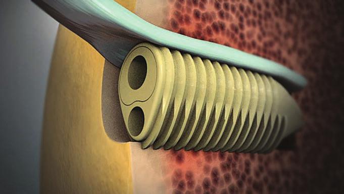
2 LATERAL PATELLOFEMORAL INSTABILITY AND THE MEDIAL PATELLOFEMORAL COMPLEX Lateral patellofemoral instability is characterized by a deficiency of the medial patellofemoral retinacular soft tissues due to ligamentous laxity and/or injury. 1,2,5 These medial retinacular structures include (1) the medial patellofemoral ligament, (2) the medial patellotibial ligament, (3) the medial patellomeniscal ligament, and (4) the superficial medial retinaculum. However, the current body of evidence indicates that the medial patellofemoral ligament (MPFL) serves as the primary passive restraint preventing lateral displacement of the patella. Clinical studies have shown the majority of acute lateral patellar dislocations result in complete or partial tears to the MPFL. 1 Likewise, conditions such as trochlear or soft tissue dysplasias can predispose a patient to recurrent patellar dislocation or subluxation and subsequent injury and hyperlaxity of the medial soft tissues. 1,2,5 INDICATION FOR MPFL Reconstruction is indicated in patients with lateral patellar instability, caused by tears or laxity of the MPFL. The patient often has a a history of acute or recurrent patellar dislocation and/or patellar subluxation. 2 AN ANATOMICAL In light of the fact that the MPFL has been identified as the primary passive restraint to patellar lateral displacement, reconstruction of the MPFL can provide the best clinical outcomes by restoring the normal limits of patellar motion. 1 Accurate anatomical reconstruction of the native anatomy and biomechanics of the MPFL is imperative to achieve an effective reconstruction. 3 Between 0 and 70 of flexion, the native MPFL is isometric in length and tension is minimal; beyond 70 of flexion, the MPFL becomes lax and tension is negligible or zero. 3 The femoral attachment site has been shown to have the greatest effect on MPFL isometry. Studies indicate that a nonanatomic reconstruction where the graft is proximally or distally malpositioned at the femoral attachment site by only 5 mm can cause significant nonisometric length changes resulting in abnormal graft tension and elevated forces in the medial patellofemoral joint. 3,4 Consequently, accurate placement of the femoral reattachment site has been identified as the most significant factor influencing surgical outcome. 4 THE TECHNIQUE The following technique, developed by Dr. Jack Farr, II, reproduces the anatomy of the MPFL by ensuring the anatomometric placement of the femoral attachment site and allowing for fine adjustments to graft length. First, two Morphix Suture Anchors are seated in the proximal two-thirds of the medial patellar margin, establishing the patellar fixation sites. The more sensitive femoral attachment site is determined by anatomometric testing. The doubled end of a semitendinosus autograft or allograft is fixated within the femoral bone tunnel. The unique, nonrotational deployment of the Eclipse Soft Tissue Anchor preserves the surgeon-desired precise orientation of the graft within the bone tunnel. Finally, the two ends of the graft are secured at length by the suture anchors in the patella, allowing for final fine adjustments to graft length. 1 SURGICAL TECHNIQUE

.")


3 SURGICAL TECHNIQUE 2 Patellar Site Preparation 1a 1b Create a longitudinal incision over the proximal two-thirds of the medial border of the patella, and dissect down to the interval between layer two (MPFL) and layer three (capsule).! Decorticate the proximal two-thirds of the medial patellar margin with a 4 mm round burr.! 1c Using a 2.5 mm Drill Bit and Drill Guide (from the Morphix Procedure Pack), drill two tunnels, one at the proximal end of the trough and the second at the mid-waist of the patella. Drill until the hard stop on the Drill Bit reaches the back of the Drill Guide to ensure appropriate tunnel depth.


4 3 SURGICAL TECHNIQUE Suture Anchor Deployment Implant two 2.5 mm Morphix Suture Anchors with 2 USP #2 Sutures in the drill holes created in the patella at the proximal end of the trough and mid-waist of the patella.! 2a With the Inserter Knob in the locked position, insert the Suture Anchor eyelet into the hole ensuring the cannula is seated on the cortex. 2b Rotate the Inserter Knob one quarter turn counter-clockwise into the unlocked position, indicated by an open padlock symbol.! Pearl #1. The wings of the Morphix Suture Anchor will deploy in the same orientation as the Inserter Handle Wings. Ensure that the device is inserted according to preferred orientation of the sutures and wings. Pearl #2. To prevent possible damage to the eyelet assembly, ensure that the insertion trajectory matches the trajectory of the hole created by the drill bit. 2c Manually depress or gently tap the Inserter Knob with a mallet to fully deploy the anchor. The anchor is fully deployed when the top of the Morphix Suture Anchor is slightly countersunk to the surface of the bone and the Inserter Knob is flush with the Inserter Handle.! 2d Unwind the suture from the suture slot in the Inserter Handle. Disengage the Inserter Handle using a counter-clockwise motion to disconnect the eyelet from the Inserter. Gently pull back the Inserter Handle to remove.


5 SURGICAL TECHNIQUE 4 Final Suture Anchor Positioning Graft Preparation 3a 3b Harvest a portion of the semitendinosus tendon through a small incision over the pes anserinus group insertion site and double the graft back upon itself. Using the Soft Tissue Sizer, measure the diameter of the graft at the doubled end. See Appendix A: Eclipse Sizing Guide for graft, bone tunnel, and Implant sizing information.!


6 5 SURGICAL TECHNIQUE Femoral Site Preparation 4a 4b Make a second, smaller incision at the femoral attachment site, located in the saddle area between the posterior border of the medial femoral epicondyle and the adductor tubercle. Place a 2.4 mm Drill-Tipped Passing Pin in the desired location in the femoral epicondyle. Confirm Passing Pin placement with fluoroscopic imaging.! 4c With forceps placed between layers two and three, pull the patellar sutures out through the femoral incision. With the knee at 20of flexion, clamp the sutures around the Passing Pin so that the sutures are taut but neither tensioned nor lax. Perform anatomometric testing in extension and flexion. Reposition the Passing Pin if necessary. 4d Once the desired location has been verified, drill over the Passing Pin with a Cannulated Reamer 1 mm larger than the graft size. Drill to a depth of 25 mm, read directly off the lasermarks of the Reamer. Refer to Appendix A: Eclipse Sizing Guide for graft, bone tunnel, and Implant sizing information.! Pearl #3. During anatomometric testing with increasing flexion, the sutures should become lax. If the sutures tension while increasing flexion, the Passing Pin is positioned too proximal on the femur. In extension, the sutures should tension only slightly. Ensure that the patella is not over-tensioned or tilted medially throughout the full range of motion.!


7 6 SURGICAL TECHNIQUE Graft Positioning 5a Using the Tendon Fork, catch the doubled end of the graft and push it fully into the bone tunnel. Verify that the graft is inserted into the base of the bone tunnel by reading the lasermark measurements on the Tendon Fork.! 5b If desired, mark the doubled end of the graft at a length equivalent to the depth of the bone tunnel. This line can then be used to indicate when the graft is fully inserted into the bone tunnel during graft positioning.! Implant Positioning 6a Position the Implant Sheath in the tunnel so that it is posterior to the graft, which exits the bone tunnel anteriorly, and the compressed profile is adjacent to the graft. Remove the Tendon Fork. Advance the Sheath into the tunnel.! 6b If necessary, mallet the metal Strike Plate on the Gun Knob to assist in advancing the Sheath into the bone tunnel. Do not twist or rotate the Deployment Gun while advancing the Implant into the bone tunnel. Pearl #4. Ensure the Sheath is 1-2 mm sub-flush in the bone tunnel, thereby maximizing Implant engagement with the cortex during fixation.


8 7 SURGICAL TECHNIQUE Gun Body Rotation 7a 7b If desired, push forward on the blue Knob Lock to allow free rotation of the Gun Body. The Gun Body can now rotate to the desired deployment position for surgeon comfort without disturbing Implant alignment in the tunnel. Push back on the Knob Lock to re-engage with the Gun Knob, preventing further rotation of the Gun Body. Implant Deployment 8a Squeeze the Deployment Trigger while pulling down on the blue Trigger Lock to release the Deployment Trigger. 8b While maintaining slight forward pressure on the Gun, squeeze the Trigger to advance the Bullet into the Sheath. When the Trigger no longer exhibits resistance, the Implant is fully deployed (approximately two pulls of the Trigger). Pearl #5. Be sure to apply slight forward pressure while pulling the Trigger to ensure that the Implant remains seated in the tunnel.


9 SURGICAL TECHNIQUE 8 Gun Body Release Push forward on the Knob Lock. Turn the Knob counterclockwise until the Gun releases from the Implant (approximately 10 turns). 9a 9b Final Graft Fixation


10 SURGICAL TECHNIQUE 9 Patellar Fixation 10a 10b Thread the free ends of the graft between layers two and three and out through the patellar incision. With the knee in 20 degrees of flexion, knot the suture threads over the free-ends of the graft several times, cinching them into place. For secure fixation, fold and suture the freeends of the graft back upon themselves. Pearl #6. In this position, the graft should experience neither tension nor laxity. With increasing flexion, the graft should become lax. In extension, the graft should tension only slightly. Ensure that the patella is not over-tensioned or tilted medially throughout the full range of motion. Closure Tie off and trim any remaining sutures. Close the incisions per surgeon preference.

11 APPENDIX A ECLIPSE SIZING GUIDE 10 Eclipse Sizing Guide Measure the diameter of the doubled end of the graft using the MedShape Soft Tissue Sizer. For MPFL reconstruction procedures, MedShape recommends selecting the Implant size that matches the diameter for the graft. If the graft measures a half size, select the Implant size 0.5 mm over the size of the graft (e.g., select a 6 mm Implant for a 5.5 mm graft). The diameter of the bone tunnel should measure 1 mm larger than the diameter of the Implant. Implant and Tunnel Sizing Guide Graft Diameter Implant Size Tunnel Diameter Tunnel Depth 5 mm 5 x 20 mm 6 mm 25 mm 6 mm 6 x 20 mm 7 mm 25 mm ECLIPSE SIZING GUIDE 7 mm 7 x 20 mm 8 mm 25 mm 8 mm 8 x 20 mm 9 mm 25 mm!


12 APPENDIX B ORDERING INFORMATION MORPHIX SUTURE ANCHOR Part No. Description Morphix, 2.5 mm Diameter, 2 USP #2 Sutures Morphix, 3.5 mm Diameter, 2 USP #2 Sutures Morphix, 2.5 mm Diameter, 1 USP #0 Suture Double Armed with Tapered Needles Morphix, 3.5 mm Diameter, 2 USP #0 Sutures Double Armed with Tapered Needles Morphix, 4.5 mm Diameter, 2 USP #2 Sutures Double Armed with Tapered Needles Morphix, 5.5 mm Diameter, 2 USP #2 Sutures Double Armed with Tapered Needles Morphix Procedure Pack includes Drill Guide and 160 mm Drill Bit. ECLIPSE SOFT TISSUE ANCHOR Part No. Description Eclipse 4 x 10 mm, with Disposable Gun Eclipse 4 x 17 mm, with Disposable Gun Eclipse 5 x 12 mm, with Disposable Gun Eclipse 5 x 20 mm, with Disposable Gun Eclipse 6 x 12 mm, with Disposable Gun Eclipse 6 x 20 mm, with Disposable Gun Eclipse 7 x 12 mm, with Disposable Gun Eclipse 7 x 20 mm, with Disposable Gun Eclipse 8 x 12 mm, with Disposable Gun Eclipse 8 x 20 mm, with Disposable Gun Eclipse 9 x 15 mm, with Disposable Gun Eclipse 9 x 20 mm, with Disposable Gun SINGLE USE INSTRUMENTS Part No. Description mm Procedure Pack, Single Use, Sterile mm Procedure Pack, Single Use, Sterile mm Procedure Pack, Single Use, Sterile SINGLE USE INSTRUMENTS Part No. Description Universal Tenodesis Procedure Pack, Sterile Cannulated Drill, 4 mm, Sterile Cannulated Acorn Reamer, 5 mm, Sterile Cannulated Acorn Reamer, 6 mm, Sterile Cannulated Acorn Reamer, 7 mm, Sterile Cannulated Acorn Reamer, 8 mm, Sterile Cannulated Acorn Reamer, 9 mm, Sterile 11 ORDERING INFORMATION Eclipse Universal Tenodesis Procedure Pack includes Soft Tissue Sizer, Lasermarked 2.4 mm SurgiBit Drill Tipped Passing Pin, Lasermarked Tendon Fork, and 0.7 mm Suture Lasso.

13 12! Dr. Jack Farr, II (MD) is a board certified orthopaedic surgeon, clinical researcher, and leading expert in cartilage and knee restoration. He holds numerous appointments and affiliations, including a clinical professorship in Orthopaedic Surgery at Indiana University Medical and board positions with the Cartilage Research Foundation and the International Cartilage Repair Society. Dr. Farr received his medical degree from Indiana University in 1979 and completed his Orthopaedic Surgery residency at Indiana University Medical Center in He has also received an honorary doctorate of Biological Engineering from Rose Hulman Institute of Technology in Terre Haute, Indiana. References: 1. Davis DK, Fithian DC. Techniques of medial retinacular repair and reconstruction. Clin Orthop Relat Res 402:38-52, Dopirak RM, Steensen RN, Maurus PB. The medial patellofemoral ligament. Orthopedics 31(4):331-38, Elias JJ, Cosgarea AJ. Technical errors during medial patellofemoral ligament reconstruction could overload medial patellofemoral cartilage: a computational analysis. Am J Sports Med 34(9): , Stephen JM, Lumpaopong P, Deehan DJ, Kader D, Amis AA. The medial patellofemoral ligament: location of femoral attachment and length change patterns resulting from anatomic and nonanatomic attachments. Am J Sports Med 40(8): , Wijdicks CA, Griffith CJ, LaPrade RF, Johansen S, Sunderland A, Arendt EA, Engebretsen L. Radiographic identification of the primary medial knee structures. J Bone Joint Surg Am 91(3):521-29, This brochure is presented to demonstrate the surgical techniques utilized by Jack Farr, II, M.D. MedShape, as the manufacturer of this device, does not practice medicine and does not recommend this or any other surgical technique for use on a specific patient. The surgeon who performs any procedure is responsible for determining and utilizing the appropriate techniques for such procedure for each individual patient. MedShape is not responsible for selection of the appropriate surgical technique to be utilized for an individual patient. Always refer to the package insert, product label, and/or product instructions prior to using any MedShape product. For further product information or to arrange a product demonstration, please contact your local MedShape representative or call Customer Service at You can also visit MedShape, Inc Northside Drive, NW Suite 440 Atlanta, GA T: F: CAUTION: Federal (USA) law restricts this device to sale by or on the order of a physician. MedShape, Inc., All rights reserved. Printed in the USA. Protected by Patent No.: US 8,069,858. Other U.S. and International Patents Pending. MK Rev 00. Issued 06/2014. Morphix is a registered trademark of MedShape, Inc. SurgiBit Technology is protected by US Patent No.: D523313, D523398, & US 7,892,2235 B2. Foreign patents and design application pending and granted. All other trademarks are trademarks of their respective owners or holders.
DEVELOPED BY MEDSHAPE, INC. IN CONJUNCTION WITH PATRICK ST. PIERRE, M.D. BICEPS TENODESIS ARTHROSCOPIC AND SUBPECTORAL SURGICAL TECHNIQUE
 ! SURGICAL TECHNIQUE! DEVELOPED BY MEDSHAPE, INC. IN CONJUNCTION WITH PATRICK ST. PIERRE, M.D. ARTHROSCOPIC AND SUBPECTORAL BICEPS TENODESIS SURGICAL TECHNIQUE BICEPS TENODESIS Indications Tenodesis of
GENERAL TECHNIQUE GUIDE
 GENERAL TECHNIQUE GUIDE PRODUCT OVERVIEW The Eclipse Soft Tissue Anchor employs MedShape s shape memory PEEK Altera technology for soft tissue fixation procedures in the shoulder, elbow, knee, hand & wrist,
The AperFix II System
 The AperFix II System A Complete Anatomic Solution Transtibial Surgical Technique 2 AperFix II System Transtibial Surgical Technique Figure 1 A Complete Anatomic Solution The Cayenne Medical AperFix and
AFX. Femoral Implant. System. The AperFix. AM Portal Surgical Technique Guide. with the. The AperFix System with the AFX Femoral Implant
 The AperFix System AFX with the Femoral Implant AM Portal Surgical Technique Guide The Cayenne Medical AperFix system with the AFX Femoral Implant is the only anatomic system for soft tissue ACL reconstruction
Medial Patellofemoral Ligament (MPFL) Surgical Technique
 Surgical Technique") Medial Patellofemoral Ligament (MPFL) Surgical Technique Medial Patellofemoral Ligament The medial patellofemoral complex, consisting of the medial patellofemoral ligament (MPFL) and the medial patellotibial
Medial Patellofemoral Ligament (MPFL) Surgical Technique Medial Patellofemoral Ligament The medial patellofemoral complex, consisting of the medial patellofemoral ligament (MPFL) and the medial patellotibial
Technique Guide. *smith&nephew N8TIVE ACL Anatomic ACL Reconstruction System
 Technique Guide *smith&nephew N8TIVE ACL Anatomic ACL Reconstruction System N8TIVE ACL System The N8TIVE ACL Anatomic Reconstruction System provides a novel and simple approach to ACL repair. The N8TIVE
Bio-Tenodesis Screw Fixation
 Bio-Tenodesis Screw Fixation in Tendon Enhanced Ankle Ligament Reconstruction Surgical Technique Kevin O'Shea, M.D. with contributions from Thomas Clanton, M.D., and William McGarvey, M.D. Bio-Tenodesis
ACL reconstruction with the ACUFEX Director Drill Guide and. ENDOBUTTON CL Fixation System. *smith&nephew. Knee Series Technique Guide ENDOBUTTON CL
 Knee Series Technique Guide *smith&nephew ENDOBUTTON CL Fixation System ACL reconstruction with the ACUFEX Director Drill Guide and ENDOBUTTON CL Fixation System Thomas D. Rosenberg, MD ACL Reconstruction
ACL Reconstruction for BTB Grafts
 Transtibial ACL Reconstruction System for BTB Grafts Surgical Technique Designed in conjunction with John C. Garrett, M.D., Atlanta, GA ACL Reconstruction for BTB Grafts Reference Anatomical Constants
SURGICAL TECHNIQUE VISUALIZE FEMORAL FIXATION 360 GRAFT TO BONE CONTACT INCREASED PULL-OUT STRENGTH
 SURGICAL TECHNIQUE VISUALIZE FEMORAL FIXATION 360 GRAFT TO BONE CONTACT INCREASED PULL-OUT STRENGTH PINN-ACL CROSSPIN SYSTEM SURGICAL TECHNIQUE INTRODUCTION The ConMed Linvatec Pinn-ACL CrossPin System
BioRCI Screw System. Surgical Technique for Hamstring and Patellar Tendon Grafts
 BioRCI Screw System Surgical Technique for Hamstring and Patellar Tendon Grafts Surgical Technique for Hamstring and Patellar Tendon Grafts Using the BioRCI Screw System The Smith & Nephew BioRCI cruciate
JOINT RULER. Surgical Technique For Knee Joint JRReplacement
 JR JOINT RULER Surgical Technique For Knee Joint JRReplacement INTRODUCTION The Joint Ruler * is designed to help reduce the incidence of flexion, extension, and patellofemoral joint problems by allowing
SureLock All-Suture Anchor System
 SureLock All-Suture Anchor System Guide for Bankart Repair Surgical Technique 2 SureLock All-Suture System Bankart Repair Surgical Technique Figure 1 *Curved and straight drill guides are available for
Figure 3 Figure 4 Figure 5
 Figure 1 Figure 2 Begin the operation with examination under anesthesia to confirm whether there are any ligamentous instabilities in addition to the posterior cruciate ligament insufficiency. In particular
ACL Reconstruction Cross-Pin Technique
 ACL Reconstruction Cross-Pin Technique Surgical Technique Lonnie E. Paulos, MD Salt Lake City, Utah 325 Corporate Drive Mahwah, NJ 07430 t: 201 831 5000 www.stryker.com A surgeon should always rely on
Abstract. Introduction. Michele Vasso 1 Katia Corona 2 Giuseppe Toro 1 Marco Rossini 1 Alfredo Schiavone Panni 1
 256 Technical Note THIEME Anatomic Double-Bundle Medial Patellofemoral Ligament Reconstruction with Autologous Semitendinosus: Aperture Fixation Both at the Femur and the Patella Michele Vasso 1 Katia
Medial Patellofemoral Ligament Reconstruction SURGICAL TECHNIQUE
 SURGICAL TECHNIQUE Contributing Surgeons R. Justin Mistovich, MD Assistant Professor Pediatric Orthopaedic Surgery Case Western Reserve University School of Medicine Cleveland, OH USA Nirav Pandya, MD
TOTAL KNEE ARTHROPLASTY SYSTEM
 SURGICAL TECHNIQUE TOTAL KNEE ARTHROPLASTY SYSTEM 90-SRK-700000 B.0 0 Contents 1. Implant Sizing 2. Surgical Technique a. Incision and Exposure b. Distal Femoral Resection c. Tibial Resection d. Femoral
TABLE OF CONTENTS. 2 (8144 Rev 2)
") 1 (8144 Rev 2) TABLE OF CONTENTS Introduction Conventus CAGE TM - Proximal Humerus...3 Indications and Contraindications...4 Surgical Summary...5 Patient Positioning & Approach...6 Surgical Technique Plate
1 (8144 Rev 2) TABLE OF CONTENTS Introduction Conventus CAGE TM - Proximal Humerus...3 Indications and Contraindications...4 Surgical Summary...5 Patient Positioning & Approach...6 Surgical Technique Plate
Distal Cut First Femoral Preparation
 Surgical Technique Distal Cut First Femoral Preparation Primary Total Knee Arthroplasty LEGION Total Knee System Femoral preparation Contents Introduction...3 DCF femoral highlights...4 Preoperative planning...6
Tibial Fixation. with TunneLoc Device. Surgical Technique by Mark J. Albritton, M.D. and Sherwin Ho, M.D.
 Tibial Fixation with TunneLoc Device Surgical Technique by Mark J. Albritton, M.D. and Sherwin Ho, M.D. Table of Contents Surgical Technique... 4 Ordering Information... 11 Indications For Use... 12 Contraindications...
Surgical Technique. CONQUEST FN Femoral Neck Fracture System
 Surgical Technique CONQUEST FN Femoral Neck Fracture System Table of Contents Introduction... 3 Indications... 3 Product Overview... 4 Surgical Technique... 5 Patient Positioning... 5 Reduce the Fracture...
ComposiTCP Anchor with BroadBand Tape
 ComposiTCP Anchor with BroadBand Tape featuring Medial Row Knot Tying Surgical Technique 2 ComposiTCP Anchor with BroadBand Tape Surgical Technique Figure 1 Figure 2 Step 1 Place the ComposiTCP punch/tap
TRUMATCH PERSONALIZED SOLUTIONS with the SIGMA High Performance Instruments
 TRUMATCH PERSONALIZED SOLUTIONS with the SIGMA High Performance Instruments Resection Guide System SURGICAL TECHNIQUE RESECTION GUIDE SURGICAL TECHNIQUE The following steps are an addendum to the SIGMA
Femoral Fixation for ACL Reconstruction. Surgical Protocol by Mark Gittins, D.O.
 Femoral Fixation for ACL Reconstruction Surgical Protocol by Mark Gittins, D.O. Features A unique weave in which a single strand of braided polyethylene is woven through itself twice in opposite directions.
Surgical Technique. VISIONAIRE FastPak Instruments for the LEGION Total Knee System
 Surgical Technique VISIONAIRE FastPak Instruments for the LEGION Total Knee System VISIONAIRE FastPak for LEGION Instrument Technique* Nota Bene The technique description herein is made available to the
Conventus CAGE PH Surgical Techniques
 Conventus CAGE PH Surgical Techniques Conventus Orthopaedics The Conventus CAGE PH (PH Cage) is a permanent implant comprised of an expandable scaffold, made from nitinol and titanium, which is deployed
10/30/18. Disclosures. Recurrent Patellar Instability. Management of Recurrent Patellar Instability
 Management of Recurrent Patellar Instability Miho J. Tanaka, MD Associate Professor Director, Women s Sports Medicine Program ORTHOPAEDIC SURGERY Disclosures None Recurrent Patellar Instability Lack of
Zimmer NexGen MIS Tibial Component. Cemented Surgical Technique IMAGE TO COME
 Zimmer NexGen MIS Tibial Component Cemented Surgical Technique IMAGE TO COME Zimmer NexGen MIS Tibial Component Cemented Surgical Technique 1 Zimmer NexGen MIS Tibial Component Cemented Surgical Technique
BICEPTOR Tenodesis System
 BICEPTOR Tenodesis System Sub-Pectoral Biceps Tenodesis A Shoulder Series Technique Guide As described by: Nikhil N. Verma, MD As described by: Nikhil N. Verma, MD Midwest Orthopedics at Rush Chicago,
BTB ACL Reconstruction with the ToggleLoc Fixation Device with ZipLoop Technology. Surgical Technique by James R. Andrews, M.D.
 BTB ACL Reconstruction with the ToggleLoc Fixation Device with ZipLoop Technology Surgical Technique by James R. Andrews, M.D. Table of Contents Femoral Tunnel Preparation... 4 Prepare ToggleLoc Device...
Arthroscopic Shoulder Repair Using the Smith & Nephew KINSA Suture Anchor
 Shoulder Series Technique Guide *smith&nephew KINSA Suture Anchor Arthroscopic Shoulder Repair Using the Smith & Nephew KINSA Suture Anchor Reviewed by: Larry D. Field, MD Mississippi Sports Medicine and
Correction System. Surgical Technique
 Nextra Hammertoe Correction System Surgical Technique Maximized Bone Purchase* Stable and Secure Phalanx Optimized Screw Design Adjustable Bone-to-Bone Apposition Progressive Ratchet Tightening Mechanism
Correction System. Surgical Technique
 Nextra Hammertoe Correction System Surgical Technique Maximized Bone Purchase* Stable and Secure Phalanx Optimized Screw Design Adjustable Bone-to-Bone Apposition Progressive Ratchet Tightening Mechanism
Surgical Technique Guide
 Sacroiliac Joint Fusion System Surgical Technique Guide Moving Life Forward Table of Contents SiCure Implant Overview...2 SiCure System Information...3 X-ray Basics...4 Patient Positioning....5 Surgical
GraftLink All-Inside ACL
 Surgical Technique GraftLink All-Inside ACL GraftLink All-Inside ACL Reconstruction with ACL TightRope ABS GraftLink Minimally Invasive ACL Reconstruction The GraftLink technique provides the ultimate
FIXED PERFORMANCE. Soft Tissue ACL Reconstruction
 ADJUSTABLE CONVENIENCE, FIXED PERFORMANCE Soft Tissue ACL Reconstruction Surgical Technique The RIGIDLOOP Adjustable Cortical System The RIGIDLOOP Adjustable Cortical System is an innovative technology
COR. Precision Targeting Cartilage Repair System. Arthroscopic Technique for Repair of Osteochondral Defects
 COR Precision Targeting Cartilage Repair System Arthroscopic Technique for Repair of Osteochondral Defects Planning the Procedure An 18-gauge spinal needle is initially used to plan a perpendicular approach
ToggleLoc. Fixation Device. Surgical Technique. Femoral Fixation for ACL Reconstruction SPORTS MEDICINE. Surgical Protocol by Mark Gittins, D.O.
 ToggleLoc Fixation Device Femoral Fixation for ACL Reconstruction Surgical Technique Surgical Protocol by Mark Gittins, D.O. SPORTS MEDICINE One Surgeon. One Patient. Over 1 million times per year, Biomet
Technique Guide. VersiTomic. ReelX STT Double-Row Achilles G-Lok. J. Martin Leland III, M.D. J. Martin Leland III, M.D. Proximal Biceps Tenodesis
 Technique Guide VersiTomic ReelX STT Double-Row Achilles G-Lok Tendon Sub-Pectoral Repair Proximal Biceps Tenodesis J. Martin Leland III, M.D. J. Martin Leland III, M.D. The opinions expressed are those
Flexibility In Action. ACL Instrumentation
 Flexibility In Action ACL Instrumentation ACL Tunnel-Preparation Instrumentation Set Reproducible graft placement with stable fixation. Stable ACL Tunnel-Preparation The Stryker Universal ACL Instrumentation
Surgical Technique. VISIONAIRE Disposable Instruments for the LEGION Total Knee System
 Surgical Technique VISIONAIRE Disposable Instruments for the LEGION Total Knee System VISIONAIRE and LEGION Disposable instrument technique* Note: All disposable instruments are interchangeable with the
Jia Li 1, Yongqian Li 1, Jingchao Wei 2, Jianzhao Wang 1, Shijun Gao 1 and Yong Shen 1*
 Li et al. Journal of Orthopaedic Surgery and Research 2014, 9:66 RESEARCH ARTICLE Open Access A simple technique for reconstruction of medial patellofemoral ligament with bone-fascia tunnel fixation at
Biologically-Assisted ACL Reconstruction. Surgical Technique
 Biologically-Assisted ACL Reconstruction Surgical Technique One Surgeon. One Patient. Over 1 million times per year, Biomet helps one surgeon provide personalized care to one patient. The science and art
CANNULINK. Intraossous Fixation System SURGICAL TECHNIQUE
 CANNULINK Intraossous Fixation System SURGICAL TECHNIQUE Contents Chapter 1 4 Introduction The CANNULINK Advantage Indications for Use Preoperative Planning Chapter 2 5 Surgical Technique CANNULINK Standard
ToggleLoc Inline Device
 ToggleLoc Inline Device with ZipLoop Technology and TunneLoc Tibial Fixation for ACL Reconstruction Surgical Technique Surgical Protocol by Jeffrey M. Conrad, M.D. SPORTS MEDICINE One Surgeon. One Patient.
Lateral Meniscus Transplant
 Lateral Meniscus Transplant Using the CONMED Meniscus Allograft Transplant (MAT) Instruments A complete guide to Lateral Meniscus Transplant using the CONMED meniscus allograft transplant Instruments.
JuggerLoc Bone-to-Bone System for Ankle Syndesmosis Fixation. Surgical Technique
 JuggerLoc Bone-to-Bone System for Ankle Syndesmosis Fixation Surgical Technique Table of Contents Position and Preparation... 2 Incision... 2 Fracture Reduction... 2 Drill Fibula and Tibia... 3 JuggerLoc
Technique Guide. *smith&nephew SPEEDSCREW Fully Threaded Knotless Implant
 Technique Guide *smith&nephew SPEEDSCREW Fully Threaded Knotless Implant SPEEDSCREW system Fully threaded knotless implant system The SPEEDSCREW system is specifically designed for rotator cuff repair
PCL GraftLink Surgical Technique
 PCL GraftLink Surgical Technique PCL GraftLink GraftLink Minimally Invasive PCL Reconstruction The GraftLink technique provides the ultimate in anatomic, minimally invasive, and reproducible PCL reconstruction
BIOKNOTLESSRC ROTATOR CUFF REPAIR SUTURE ANCHOR SURGICAL TECHNIQUE. Surgical Technique for Arthroscopic Rotator Cuff Repair. Raymond Thal, M.D.
 SURGICAL TECHNIQUE ROTATOR CUFF REPAIR BIOKNOTLESSRC SUTURE ANCHOR Surgical Technique for Arthroscopic Rotator Cuff Repair Raymond Thal, M.D. Town Center Orthopaedic Associates Reston, Virginia Surgical
Surgical Technique. Customer Service:
 Patent and Patent Pending CAUTION: Federal Law (USA) restricts this device to sale by or on the order of a physician. INDICATIONS FOR USE The Axis Charcot Fixation System in diameters of 5.5, 6.5 and 7.5mm
Subpectoral Biceps Tenodesis using Cortical Buttons Surgical Technique
 Subpectoral Biceps Tenodesis using Cortical Buttons Surgical Technique Subpectoral Biceps Tenodesis Subpectoral Biceps Tenodesis using Cortical Buttons Introduction Subpectoral biceps tenodesis using cortical
Surgical Technique Guide
 Surgical Technique Guide Minimally Invasive, Intramedullary Device For Distal Radius Fragility Fractures The Sonoma WRx Wrist Fracture Repair Device is flexible, inserting easily through a small incision
Knee Preservation System
 Knee Preservation System Anatomic Patellar Tendon ACL Reconstruction using the Bullseye Cruciate System SURGICAL TECHNIQUE Anatomic Patellar Tendon ACL Reconstruction using the Bullseye Cruciate System
Distal Radius Plate Instrument and Implant Set. Discontinued December 2017 DSUS/TRM/0916/1063(1)
") Distal Radius Plate Instrument and Implant Set Surgical Technique Discontinued December 2017 DSUS/TRM/0916/1063(1) The Distal Radius Plates Indications For fixation of fractures and osteotomies, including
Distal Radius Plate Instrument and Implant Set Surgical Technique Discontinued December 2017 DSUS/TRM/0916/1063(1) The Distal Radius Plates Indications For fixation of fractures and osteotomies, including
MIS Cemented Tibial Component
 MIS Cemented Tibial Component NexGen Complete Knee Solution Surgical Technique Table of Contents Surgical Exposure... 2 Finish the Tibia... 2 Position Based on Anatomic Landmarks... 3 Lateral Posterior
Surgical Technique 4.5/8.5MM BEAMING SYSTEM. Customer Service:
 Patent and Patent Pending CAUTION: Federal Law (USA) restricts this device to sale by or on the order of a physician. INDICATIONS FOR USE The 4.5/8.5 screw system is intended for fixation arthrodesis of
Signature Personalized Patient Care
 Surgical Technique Acetabular Guide System Contents One Surgeon. One Patient. Over 1 million times per year, Biomet helps one surgeon provide personalized care to one patient. The science and art of medical
Double Bundle ACL Reconstruction using the Smith & Nephew Outside-In Anatomic ACL Guide System
 Knee Series Technique Guide Double Bundle ACL Reconstruction using the Smith & Nephew Outside-In Anatomic ACL Guide System Luigi Adriano Pederzini, MD Massimo Tosi, MD Mauro Prandini, MD Luigi Milandri,
Technique Guide. MULTIFIX P PEEK 4.5mm Knotless Fixation Implant
 Technique Guide MULTIFIX P PEEK 4.5mm Knotless Fixation Implant MULTIFIX P system With the ability to accommodate 2-4 suture strands and lock suture internally, the 4.5mm MULTIFIX P system gives the surgeon
S U R G I C A L T E C H N I Q U E David A. McQueen, MD Return to Menu
 S U R G I C A L T E C H N I Q U E David A. McQueen, MD TOTAL KNEE INSTRUMENTS Wichita Fusion Nail Introduction...1 Preoperative Planning...2 Surgical Technique...3-8 Wichita Fusion Nail Surgical Technique
ACL Primary Repair Surgical Technique
 ACL Primary Repair Surgical Technique ACL Primary Repair ACL Primary Repair BioComposite SwiveLock and Labral Scorpion Suture Passing Technology There has been a recent resurgence of interest in the possibility
LCP Medial Distal Tibia Plate, without Tab. The Low Profile Anatomic Fixation System with Angular Stability and Optimal Screw Orientation.
 LCP Medial Distal Tibia Plate, without Tab. The Low Profile Anatomic Fixation System with Angular Stability and Optimal Screw Orientation. Technique Guide LCP Small Fragment System Table of Contents Introduction
CAREFULLY READ ALL INSTRUCTIONS PRIOR TO USE
 CAREFULLY READ ALL INSTRUCTIONS PRIOR TO USE INDICATIONS FOR USE The LATERA Absorbable Nasal Implant is indicated for supporting upper and lower lateral nasal cartilage. CAUTION: Federal law restricts
PAL Pelvic Alignment Level
 PAL Pelvic Alignment Level Surgical Protocol For consistency during surgery Pelvic Alignment Level (PAL) Features Pelvic Alignment Level Surgical Protocol To Table To Floor 1. Patient Positioning & Preparation
Orthopedic Bone Nail System - Distal Femoral Nail Surgical Technique Manual
 Orthopedic Bone Nail System - Distal Femoral Nail Surgical Technique Manual Note: The surgical procedures should be performed under the guidance of qualified skilled orthopedic surgeons, and this surgical
Patella Planing System
 REFERENCE GUIDE AND SURGICAL TECHNIQUE Patella Planing System SPECIALIST INSTRUMENTS AN ONLAY PATELLA PREPARATION SYSTEM TECHNIQUE FOR PRIMARY PATELLAR RESURFACING The Specialist 2 Patellar Planer System
MCL Reconstruction Surgical Protocol by Tarek Fahl, M.D.
 MCL Reconstruction Surgical Protocol by Tarek Fahl, M.D. Features A unique weave in which a single strand of braided polyethylene is woven through itself twice in opposite directions This construct allows
AcUMEDr. LoCKING CLAVICLE PLATE SYSTEM
 AcUMEDr LoCKING CLAVICLE PLATE SYSTEM LoCKING CLAVICLE PLATE SYSTEM Since 1988 Acumed has been designing solutions to the demanding situations facing orthopedic surgeons, hospitals and their patients.
Bio-TransFix ACL Reconstruction. Surgical Technique
 Bio-TransFix ACL Reconstruction Surgical Technique Bio-TransFix ACL Reconstruction 1 Semitendinosus and gracilis tendon autografts or tibialis tendon allografts are mounted on the GraftPro workstation.
Surgical Technique Carpal Fusion
 Carpal Fusion Patent and Patent Pending CAUTION: Federal Law (USA) restricts this device to sale by or on the order of a physician. INDICATIONS FOR USE The Extremity Medical Lag Screw and X-Post System
Rotator Cuff Repair using JuggerKnot Soft Anchor 2.9mm Surgical Technique
 Rotator Cuff Repair using JuggerKnot Soft Anchor 2.9mm Surgical Technique It s small. It s strong. And it's all suture. The JuggerKnot Soft Anchor represents the next generation of suture anchor technology.
*smith&nephew ENDOBUTTON CL. Knee Series Technique Guide. Fixation System
 Knee Series Technique Guide *smith&nephew ENDOBUTTON CL Fixation System Double Bundle ACL Reconstruction using the Smith & Nephew ACUFEX Director Set for Anatomic ACL Reconstruction French Anatomic ACL-R
Patellofemoral Instability Jacqueline Munch, MD April 23, 2016
 Patellofemoral Instability Jacqueline Munch, MD April 23, 2016 With many thanks to Beth Shubin Stein, MD What is the Problem??? THIS IS THE PROBLEM Patella Stability Factors contributing to stability Articular
RESECTION GUIDE SYSTEM. TRUMATCH Personalized Solutions Surgical Technique with ATTUNE Knee INTUITION Instruments
 RESECTION GUIDE SYSTEM TRUMATCH Personalized Solutions Surgical Technique with ATTUNE Knee INTUITION Instruments RESECTION GUIDE SURGICAL TECHNIQUE The following steps are an addendum to the ATTUNE Knee
ZMR Over-the-Junction Instruments for Revision Hip Arthroplasty. Surgical Technique IMAGE TO COME
 ZMR Over-the-Junction Instruments for Revision Hip Arthroplasty Surgical Technique IMAGE TO COME ZMR Over-the-Junction Instruments for Revision Hip Arthroplasty Introduction The ZMR Over-the-Junction (OTJ)
Patellofemoral Pathology
 Patellofemoral Pathology Matthew Murray, MD UT Health Science Center/UT Medicine Sports Medicine and Arthroscopic Surgery I have disclosed that I am a consultant for Biomet Orthopaedics. Anterior Knee
ADJUSTABLE CONVENIENCE, FIXED PERFORMANCE
 ADJUSTABLE CONVENIENCE, FIXED PERFORMANCE Soft Tissue ACL Reconstruction This publication is not intended for distribution in the USA. SURGICAL TECHNIQUE THE RIGIDLOOP ADJUSTABLE CORTICAL SYSTEM The RIGIDLOOP
TRUMATCH PERSONALIZED SOLUTIONS with the SIGMA High Performance Instruments
 TRUMATCH PERSONALIZED SOLUTIONS with the SIGMA High Performance Instruments Pin Guide System SURGICAL TECHNIQUE PIN GUIDE SURGICAL TECHNIQUE The following steps are an addendum to the SIGMA High Performance
MULTIFIX S Knotless Implants
 Technique Guide MULTIFIX S Knotless Implants The MULTIFIX S system offers 5.5mm and 6.5mm knotless, all-peek, poundin implants. The system provides multiple fixation options, a streamlined technique, and
Zimmer NexGen Trabecular Metal Tibial Tray
 Zimmer NexGen Trabecular Metal Tibial Tray Surgical Technique Zimmer NexGen Trabecular Metal Tibial Tray Surgical Technique Give Bone A Solid Hold Zimmer NexGen Trabecular Metal Tibial Tray Surgical Technique
Medial Portal ACL Reconstruction
 Medial Portal ACL Reconstruction with Precision Flexible Reaming Instrumentation Surgical Technique by William Prickett, M.D. and Gregory J. Loren, M.D., FAAOS Table of Contents Introduction... 2 Portals...
Torn ACL - Anatomic Footprint ACL Reconstruction
 Torn ACL - Anatomic Footprint ACL Reconstruction The anterior cruciate ligament (ACL) is one of four ligaments that are crucial to the stability of your knee. It is a strong fibrous tissue that connects
Locking Radial Head Plates
 Locking Radial Head Plates Locking Radial Head Plates Since 1988, Acumed has been designing solutions to the demanding situations facing orthopaedic surgeons, hospitals and their patients. Our strategy
AcUMEDr. FoREARM ROD SYSTEM
 AcUMEDr FoREARM ROD SYSTEM FoREARM ROD SYSTEM Since 1988 Acumed has been designing solutions to the demanding situations facing orthopedic surgeons, hospitals and their patients. Our strategy has been
Technique Guide. 2.4 mm Variable Angle LCP Distal Radius System. For fragment-specific fracture fixation with variable angle locking technology.
 Technique Guide 2.4 mm Variable Angle LCP Distal Radius System. For fragment-specific fracture fixation with variable angle locking technology. Table of Contents Introduction 2.4 mm Variable Angle LCP
Biomet Large Cannulated Screw System
 Biomet Large Cannulated Screw System s u r g i c a l t e c h n i q u e A Complete System for Simplified Fracture Fixation 6.5mm & 7.3mm The Titanium, Self-drilling, Self-tapping Large Cannulated Screw
Imola Lateral IBF System Surgical Technique
 Imola Lateral IBF System Surgical Technique IMOLA CIRCUIT TABLE OF CONTENTS Design Rationale Instructions for Use Surgical Technique 1. Table Mounting 2. Surgical Planning & Targeting 3. Access and Preparation
CrossFix II. All-Suture, All-Inside Meniscal Repair System. Surgical Technique
 CrossFix II All-Suture, All-Inside Meniscal Repair System Surgical Technique 3 CrossFix II All- Suture Repair System Surgical Technique All-Inside Repair with Inside-Out Results The CrossFix II Meniscal
Resurfacing Distal Femur. Orthopaedic Salvage System
 Resurfacing Distal Femur Orthopaedic Salvage System Primary Arthroplasty OSS 3cm Resurfacing Distal Femur Distal Femoral Resection Drill and ream the distal femur in the following sequence: (Figure 1)
Active, yet simple deployment, now with a curved approach. SUTUREFIX All-Suture Anchor
 Active, yet simple deployment, now with a curved approach SUTUREFIX All-Suture Anchor Active, yet simple deployment, now with a curved delivery system The SUTUREFIX All-Suture Anchor delivery system tells
CROSS PIN TECHNOLOGY FOR AN ANATOMIC ACL
 CROSS PIN TECHNOLOGY FOR AN ANATOMIC ACL INTRODUCTION The RIGIDFIX Curve Cross Pin System is designed specifically for use with the anteromedial (AM) portal approach to achieve a more anatomic soft tissue
Dr. Pablo E. Gelber MD PhD Hospital de la Santa Creu i Sant Pau and ICATME Hospital Universitari Quirón Dexeus (Barcelona, Spain) Figure 2a-d
 Figure 2a-d") A GUIDE TO MASTERING THE GRAFTMAX BUTTON ADJUSTABLE CORTICAL FIXATION DEVICE An examination of surgical learnings during the first 6 months of clinical usage Dr. Pablo E. Gelber MD PhD Hospital de la Santa
A GUIDE TO MASTERING THE GRAFTMAX BUTTON ADJUSTABLE CORTICAL FIXATION DEVICE An examination of surgical learnings during the first 6 months of clinical usage Dr. Pablo E. Gelber MD PhD Hospital de la Santa
PIN GUIDE SYSTEM SURGICAL TECHNIQUE. with the SIGMA High Performance Instruments System. This publication is not intended for distribution in the USA.
 PIN GUIDE SYSTEM with the SIGMA High Performance Instruments System This publication is not intended for distribution in the USA. SURGICAL TECHNIQUE Pin Guide Surgical Technique The following steps are
The Ceterix NovoStitch Disposable Suture Passer. Do Not Reuse
 The Ceterix NovoStitch Disposable Suture Passer Ceterix Orthopaedics 959 Hamilton Avenue Menlo Park, CA 94025 Customer Service: (650) 241-1748 IK Sterile D Do Not Reuse i See Instructions for Use THE CETERIX
Precisely locate anatomic femoral ACL insertion sites 1. ACUFEX PINPOINT Anatomic ACL Guide System
 Precisely locate anatomic femoral ACL insertion sites 1 ACUFEX PINPOINT Anatomic ACL Guide System This versatile and easy-to-use guide system enables visualization of the size and placement of femoral
Technique Guide. 3.5 mm LCP Low Bend Medial Distal Tibia Plates. Part of the Synthes locking compression plate (LCP) system.
 system.") Technique Guide 3.5 mm LCP Low Bend Medial Distal Tibia Plates. Part of the Synthes locking compression plate (LCP) system. Table of Contents Introduction 3.5 mm LCP Low Bend Medial Distal Tibia Plates
Technique Guide 3.5 mm LCP Low Bend Medial Distal Tibia Plates. Part of the Synthes locking compression plate (LCP) system. Table of Contents Introduction 3.5 mm LCP Low Bend Medial Distal Tibia Plates
GraftLink All-Inside ACL Reconstruction with ACL TightRope ABS Surgical Technique
 GraftLink All-Inside ACL Reconstruction with ACL TightRope ABS Surgical Technique GraftLink All-Inside ACL GraftLink Minimally Invasive ACL Reconstruction The GraftLink technique provides the ultimate