Spine MRI in Trauma Patients
|
|
|
- Barnaby Garrison
- 5 years ago
- Views:
Transcription

1 Spine MRI in Trauma Patients 4th Musculoskeletal MRI meeting 2017: Spine MRI 6th May, 2017 Gustav Andreisek, MD, MBA Ospedale Regionale di Lugano, Civico, Aula Magna Professor of Radiology, University of Zurich and Head of Radiology Spital Thurgau, Cantonal Hospital Münsterlingen, Switzerland
2 Disclosures Gustav Andreisek was co-worker of a study which resulted in US patent (USPTO Number 12/947,256); received grants from Swiss National Science Foundation (SNCF), Holcim, and Siemens; is currently Co-PI or Sub-PI in several third party funded clinical trials at the University of Zurich (Sponsors include: Millennium Pharmaceuticals, Eli Lilly, GlaxoSmithKline, Cytheris SA, Roche, BioChemics, Novartis, Bristol-Meyers Squibb, TopoTarget, and Merck Sharp & Dohme) and where money is paid to the department Gustav Andreisek works for. The department also receives grants from Bayer and Guerbet and has ongoing research collaborations with Siemens and Philips. has given workshops and talks at a congress which was sponsored by Mepha Pharma AG, Switzerland, and received a speaker fee. He also gives talks at Lunch symposia and CME courses, which are organized and sponsored by Guerbet, and receives speakers fees. Gustav Andreisek served as a consultant for Otsuka Pharmaceutical Europe Ltd at a one-day meeting in London, and received a consultant fee and reimbursement of travel costs. Gustav Andreisek was invited by GE, Philips and Siemens for official company receptions at international radiological congresses (RSNA).


3 53 ys old lady after minor trauma Initially seen by familiy doctor, referred to external hospital CC: fall on soft ground day before, now back pain PMH: n/a Two weeks later, persistent pain


4 Content minpersonal use only MRI Challenges CT Guidelines and Reporting Strategy Future Directions
5 Challenges
 international situations and")
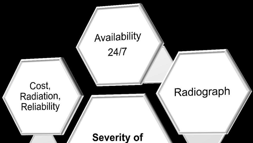
6 Availablity 24 / 7 / 365 Imaging modalities Radiographs CT MR Radiologist on-call Experienced technicians and radiologists on-call for emergency MRI Human resources, costs, reimbursement, outsourcing Guidelines must cover national and (best) international situations and infra-structure
 Diffusion-weighted imaging for spinal cord Source")
7 Full service or fast track imaging Image acquisition Plain films CT Axials, sagittals, coronals 3D volume rendering Angio / perfusion Surgical planning simulations MRI Angio (cervical spine) Diffusion-weighted imaging for spinal cord Source USZ
8 Full service or fast track imaging Image interpretation Full image evaluation (incl. all degenerative changes) Fast track image evaluation (only focussed on trauma) step-wise approach with preliminary image reading and subsequent full report (within 24hrs) (Semi-) quantitative analysis Data transfer PACS Reporting in-house vs externally Source USZ
 limited information regarding soft tissues Spine?")
9 Plain Film Widely available, cheap, fast Mainstay of bone and joint imaging, particularly in trauma Disadvantages uses ionising radiation (x rays) limited information regarding soft tissues Spine?? Source USZ


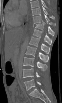
10 Computed Tomography (CT) Cross sectional imaging capability Reformatting in other planes and 3D Best for bony cortex and calcification Good at evaluation of comminuted fractures to complex structures Pelvis Calcaneus Wrist Spine Source USZ Source USZ

11 Quelle: 20min.ch


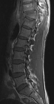
12 Magnetic Resonance (MR) Imaging Multiplanar imaging Excellent soft tissue contrast Ideally for radiographically occult fractures Source USZ


13 Added value of MRI A 37-year-old woman after a bicycle accident. Pe rso na lu se AO A1.2 on ly vs AO B1.2

 injury severity score (ISS) 1")
14 Change of therapy due to MRI Pe 53-year-old man after a car accident rso na A3.1 lu AO vs se B1.2 on ly Thoraco-Lumbar Injury Classification and Severity (TLICS) injury severity score (ISS) 1 vs 7


15 24 ys old male with cervical spine trauma Pe Neck pain, no neurological deficits rso na lu se on ly Cervical Spine = CT Source USZ


16 Nuclear Medicine entire skeleton at once bone scan is an indicator of bone turn over very sensitive, not specific fracture tumour arthritis infection metabolic bone disease multiple metastases Source UHN, Toronto
17 Typical Report of Bone Scan Non-specific uptake xiphoid process region of the sternum. Correlation with clinical examination suggested. Unless there has been trauma to these sites I cannot exclude metastatic disease and further radiologic correlation is recommended. This likely represents a normal variant, however, correlation with x-ray is recommended to rule out loosening or other pathology. Clinical correlation and further investigation with a left shoulder radiograph is recommended. Suspected degenerative change midcervical spine, radiograph would be confirmatory. Possible traumatic injury to the sternoclavicular joints bilaterally. Radiographic correlation is recommended. Mild focal activity within the left acetabulum anteriorly which is non-specific and could be related to either degenerative changes or a metastatic deposit.
18 Typical Report of Bone Scan Non-specific uptake xiphoid process region of the sternum. Correlation with clinical examination suggested. Unless there has been trauma to these sites I cannot exclude metastatic disease and further radiologic correlation is recommended. This likely represents a normal variant, however, correlation with x-ray is recommended to rule out loosening or other pathology. Clinical correlation and further investigation with a left shoulder radiograph is recommended. Suspected degenerative change midcervical spine, radiograph would be confirmatory. Possible traumatic injury to the sternoclavicular joints bilaterally. Radiographic correlation is recommended. Mild focal activity within the left acetabulum anteriorly which is non-specific and could be related to either degenerative changes or a metastatic deposit.
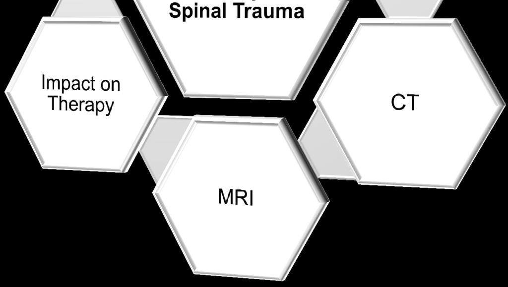
19 Impact on Therapy

20 Cost, Radiation, Reliability Lack of cost-efficacy studies with regard to CT and/or MR in acute spinal trauma Huge variability in radiation exposure even within a small, well developed country No prospective controlled study on the reliability of different imaging techniques in different clinical scenarios.
21 Content minpersonal use only MRI Challenges CT Guidelines and Reporting Strategy Future Directions
22 Evidence-based guidelines increasing role in patient care and reimbursement decisions federal and state agencies and third-party payers look to evidence-based recommendations to improve quality of care and halt the increase in health care costs Joshi GP. How Important Is Evidence-Based Medicine in Epidural Injection for Low Back Pain? Practical pain management. First published on: March 1, 2014

23 ACR Appropriatness Criteria

24 Clinical Scenarios > age14 Variant 1-8 = cervical spine Variant 9, 10 = adults, thoraco-lumbar Variant 11-14, age <14 yrs

25 Clinical Scenarios > age14


26 Severity of thoraco-lumbar trauma Compression Type Flexion Extension Distraction Type 20% of spinal fractures are multiple 95% of spinal fractures are at continuous levels Most thoracolumbar spinal fractures occur in the Th10-L2 region Multidirectional Rotation - Translation Type
27 Magerl AO Classification (1994) This Swiss system classifies thoracolumbar fractures into 3 groups, based on the mechanism of injury: A. Compression or Burst A1: Wedge A2: Split or coronal A3: Burst B. Flexion - Distraction B1: Distraction of the posterior soft tissues (subluxation) B2: Distraction of the posterior arch (Chance fracture) B3: Distraction of the anterior disc (extension spondylolysis) C. Multi-directional with translation C1: Anterior-posterior (dislocation) C2: Lateral (lateral shear) C3: Rotational (rotational burst)

 is")
28 Type A Fractures (65%) Injury to spinal cord (due to displacement of posterior fragments) is common


29 Type B Fractures (15%) Chance Fracture
 are torn with this mechanism Up to 65% have intra-abdominal injury, especially bowel Neurological")
30 Flexion - Distraction Fracture Seat belt fracture; Chance fracture; Anterior wedging of low thoracic or upper lumbar vertebrae Focal kyphosis, facet and vertebra subluxation Stabilizing ligaments (anterior, posterior longitudinal, capsular, ligamenta flavum) are torn with this mechanism Up to 65% have intra-abdominal injury, especially bowel Neurological damage in 30%


31 Flexion - Distraction Fracture Typically located at thoracolumbar junction or upper lumbar spine Must obtain CT once plain film findings suggest fracture, or show focal kyphosis; look for intra-abdominal injury MR to evaluate cord injury, compression > 15 degrees of kyphosis indicates instability T2 STIR T1

32 Type C Fractures (20%)
33 Content minpersonal use only MRI Challenges CT Guidelines and Reporting Strategy Future Directions Emergency MRI Dual-energy MDCT

34 Emergency MR Imaging Recent literature shows a significant added value of complimentary emergency MRI especially with regard to patient management which is frequently changed after MRI. Fracture classification Associated findings Occult fractures / Bone bruise Myelopathy and false positive CT Winklhofer et al. Magnetic resonance imaging frequently changes classification of acute traumatic thoracolumbar spine injuries. Skeletal Radiol 2012 Pizones et al. Impact of magnetic resonance imaging on decision making for thoracolumbar traumatic fracture diagnosis and treatment. Eur Spine J. 2011;20 Suppl. 3: Crosby et al. Diagnostic abilities of magnetic resonance imaging in traumatic injury to the posterior ligamentous complex: the effect of years in training. The Spine Journal 2011

35 Dual-energy MDCT
36 Conclusion CT is the mainstay in spinal trauma imaging. Emergency MRI provides complementary information and is indicated in all patients with neurologic deficits. It should also used in patients without neurologic deficits.
MR Neurography: Cervical Plexus and Shoulder Girdle
 MR Neurography: Cervical Plexus and Shoulder Girdle Gustav Andreisek, MD, MBA 3rd MSK MRI Meeting 2016 Date: April 23th, 2016 Time: 11:10-11:30 AM Head MSK and MR Imaging Department of Radiology University
Chronic sport injuries of the knee
 Chronic sport injuries of the knee 5th Musculoskeletal MRI meeting 2018: Knee MRI Gustav Andreisek, MD, MBA Saturday, May 5th, 2018; 14:00-16:00 Professor of Radiology, University of Zurich and Head of
Thoracolumbar Spine Fractures
 Thoracolumbar Spine Fractures C. Craig Blackmore, MD, MPH Professor of Radiology Adjunct Professor of Health Services Harborview Injury Prevention and Research Center University of Washington Outline Who
102 Results RESULTS. Age Mean=S.D Range 42= years -84 years Number % <30 years years >50 years
 102 Results RESULTS A total of 50 cases were studied 39 males and 11females.Their age ranged between 16 years and 84 years (mean 42years). T1 and T2WI were acquired for all cases in sagittal and axial
Classification of Thoracolumbar Spine Injuries
 Classification of Thoracolumbar Spine Injuries Guillem Saló Bru 1 IMAS. Hospitals del Mar i de l Esperança. ICATME. Institut Universitari Dexeus USP. UNIVERSITAT AUTÒNOMA DE BARCELONA Objectives of classification
Fractures of the thoracic and lumbar spine and thoracolumbar transition
 Most spinal column injuries occur in the thoracolumbar transition, the area between the lower thoracic spine and the upper lumbar spine; over half of all vertebral fractures involve the 12 th thoracic
SUBAXIAL CERVICAL SPINE TRAUMA- DIAGNOSIS AND MANAGEMENT
 SUBAXIAL CERVICAL SPINE TRAUMA- DIAGNOSIS AND MANAGEMENT 1 Anatomy 3 columns- Anterior, middle and Posterior Anterior- ALL, Anterior 2/3 rd body & disc. Middle- Posterior 1/3 rd of body & disc, PLL Posterior-
Imaging of Cervical Spine Trauma Tudor H Hughes, M.D.
 Imaging of Cervical Spine Trauma Tudor H Hughes, M.D. General Considerations Most spinal fractures are due to a single episode of major trauma. Fatigue fractures of the spine are unusual except in the
factor for identifying unstable thoracolumbar fractures. There are clinical and radiological criteria
 NMJ-Vol :2/ Issue:1/ Jan June 2013 Case Report Medical Sciences Progressive subluxation of thoracic wedge compression fracture with unidentified PLC injury Dr.Thalluri.Gopala krishnaiah* Dr.Voleti.Surya
AO CLASSIFICATIONS THORACO-LUMBAR SPINAL INJURIES
 AO CLASSIFICATIONS THORACO-LUMBAR SPINAL INJURIES T H E A O / A S I F ( A R B E I T S G E M E I N S C H A F T F Ü R O S T E O S Y N T H E S E F R A G E N / A S S O C I A T I O N F O R T H E S T U D Y O
Subaxial Cervical Spine Trauma. Introduction. Anatomic Considerations 7/23/2018
 Subaxial Cervical Spine Trauma Sheyan J. Armaghani, MD Florida Orthopedic Institute Assistant Professor USF Dept of Orthopedics Introduction Trauma to the cervical spine accounts for 5 of all spine injuries
Diagnostic accuracy of MRI in detecting posterior ligamentous complex injury in thoracolumbar vertebral fractures
 Diagnostic accuracy of MRI in detecting posterior ligamentous complex injury in thoracolumbar vertebral fractures Poster No.: C-1726 Congress: ECR 2011 Type: Scientific Exhibit Authors: E. Aguirre, P.
Fractures of the Thoracic and Lumbar Spine
 A spinal fracture is a serious injury. Nader M. Hebela, MD Fellow of the American Academy of Orthopaedic Surgeons http://orthodoc.aaos.org/hebela Cleveland Clinic Abu Dhabi Cleveland Clinic Abu Dhabi Neurological
Thoracolumbar spine trauma classifications: evolution or more confusion
 Thoracolumbar spine trauma classifications: evolution or more confusion Poster No.: C-1713 Congress: ECR 2012 Type: Educational Exhibit Authors: J. P. Salazar, J. Halaburda Berni, C. Torrents, L. Casas;
Imaging of Trauma to the Spine. Orthopedic Diplomate Program University of Bridgeport College of Chiropractic
 Imaging of Trauma to the Spine Orthopedic Diplomate Program University of Bridgeport College of Chiropractic Jefferson Fracture Yee, LL: The Jefferson Fracture, Radiology Cases in Pediatric Emergency Medicine.
Spinal injury is very common in Ireland: 19 per 100,000 (1). It poses a significant disease burden.
. It poses a significant disease burden.") MRI in traumatic spinal cord injury: a single national spinal centre experience and study of imaging features with clinical correlation with ASIA score and outcome Poster No.: C-1235 Congress: ECR 2011
MRI in traumatic spinal cord injury: a single national spinal centre experience and study of imaging features with clinical correlation with ASIA score and outcome Poster No.: C-1235 Congress: ECR 2011
Outline. Epidemiology Indications for C-spine imaging Modalities Interpretation Types of fractures
 C-Spine Plain Films Outline Epidemiology Indications for C-spine imaging Modalities Interpretation Types of fractures Epidemiology 7000-10000 c-spine injuries treated each year Additional 5000 die at the
Comprehension of the common spine disorder.
 Objectives Comprehension of the common spine disorder. Disc degeneration/hernia. Spinal stenosis. Common spinal deformity (Spondylolisthesis, Scoliosis). Osteoporotic fracture. Anatomy Anatomy Anatomy
Ligamentous Integrity in Spinal Cord Injury without Radiographic Abnormality. Dr Anria Horn Dr Stewart Dix-Peek
 Ligamentous Integrity in Spinal Cord Injury without Radiographic Abnormality Dr Anria Horn Dr Stewart Dix-Peek Introduction Spinal Cord Injury Without Radiographic Abnormality SCIWORA Pang, Wilberger 1982
ESSENTIALS OF PLAIN FILM INTERPRETATION: SPINE DR ASIF SAIFUDDIN
 ESSENTIALS OF PLAIN FILM INTERPRETATION: SPINE DR ASIF SAIFUDDIN Consultant Musculoskeletal Radiologist Royal National Orthopaedic Hospital Stanmore,UK. INTRODUCTION 2 INTRODUCTION 3 INTRODUCTION Spinal
National Imaging Associates, Inc. Clinical guidelines
 National Imaging Associates, Inc. Clinical guidelines Original Date: September 1997 THORACIC SPINE CT Page 1 of 5 CPT Codes: 72128, 72129, 72130 Last Review Date: May 2013 Guideline Number: NIA_CG_043
THORACO-LUMBAR SPINE TRAUMA NORDIC TRAUMA COURSE 2016, AARHUS
 THORACO-LUMBAR SPINE TRAUMA NORDIC TRAUMA COURSE 2016, AARHUS Ken F. Linnau, MD, MS Emergency Radiology Harborview Medical Center University of Washington Seattle, WA Thanks to Quynh T. Nguyen, MHS, PA-C
SCIWORA Rozlyn McTeer BSN, RN, CEN Pediatric Trauma Coordinator Trauma Services OBJECTIVES DEFINITION 11/8/2017. Identify SCIWORA.
 SCIWORA Rozlyn McTeer BSN, RN, CEN Pediatric Trauma Coordinator Trauma Services Identify SCIWORA. OBJECTIVES Identify the population at risk. To identify anatomic and physiologic reasons for SCIWORA. To
Complex Fractures and Hip Dislocations
 IMAGING OF HIP PAIN Patients may present with acute (< 2 weeks) or chronic hip pain. Acute pain may be related or not related to an acute traumatic event such as fall or trauma from a motor vehicle accident.
Upper Cervical Spine - Occult Injury and Trigger for CT Exam
 Upper Cervical Spine - Occult Injury and Trigger for CT Exam Main Menu Introduction Clinical clearance of C-SpineC Radiographic evaluation Norms for C-spineC Triggers for CT exam: Odontoid Lateral view
Prof. Dr. NAGUI M. ABDELWAHAB,M.D.; MARYSE Y. AWADALLAH, M.D. AYA M. BASSAM, Ms.C.
 Role of Whole-body Diffusion MR in Detection of Metastatic lesions Prof. Dr. NAGUI M. ABDELWAHAB,M.D.; MARYSE Y. AWADALLAH, M.D. AYA M. BASSAM, Ms.C. Cancer is a potentially life-threatening disease,
Subaxial Cervical Spine Trauma Dr Hesarikia BUMS
 Subaxial Cervical Spine Trauma Dr. Hesarikia BUMS Subaxial Cervical Spine From C3-C7 ROM Majority of cervical flexion Lateral bending Approximately 50% rotation Ligamentous Anatomy Anterior ALL, PLL, intervertebral
Objectives. Comprehension of the common spine disorder
 Objectives Comprehension of the common spine disorder Disc degeneration/hernia Spinal stenosis Common spinal deformity (Spondylolisthesis, Scoliosis) Osteoporotic fracture Destructive spinal lesions Anatomy
Message of the Month for GPs June 2013
 Message of the Month for GPs June 2013 Dr Winn : Consultant Musculoskeletal Radiologist, Manchester Royal Infirmary Imaging of the musculoskeletal system Musculoskeletal pain is a common problem in the
JUSTIFICATION PROTOCOLS FOR CT SCANNING ALBURY WODONGA HEALTH WODONGA CAMPUS
 JUSTIFICATION PROTOCOLS FOR CT SCANNING ALBURY WODONGA HEALTH WODONGA CAMPUS JUSTIFICATION PROTOCOLS FOR CT SCANNING INTRODUCTION: In accordance with the Victorian Radiation Act 2005 Wodonga Medical Imaging,
A Patient s Guide to Diffuse Idiopathic Skeletal Hyperostosis (DISH)
") A Patient s Guide to Diffuse Idiopathic Skeletal Hyperostosis (DISH) 6565 Fannin Street Houston, TX 77030 Phone: 713-790-3333 DISCLAIMER: The information in this booklet is compiled from a variety of sources.
A Patient s Guide to Diffuse Idiopathic Skeletal Hyperostosis (DISH) 6565 Fannin Street Houston, TX 77030 Phone: 713-790-3333 DISCLAIMER: The information in this booklet is compiled from a variety of sources.
Am I eligible for the TOPS study? Possibly, if you suffer from one or more of the following conditions:
 Am I eligible for the TOPS study? Possibly, if you suffer from one or more of the following conditions: Radiating leg pain Greater leg / buttock pain than back pain Severe pain sets in when walking as
Chance Fracture Joseph Junewick, MD FACR
 Chance Fracture Joseph Junewick, MD FACR 08/02/2010 History Restrained teenager involved in motor vehicle accident. Diagnosis Chance Fracture (Hyperflexion-Distraction Injury) Discussion Chance-type spinal
Thoracic and Lumbar Spine Fractures and Dislocations: Assessment and Classification
 Thoracic and Lumbar Spine Fractures and Dislocations: Assessment and Classification Mark L Prasarn MD University of Texas Dept of Orthopaedic Surgery Houston, Texas Updated 7/2016 Anatomy of the Spine
Spine MRI and Spine CT Test Request Tip Sheet
 Spine MRI and Spine CT With/Without Contrast CT, MRI Studies should NOT be ordered simultaneously as dual studies (i.e., with and without contrast). Radiation exposure is doubled and both views are rarely
Subaxial Cervical Spine Trauma
 Subaxial Cervical Spine Trauma Pooria Salari, MD Assistant Professor Of Orthopaedics Department of Orthopaedic Surgery St. Louis University School of Medicine St. Louis, Missouri, USA Initial Evaluation
MDCT and MRI evaluation of cervical spine trauma
 Insights Imaging (2014) 5:67 75 DOI 10.1007/s13244-013-0304-2 PICTORIAL REVIEW MDCT and MRI evaluation of cervical spine trauma Michael Utz & Shadab Khan & Daniel O Connor & Stephen Meyers Received: 10
Imaging and Management of the Charcot Spine Following Spinal Injury
 Imaging and Management of the Charcot Spine Following Spinal Injury Poster No.: P-0023 Congress: ESSR 2012 Type: Scientific Exhibit Authors: A. Isaac, P. A. Tyler; Stanmore/UK Keywords: Musculoskeletal
How to interpret computed tomography of the lumbar spine
 REVIEW Ann R Coll Surg Engl 2014; 96: 502 507 doi 10.1308/003588414X13946184902361 How to interpret computed tomography of the lumbar spine Z Ahmad 1, R Mobasheri 2,TDas 3, S Vaidya 4, S Mallik 5, M El-Hussainy
Spinal Cord Injuries: The Basics. Kadre Sneddon POS Rounds October 1, 2003
 Spinal Cord Injuries: The Basics Kadre Sneddon POS Rounds October 1, 2003 Anatomy Dorsal columntouch, vibration Corticospinal tract- UMN Anterior horn-lmn Spinothalamic tractpain, temperature (contralateral)
Departement of Neurosurgery A.O.R.N A. Cardarelli- Naples.
 Percutaneous posterior pedicle screw fixation in the treatment of thoracic, lumbar and thoraco-lumbar junction (T12-L1) traumatic and pathological spine fractures. Report of 45 cases. G. Vitale, A. Punzo,
Hidayatullah Hamidi. MD Consultant Radiologist. Lumbar Spine MR Imaging Interpretation
 Hidayatullah Hamidi. MD Consultant Radiologist Lumbar Spine MR Imaging Interpretation 13/12/2018 Presenter Hidayatullah Hamidi Consultant Radiologist, Radiology PGME program director, FMIC, Kabul, Afghanistan
Chapter 3 Diagnostic Imaging. 1 Diagnostic Imaging
 Chapter 3 Diagnostic Imaging 1 Diagnostic Imaging Radiographic Examination: Standards and Indications Position Statement Radiography should only be performed on the basis of clinical necessity as judged
J of Evolution of Med and Dent Sci/ eissn , pissn / Vol. 4/ Issue 34/ Apr 27, 2015 Page 5797
 THE CORRELATION OF RADIOLOGICAL EXAMINATION AND VOLITIONAL VOIDING IN THORACO-LUMBAR FRACTURES AND SPINAL INJURY Mathangi Santhosh Kumar 1, Aastha 2, David Mohan 3, Suranjan Bhattacharji 4 HOW TO CITE
Functional Orthopedic Imaging Capturing Motion, Flow and Perfusion. Case Study Brochure Centre University Hospital Nancy.
 Capturing Motion, Flow and Perfusion dynamic volume CT Case Study Brochure Centre University Hospital Nancy http://www.toshibamedicalsystems.com Toshiba Medical Systems Corporation 2013. All rights reserved.
Primary care referral criteria for musculoskeletal MRI scans
 Appendix 1 Primary care referral criteria for musculoskeletal MRI scans Accepted Criteria for Direct Access MRI Body Part Symptoms Imaging indicated Lumbar Spine Low Back Pain with adverse symptoms or
FOR CMS (MEDICARE) MEMBERS ONLY NATIONAL COVERAGE DETERMINATION (NCD) FOR MAGNETIC RESONANCE IMAGING:
 MEMBERS ONLY NATIONAL COVERAGE DETERMINATION (NCD) FOR MAGNETIC RESONANCE IMAGING:") National Imaging Associates, Inc. Clinical guidelines BONE MARROW MRI Original Date: July 2008 Page 1 of 5 CPT Codes: 77084 Last Review Date: September 2014 NCD 220.2 MRI Last Effective Date: July 2011
National Imaging Associates, Inc. Clinical guidelines BONE MARROW MRI Original Date: July 2008 Page 1 of 5 CPT Codes: 77084 Last Review Date: September 2014 NCD 220.2 MRI Last Effective Date: July 2011
Module 1: Basic Comprehensive Course
 The Hellenic Spine Society organize 5 modules according to the following program, which is based on the Eurospine program Module 1: Basic Comprehensive Course SESSION1: SPINE THE BIGGER PICTURE Evidence
The role of multimodality imaging in Multiple Myeloma: Past, Present and Future
 The role of multimodality imaging in Multiple Myeloma: Past, Present and Future Poster No.: C-1661 Congress: ECR 2015 Type: Educational Exhibit Authors: J. Niza, R. Gil, P. Pereira, C. Oliveira ; Setúbal/PT,
Imaging of spine trauma
 Imaging of spine trauma RD Magazine, 44, 514, 23-24 Dr Matthew Jaring Speciality registrar in clinical radiology Dr Roland Watura onsultant musculoskeletal radiologist Southmead Hospital, ristol Introduction
Classification? Classification system should be: Comprehensive Usable Accurate Predictable Able to guide intervention
 Moderator: Dr. P.S. Chandra Dr. Dr Deepak Gupta Classification? Classification system should be: Comprehensive Usable Accurate Predictable Able to guide intervention A precise, comprehensive, ideal
Imaging of Cervical Spine Trauma
 Imaging of Cervical Spine Trauma C Craig Blackmore, MD, MPH Professor of Radiology and Adjunct Professor of Health Services University of Washington, Harborview Medical Center Salary support: AHRQ grant
17. Imaging and interventional radiology
 17. Imaging and interventional radiology These guidelines have been adapted from the Leeds Major Trauma Centre Imaging in Paediatric Major Trauma guidelines Written by Dr Annmarie Jeanes (Consultant Paediatric
Spine MRI and Spine CT Test Request Tip Sheet
 Spine MRI and Spine CT With/Without Contrast CT, MRI The study considered best for a specific clinical scenario should be ordered. The second study should be done ONLY if the first study does not provide
Case Report Traumatic Death due to Simultaneous Double Spine Fractures in Patient with Ankylosing Spondylitis
 Case Reports in Orthopedics Volume 2015, Article ID 590935, 4 pages http://dx.doi.org/10.1155/2015/590935 Case Report Traumatic Death due to Simultaneous Double Spine Fractures in Patient with Ankylosing
ASJ. A Rare Hyperextension Injury in Thoracic Spine Presenting with Delayed Paraplegia. Asian Spine Journal. Introduction
 sian Spine Journal 126 Dong-Eun Case Shin Report et al. http://dx.doi.org/10.4184/asj.2013.7.2.126 Rare Hyperextension Injury in Thoracic Spine Presenting with Delayed Paraplegia Dong-Eun Shin, Ki-Sik
Thoracolumbar Spinal Injuries
 31 Thoracolumbar Spinal Injuries Michael Heinzelmann, Guido A. Wanner Fractures Section 883 Core Messages Spinal fractures are frequently located at the thoracolumbar junction for biomechanical reasons
Original article: Multidetector computed tomographic evaluation of cervical spine trauma
 Original article: Multidetector computed tomographic evaluation of cervical spine trauma 1Sajid Ansari *, 2 R.K. Rauniyar, 3 Kaleem Ahmad, 4 Mukesh Kumar Gupta 1Assistant Professor, Department of Radiodiagnosis,
Disclosures: T. Yoshii: None. T. Yamada: None. T. Taniyama: None. S. Sotome: None. T. Kato: None. S. Kawabata: None. A. Okawa: None.
 Dynamic Changes in Spinal Cord Compression by Cervical Ossification of the Posterior Longitudinal Ligament Evaluated by Kinematic Computed Tomography Myelogram Toshitaka Yoshii, Tsuyoshi Yamada, Takashi
Revised Dec Spine MR Protocols
 Spine MR Protocols Sp 1: Cervical spine MRI without contrast Sp 2: Pre- and post-contrast cervical spine MRI Sp 3: Pre- and post-contrast cervical spine MRI (multiple sclerosis protocol) Sp 4: Thoracic
Degenerative Disease of the Spine
 Degenerative Disease of the Spine Introduction: I. Anatomy Talk Overview II. Overview of Disease Processes: A. Spondylosis B. Intervertebral Disc Disease III. Diagnosis IV. Therapy Introduction: Myelopathy
3/10/17 Spinal a Injury 1
 Spinal Injury 1 'Paralysed' Watmough vows he'll have the backbone for Game Two after treatment for neck injury Watmough will have cortisone injected into his spine this morning to speed up the recovery
Metastatic Spinal Disease
 Metastatic Spinal Disease Mr Neil Chiverton Consultant Spinal Surgeon, Sheffield Objectives The scale and nature of the problem NICE recommendations Surgical decision making Case illustrations Incidence
Common fracture & dislocation of the cervical spine. Theerachai Apivatthakakul Department of Orthopaedic Chiangmai University
 Common fracture & dislocation of the cervical spine Theerachai Apivatthakakul Department of Orthopaedic Chiangmai University Objective Anatomy Mechanism and type of injury PE.and radiographic evaluation
Spine MRI and Spine CT Test Request Tip Sheet
 Spine MRI and Spine CT With/Without Contrast CT, MRI The study considered best for a specific clinical scenario should be ordered. The second study should be done ONLY if the first study does not provide
Effective Utilization of Imaging. John V. Roberts, M.D. Premier Radiology Abdominal Imaging
 Effective Utilization of Imaging John V. Roberts, M.D. Premier Radiology Abdominal Imaging Safety Contrast and Radiation What to order Abdomen/Pelvis Brain/Spine Chest Musculoskeletal Ob/Gyn Head and Neck
ORIGINAL PAPER. Department of Orthopedic Surgery,Nagoya University Graduate School of Medicine,Nagoya,Japan 2
 Nagoya J. Med. Sci. 80. 583 589, 2018 doi:10.18999/nagjms.80.4.583 ORIGINAL PAPER Evaluation of sagittal alignment and range of motion of the cervical spine using multi-detector- row computed tomography
New Dual-energy X-ray Absorptiometry Machines (idxa) and Vertebral Fracture Assessment
 and Vertebral Fracture Assessment") Case 1 New Dual-energy X-ray Absorptiometry Machines (idxa) and Vertebral Fracture Assessment (VFA) History and Examination Your wealthy friend who is a banker brings his 62-year-old mother to your office
Case 1 New Dual-energy X-ray Absorptiometry Machines (idxa) and Vertebral Fracture Assessment (VFA) History and Examination Your wealthy friend who is a banker brings his 62-year-old mother to your office
Advances in Emergency Imaging
 Hampton Symposium,, October 16 th, 2010 Advances in Emergency Imaging Robert A. Novelline, MD Professor of Radiology, Harvard Medical School Director of Emergency Radiology, Massachusetts General Hospital
Kanji Mori, Kazuya Nishizawa, Akira Nakamura, and Shinji Imai. 1. Introduction. 2. Case Presentation
 Case Reports in Orthopedics Volume 2015, Article ID 301858, 4 pages http://dx.doi.org/10.1155/2015/301858 Case Report Atraumatic Occult Odontoid Fracture in Patients with Osteoporosis-Associated Thoracic
CLINICAL CONCEPTS FOR ORTHOPEDICS. CMS Clinical Concepts
 CLINICAL CONCEPTS FOR ORTHOPEDICS CMS Clinical Concepts ICD 10 LESSONS FROM OFFICE DOCUMENTATION Presented by Dr. Frankeny OUR CHALLENGE: CHANGING OUR DOCUMENTATION ICD 10 Learn the nomenclature Documenting
CLINICAL PRESENTATION AND RADIOLOGY QUIZ QUESTION
 Donald L. Renfrew, MD Radiology Associates of the Fox Valley, 333 N. Commercial Street, Suite 100, Neenah, WI 54956 11/24/2012 Radiology Quiz of the Week # 100 Page 1 CLINICAL PRESENTATION AND RADIOLOGY
A rare case of spinal injury: bilateral facet dislocation without fracture at the lumbosacral joint
 J Orthop Sci (2012) 17:189 193 DOI 10.1007/s00776-011-0082-y CASE REPORT A rare case of spinal injury: bilateral facet dislocation without fracture at the lumbosacral joint Kei Shinohara Shigeru Soshi
SPINAL MAGNETIC RESONANCE IMAGING INTERPRETATION
 CLINICAL VIGNETTE 2017; 3:2 SPINAL MAGNETIC RESONANCE IMAGING INTERPRETATION Editor-in-Chief: Idowu, Olufemi E. Neurological surgery Division, Department of Surgery, LASUCOM/LASUTH, Ikeja, Lagos, Nigeria.
Digital tomosynthesis (DT) has been well described as a
 has been well described as a") Case Report The Usefulness of Digital Tomosynthesis (DT) in Assisting in Cases of Doubtful Routine Radiography and/or Computed Tomography (CT) Image. Abstract Digital tomosynthesis is useful in assisting
Case Report The Usefulness of Digital Tomosynthesis (DT) in Assisting in Cases of Doubtful Routine Radiography and/or Computed Tomography (CT) Image. Abstract Digital tomosynthesis is useful in assisting
Magnetic resonance imaging in acute spinal trauma: Pictorial essay
 Magnetic resonance imaging in acute spinal trauma: Pictorial essay Poster No.: C-1463 Congress: ECR 2013 Type: Educational Exhibit Authors: S. Khurana 1, S. Manchanda 1, N. Rajpal 1, S. Agrawal 1, S. Gupta
CT Findings of Traumatic Posterior Hip Dislocation after Reduction 1
 CT Findings of Traumatic Posterior Hip Dislocation after Reduction 1 Sung Kyoung Moon, M.D., Ji Seon Park, M.D., Wook Jin, M.D. 2, Kyung Nam Ryu, M.D. Purpose: To evaluate the CT images of reduced hips
Pediatric Imaging Spine MRI and Spine CT Test Request Tip Sheet
 Pediatric Imaging Spine MRI and Spine CT MRI is almost always preferred over CT scan; if ordering CT, CLEARLY document why MRI is not appropriate. In cases of back pain without red flags, six weeks of
REVIEW QUESTIONS ON VERTEBRAE, SPINAL CORD, SPINAL NERVES
 REVIEW QUESTIONS ON VERTEBRAE, SPINAL CORD, SPINAL NERVES 1. A 28-year-old-women presented to the hospital emergency room with intense lower back spasms in the context of coughing during an upper respiratory
CLINICAL PRESENTATION AND RADIOLOGY QUIZ QUESTION
 Donald L. Renfrew, MD Radiology Associates of the Fox Valley, 333 N. Commercial Street, Suite 100, Neenah, WI 54956 12/29/2012 Radiology Quiz of the Week # 105 Page 1 CLINICAL PRESENTATION AND RADIOLOGY
Learning from Discrepancies Meetings - What we've learned from Musculoskeletal Diagnostic Errors in 2014
 Learning from Discrepancies Meetings - What we've learned from Musculoskeletal Diagnostic Errors in 2014 Poster No.: P-0104 Congress: ESSR 2015 Type: Scientific Poster Authors: B. Batohi, R. Chhabra, S.
Spine. Neuroradiology. Spine. Spine Pathology. Distribution of fractures. Radiological algorithm. Role of radiology 18/11/2015
 Spine Neuroradiology Spine Prof.Dr.Nail Bulakbaşı X Ray: AP/L/Oblique Vertebra & disc spaces CT & CTA Vertebra, discs, vessels MRI & MRA Vertebra, disc, vessels, meninges Spinal cord & nerves Myelography
Spinal Trauma: Imaging, Diagnosis, And Management READ ONLINE
 Spinal Trauma: Imaging, Diagnosis, And Management READ ONLINE Jul 22, 2013 Thoracic Spinal Trauma Imaging. who have sustained thoracic spinal trauma is to for the diagnosis of a thoracic spinal fracture
SPINE EVALUATION AND CLEARANCE Basic Principles
 SPINE EVALUATION AND CLEARANCE Basic Principles General 1. Entire spine is immobilized during primary survey. 2. Radiographic clearance of the spine is not required before emergent surgical procedures.
Neck Pain: Help! Eric M. Massicotte, MD, MSc, MBA, FRCSC Associate Professor University of Toronto
 Neck Pain: Help! Eric M. Massicotte, MD, MSc, MBA, FRCSC Associate Professor University of Toronto Copyright 2017 by Sea Courses Inc. All rights reserved. No part of this document may be reproduced, copied,
Spinal Trauma. Dr T G Kruger
 Spinal Trauma Dr T G Kruger Epidemiology Spine injury in 6% of trauma patients Multiple levels involved in 20% of cases 80% of spinal cord injury patients have concurrent other system injuries 41% have
IMAGISTICÃ. Magnetic resonance imaging assessment of spinal injury
 IMGISTICÃ Magnetic resonance imaging assessment of spinal injury NICOLE OLOG, M.D., IRINEL ONCE, M.D. Radiology & Imaging Department, ucharest Emergency Clinical Hospital uthor for correspondence: NICOLE
Spinal canal stenosis Degenerative diseases F 06
 What is spinal canal stenosis? The condition known as spinal canal stenosis is a narrowing (stenosis) of the spinal canal that in most cases develops due to the degenerative (wear-induced) deformation
Dr Ajit Singh Moderator Dr P S Chandra Dr Rajender Kumar
 BIOMECHANICS OF SPINE Dr Ajit Singh Moderator Dr P S Chandra Dr Rajender Kumar What is biomechanics? Biomechanics is the study of the consequences of application of external force on the spine Primary
APPROPRIATE USE GUIDELINES
 APPROPRIATE USE GUIDELINES Appropriateness of Advanced Imaging Procedures (MRI, CT, Bone Scan/PET) in Patients with Neck Pain CDI QUALITY INSTITUTE: PROVIDER LED ENTITY (PLE) Updated June, 2017 Contents
Pott disease (spinal tuberculosis): MR and CT imaging
: MR and CT imaging") Pott disease (spinal tuberculosis): MR and CT imaging Poster No.: C-1422 Congress: ECR 2016 Type: Educational Exhibit Authors: G. Beretis, K. Giannaki, M. Fasoula, D. Kypriotis, T. Dagla, T.-P. Mantzouranis;
Pott disease (spinal tuberculosis): MR and CT imaging Poster No.: C-1422 Congress: ECR 2016 Type: Educational Exhibit Authors: G. Beretis, K. Giannaki, M. Fasoula, D. Kypriotis, T. Dagla, T.-P. Mantzouranis;
4/28/2010. Fractures. Normal Bone and Normal Ossification Bone Terms. Epiphysis Epiphyseal Plate (physis) Metaphysis
 Metaphysis") Fractures Normal Bone and Normal Ossification Bone Terms Epiphysis Epiphyseal Plate (physis) Metaphysis Diaphysis 1 Fracture Classifications A. Longitudinal B. Transverse C. Oblique D. Spiral E. Incomplete
Fractures Normal Bone and Normal Ossification Bone Terms Epiphysis Epiphyseal Plate (physis) Metaphysis Diaphysis 1 Fracture Classifications A. Longitudinal B. Transverse C. Oblique D. Spiral E. Incomplete
Diagnostic Imaging Exams
 Guide for Chiropractors Diagnostic Imaging Exams CREATED FOR OUR CHIROPRACTIC PARTNERS This document has been prepared by the specialized, board-certified radiologists who interpret patient exams for Center
University of Groningen. Thoracolumbar spinal fractures Leferink, Vincentius Johannes Maria
 University of Groningen Thoracolumbar spinal fractures Leferink, Vincentius Johannes Maria IMPORTANT NOTE: You are advised to consult the publisher's version (publisher's PDF) if you wish to cite from
The imaging features of spondylolisthesis : what the clinician needs to know
 The imaging features of spondylolisthesis : what the clinician needs to know Poster No.: C-1018 Congress: ECR 2011 Type: Authors: Educational Exhibit D. Shah 1, C. J. Burke 1, A. C. andi 2, R. Houghton
Spine MRI and Spine CT Test Request Tip Sheet
 Spine MRI and Spine CT MRI is almost always preferred over CT scan; if ordering CT, CLEARLY document why MRI is not appropriate. In cases of back pain without red flags, six weeks of multimodality supervised
VAriation. Orthotics and Me (?surgeons) Greg Etherington Spine Surgeon. Orthopaedic & Neurosurgery backgrounds. Subspeciality training
 Greg Etherington Spine Surgeon. Orthopaedic & Neurosurgery backgrounds. Subspeciality training") Orthotics and Me (?surgeons) Greg Etherington Spine Surgeon Orthopaedic & Neurosurgery backgrounds Subspeciality training spine, upper limb, trauma, pelvis. What do you do in spine? Lumbar Cervical Trauma
Orthotics and Me (?surgeons) Greg Etherington Spine Surgeon Orthopaedic & Neurosurgery backgrounds Subspeciality training spine, upper limb, trauma, pelvis. What do you do in spine? Lumbar Cervical Trauma
Original Date: February 2006 PLAIN FILM X-RAYS
 Magellan Healthcare Clinical guidelines Original Date: February 2006 PLAIN FILM X-RAYS Page 1 of 5 Adopted Date 1 : April 2016 Physical Medicine Clinical Decision Making Last Review Date: August 2015 Guideline
Thorasic and lumbar spinal injury. Dr.Abrisham
 Thorasic and lumbar spinal injury Dr.Abrisham Goal : alignment Stability Preserve neuologic function early mobilization Incidence: most site is thoraco lumbar 50% T 11 to L 1 30% L 2 to L 5 Motor vehicle
B. CT protocols for the spine
 B. CT protocols for the spine Poster No.: A-003 Congress: ECR 2010 Type: Invited Speaker Topic: Neuro Authors: B. Tins; Oswestry/UK Keywords: CT, spine, diagnostic imaging protocol DOI: 10.1594/ecr2010/A-003