Chance Fracture Joseph Junewick, MD FACR
|
|
|
- Eustacia Walker
- 6 years ago
- Views:
Transcription
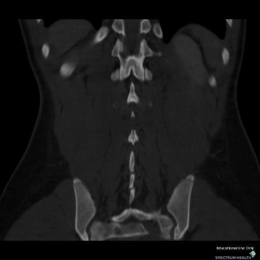
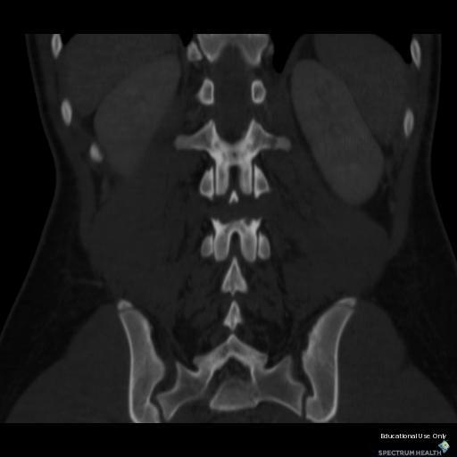
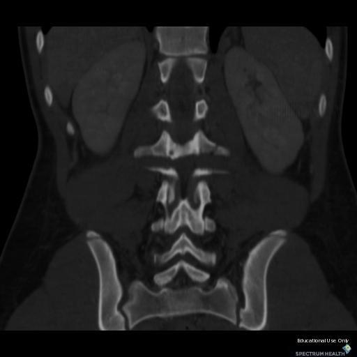
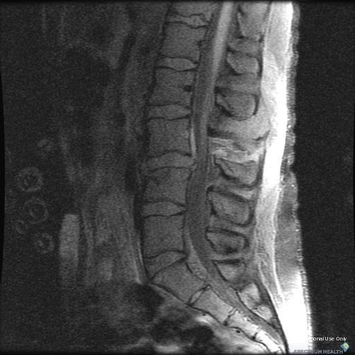
1 Chance Fracture Joseph Junewick, MD FACR 08/02/2010 History Restrained teenager involved in motor vehicle accident. Diagnosis Chance Fracture (Hyperflexion-Distraction Injury) Discussion Chance-type spinal injuries are the result of hyperflexion and distraction of the spine around a fulcrum. Initially this injury was described in motor vehicle accident victims restrained by a lap belt. The classic radiographic features of this injury include horizontal fractures of the pedicles extending transversely through the vertebral body. In cases in which reduction of the fracture is incomplete, the posterior vertebral body height is increased in relation to the adjacent inferior vertebral body. The wedge-shaped compression fracture of the anterosuperior vertebral body suggests that the fulcrum point is located behind the anterior column. The key indication that a flexion-distraction injury has occurred is posterior osteoligamentous complex disruption in the presence of minimal vertebral malalignment. MR imaging allows depiction of the injured posterior osteoligamentous component. There is widespread damage to the soft tissues, as highlighted by edema and hemorrhage in the subcutaneous fat and paraspinous muscles at multiple contiguous vertebral levels. Supraspinous, interspinous, and flaval ligaments are disrupted. Cord injury is common at the site of spinal injury related to profound distractive tension. Contiguous vertebral injury due to compressive forces, with bone edema occurring in the anterosuperior aspect of the subjacent vertebral body and noncontiguous vertebral injury are common. Disc herniation and anterior longitudinal ligament was disruption are also often seen. It is important to remember that Chance fractures are highly associated with injuries of the abdominal wall, bowel, mesentery, solid viscera and abdominal great vesselsvisceral injuries including. Findings CT-Sagittal and coronal reformated images demonstrate a transverse fracture of the posterior elements of L3, kyphosis at L3-L4 and superior compression deformity of L4. MR-Sagittal fat-suppressed T1, T2 and inversion recovery and axial T2 images show disc herniation at L3-L4, elevation of the posterior longitudinal ligament by blood, compression deformity of L4, posterior element fracture of L3 and extensive paraspinous edema. Reference Groves CJ, Cassar-Pullicino VN, Tins BJ, Tyrrell PNM, McCall IW. Chance-Type Flexion-Distraction Injuries in the Thoracolumbar Spine: MR Imaging. Radiology (2005); 236:

2
3
4
5

6
7
8

9
10 Sponsored By Disclaimer This teaching site is partially funded by an educational grant from GE Healthcare and Advanced Radiology Services, PC. The material on this site is independently controlled by Advanced Radiology Services, PC, and GE Healthcare and Spectrum Health have no influence over the content of this site Content Download Agreement The cases and images on this website are owned by Spectrum Health. Permission is granted (for nonprofit educational purposes) to download and print materials to distribute for the purpose of facilitating the education of health professionals. The authors retain all rights to the material and users are requested to acknowledge the source of the material. Site Disclaimer This site is developed to reach healthcare professionals and medical students. Nothing this site should be considered medical advice. Only your own doctor can help you make decisions about your medical care. If you have a specific medical question or are seeking medical care, please contact your physician. The information in this website is provided for general medical education purposes only and is not meant to substitute for the independent medical judgment of a physician relative to diagnostic and treatment options of a specific medical condition. The viewpoints expressed in these cases are those of the authors. They do not represent an endorsement. In no event will Advanced Radiology Associates, PC, Spectrum Health Hospitals (Helen Devos Children's Hospital) or GE Healthcare be liable for any decision made or action taken in reliance upon the information provided through this website.
Atlanto-occipital Dislocation Joseph Junewick, MD FACR
 Atlanto-occipital Dislocation Joseph Junewick, MD FACR 09/23/2009 History 12 year old male restrained back seat passenger in a car hit by a snowplow. Diagnosis Atlanto-occipital Dislocation Discussion
Spinal LCH Joseph Junewick, MD FACR
 Spinal LCH Joseph Junewick, MD FACR 05/16/2009 History 16 year old female with multiply recurrent Langerhans Cell Histiocytosis now with severe left sided neck pain. Diagnosis Langerhans Cell Histiocytosis
Diskitis Joseph Junewick, MD FACR
 Diskitis Joseph Junewick, MD FACR 09/20/2010 History 2 year old with fever, back pain and elevated sedimentation rate. Diagnosis Diskitis Discussion Diskitis is an inflammatory process of the intervertebral
Term Hypoxic Ischemic Injury Joseph Junewick, MD FACR
 Term Hypoxic Ischemic Injury Joseph Junewick, MD FACR 08/11/2010 History Term infant with perinatal distress and attempted forceps delivery. Diagnosis Term Hypoxic Ischemic Injury Discussion Encephalopathy
Transverse Dural Sinus Thrombosis Joseph Junewick, MD FACR
 Transverse Dural Sinus Thrombosis Joseph Junewick, MD FACR 03/19/2010 History Child with headache and otomastoiditis. Diagnosis Dural venous thrombosis secondary to mastoiditis Discussion The cerebral
Paraspinal Venous Malformation Joseph Junewick, MD FACR
 Paraspinal Venous Malformation Joseph Junewick, MD FACR 06/04/2010 History 2 year old with history of fall. Rule out spinal injury. Diagnosis Paraspinal Venous Malformation Additional Clinical CT of the
Ulcerative Colitis Joseph Junewick, MD FACR
 Ulcerative Colitis Joseph Junewick, MD FACR 06/04/2010 History 16 year old male with hematochezia and anemia. Diagnosis Ulcerative Colitis Additional Clinical History of ulcerative colitis. Discussion
Chiari III Joseph Junewick, MD FACR
 Chiari III Joseph Junewick, MD FACR 07/02/2010 History Newborn with suboccipital mass. Diagnosis Chiari III Additional Clinical Surgery-Skin covered suboccipital cystic mass confined by the dura. Pathology-Leptomeningeal
Pleural Empyema Joseph Junewick, MD FACR
 Pleural Empyema Joseph Junewick, MD FACR 03/19/2010 History Teenager with persistent fever and cough. Pneumonia diagnosed 1 week ago. Diagnosis Pleural Empyema Additional Clinical Surgery-Clear fluid with
Neuroblastoma Joseph Junewick, MD FACR
 Neuroblastoma Joseph Junewick, MD FACR 03/18/2011 History 15 month old with anemia. Diagnosis Neuroblastoma Discussion Neuroblastic tumors derive from primordial neural crest cells destined for sympathetic
Presacral Neuroblastoma Joseph Junewick, MD FACR
 Presacral Neuroblastoma Joseph Junewick, MD FACR 01/12/2010 History 16 month old male with irritability. Diagnosis Presacral Neuroblastoma Additional Clinical Initial US to evaluate for intussusception
Vein of Galen Malformation Joseph Junewick, MD FACR
 Vein of Galen Malformation Joseph Junewick, MD FACR 04/14/2018 History Midline cystic intracranial mass on prenatal ultrasound. Diagnosis Vein of Galen Malformation Discussion In normal fetal development,
Radiation Pneumonitis Joseph Junewick, MD FACR
 Radiation Pneumonitis Joseph Junewick, MD FACR 03/19/2010 History 16 year old with history of relapsed stage IV-A Hodgkin disease. Prior pulmonary involvement was irradiated. Diagnosis Radiation Pneumonitis
Bilateral Retinoblastoma Joseph Junewick, MD FACR
 Bilateral Retinoblastoma Joseph Junewick, MD FACR 06/11/2010 History 17 month old adopted female with proptosis. Diagnosis Bilateral Retinoblastoma Discussion Retinoblastoma is the most common pediatric
Posterior Slipped Capital Femoral Epiphysis Joseph Junewick, MD FACR
 Posterior Slipped Capital Femoral Epiphysis Joseph Junewick, MD FACR 08/11/2010 History 6 year old male with intermittent hip pain for several months, acutely worsened after climbing the sand dunes. Diagnosis
Pituitary Macroadenoma Joseph Junewick, MD FACR
 Pituitary Macroadenoma Joseph Junewick, MD FACR 08/13/2010 History 12 year old female with headache and visual disturbance. Diagnosis Pituitary Macroadenoma Additional Clinical Markedly elevated growth
Retroperitoneal Teratoma Heather Borders, MD
 Retroperitoneal Teratoma Heather Borders, MD 03/04/2012 History Newborn with congenitally diagnosed mass. No other clinical symptoms. Diagnosis Retroperitoneal Teratoma; Immature teratoma, grade 1, with
Scrofula Joseph Junewick, MD FACR
 Scrofula Joseph Junewick, MD FACR 06/20/2012 History 4 year old male with refractory cervical adenopathy Diagnosis Scrofula Additional Clinical Positive PPD skin test. Discussion Scrofula refers to tuberculous
Gastrointestinal Hemangiomatosis Joseph Junewick, MD FACR
 Gastrointestinal Hemangiomatosis Joseph Junewick, MD FACR 03/06/2010 History 3 month old with protuberant abdomen and anemia. Diagnosis Gastrointestinal Hemangiomatosis Discussion Gastrointestinal hemangiomatosis
Thoracolumbar Spine Fractures
 Thoracolumbar Spine Fractures C. Craig Blackmore, MD, MPH Professor of Radiology Adjunct Professor of Health Services Harborview Injury Prevention and Research Center University of Washington Outline Who
Tuberculous Meningitis Joseph Junewick, MD FACR
 Tuberculous Meningitis Joseph Junewick, MD FACR 08/11/2010 History 14 month old with fever and increasing lethargy. Diagnosis Tuberculous Meningitis Additional Clinical Grandmother with active tuberculosis.
Fallopian tube torsion and paratubal cyst Heather Borders, MD
 Fallopian tube torsion and paratubal cyst Heather Borders, MD 01/24/2012 History 13 year old female with one week of pelvic pain Diagnosis Fallopian tube torsion with paratubal cyst Additional Clinical
Testicular Microlithiasis related to McCune-Albright Syndrome Joseph Junewick, MD FACR
 Testicular Microlithiasis related to McCune-Albright Syndrome Joseph Junewick, MD FACR 04/25/2010 History 12 year old with McCune-Albright syndrome. Diagnosis Testicular Microlithiasis related to Mcune-Albright
Thymic Involvement in Chronic Granulomatous Disease of Childhood
 Thymic Involvement in Chronic Granulomatous Disease of Childhood Joseph Junewick, MD FACR 07/16/2010 History 3 year old male with multifocal osteomyelitis. Diagnosis Thymic Involvement in Chronic Granulomatous
Pediatric Thoracic Spine Injuries: A Single-Institution Experience
 Pediatric Imaging Review Junewick et al. Pediatric Thoracic Spine Injuries Pediatric Imaging Review Joseph J. Junewick 1,2,3 Heather L. orders 1,2,3 lan T. Davis 4,5 Junewick JJ, orders HL, Davis T Keywords:
AO CLASSIFICATIONS THORACO-LUMBAR SPINAL INJURIES
 AO CLASSIFICATIONS THORACO-LUMBAR SPINAL INJURIES T H E A O / A S I F ( A R B E I T S G E M E I N S C H A F T F Ü R O S T E O S Y N T H E S E F R A G E N / A S S O C I A T I O N F O R T H E S T U D Y O
Fractures of the thoracic and lumbar spine and thoracolumbar transition
 Most spinal column injuries occur in the thoracolumbar transition, the area between the lower thoracic spine and the upper lumbar spine; over half of all vertebral fractures involve the 12 th thoracic
102 Results RESULTS. Age Mean=S.D Range 42= years -84 years Number % <30 years years >50 years
 102 Results RESULTS A total of 50 cases were studied 39 males and 11females.Their age ranged between 16 years and 84 years (mean 42years). T1 and T2WI were acquired for all cases in sagittal and axial
Fractures of the Thoracic and Lumbar Spine
 A spinal fracture is a serious injury. Nader M. Hebela, MD Fellow of the American Academy of Orthopaedic Surgeons http://orthodoc.aaos.org/hebela Cleveland Clinic Abu Dhabi Cleveland Clinic Abu Dhabi Neurological
SUBAXIAL CERVICAL SPINE TRAUMA- DIAGNOSIS AND MANAGEMENT
 SUBAXIAL CERVICAL SPINE TRAUMA- DIAGNOSIS AND MANAGEMENT 1 Anatomy 3 columns- Anterior, middle and Posterior Anterior- ALL, Anterior 2/3 rd body & disc. Middle- Posterior 1/3 rd of body & disc, PLL Posterior-
factor for identifying unstable thoracolumbar fractures. There are clinical and radiological criteria
 NMJ-Vol :2/ Issue:1/ Jan June 2013 Case Report Medical Sciences Progressive subluxation of thoracic wedge compression fracture with unidentified PLC injury Dr.Thalluri.Gopala krishnaiah* Dr.Voleti.Surya
Subaxial Cervical Spine Trauma. Introduction. Anatomic Considerations 7/23/2018
 Subaxial Cervical Spine Trauma Sheyan J. Armaghani, MD Florida Orthopedic Institute Assistant Professor USF Dept of Orthopedics Introduction Trauma to the cervical spine accounts for 5 of all spine injuries
Classification of Thoracolumbar Spine Injuries
 Classification of Thoracolumbar Spine Injuries Guillem Saló Bru 1 IMAS. Hospitals del Mar i de l Esperança. ICATME. Institut Universitari Dexeus USP. UNIVERSITAT AUTÒNOMA DE BARCELONA Objectives of classification
Outline. Epidemiology Indications for C-spine imaging Modalities Interpretation Types of fractures
 C-Spine Plain Films Outline Epidemiology Indications for C-spine imaging Modalities Interpretation Types of fractures Epidemiology 7000-10000 c-spine injuries treated each year Additional 5000 die at the
Diagnostic accuracy of MRI in detecting posterior ligamentous complex injury in thoracolumbar vertebral fractures
 Diagnostic accuracy of MRI in detecting posterior ligamentous complex injury in thoracolumbar vertebral fractures Poster No.: C-1726 Congress: ECR 2011 Type: Scientific Exhibit Authors: E. Aguirre, P.
The vault bones Frontal Parietals Occiput Temporals Sphenoid Ethmoid
 The Vertebral Column Head, Neck and Spine Bones of the head Some consider the bones of the head in terms of the vault bones and the facial bones hanging off the front of them The vault bones Frontal Parietals
SCIWORA Rozlyn McTeer BSN, RN, CEN Pediatric Trauma Coordinator Trauma Services OBJECTIVES DEFINITION 11/8/2017. Identify SCIWORA.
 SCIWORA Rozlyn McTeer BSN, RN, CEN Pediatric Trauma Coordinator Trauma Services Identify SCIWORA. OBJECTIVES Identify the population at risk. To identify anatomic and physiologic reasons for SCIWORA. To
Imaging of Cervical Spine Trauma Tudor H Hughes, M.D.
 Imaging of Cervical Spine Trauma Tudor H Hughes, M.D. General Considerations Most spinal fractures are due to a single episode of major trauma. Fatigue fractures of the spine are unusual except in the
Spinal Cord Injuries: The Basics. Kadre Sneddon POS Rounds October 1, 2003
 Spinal Cord Injuries: The Basics Kadre Sneddon POS Rounds October 1, 2003 Anatomy Dorsal columntouch, vibration Corticospinal tract- UMN Anterior horn-lmn Spinothalamic tractpain, temperature (contralateral)
VERTEBRAL COLUMN VERTEBRAL COLUMN
 VERTEBRAL COLUMN FUNCTIONS: 1) Support weight - transmits weight to pelvis and lower limbs 2) Houses and protects spinal cord - spinal nerves leave cord between vertebrae 3) Permits movements - *clinical
Imaging of Trauma to the Spine. Orthopedic Diplomate Program University of Bridgeport College of Chiropractic
 Imaging of Trauma to the Spine Orthopedic Diplomate Program University of Bridgeport College of Chiropractic Jefferson Fracture Yee, LL: The Jefferson Fracture, Radiology Cases in Pediatric Emergency Medicine.
Magnetic resonance imaging in acute spinal trauma: Pictorial essay
 Magnetic resonance imaging in acute spinal trauma: Pictorial essay Poster No.: C-1463 Congress: ECR 2013 Type: Educational Exhibit Authors: S. Khurana 1, S. Manchanda 1, N. Rajpal 1, S. Agrawal 1, S. Gupta
Posterior longitudinal ligament status in cervical spine bilateral facet dislocations
 Thomas Jefferson University Jefferson Digital Commons Department of Orthopaedic Surgery Faculty Papers Department of Orthopaedic Surgery November 2005 Posterior longitudinal ligament status in cervical
MR Imaging Findings in Spinal Ligamentous Injury. Philip F. Benedetti 1, Linda M. Fahr 2, Lawrence R. Kuhns 3, L.
 Pictorial Essay MR Imaging Findings in Spinal Ligamentous Injury Fig. 1. Normal anatomy in 21-year-old man. Sagittal T1-weighted MR image (TR/TE, 510/25) obtained on 0.3-T scanner shows normal apical ligament
SPINAL MAGNETIC RESONANCE IMAGING INTERPRETATION
 CLINICAL VIGNETTE 2017; 3:2 SPINAL MAGNETIC RESONANCE IMAGING INTERPRETATION Editor-in-Chief: Idowu, Olufemi E. Neurological surgery Division, Department of Surgery, LASUCOM/LASUTH, Ikeja, Lagos, Nigeria.
2. The vertebral arch is composed of pedicles (projecting from the body) and laminae (uniting arch posteriorly).
 and laminae (uniting arch posteriorly).") VERTEBRAL COLUMN 2018zillmusom I. VERTEBRAL COLUMN - functions to support weight of body and protect spinal cord while permitting movements of trunk and providing for muscle attachments. A. Typical vertebra
VERTEBRAL COLUMN 2018zillmusom I. VERTEBRAL COLUMN - functions to support weight of body and protect spinal cord while permitting movements of trunk and providing for muscle attachments. A. Typical vertebra
THORACO-LUMBAR SPINE TRAUMA NORDIC TRAUMA COURSE 2016, AARHUS
 THORACO-LUMBAR SPINE TRAUMA NORDIC TRAUMA COURSE 2016, AARHUS Ken F. Linnau, MD, MS Emergency Radiology Harborview Medical Center University of Washington Seattle, WA Thanks to Quynh T. Nguyen, MHS, PA-C
How to interpret computed tomography of the lumbar spine
 REVIEW Ann R Coll Surg Engl 2014; 96: 502 507 doi 10.1308/003588414X13946184902361 How to interpret computed tomography of the lumbar spine Z Ahmad 1, R Mobasheri 2,TDas 3, S Vaidya 4, S Mallik 5, M El-Hussainy
Dr Ajit Singh Moderator Dr P S Chandra Dr Rajender Kumar
 BIOMECHANICS OF SPINE Dr Ajit Singh Moderator Dr P S Chandra Dr Rajender Kumar What is biomechanics? Biomechanics is the study of the consequences of application of external force on the spine Primary
Subaxial Cervical Spine Trauma
 Subaxial Cervical Spine Trauma Pooria Salari, MD Assistant Professor Of Orthopaedics Department of Orthopaedic Surgery St. Louis University School of Medicine St. Louis, Missouri, USA Initial Evaluation
THE VERTEBRAL COLUMN. Average adult length: In male: about 70 cms. In female: about 65 cms.
 THE VERTEBRAL COLUMN Average adult length: In male: about 70 cms. In female: about 65 cms. 1 Vertebral Column (Regions and Curvatures) Curvatures of the vertebral column: A. Primary curvature: C-shaped;
Sir William Asher ANATOMY
 SPINAL CORD INJURY BASICS RELATED TO LIFE CARE PLANNING Lesson 1 Sir William Asher Picture the pathetic patient lying long abed, the urine leaking from his distended bladder, the lime draining from his
Pediatric Trauma Karim Rafaat, MD
 Pediatric Trauma Karim Rafaat, MD Goals Time is short I m going to presume you know your basic ATLS (that s that whole ABCD thing, by the way) Discuss each general trauma susceptible region Focus on: Epidemiology
Non-Contiguous Spinal Injury in Cervical Spinal Trauma: Evaluation with Cervical Spine MRI
 Non-Contiguous Spinal Injury in Cervical Spinal Trauma: Evaluation with Cervical Spine MRI Soo-Jung Choi, MD 1 Myung Jin Shin, MD 2 Sung Moon Kim, MD 1 Sang-Jin Bae, MD 3 Index terms: Spine, MR Spine,
Transitioning to the Suboccipital Triangle. Suboccipital Triangle
 Transitioning to the Suboccipital Triangle Syllabus p. 14-15 Suboccipital Triangle Borders -Rectus capitis posterior major -Obliquus capitis superior -Obliquus capitis inferior Contents -Vertebral artery
REVIEW QUESTIONS ON VERTEBRAE, SPINAL CORD, SPINAL NERVES
 REVIEW QUESTIONS ON VERTEBRAE, SPINAL CORD, SPINAL NERVES 1. A 28-year-old-women presented to the hospital emergency room with intense lower back spasms in the context of coughing during an upper respiratory
Thorasic and lumbar spinal injury. Dr.Abrisham
 Thorasic and lumbar spinal injury Dr.Abrisham Goal : alignment Stability Preserve neuologic function early mobilization Incidence: most site is thoraco lumbar 50% T 11 to L 1 30% L 2 to L 5 Motor vehicle
Comparative Study of Surgical Approaches for Distractive Flexion Injuries of Sub-Axial Cervical Spine
 Open Journal of Modern Neurosurgery, 2018, 8, 342-351 http://www.scirp.org/journal/ojmn ISSN Online: 2163-0585 ISSN Print: 2163-0569 Comparative Study of Surgical Approaches for Distractive Flexion Injuries
Radiology of Cervical Spine Trauma. Cervical Spine Trauma. Imaging Standards. Canadian C. Spine Rule 11/28/2016
 Radiology of Cervical Spine Trauma Dr. Steven J. Gould, D.C. Board Certified Chiropractic Radiologist Cleveland Chiropractic College, KC. MO. Radiology Residency at CCC, KC Cervical Spine Trauma Vertebral
Functional Anatomy and Exam of the Lumbar Spine. Thomas Hunkele MPT, ATC, NASM-PES,CES Coordinator of Rehabilitation
 Functional Anatomy and Exam of the Lumbar Spine Thomas Hunkele MPT, ATC, NASM-PES,CES Coordinator of Rehabilitation Disclosure Anatomical Review Quick Review of Bony and Ligamentous structures Discal anatomy
It consist of two components: the outer, laminar fibrous container (or annulus), and the inner, semifluid mass (the nucleus pulposus).
, and the inner, semifluid mass (the nucleus pulposus).") Lumbar Spine The lumbar vertebrae are the last five vertebrae of the vertebral column. They are particularly large and heavy when compared with the vertebrae of the cervical or thoracicc spine. Their bodies
Lumbar Spine The lumbar vertebrae are the last five vertebrae of the vertebral column. They are particularly large and heavy when compared with the vertebrae of the cervical or thoracicc spine. Their bodies
Flexion Distraction Injuries in the Thoracolumbar Spine: An In Vitro Study of the Relation Between Flexion Angle and the Motion Axis of Fracture
 Journal of Spinal Disorders & Techniques Vol. 15, No. 2, pp. 139 143 2002 Lippincott Williams & Wilkins, Inc., Philadelphia Flexion Distraction Injuries in the Thoracolumbar Spine: An In Vitro Study of
Posterior. Lumbar Fusion. Disclaimer. Integrated web marketing. Multimedia Health Education
 Posterior Lumbar Fusion Disclaimer This movie is an educational resource only and should not be used to make a decision on. All decisions about surgery must be made in conjunction with your surgeon or
Ligamentous Integrity in Spinal Cord Injury without Radiographic Abnormality. Dr Anria Horn Dr Stewart Dix-Peek
 Ligamentous Integrity in Spinal Cord Injury without Radiographic Abnormality Dr Anria Horn Dr Stewart Dix-Peek Introduction Spinal Cord Injury Without Radiographic Abnormality SCIWORA Pang, Wilberger 1982
Ligaments of the vertebral column:
 In the last lecture we started talking about the joints in the vertebral column, and we said that there are two types of joints between adjacent vertebrae: 1. Between the bodies of the vertebrae; which
Spine. Neuroradiology. Spine. Spine Pathology. Distribution of fractures. Radiological algorithm. Role of radiology 18/11/2015
 Spine Neuroradiology Spine Prof.Dr.Nail Bulakbaşı X Ray: AP/L/Oblique Vertebra & disc spaces CT & CTA Vertebra, discs, vessels MRI & MRA Vertebra, disc, vessels, meninges Spinal cord & nerves Myelography
Spinal Trauma at the Pediatric Age
 Spinal Trauma at the Pediatric Age Burçak B LG NER Nejat AKALAN ABSTRACT Spinal trauma is relatively rare in pediatric patients. The anatomy and biomechanics of the growing spine produce failure patterns
Clinical Analysis of Minimally Invasive Single-segment Reduction and Internal Fixation in Patients with Thoracolumbar Fractures
 Journal of Clinical and Nursing Research 2018, 2(1): 23-27 Journal of Clinical and Nursing Research Clinical Analysis of Minimally Invasive Single-segment Reduction and Internal Fixation in Patients with
MEDICAL IMAGING OF THE VERTEBRAE
 MEDICAL IMAGING OF THE VERTEBRAE Vertebrae are your friends Matthew Harper MS-IV LECTURE OBJECTIVES INTRODUCE THE MOST COMMON MODALITIES OF MEDICAL IMAGING AND BASIC TECHNIQUES FOR READING THESE IMAGES
Thoracolumbar spine trauma classifications: evolution or more confusion
 Thoracolumbar spine trauma classifications: evolution or more confusion Poster No.: C-1713 Congress: ECR 2012 Type: Educational Exhibit Authors: J. P. Salazar, J. Halaburda Berni, C. Torrents, L. Casas;
PROTOCOLS. Lap-belt syndrome. Principal investigator. Background
 Lap-belt syndrome Principal investigator Claude Cyr, MD, Centre hospitalier universitaire de Sherbrooke, 3001 12 e Avenue Nord, Sherbrooke QC J1H 5N4; tel.: (819) 346-1110, ext. 14634; fax: (819) 564-5398;
1 Normal Anatomy and Variants
 1 Normal Anatomy and Variants 1.1 Normal Anatomy MR Technique. e standard MR protocol for a routine evaluation of the spine always comprises imaging in sagittal and axial planes, while coronal images are
ASJ. A Rare Hyperextension Injury in Thoracic Spine Presenting with Delayed Paraplegia. Asian Spine Journal. Introduction
 sian Spine Journal 126 Dong-Eun Case Shin Report et al. http://dx.doi.org/10.4184/asj.2013.7.2.126 Rare Hyperextension Injury in Thoracic Spine Presenting with Delayed Paraplegia Dong-Eun Shin, Ki-Sik
Thoracolumbar Spinal Injuries
 31 Thoracolumbar Spinal Injuries Michael Heinzelmann, Guido A. Wanner Fractures Section 883 Core Messages Spinal fractures are frequently located at the thoracolumbar junction for biomechanical reasons
Thoracic and Lumbar Spine Anatomy.
 Thoracic and Lumbar Spine Anatomy www.fisiokinesiterapia.biz Thoracic Vertebrae Bodies Pedicles Laminae Spinous Processes Transverse Processes Inferior & Superior Facets Distinguishing Feature Costal Fovea
Spine MRI in Trauma Patients
 Spine MRI in Trauma Patients 4th Musculoskeletal MRI meeting 2017: Spine MRI 6th May, 2017 Gustav Andreisek, MD, MBA Ospedale Regionale di Lugano, Civico, Aula Magna Professor of Radiology, University
MDCT and MRI evaluation of cervical spine trauma
 Insights Imaging (2014) 5:67 75 DOI 10.1007/s13244-013-0304-2 PICTORIAL REVIEW MDCT and MRI evaluation of cervical spine trauma Michael Utz & Shadab Khan & Daniel O Connor & Stephen Meyers Received: 10
Objectives. Comprehension of the common spine disorder
 Objectives Comprehension of the common spine disorder Disc degeneration/hernia Spinal stenosis Common spinal deformity (Spondylolisthesis, Scoliosis) Osteoporotic fracture Destructive spinal lesions Anatomy
Comprehension of the common spine disorder.
 Objectives Comprehension of the common spine disorder. Disc degeneration/hernia. Spinal stenosis. Common spinal deformity (Spondylolisthesis, Scoliosis). Osteoporotic fracture. Anatomy Anatomy Anatomy
Bony framework of the vertebral column Structure of the vertebral column
 5.1: Vertebral column & back. Overview. Bones o vertebral column. o typical vertebra. o vertebral canal. o spinal nerves. Joints o Intervertebral disc. o Zygapophyseal (facet) joint. Muscles o 2 compartments:
AOSpine Severity. This. Disclaimer:
 AOSpine Classification and Injury Severity System for Traumatic Fractures of the Thoracolumbarr Spine This is the present form of the classification and injury severity system the AOSpine Knowledge Forum
VERTEBRAL COLUMN ANATOMY IN CNS COURSE
 VERTEBRAL COLUMN ANATOMY IN CNS COURSE Vertebral body Sections of the spine Atlas (C1) Axis (C2) What type of joint is formed between atlas and axis? Pivot joint What name is given to a fracture of both
KumaFix fixation for thoracolumbar burst fractures: a prospective study on selective consecutive patients
 Xi an Hong Hui Hospital Xi an, Shaanxi, China KumaFix fixation for thoracolumbar burst fractures: a prospective study on selective consecutive patients Dingjun Hao, Baorong He, Liang Yan Hong Hui Hospital,
Asymmetric T5 Pedicle Subtraction Osteotomy (PSO) for complex posttraumatic deformity
 for complex posttraumatic deformity") Eur Spine J (2013) 22:2130 2135 DOI 10.1007/s00586-013-2942-y OPEN OPERATING THEATRE (OOT) Asymmetric T5 Pedicle Subtraction Osteotomy (PSO) for complex posttraumatic deformity Ibrahim Obeid Fethi Laouissat
Eur Spine J (2013) 22:2130 2135 DOI 10.1007/s00586-013-2942-y OPEN OPERATING THEATRE (OOT) Asymmetric T5 Pedicle Subtraction Osteotomy (PSO) for complex posttraumatic deformity Ibrahim Obeid Fethi Laouissat
Thoracic and Lumbar Spine Fractures and Dislocations: Assessment and Classification
 Thoracic and Lumbar Spine Fractures and Dislocations: Assessment and Classification Mark L Prasarn MD University of Texas Dept of Orthopaedic Surgery Houston, Texas Updated 7/2016 Anatomy of the Spine
Single-Level Fixation of Flexion Distraction Injuries
 Journal of Spinal Disorders & Techniques Vol. 16, No. 3, pp. 236 242 2003 Lippincott Williams & Wilkins, Inc., Philadelphia Single-Level Fixation of Flexion Distraction Injuries * Joel A. Finkelstein,
Cervical Spine Trauma 2016 Nordic Trauma Society
 Cervical Spine Trauma 2016 Nordic Trauma Society Stuart E. Mirvis. M.D., FACR Department of Radiology and Maryland Shock-Trauma Center University of Maryland School of Medicine Topics to Review Definition
Upper Cervical Spine - Occult Injury and Trigger for CT Exam
 Upper Cervical Spine - Occult Injury and Trigger for CT Exam Main Menu Introduction Clinical clearance of C-SpineC Radiographic evaluation Norms for C-spineC Triggers for CT exam: Odontoid Lateral view
Copyright 2010 Pearson Education, Inc. Copyright 2010 Pearson Education, Inc. Figure Sectioned spinous process. Interspinous.
 PowerPoint Lecture Slides prepared by Janice Meeking, Mount Royal College C H A P T E R 7 The Skeleton: Part B Vertebral Column Transmits weight of trunk to lower limbs Surrounds and protects spinal cord
Module 1: Basic Comprehensive Course
 The Hellenic Spine Society organize 5 modules according to the following program, which is based on the Eurospine program Module 1: Basic Comprehensive Course SESSION1: SPINE THE BIGGER PICTURE Evidence
Subaxial Cervical Spine Trauma Dr Hesarikia BUMS
 Subaxial Cervical Spine Trauma Dr. Hesarikia BUMS Subaxial Cervical Spine From C3-C7 ROM Majority of cervical flexion Lateral bending Approximately 50% rotation Ligamentous Anatomy Anterior ALL, PLL, intervertebral
Pediatric Abdomen Trauma
 Pediatric Abdomen Trauma Susan D. John, MD, FACR Pediatric Trauma Trauma is leading cause of death and disability in children and adolescents Causes and effects vary between age groups Blunt trauma predominates
A Pictorial Review of the Biomechanics and Imaging Findings in Cervical Spine Injuries
 A Pictorial Review of the Biomechanics and Imaging Findings in Cervical Spine Injuries Award: Certificate of Merit Poster No.: C-1741 Congress: ECR 2011 Type: Educational Exhibit Authors: A. Adams, A.
Case Report Adjacent Lumbar Disc Herniation after Lumbar Short Spinal Fusion
 Case Reports in Orthopedics, Article ID 456940, 4 pages http://dx.doi.org/10.1155/2014/456940 Case Report Adjacent Lumbar Disc Herniation after Lumbar Short Spinal Fusion Koshi Ninomiya, Koichi Iwatsuki,
Traumatic Thoracolumbar Spine Injuries: What the Spine Surgeon Wants to Know 1
 Note: This copy is for your personal non-commercial use only. To order presentation-ready copies for distribution to your colleagues or clients, contact us at www.rsna.org/rsnarights. TRAUMA/EMERGENCY
River North Pain Management Consultants, S.C., Axel Vargas, M.D., Regional Anesthesiology and Interventional Pain Management.
 River North Pain Management Consultants, S.C., Axel Vargas, M.D., Regional Anesthesiology and Interventional Pain Management. Chicago, Illinois, 60611 Phone: (888) 951-6471 Fax: (888) 961-6471 Clinical
Spine Trauma- Part B
 Spine Trauma- Part B Cervical Spine Injuries Atlanto- Occipital Dislocation Hyperextension and distraction mechanism Down s syndrome, RA more susceptible Asymmetric lateral masses on odontoid view Widened
IMAGISTICÃ. Magnetic resonance imaging assessment of spinal injury
 IMGISTICÃ Magnetic resonance imaging assessment of spinal injury NICOLE OLOG, M.D., IRINEL ONCE, M.D. Radiology & Imaging Department, ucharest Emergency Clinical Hospital uthor for correspondence: NICOLE
Partial vertebrectomy with vertebral shortening for. Key words: thoraco-lumbar fracturedislocation,
 Romanian Neurosurgery (2014) XXI 2: 183-187 183 Partial vertebrectomy with vertebral shortening for thoraco-lumbar fracture-dislocation. Case report and technical note Bogdan Costachescu 1,2, Cezar E.
Departement of Neurosurgery A.O.R.N A. Cardarelli- Naples.
 Percutaneous posterior pedicle screw fixation in the treatment of thoracic, lumbar and thoraco-lumbar junction (T12-L1) traumatic and pathological spine fractures. Report of 45 cases. G. Vitale, A. Punzo,