Human Systems. Technology - Ultrasounds
|
|
|
- Susanna Franklin
- 5 years ago
- Views:
Transcription

1 Human Systems Technology - Ultrasounds
2 What is General Ultrasound Imaging? Ultrasound imaging, also called ultrasound scanning or sonography, involves exposing part of the body to high-frequency sound waves to produce pictures of the inside of the body. Ultrasound exams do not use ionizing radiation. Because ultrasound images are captured in realtime, they can show the structure and movement of the body's internal organs, as well as blood flowing through blood vessels.
3 Ultrasound imaging is a noninvasive medical test that helps physicians diagnose and treat medical conditions. Conventional ultrasound displays the images in thin, flat sections of the body. Advancements in ultrasound technology include: Three-dimensional (3-D) ultrasound Four-dimensional (4-D) ultrasound is 3-D ultrasound in motion. A Doppler ultrasound study may be part of an ultrasound examination.
4 Doppler Ultrasound Doppler ultrasound is a special ultrasound technique that evaluates blood velocity as it flows through a blood vessel, including the body's major arteries and veins in the abdomen, arms, legs and neck. There are three types of Doppler ultrasound: Color Doppler Power Doppler: more sensitive providing greater detail of blood flow, however, does not determine the direction of blood flow Spectral Doppler: displays blood flow measurements graphically, in terms of the distance traveled per unit of time.
5 Common Uses Ultrasound examinations can help to diagnose a variety of conditions and to assess organ damage following illness. Ultrasound is used to help physicians evaluate symptoms such as: pain swelling infection
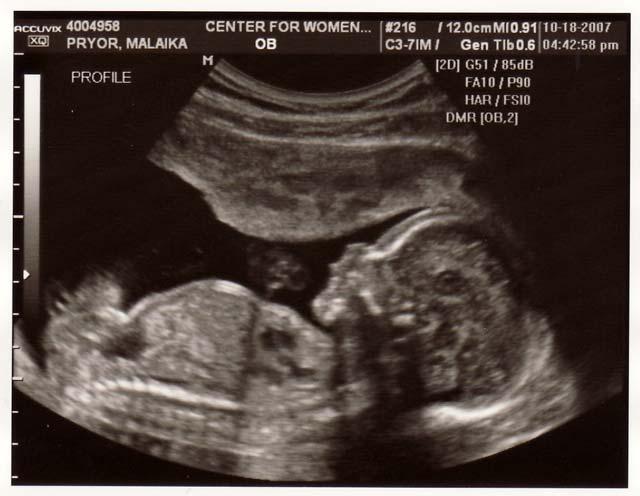
6 Organs Viewed Ultrasound is a useful way of examining many of the body's internal organs, including but not limited to the: heart and blood vessels, including the abdominal aorta and its major branches liver gallbladder spleen pancreas kidneys bladder uterus, ovaries, and unborn child (fetus) in pregnant patients eyes thyroid and parathyroid glands scrotum (testicles)
7 Other Uses Ultrasound is also used to: guide procedures such as needle biopsies, in which needles are used to extract sample cells from an abnormal area for laboratory testing. image the breasts and to guide biopsy of breast cancer. diagnose a variety of heart conditions and to assess damage after a heart attack or diagnose for valvular heart disease. Doppler ultrasound images can help the physician to see and evaluate: blockages to blood flow (such as clots). narrowing of vessels (which may be caused by plaque). tumors and congenital malformation.
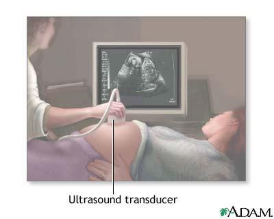
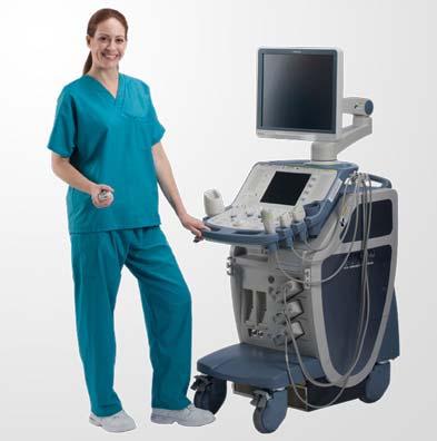
8 Equipment Ultrasound scanners consist of a console containing a computer and electronics, a video display screen and a transducer that is used to scan the body and blood vessels. The transducer is a small hand-held device that resembles a microphone, attached to the scanner by a cord. The transducer sends out high frequency sound waves into the body and then listens for the returning echoes from the tissues in the body. The principles are similar to sonar used by boats and submarines.
9 The ultrasound image is immediately visible on a nearby screen that looks much like a computer or television monitor. The image is created based on: the amplitude (strength), frequency and time it takes for the sound signal to return from the patient to the transducer.
10 Benefits Most ultrasound scanning is noninvasive (no needles or injections) and is usually painless. Ultrasound is widely available, easy-to-use and less expensive than other imaging methods. Ultrasound imaging uses no ionizing radiation. Ultrasound scanning gives a clear picture of soft tissues that do not show up well on x-ray images. Ultrasound causes no health problems and may be repeated as often as is necessary. Ultrasound is the preferred imaging modality for the diagnosis and monitoring of pregnant women and their unborn babies. Ultrasound provides real-time imaging, making it a good tool for guiding minimally invasive procedures such as needle biopsies and needle aspiration.
11 Risks For standard diagnostic ultrasound there are no known harmful effects on humans.
12 Limitations Ultrasound waves are disrupted by air or gas; therefore ultrasound is not an ideal imaging technique for the bowel or organs obscured by the bowel. Large patients are more difficult to image because tissue attenuates (weakens) the sound waves as they pass deeper into the body. Ultrasound has difficulty penetrating bone and therefore can only see the outer surface of bony structures and not what lies within.
13
14
15 How it works video eature=related &feature=related
16
17 Resources Regular Ultrasound btnq 4-D scan uijm
Abdominal Ultrasound
 Abdominal Ultrasound What is Ultrasound Imaging of the Abdomen? What are some common uses of the procedure? How should I prepare? What does the equipment look like? How does the procedure work? How is
General Ultrasound. What is General Ultrasound Imaging?
 Scan for mobile link. General Ultrasound What is General Ultrasound Imaging? Ultrasound is safe and painless, and produces pictures of the inside of the body using sound waves. Ultrasound imaging, also
General Ultrasound. What is General Ultrasound Imaging?
 Scan for mobile link. General Ultrasound Ultrasound imaging uses sound waves to produce pictures of the inside of the body. It is used to help diagnose the causes of pain, swelling and infection in the
An abdominal ultrasound produces a picture of the organs and other structures in the upper abdomen.
 Scan for mobile link. Ultrasound - Abdomen Ultrasound imaging of the abdomen uses sound waves to produce pictures of the structures within the upper abdomen. It is used to help diagnose pain or distention
Children's (Pediatric) Ultrasound - Abdomen
 Ultrasound - Abdomen") Scan for mobile link. Children's (Pediatric) Ultrasound - Abdomen Children s (pediatric) ultrasound imaging of the abdomen is a safe, noninvasive test that uses sound waves to produce a clear picture of
Scan for mobile link. Children's (Pediatric) Ultrasound - Abdomen Children s (pediatric) ultrasound imaging of the abdomen is a safe, noninvasive test that uses sound waves to produce a clear picture of
Ultrasound imaging is a noninvasive medical test that helps physicians diagnose and treat medical conditions.
 CAROTID ULTRASOUND What is Carotid Ultrasound Imaging? Ultrasound imaging, also called ultrasound scanning or sonography, involves exposing part of the body to highfrequency sound waves to produce pictures
Ultrasound - Pelvis. What is Pelvic Ultrasound Imaging?
 Scan for mobile link. Ultrasound - Pelvis Ultrasound imaging of the pelvis uses sound waves to produce pictures of the structures and organs in the lower abdomen and pelvis. There are three types of pelvic
General Ultrasound. What is General Ultrasound Imaging?
 Scan for mobile link. General Ultrasound Ultrasound imaging uses sound waves to produce pictures of the inside of the body. It is used to help diagnose the causes of pain, swelling and infection in the
Ultrasound - Musculoskeletal
 Ultrasound - Musculoskeletal What is Ultrasound Imaging of the Musculoskeletal System? Ultrasound imaging, also called ultrasound scanning or sonography, involves exposing part of the body to high-frequency
Ultrasound - Prostate
 Scan for mobile link. Ultrasound - Prostate Ultrasound of the prostate uses sound waves to produce pictures of a man s prostate gland and to help diagnose symptoms such as difficulty urinating or an elevated
Pelvic Ultrasound.
 Pelvic Ultrasound Before Your Exam: Drink 32 oz. of water one hour before your examination time. Try to drink all the liquid within 30 minutes. Do not urinate before the exam. Arrive for your exam with
Ureteral Stenting and Nephrostomy
 Scan for mobile link. Ureteral Stenting and Nephrostomy Ureteral stenting and nephrostomy help restore urine flow through blocked ureters and return the kidney to normal function. Ureters are long, narrow
relieve pressure on the lungs treat symptoms such as shortness of breath and pain determine the cause of excess fluid in the pleural space.
 Scan for mobile link. Thoracentesis Thoracentesis uses imaging guidance and a needle to help diagnose and treat pleural effusions, a condition in which the space between the lungs and the inside of the
FOR APPOINTMENT: MULTIMEDIA HEALTH EDUCATION
 P R E S E N T S MULTIMEDIA HEALTH EDUCATION FOR APPOINTMENT: Tel: 215-997-1660 Email: info@colmarimaging.com Location: 182 BETHLEHEM PIKE COLMAR, PA 18915 MULTIMEDIA HEALTH EDUCATION MANUAL TABLE OF CONTENTS
Children's (Pediatric) Contrast-enhanced Voiding Urosonography
 Contrast-enhanced Voiding Urosonography") Scan for mobile link. Children's (Pediatric) Contrast-enhanced Voiding Urosonography Pediatric contrast-enhanced voiding urosonography uses ultrasound to examine a child's bladder and urinary tract. It
Scan for mobile link. Children's (Pediatric) Contrast-enhanced Voiding Urosonography Pediatric contrast-enhanced voiding urosonography uses ultrasound to examine a child's bladder and urinary tract. It
relieve pressure on the lungs treat symptoms such as shortness of breath and pain determine the cause of excess fluid in the pleural space.
 Scan for mobile link. Chest Interventions What are Chest Interventions? Chest interventions are minimally invasive procedures used to diagnose and treat pleural effusions, a condition in which there is
Vascular Testing. and. You
 Vascular Testing and You V ASCULAR ULTRASOUND is an important diagnostic tool used in the diagnosis and detection of blood vessel problems. Ultrasound is also used to detect heart problems. Your vascular
DIGITAL IMAGE PROCESSING IN ULTRASOUND IMAGES
 DIGITAL IMAGE PROCESSING IN ULTRASOUND IMAGES Kamaljeet Kaur Computer Science & Engineering Department Guru Nanak Dev Engg. College, Ludhiana. Punjab-India meetk.89@gmail.com ABSTRACT-- Image processing
Intravascular Ultrasound
 Scan for mobile link. Intravascular Ultrasound Intravascular ultrasound (IVUS) uses a transducer or probe to generate sound waves and produce pictures of the coronary arteries. IVUS can show the entire
Medical Sonography Program Information PowerPoint
 Medical Sonography Program 2019 Information PowerPoint Sonographers are skilled health care professional who uses high frequency sound waves to produce dynamic visual images of organs, tissues, or blood
Ultrasound Table of contents
 Ultrasound Table of contents Ultrasound-Abdomen (Gallbladder, Liver, Pancreas, Spleen) Ultrasound-Amniocentesis Ultrasound-Aorta Ultrasound-Biophysical Profile Ultrasound-Breast Ultrasound-Breast Needle
Biopsies - Overview. What are biopsies? What are some common uses of the procedure?
 Scan for mobile link. Biopsies - Overview A biopsy is the removal of tissue from any part of the body to examine it for disease. Some may remove a small tissue sample with a needle while others may surgically
Arteriogram An X-ray of an artery after the injection of dye.
 A Abscess A localized collection of pus in any part of the body, usually surrounded by inflamed tissue. Anesthetic An agent that causes loss of sensation with or without the loss of consciousness. Angiography,
Contrast Materials Patient Safety: What are contrast materials and how do they work?
 Contrast Materials Patient Safety: What are contrast materials and how do they work? Which imaging exams use contrast materials? How safe are contrast materials? How should I prepare for my imaging procedure
Job Task Analysis for ARDMS Abdomen Data Collected: June 30, 2011
 Job Task Analysis for ARDMS Abdomen Data Collected: June 30, 2011 Reported: Analysis Summary for: Abdomen Examination Survey Dates 06/13/2011-06/26/2011 Invited Respondents 6,000 Surveys with Demographics
Patient Education. Ultrasound
 Patient Education Ultrasound What you should know about your Abdominal Ultrasound. An abdominal ultrasound is performed to examine your abdominal organs such as liver, spleen, gall bladder or pancreas.
Duplex Ultrasound. A Detailed Look at Your Blood Vessels
 Duplex Ultrasound A Detailed Look at Your Blood Vessels What Is Duplex Ultrasound? Ultrasound is a test that uses sound waves to create detailed pictures of the inside of your body. Duplex ultrasound is
Imaging Patient Education. Magnetic Resonance Imaging (MRI)
") Magnetic Resonance Imaging (MRI) What you should know about your Body MRI exam: Purpose: Magnetic Resonance Imaging (MRI) uses radio waves and a strong magnetic field to provide clear and detailed images
Magnetic Resonance Imaging (MRI) What you should know about your Body MRI exam: Purpose: Magnetic Resonance Imaging (MRI) uses radio waves and a strong magnetic field to provide clear and detailed images
Abdomen Sonography Examination Content Outline
 Abdomen Sonography Examination Content Outline (Outline Summary) # Domain Subdomain Percentage 1 2 3 Anatomy, Perfusion, and Function Pathology, Vascular Abnormalities, Trauma, and Postoperative Anatomy
Lower Extremity Arterial Disease
 Lower Extremity Arterial Disease Circulating the Facts About Peripheral Disease Brought to you by the Education Committee of the Society for 1 www.svnnet.org Peripheral Artery Disease (PAD) Many people
Sonography. 1. Introduction. 2. Documentation of Compliance. 3. Didactic Competency Requirements. 4. Clinical Competency Requirements
 PRIMARY CERTIFICATION Sonography 1. Introduction Candidates for certification and registration are required to meet the Professional Education Requirements specified in the ARRT Rules and Regulations.
Computed Tomography (CT) - Body
 - Body") Scan for mobile link. Computed Tomography (CT) - Body Computed tomography (CT) of the body uses special x-ray equipment to help detect a variety of diseases and conditions. CT scanning is fast, painless,
Scan for mobile link. Computed Tomography (CT) - Body Computed tomography (CT) of the body uses special x-ray equipment to help detect a variety of diseases and conditions. CT scanning is fast, painless,
Elastography in the. technically difficult patient. EPIQ ultrasound system. Ultrasound
 Ultrasound Elastography in the technically difficult patient EPIQ ultrasound system Chairman Department of Diagnostic Radiology Allegheny General Hospital Pittsburgh, PA, USA You can offer more information
Computed Tomography (CT) - Body
 - Body") Computed Tomography (CT) - Body What is CT Scanning of the Body? CT scanning sometimes called CAT scanning is a noninvasive medical test that helps physicians diagnose and treat medical conditions. CT
Computed Tomography (CT) - Body What is CT Scanning of the Body? CT scanning sometimes called CAT scanning is a noninvasive medical test that helps physicians diagnose and treat medical conditions. CT
Computed Tomographic Angiography (CTA)
") Computed Tomographic Angiography (CTA) A Detailed Look at Your Blood Vessels What Is CTA? CTA (computed tomographic angiography) is a test that creates detailed pictures of your blood vessels. During a
Computed Tomographic Angiography (CTA) A Detailed Look at Your Blood Vessels What Is CTA? CTA (computed tomographic angiography) is a test that creates detailed pictures of your blood vessels. During a
What is an ultrasound scan?
 Freephone helpline 0808 808 5555 information@lymphoma-action.org.uk www.lymphoma-action.org.uk Ultrasound scan An ultrasound scan uses sound waves to make pictures of the inside of your body. It can help
Cryotherapy. What is Cryotherapy? What are some common uses of the procedure?
 Scan for mobile link. Cryotherapy Cryotherapy uses imaging guidance, a needle-like applicator called a cryoprobe, and liquid nitrogen or argon gas to create intense cold to freeze and destroy diseased
COMENIUS-Project: SM&CLIL Radiation & Medicine
 Medical imaging refers to the techniques and processes used to create images of the human body (or parts thereof) for clinical purposes. Thanks to modern mathematics and computer technology, medical imaging
Computed Tomography (CT) - Abdomen and Pelvis
 - Abdomen and Pelvis") Scan for mobile link. Computed Tomography (CT) - Abdomen and Pelvis Computed tomography (CT) of the abdomen and pelvis is a diagnostic imaging test used to help detect diseases of the small bowel, colon
Scan for mobile link. Computed Tomography (CT) - Abdomen and Pelvis Computed tomography (CT) of the abdomen and pelvis is a diagnostic imaging test used to help detect diseases of the small bowel, colon
Having an Ultrasound Scan
 Having an Ultrasound Scan Information for Patients In this leaflet: Introduction 2 What is an Ultrasound scan?....2 How does it work?... 2 Are there any risks?.2 What do I need to do before my scan?.....3
Abdominal Vascular Arterial Disease
 Abdominal Vascular Arterial Disease Abdominal Vascular Arterial Disease People take better care of their health when they know what s going on in their bodies. For those with abdominal vascular arterial
Breast and Ovarian Cancer
 Patient Education Breast and Ovarian Cancer Screening and detection The goal of screening for cancer is to find it as early as possible, when it is easiest to cure. This handout describes the symptoms
RADIOLOGY (MEDICAL IMAGING)
") RADIOLOGY (MEDICAL IMAGING) Radiology is the study of the diagnosis of disease by the use of radiant energy (radiation). In the past this meant the use of X-rays to make an image. Today many other forms
RADIOLOGY (MEDICAL IMAGING) Radiology is the study of the diagnosis of disease by the use of radiant energy (radiation). In the past this meant the use of X-rays to make an image. Today many other forms
Cervical cancer is a disease in which malignant (cancer) cells form in the tissues of the cervix.
 cells form in the tissues of the cervix.") Cervical Cancer Cervical cancer is a disease in which malignant (cancer) cells form in the tissues of the cervix. The cervix is the lower, narrow end of the uterus (the hollow, pear-shaped organ where
Cervical Cancer Cervical cancer is a disease in which malignant (cancer) cells form in the tissues of the cervix. The cervix is the lower, narrow end of the uterus (the hollow, pear-shaped organ where
General Information Key Points
 The content of this booklet was adapted from content originally published by the National Cancer Institute. Male Breast Cancer Treatment (PDQ ) Patient Version. Updated September 29,2017. https://www.cancer.gov/types/breast/patient/male-breast-treatment-pdq
Ultrasound. Information for patients and families
 Ultrasound Information for patients and families Read this booklet to learn: what an ultrasound is the different types and how to prepare what to bring to your ultrasound appointment what to expect where
Lymphoscintigraphy is a special type of nuclear medicine imaging that provides pictures called scintigrams of the lymphatic system.
 Scan for mobile link. Lymphoscintigraphy Lymphoscintigraphy helps evaluate your body s lymphatic system for disease using small amounts of radioactive materials called radiotracers that are typically injected
What Is an Endoscopic Ultrasound (EUS)?
?") ENDOSCOPIC ULTRASOUND (EUS) What Is an Endoscopic Ultrasound (EUS)? An endoscopic ultrasound (EUS) is a specialized procedure that blends: Endoscopy use of a scope to look at the inside lining of the gastrointestinal
ENDOSCOPIC ULTRASOUND (EUS) What Is an Endoscopic Ultrasound (EUS)? An endoscopic ultrasound (EUS) is a specialized procedure that blends: Endoscopy use of a scope to look at the inside lining of the gastrointestinal
Intravenous Pyelogram (IVP)
") Scan for mobile link. Intravenous Pyelogram (IVP) Intravenous pyelogram (IVP) is an x-ray exam that uses an injection of contrast material to evaluate your kidneys, ureters and bladder and help diagnose
Scan for mobile link. Intravenous Pyelogram (IVP) Intravenous pyelogram (IVP) is an x-ray exam that uses an injection of contrast material to evaluate your kidneys, ureters and bladder and help diagnose
Diagnostic Ultrasound. Sutiporn Khampunnip, M.D.
 Diagnostic Ultrasound Sutiporn Khampunnip, M.D. Definition of Ultrasound Ultrasound is simply sound waves, like audible sound. High-frequency sound and refers to mechanical vibrations above 20 khz. Human
Cancer , The Patient Education Institute, Inc. ocf80101 Last reviewed: 06/08/2016 1
 Cancer Introduction Cancer begins in your cells, which are the building blocks of your body. Extra cells can form a mass called a tumor. Some tumors aren t cancerous, while other ones are. Cells from cancerous
Breast Cancer Screening
 Scan for mobile link. Breast Cancer Screening What is breast cancer screening? Screening examinations are tests performed to find disease before symptoms begin. The goal of screening is to detect disease
Abdominal Exam: The examination of the abdomen used by physicians to detect an abdominal aortic aneurysm.
 Glossary of Terms Abdominal Exam: The examination of the abdomen used by physicians to detect an abdominal aortic aneurysm. Angiogram: A diagnostic test requiring the insertion of a catheter into an artery
RADIOLOGIC TECHNOLOGY (526)
") RADIOLOGIC TECHNOLOGY (526) 526-133 DMS General Procedures 2 Radiologic Technology (526) 1 526-130 Introduction to Diagnostic Medical Sonography This course introduces the student to the history of ultrasound
RADIOLOGIC TECHNOLOGY (526) 526-133 DMS General Procedures 2 Radiologic Technology (526) 1 526-130 Introduction to Diagnostic Medical Sonography This course introduces the student to the history of ultrasound
Radiofrequency Ablation (RFA) / Microwave Ablation (MWA) of Liver Tumors
 / Microwave Ablation (MWA) of Liver Tumors") Scan for mobile link. Radiofrequency Ablation (RFA) / Microwave Ablation (MWA) of Liver Tumors Radiofrequency ablation (RFA) and microwave ablation (MWA) are treatments that use image guidance to place
Scan for mobile link. Radiofrequency Ablation (RFA) / Microwave Ablation (MWA) of Liver Tumors Radiofrequency ablation (RFA) and microwave ablation (MWA) are treatments that use image guidance to place
Magnetic Resonance Imaging (MRI) - Body
 - Body") Scan for mobile link. Magnetic Resonance Imaging (MRI) - Body What is MRI of the Body? Magnetic resonance imaging (MRI) is a noninvasive medical test that physicians use to diagnose and treat medical conditions.
Scan for mobile link. Magnetic Resonance Imaging (MRI) - Body What is MRI of the Body? Magnetic resonance imaging (MRI) is a noninvasive medical test that physicians use to diagnose and treat medical conditions.
Terminology Tissue Appearance
 By Marc Nielsen, MD Advantages/Disadvantages Generation of Image Ultrasound Machine/Transducer selection Modes of Ultrasound Terminology Tissue Appearance Scanning Technique Real-time Portable No ionizing
Pancreatic Cancer. What is pancreatic cancer?
 Scan for mobile link. Pancreatic Cancer Pancreatic cancer is a tumor of the pancreas, an organ that is located behind the stomach in the abdomen. Pancreatic cancer does not always cause symptoms until
Improving Methods for Breast Cancer Detection and Diagnosis. The National Cancer Institute (NCI) is funding numerous research projects to improve
 is funding numerous research projects to improve") CANCER FACTS N a t i o n a l C a n c e r I n s t i t u t e N a t i o n a l I n s t i t u t e s o f H e a l t h D e p a r t m e n t o f H e a l t h a n d H u m a n S e r v i c e s Improving Methods for
CANCER FACTS N a t i o n a l C a n c e r I n s t i t u t e N a t i o n a l I n s t i t u t e s o f H e a l t h D e p a r t m e n t o f H e a l t h a n d H u m a n S e r v i c e s Improving Methods for
Cholangiocarcinoma (Bile Duct Cancer)
") Cholangiocarcinoma (Bile Duct Cancer) The Bile Duct System (Biliary Tract) A network of bile ducts (tubes) connects the liver and the gallbladder to the small intestine. This network begins in the liver
Cholangiocarcinoma (Bile Duct Cancer) The Bile Duct System (Biliary Tract) A network of bile ducts (tubes) connects the liver and the gallbladder to the small intestine. This network begins in the liver
Q1. The diagram shows an ultrasound monitor being used to scan a fetus.
 Q1. The diagram shows an ultrasound monitor being used to scan a fetus. The table shows the velocity of ultrasound waves in different tissues of the fetus. Tissue Amniotic fluid (liquid surrounding fetus)
Interventional Radiology (IR)
") Interventional Radiology (IR) Risk information for inpatients UHN Read this brochure to learn about: Interventional Radiology (IR) procedures The risks of IR procedures Problems to what to watch for Please
Interventional Radiology (IR) Risk information for inpatients UHN Read this brochure to learn about: Interventional Radiology (IR) procedures The risks of IR procedures Problems to what to watch for Please
What is Thyroid Cancer?
 Thyroid Cancer What is Thyroid Cancer? The thyroid is a gland at the base of the throat near the trachea (windpipe). It is shaped like a butterfly, with a right lobe and a left lobe. The isthmus, a thin
Chapter Overview. Chapter 1. Anatomy. Physiology
 Chapter Overview Chapter 1 An Introduction to the Human Body Define Anatomy and Physiology Levels of Organization Characteristics of Living Things Homeostasis Anatomical Terminology 1 2 Anatomy Describes
PERCUTANEOUS CRYOABLATION
 PERCUTANEOUS CRYOABLATION IS A MINIMALLY INVASIVE, IMAGE-GUIDED TREATMENT THAT DESTROYS (ABLATES) TUMORS AND OTHER TARGETED TISSUE WITH EXTREME COLD WHILE SPARING SURROUNDING HEALTHY TISSUE. What are some
Children's (Pediatric) Nuclear Medicine
 Nuclear Medicine") Scan for mobile link. Children's (Pediatric) Nuclear Medicine Children s (pediatric) nuclear medicine imaging uses small amounts of radioactive materials called radiotracers, a special camera and a computer
Scan for mobile link. Children's (Pediatric) Nuclear Medicine Children s (pediatric) nuclear medicine imaging uses small amounts of radioactive materials called radiotracers, a special camera and a computer
Ultrasound in Medicine
 Ultrasound in Medicine Experimental Equipment for Medical Education Universities Colleges Medical Schools Medical and Med-Technical Training Education can befun! WELCOME TO GAMPT Devices and accessories
Physical Principles of Ultrasound
 Physical Principles of Ultrasound Grateful appreciation to Richard A. Lopchinsky, MD, FACS and Nancy H. Van Name, RDMS, RTR, and MarleneKattaron, RDMS 2000 UIC All Rights Reserved. Course Objectives Identify
Nuclear Medicine Imaging of Liver, Spleen and Gallbladder
 Nuclear Medicine Imaging of Liver, Spleen and Gallbladder An Introductory Guide For Patients And Their Families You may be wondering why your doctor ordered a nuclear medicine scan of your liver and spleen
Magnetic Resonance Imaging (MRI) - Body
 - Body") Scan for mobile link. Magnetic Resonance Imaging (MRI) - Body Magnetic resonance imaging (MRI) of the body uses a powerful magnetic field, radio waves and a computer to produce detailed pictures of the
Scan for mobile link. Magnetic Resonance Imaging (MRI) - Body Magnetic resonance imaging (MRI) of the body uses a powerful magnetic field, radio waves and a computer to produce detailed pictures of the
Computed Tomography (CT) - Sinuses
 - Sinuses") Scan for mobile link. Computed Tomography (CT) - Sinuses Computed tomography (CT) of the sinuses uses special x-ray equipment to evaluate the paranasal sinus cavities hollow, air-filled spaces within the
Scan for mobile link. Computed Tomography (CT) - Sinuses Computed tomography (CT) of the sinuses uses special x-ray equipment to evaluate the paranasal sinus cavities hollow, air-filled spaces within the
created by high-voltage devices Examples include medical and dental x-rays, light, microwaves and nuclear energy
 What is radiation? Radiation is energy emitted from a source, that travels through space and can penetrate matter. Listed below are two types that we are exposed to and contribute to our overall radiation
General Nuclear Medicine
 General Nuclear Medicine What is General Nuclear Medicine? What are some common uses of the procedure? How should I prepare? What does the equipment look like? How does the procedure work? How is the procedure
What Is Nuclear Medicine?
 What Is Nuclear Medicine? An Introductory Guide For Patients And Their Families What is nuclear medicine? Nuclear medicine is a type of medical imaging that uses small amounts of radioactive material (called
Vaginal cancer: Know what to expect
 Vaginal cancer: Know what to expect For women with vaginal cancer What is the vagina? The vagina is a hollow canal that connects the cervix and the uterus to the outside. of the body. When a woman gives
X-ray (Radiography) - Bone
 - Bone") Scan for mobile link. X-ray (Radiography) - Bone Bone x-ray uses a very small dose of ionizing radiation to produce pictures of any bone in the body. It is commonly used to diagnose fractured bones or
Scan for mobile link. X-ray (Radiography) - Bone Bone x-ray uses a very small dose of ionizing radiation to produce pictures of any bone in the body. It is commonly used to diagnose fractured bones or
Introduction to Biomedical Imaging
 Alejandro Frangi, PhD Computational Imaging Lab Department of Information & Communication Technology Pompeu Fabra University www.cilab.upf.edu Basic principles. Comparison to X-rays Ultrasound > 20kHz
Principles of Ultrasound. Cara C. Prideaux, M.D. University of Utah PM&R Sports Medicine Fellow March 14, 2012
 Principles of Ultrasound Cara C. Prideaux, M.D. University of Utah PM&R Sports Medicine Fellow March 14, 2012 None Disclosures Outline Introduction Benefits and Limitations of US Ultrasound (US) Physics
Introduction to Health Care & Careers. Chapter 22. Answers to Checkpoint and Review Questions
 Introduction to Health Care & Careers Chapter 22 Answers to Checkpoint and Review Questions Checkpoints 1. What education and training are needed to become a cardiographic technician? Cardiographic technicians
Ultrasonography of the Neck as an Adjunct to FNA. Nicole Massoll M.D.
 Ultrasonography of the Neck as an Adjunct to FNA Nicole Massoll M.D. Basic Features of Head and Neck Ultrasound and Anatomy Nicole Massoll M.D. University of Arkansas for Medical Sciences, Little Rock
Computed Tomography (CT) - Chest
 - Chest") Scan for mobile link. Computed Tomography (CT) - Chest What is CT Scanning of the Chest? Computed tomography, more commonly known as a CT or CAT scan, is a diagnostic medical test that, like traditional
Scan for mobile link. Computed Tomography (CT) - Chest What is CT Scanning of the Chest? Computed tomography, more commonly known as a CT or CAT scan, is a diagnostic medical test that, like traditional
Computed Tomography (CT) - Spine
 - Spine") Scan for mobile link. Computed Tomography (CT) - Spine Computed tomography (CT) of the spine is a diagnostic imaging test used to help diagnose or rule out spinal column damage in injured patients. CT
Scan for mobile link. Computed Tomography (CT) - Spine Computed tomography (CT) of the spine is a diagnostic imaging test used to help diagnose or rule out spinal column damage in injured patients. CT
SAY HELLO TO GOOD HEALTH.
 SAY HELLO TO GOOD HEALTH. WHY HEALTH CHECK-UP Good health is the foundation of a happy, productive and rewarding life. Most health problems can be managed more effectively if detected early. The modern
Deep Vein Thrombosis
 Deep Vein Thrombosis Introduction Deep vein thrombosis (DVT) is a blood clot in a vein. This condition can affect men and women of any age and race. DVT is a potentially serious condition. If not treated,
Ovarian Cancer Includes Epithelial, Fallopian Tube, Primary Peritoneal Cancer, and Ovarian Germ Cell Tumors
 Ovarian Cancer Includes Epithelial, Fallopian Tube, Primary Peritoneal Cancer, and Ovarian Germ Cell Tumors Overview Ovarian epithelial cancer, fallopian tube cancer, and primary peritoneal cancer are
Catheter-directed Thrombolysis
 Scan for mobile link. Catheter-directed Thrombolysis Catheter-directed thrombolysis treats vascular blockages and improves blood flow by dissolving abnormal blood clots. A blood clot, or thrombus, can
Seeing the Unseen Clinical advances and future directions of SMI
 Seeing the Unseen Clinical advances and future directions of SMI Jiro Hata, M.D., Ph. D. Professor Department of Endoscopy and Ultrasound Kawasaki Medical School Okayama, Japan Introduction Superb Micro-vascular
EUROSON SCHOOL 2019 January 18-19, 2019, Athens-Greece Preliminary Programme
 EUROSON SCHOOL 2019 January 18-19, 2019, Athens-Greece Preliminary Programme 08:00-09:00 Registration Friday, January 18 Theoretical Course / PHYSICS AND TECHNOLOGY 09:00-09:15 Basics in US Physics 09:15-09:30
Magnetic Resonance Imaging (MRI) - Body
 - Body") Magnetic Resonance Imaging (MRI) - Body What is MRI of the Body? Magnetic resonance imaging (MRI) is a noninvasive medical test that helps physicians diagnose and treat medical conditions. MR imaging uses
Magnetic Resonance Imaging (MRI) - Body What is MRI of the Body? Magnetic resonance imaging (MRI) is a noninvasive medical test that helps physicians diagnose and treat medical conditions. MR imaging uses
Computed Tomography (CT) - Chest
 - Chest") Scan for mobile link. Computed Tomography (CT) - Chest Computed tomography (CT) of the chest uses special x-ray equipment to examine abnormalities found in other imaging tests and to help diagnose the
Scan for mobile link. Computed Tomography (CT) - Chest Computed tomography (CT) of the chest uses special x-ray equipment to examine abnormalities found in other imaging tests and to help diagnose the
Course specification
 Al-Azhar University Faculty of Medicine for Men Course specification For Master of Radiodiagnosis ( 2014 2015 ) University : Al-Azhar Faculty : Medicine for men Course specification - Programmers on which
Cerebrovascular. Disease
 Cerebrovascular Disease People take better care of their health when they know what s going on in their bodies. For those with cerebrovascular disease, this means understanding how the arteries work and
From Head to Toe Use of Advanced Dynamic Flow in prenatal ultrasound
 From Head to Toe Use of Advanced Dynamic Flow in prenatal ultrasound Without doubt, the B- Schwerdtfeger, R. tant diagnostic instrument. Furthermore, we use colour in feto- mode imaging is the most important
Breast Imaging & You
 Breast Imaging & You What s Inside: Breast Imaging... 2 Digital Breast Tomosynthesis (DBT) mammograms... 4 Breast cancer screening... 6 Dense breast tissue... 8 Automated breast ultrasound (ABUS)... 9
Upper Gastrointestinal (GI) Tract X-ray (Radiography)
 Tract X-ray (Radiography)") Upper Gastrointestinal (GI) Tract X-ray (Radiography) What is Upper Gastrointestinal (GI) Tract Radiography? What are some common uses of the procedure? How should I prepare? What does the equipment look
Upper Gastrointestinal (GI) Tract X-ray (Radiography) What is Upper Gastrointestinal (GI) Tract Radiography? What are some common uses of the procedure? How should I prepare? What does the equipment look
1 Fundamentals. Basic Definitions and Physics Principles. Fundamentals
 1 To become versed in the language of ultrasonography, it is necessary to review some of the basic principles of physics. The wave physics principles of ordinary (i.e., audible) sound apply to ultrasound
 P R E S E N T S Dr. Mufa T. Ghadiali is skilled in all aspects of General Surgery. His General Surgery Services include: General Surgery Advanced Laparoscopic Surgery Surgical Oncology Gastrointestinal
Lymphoma is a cancer that develops in the white blood cells (lymphocytes) of the lymphatic system, which is part of the body's immune system.
 of the lymphatic system, which is part of the body's immune system.") Scan for mobile link. Lymphoma Lymphoma is a cancer that develops in the white blood cells of the lymphatic system. Symptoms may include enlarged lymph nodes, unexplained weight loss, fatigue, night sweats
Scan for mobile link. Lymphoma Lymphoma is a cancer that develops in the white blood cells of the lymphatic system. Symptoms may include enlarged lymph nodes, unexplained weight loss, fatigue, night sweats
What is Testicular cancer?
 Testicular Cancer What is Testicular cancer? Testicular cancer is a disease in which cancer cells form in the tissues of one or both testicles. The testicles are 2 egg-shaped glands located inside the