Terminology Tissue Appearance
|
|
|
- Clementine Daniels
- 6 years ago
- Views:
Transcription
1 By Marc Nielsen, MD
2 Advantages/Disadvantages Generation of Image Ultrasound Machine/Transducer selection Modes of Ultrasound Terminology Tissue Appearance Scanning Technique

3 Real-time Portable No ionizing radiation Inexpensive? Side-side comparison Patients love it! Guide procedures
4 Operator dependent Expensive? Limited penetration through bone and air Imaging deep structures

5 Electric field applied to piezoelectric crystals located on transducer surface Mechanical vibration of crystals creates sound waves Each crystal produces an US wave Summation of all waves forms the US beam Wave reflects as echo that vibrates transducer Vibrations produce electrical pulses Scanner processes and transforms to image
6 Frequency 1-18 megahertz (mhz) Lower frequencies = less resolution but deeper penetration E.G. kidney, liver 1-6 mhz Higher frequencies = smaller wavelength capable of reflecting from smaller structures Readily absorbed by tissue = less penetration Higher frequency = higher resolution E.G. Muscles, tendons 7-18 mhz
7 Pulser - applies high amplitude voltage to energize crystals Transducer Converts electrical energy to mechanical (ultrasound) energy and vice versa Two types of transducers Linear - sound wave is propagated in a linear fashion parallel to the transducer surface Ideal for MSK US Curvilinear increases field of view. Ideal for visualization of deeper structures Receiver detects and amplifies signals

8 Which probe to pick? Surface area of skin/transducer Frequency of emitted sound wave
9 Which probe to pick? Surface area of skin/transducer Frequency of emitted sound wave
10 Large foot print Low Frequency = Increased Depth Abdominal US
 Low frequency Echo or abdominal")
11 Smaller foot print (fits between the ribs) Low frequency Echo or abdominal US

12 Flat foot print High frequency Maximum depth cm Musculoskeletal or vessel US
13
14 B-mode (2D mode) linear array of transducers simultaneously scan a plane through the body Two-dimensional cross-section of tissue Doppler mode Measuring and visualizing blood flow Duplex simultaneous presentation of 2D and doppler information
15 Acoustical impedance Sound wave encounters material with different density, and wave is reflected back as an echo Gas or solids Most of the acoustic energy is reflected - impossible to see deeper
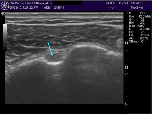
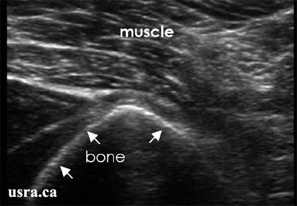
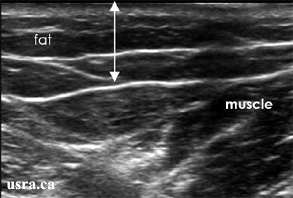
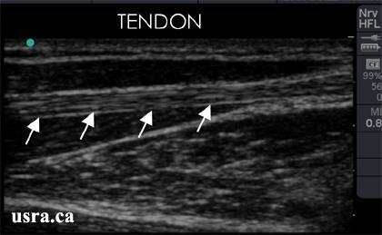
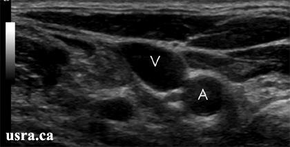
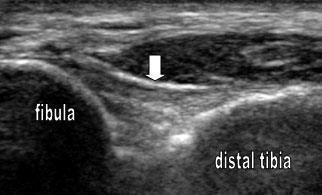
16 Reflection some of sound energy strikes a boundary between media and is returned to the transducer > degree of impedance mismatch at tissue interface = >amount of reflection Refraction - change in direction of wave propagation when traveling from one medium to another
17 Angle of incidence US wave hitting a smooth interface at 90 angle will result in a perpendicular reflection Strong, bright signal US wave hitting the surface at an angle < 90 will result in the wave being deflected away from the transducer an an angle equal to the angle of incidence but in the opposite direction (angle of reflection) Weak, darker image
18
19 Echogenicity a method of describing the reflecting echos Hypoechoic Darker (less reflection) Hyperechoic Brighter (more reflection) Anechoic Black (No echos) Isoechoic Equal
20 Attenuation: A decrease in intensity, power and amplitude as a sound wave travels Gain: Adjusting the intensity of the acoustic pulse, with the result being a stronger echo
21 Near field Top of screen Far field Bottom of screen
22 Anisotropy Tissue NOT imaged perpendicular to the sound beam Appears artifactually hypoechoic Can be confused with pathology
23
24 TISSUE US IMAGE Veins Arteries Fat Muscle Tendon Ligaments Bone Nerves Anechoic, compressible Anechoic, pulsatile Hyopechoic with irregular hyperechoic lines Hypoechoic, but separated with hyperechoic septa Hyerechoic & fiber-like Like tendons, but more compact Hyperechoic lines with a hypoechoic shadow Starry night appearance: transverse
25
26
27
28 Select transducer Hold transducer between thumb and fingers of dominant hand. Stabilize transducer on the patient with the small finger of the heel of the imaging hand Apply gel to the transducer Eliminates air between the probe and skin surface Air is the enemy Palpate area of interest and identify what you are looking for
29 Locate area of interest with ultrasound Adjust the depth of the sound beam Structure of interest is visible and centered in the image Select desired optimization setting from onscreen menu Res = best resolution possible Gen = balance between resolution and penetration Pen = best penetration possible
30 Adjust gain Amplifies return echos (adjusts brightness of the image) May use autogain May adjust for near or far field
31 Image orientation Index Mark Orientates probe to your screen Keep on left side of the screen Proximal aspect of a structure on the left side of the image and the distal aspect on the right Describing orientation Two views Transverse Cross section Longitudinal Long axis
32 Heel-toe when transducer is rocked or angled along the long axis of the transducer Toggle transducer is angled from side to side
33 Use highest frequency that allows US to image the depth of the structures of clinical interest Image depth decreases as frequency of US increases Move focal zone on screen to level at area of interest Youtube Excellent source for Sonsosite product information and instruction on ultrasound
34 Four parts Introduction to US OB US MSK US FAST (Focused Assessment with Sonography for Trauma) Will include venous US gfresidency.com
35 Arntfield, Robert, EML Ultrasound Rounds Dixon, J. Bryan Basics of Musculoskeletal Ultrasound Hecht, Suzanne Musculoskeletal Ultrasound Jacobson Fundamentals of Musculoskeletal Ultrasound Jacobson Primack, Scott J. Musculoskeletal Ultrasound SonoSite M-Turbo Product Training Part 3: How to Perform and Exam Wikipedia Medical Ultrasonography
Principles of Ultrasound. Cara C. Prideaux, M.D. University of Utah PM&R Sports Medicine Fellow March 14, 2012
 Principles of Ultrasound Cara C. Prideaux, M.D. University of Utah PM&R Sports Medicine Fellow March 14, 2012 None Disclosures Outline Introduction Benefits and Limitations of US Ultrasound (US) Physics
Basic Physics of Ultrasound and Knobology
 WELCOME TO UTMB Basic Physics of Ultrasound and Knobology By Daneshvari Solanki, FRCA Laura B. McDaniel Distinguished Professor Anesthesiology and Pain Medicine University of Texas Medical Branch Galveston,
3/20/2017. Disclosures. Ultrasound Fundamentals. Ultrasound Fundamentals. Bone Anatomy. Tissue Characteristics
 Disclosures Images of ultrasound equipment in this presentation are not an endorsement Fundamentals of Musculoskeletal Ultrasound Physics and Knobology Shane A. Shapiro, M.D. Assistant Professor Orthopedic
Preamble (disclaimer)
") Preamble (disclaimer) PHYSICS AND PRINCIPLES OF HEAD/NECK ULTRASOUND Joseph C. Sniezek, MD FACS LTC, MC, USA Otolaryngology/H&N Surgery Tripler Army Medical Center 1. I am not a physicist 2. ACS has recommended
Preamble (disclaimer) PHYSICS AND PRINCIPLES OF HEAD/NECK ULTRASOUND Joseph C. Sniezek, MD FACS LTC, MC, USA Otolaryngology/H&N Surgery Tripler Army Medical Center 1. I am not a physicist 2. ACS has recommended
The Essentials Tissue Characterization and Knobology
 The Essentials Tissue Characterization and Knobology Randy E. Moore, DC, RDMS RMSK No relevant financial relationships Ultrasound The New Standard of Care Musculoskeletal sonography has become the standard
The Physics of Ultrasound. The Physics of Ultrasound. Claus G. Roehrborn. Professor and Chairman. Ultrasound Physics
 The Physics of Ultrasound Pipe Organ 10-8000 Emission Dog 452-1080 Man 85-1100 Spectrum Bat 10,000-120,000 Porpoise 7000-120,000 Claus G. Roehrborn Professor and Chairman 10 20 Cycles per second Reception
Basic of Ultrasound Physics E FAST & Renal Examination. Dr Muhammad Umer Ihsan MBBS,MD, DCH CCPU,DDU1,FACEM
 Basic of Ultrasound Physics E FAST & Renal Examination Dr Muhammad Umer Ihsan MBBS,MD, DCH CCPU,DDU1,FACEM What is Sound? Sound is Mechanical pressure waves What is Ultrasound? Ultrasounds are sound waves
Diagnostic Ultrasound. Sutiporn Khampunnip, M.D.
 Diagnostic Ultrasound Sutiporn Khampunnip, M.D. Definition of Ultrasound Ultrasound is simply sound waves, like audible sound. High-frequency sound and refers to mechanical vibrations above 20 khz. Human
Ultrasound Knobology
 Ultrasound Knobology Raj Dasgupta MD, FACP, FCCP, FASSM Assistant Professor of Clinical Medicine Pulmonary / Critical Care / Sleep Medicine University of Southern California (USC) Objectives Physics of
1 Fundamentals. Basic Definitions and Physics Principles. Fundamentals
 1 To become versed in the language of ultrasonography, it is necessary to review some of the basic principles of physics. The wave physics principles of ordinary (i.e., audible) sound apply to ultrasound
Ultrasound Principles cycle Frequency Wavelength Period Velocity
 ! Teresa S. Wu, MD, FACEP Director, EM Ultrasound Program & Fellowship Co-Director, Simulation Based Training Program & Fellowship Associate Program Director, EM Residency Program Maricopa Medical Center
Ultrasound Physics & Terminology
 Ultrasound Physics & Terminology This module includes the following: Basic physics terms Basic principles of ultrasound Ultrasound terminology and terms Common artifacts seen Doppler principles Terms for
Introduction to Ultrasound Guided Region Anesthesia
 Introduction to Ultrasound Guided Region Anesthesia Brian D. Sites, MD Dept of Anesthesiology Dartmouth-Hitchcock Medical Center INTRODUCTION Welcome to Introduction to Ultrasound Guided Regional Anesthesia.
Ultrasound Physics and Knobology Alan Macfarlane. Consultant Anaesthetist Glasgow Royal Infirmary
 Ultrasound Physics and Knobology Alan Macfarlane Consultant Anaesthetist Glasgow Royal Infirmary RAPM 2009; 34: 40-46 Ultrasound Proficiency Understanding US image generation and device operation Image
WELCOME! Introduction to Bedside Ultrasound
 WELCOME! Introduction to Bedside Ultrasound TEACHERS University of California-Irvine School of Medicine Nathan Molina nathan.d.molina@gmail.com Trevor Plescia taplescia90@gmail.com Jack Silva jpsilva42@gmail.com
Introduction & Physics of ED Ultrasound. Objectives. What? - Limited Studies. Who? - ED Docs
 Introduction & Physics of ED Ultrasound Martine Sargent, MD Ultrasound Director, Assistant Professor UCSF Department of Emergency Medicine San Francisco General Hospital & Trauma Center Objectives Who?
Point-of-Care Ultrasound: An Introduction
 Point-of-Care Ultrasound: An Introduction Delegation Teaching Package for Registered Respiratory Therapists and Anesthesia Assistants Developed by: Rob Bryan RRT, AA Edited by: Kelly Hassall RRT, FCSRT,
Physical Principles of Ultrasound
 Physical Principles of Ultrasound Grateful appreciation to Richard A. Lopchinsky, MD, FACS and Nancy H. Van Name, RDMS, RTR, and MarleneKattaron, RDMS 2000 UIC All Rights Reserved. Course Objectives Identify
Ultrasound Physics & Doppler
 Ultrasound Physics & Doppler Endocrine University 2018 Mark Lupo, MD, FACE, ECNU Objectives Review the essential components of ultrasound physics in neck sonography Demonstrate the importance of ultrasound
Ultrasound. Principles of Medical Imaging. Contents. Prof. Dr. Philippe Cattin. MIAC, University of Basel. Oct 17th, 2016
 Ultrasound Principles of Medical Imaging Prof. Dr. Philippe Cattin MIAC, University of Basel Contents Abstract 1 Image Generation Echography A-Mode B-Mode M-Mode 2.5D Ultrasound 3D Ultrasound 4D Ultrasound
CONTENTS. Test Number cpd Tanya Reynolds (Nat. Dip. Diag. Rad., B. Tech. Diag. Rad., B. Tech. Ultrasound)
") CONTENTS page 1-15 page 16 BASIC 2-DIMENSIONAL ULTRASOUND PRINCIPLES Multiple Choice Test Test Number cpd 41640 Tanya Reynolds (Nat. Dip. Diag. Rad., B. Tech. Diag. Rad., B. Tech. Ultrasound) Tanya is
CONTENTS page 1-15 page 16 BASIC 2-DIMENSIONAL ULTRASOUND PRINCIPLES Multiple Choice Test Test Number cpd 41640 Tanya Reynolds (Nat. Dip. Diag. Rad., B. Tech. Diag. Rad., B. Tech. Ultrasound) Tanya is
Ultrasound in Anesthesia: Applying Scientific Principles to Clinical Practice
 AANA Journal Course Update for Nurse Anesthetists 3 6 CE Credits* Ultrasound in Anesthesia: Applying Scientific Principles to Clinical Practice Christian R. Falyar, CRNA, DNAP The use of ultrasound as
What is Ultrasound? What is Ultrasound? B A. Basic Principles of Ultrasound. Basic Principles of Ultrasound. Basic Principles of Ultrasound
 Introduction to Ultrasound Principles Mani Montazemi, RDMS Baylor College of Medicine Division of Maternal-Fetal Medicine Department of Obstetrics and Gynecology Manager, Maternal Fetal Center Imaging
INTRODUCTION. Getting the best scan. Choosing a probe. Choosing the frequency
 Getting the best scan Choosing a probe Select the most appropriate probe for the particular scan required. s vary in their: operating frequency range higher ultrasound frequencies provide better discrimination
High resolution ultrasound scanner for skin imaging
 High resolution ultrasound scanner for skin imaging Christine Turlat Sales Director Atys medical 17 Parc d Arbora 69510 SOUCIEU EN JARREST Atys company Principle of ultrasound imaging DERMCUP Normal image
Descriptions of NDT Projects Fall 2004 October 31, 2004
 Descriptions of NDT Projects Fall 2004 October 31, 2004 Introduction There are two separate NDT labs in Magister: ULTRA for ultrasound and EDDY for eddy current. Both labs are equipped with mechanical
Lesson 07: Ultrasound Transducers. This lesson contains 73 slides plus 16 multiple-choice questions.
 Lesson 07: Ultrasound Transducers This lesson contains 73 slides plus 16 multiple-choice questions. This lesson was derived from pages 33 through 42 in the textbook: Ultrasound Transducers Ultrasound Transducers
ULTRASOUND IMAGING EE 472 F2018. Prof. Yasser Mostafa Kadah
 ULTRASOUND IMAGING EE 472 F2018 Prof. Yasser Mostafa Kadah www.k-space.org Recommended Textbook Diagnostic Ultrasound: Physics and Equipment, 2nd ed., by Peter R. Hoskins (Editor), Kevin Martin (Editor),
What is Ultrasound? Resolution Image production Attenuation Imaging modes Ultrasound artifacts... 7
 What is Ultrasound?... 1 Resolution... 3 Image production... 3 Attenuation... 4 Imaging modes... 5 Ultrasound artifacts... 7 0 What is Ultrasound? High frequency sound of frequencies 2-50 MHz is used in
Table of contents. Foreword. Preface. 1 Introduction Historical Perspective 00
 Table of contents Foreword Preface 1 Introduction 00 1.1 Historical Perspective 00 2 Fundamentals of musculoskeletal ultrasound 00 2.1 Frequency and wavelength 00 2.2 Generating ultrasound waves 00 2.3
Sound in medicine. CH.12. Dr.Rajaa أ.م.د. رجاء سهيل جنم جامعة تكريت كلية طب االسنان. General Properties of Sound
 CH.12. Dr.Rajaa Sound in medicine أ.م.د. رجاء سهيل جنم جامعة تكريت كلية Sound : It is the audible waves of frequency between 20 Hz and 20 khz. Infrasound : refers to the sound of frequency below the normal
Emergency Medicine Interest Group (EMIG) 2016
 2016") Emergency Medicine Interest Group (EMIG) 2016 Welcome to the flipped classroom (learning objectives summary) for the 2016 Emergency Medicine Interest Group (EMIG) Procedures Workshop. Overview - Tuesday
Emergency Medicine Interest Group (EMIG) 2016 Welcome to the flipped classroom (learning objectives summary) for the 2016 Emergency Medicine Interest Group (EMIG) Procedures Workshop. Overview - Tuesday
Background & Indications Probe Selection
 Teresa S. Wu, MD, FACEP Director, EM Ultrasound Program & Fellowship Co-Director, Simulation Based Training Program & Fellowship Associate Program Director, EM Residency Program Maricopa Medical Center
Ultrasonography of the Neck as an Adjunct to FNA. Nicole Massoll M.D.
 Ultrasonography of the Neck as an Adjunct to FNA Nicole Massoll M.D. Basic Features of Head and Neck Ultrasound and Anatomy Nicole Massoll M.D. University of Arkansas for Medical Sciences, Little Rock
DIGITAL IMAGE PROCESSING IN ULTRASOUND IMAGES
 DIGITAL IMAGE PROCESSING IN ULTRASOUND IMAGES Kamaljeet Kaur Computer Science & Engineering Department Guru Nanak Dev Engg. College, Ludhiana. Punjab-India meetk.89@gmail.com ABSTRACT-- Image processing
Dr Emma Chung. Safety first - Physical principles for excellent imaging
 Safety first - Physical principles for excellent imaging Dr Emma Chung Lecturer in Medical Physics, University of Leicester Clinical Scientist, University Hospitals of Leicester NHS Trust Thanks to Caroline
Application of Phased Array Radar Theory to Ultrasonic Linear Array Medical Imaging System
 Application of Phased Array Radar Theory to Ultrasonic Linear Array Medical Imaging System R. K. Saha, S. Karmakar, S. Saha, M. Roy, S. Sarkar and S.K. Sen Microelectronics Division, Saha Institute of
Abdominal Ultrasound
 Abdominal Ultrasound Imaging Control Buttons Depth The organ imaged should take up 3/4 of the screen Frequency = Penetration Use high frequencies (harmonics) for fluid filled and superficial structures
4.17. RESEARCHING MODELS WITH AN ULTRASONIC ECHOSCOPE
 4.17. RESEARCHING MODELS WITH AN ULTRASONIC ECHOSCOPE Purpose of experiment Determine the main characteristics of ultrasound waves, and the distances and positions of models using an ultrasonic echoscope.
4.17. RESEARCHING MODELS WITH AN ULTRASONIC ECHOSCOPE
 4.17. RESEARCHING MODELS WITH AN ULTRASONIC ECHOSCOPE Purpose of experiment Determine the main characteristics of ultrasound waves, and the distances and positions of models using an ultrasonic echoscope.
ULTRASOUND. OB/Gyn (Core) Ultrasound PIEZOELECTRIC EFFECT. Principles of Ultrasound Physics and Instrumentation. Nathan Pinkney, BS, CDOS
 Ultrasound PIEZOELECTRIC EFFECT. Principles of Ultrasound Physics and Instrumentation. Nathan Pinkney, BS, CDOS") 1 OB/Gyn (Core) Ultrasound Principles of Ultrasound Physics and Instrumentation Nathan Pinkney, BS, CDOS Philadelphia College of Osteopathic Medicine 2016 ULTRASOUND CATEGORIES OF SOUND INFRASOUND = below
1 OB/Gyn (Core) Ultrasound Principles of Ultrasound Physics and Instrumentation Nathan Pinkney, BS, CDOS Philadelphia College of Osteopathic Medicine 2016 ULTRASOUND CATEGORIES OF SOUND INFRASOUND = below
An Overview of Ultrasound Testing For Lesion Detection in Human Kidney
 Journal of Tomography System & Sensors Application Vol.1, Issue 1, June 2018 An Overview of Ultrasound Testing For Lesion Detection in Human Kidney Aina Fadhilah Abd Rahim 1, Zawin Najah Abd Halim 1, Jaysuman
Ultrasound guidance in regional anesthesia has
 Ultrasound and Regional Anesthesia Artifacts and Pitfall Errors Associated With Ultrasound-Guided Regional Anesthesia. Part I: Understanding the Basic Principles of Ultrasound Physics and Machine Operations
Ultrasound in Peripheral Nerve Interventions
 Ultrasound in Peripheral Nerve Interventions John L. Lin, M.D. Shepherd Center Assistant Clinical Professor Emory University, School of Medicine Outline Ultrasound basics Nerve blocks in physiatric setting
ULTRASOUND NOMENCLATURE
 Chapter 1: Ultrasound Nomenclature, Image Orientation, and Basic Instrumentation CYNTHIA SIKOWSKI Ultrasound waves are sound waves that have a frequency exceeding 20,000 Hz. When sound waves are transmitted
Breast Imaging Essentials
 Breast Imaging Essentials Module 9 Transcript 2016 ASRT. All rights reserved. Breast Imaging Essentials Module 9 Breast Ultrasound 1. ASRT Animation 2. Welcome Welcome to Module 9 of Breast Imaging Essentials
Diploma of Medical Ultrasonography (DMU) Physical Principles of Ultrasound and Instrumentation Syllabus
 Physical Principles of Ultrasound and Instrumentation Syllabus") Diploma of Medical Ultrasonography (DMU) Physical Principles of Ultrasound and Instrumentation Syllabus Page 1 of 7 11/18 Candidates are expected to cover all of the content of this syllabus when preparing
Diploma of Medical Ultrasonography (DMU) Physical Principles of Ultrasound and Instrumentation Syllabus Page 1 of 7 11/18 Candidates are expected to cover all of the content of this syllabus when preparing
Ultrasonic Testing Level I:
 Ultrasonic Testing Level I: 1- Sound Wave - Introduction - ASNT Level I - Sound Wave Propagation - Velocity / Frequency / Wave Length - Acoustic Impedance - Energy / Intensity 2- Ultrasound Wave Modes
DOWNLOAD OR READ : ULTRASONOGRAPHY AN INTRODUCTION TO NORMAL STRUCTURE AND FUNCTIONAL ANATOMY PDF EBOOK EPUB MOBI
 DOWNLOAD OR READ : ULTRASONOGRAPHY AN INTRODUCTION TO NORMAL STRUCTURE AND FUNCTIONAL ANATOMY PDF EBOOK EPUB MOBI Page 1 Page 2 ultrasonography an introduction to normal structure and functional anatomy
Introduction to Biomedical Imaging
 Alejandro Frangi, PhD Computational Imaging Lab Department of Information & Communication Technology Pompeu Fabra University www.cilab.upf.edu Basic principles. Comparison to X-rays Ultrasound > 20kHz
Chapter 14. Imaging Artifacts
 Chapter 14 Image Artifacts The complex physical interactions that occur between an ultrasound beam and human anatomy and the intricate and sophisticated technological components of a sonographic imaging
Learning Objectives. Ultrasound for the Primary Care Provider. Portable Ultrasound: Laptops, Tablets, Plug-in Probes, and Pocket devices
 Learning Objectives Ultrasound for the Primary Care Provider Richard Hoppmann, MD, FACP University of South Carolina School of Medicine Assess the main components and functions of a portable ultrasound
Medical Imaging. By: Engr. Joseph Ronald Canedo
 Medical Imaging By: Engr. Joseph Ronald Canedo Medical Sonography (Ultrasound) is an ultrasound-based diagnostic imaging technique used to visualize muscles and internal organs, their size, structures
Supplement (videos)
") Supplement (videos) Ruben s tube (sound): http://www.youtube.com/watch?v=gpcquuwqayw Doppler US (diagnostic use): http://www.youtube.com/watch?v=fgxzg-j_hfw http://www.youtube.com/watch?v=upsmenyoju8 High
Supplement (videos) Ruben s tube (sound): http://www.youtube.com/watch?v=gpcquuwqayw Doppler US (diagnostic use): http://www.youtube.com/watch?v=fgxzg-j_hfw http://www.youtube.com/watch?v=upsmenyoju8 High
Exam Practice Guide. Units 1 & 2 Physics: Detailed Study 5 - Investigations: Medical physics Examination Questions
 Exam Practice Guide Units 1 & 2 Physics: Detailed Study 5 - Investigations: Medical physics Examination Questions Key Features: 22 original examination style questions on all examinable topics. Full solutions
Ultrasound in Sports Medicine
 Ultrasound in Sports Medicine CASES AND USES T I F FA N Y T S AY, M D T O W S O N O R T H O PA E D I C A S S O C I AT E S T H E P R I M A R Y C A R E A P P R O A C H T O T R E AT I N G T H E I N J U R
for the Veterinary Technician
 An Overview of Abdominal Ultrasound for the Veterinary Technician Valerie Gates, CVT, VTS (ECC) Learning Objective: The reader should gain a basic understanding of ultrasound, including physics, terminology,
Introduction to Musculoskeletal Ultrasound. Disclosures. Evidence Based Medicine Key References 8/30/2017
 Introduction to Musculoskeletal Ultrasound Johannes Roth MD, PhD, FRCPC, RhMSUS Professor of Pediatrics University of Ottawa Gurjit S Kaeley MBBS, MRCP, RhMSUS Professor of Medicine Division Chief Director
Chapter 2 Pitfalls in Musculoskeletal Ultrasound
 Chapter 2 Pitfalls in Musculoskeletal Ultrasound Violeta Maria Vlad MD, PhD Introduction Taking a good ultrasound (US) picture is an art. Interpreting it is a science. This is in fact everything US is
Abdominal Ultrasound
 Abdominal Ultrasound What is Ultrasound Imaging of the Abdomen? What are some common uses of the procedure? How should I prepare? What does the equipment look like? How does the procedure work? How is
FAST Focused Assessment with Sonography in Trauma
 FAST Focused Assessment with Sonography in Trauma Wilma Rodriguez Mojica,MD,FACR Professor of Radiology UPR School of Medicine Ultrasound Section - Radiological Sciences Department OBJECTIVES Understand
NCVH. Ultrasongraphy: State of the Art Vein Forum 2015 A Multidisciplinary Approach to Otptimizing Venous Circulation From Wounds to WOW
 Ultrasongraphy: State of the Art 2015 NCVH New Cardiovascular Horizons Vein Forum 2015 A Multidisciplinary Approach to Otptimizing Venous Circulation From Wounds to WOW Anil K. Chagarlamudi, M.D. Cardiovascular
Ultrasound Guided Injections
 Ultrasound Guided Injection Technique More accurate injections Better Results! 1 Benefits: Increased Level of Certainty ie : really know how accurate PRP/Prolotherapy Avoid damage to articular cartilage
Lesson 03: Sound Wave Propagation and Reflection. This lesson contains 15 slides plus 14 multiple-choice questions.
 Lesson 03: Sound Wave Propagation and Reflection This lesson contains 15 slides plus 14 multiple-choice questions. Accompanying text for the slides in this lesson can be found on pages 8 through 14 in
Physical Principles of Ultrasound
 Physical Principles of Ultrasound Pat F. Fulgham 2 Introduction The use of ultrasound is fundamental to the practice of urology. In order for urologists to best use this technology on behalf of their patients,
Flaw Assessment Using Shear wave Phased array Ultrasonic Transducer
 18th World Conference on Nondestructive Testing, 16-20 April 2012, Durban, South Africa Flaw Assessment Using Shear wave Phased array Ultrasonic Transducer Byungsik YOON AUTHOR 1, Hee-Jong LEE CO-AUTHOR
Diagnostic approach to heart disease
 Diagnostic approach to heart disease Initial work up History Physical exam Chest radiographs ECG Special studies Echocardiography Cardiac catheterization Echocardiography principles Technique of producing
Pulse-Echo Ultrasound Imaging. Resolution in Ultrasound Imaging. Doppler Ultrasound. Resolution vs Penetration. Medical Imaging (EL582/BE620/GA4426)
") Medical Imaging (EL582/BE620/GA4426) Pulse-Echo Ultrasound Imaging Ultrasound Imaging Lecture 2 Daniel (Dan) Turnbull, Ph.D. Skirball Institute and Dept of Radiology NYU School of Medicine (daniel.turnbull@med.nyu.edu)
Medical Imaging (EL582/BE620/GA4426) Pulse-Echo Ultrasound Imaging Ultrasound Imaging Lecture 2 Daniel (Dan) Turnbull, Ph.D. Skirball Institute and Dept of Radiology NYU School of Medicine (daniel.turnbull@med.nyu.edu)
Human Systems. Technology - Ultrasounds
 Human Systems Technology - Ultrasounds What is General Ultrasound Imaging? Ultrasound imaging, also called ultrasound scanning or sonography, involves exposing part of the body to high-frequency sound
S1Stephanie J. Doniger
 Section 1 Ultrasound fundamentals Introduction S1Stephanie J. Doniger Pediatric Emergency Medicine is a relatively new field of medicine developed in the 1980s. Since its inception, several advancements
Learning Objectives. Frequency: resolution and depth. The Evolution of Ultrasound Technology. Systems are smaller and portable
 9:45 10:45am Ultrasound for the PCP SPEAKER Richard Hoppmann, MD, FACP Presenter Disclosure Information The following relationships exist related to this presentation: Richard Hoppmann, MD, FACP, has no
CSB 046 Complementary Imaging Techniques
 CSB 046 Complementary Imaging Techniques - Quizzes are only ultrasound, final includes nuc med and ultrasound Week 1 Intro to Ultrasound Physics - Uses 1 to 20 MHz frequencies, which is way above the sound
Ultrasound Evaluation of the Posterior Segment of the Eye A Ready Reckoner
 180 Kerala Journal of Ophthalmology Vol. XX, No. 2 OPHTHALMIC INSTRUMENTATION Ultrasound Evaluation of the Posterior Segment of the Eye A Ready Reckoner Dr. Mahesh G. MS DO DNB FRCSEd., Dr. A. Giridhar
Basic Physics of Ultrasound in Transesophageal Echocardiography
 SPECIAL ARTICLE IJUTPC Basic Physics of Ultrasound in Transesophageal Echocardiography Basic Physics of Ultrasound in Transesophageal Echocardiography 1 Mary Korula, 2 Ravi Hebballi 1 Senior Consultant,
Musculoskeletal Ultrasound: Basics, Utility, and Clinical Applications
 Musculoskeletal Ultrasound: Basics, Utility, and Clinical Applications Andrew Lavigne, MD, FRCPC Physical Medicine and Rehabilitation CSCN Diplomat (EMG) Dip Sport Medicine Eugene Maida, MD, PGY-4 Resident
Basic Ultrasound Physics Board Review Questions
 Basic Ultrasound Physics Board Review Questions Sidney K. Edelman, PhD ESP Ultrasound The Woodlands, TX Question 1 What is the wavelength of 2 MHz sound in soft tissue? 1. 1.54 mm 2. 0.75 mm 3. 0.75 cm
The 2 nd Cambridge Advanced Emergency Ultrasound Course
 The 2 nd Cambridge Advanced Emergency Ultrasound Course Addenbrooke s Hospital Cambridge Sept 2008 1 2 Faculty! UK! USA! Australia! Toshiba! Emergency Medicine! Radiology 3 Programme! Day 1 Introduction
Knobology for Dummies
 Knobology for Dummies Power On/Off Preset button Patient Information Entry Choose preset Transducer probes Connect and disconnect transducer Approach to the patient (machine placement, comfort, draping,
Ultrasound in Medicine
 Ultrasound in Medicine Experimental Equipment for Medical Education Universities Colleges Medical Schools Medical and Med-Technical Training Education can befun! WELCOME TO GAMPT Devices and accessories
Ultrasound Applied Physics
 Ultrasound Applied Physics University of Toronto Department of Medical Imaging Applied Physics Mini-Course #3 2016 Ultrasound Laboratory Manual and Examination Booklet 1/21/2016 Ultrasound Applied Physics
This test contains questions that are borrowed from other sources. It was not accepted to the exchange but is included in this folder because it was
 This test contains questions that are borrowed from other sources. It was not accepted to the exchange but is included in this folder because it was the only test submitted for this event. Sounds of Music
1. SCOPE ELIGIBILITY EXAMINATION CONTENT RENEWAL & RECERTIFICATION PROCEDURE ESSENTIAL READING...
 Certification Services Division Newton Building, St George s Avenue Northampton, NN2 6JB United Kingdom Tel: +44(0)1604-893-811. Fax: +44(0)1604-893-868. E-mail: pcn@bindt.org PCN/GEN ISO 20807 Appendix
Image optimization for critical care US
 Image optimization for critical care US 1 Although we assume you are already familiar with focused US in the ED, it might not hurt to revise the basics: Machines & transducers US appearance of normal tissues
Ultrasound: Past and Present. Lecturer: Dr. John M Hudson, PhD
 Ultrasound: Past and Present Lecturer: Dr. John M Hudson, PhD Disclosures 2 No conflicts of interest to declare Course Outline 3 1. Survey of ultrasound physics & applications 2. (Sep 21) 3. (Sep 28) 4.
An abdominal ultrasound produces a picture of the organs and other structures in the upper abdomen.
 Scan for mobile link. Ultrasound - Abdomen Ultrasound imaging of the abdomen uses sound waves to produce pictures of the structures within the upper abdomen. It is used to help diagnose pain or distention
Basics of US Regional Anaesthesia. November 2008
 Basics of US Regional Anaesthesia November 2008 Essential Physics HIGH frequency = great resolution but poor penetration LOW frequency = poor resolution but great penetration Potential Advantages of US
Ultrasonic Testing. Basic Principles
 Ultrasonic Testing Ultrasonic Testing (UT) uses high frequency sound waves (typically in the range between 0.5 and 15 MHz) to conduct examinations and make measurements. Besides its wide use in engineering
Development of Ultrasound Based Techniques for Measuring Skeletal Muscle Motion
 Development of Ultrasound Based Techniques for Measuring Skeletal Muscle Motion Jason Silver August 26, 2009 Presentation Outline Introduction Thesis Objectives Mathematical Model and Principles Methods
Guide to Small Animal Vascular Imaging using the Vevo 770 Micro-Ultrasound System
 Guide to Small Animal Vascular Imaging using the Vevo 770 Micro-Ultrasound System January 2007 Objectives: After completion of this module, the participant will be able to accomplish the following: Understand
BASIC PRINCIPLES OF DIAGNOSTIC ULTRASONOGRAPHY
 EXTENSION ARTICLE Pakistan Vet. J.. 17 (3): 1997 BASIC PRINCIPLES OF DIAGNOSTIC ULTRASONOGRAPHY Nazir Ahmad Department of Animal Reproduction, University of Agriculture, Faisalabad-38040, Pakistan INTRODUCTION
Basic Training Programme. 16 Februrary 2018, ROTTERDAM. Pre and Post-Course Test Answers
 Basic Training Programme 16 Februrary 2018, ROTTERDAM Pre and Post-Course Test Answers Your details: Name: Conference registration number/ BT delegate number: Email address: Are you already performing
The table below shows the density and velocity of waves in two different substances. Density / kg m 3 Velocity / m s 1
 Q1.(a) When ultrasound is incident at an interface between two different media some energy is transmitted and some is reflected. The ratio of the reflected energy intensity I r to the incident energy intensity
ADVANCED PHASED ARRAY TECHNOLOGIES
 IRNDT 2016 3rd Iranian International NDT Conference ADVANCED PHASED ARRAY TECHNOLOGIES Wolfram A. Karl Deutsch Karl Deutsch Pruef- und Messgeraetebau GmbH + Co KG, Wuppertal, Germany, E-Mail: info@karldeutsch.de
Needle visualization with ZONARE ultrasound systems
 Needle visualization with ZONARE ultrasound systems This material provides a general overview of ultrasound guided needle imaging and techniques and is not intended to replace formal training or education
Ultrasound basics Part 1
 Ultrasound basics Part 1 'Ultrasound enhanced critical care medicine' Rohit Patel, MD University of Florida Health Director, Critical Care Ultrasound Surgical ICU Center for Intensive Care Gainesville,
Roaa M.Hussein Professor, Department of Physics, College of Science, Ramadi, Iraq. Abstract:
 Abstract: Ultrasound Waves Employment in the Medical Diagnostic for Lıver and Gallbladder Faik H. Antar Professor, Department of Physics, College of Science, AL- Anbar University, Ramadi, Iraq. Ultrasound
Chapter 3 Physical Principles of Ultrasound of the Male Genitalia
 Chapter 3 Physical Principles of Ultrasound of the Male Genitalia Bruce R. Gilbert and Pat Fox Fulgham Introduction The value of ultrasound evaluation of the male genitalia depends, in large part, on the
Chapter 3. Sonographic Image Interpretation
 Chapter 3 Sonographic Image Interpretation Sonograms are two-dimensional gray-scale images that allow assessment and diagnosis of many anatomic and pathologic changes that can occur in the human body.
Endobronchial Ultrasound
 Endobronchial Ultrasound l Armin Ernst Editors Felix J.F. Herth Endobronchial Ultrasound An Atlas and Practical Guide 13 Editors Armin Ernst, MD Chief, Section of Interventional Pulmonology; Director,
Category Term Definition Comments 1 Major Categories 1a
 Working Lexicon Categories, Terms & Definitions Category Term Definition Comments 1 Major Categories 1a Physiologic Category (consistent with normal ovarian physiology) Follicle Simple 3 cm in premenopausal