Amber Priority. Image Library
|
|
|
- Percival Tyler
- 5 years ago
- Views:
Transcription

1 Amber Priority Image Library
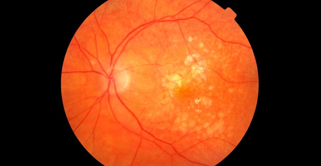
2 Amber flag Diabetic Maculopathy (M1) Pre-proliferative Diabetic Retinopathy (R2) Old, treated and now inactive DR (R1/M0/P1or R0/M0/P1) Where only partial or incomplete images can be achieved Other ocular conditions with retinal signs ALWAYS MAKE NOTES TO INFORM THE PROGRAMME WHY YOU HAVE FLAGGED AS AMBER
3 Diabetic Maculopathy (M1) Exudates within macula Flag as amber if any exudates at all on macular image If unsure whether they are drusen, still flag as amber Blood spot within 1 disc diameter (DD) of the fovea with a VA of or worse Usually significant background diabetic retinopathy (BDR) as well

4 Inner Circle: <1DD; Outer Circle: rough confines of the macula Exudates


5 Within 1 DD of fovea Subtle Exudates

6 VA still good; Younger patient with prominent surface reflexes Para-foveal exudate

7 Exudate patch

8 Exudates, trailing towards fovea

9 Multiple exudate patches Exudates

10 Fellow eye; Note larger blot haemorrhages as well Exudates
11 Pre-proliferative DR (R2) Multiple blot haemorrhages Flag as amber if there are more than 5 blood spots visible or more than one large haemorrhage Vessel changes Flag as amber if there are any unusual vessels Intra-retinal Micro vascular anomaly (IRMA) Venous Beading Venous reduplication Cotton wool spots Flag as amber if there are any white, fluffy lesions

12 Nasal to disc; Classic position IRMA

13 Subtle, but note significant BDR and Cotton-wool spot nearby IRMA

14 Note some blots centrally; More significant DR Larger and more numerous haemorrhages

15 Also, exudates Multiple blots and CWS

16 Also exudate Multiple blots and cotton wool spots

17 Note narrowing of vessel as well Venous Beading

18 Several IRMA, beading, venous dilatation, narrowing and sheathing Multiple nasal vessel changes

19 Reduplication with early beading

20 In more detail Reduplication
21 Old laser scars Always flag at least as amber if there are any signs of old laser scars on any images If there also appears to be New vessels or fibrosis: Flag as Red Follow the reporting protocol for red cases in Section 13 of the Screening Protocol

22 Mild, but significant BDR Mild laser photo-coagulation

23 Mild BDR Peripheral scatter laser photo-coagulation

24 Signs of peripheral scatter Laser Photocoagulation Very mild BDR otherwise; Still requires amber flag
25 No NVD/NVE or Fibrosis, so should only be set as amber Extensive laser scarring
26 Other ocular conditions Flag as amber if there are any signs of known non-dr conditions on the images Flag as amber if there are any signs that could indicate non-dr conditions on the images
27 Other ocular conditions Vein Occlusions Usually asymmetrical lesions Embolism Wet AMD Dry AMD Glaucoma Disc splinter haemorrhage Pale, cupped disc Optic Nerve disorders Choroiditis Vitreous disorders

28 Note collateral disc vessels Central Retinal Vein Occlusion

29 Gold embolism

30 Glaucoma

31 Glaucoma

32 Suspicious for glaucoma Disc haemorrhage, not DR

33 VA moderately reduced or no change Papilloedema

34 Fellow eye, almost always bilateral Papilloedema

35 More likely to be unilateral and VA reduced Papillitis


36 Comparison with previous image Left side: Last screening episode Right side: current episode

37 Central swelling Wet AMD

38 Central exudation and pigment dispersion Wet AMD

39 Pigment dispersion and Drusen Dry AMD
40 Dry AMD

41 Chorio-retinal scar

42 Melanoma

43 Retino-Schisis

44 Retino-Schisis
 Cellophane")
45 AKA Macular Pucker/Epi-macular membrane (EMM) Cellophane Maculopathy
Central Mersey Diabetic Retinopathy Screening Programme. Referring patients for Diabetic Retinopathy Screening
 Central Mersey Diabetic Retinopathy Screening Programme Referring patients for Diabetic Retinopathy Screening Information for GPs in Halton & St Helens, Knowsley and Warrington PCT Version: June 2008 Review
Diabetic Retinopathy Screening in Hong Kong. Dr. Rita Gangwani M.S, FRCS (Ophth), FCOphth(HK), FHKAM Eye Institute, The University of Hong Kong
, FCOphth(HK), FHKAM Eye Institute, The University of Hong Kong") Diabetic Retinopathy Screening in Hong Kong Dr. Rita Gangwani M.S, FRCS (Ophth), FCOphth(HK), FHKAM Eye Institute, The University of Hong Kong Co-Investigators Prof. David Wong Prof. Sarah McGhee Dr. Wico
Diabetic Retinopathy Screening in Hong Kong Dr. Rita Gangwani M.S, FRCS (Ophth), FCOphth(HK), FHKAM Eye Institute, The University of Hong Kong Co-Investigators Prof. David Wong Prof. Sarah McGhee Dr. Wico
Year 2 MBChB Clinical Skills Session Ophthalmoscopy. Reviewed & ratified by: Mr M Batterbury Consultant Ophthalmologist
 Year 2 MBChB Clinical Skills Session Ophthalmoscopy Reviewed & ratified by: o Mr M Batterbury Consultant Ophthalmologist Learning objectives o To understand the anatomy and physiology of the external and
Optical Coherence Tomography in Diabetic Retinopathy. Mrs Samantha Mann Consultant Ophthalmologist Clinical Lead of SEL-DESP
 Optical Coherence Tomography in Diabetic Retinopathy Mrs Samantha Mann Consultant Ophthalmologist Clinical Lead of SEL-DESP Content OCT imaging Retinal layers OCT features in Diabetes Some NON DR features
Central venous occlusion
 Central venous occlusion Central venous occlusion (right eye) There are dark haemorrhages at the macula and all over the retina. Choroidal haemangioma A choroidal haemangioma has salmon pink colour. There
Mild NPDR. Moderate NPDR. Severe NPDR
 Diabetic retinopathy Diabetic retinopathy is the most common cause of blindness in adults aged 35-65 years-old. Hyperglycaemia is thought to cause increased retinal blood flow and abnormal metabolism in
Step 4: Ask permission to turn off lights or draw the curtains
 STEPS OF EYE EXAMINATION - FUNDUS Step 1: Approach the patient Read the instructions carefully for clues Shake hands, introduce yourself Ask permission to examine him I would like to examine your eyes,
Dr/ Marwa Abdellah EOS /16/2018. Dr/ Marwa Abdellah EOS When do you ask Fluorescein angiography for optic disc diseases???
 When do you ask Fluorescein angiography for optic disc diseases??? 1 NORMAL OPTIC DISC The normal optic disc on fluorescein angiography is fluorescent due to filling of vessels arising from the posterior
PART 1: GENERAL RETINAL ANATOMY
 PART 1: GENERAL RETINAL ANATOMY General Anatomy At Ora Serrata At Optic Nerve Head Fundoscopic View Of Normal Retina What Is So Special About Diabetic Retinopathy? The WHO definition of blindness is
FRANZCO, MD, MBBS. Royal Darwin Hospital
 Diabetes and Eye By Dr. Nishantha Wijesinghe FRANZCO, MD, MBBS Consultant Ophthalmologist Royal Darwin Hospital 98% of Diabetics do not need to suffer from severe visual loss Yet Diabetic eye disease is
Documentation, Codebook, and Frequencies
 Documentation, Codebook, and Frequencies Ophthalmology Retinal Imaging Examination Survey Years: 2005 to 2006 SAS Transport File: OPXRET_D.XPT December 2008 NHANES 2005 2006 Data Documentation Exam Component:
ZEISS AngioPlex OCT Angiography. Clinical Case Reports
 Clinical Case Reports Proliferative Diabetic Retinopathy (PDR) Case Report 969 PROLIFERATIVE DIABETIC RETINOPATHY 1 1-year-old diabetic female presents for follow-up of proliferative diabetic retinopathy
Diabetic Eye Disease Visual Recognition & Interpretation of Clinical Signs
 + Diabetic Eye Disease Visual Recognition & Interpretation of Clinical Signs Quiz created by CLEARVIEW Training Jane Macnaughton MCOptom & Peter Chapman MCOptom FBDO + CET Accreditation C 19106 2 CET Points
Diabetic Retinopathy. Barry Emara MD FRCS(C) Giovanni Caboto Club October 3, 2012
 Giovanni Caboto Club October 3, 2012") Diabetic Retinopathy Barry Emara MD FRCS(C) Giovanni Caboto Club October 3, 2012 Outline Statistics Anatomy Categories Assessment Management Risk factors What do you need to do? Objectives Summarize the
Diabetic Retinopathy Barry Emara MD FRCS(C) Giovanni Caboto Club October 3, 2012 Outline Statistics Anatomy Categories Assessment Management Risk factors What do you need to do? Objectives Summarize the
7.1 Grading Diabetic Retinopathy
 Chapter 7 DIABETIC RETINOPATHYGRADING -------------------------------------------------------------------------------------------------------------------------------------- A consistent approach to the
Diabetic Retinopathy
 Diabetic Retinopathy Introduction People with diabetes are more likely to have eye problems that can lead to blindness. Diabetic retinopathy is a disease of the eye s retina that is caused by diabetes.
EyePACS Grading System (Part 2): Detecting Presence and Severity of Background (Non-Proliferative) Diabetic Retinopathy Lesion
: Detecting Presence and Severity of Background (Non-Proliferative) Diabetic Retinopathy Lesion") EyePACS Grading System (Part 2): Detecting Presence and Severity of Background (Non-Proliferative) Diabetic Retinopathy Lesion George Bresnick MD MPA Jorge Cuadros OD PhD Anatomy of the eye: 3 Normal Retina
EyePACS Grading System (Part 2): Detecting Presence and Severity of Background (Non-Proliferative) Diabetic Retinopathy Lesion George Bresnick MD MPA Jorge Cuadros OD PhD Anatomy of the eye: 3 Normal Retina
Marcus Gonzales, OD, FAAO Cedar Springs Eye Clinic
 Marcus Gonzales, OD, FAAO Cedar Springs Eye Clinic 25.6 million adults 11.3% of the adult population 10.9 million adults 65 years and older 26.9% of this age population 79 million people are Pre-diabetic!!
X-Plain Diabetic Retinopathy Reference Summary
 X-Plain Diabetic Retinopathy Reference Summary Introduction Patients with diabetes are more likely to have eye problems that can lead to blindness. Diabetic retinopathy is a disease of the eye s retina
Common Causes of Vision Loss
 Common Causes of Vision Loss Learning Objectives To identify the most common causes of vision loss in the United States To differentiate the most common forms of agerelated macular degeneration and diabetic
For details on measurement and recording of visual acuity, refer to Annex 1. VISION INTERPRETING RESULTS ABSTRACT
 management update on functional decline in older adults 2012 Unit No. 5 VISION Dr Au Eong Kah Guan, Ms Yulianti, Ms Fifiana ABSTRACT Among Singaporean adults of Chinese origin aged 40 to 79 years old,
Medical Retina 2011 Nicholas Lee
 Medical Retina 2011 Nicholas Lee 1 Diabetic Retinopathy Epidemiology 1000 registered blind each year 2% diabetics registered as blind (8% of all Blind Registrations) 42% with Mild Background DR will progress
Leo Semes, OD, FAAO UAB Optometry
 Leo Semes, OD, FAAO UAB Optometry Safe; inert Has long track record - over 45 years Mixes with plasma and highlights blood vessel compromise Using specific exciting (490 nm)and absorption (510 nm) filters
Eyes on Diabetics: How to Avoid Blindness in Diabetic Patient
 Eyes on Diabetics: How to Avoid Blindness in Diabetic Patient Rova Virgana FK Unpad Pusat Mata Nasional RS Mata Cicendo Bandung Eye Center (Hospital and Clinic) PIT IDI Jabar 2018 Keys Facts from WHO
World Sight Day Case Studies. Mark Frost Screening Manager South East London DESP
 World Sight Day 2015 Case Studies Mark Frost Screening Manager South East London DESP Introduction All of the following cases have been identified in our screening programme over the last 3 years. The
EyePACS Grading System (Part 3): Detecting Proliferative (Neovascular) Diabetic Retinopathy. George Bresnick MD MPA Jorge Cuadros OD PhD
: Detecting Proliferative (Neovascular) Diabetic Retinopathy. George Bresnick MD MPA Jorge Cuadros OD PhD") EyePACS Grading System (Part 3): Detecting Proliferative (Neovascular) Diabetic Retinopathy George Bresnick MD MPA Jorge Cuadros OD PhD Anatomy of the eye: 3 Normal Retina Retinal Arcades Macula Optic
EyePACS Grading System (Part 3): Detecting Proliferative (Neovascular) Diabetic Retinopathy George Bresnick MD MPA Jorge Cuadros OD PhD Anatomy of the eye: 3 Normal Retina Retinal Arcades Macula Optic
OCT Angiography in Primary Eye Care
 OCT Angiography in Primary Eye Care An Image Interpretation Primer Julie Rodman, OD, MS, FAAO and Nadia Waheed, MD, MPH Table of Contents Diabetic Retinopathy 3-6 Choroidal Neovascularization 7-9 Central
INTRODUCTION AND SYMPTOMS
 CHAPTER 1 INTRODUCTION AND SYMPTOMS Introduction of Diabetic Retinopathy Diabetic retinopathy (DR) is a potentially blinding complication of diabetes. It is defined as presence of one or more definite
Diabesity A Public Health Crisis: AOA Evidence Based Translation to Care Series
 Diabesity A Public Health Crisis: AOA Evidence Based Translation to Care Series Joseph J. Pizzimenti, OD, FAAO Associate Professor Nova Southeastern University The Eye Care Institute pizzimen@nova.edu
DIABETIC RETINOPATHY
 DIABETIC RETINOPATHY C. L. B. Canny, MD FRCSC Diabetic retinopathy is the most serious eye manifestation of diabetes and is responsible for most of the blindness caused by diabetes. Diabetic retinopathy
Diabetic Management beyond traditional risk factors and LDL-C control: Can we improve macro and microvascular risks?
 Retinopathy Diabetes has a negative effect on eyes in many ways, increasing the risk of cataracts for example, but the most common and serious ocular complication of diabetes is retinopathy. Diabetic retinopathy
Posterior Segment Update
 Posterior Segment Update Featured Speaker: Dr. Kyle Cheatham, FAAO, DIP ABO DISCLOSURE STATEMENT We have no direct financial or proprietary interest in any companies, products or services mentioned in
Management of diabetic eye disease: an overview
 Peter Blows Photographic screening for diabetic eye disease can identify patients who will benefit from laser treatment. BOTSWANA MANAGEMENT Management of diabetic eye disease: an overview Laser for DR
OCT Angiography The Next Frontier
 Choroid Retina avascular 5/13/2017 OCT Angiography The Next Frontier Pierce Kenworthy OD, FAAO June 9, 2017 OCT Angiography (OCTA) 2016 Non-invasive, motion contrast imaging Represents erythrocyte movement
OCCLUSIVE VASCULAR DISORDERS OF THE RETINA
 OCCLUSIVE VASCULAR DISORDERS OF THE RETINA Learning outcomes By the end of this lecture the students would be able to Classify occlusive vascular disorders (OVD) of the retina. Correlate the clinical features
evaluation of vitreoretinal adhesions in exudative AMD using optical coherence tomography
 evaluation of vitreoretinal adhesions in exudative AMD using optical coherence tomography Dr. Mahmoud Alaa Abouhusssein, FRCO Lecturer of ophthalmology, Alexandria university Dr. Amir Ramadan Gomaa, MD
Vascular Disease Ocular Manifestations of Systemic Hypertension
 Vascular Disease Ocular Manifestations of Systemic Hypertension Maynard L. Pohl, OD, FAAO Pacific Cataract & Laser Institute 10500 NE 8 th Street, Suite 1650 Bellevue, WA 98004 USA 425-462-7664 Cerebrovascular
RANZCO Screening and Referral Pathway for Diabetic Retinopathy #
 RANZCO Screening and Referral Pathway for Diabetic Retinopathy # Patient Presents a. Screen for Diabetic Retinopathy every 2 years b. Begin screening at diagnosis of Diabetes * Clinical Modifi ers Yearly
Chris Brown, M.D. Eye Specialty Group, PLC Continuing Education Series
 Chris Brown, M.D. Eye Specialty Group, PLC 2018 Continuing Education Series Disclaimer I have no financial interests in this lecture or any information discussed therein Objectives Fluorescein Angiogram
OCULAR HEMORRHAGES. ROSCOE J. KENNEDY, M.D. Department of Ophthalmology
 OCULAR HEMORRHAGES ROSCOE J. KENNEDY, M.D. Department of Ophthalmology Ocular hemorrhages are important not only because they produce visual loss but also because they usually indicate a disorder elsewhere
Clinical Case Presentation. Branch Retinal Vein Occlusion. Sarita M. Registered Nurse Whangarei Base Hospital
 Clinical Case Presentation on Branch Retinal Vein Occlusion Sarita M. Registered Nurse Whangarei Base Hospital Introduction Case Study Pathogenesis Clinical Features Investigations Treatment Follow-up
Diabetic Retinopathy A Presentation for the Public
 Diabetic Retinopathy A Presentation for the Public Ray M. Balyeat, MD The Eye Institute Tulsa, Oklahoma The Healthy Eye Light rays enter the eye through the cornea, pupil and lens. These light rays are
EFFICACY OF ANTI-VASCULAR ENDOTHELIAL GROWTH FACTOR AGENTS IN RETINAL DISORDER FOR BETTER VISUAL ACUITY
 EFFICACY OF ANTI-VASCULAR ENDOTHELIAL GROWTH FACTOR AGENTS IN RETINAL DISORDER FOR BETTER VISUAL ACUITY Diwakar chaudhary *1, 2, Hu shuqiong, Long Yuan and Xiong kun 1 Yangtze University, 1 Nanhuan Road
VASCULAR DISEASE IN OPTOMETRIC PRACTICE
 VASCULAR DISEASE IN OPTOMETRIC PRACTICE Outline notes to accompany City University 3 rd year undergraduate Clinical Practice course lecture Introduction Dr PhD BSc FCOptom FAAO DCLP Optometrists in the
 measure of your overall performance. An isolated glucose test is helpful to let you know what your sugar level is at one moment, but it doesn t tell you whether or not your diabetes is under adequate control
Grading Diabetic Retinopathy R2 - dots, blots and multiple headaches!
 Grading Diabetic Retinopathy R2 - dots, blots and multiple headaches! Simon Harding FRCOphth FRCS MD Professor of Clinical Ophthalmology University of Liverpool BARS 2010 Manchester English National Grading
DR Screening In Singapore: Achievements & Future Challenges
 DR Screening In Singapore: Achievements & Future Challenges Ecosse Lamoureux Director, Population Research Platform Singapore Eye Research Institute (SERI) Background About 600,000 of Singaporeans aged
Sponsored by. Shared care and referral pathways. Part 2: diabetes screening leading from the front
 CET CONTINUING Sponsored by 1 CET POINT Shared care and referral pathways Part 2: diabetes screening leading from the front Chris Steele, BSc (Hons), FCOptom, DCLP, DipOC, DipTp(IP), FBCLA The alarming
Fundus Autofluorescence. Jonathan A. Micieli, MD Valérie Biousse, MD
 Fundus Autofluorescence Jonathan A. Micieli, MD Valérie Biousse, MD The retinal pigment epithelium (RPE) has many important functions including phagocytosis of the photoreceptor outer segments Cone Rod
Red fundus lesions are a
 Diagnosis and management of red fundus lesions Sam Khandhadia and Richard Newsom discuss common red fundus lesions, how to identify them and the most appropriate clinical management. Module C8569, two
Diagnosis and treatment of diabetic retinopathy. Blake Cooper MD Ophthalmologist Vitreoretinal Surgeon Retina Associates Kansas City
 Diagnosis and treatment of diabetic retinopathy Blake Cooper MD Ophthalmologist Vitreoretinal Surgeon Retina Associates Kansas City Disclosures Consulted for Novo Nordisk 2017,2018. Will be discussing
Diabetes and Eye Health more than meets the eye Vision Initiative - in association with PSA
 Diabetes and Eye Health more than meets the eye Vision Initiative - in association with PSA Vision 2020 Australia Vision Initiative RANZCO & OAA (Vic) Proud members of Vision 2020 Australia Outline Vision
Clinically Significant Macular Edema (CSME)
") Clinically Significant Macular Edema (CSME) 1 Clinically Significant Macular Edema (CSME) Sadrina T. Shaw OMT I Student July 26, 2014 Advisor: Dr. Uwaydat Clinically Significant Macular Edema (CSME) 2
Clinically Significant Macular Edema (CSME) 1 Clinically Significant Macular Edema (CSME) Sadrina T. Shaw OMT I Student July 26, 2014 Advisor: Dr. Uwaydat Clinically Significant Macular Edema (CSME) 2
Diabetic Retinopathy
 Diabetic Retinopathy Diabetes mellitus is one of the leading causes of irreversible blindness worldwide. In the United States, it is the most common cause of blindness in people younger than 65 years.
NEPTUNE RED BANK BRICK
 NEPTUNE RED BANK BRICK Diabetes & The Eye Diabetics are more likely to develop Cataracts at a younger age. Diabetics are twice as likely to develop Glaucoma when compared to non-diabetics. The primary
Ophthalmology Macular Pathways
 Ophthalmology Macular Pathways Age related Macular Degeneration Diabetic Macular Oedema Macular Oedema secondary to Central Retinal Macular Oedema secondary to Branch Retinal CNV associated with pathological
Diabetes Retinopathy Extension Dataset
 For reference only Do Not Use For more information contact: cdsis@nhs.net Diabetes Retinopathy Extension Dataset March 2006 National Clinical Dataset Development Programme (NCDDP) Support Team Information
DIABETES AND YOUR EYES. Presented by Dr. Andrea Hagler
 DIABETES AND YOUR EYES Presented by Dr. Andrea Hagler Tahlequah, OK Forest Grove, OR Brief Review of Diabetes The body s endocrine system is responsible for regulating growth, reproduction, and tissue
OCT Image Analysis System for Grading and Diagnosis of Retinal Diseases and its Integration in i-hospital
 Progress Report for1 st Quarter, May-July 2017 OCT Image Analysis System for Grading and Diagnosis of Retinal Diseases and its Integration in i-hospital Milestone 1: Designing Annotation tool extraction
Five Things You re Missing with Your Fundus Camera
 ebook Five Things You re Missing with Your Fundus Camera By Donald J. Siegel, OD, Sun City West Eye Care Sponsored by: Before I began incorporating EIDON true-color imaging into my practice, my retinal
Diabetic Eye Disease
 Manchester Royal Eye Hospital Medical Retinal Services Information for Patients Diabetic Eye Disease This leaflet sets out to answer some of your questions about diabetic eye disease. You may wish to discuss
Diabetic Retinopathy in Primary Care
 Diabetic Retinopathy in Primary Care Epidemiology Diabetes is one of the most serious challenges to health care world rld-wide. According to recent projections it will affect 239 million people e by 2010-
NO FINANCIAL INTERESTS ARE DISCLOSED BY THE AUTHORS
 OCULAR FINDINGS IN APLASTIC ANEMIA: MULTICENTER STUDY & LITERATURE REVIEW Ahmad M Mansour1, MD, Jong Wook Lee, MD, PhD, Seung Ah Yahng, MD, Kyu Seop Kim, MD, Maha Shahin, MD, Nelson Hamerschlak, MD, PhD,
Do You See What I See!!! Shane R. Kannarr, OD
 Do You See What I See!!! Shane R. Kannarr, OD skannarr@kannarreyecare.com Define Specialty Testing Additional Test to: Prove/Disprove Diagnosis To monitor progression of a condition To document a condition
The Human Eye. Cornea Iris. Pupil. Lens. Retina
 The Retina Thin layer of light-sensitive tissue at the back of the eye (the film of the camera). Light rays are focused on the retina then transmitted to the brain. The macula is the very small area in
Ocular Pathology. I. Congenital and/or developmental. A. Trisomy 21. Hypertelorism (widely spaced eyes) Keratoconus (cone shaped cornea)
 Keratoconus (cone shaped cornea)") I. Congenital and/or developmental Robbins Pathologic Basis of Disease, 6 th Ed. A. Trisomy 21 Hypertelorism (widely spaced eyes) Keratoconus (cone shaped cornea) Focal hypoplasia of iris Cataracts frequently
I. Congenital and/or developmental Robbins Pathologic Basis of Disease, 6 th Ed. A. Trisomy 21 Hypertelorism (widely spaced eyes) Keratoconus (cone shaped cornea) Focal hypoplasia of iris Cataracts frequently
One-Field of View vs. Two-Fields of View; Is a Macula Centered Photograph Adequate for Diabetic Retinopathy Screening? -
 International Journal of Ophthalmology & Vision Research Research Article One-Field of View vs. Two-Fields of View; Is a Macula Centered Photograph Adequate for Diabetic Retinopathy Screening? - Mirriam
Neuro-Ocular Grand Rounds Anthony B. Litwak,OD, FAAO VA Medical Center Baltimore, Maryland
 Neuro-Ocular Grand Rounds Anthony B. Litwak,OD, FAAO VA Medical Center Baltimore, Maryland Dr. Litwak is on the speaker and advisory boards for Alcon and Zeiss Meditek COMMON OPTIC NEUROPATHIES THAT CAN
Abstract title: Vision loss from myelinated retinal nerve fiber layer with maculopathy. Authors: Man Kin (Eric) Chow, OD Lori Vollmer, OD, FAAO
 Chow, OD Lori Vollmer, OD, FAAO") Abstract title: Vision loss from myelinated retinal nerve fiber layer with maculopathy. Authors: Man Kin (Eric) Chow, OD Lori Vollmer, OD, FAAO Joseph Sowka, OD, FAAO General Topic: Ocular Disease Primary
Abstract title: Vision loss from myelinated retinal nerve fiber layer with maculopathy. Authors: Man Kin (Eric) Chow, OD Lori Vollmer, OD, FAAO Joseph Sowka, OD, FAAO General Topic: Ocular Disease Primary
Facts About Diabetic Eye Disease
 Facts About Diabetic Eye Disease Points to Remember 1. Diabetic eye disease comprises a group of eye conditions that affect people with diabetes. These conditions include diabetic retinopathy, diabetic
FROM OUTDATED TO UPDATED Eminence-Based Medicine
 FROM OUTDATED TO UPDATED Eminence-Based Medicine Evidence-Based Medicine A REVIEW OF KEY CLINICAL TRIALS Anthony DeWilde, OD FAAO 1 EMINENCE BASED MEDICINE 2 EVIDENCE BASED MEDICINE 3 4 CLINICAL TRIALS
Diabetic retinopathy damage to the blood vessels in the retina. Cataract clouding of the eye s lens. Cataracts develop at an earlier age in people
 Diabetic Retinopathy What is diabetic eye disease? Diabetic eye disease refers to a group of eye problems that people with diabetes may face as a complication of diabetes. All can cause severe vision loss
Tiffany L. Kruger, D.O. Children s Hospital of Michigan Wayne State University/Kresge Eye Institute
 Pediatric Cases Nt Not To Be Missed Tiffany L. Kruger, D.O. Pediatric Ophthalmology Fellow Children s Hospital of Michigan Wayne State University/Kresge Eye Institute Case Presentation CC: Left eye turns
Retinal Vein Occlusion (RVO) Treatment pathway- Northeast England. Retinal Vein Occlusion (RVO) with Macular oedema (MO)
 Treatment pathway- Northeast England. Retinal Vein Occlusion (RVO) with Macular oedema (MO)") Retinal Vein Occlusion (RVO) Treatment pathway- Northeast England (Royal Victoria Infirmary, Sunderland Eye Infirmary, James Cook University Hospital, Darlington Memorial Hospital, University Hospital
Retinal Vein Occlusion (RVO) Treatment pathway- Northeast England (Royal Victoria Infirmary, Sunderland Eye Infirmary, James Cook University Hospital, Darlington Memorial Hospital, University Hospital
Understanding Diabetic Retinopathy
 Understanding Diabetic Retinopathy What Is Diabetic Retinopathy? Diabetes damages blood vessels in the rear of the eye. This condition is called diabetic retinopathy. It can lead to vision loss or blindness.
Diabetic Retinopathy
 Diabetic Retinopathy Overview This presentation covers the following topics: Definitions Epidemiology of diabetic retinopathy Evidence for public health approaches Screening for diabetic retinopathy Health
4/27/2010 INTRODUCTION TO RETINAL VASCULAR DISEASE VENOUS/VENULAR CENTRAL RETINAL VEIN OBSTRUCTION / CRVO ADDITIONAL FEATURES /COMPLICATIONS
 INTRODUCTION TO RETINAL VASCULAR DISEASE VENOUS/VENULAR Leo Semes, OD Professor, UAB Optometry 2 CENTRAL RETINAL VEIN OBSTRUCTION CENTRAL RETINAL VEIN OBSTRUCTION / OCCLUSION (CRVO) obstruction of the
Diabetic and the Eye: An Introduction
 Diabetic and the Eye: An Introduction Lawrence Iu FRCSEd (Ophth), FCOphthHK, FHKAM (Ophthalmology) Department of Ophthalmology, Grantham Hospital & Queen Mary Hospital Background Diabetes mellitus (DM)
Building The Retina Company
 Building The Retina Company Optos devices produce ultra-widefield (UWF ), high resolution images (optomap ) of approximately 82% (200 ) of the retina. A single optomap can document the retina from the
GRADUAL LOSS OF VISION. Teresa Anthony Consultant Ophthalmologist Emersons Green NHS Treatment Centre Bristol
 GRADUAL LOSS OF VISION Teresa Anthony Consultant Ophthalmologist Emersons Green NHS Treatment Centre Bristol CVI Registration Low Vision: 6/60 Sight impaired: 3/60 Severe sight impaired:
OCT Fundal Angiography Initial Experience The new era in Medical Retina Imaging Based on Cirrus 5000 AngioPlex 2016 Model Sheena George & Nicholas
 OCT Fundal Angiography Initial Experience The new era in Medical Retina Imaging Based on Cirrus 5000 AngioPlex 2016 Model Sheena George & Nicholas Lee Consultants Ophthalmologist at The Hillingdon Hospital
Contractor Information. LCD Information. Local Coverage Determination (LCD): Ophthalmic Angiography (Fluorescein and Indocyanine Green) (L34426)
: Ophthalmic Angiography (Fluorescein and Indocyanine Green) (L34426)") Local Coverage Determination (LCD): Ophthalmic Angiography (Fluorescein and Indocyanine Green) (L34426) Links in PDF documents are not guaranteed to work. To follow a web link, please use the MCD Website.
Local Coverage Determination (LCD): Ophthalmic Angiography (Fluorescein and Indocyanine Green) (L34426) Links in PDF documents are not guaranteed to work. To follow a web link, please use the MCD Website.
Preparing for laser treatment for diabetic retinopathy and maculopathy
 Preparing for laser treatment for diabetic retinopathy and maculopathy Information for patients Preparing for laser treatment for diabetic retinopathy and maculopathy. This leaflet sets out to answer the
Patient AB. Born in 1961 PED
 Clinical Atlas Patient AB Born in 1961 PED Autofluorescence Dilated 45 EasyScan Zero-dilation IR 45 Fundus Dilated 45 In the fundus photos (Canon CX1) the PED is not able to be seen. However, the extent
Use of the Free Electron Laser for the Noninvasive Determination of Retinal Oxyhemoglobin Saturation by Near Infrared Reflectance Spectrophotometry
 Use of the Free Electron Laser for the Noninvasive Determination of Retinal Oxyhemoglobin Saturation by Near Infrared Reflectance Spectrophotometry Ref: Eye, M.C. Escher, 1946 Ref: Eye, M.C. Escher, 1946
CHAPTER 8 EVALUATION OF FUNDUS IMAGE ANALYSIS SYSTEM
 CHAPTER 8 EVALUATION OF FUNDUS IMAGE ANALYSIS SYSTEM Diabetic retinopathy is very common retinal disease associated with diabetes. Efforts to prevent diabetic retinopathy though have yielded some results;
Jay M. Haynie, O.D.; F.A.A.O. Olympia Tacoma Renton Kennewick Washington
 Jay M. Haynie, O.D.; F.A.A.O. Olympia Tacoma Renton Kennewick Washington I Jay M. Haynie, OD, FAAO have received honoraria from the following companies: Reichert Technologies Notal Vision Carl Zeiss Meditec
Vitreomacular interface disorders. Ghanbari MD 1393:10:25
 Vitreomacular interface disorders Ghanbari MD 1393:10:25 Human vitreous after dissection of the sclera, choroid, and retina. Lamellar structure of the posterior vitreous cortex (PVC) in the monkey. V =
ARIC Neurocognitive Study RETINAL GRADING PROTOCOL
 ARIC Neurocognitive Study RETINAL GRADING PROTOCOL Table of Contents 1 INTRODUCTION... 1 1.1 EQUIPMENT AND MATERIALS... 1 2 GRADING PROCEDURES AND RULES... 2 2.1 THE GRADING FORM... 3 2.2 PRELIMINARY GRADING...
Diabetes & Your Eyes
 Diabetes & Your Eyes Diabetes is a disease that occurs when the pancreas does not secrete enough insulin or the body is unable to process it properly. Insulin is the hormone that regulates the level of
Implementing New & Revised ICD-10 Codes John A. McGreal Jr., O.D. Missouri Eye Associates McGreal Educational Institute
 Implementing New & Revised ICD-10 Codes John A. McGreal Jr., O.D. Missouri Eye Associates McGreal Educational Institute Excellence in Optometric Education John A. McGreal Jr., O.D. Missouri Eye Associates
A Little Physics. How Does It Work? Radiation Therapy for Choroidal Neovascularisation in AMD A Review. => cell death
 Radiation Therapy for Choroidal Neovascularisation in AMD A Review Jen Anikina ST4 Hillingdon Hospital A Little Physics One gray is the absorption of one joule of ionizing radiation energy per kilogram
optic disc neovascularisation
 British Journal of Ophthalmology, 1979, 63, 412-417 A comparative study of argon laser and krypton laser in the treatment of diabetic optic disc neovascularisation W. E. SCHULENBURG, A. M. HAMILTON, AND
The Era of anti- - - VEGF Kirk L. Halvorson, OD
 The Era of anti- - - VEGF Kirk L. Halvorson, OD Introduction: Anti- - - Vascular Endothelial Growth Factor (Anti- - - VEGF) medication is a relatively a new line of medications used in treating a variety
Implementing New & Revised ICD-10 Codes
 Implementing New & Revised ICD-10 Codes John A. McGreal Jr., O.D. Missouri Eye Associates cgreal Educational Institute Excellence in Optometric Education John A. McGreal Jr., O.D. Missouri Eye Associates
Manual. Manual Welsh Eye Care Initiative. A Welsh Eye Care Initiative. Protocol. The Assessment and Management of Age-related Macular Degeneration
 A Protocol 1.0 Definitions The following terms are important in this text: Wet Macular Degeneration Condition caused by the growth of abnormal blood vessels under the retina. Symptoms appear suddenly and
Neuro-Ocular Grand Rounds
 Neuro-Ocular Grand Rounds Anthony B. Litwak,OD, FAAO VA Medical Center Baltimore, Maryland Dr. Litwak is on the speaker and advisory boards for Alcon and Zeiss Meditek COMMON OPTIC NEUROPATHIES THAT CAN
Photocoagulation of disciform macular lesions
 British Journal of Ophthalmology, 1979, 63, 669-673 Photocoagulation of disciform macular lesions with krypton laser A. C. BIRD AND R. H. B. GREY From the Institute of Ophthalmology, Moorfields Eye Hospital,
Course # Flashes and Floaters and Curtains, Oh My!
 Course # 132 Flashes and Floaters and Curtains, Oh My! FLASHES and FLOATERS and CURTAINS, OH MY!!! FLASHES OF LIGHT Vitreous is the villain Retinal traction Retinal hole Retinal tear Migraine Classic migraine
Course # Flashes and Floaters and Curtains, Oh My!
 Course # 132 Flashes and Floaters and Curtains, Oh My! FLASHES and FLOATERS and CURTAINS, OH MY!!! FLASHES OF LIGHT Vitreous is the villain Retinal traction Retinal hole Retinal tear Migraine Classic migraine