CT Evaluation of Bowel Wall Thickening. Dr: Adel El Badrawy; M.D. Lecturer of Radio Diagnosis Faculty of Medicine Mansoura University.
|
|
|
- Brice Gerard Pearson
- 6 years ago
- Views:
Transcription
1 CT Evaluation of Bowel Wall Thickening By Dr: Adel El Badrawy; M.D. Lecturer of Radio Diagnosis Faculty of Medicine Mansoura University.
2 The CT findings of bowel wall thickening includes 1 Degree of thickening. 2 Pattern of attenuation (homogeneous or heterogeneous). 3 Symmetric versus asymmetric thickening. 4 Length of thickening. 5 Associated peri enteric abnormalities.
3 Degree of Bowel Wall Thickening I Mild thickening ( < 2 cm). A Common 1 Infectious enterocolitis. 2 Ulcerative colitis. 3 Crohn s disease. 4 Radiation injury. 5 Ischaemia. 6 Bowel edema in cirrhosis. 7 Submucosal haemorrhage. B Uncommon 1 Adenocarcinoma. 2 Lymphoma.
4 II Marked thickening (>2 cm) A Common 1 Adenocarcinoma, G.I. stromal tumor, metastases, lymphoma. 2 Severe colitis. 3 S.L.E. B Uncommon 1 Crohn s disease, T.B., histoplasmosis, cytomegalovirus. 2 Submucosal hemorrhage.
5 Pattern of attenuation in bowel wall thickening I Homogeneous A Common 1 Submucosal hemorrhage. 2 Lymphoma. 3 Small adenocarcinoma. B Uncommon 1 Infarcted bowel. 2 Pitfalls related to residual fluid. 3 Chronic Crohn s disease. 4 Chronic radiation injury.
6 II Heterogeneous A Stratified attenuation 1 Common a Ischemia. b Infectious enterocolitis. c Crohn s disease, ulcerative colitis. d Vasculitis, Lupus, Henoch Schonlein purpura. e Radiation. f Bowel edema related to cirrhosis or low protien state. 2 Uncommon a Infiltrating scirrhous carcinoma (usually stomach or rectum). b Residual fluid and contrast material. c Submucosal fat deposition. d Pneumatosis.
7 II Heterogeneous B Mixed attenuation (Common) 1 Large adenocarcinoma. 2 G.I. stromal tumour. 3 Mucinous adenocarcinoma.
8 ymmetry of bowel wall thickening I Symmetric A Infections of the small & large bowel. B Ulcerative colitis. C Crohn s disease. D Radiation injury. E Ischaemia. F Bowel edema in cirrhosis. G Lymphoma. H Submucosal haemorrhage. II Asymmetric A Adenocarcinoma B G.I. stromal tumor.
9 B Uncommon S.L.E. Length of bowel wall thickening I Focal ( < 10 cm) A Common: 1 Diverticulitis, appendicitis. 2 Adenocarcinoma. B Uncommon 1 Lymphoma. 2 T.B. 3 Crohn s disease. II Segmental ( cm) A Common 1 Lymphoma. 2 Crohn s disease. 3 Infectious ileitis. 4 Radiation. 5 Submucosal haemorrhage. 6 Ischaemia.
10 Length of bowel wall thickness III Diffuse A Common 1 U.C. 2 Infectious enterocolitis. 3 Edema from low protien & cirrhosis. 4 S.L.E. B Uncommon: ischaemia.
11 Morphologic criteria aiding interpretation of CT scans in intestinal disease Characteristic Benign Malignant Edges Tapered Heaped up Thickness Valvulae conniventes or haustration Uniform Symmetric Thinner Present Variable Asymmetric Thicker Absent

12 Deposition of fat in submucosa chronic ulcerative colitis..
 and outer enhancement of")
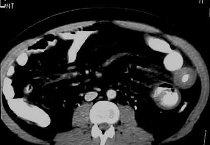
13 Target" sign ulcerative colitis. Mild wall thickening with classic target appearance and inner enhancement of mucosa (short white arrow) and outer enhancement of muscular layer (long white arrow) surrounding low attenuation edematous submucosa (black arrow).

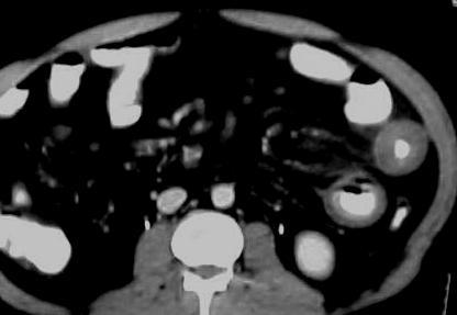
14 acute ulcerative colitis. The thickened wall of the rectosigmoid segment demonstrates uniform increased enhancement similar to the attenuation of the external iliac vein Pericolonic vessels are dilated (white arrow).
.")
15 Accordion" sign Clostridium difficile colitis. Marked thickening of haustra (arrowheads). Barium (arrow) trapped between thickened haustra mimic appearance of accordion

16 Focal asymmetrically thickened ulcerated mass (arrow) on nondependent wall of rectum. Biopsy revealed rectal adenocarcinoma.

17 Water halo sign in pseudomembranous colitis. On an intravenous contrast enhanced CT scan, the thickened inner layer of the rectosigmoid (straight arrows) is surrounded by a thicker outer layer (curved arrows).


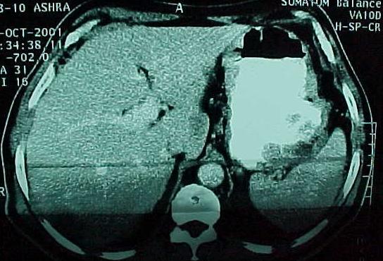
18 Case 1: Gastric Carcinoma With Hepatic Metastases

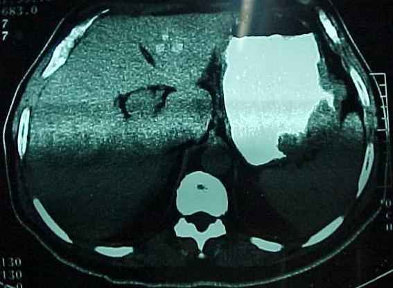
19 Case 2: Gastric carcinoma


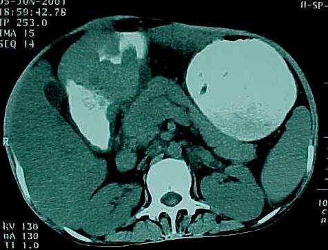
20 Case 3: Small bowel lymphoma L.N.


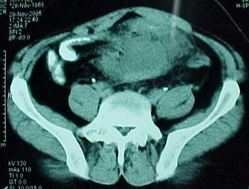
21 Case 4: Ascending Colonic Carcinoma


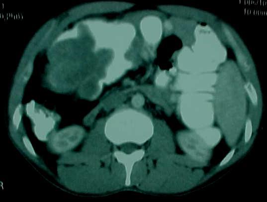
22 Case 5: Gastric & Duodenal Lymphoma

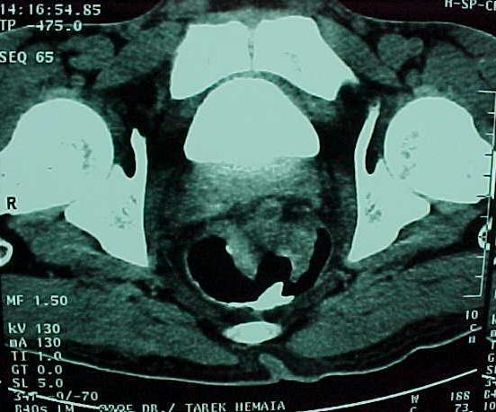
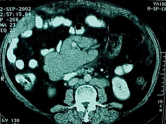
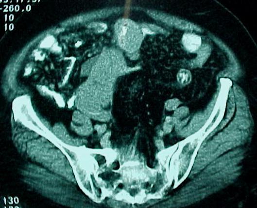
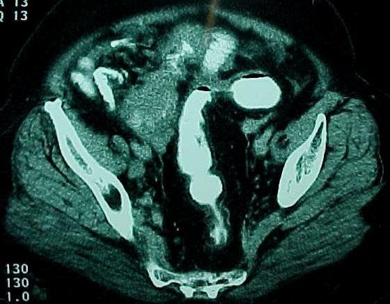
23 Case 6: Rectal Carcinoma

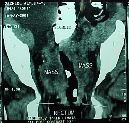
24 Case: Rectal carcinoma
25 Case 7: Intestinal Lymphoma


26 Case 8: Gastric & Duodenal Lymphoma


27 Case 9: Ascending Colonic Carcinoma


28 Case 10: Carcinoma Transverse Colon

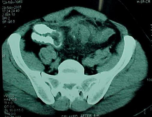
29 Case 11: Ileal lymphoma


30 Case 12: Schwannoma of Vagus Nerve of the Stomach.


31 Case 13 : Crohn s disease with Mesenteric abscess


32 Case 14 : Crohn s disease with Mesenteric abscess


33 Case 15: CT Scan S.M.V.T. With intestinal congestion


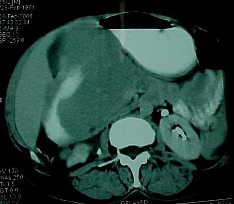
34 Case 16 :Ceacal Carcinoma with Hepatic Metastases.


35 Case 17: CT Scan Gastric varices


36
ENTEROCOLITIDES CAN YOU TELL THEM APART ON MDCT? Richard M. Gore, MD North Shore University Medical Center University of Chicago Evanston, Illinois
 ENTEROCOLITIDES CAN YOU TELL THEM APART ON MDCT? Richard M. Gore, MD North Shore University Medical Center University of Chicago Evanston, Illinois SCBT/MR 2010 San Diego, California March 8, 2010 13:40-14:00
Medical application of transabdominal ultrasound in gastrointestinal diseases
 Medical application of transabdominal ultrasound in gastrointestinal diseases Hsiu-Po Wang Department of Emergency Medicine National Taiwan University Hospital Real-time ultrasound has become a standard
CT of Bowel Wall Thickening: Significance and Pitfalls of Interpretation
 CT of owel Wall Thickening: Significance and Pitfalls of Interpretation C T has become the most important imaging technique for evaluating the abdomen and pelvis. CT is used to examine patients with acute
Gastrointestinal Tract. Anatomy of GI Tract. Anatomy of GI Tract. (Effective February 2007) (1%-5%)
 (1%-5%)") Gastrointestinal Tract (Effective February 2007) (1%-5%) Anatomy of GI Tract Esophagus bulls-eye or target EG junction seen on sagittal scan posterior to left lobe of liver and anterior to aorta Anatomy
Gastrointestinal Tract (Effective February 2007) (1%-5%) Anatomy of GI Tract Esophagus bulls-eye or target EG junction seen on sagittal scan posterior to left lobe of liver and anterior to aorta Anatomy
Thickened gastrointestinal wall findings on computed tomography: simplifying the diagnosis.
 Thickened gastrointestinal wall findings on computed tomography: simplifying the diagnosis. Poster No.: C-0419 Congress: ECR 2015 Type: Educational Exhibit Authors: W. Mnari, O. Fkih, M. Maatouk, A. Zrig,
GASTROINTESTINAL IMAGING STUDY GUIDE
 GASTROINTESTINAL IMAGING STUDY GUIDE Pharynx Diverticula Foreign bodies Trauma o Motility Disorders Esophagus Diverticula Trauma Esophagitis Barrett esophagus Rings, webs, and strictures Varices Benign
... Inflammatory disorder of the colon that occurs as a complication of antibiotic treatment.
 Definition Inflammatory disorder of the colon that occurs as a complication of antibiotic treatment. " Epidemiology Humans represent the main reservoir of Clostridium difficile, which is not part of the
Gastroenterology Tutorial
 Gastroenterology Tutorial Gastritis Poorly defined term that refers to inflammation of the stomach. Infection with H. pylori is the most common cause of gastritis. Most patients remain asymptomatic Some
LOOKING FOR AIR IN ALL THE WRONG PLACES Richard M. Gore, MD North Shore University Health System University of Chicago Evanston, IL
 SIGNIFICANCE OF EXTRALUMINAL ABDOMINAL GAS: LOOKING FOR AIR IN ALL THE WRONG PLACES Richard M. Gore, MD North Shore University Health System University of Chicago Evanston, IL SCBT/MR 2012 October 26,
Contrast-enhanced small bowel ultrasound in the assessment of the small bowel in patients with Crohn s Disease
 Contrast-enhanced small bowel ultrasound in the assessment of the small bowel in patients with Crohn s Disease C.F. Healy 1, D. Ferguson 1, S. Jepson 1, B. Salh 2, F. Donnellan 2, N. Chatur 2, A. C. Harris
CT EVALUATION OF GASTRIC LESIONS:
 CT EVALUATION OF GASTRIC LESIONS: Pictural essay Hasni Bouraoui I, Kahloun A, Jemni H, Elouni F, Moulahi H, Daadoucha A, Ben Ali A, Sriha B, Tlili Graies K Departments of Radiology, Gastro enterology,
Pitfalls in the CT diagnosis of appendicitis
 The British Journal of Radiology, 77 (2004), 792 799 DOI: 10.1259/bjr/95663370 E 2004 The British Institute of Radiology Pictorial review Pitfalls in the CT diagnosis of appendicitis 1 C D LEVINE, 2 O
Evaluating the CT Diagnosis of Clostridium difficile Colitis: Should CT Guide Therapy?
 Iain D. C. Kirkpatrick 1 Howard M. Greenberg Received April 7, 2000; accepted after revision August 24, 2000. 1 Both authors: Department of Radiology, University of Manitoba, Health Sciences Centre, 820
Radiology of GI system diseases
 GI Cycle - Lecture 12 436 Teams Radiology of GI system diseases Objectives 1. 2. 3. To know common GIT Pathologies presentation. To understand step wise approach in requesting GIT Radiology Investigations.
Utility of CT enterography in the evaluation of small bowel pathologies
 International Journal of Advances in Medicine Varma RU et al. Int J Adv Med. 2017 Oct;4(5):1328-1332 http://www.ijmedicine.com pissn 2349-3925 eissn 2349-3933 Original Research Article DOI: http://dx.doi.org/10.18203/2349-3933.ijam20174190
Alison Douglass Gillian Lieberman, MD. November. Colon Cancer. Alison Douglass, Harvard Medical School Year III Gillian Lieberman, MD
 November Colon Cancer Alison Douglass, Harvard Medical School Year III Our Patient Mr. K. is a 67 year old man with no prior medical problems other than hemorrhoids which have caused occasional rectal
8. The polyp in the illustration can be described as (circle all that apply) a. Exophytic b. Pedunculated c. Sessile d. Frank
 a. Exophytic b. Pedunculated c. Sessile d. Frank") Quiz 1 Overview 1. Beginning with the cecum, which is the correct sequence of colon subsites? a. Cecum, ascending, splenic flexure, transverse, hepatic flexure, descending, sigmoid. b. Cecum, ascending,
Quiz 1 Overview 1. Beginning with the cecum, which is the correct sequence of colon subsites? a. Cecum, ascending, splenic flexure, transverse, hepatic flexure, descending, sigmoid. b. Cecum, ascending,
Imaging abdominal vascular emergencies. V.Stoynova
 Imaging abdominal vascular emergencies V.Stoynova Abdominal vessels V. Stoynova 2 Acute liver bleeding trauma anticoagulant therapy liver disease : HCC, adenoma, meta, FNH, Hemangioma Diagnosis :CT angiography
Colitis: How to make a correct differential diagnosis in CT
 Colitis: How to make a correct differential diagnosis in CT Poster No.: C-1578 Congress: ECR 2013 Type: Educational Exhibit Authors: C. de la Torre, C. Cañete, R. Rodriguez; Malaga/ES Keywords: Inflammation,
Original Research Article
 Original Research Article Characterization of Kalpesh K. Patel 1, Mayur V. Khandhedia 2, Vishalkumar H. Bhardava 3 1 Assistant Professor, 2 Associate Professor, 3 Assistant Professor, Department of Radiology,
IMAGING GUIDELINES - COLORECTAL CANCER
 IMAGING GUIDELINES - COLORECTAL CANCER DIAGNOSIS The majority of colorectal cancers are diagnosed on colonoscopy, with some being diagnosed on Ba enema, ultrasound or CT. STAGING CT chest, abdomen and
ד"ר דוד ירדני המכון לגסטרואנטרולוגיה ומחלות כבד מרכז רפואי סורוקה
 ד"ר דוד ירדני המכון לגסטרואנטרולוגיה ומחלות כבד מרכז רפואי סורוקה Presentaion: S.A is 38 years old. Referred for rectal bleeding investigation. Describes several occasions of bleeding and abdominal pain.
Imaging in gastric cancer
 Imaging in gastric cancer Gastric cancer remains a deadly disease because of late diagnosis. Adenocarcinoma represents 90% of malignant tumors. Diagnosis is based on endoscopic examination with biopsies.
Emergency MDCT in case of right lower quadrant pain
 Emergency MDCT in case of right lower quadrant pain Poster No.: C-0563 Congress: ECR 2015 Type: Educational Exhibit Authors: M. Lisitskaya, V. Sinitsyn; Moscow/RU Keywords: Abdomen, Emergency, Gastrointestinal
Pneumatosis intestinalis, not always a surgical emergency
 Pneumatosis intestinalis, not always a surgical emergency Poster No.: C-2233 Congress: ECR 2012 Type: Educational Exhibit Authors: E. Vanhoutte, M. Lefere, R. Vanslembrouck, D. Bielen, G. De 1 1 2 1 1
[A RESEARCH COORDINATOR S GUIDE]
![[A RESEARCH COORDINATOR S GUIDE]](/thumbs/88/117127924.jpg "[A RESEARCH COORDINATOR S GUIDE]") 2013 COLORECTAL SURGERY GROUP Dr. Carl J. Brown Dr. Ahmer A. Karimuddin Dr. P. Terry Phang Dr. Manoj J. Raval Authored by Jennifer Lee A cartoon about colonoscopies. 1 [A RESEARCH COORDINATOR S GUIDE]
2013 COLORECTAL SURGERY GROUP Dr. Carl J. Brown Dr. Ahmer A. Karimuddin Dr. P. Terry Phang Dr. Manoj J. Raval Authored by Jennifer Lee A cartoon about colonoscopies. 1 [A RESEARCH COORDINATOR S GUIDE]
Plain abdomen The standard films are supine & erect AP views (alternative to erect, lateral decubitus film is used in ill patients).
.") Plain abdomen The standard films are supine & erect AP views (alternative to erect, lateral decubitus film is used in ill patients). The stomach can be readily identified by its location, gastric rugae
Plain abdomen The standard films are supine & erect AP views (alternative to erect, lateral decubitus film is used in ill patients). The stomach can be readily identified by its location, gastric rugae
Stomach Computerized Tomography indications, technique, examples. VUH SK Radiology and nuclear medicine center Radiologist Dileta Rutkauskaitė
 Stomach Computerized Tomography indications, technique, examples VUH SK Radiology and nuclear medicine center Radiologist Dileta Rutkauskaitė Stomach Computerized Tomography gastroente rologist Oncologist
Role of imaging in the evaluation of the acute abdomen
 Prof. András Palkó MD, PhD Role of imaging in the evaluation of the acute abdomen Faculty of General Medicine University of Szeged Hungary 1 Definition Sudden onset of severe symptoms requiring emergency
Disclosure. Acknowledgement. What is the Best Workup for Rectal Cancer Staging: US/MRI/PET? Rectal cancer imaging. None
 What is the Best Workup for Rectal Cancer Staging: US/MRI/PET? Zhen Jane Wang, MD Assistant Professor in Residence UC SF Department of Radiology Disclosure None Acknowledgement Hueylan Chern, MD, Department
BOWEL WALL ATTENUATION- NOT JUST ANOTHER SHADE OF GREY. T Barrow, P Arora, S Sukumar, V Rudralingam
 OWEL WLL TTENUTION- NOT JUST NOTHER SHDE OF GREY T arrow, P rora, S Sukumar, V Rudralingam LERNING OJECTIVES Gain a better appreciation of the variation of bowel wall attenuation seen in a range of bowel
Inflammatory Bowel Disease When is diarrhea not just diarrhea?
 Inflammatory Bowel Disease When is diarrhea not just diarrhea? Jackie Kazik, MA, PA C CME Resources CAPA Annual Conference, 2011 Inflammatory Bowel Disease Objectives Discuss what is known about the pathophysiology
X-ray Corner. Imaging of the Small Bowel. Pantongrag-Brown L. Case 1. A 63-year-old man presented with abdominal pain, nausea and vomiting.
 THAI J 42 Imaging of the Small Bowel GASTROENTEROL 2015 X-ray Corner Imaging of the Small Bowel Pantongrag-Brown L Small bowel is the longest tubular organ in the body, about 18-22 feet. It is anchored
Gastrointestinal Disorders. Disorders of the Esophagus 3/7/2013. Congenital Abnormalities. Achalasia. Not an easy repair. Types
 Gastrointestinal Disorders Congenital Abnormalities Disorders of the Esophagus Types Stenosis Atresia Fistula Newborn aspirates while feeding. Pneumonia Not an easy repair Achalasia Lack of relaxation
Histopathology: gastritis and peptic ulceration
 Histopathology: gastritis and peptic ulceration These presentations are to help you identify, and to test yourself on identifying, basic histopathological features. They do not contain the additional factual
Images In Gastroenterology
 Images In Gastroenterology Thong-Ngam D, et al. THAI J GASTROENTEROL 2005 Vol. 6 No. 2 May - Aug. 2005 105 Imaging of Gastrointestinal Stromal Tumors Pornpim Fuangtharnthip, M.D. Narumol Hargroove, M.D.
APPENDICITIS AND ITS APPEARANCES ON CT
 APPENDICITIS AND ITS APPEARANCES ON CT APPENDICITIS Results from acute inflammation of the appendix. Most common abdominal surgical emergencies. Diagnosis usually clinical based on physical exam and lab
MESENTERIC ISCHEMIA THE FORGOTTEN DIAGNOSIS. Richard M. Gore, MD North Shore University Health System University of Chicago Evanston, Illinois
 MESENTERIC ISCHEMIA THE FORGOTTEN DIAGNOSIS Richard M. Gore, MD North Shore University Health System University of Chicago Evanston, Illinois SCBT/MR 2010 San Diego, California March 8, 2010 16:00-16:10
Nordic Forum - Trauma & Emergency Radiology. Bowel Obstruction: Imaging Update
 Nordic Forum - Trauma & Emergency Radiology Bowel Obstruction: Imaging Update Borut Marincek Institute of Diagnostic Radiology University Hospital Zurich, Switzerland Acute Abdomen Bowel Obstruction Bowel
UNDERSTANDING X-RAYS: ABDOMINAL IMAGING THE ABDOMEN
 UNDERSTANDING X-RAYS: ABDOMINAL IMAGING THE ABDOMEN Radiology Enterprises radiologyenterprises@gmail.com www.radiologyenterprises.com STOMACH AND SMALL BOWEL STOMACH AND SMALL BOWEL Swallowed air is a
Imaging Evaluation of Polyps. CT Colonography: Sessile Adenoma. Polyps, DALMs & Megacolon Objectives
 Polyps, DALMs & Megacolon: Pathology and Imaging of the Colon and Rectum Angela D. Levy and Leslie H. Sobin Washington, DC Drs. Levy and Sobin have indicated that they have no relationships which, in the
elical CT plays an important role
 bdominal Imaging Yu et al. Helical CT of cute RLQ Pain Pictorial Essay Jinxing Yu 1 nn S. Fulcher Mary nn Turner Robert. Halvorsen Yu J, Fulcher S, Turner M, Halvorsen R Helical CT Evaluation of cute Right
Spontaneous perforation of the colon: CT findings and clinical characteristics
 Spontaneous perforation of the colon: CT findings and clinical characteristics Poster No.: C-0724 Congress: ECR 2012 Type: Scientific Exhibit Authors: H. Cho, H. Y. Han, T. J. Chun, I. K. Yu ; Daejon/KR,
MDCT Features of Angiotensin- Converting Enzyme Inhibitor Induced Visceral Angioedema
 Gastrointestinal Imaging Pictorial Essay Vallurupalli and Coakley MDCT of Visceral ngioedema Gastrointestinal Imaging Pictorial Essay Kalyani Vallurupalli 1 Kevin J. Coakley 2 Vallurupalli K, Coakley KJ
Nasogastric tube. Stomach. Pylorus. Duodenum 1. Duodenum 2. Duodenum 3. Duodenum 4
 Esophagus Barium Swallow Stomach and Duodenum 4 year old Upper GI Nasogastric tube Stomach and Duodenum 4 year old Upper GI Nasogastric tube Stomach Pylorus Duodenum 1 Duodenum 2 Duodenum 3 Duodenum 4
Disorders of Cell Growth & Neoplasia. Histopathology Lab
 Disorders of Cell Growth & Neoplasia Histopathology Lab Paul Hanna April 2010 Case #84 Clinical History: 5 yr-old, West Highland White terrier. skin mass from axillary region. has been present for the
하부위장관비종양성질환의 감별진단 주미인제의대일산백병원
 하부위장관비종양성질환의 감별진단 주미인제의대일산백병원 Solutions for diagnostic problems in Colitis : Please ask yourself five questions Normal or Inflamed? Acute or Chronic? IBD or Other chronic colitis? Ulcerative colitis or
Pitfalls in the Diagnosis of Inflammatory Bowel Disease
 Pitfalls in the Diagnosis of Inflammatory Bowel Disease Robert H Riddell MD Mt Sinai Hospital Toronto Prof of Lab. Medicine and Pathobiology University of Toronto Atypical gross / endoscopic distribution
The Role of Ultrasound in the Assessment of Inflammatory Bowel Disease
 The Role of Ultrasound in the Assessment of Inflammatory Bowel Disease Dr. Richard A. Beable Consultant Gastrointestinal Radiologist Queen Alexandra Hospital Portsmouth Hospitals NHS Trust Topics for Discussion
Abdominal Complications After Bone Marrow Transplantation in Children: Sonographic and CT Findings
 1023 Pictorial Essay Abdominal Complications After Bone Marrow Transplantation in Children: Sonographic and CT Findings Ellen C. Benya,1 2 Carlos J. Sivit, 2 and Ralph R. Quinones2 3 Bone marrow transplantation
Emergency radiology of the large-bowel: What radiologists should know
 Emergency radiology of the large-bowel: What radiologists should know Poster No.: C-1659 Congress: ECR 2016 Type: Educational Exhibit Authors: A. Falkowski, D. Boll; Basle/CH Keywords: Colon, Emergency,
ACUTE ABDOMEN IN OLDER CHILDREN. Carlos J. Sivit M.D.
 ACUTE ABDOMEN IN OLDER CHILDREN Carlos J. Sivit M.D. ACUTE ABDOMEN Clinical condition characterized by severe abdominal pain developing over several hours ACUTE ABDOMINAL PAIN Common childhood complaint
Bowel emergencies. Bruce Lehnert MD. Stomach. Gastric : Bleeding varices. Stomach
 Bowel emergencies Bruce Lehnert MD Stomach Gastric : Bleeding varices Stomach 1 Stomach Upper GI bleed Proximal to the ligament of Treitz 5X more common than lower GIB High volume Endoscopy identifies
Abdominal Imaging: Luminal organs. Rowland Illing MA BMBCh DM FLS MRCS(Eng) FRCR
 FRCR") Abdominal Imaging: Luminal organs Rowland Illing MA BMBCh DM FLS MRCS(Eng) FRCR Aims Reference text & resources Management of a patient Imaging what and when to use What to ask and how to describe Segments
Abdominal Imaging: Luminal organs Rowland Illing MA BMBCh DM FLS MRCS(Eng) FRCR Aims Reference text & resources Management of a patient Imaging what and when to use What to ask and how to describe Segments
11/21/13 CEA: 1.7 WNL
 Case Scenario 1 A 70 year-old white male presented to his primary care physician with a recent history of rectal bleeding. He was referred for imaging and a colonoscopy and was found to have adenocarcinoma.
Patho Basic Chronic Inflammatory Bowel Diseases. Jürg Vosbeck Pathology
 Patho Basic Chronic Inflammatory Bowel Diseases Jürg Vosbeck Pathology General Group of chronic relapsing diseases with chronic bloody or watery diarrhea Usually ulcerative colitis (UC) or Crohn s disease
2015 복영증례 51/M C.C. Past Hx: DM, HTN (1998), Lab: WBC (11500/ μl ), CRP (0.71 mg/dl) 순천향서울병원황지영, 홍성숙 APCT (HAD #1) APCT (HAD#1) APCT (HAD #15)
, Lab: WBC (11500/ μl ), CRP (0.71 mg/dl) 순천향서울병원황지영, 홍성숙 APCT (HAD #1) APCT (HAD#1) APCT (HAD #15)") Case 1 2015 복영증례 순천향서울병원황지영, 홍성숙 51/M C.C Abdominal pain and chilling (1 week ago) Diarrhea (a month ago) Past Hx: DM, HTN (1998), Alcoholic liver disease (2008) Lab: WBC (11500/ μl ), CRP (0.71 mg/dl)
Case 1 2015 복영증례 순천향서울병원황지영, 홍성숙 51/M C.C Abdominal pain and chilling (1 week ago) Diarrhea (a month ago) Past Hx: DM, HTN (1998), Alcoholic liver disease (2008) Lab: WBC (11500/ μl ), CRP (0.71 mg/dl)
INVESTIGATIONS OF GASTROINTESTINAL DISEAS
 INVESTIGATIONS OF GASTROINTESTINAL DISEAS Lecture 1 and 2 دز اسماعيل داود فرع الطب كلية طب الموصل Radiological tests of structure (imaging) Plain X-ray: May shows soft tissue outlines like liver, spleen,
Radiographic Findings of Gastrointestinal Anisakiasis:
 Radiographic Findings of Gastrointestinal Anisakiasis: Clinical and Pathologic Correlation 1 Tae Woong Chung, M.D., Heoung Keun Kang, M.D., Yong Yeon Jeong, M.D., Gwang Woo Jeong, Ph.D., Jeong Jin Seo,
Mohamed EL-hemaly Gastro- intestinal surgical center, Mansoura University.
 Mohamed EL-hemaly Gastro- intestinal surgical center, Mansoura University. Chronic transmural inflammatory process of the bowel & affects any part of the gastro -intestinal tract from the mouth to the
A916: rectum: adenocarcinoma
 General facts of colorectal cancer The colon has cecum, ascending, transverse, descending and sigmoid colon sections. Cancer can start in any of the r sections or in the rectum. The wall of each of these
U Lecture Objectives. U Nordic Forum Trauma & Emergency Radiology. Bowel obstruction. U Bowel Obstruction: Etiologies
 Nordic Forum Trauma & Emergency Radiology Lecture Objectives Bowel Obstruction To illustrate the spectrum of acute obstruction of the small and the large bowel To explain how these bowel obstructions may
Chapter 32 Gastroenterology General Pathophysiology General Risk Factors for GI emergencies: Excessive Consumption Excessive Smoking Increased
 1 2 3 4 5 6 7 Chapter 32 Gastroenterology General Pathophysiology General Risk Factors for GI emergencies: Excessive Consumption Excessive Smoking Increased Ingestion of Caustic Substances Poor Bowel Habits
Chronic diarrhea. Dr.Nasser E.Daryani Professor of Tehran Medical University
 1 Chronic diarrhea Dr.Nasser E.Daryani Professor of Tehran Medical University Timing Acute diarrhea: 4 weeks Definitions Derived from Greek
Case History B Female patient 1970 Clinical History : crampy abdominal pain and episodes of bloody diarrhea Surgical treatment
 Case History B-1325945 Female patient 1970 Clinical History : crampy abdominal pain and episodes of bloody diarrhea Surgical treatment Case History B-1325945 Pathology Submucosa & Muscularis Endometriosis
Clinical Management of Obscure- Overt Gastrointestinal Bleeding. Presented by Dr. 張瀚文
 Clinical Management of Obscure- Overt Gastrointestinal Bleeding Presented by Dr. 張瀚文 Definition Obscure: : hard to understand; not clear. Overt: : public; not secret. Occult: : hidden from the knowledge
Polyps in general: is a descriptive term of forming a mass that is exophytic & polypoid.
 ميحرلا نمحرلا هللا مسب Gastric Tumors: Benign tumours & tumor-like conditions: -Mucosal: Gastric polyps (they are uncommon) -Mesenchymal tumours: Leiomyoma & Lipoma (can occur anywhere in the body) Malignant:
CT imaging findings of acute mesenteric ischemia and ischemic colitis. A brief pictorial essay.
 CT imaging findings of acute mesenteric ischemia and ischemic colitis. A brief pictorial essay. Poster No.: C-0750 Congress: ECR 2011 Type: Educational Exhibit Authors: Y. Arias Morales, J. P. Giraldo
in Patients Without Overt Gastrointestinal Disease
 Gastrointestinal Imaging Original Research Gervaise et al. Gastric Wall Fatty Infiltration Gastrointestinal Imaging Original Research Alban Gervaise 1 Pierre Naulet 1 Christelle Gervaise-Henry 2 Camille
Cross-sectional Imaging of Neuroendocrine Tumors of the Gastrointestinal Tract
 Cross-sectional Imaging of Neuroendocrine Tumors of the Gastrointestinal Tract Eric J. May 1, Shannon P. Sheedy 1, Joel G. Fletcher 1, Mark J. Truty 2, Thomas C. Smyrk 3, Jeff L. Fidler 1 1. Radiology,
A. Incorrect! The esophagus connects the pharynx and the stomach.
 Human Physiology - Problem Drill 19: Digestive Physiology and Nutrition Question No. 1 of 10 Instructions: (1) Read the problem and answer choices carefully, (2) Work the problems on paper as 1. This organ
Diagnosis of uncomplicated stercoral colitis: CT findings
 Diagnosis of uncomplicated stercoral colitis: CT findings Poster No.: C-404 Congress: ECR 2009 Type: Educational Exhibit Topic: Abdominal and Gastrointestinal Authors: A. Linda 1, J. Heiken 2 ; 1 Udine/IT,
IBD. Crohn s. Outline. Ulcerative colitis versus Crohn s disease: is biopsy useful? UC vs. Crohn s? Is it easy? Biopsy settings 21/07/2017 IBD
 Outline Ulcerative colitis versus Crohn s disease: is biopsy useful? Roger Feakins Colorectal biopsies Ileal and upper GI biopsies Special situations New techniques Summary Inflammatory bowel disease (IBD)
Diseases of the Colon. Jack Bragg, D.O., F.A.C.O.I.
 Diseases of the Colon Jack Bragg, D.O., F.A.C.O.I. I have no disclosures I work for the Curators of the University of Missouri Inflammatory Bowel Disease ULCERATIVE COLITIS CROHN S DISEASE Transmural Inflammation
ABDOMEN - GI. Duodenum
 TALA SALEH ABDOMEN - GI Duodenum - Notice the shape of the duodenum, it looks like capital G shape tube which extends from the pyloroduodenal junction to the duodenojejunal junction. - It is 10 inches
NON INVASIVE MONITORING OF MUCOSAL HEALING IN IBD. THE ROLE OF BOWEL ULTRASOUND. Fabrizio Parente
 NON INVASIVE MONITORING OF MUCOSAL HEALING IN IBD. THE ROLE OF BOWEL ULTRASOUND Fabrizio Parente Gastrointestinal Unit, A.Manzoni Hospital, Lecco & L.Sacco School of Medicine,University of Milan - Italy
Table S2 Study group sample sizes for CEA, CYFRA21-1 and CA125 determinations.
 Supplementary Data Table S Clinico-pathological data associated with malignant and benign cases Primary site Early stage Late stage Caecum 3 (5%) 4 (20%) (5%) Ascending colon 6 (30%) 2 (0%) 0 (0%) Transverse
COLORECTAL CANCER FAISALGHANISIDDIQUI MBBS; FCPS; PGDIP-BIOETHICS; MCPS-HPE
 COLORECTAL CANCER FAISALGHANISIDDIQUI MBBS; FCPS; PGDIP-BIOETHICS; MCPS-HPE PROFESSOR OF SURGERY & DIRECTOR, PROFESSIONAL DEVELOPMENT CENTRE J I N N A H S I N D H M E D I C A L U N I V E R S I T Y faisal.siddiqui@jsmu.edu.pk
Perforation of a Duodenal Diverticulum. Elective Student S. C.
 Perforation of a Duodenal Diverticulum 2008 4 Elective Student S. C. Case History An elderly male presented to the Emergency Department with abdominal pain. Chief Complaint: Worsening, diffuse abdominal
Malignant Focal Liver Lesions
 Malignant Focal Liver Lesions Other Than HCC Pablo R. Ros, MD, MPH, PhD Departments of Radiology and Pathology University Hospitals Cleveland Medical Center Case Western Reserve University Pablo.Ros@UHhospitals.org
Index. Note: Page numbers of article titles are in boldface type.
 Index Note: Page numbers of article titles are in boldface type. A Abdominal pain, abdominal considerations in, 183 184 antiemetics in, 182 auscultation in, 170 C-reactive protein in, 174 175 character
TIPS AND PITFALLS IN PLAIN FILM INTERPRETATION
 TIPS AND PITFALLS IN PLAIN FILM INTERPRETATION Dr Philip Touska MBBS, BMedSci(Hons), MRCS, DO-HNS, FRCR Radiology Fellow Guy s & St Thomas Hospitals LEARNING OBJECTIVES Where do we go wrong? Common pitfalls
Supplementary Online Content
 Supplementary Online Content Tran AH, Ngor EWM, Wu BU. Surveillance colonoscopy in elderly patients: a retrospective cohort study. JAMA Intern Med. Published online August 11, 2014. doi:10.1001/jamainternmed.2014.3746
The focus of this week s lab will be pathology of the gastrointestinal and hepatobiliary systems.
 GASTROINTESTINAL AND HEPATOBILIARY SYSTEMS The focus of this week s lab will be pathology of the gastrointestinal and hepatobiliary systems. GASTROINTESTINAL SYSTEM AND HEPATOBILIARY SYSTEM We will examine
Staging Challenges in Lower GI Cancers. Disclosure of Relevant Financial Relationships. AJCC 8 th edition and CAP protocol updates
 Staging Challenges in Lower GI Cancers Sanjay Kakar, MD University of California, San Francisco March 05, 2017 Disclosure of Relevant Financial Relationships USCAP requires that all planners (Education
Gastric Carcinoma in Patients with Crohn Disease: Report of Four Cases
 311 0361-803X/91/1 572-0311 C American Roentgen Ray Society Seth N. GIick1 Received January 1 7, 1991 ; accepted after re vision March 1 2, 1991. 1 Department of Diagnostic Radiology, Hahnemann University
Surgical Management of IBD. Val Jefford Grand Rounds October 14, 2003
 Surgical Management of IBD Val Jefford Grand Rounds October 14, 2003 Introduction Important Features Clinical Presentation Evaluation Medical Treatment Surgical Treatment Cases Overview Introduction Two
Duodenum retroperitoneal
 Duodenum retroperitoneal C shaped Initial region out of stomach into small intestine RETROperitoneal viscus Superior 1 st part duodenal cap ; moves upwards and backwards to lie on the R crura medial to
Ulcerative Colitis. ulcerative colitis usually only affects the colon.
 Ulcerative Colitis Introduction Ulcerative colitis is an inflammatory bowel disease. It is one of the 2 most common inflammatory bowel diseases. The other one is Crohn s disease. Ulcerative colitis and
Appendix 9: Endoscopic Ultrasound in Gastroenterology
 Appendix 9: Endoscopic Ultrasound in Gastroenterology This curriculum is intended for clinicians who perform endoscopic ultrasonography (EUS) in gastroenterology. It includes standards for theoretical
Abdomen and Pelvis CT (1) By the end of the lecture students should be able to:
 By the end of the lecture students should be able to:") RAD 451 Abdomen and Pelvis CT (1) By the end of the lecture students should be able to: State the common indications for Abdomen and pelvis CT exams Identify possible contra indications for Abdomen and
RAD 451 Abdomen and Pelvis CT (1) By the end of the lecture students should be able to: State the common indications for Abdomen and pelvis CT exams Identify possible contra indications for Abdomen and
Table 0: Description of Grading System for Anatomic Severity of Disease in Emergency. Local disease confined to the organ with minimal abnormality
 Table 0: of Grading System for Anatomic Severity of Disease in Emergency Local disease confined to the organ with minimal Local disease confined to the organ with severe Local extension Table 1: Universal
Lab Monitor Images Dissection of the Abdominal Vasculature + Lower Digestive System
 Lab Monitor Images Dissection of the Abdominal Vasculature + Lower Digestive System Stomach & Duodenum Frontal (AP) View Nasogastric tube 2 1 3 4 Stomach Pylorus Duodenum 1 Duodenum 2 Duodenum 3 Duodenum
Colon and Rectum. Protocol revision date: January 2005 Based on AJCC/UICC TNM, 6th edition
 Colon and Rectum Protocol applies to all invasive carcinomas of the colon and rectum. Carcinoid tumors, lymphomas, sarcomas, and tumors of the vermiform appendix are excluded. Protocol revision date: January
Essentials of Clinical MR, 2 nd edition. 73. Urinary Bladder and Male Pelvis
 73. Urinary Bladder and Male Pelvis Urinary bladder carcinoma is best locally staged with MRI. It is important however to note that a thickened wall (> 5 mm) is a non-specific finding seen in an underfilled
Gastro Intestinal Pathology
 Duration: 04 weeks (20 days) Gastro Intestinal 3/SBM-4/01 Alimentation in health Topic/ Concept Objectives Time Dept. 1. recall digestion, absorption and metabolism relating to, carbohydrates, proteins,
Disorders of the kidney. Urine analysis. Nephrotic and nephritic syndrome.
 Disorders of the kidney. Urine analysis. Nephrotic and nephritic syndrome. Azotemia and Urinary Abnormalities Disturbances in urine volume oliguria, anuria, polyuria Abnormalities of urine sediment red
Brief History. Identification : Past History : HTN without regular treatment.
 Brief History Identification : Name : 陳 x - Admission : 94/10/06 Gender : male Age : 75 y/o Chief Complaint : Urinary difficulty for months. Past History : HTN without regular treatment. Brief History
DR JAIKISHOR JOTHIRAJ MD POST GRADUATE DEPT OF RADIODIAGNOSIS
 DR JAIKISHOR JOTHIRAJ MD POST GRADUATE DEPT OF RADIODIAGNOSIS YASHODAMMAL 70 YRS OD LADY had C/o diffuse lower abdominal pain 20 days h/o blood in stools 4 days h/o vomiting 2 days h/o burning micturation
What do we need for diagnosis of IBD
 What do we need for diagnosis of IBD Kaichun Wu Dept. of Gastroenterology, Xijing Hospital Fourth Military Medical University Xi an an,, China In China UC 11.6/10 5,CD 1.4/10 5 Major cause of chronic diarrhea