Stomach Computerized Tomography indications, technique, examples. VUH SK Radiology and nuclear medicine center Radiologist Dileta Rutkauskaitė
|
|
|
- Reynold Watkins
- 5 years ago
- Views:
Transcription
1 Stomach Computerized Tomography indications, technique, examples VUH SK Radiology and nuclear medicine center Radiologist Dileta Rutkauskaitė
2 Stomach Computerized Tomography gastroente rologist Oncologist endoscopist surgeon radiologist

3 Stomach Computerized Tomography appropriate preparation technique of examination examples of pathology
4 Indications for stomach CT Preoperative evaluaton with pathologicaly proven gastric cancers Evaluation of the disease progresion/response to chemotherapy Detection and differentiation of submucosal lesions H. S. Kim, H. Y. Han et Preoperative evaluation of gastric cancer: value of spiral CT during gastric arteriography (CTGA) ; Abdom Imaging 26: (2001)


5 Anamnesis Patient anamnesis Previous gastric operations Endoscopic results Histopathological results


6 Stomach CT- preparation Fasting for 6-8 hour. Current Role of CT in Imaging of the Stomach Karen M. Horton, MD Elliot K. Fishman, MD RadioGraphics

7 Stomach CT- preparation We use pure water as an oral contrast agent (~ ml) - well tolerated - good distention of the stomach wall - allows good visualization of the enhancing wall Current Role of CT in Imaging of the Stomach1 Karen M. Horton, MD Elliot K. Fishman, MD RadioGraphics


8 Stomach CT- preparation For imaging of the stomach, adequate distention is essential. If the entire stomach is not well distended, disease may be overlooked or the collapsed gastric wall may mimic disease.


9 Stomach CTpatient positioning Supine position Right lateral decubitus position Patient are positioned according to the presumed location of the lesion seen at endoscopy
, - Portal venous ph. (70 s) - Delayed ph. (150 s). Current Role of CT in Imaging of the Stomach Karen M. Horton, MD Elliot K.")
10 Stomach CTscanning Scanning - unenhanced scan (to evaluate gastric distention) - enhanced scan after i/v c/m injection: 2 phases: - Late arterial ph. (35-40s), - Portal venous ph. (70 s) - Delayed ph. (150 s). Current Role of CT in Imaging of the Stomach Karen M. Horton, MD Elliot K. Fishman, MD RadioGraphics
 - musculiaris propria and serosa (enhancing layer) Normaly no more 5 mm in")
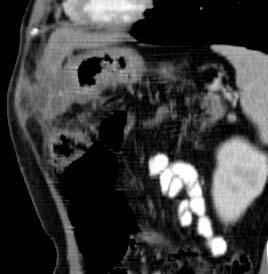
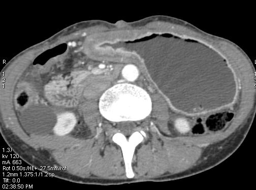
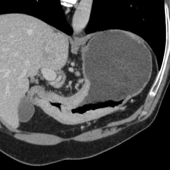
11 Stomach CT Normal gastric wall cearly visible as two or three layers; - mucosa (intensively enhancing layer) - submucosa (low density layer) - musculiaris propria and serosa (enhancing layer) Normaly no more 5 mm in thickness
12 Gastric pathology
13 Gastric adenoca CT signs: - Enhacement changes in the wall - Local or diffuse wall thickening - Infiltration into surrounding structures - liver, l/n mts
14 CT Criteria for T and N Staging of Gastric Cancer Stage T stage CT Criteria T1 Neoplasm shows focal thickening of inner layer, is almost well enhanced, and has visible low-attenuation-strip outer layer of gastric wall and clear fat plane around tumor T2 T3 T4 Neoplasm shows focal or diffuse thickening of gastric wall with transmural involvement, is almost well enhanced, and has smooth outer wall border and clear fat plane around tumor Transmural tumor with irregular or nodular outer border and/or perigastric fat infiltration Obliteration of fat plane between gastric tumor and adjacent organ or invasion of adjacent organ N stage N0 N1 N2 N3 No regional lymph node metastases Metastases in 1 6 regional lymph nodes Metastases in 7 15 regional lymph nodes Metastases in 15 regional lymph nodes Greene FL, Page DL, Fleming ID, et al, eds. AJCC manual of staging of cancer. 6th ed. New York, NY: Springer-Verlag, 2002

15 Gastric adenoca in situ


16 Early gastric cancer - T1

17 Gastric adenoca T2

18 Gastric adenoca T3

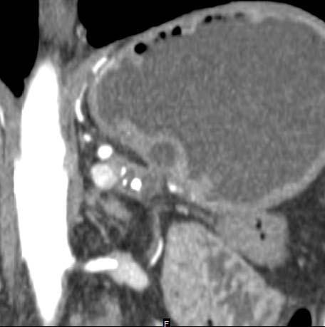
19 Gastric adenoca T4 N1M1
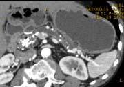
20 Gastric infiltrative adenoca

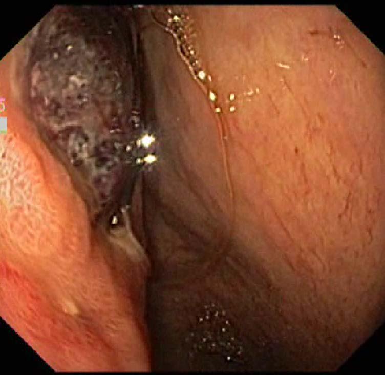
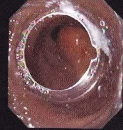
21 Gastric lymphoma Diffuse or segmental wall infiltration Usualy we see enlarged l/n Ct feature can be like adenoca

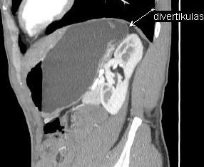
22 Gastric lymphoma


23 Gastric GIST

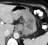
24 Gastric GIST During abdominal echoscopy was suspected panceatic lesion


25 Gastric ulcerated GIST in antrum

26 Gastric endocrinal tumors Carcinoid in gastric body mostly like small polyps mostly intensively enhancing

27 Gastric neuroendocrine Ca pt2n1 Histo carcinoid Transmural c/m enhancement Enlarged, round of shape, enhancing l/n
28 Intramural pseudocyst into gastric wall

29 Gastric antrum lipoma


30 Endoscopy findings gastric fundus submucosal lesion CT - polysplenism
31 Gastric diverticulum
32 Conclusions - Correct patient preparation for gastric CT examination is essential - gives a lot of information about the changes in gastric wall - The knowledge in characteristics of CT findings in normal gastric wall and pathological changes is crucial for the accurate diagnosis, treatment choice and planing
33 Thank you for Your attention
Imaging in gastric cancer
 Imaging in gastric cancer Gastric cancer remains a deadly disease because of late diagnosis. Adenocarcinoma represents 90% of malignant tumors. Diagnosis is based on endoscopic examination with biopsies.
Abstracting Upper GI Cancer Incidence and Treatment Data Quiz 1 Multiple Primary and Histologies Case 1 Final Pathology:
 Abstracting Upper GI Cancer Incidence and Treatment Data Quiz 1 Multiple Primary and Histologies Case 1 A 74 year old male with a history of GERD presents complaining of dysphagia. An esophagogastroduodenoscopy
CT EVALUATION OF GASTRIC LESIONS:
 CT EVALUATION OF GASTRIC LESIONS: Pictural essay Hasni Bouraoui I, Kahloun A, Jemni H, Elouni F, Moulahi H, Daadoucha A, Ben Ali A, Sriha B, Tlili Graies K Departments of Radiology, Gastro enterology,
Case Scenario 1. The patient has now completed his neoadjuvant chemoradiation and has been cleared for surgery.
 Case Scenario 1 July 10, 2010 A 67-year-old male with squamous cell carcinoma of the mid thoracic esophagus presents for surgical resection. The patient has completed preoperative chemoradiation. This
Gastric Cancer Histopathology Reporting Proforma
 Gastric Cancer Histopathology Reporting Proforma Mandatory questions (i.e. protocol standards) are in bold (e.g. S1.01). S1.01 Identification Family name Given name(s) Date of birth Sex Male Female Intersex/indeterminate
Unusual Manifestation of Stomach Cancer; Pictorial review and review of the literature
 Unusual Manifestation of Stomach Cancer; Pictorial review and review of the literature Poster No.: C-1386 Congress: ECR 2014 Type: Educational Exhibit Authors: D. H. Lee, S. K. Moon; Seoul/KR Keywords:
Patient. Male 76 year old C.C: abdominal pain
 Patient Male 76 year old C.C: abdominal pain Bowel stool retention Suspected pulmonary TB at right upper lung Infiltration in right lower lung Pleural thickening at the Right chest Localized dilated small
A916: rectum: adenocarcinoma
 General facts of colorectal cancer The colon has cecum, ascending, transverse, descending and sigmoid colon sections. Cancer can start in any of the r sections or in the rectum. The wall of each of these
Unusual Manifestation of Stomach Cancer; Pictorial review and review of the literature
 Unusual Manifestation of Stomach Cancer; Pictorial review and review of the literature Poster No.: C-1386 Congress: ECR 2014 Type: Educational Exhibit Authors: D. H. Lee, S. K. Moon; Seoul/KR Keywords:
CT Evaluation of Bowel Wall Thickening. Dr: Adel El Badrawy; M.D. Lecturer of Radio Diagnosis Faculty of Medicine Mansoura University.
 CT Evaluation of Bowel Wall Thickening By Dr: Adel El Badrawy; M.D. Lecturer of Radio Diagnosis Faculty of Medicine Mansoura University. The CT findings of bowel wall thickening includes 1 Degree of thickening.
[A RESEARCH COORDINATOR S GUIDE]
![[A RESEARCH COORDINATOR S GUIDE]](/thumbs/88/117127924.jpg "[A RESEARCH COORDINATOR S GUIDE]") 2013 COLORECTAL SURGERY GROUP Dr. Carl J. Brown Dr. Ahmer A. Karimuddin Dr. P. Terry Phang Dr. Manoj J. Raval Authored by Jennifer Lee A cartoon about colonoscopies. 1 [A RESEARCH COORDINATOR S GUIDE]
2013 COLORECTAL SURGERY GROUP Dr. Carl J. Brown Dr. Ahmer A. Karimuddin Dr. P. Terry Phang Dr. Manoj J. Raval Authored by Jennifer Lee A cartoon about colonoscopies. 1 [A RESEARCH COORDINATOR S GUIDE]
Diagnosis of Gastric Cancer with MDCT Using the Water-Filling Method and Multiplanar Reconstruction: CT Histologic Correlation
 MDCT of Gastric Cancer Gastrointestinal Imaging Original Research Downloaded from www.ajronline.org by 46.3.198.21 on 01/21/18 from IP address 46.3.198.21. Copyright RRS. For personal use only; all rights
GIT RADIOLOGY. Water-soluble contrast media (e.g. gastrograffin) are the other available agents.which doesn t cause inflammatory peritonitis..
 are the other available agents.which doesn t cause inflammatory peritonitis..") GIT RADIOLOGY Imaging techniques-general principles: Contrast examinations: Barium sulphate is the best contrast for GIT (with good mucosal coating & excellent opacification & being inert); but is contraindicated
GIT RADIOLOGY Imaging techniques-general principles: Contrast examinations: Barium sulphate is the best contrast for GIT (with good mucosal coating & excellent opacification & being inert); but is contraindicated
Gastric Cancer Staging AJCC eighth edition. Duncan McLeod Westmead Hospital, NSW
 Gastric Cancer Staging AJCC eighth edition Duncan McLeod Westmead Hospital, NSW Summary of changes New clinical stage prognostic groups, ctnm Postneoadjuvant therapy pathologic stage groupings, yptnm -
Alison Douglass Gillian Lieberman, MD. November. Colon Cancer. Alison Douglass, Harvard Medical School Year III Gillian Lieberman, MD
 November Colon Cancer Alison Douglass, Harvard Medical School Year III Our Patient Mr. K. is a 67 year old man with no prior medical problems other than hemorrhoids which have caused occasional rectal
Incidental Esophageal Findings on Chest CT. Amira Hussien, MD, Elliot Fishman, MD, Bouchra Younes, MD, Ahmed Hatw. Johns Hopkins Medical Institution
 Incidental Esophageal Findings on Chest CT Amira Hussien, MD, Elliot Fishman, MD, ouchra Younes, MD, Ahmed Hatw. Johns Hopkins Medical Institution I have nothing to disclose. DISCLOSURE INTRODUCTION Although
Images In Gastroenterology
 Images In Gastroenterology Thong-Ngam D, et al. THAI J GASTROENTEROL 2005 Vol. 6 No. 2 May - Aug. 2005 105 Imaging of Gastrointestinal Stromal Tumors Pornpim Fuangtharnthip, M.D. Narumol Hargroove, M.D.
malignant polyp Daily Challenges in Digestive Endoscopy for Endoscopists and Endoscopy Nurses BSGIE Annual Meeting 18/09/2014 Mechelen
 Plan Incidental finding of a malignant polyp 1. What is a polyp malignant? 2. Role of the pathologist and the endoscopist 3. Quantitative and qualitative risk assessment 4. How to decide what to do? Hubert
X-Ray Corner. Imaging of the Stomach. Pantongrag-Brown L
 THAI J 178 Imaging of the Stomach GASTROENTEROL 2014 X-Ray Corner Imaging of the Stomach Pantongrag-Brown L Imaging modalities used in stomach include plain radiographs, UGI study, US, CT, PET CT and MRI.
Efficacy of High Resolution Transabdominal Sonography of the Fluid Filled Stomach in the Evaluation of Gastric Carcinomas
 4-67 421 Efficacy of High Resolution Transabdominal Sonography of the Fluid Filled Stomach in the Evaluation of Gastric Carcinomas S SINGH, V CHOWDHURY ABSTRACT AIM: To evaluate the efficacy of high-resolution
Glomus Tumor of the Stomach
 J Radiol Sci 2011; 36: 49-53 Glomus Tumor of the Stomach Yuan-Chun Huang 1 Chen-Te Chou 2 Yu-Cheng Hong 1 Shang-Yun Ho 1 Kwo-Whei Lee 1 Hwa-Koon Wu 1 Department of Medical Imaging 1, Changhua Christian
Quiz Adenocarcinoma of the distal stomach has been increasing in the last 20 years. a. True b. False
 Quiz 1 1. Which of the following are risk factors for esophagus cancer. a. Obesity b. Gastroesophageal reflux c. Smoking and Alcohol d. All of the above 2. Adenocarcinoma of the distal stomach has been
Gastrointestinal Tract Cancer
 Gastrointestinal Tract Cancer Tumors of the Stomach Gastric adenocarcinoma Incidence and Epidemiology Incidence mortality rates USA High incidence: Japan, China, Chile, Ireland risk lower socioeconomic
Rui-Jia Sun 1, Lei Tang 1, Ying Chen 1, Xiao-Ting Li 1, Yu Sun 2, Zi-Yu Li 3, Ying-Shi Sun 1. Original Article. Abstract.
 Original Article Feasibility of differentiating T3 from T4a gastric cancer in different Lauren classification by determining serosa invasion: Diagnostic performance of high enhanced serosa sign Rui-Jia
Four Cases of Large Cell Neuroendocrine Carcinoma of the Stomach: Findings on CT and Barium Studies 1
 Four Cases of Large Cell Neuroendocrine Carcinoma of the Stomach: Findings on CT and arium Studies 1 Hee Jung Kim, M.D., Dongil Choi, M.D., Soon Jin Lee, M.D., Won Jae Lee, M.D., Sung Kim, M.D. 2, Jae
Disorders of Cell Growth & Neoplasia. Histopathology Lab
 Disorders of Cell Growth & Neoplasia Histopathology Lab Paul Hanna April 2010 Case #84 Clinical History: 5 yr-old, West Highland White terrier. skin mass from axillary region. has been present for the
Original Report. Carcinoid Tumors of the Stomach: A Clinical and Radiographic Study
 Aaron J. Binstock 1 C. Daniel Johnson 1 David H. Stephens 1 Ricardo V. Lloyd 2 Joel G. Fletcher 1 Received July 25, 2000; accepted after revision September 29, 2000. 1 Department of Radiology, Mayo Clinic,
COLLECTING CANCER DATA: STOMACH AND ESOPHAGUS
 COLLECTING CANCER DATA: STOMACH AND ESOPHAGUS 2017 2018 NAACCR WEBINAR SERIES Q&A Please submit all questions concerning webinar content through the Q&A panel. Reminder: If you have participants watching
Cross-sectional Imaging of Neuroendocrine Tumors of the Gastrointestinal Tract
 Cross-sectional Imaging of Neuroendocrine Tumors of the Gastrointestinal Tract Eric J. May 1, Shannon P. Sheedy 1, Joel G. Fletcher 1, Mark J. Truty 2, Thomas C. Smyrk 3, Jeff L. Fidler 1 1. Radiology,
Contrast-enhanced small bowel ultrasound in the assessment of the small bowel in patients with Crohn s Disease
 Contrast-enhanced small bowel ultrasound in the assessment of the small bowel in patients with Crohn s Disease C.F. Healy 1, D. Ferguson 1, S. Jepson 1, B. Salh 2, F. Donnellan 2, N. Chatur 2, A. C. Harris
Esophageal Cancer Staging Essentials: The New TNM Staging System (7th edition) and Clinicoradiologic Implications
 and Clinicoradiologic Implications") Esophageal Cancer Staging Essentials: The New TNM Staging System (7th edition) and Clinicoradiologic Implications Poster No.: E-0060 Congress: ESTI 2012 Type: Scientific Exhibit Authors: K. Lee, T. J.
Esophageal Cancer Staging Essentials: The New TNM Staging System (7th edition) and Clinicoradiologic Implications Poster No.: E-0060 Congress: ESTI 2012 Type: Scientific Exhibit Authors: K. Lee, T. J.
C. CT scan shows ascites and thin enhancing parietal peritoneum
 291 A B Fig. 1. A 55-year-old gastric cancer patient with peritoneal carcinomatosis. At surgery, there was large amount of ascites in peritoneal cavity and there were multiple small metastatic nodules
Practical considerations in preoperative evaluation of gastric cancer with MDCT: Guideline for the radiological report.
 Practical considerations in preoperative evaluation of gastric cancer with MDCT: Guideline for the radiological report. Poster No.: C-1549 Congress: ECR 2015 Type: Educational Exhibit Authors: I. Rubio
Polyps in general: is a descriptive term of forming a mass that is exophytic & polypoid.
 ميحرلا نمحرلا هللا مسب Gastric Tumors: Benign tumours & tumor-like conditions: -Mucosal: Gastric polyps (they are uncommon) -Mesenchymal tumours: Leiomyoma & Lipoma (can occur anywhere in the body) Malignant:
ENTEROCOLITIDES CAN YOU TELL THEM APART ON MDCT? Richard M. Gore, MD North Shore University Medical Center University of Chicago Evanston, Illinois
 ENTEROCOLITIDES CAN YOU TELL THEM APART ON MDCT? Richard M. Gore, MD North Shore University Medical Center University of Chicago Evanston, Illinois SCBT/MR 2010 San Diego, California March 8, 2010 13:40-14:00
CT role in distinguishing GIST from non-gist mesenchymal gastric tumors
 CT role in distinguishing GIST from non-gist mesenchymal gastric tumors Poster No.: C-0686 Congress: ECR 2016 Type: Educational Exhibit Authors: N. Almeida Costa, M. J. Magalhães, J. Abreu e Silva, M.
Lesions of the pancreaticoduodenal groove, a pictorial review
 Lesions of the pancreaticoduodenal groove, a pictorial review Poster No.: C-2131 Congress: ECR 2013 Type: Educational Exhibit Authors: E. Ni Mhurchu, L. Lavelle, I. Murphy, S. Skehan ; IE, Dublin/ IE Keywords:
Common and unusual CT and MRI manifestations of pancreatic adenocarcinoma: a pictorial review
 Review Article Common and unusual CT and MRI manifestations of pancreatic adenocarcinoma: a pictorial review Min-Jie Yang, Su Li, Yong-Guang Liu, Na Jiao, Jing-Shan Gong Department of Radiology, Shenzhen
A Gastric Schwannoma Misdiagnosed as B Cell Lymphoma
 계명의대학술지제 31 권 2 호 Keimyung Med J Vol. 31, No. 2, December, 2012 A Gastric Schwannoma Misdiagnosed as B Cell Lymphoma Kyung lim Koo, Yu Na Kang 1, M.D. Undergraduate Student, Department of Pathology 1,
8. The polyp in the illustration can be described as (circle all that apply) a. Exophytic b. Pedunculated c. Sessile d. Frank
 a. Exophytic b. Pedunculated c. Sessile d. Frank") Quiz 1 Overview 1. Beginning with the cecum, which is the correct sequence of colon subsites? a. Cecum, ascending, splenic flexure, transverse, hepatic flexure, descending, sigmoid. b. Cecum, ascending,
Quiz 1 Overview 1. Beginning with the cecum, which is the correct sequence of colon subsites? a. Cecum, ascending, splenic flexure, transverse, hepatic flexure, descending, sigmoid. b. Cecum, ascending,
IMAGING GUIDELINES - COLORECTAL CANCER
 IMAGING GUIDELINES - COLORECTAL CANCER DIAGNOSIS The majority of colorectal cancers are diagnosed on colonoscopy, with some being diagnosed on Ba enema, ultrasound or CT. STAGING CT chest, abdomen and
The Role of Spiral Computed Tomography in the Detection & Staging of Gastric Malignancy
 The Role of Spiral Computed Tomography in the Detection & Staging of Gastric Malignancy *Shawki Yousif Fawzi FRCS ** Rita Leon Baghdasar MomjianFICMS(Diag Rad) *** Inaam Azeez Khaleel DMRD ABSTRACT Background
Regression of Advanced Gastric MALT Lymphoma after the Eradication of Helicobacter pylori
 Gut and Liver, Vol. 6, No. 2, April 2012, pp. 270-274 CASE REPORT Regression of Advanced Gastric MALT Lymphoma after the Eradication of Helicobacter pylori Soo-Kyung Park, Hwoon-Yong Jung, Do Hoon Kim,
Pattern based approach for differential diagnosis of small bowel neoplasms using MDCT
 Pattern based approach for differential diagnosis of small bowel neoplasms using MDCT Poster No.: C-1400 Congress: ECR 2014 Type: Educational Exhibit Authors: P. Bhari Thippeswamy, C. Anuradha, A. Polimood,
Local staging of colon cancer: the current role of CT
 Local staging of colon cancer: the current role of CT Poster No.: C-2699 Congress: ECR 2018 Type: Authors: Keywords: DOI: Educational Exhibit A. P. Pissarra, R. R. Domingues Madaleno, C. Sanches, L. Curvo-
A218 : Esophagus cancer tissues. (formalin fixed)
") (formalin fixed) For research use only Specifications: No. of cases: 40 Tissue type: Esophagus cancer tissues No. of spots: 2 spots from each cancer case (80 spots) 4 non-neoplastic spots (4 spots) Total
(formalin fixed) For research use only Specifications: No. of cases: 40 Tissue type: Esophagus cancer tissues No. of spots: 2 spots from each cancer case (80 spots) 4 non-neoplastic spots (4 spots) Total
CT evaluation of small bowel carcinoid tumors
 CT evaluation of small bowel carcinoid tumors Poster No.: C-0060 Congress: ECR 2015 Type: Educational Exhibit Authors: N. V. V. P. Costa, L. Nascimento, T. Bilhim ; Estoril/PT, PT, 1 2 3 1 2 3 Lisbon/PT
Differentiation of gastric ulcers with MDCT
 Abdominal Imaging ª Springer Science+Business Media, LLC 2007 Published online: 7 December 2006 Abdom Imaging (2007) 32:688 693 DOI: 10.1007/s00261-006-9162-4 Differentiation of gastric ulcers with MDCT
Gastroenterology Tutorial
 Gastroenterology Tutorial Gastritis Poorly defined term that refers to inflammation of the stomach. Infection with H. pylori is the most common cause of gastritis. Most patients remain asymptomatic Some
Greater Manchester & Cheshire Guidelines for Pathology Reporting for Oesophageal and Gastric Malignancy
 Greater Manchester & Cheshire Guidelines for Pathology Reporting for Oesophageal and Gastric Malignancy Authors: Dr Gordon Armstrong, Dr Sue Pritchard 1. General Comments 1.1 Cancer reporting: Biopsies
Breast Imaging Lexicon
 9//201 200 BI RADS th Edition 201 BI RADS th Edition Breast Imaging Lexicon Mammographic Pathology and Assessment Categories Deborah Thames, R.T.(R)(M)(QM) The Advanced Health Education Center Nonmember:
Thickened gastrointestinal wall findings on computed tomography: simplifying the diagnosis.
 Thickened gastrointestinal wall findings on computed tomography: simplifying the diagnosis. Poster No.: C-0419 Congress: ECR 2015 Type: Educational Exhibit Authors: W. Mnari, O. Fkih, M. Maatouk, A. Zrig,
GASTRIC ULTRASOUND. A Point-of-care tool for aspiration risk assessment.
 GASTRIC ULTRASOUND A Point-of-care tool for aspiration risk assessment edu@gastricultrasound.org Indications Any clinical situation where aspiration risk is uncertain. For example: Lack of adherence to
RECTAL CARCINOMA: A DISTANCE APPROACH. Stephanie Nougaret
 RECTAL CARCINOMA: A DISTANCE APPROACH Stephanie Nougaret stephanienougaret@free.fr Despite the major improvements that have been made due to total mesorectal excision (TME) management of rectal cancer
Role of multidetector computed tomography (MDCT) in diagnosis and staging of gall bladder carcinoma
 in diagnosis and staging of gall bladder carcinoma") The Egyptian Journal of Radiology and Nuclear Medicine (2013) 44, 1 7 Egyptian Society of Radiology and Nuclear Medicine The Egyptian Journal of Radiology and Nuclear Medicine www.elsevier.com/locate/ejrnm
The Egyptian Journal of Radiology and Nuclear Medicine (2013) 44, 1 7 Egyptian Society of Radiology and Nuclear Medicine The Egyptian Journal of Radiology and Nuclear Medicine www.elsevier.com/locate/ejrnm
General Data. Gender : Male Birthday and age : 12/07/24,80 y/o Occupation : 無 Date of Admission :
 General Data Gender : Male Birthday and age : 12/07/24,80 y/o Occupation : 無 Date of Admission : 92-07-09 1 Chief complaint Upper abdominal fullness 30 minutes after having foods with sometimes epigastralgia
We are IntechOpen, the world s leading publisher of Open Access books Built by scientists, for scientists. International authors and editors
 We are IntechOpen, the world s leading publisher of Open Access books Built by scientists, for scientists 3,700 108,500 1.7 M Open access books available International authors and editors Downloads Our
Imaging techniques in the diagnosis, staging and follow up of GI cancers. Moderators: Banke Agarwal, MD and Paul Schultz, MD
 Imaging techniques in the diagnosis, staging and follow up of GI cancers Moderators: Banke Agarwal, MD and Paul Schultz, MD Panelists Axel Grothey, MD Professor of Oncology Division of Medical Oncology
Gastrointestinal Tract. Anatomy of GI Tract. Anatomy of GI Tract. (Effective February 2007) (1%-5%)
 (1%-5%)") Gastrointestinal Tract (Effective February 2007) (1%-5%) Anatomy of GI Tract Esophagus bulls-eye or target EG junction seen on sagittal scan posterior to left lobe of liver and anterior to aorta Anatomy
Gastrointestinal Tract (Effective February 2007) (1%-5%) Anatomy of GI Tract Esophagus bulls-eye or target EG junction seen on sagittal scan posterior to left lobe of liver and anterior to aorta Anatomy
Case Report Ileocecal Intussusception due to a Lipoma in an Adult
 Case Reports in Surgery Volume 2012, Article ID 684298, 4 pages doi:10.1155/2012/684298 Case Report Ileocecal Intussusception due to a Lipoma in an Adult Mehmet Bilgin, 1 Huseyin Toprak, 1 Issam Cheikh
Clinical Study Small Bowel Tumors: Clinical Presentation, Prognosis, and Outcomein33PatientsinaTertiaryCareCenter
 Hindawi Publishing Corporation Journal of Oncology Volume 2008, Article ID 212067, 5 pages doi:10.1155/2008/212067 Clinical Study Small Bowel Tumors: Clinical Presentation, Prognosis, and Outcomein33PatientsinaTertiaryCareCenter
Small Bowel Intussusception in an Adult due to Lipoma: a Rare Cause of Obstruction. Case report and Literature Review
 ISPUB.COM The Internet Journal of Surgery Volume 25 Number 1 Small Bowel Intussusception in an Adult due to Lipoma: a Rare Cause of Obstruction. Case report and Literature Review Yashpal, M Bansal, A Kudva
Original Article Differential diagnosis of primary gastric lymphoma on multi-detector computed tomography
 Int J Clin Exp Med 2016;9(2):1878-1883 www.ijcem.com /ISSN:1940-5901/IJCEM0012261 Original Article Differential diagnosis of primary gastric lymphoma on multi-detector computed tomography Songshi Quan
Application of Magnetic Resonance Images in Gastrointestinal Malignancies
 Chin J Radiol 2003; 28: 269-275 269 Application of Magnetic Resonance Images in Gastrointestinal Malignancies SHENG-LAN YU 1 YUK-MING TSANG 1 PO-CHIN LIANG 1 HUNG-JUNG WANG 1 CHIEN-YAO HSU 1 TA-CHENG WEI
Anatomical and Functional MRI of the Pancreas
 Anatomical and Functional MRI of the Pancreas MA Bali, MD, T Metens, PhD Erasme Hospital Free University of Brussels Belgium mbali@ulb.ac.be Introduction The use of MRI to investigate the pancreas has
Effect of Adjusted Positioning on Gastric Distention and Fluid Distribution During CT Gastrography
 CT Gastrograph y Gastrointestinal Imaging Technical Innovation Se Hyung Kim 1 Jeong Min Lee 1,2 Joon Koo Han 1,2 Jae Young Lee 1,2 Han Kwang Yang 3 Hyuk-Joon Lee 3 Kyung-Sook Shin 4 Byung Ihn Choi 1,2
Imaging abdominal vascular emergencies. V.Stoynova
 Imaging abdominal vascular emergencies V.Stoynova Abdominal vessels V. Stoynova 2 Acute liver bleeding trauma anticoagulant therapy liver disease : HCC, adenoma, meta, FNH, Hemangioma Diagnosis :CT angiography
Dr Claire Smith, Consultant Radiologist St James University Hospital Leeds
 Dr Claire Smith, Consultant Radiologist St James University Hospital Leeds Imaging in jaundice and 2ww pathway Image protocol Staging Limitations Pancreatic cancer 1.2.4 Refer people using a suspected
Essentials of Clinical MR, 2 nd edition. 73. Urinary Bladder and Male Pelvis
 73. Urinary Bladder and Male Pelvis Urinary bladder carcinoma is best locally staged with MRI. It is important however to note that a thickened wall (> 5 mm) is a non-specific finding seen in an underfilled
Digestive system L 2. Lecturer Dr. Firdous M. Jaafar Department of Anatomy/Histology section
 Digestive system L 2 Lecturer Dr. Firdous M. Jaafar Department of Anatomy/Histology section objectives 1-Describe the general structure of digestive tract: a-mucosa. b-submucosa. c-muscularis externa d-adventitia
CT imaging findings of acute mesenteric ischemia and ischemic colitis. A brief pictorial essay.
 CT imaging findings of acute mesenteric ischemia and ischemic colitis. A brief pictorial essay. Poster No.: C-0750 Congress: ECR 2011 Type: Educational Exhibit Authors: Y. Arias Morales, J. P. Giraldo
A LEADER IN ADVANCED ENDOSCOPY AND HEPATOBILIARY SURGERY
 A LEADER IN ADVANCED ENDOSCOPY AND HEPATOBILIARY SURGERY St. Peter s Hospital Advanced Endoscopy & Hepatobiliary Center Welcome The St. Peter s Hospital Advanced Endoscopy & Hepatobiliary Center is a leader
Emergency radiology of the large-bowel: What radiologists should know
 Emergency radiology of the large-bowel: What radiologists should know Poster No.: C-1659 Congress: ECR 2016 Type: Educational Exhibit Authors: A. Falkowski, D. Boll; Basle/CH Keywords: Colon, Emergency,
METASTASES FROM GASTRIC CARCINOMA TO COLON LESIONS: A CASE REPORT IN THE FORM OF MULTIPLE FLAT ELEVATED CASE PRESENTATION
 H.C. Lee, M.T. Yang, K.Y. Lin, et al METASTASES FROM GASTRIC CARCINOMA TO COLON IN THE FORM OF MULTIPLE FLAT ELEVATED LESIONS: A CASE REPORT Hsi-Chang Lee, Min-Ta Yang, 1 Kuang-Yang Lin, 1 Hsing-Yang Tu,
Principles of diagnosis, work-up and therapy The Gastroenterologist s role
 Principles of diagnosis, work-up and therapy The Gastroenterologist s role Dr. Christos G. Toumpanakis MD PhD FRCP Consultant in Gastroenterology/Neuroendocrine Tumours Hon. Senior Lecturer University
I appreciate the courtesy of Kusumoto at NCC for this presentation. What is Early Lung Cancers. Early Lung Cancers. Early Lung Cancers 18/10/55
 I appreciate the courtesy of Kusumoto at NCC for this presentation. Dr. What is Early Lung Cancers DEATH Early period in its lifetime Curative period in its lifetime Early Lung Cancers Early Lung Cancers
Interesting Pediatric ultrasound cases. Presented by: Falguni Patel (RDMS, RVT)
") Interesting Pediatric ultrasound cases Presented by: Falguni Patel (RDMS, RVT) Role of ultrasound to rule out Appendicitis Overview: Ultrasound is relatively inexpensive, safe and quick solution to rule
Interesting Pediatric ultrasound cases Presented by: Falguni Patel (RDMS, RVT) Role of ultrasound to rule out Appendicitis Overview: Ultrasound is relatively inexpensive, safe and quick solution to rule
The Radiologic Features of Xanthogranulomatous Cholecystitis: An Important Mimic of Gallbladder Carcinoma
 The Radiologic Features of Xanthogranulomatous Cholecystitis: An Important Mimic of Gallbladder Carcinoma Poster No.: C-0691 Congress: ECR 2014 Type: Authors: Keywords: DOI: Educational Exhibit H. L. khosa
SEER Summary Stage Still Here!
 SEER Summary Stage Still Here! CCRA NORTHERN REGION STAGING SYMPOSIUM SEPTEMBER 20, 2017 SEER Summary Stage Timeframe: includes all information available through completion of surgery(ies) in the first
Radiologist Performance in Differentiating Polypoid Early From Advanced Gastric Cancer Using Specific CT Criteria: Emphasis on Dimpling Sign
 Gastrointestinal Imaging Original Research Lee et al. CT of Gastric Cancer Gastrointestinal Imaging Original Research Eun Sun Lee 1 Se Hyung Kim 1,2 Jae Young Lee 1,2 Soo Jin Kim 1 Min A Kim 3 Jeong Min
Gastric Cancer in a Young Postpartum Female. Kings County Hospital Center SUNY Downstate Case Conference May 24, 2012
 Gastric Cancer in a Young Postpartum Female Kings County Hospital Center SUNY Downstate Case Conference May 24, 2012 Case HPI: 31 yo F, G5P3, 3 weeks s/p C-section, with gastric outlet obstruction. Pt
Colorectal Pathway Board (Clinical Subgroup): Imaging Guidelines September 2015
: Imaging Guidelines September 2015") Colorectal Pathway Board (Clinical Subgroup): Imaging Guidelines September 2015 1 Contents Page No. 1. Objective 3 2. Imaging Techniques 3 3. Staging of Colorectal Cancer 5 4. Radiological Reporting 6
Colorectal Pathway Board (Clinical Subgroup): Imaging Guidelines September 2015 1 Contents Page No. 1. Objective 3 2. Imaging Techniques 3 3. Staging of Colorectal Cancer 5 4. Radiological Reporting 6
Complicated Meckel`s diverticulum; to be considered as a differential diagnosis in the acute abdominal pain. Ultrasound and MDCT imaging finding
 Complicated Meckel`s diverticulum; to be considered as a differential diagnosis in the acute abdominal pain. Ultrasound and MDCT imaging finding Poster No.: C-0174 Congress: ECR 2013 Type: Educational
Utility of CT enterography in the evaluation of small bowel pathologies
 International Journal of Advances in Medicine Varma RU et al. Int J Adv Med. 2017 Oct;4(5):1328-1332 http://www.ijmedicine.com pissn 2349-3925 eissn 2349-3933 Original Research Article DOI: http://dx.doi.org/10.18203/2349-3933.ijam20174190
Imaging in breast cancer. Mammography and Ultrasound Donya Farrokh.MD Radiologist Mashhad University of Medical Since
 Imaging in breast cancer Mammography and Ultrasound Donya Farrokh.MD Radiologist Mashhad University of Medical Since A mammogram report is a key component of the breast cancer diagnostic process. A mammogram
CASE REPORT GASTRIC ADENOCARCINOMA METASTASIS TO THE BREAST- A DIFFERENTIAL DIAGNOSIS WITH PRIMARY BREAST ADENOCARCINOMA AND REVIEW OF LITERATURE.
 GASTRIC ADENOCARCINOMA METASTASIS TO THE BREAST- A DIFFERENTIAL DIAGNOSIS WITH PRIMARY BREAST ADENOCARCINOMA AND REVIEW OF LITERATURE. Ashwin Hebbar.K 1, Shashidar. K 2, Kamal Kishor 3, Manjunath 4, Shivakumar.H.
Gastrointestinal pathology 2018 lecture 4. Dr Heyam Awad FRCPath
 Gastrointestinal pathology 2018 lecture 4 Dr Heyam Awad FRCPath Topics to be covered Peptic ulcer disease Hiatal hernia Gastric neoplasms Peptic ulcer disease (PUD)= chronic gastric ulcer Causes H pylori
Advancing Diagnosis and Treatment for Gastric Cancer
 Advancing Diagnosis and Treatment for Gastric Cancer The Gastric Cancer Care Program of NewYork-Presbyterian/Columbia University Medical Center provides a comprehensive strategy of early detection, multidisciplinary
Comparison of multidetector-row computed tomography findings of IgG4-related sclerosing cholangitis and cholangiocarcinoma
 Comparison of multidetector-row computed tomography findings of IgG4-related sclerosing cholangitis and cholangiocarcinoma Poster No.: C-0245 Congress: ECR 2014 Type: Scientific Exhibit Authors: M. Yata,
Pitfalls in the CT diagnosis of appendicitis
 The British Journal of Radiology, 77 (2004), 792 799 DOI: 10.1259/bjr/95663370 E 2004 The British Institute of Radiology Pictorial review Pitfalls in the CT diagnosis of appendicitis 1 C D LEVINE, 2 O
CT findings of tumors and tumor-like conditions of small intestine.
 CT findings of tumors and tumor-like conditions of small intestine. Poster No.: C-1100 Congress: ECR 2012 Type: Educational Exhibit Authors: T. Tsuda, M. Takechi, H. Tanaka, T. Mochizuki; Ehime/ Keywords:
Title: Painless jaundice as an initial presentation of lung adenocarcinoma
 Title: Painless jaundice as an initial presentation of lung adenocarcinoma Authors: Irene Andaluz García, Irene González Partida, Javier Lucas Ramos, Jorge Yebra Carmona DOI: 10.17235/reed.2018.5587/2018
CT findings of tumors and tumor-like conditions of small intestine.
 CT findings of tumors and tumor-like conditions of small intestine. Poster No.: C-1100 Congress: ECR 2012 Type: Educational Exhibit Authors: T. Tsuda, M. Takechi, H. Tanaka, T. Mochizuki; Ehime/JP Keywords:
Tumours of the Oesophagus & Gastro-Oesophageal Junction Histopathology Reporting Proforma
 Tumours of the Oesophagus & Gastro-Oesophageal Junction Histopathology Reporting Proforma Mandatory questions (i.e. protocol standards) are in bold (e.g. S1.01). S1.01 Identification Family name Given
Case Scenario year-old white male presented to personal physician with dyspepsia with reflux.
 Case Scenario 1 57-year-old white male presented to personal physician with dyspepsia with reflux. 7/12 EGD: In the gastroesophageal junction we found an exophytic tumor. The tumor occupies approximately
Radiology of hepatobiliary diseases
 GI cycle - Lecture 14 436 Teams Radiology of hepatobiliary diseases Objectives 1. To Interpret plan x-ray radiograph of abdomen with common pathologies. 2. To know the common pathologies presentation.
CT 101 :Pancreas and Spleen
 CT 101 :Pancreas and Spleen Shikha Khullar,, MD, MPH Division of Radiology University of South Alabama The Pancreas Normal Pancreas 3 Phase Pancreatic CT Non contrast Arterial phase : 30-35 35 second
Histopathology: gastritis and peptic ulceration
 Histopathology: gastritis and peptic ulceration These presentations are to help you identify, and to test yourself on identifying, basic histopathological features. They do not contain the additional factual
Acute Aortic Syndromes
 Acute Aortic Syndromes Carole J. Dennie, MD Acute Thoracic Aortic Syndromes Background Non-Traumatic Acute Thoracic Aortic Syndromes Carole Dennie MD FRCPC Associate Professor of Radiology and Cardiology
Alimentary Canal (I)
") Alimentary Canal (I) Esophagus and Stomach (Objectives) By the end of this lecture, the student should be able to discuss the microscopic structure in correlation with the function of the following organs:
Alimentary Canal (I) Esophagus and Stomach (Objectives) By the end of this lecture, the student should be able to discuss the microscopic structure in correlation with the function of the following organs: