Treatment of Comminuted Subtrochanteric Fractures by Dynamic Hip Screw
|
|
|
- Joleen Cameron
- 6 years ago
- Views:
Transcription
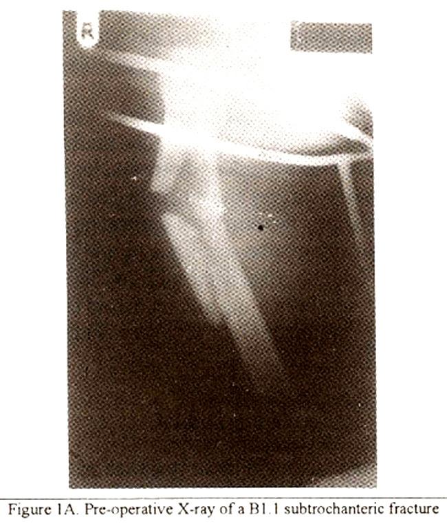
1 Treatment of Comminuted Subtrochanteric Fractures by Dynamic Hip Screw Pages with reference to book, From 212 To 215 Modood Ali ( Department of Surgery, College of Medicine, King Saud University, P.O. Box 641, Abha, Saudi Arabia. ) Abstract Twenty four cases of fracture of the proximal femoral shaft treated with dynamic hip screw fixation are included. All had severe comminution with subtrochanteric extensions. Dynamic hip screw provided stable osteosynthesis and helped to achieve bone union within 18 weeks in 19 of 21 followed-up cases. The anatomical configuration of the femur was maintained with minimal limb length discrepancy in one case and knee flexion of more than 110 degrees in all. Weight bearing was achieved after a mean period of 7.5 weeks post-fixation. Complications of treatment are also presented. Different implants are used to treat this injury with variable results. Dynamic hip screw system is a satisfactory implant for internal fixation in patients with these difficult fractures (JPMA 45:212, 1995). Introduction The proximal femur includes the head, neck and the trochanters with the adjoining region. The precise extent ofthe region designated the subtrochanteric area remains undefined. Commonly this includes the area from the level of the lesser trochanter to within the center of the isthmus of the femoral shaft 1 and fractures in this area are believed to have a high incidence of unsatisfactory results alter operative treatment. This paper analyses the result of treatment with Dynamic Hip Screw (DHS) system of extensively comminuted fractures. of the upper femoral shaft with subtrochanteric extension. The fractures reviewed in this study do not include the intertrochanteric or subtrochanteric fractures that are considered within the premise of hip fractures nor are all fractures limited to the upper third of the femoral shaft. Inclusion of trochantenc fractures is believed to confuse the analysis of results of treatment 2. Patients and Methods This is a retrospective study of 24 cases treated at Asir Central Hospital, Abha, Saudi Arabia, between January, 1988 and June, The sample included 5 females and 19 males. The age ranged between 20 and 60 years, the mean being 36.5 years. The peak incidence was observed to occur between 20 to 40 years. Twenty- three patients had been involved in road traffic accidents and one had fallen from a height. Left sided fractures accounted for 15 cases and right for 9 cases. There were no open fractures. Fourteen patients had associated osseous and/or torso injuries. Patients were treated with DHS fixation using a long barrel plate. The type of fractures treated, were broadly of the types B 1.1, B2.1, B3. land C3.l of the AO classification 3. All fractures involved the proximal shalt of femur with upward extension. They were severely comminuted (Figure 1).
2
.")
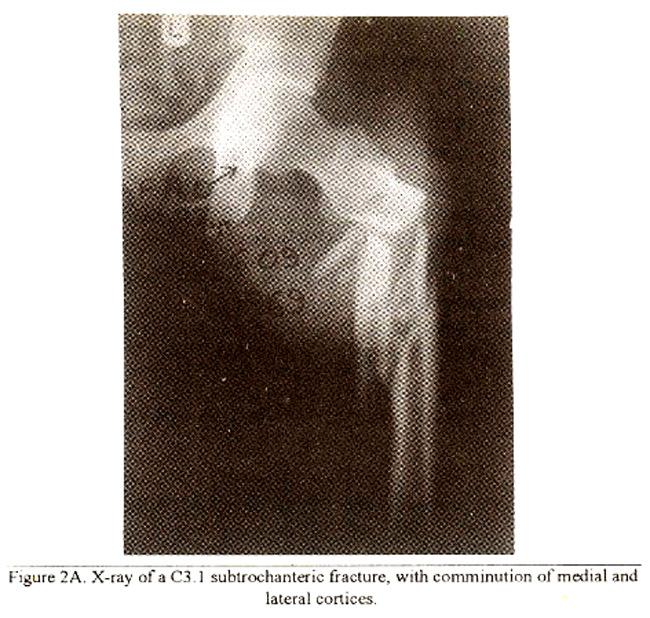
3 Fifteen cases had dismptionof the medial cortex, two of these had fractures of both medial and lateral cortices (Figure 2).
4
.")
5 Comminution of the lateral cortex was present in 5 cases and vertical splitting of the femoral shalt in 4 cases, one of which was associated with anterior cortex separation. Twenty one cases were followed up in the clinic over a mean period of 14.8 months (range 8-28 months). Surgical Treatment
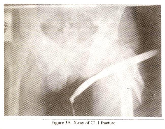
6 The patients were positioned on a fracture table. Fracture fragments were manipulated and checked under image intensifier until gross bony alignment was achieved. The fracture was approached through a lateral incision 4. Vastus lateralis muscle was released at its upper attachment, mobilized and retracted to expose the fracture. Through a combination of image intensification and visual inspection., the fracture was reduced and fragments repositioned anatomically. Caution was exercised to avoid excessive soft tissue dissection from the fragments particularly on the medial side. Bone clamps were used to hold reduction. Interfragnientary screws or cerciage wire were used, when necessary, to keep fragments in position. Under intensifier control, a cancellous hip screw, of suitable length, was inserted into the neck and head of femur. Along, 10 to 12 hole, barrel plate was then coupled and held in position with screws. Cancellous bone grafts for medial cortex deficiency were used in 7 cases only. The wound was closed when stability of the fracture was achieved. Active movements of the knee were instituted on the first postoperative day. Non-weight-bearing ambulation was allowed on the third post operative day and graduated weight bearing was allowed in accordance with the appearance of radiological signs of union. Full weight bearing was achieved at a mean period of 7.5 weeks (range 6-16 weeks). Patients were allowed to return to sedentary jobs once they were fully weight bearing. Mobilization was delayed in patients with associated head or upper limb injuries. However, active and passive movements were encouraged in bed, before upright mobilization was allowed. An antibiotic (Cephalosporin) was administered prophylactically in all patients. Results Union was achieved in 19 patients between 8 and 18 weeks (mean 11.5 weeks) (Figure 3).
7

8 None of the patients had less than 110 degrees of knee flexion. Pseudomonas infection was encountered in one case with uncontrolled diabetes mellitus. This resulted in delayed healing of the fracture and was treated with debndenient, insertion of antibiotic impregnated cement beads and repeated lavage with systemic antibiotic therapy. The fracture healed 20 months afterfixation and did
9 not require removal of implant. One patient developed non-union. This patient, in retrospect, should have had a primary bone graft for his medial comminution. He was subsequently treated by bone graft 18 months after his first surgery and proceeded to healing, though with avams deformity resulting in a shortening of 2.5 cms. Deep vein thrombosis occurred in one case who was confined in bed due to associated injuries. This delayed the time to achieve weight-bearing and increased the duration of hospitalisation. He made full recovery with appropriate medication. Discussion The limiting factors in these fractures were the amoutit of bone mass involved and the degree of comminution. The linear extension into the proximal subtrochanteric region carried the diaphyseal fractures into an area of high stress concentration with compression on the inside and tension on the outside 5. These factors posed additional problems in achieving adequate osteosynthesis. The variety of methods available for the management of such fractures include intramedullaiy nails, with or without cerclage wire or tangential screw fixation, intramedullary nails with locking 1,6, Smith-Peterson pin and plate, fixed angled nail plate 2,7, condylar blade plate 8,9, Zickel nail 10, codviocephalic insertion of malleable rods 11,12, dynamic condylar screw 13 and sliding screw system 14. This spectrum reflects the difficulty in the management of such fractures 15. Each of these modalities has listed advantages for its use and variable results have been obtained by each However, no single option of treatment can be recommended for all cases 20. Favourable results have been obtained by the use of different sliding screws14,21,22. Head penetration may be avoided and the rigid plate enables stabilization of the fracture. Failure generally result from technical 20 or mechanical 23 errors. In our patients DHS allowed a firm hold on the proximal fragment. When coupled with a long barrel plate, firm fixation was obtained. Reposition of the medial cortex under direct and intensified visualization reduced soft tissue stripping from the fracture fragments. This minimized the chance of avascular necrosis and enabled medialization of the cortex. Primary union in 19 of these cases, achieved within 18 weeks implies that the judicious use of the DHS system can favourably influence osteosynthesis of femoral shaft fractures irrespective of the degree ofcomminution and location. An anatomical reduction, stable fixation and restoration of the medial cortex are important for tile success of the dynamic hip screw 21,24. A predominantly satisfactory clinical result obtaified in the majority of our patients, all of whom were victims of severe tmuma, supports this contention. It is concluded that with appropriate reduction and adequate bone contact, fixation with compression hip screw can allow stable osteosynthesis and ensure satisfactory bone healing in such difficult fractures. References 1. Russell, T. A. Fractures of Hip and Pelvis. In 8th ed. Crenshaw, A. H., ed. Campbell s Operative Orthopaedics. St. Louis, Missouri, Mosby Year Book Inc., 1992; pp DeLee, J. C. Fractures and Dislocations ofthe Hip. In: 2nd ed. Rockwood, C. A., Green, D. P eds. Fractures in Adults. Philadelphia, J. B. Lippincott, 1984, pp Muller, M. E., Nazarian, S., Koch, P. et al. The comprehensive classification of Fractures of Long Bones. Berlin, Heidelberg, Springer-Verlag, 1990, pp Ruedi, 1., von Hochstetter, A. H. C. and Schlumpf, R. Surgical Approaches for Internal Fixation. Berlin Heidelberg, Springer- Verlag, 1984, pp Inman, V. T. Functional Aspects of the Abductor Muscles of the Hip. J. Bone Joint Surg., 1 947:29: Wiss, D. A, and Brien, W. W. Subtrochanteric fractures of the femur, Results of treatment by
10 interlocking nailing. Clin. Ortho., 1992, Iqbal, M. Q. An Appraisal of Treatment of Femoral Shaft Fractures, Open Vs. Closed. Med. J. Aust., 1976;1 : Muller, M. E., Ailgower, M.. Schneider, R. et al. Manual of Internal Fixation-Techniques Recommended by the AO-ASIF Crroup. Berlin Heidelberg, Springer-Verlag. 1992, pp Kinast, C., Bolhofner, B. R., Mast, J. W. et al. Subtrochanteric fractures of the femur. Results of treatment with the 95 degrees condylar blade-plate. Clin. Orthop., 1989;238: Zickel, R. E. An Intramedullary Fixation Device for the Proximal Part of the Femur. J. Bone Joint Surg., 1976;58-A: Kudema, H., Bohler, N. and Collon, D. J. Treatment of lntertrochanteric and Subtrochanteric Fractures of the Hip by the Ender Method, J. Bone Joint Surg., 1976;58-A: Harris, L. J. Condylocephalic Nailing of Proximal Femoral Fractures. Instr CourseLect., 1983;32: Sanders, R. and Regazzoni, P. Treatment of subtrochanterie femur fractures using the dynamic condylar screw. J. Orthop. Trauma, 1989;3: Massie, W. K Extracapsular Fractures of the I-Lip Treated by Impaction Using a Sliding Nail Plate. Clin. Orthop., 1962;22: Waddel, J. P. Altematives in the management ofsuhtrochanterit fractures. Instr. Course Lect., 1984,33: Papagiannopoulos, G., Stewart, H. D. and Lunn. P.G. Treatment ofsubtrochanteric fractures of the femur: a study of intramedullary compression nailing Injury, 1989;20: Thomas, W. G. and Villar, R. N. Suhtroehanteric fractures- Zickel nail or nail-plate? J. Bone Joint Surg. (Br.), 1986:68: Whitelaw, G. P., Segal, D., Sanzone, C. F. et al. Unstable intertrochanteric/subtrochanteric fractures of the femur. Clin. Orthop., 1990; Dobozi, W.R., Larson, B. J., Zindriek, M. et al. Flexible intramedullary nailing ofsubtrochanteric fractures of the femur. A multicenter analysis. Clin. Orthop., 1986; Simpson, A.H.R.W., Varthy, K., Dood, C.A.F. Sliding Hip Screws: Modes of failure. injury, 1989;20: Senter, B., Kendig, R. and Savoie, F. H. Operative stabilization of subtrochanteric fractures of the femur, J. Orthop. Trauma, 1990;4: Gruss, M. and Traut, R. Management of unstable pertrochanteric and per-to subtrochanteric femoral fractures with the dynamic hip screw. Aktuel. Traumatol., 1992;22; Mainds, C. C. and Newman, R J. Implant Failures in Patients with Proximal Fernoral Fractures Treated with a Sliding Screw Device. Injury, 1989;20: Kyle, R. F Fractures of the Proximal Part of the Femur. J. Bone Joint Surg. (Am.), 1994;76-A :
JMSCR Vol. 03 Issue 08 Page August 2015
 www.jmscr.igmpublication.org Impact Factor 3.79 Index Copernicus Value: 5.88 ISSN (e)-2347-176x ISSN (p) 2455-0450 DOI: http://dx.doi.org/10.18535/jmscr/v3i8.08 Study of Functional and Radiological Outcome
Fractures of the tibia shaft treated with locked intramedullary nail Retrospective clinical and radiographic assesment
 ARS Medica Tomitana - 2013; 4(75): 197-201 DOI: 10.2478/arsm-2013-0035 Șerban Al., Botnaru V., Turcu R., Obadă B., Anderlik St. Fractures of the tibia shaft treated with locked intramedullary nail Retrospective
A comparative study of 30 cases of trochanteric fracture femur treated with dynamic hip screw and proximal femoral nailing
 Original Article A comparative study of 30 cases of trochanteric fracture femur treated with dynamic hip screw and proximal femoral nailing Jaswinder Pal Singh Walia *, Himanshu Tailor**, H S Mann ***,
Failed Subtrochanteric Fracture How I Decide What to Do?
 Failed Subtrochanteric Fracture How I Decide What to Do? Gerald E. Wozasek Thomas M. Tiefenboeck 5 October 2016, Washington Medical University of Vienna, Department of Trauma Surgery ordination @wozasek.at
MRIMS Journal of Health Sciences 2016;4(1) pissn: , eissn:
 pissn: , eissn:") MRIMS Journal of Health Sciences 216;4(1) pissn: 2321-76, eissn: 2321-7294 http://www.mrimsjournal.com/ Original Article A comparative study of proximal femoral nailing and dynamic hip screw in the management
MRIMS Journal of Health Sciences 216;4(1) pissn: 2321-76, eissn: 2321-7294 http://www.mrimsjournal.com/ Original Article A comparative study of proximal femoral nailing and dynamic hip screw in the management
Distal femoral fracture with subsequent ipsilateral proximal femoral fracture
 Distal femoral fracture with subsequent ipsilateral proximal femoral fracture by M Agarwal, MS FRCS, AA Syed, FRCSI, PV Giannoudis (!), BSc,MB,MD,EEC(Orth) Dept. of Orthopaedics and Trauma, St.James University
Technique Guide. LCP Proximal Femoral Hook Plate 4.5/5.0. Part of the LCP Periarticular Plating System.
 Technique Guide LCP Proximal Femoral Hook Plate 4.5/5.0. Part of the LCP Periarticular Plating System. Table of Contents Introduction Features and Benefits 2 AO ASIF Principles 4 Indications 5 Surgical
Salvage of failed dynamic hip screw fixation of intertrochanteric fractures
 Injury, Int. J. Care Injured (2005) xxx, xxx xxx 1 www.elsevier.com/locate/injury 2 3 4 5 6 7 8 8 9 10 11 12 13 14 15 Salvage of failed dynamic hip screw fixation of intertrochanteric s G.Z. Said, O. Farouk
THE RING BUTTERFLY FRAGMENT
 THE RING BUTTERFLY FRAGMENT Pages with reference to book, From 80 To 82 Philip D`Sousa, Masood Umer, Riaz Hussain Lakdawala ( Department of Surgery (Orthopaedics), The Aga Khan University, Karachi. ) In
Use of Unlocked Intramedullary Nailing in Winquist Type I and II Femoral Isthmus Fracture
 Use of Unlocked Intramedullary Nailing in Winquist Type I and II Femoral Isthmus Fracture HT Ling, MBBS (UM), WM Ng, MS Ortho (UM), MK Kwan, MS Ortho (UM), LK Fathi Aizuddeen, MBBS (UM), PCM Tay, MBBS
Comparitive Study between Proximal Femoral Nailing and Dynamic Hip Screw in Intertrochanteric Fracture of Femur *
 Open Journal of Orthopedics, 2013, 3, 291-295 Published Online November 2013 (http://www.scirp.org/journal/ojo) http://dx.doi.org/10.4236/ojo.2013.37053 291 Comparitive Study between Proximal Femoral Nailing
9/24/2015. When Can I Use a SHS? When CAN T I Use a SHS? Sliding Hip Screw. Time proven. Technically simple. Cheap. Quick
 When Can I Use a SHS? Frank A. Liporace, MD Associate Professor Director of Orthopaedic Trauma Research Director of Orthopaedic Trauma Jersey City Medical Center New York University / Hospital for Joint
Valgus subtrochanteric osteotomy for malunited intertrochanteric fractures : Our experience in 5 cases
 Original article : Valgus subtrochanteric osteotomy for malunited intertrochanteric fractures : Our experience in 5 cases Rajendraprasad Butala *, Sunirmal Mukherjee, Prakash Samant, Ravindra Khedekar
INTERTROCHANTERIC FEMORAL FRACTURES TREATED BY DYNAMIC HIP SCREW
 29 INTERTROCHANTERIC FEMORAL FRACTURES TREATED BY DYNAMIC HIP SCREW Muhammad Ayoub Laghari, Asadullah Makhdoom, Pir Abdul latif Qureshi, Abbass Memon, Faheem Ahmed Memon, Professor Khaleeque Ahmed Siddiqui
Assessment of Prognosis of Patients with Intertrochanteric Fractures Undergoing Treatment with PFN: An Observational Study
 Original article: Assessment of Prognosis of Patients with Intertrochanteric Fractures Undergoing Treatment with PFN: An Observational Study Gajraj Singh 1, Sandhya Gautam 2 1Assistant Professor, Department
Vasu Pai FRACS, MCh, MS, Nat Board Ortho Surgeon Gisborne
 Vasu Pai FRACS, MCh, MS, Nat Board Ortho Surgeon Gisborne FRACTURE MANAGEMENT I Simple closed fracture : Complete or Incomplete Stable or unstable II Open fracture III Multiple fracture IV Polytrauma Fractures
BRIDGE PLATING OF COMMINUTED SHAFT OF FEMUR FRACTURES
 BRIDGE PLATING OF COMMINUTED SHAFT OF FEMUR FRACTURES Mohammad Abul kalam, Pradeep Kumar, Mohammad Afzal Hussain and Iqbal Ahmad Abstract A prospective study of forty comminuted femoral shaft fractures,
Provision of Rotational Stability: Prevention of Collapse: Closed Fracture Reduction: Minimally Invasive Surgery with no Exposure of the Fracture:
 INTRODUCTION Percutaneous Compression Plating was developed by considering each of the stages in the surgical procedure for pertrochanteric fractures and the ways in which these might be improved. Primary
The Lateral Trochanteric Wall A Key Element in the Reconstruction of Unstable Pertrochanteric Hip Fractures
 CLINICAL ORTHOPAEDICS AND RELATED RESEARCH Number 425, pp. 82 86 2004 Lippincott Williams & Wilkins The Lateral Trochanteric Wall A Key Element in the Reconstruction of Unstable Pertrochanteric Hip Fractures
Internal fixation of femoral neck fractures
 Acta Orthop Scand 55, 423-429, 1984 Internal fixation of femoral neck fractures Compression screw compared with nail plate fixation In a prospective, randomized study of femoral neck fracture operations,
Comparative Study of Fixation Devices for Intertrochanteric Fractures
 Comparative Study of Fixation Devices for Intertrochanteric Fractures C. Sticlaru * A. Davidescu Politehnica University of Timişoara Politehnica University of Timişoara Timişoara, România Timişoara, România
Types of Plates 1. New Dynamic Compression Plate: Diaphyseal fracture: Radius, Ulna, Humerus, Rarely tibia
 Types of Plates 1. New Dynamic Compression Plate: DCP Diaphyseal fracture: Radius, Ulna, Humerus, Rarely tibia 1. Undercut adjacent to the holes low contact: less stress shield 2. Undercut at the undersurface
Indirect Reduction with Sliding Compression Screw Stabilization for Subtrochanteric Fractures
 Original Article 190 Indirect Reduction with Sliding Compression Screw Stabilization for Subtrochanteric Fractures Yu-Tun Hsu, MD; Chi-Chuan Wu, MD; Chun-Yi Su, MD; Kuo-Fun Fan, MD; I-Chuan Tseng, MD;
DISLOCATION AND FRACTURES OF THE HIP. Dr Károly Fekete
 DISLOCATION AND FRACTURES OF THE HIP Dr Károly Fekete 1 OUTLINE Epidemiology Incidence Anatomy Patient s examination, clinical symptons Diagnosis Classification Management Special complications 2 EPIDEMIOLOGY,
Biomet Large Cannulated Screw System
 Biomet Large Cannulated Screw System s u r g i c a l t e c h n i q u e A Complete System for Simplified Fracture Fixation 6.5mm & 7.3mm The Titanium, Self-drilling, Self-tapping Large Cannulated Screw
A Clinical Study For Evaluation Of Results Of Closed Interlocking Nailing Of Fractures Of The Shaft Of The Tibia
 ISPUB.COM The Internet Journal of Orthopedic Surgery Volume 17 Number 2 A Clinical Study For Evaluation Of Results Of Closed Interlocking Nailing Of Fractures Of The Shaft Of R Gupta, T Motten, N Kalsotra,
Closed reduction and internal fixation of fractures of the shaft of the femur by the Titanium Elastic Nailing System in children.
 ISPUB.COM The Internet Journal of Orthopedic Surgery Volume 17 Number 1 Closed reduction and internal fixation of fractures of the shaft of the femur by the Titanium Elastic Nailing System in children.
EXTENDED TROCHANTERIC OSTEOTOMY SURGICAL TECHNIQUE FPO EXTENSIVELY COATED FIXATION
 EXTENDED TROCHANTERIC OSTEOTOMY SURGICAL TECHNIQUE FPO EXTENSIVELY COATED FIXATION SINCE 1983 PREOPERATIVE PLANNING EXPLANTATION OPTIONS the cement from inside the cement canal until the bone/ cement bond
Type I : At the level of lesser trochanter Type II : Less than 2.5 cm below lesser trochanter. Type III : cm below lesser trochanter
 Type II : Major fracture line along the intertrochanteric line with communition in coronal plain. Type III : Fracture at the level of lesser trochanter with variable communition and extension in subtrochanteric
Ipsilateral femoral neck and shaft fractures: a retrospective analysis of two treatment methods
 J Orthopaed Traumatol (2008) 9:141 147 DOI 10.1007/s10195-008-0025-3 ORIGINAL ARTICLE Ipsilateral femoral neck and shaft fractures: a retrospective analysis of two treatment methods Roop Singh Æ Rajesh
Trochanter Stabilization Plate for DHS Implants
 Extends DHS Plate Construct to Help Stabilize Greater Trochanter Trochanter Stabilization Plate for DHS Implants Surgical Technique Table of Contents Introduction Trochanter Stabilization Plate for DHS
Cannulated Pediatric Osteotomy System (CAPOS)
") A Single System of Osteotomy Blade Plates and Cannulated Instrumentation Cannulated Pediatric Osteotomy System (CAPOS) Surgical Technique Table of Contents Introduction Cannulated Pediatric Osteotomy System
A Single System of Osteotomy Blade Plates and Cannulated Instrumentation Cannulated Pediatric Osteotomy System (CAPOS) Surgical Technique Table of Contents Introduction Cannulated Pediatric Osteotomy System
HOW TO CITE THIS ARTICLE:
 A COMPARATIVE STUDY OF FUNCTIONAL OUTCOME BETWEEN DYNAMIC HIP SCREW AND PROXIMAL FEMORAL NAIL IN SURGICAL MANAGEMENT OF PER-TROCHANTERIC FRACTURES Umesh M. Shivanna 1, Girish H. Rudrappa 2 HOW TO CITE
Unlocked Nailing vs. Interlocking Nailing for Winquist Type I and II Femoral Isthmus Fractures. Is there a Difference?
 Unlocked Nailing vs. Interlocking Nailing for Winquist Type I and II Femoral Isthmus Fractures. Is there a Difference? CK Yu, MBBS (UM), HY Wong*, MD (UKM), AS Vivek, FRCS (Edin), BC Se To*, FRCS (Edin)
ILIZAROV TECHNIQUE IN CORRECTING LIMBS DEFORMITIES: PRELIMINARY RESULTS
 Bahrain Medical Bulletin, Volume 17, Number 2, June 1995 Original ILIZAROV TECHNIQUE IN CORRECTING LIMBS DEFORMITIES: PRELIMINARY RESULTS Saleh W. Al-Harby, FRCS(Glasg)* This is a prospective study of
Pre-Operative Planning. Positioning of the Patient
 Surgical Technique Pre-Operative Planning Decide upon the size and angle of the barrel plate to be used from measuring the x-rays. To maximise the sliding action when using shorter lag screws, the Short
COMPARATIVE STUDY OF MANAGEMENT OF DIAPHYSEAL FEMUR FRACTURE WITH INTRAMEDULLARY INTERLOCKING NAIL AND K. NAIL
 International Journal of Innovation and Applied Studies ISSN 2028-9324 Vol. 15 No. 3 Apr. 2016, pp. 560-564 2016 Innovative Space of Scientific Research Journals http://www.ijias.issr-journals.org/ COMPARATIVE
Treatment of traumatic conditions of the femur using the Huckstep nail
 East and Central African Journal of Sulgey Vol. 2, No. I Treatment of traumatic conditions of the femur using the Huckstep nail T F Wisniewski MD PhD(0rth) Department of Orthopaedic Surgery and Bone and
ORIGINAL ARTICLE. INTER TROCHANTERIC # NECK FEMUR FIXATION WITH TFN 250 CASES. Prasad Vijaykumar Joshi, Chandrashekar Yadav.
 INTER TROCHANTERIC # NECK FEMUR FIXATION WITH TFN 250 CASES. Prasad Vijaykumar Joshi, Chandrashekar Yadav. 1. Assistant Professor. Department of Orthopaedics, Joshi Hospital Pvt. Ltd. Phaltan, Maharashtra.
TECHNIQUE GUIDE. DHS /DCS Dynamic Hip and Condylar Screw System
 DHS /DCS Dynamic Hip and Condylar Screw System TECHNIQUE GUIDE Original Instruments and Implants of the Association for the Study of Internal Fixation A0 ASIF Contents Introduction to the Dynamic Hip Screw
Technique Guide. DHS Blade. For osteoporotic bone.
 Technique Guide DHS Blade. For osteoporotic bone. Table of Contents Introduction Features and Benefits 2 Indications and Contraindications 4 Clinical Cases 5 Surgical Technique Implantation 6 Implant
CASE REPORT. Bone transport utilizing the PRECICE Intramedullary Nail for an infected nonunion in the distal femur
 PRODUCTS CASE REPORT Bone transport utilizing the PRECICE Intramedullary Nail for an infected nonunion in the distal femur Robert D. Fitch, M.D. Duke University Health System 1 1 CONDITION Infected nonunion
The Journal of the Korean Society of Fractures Vol.13, No.3, July, 2000
 The Journal of the Korean Society of Fractures Vol13, No3, July, 2000 2, 3 ) : 40-12, Tel : (02) 966-1616 Fax : (02) 968-2394 E-mail : adkajs@thrunetcom 471 8, 1 2 ) (Table 1) 1 6 14, 2 1 2 1 Ender 29
The Journal of the Korean Society of Fractures Vol.16, No.1, January, 2003
 The Journal of the Korean Society of Fractures Vol16, No1, January, 2003 : 351 ( )463-712 TEL: (031) 780-5270/5271 FAX : (031) 708-3578 E-mail: bskima@netsgocom 16,, ( > 20mm ) 5, ) 20 % 1 ), 6,, 3 8 8
ICUC Paper. The treatment of trochanteric fractures revisited: Pietro Regazzoni, Alberto Fernandez, Dominik Heim, Stephan M. Perren.
 The treatment of trochanteric fractures revisited: Pietro Regazzoni, Alberto Fernandez, Dominik Heim, Stephan M. Perren September 2016 An optimal treatment of hip fractures is crucial because of the great
LCP Low Bend Medial Distal Tibia Plates 3.5 mm. Anatomic plates with low profile head for intra- and extraarticular fractures.
 LCP Low Bend Medial Distal Tibia Plates 3.5 mm. Anatomic plates with low profile head for intra- and extraarticular fractures. Surgical Technique This publication is not intended for distribution in the
Technique Guide. 3.5 mm LCP Periarticular Proximal Humerus Plate. Part of the Synthes locking compression plate (LCP) system.
 system.") Technique Guide 3.5 mm LCP Periarticular Proximal Humerus Plate. Part of the Synthes locking compression plate (LCP) system. Table of Contents Introduction 3.5 mm LCP Proximal Humerus Plate 2 AO Principles
Technique Guide 3.5 mm LCP Periarticular Proximal Humerus Plate. Part of the Synthes locking compression plate (LCP) system. Table of Contents Introduction 3.5 mm LCP Proximal Humerus Plate 2 AO Principles
LISS DF and LISS PLT. Less Invasive Stabilization Systems for Distal Femur and Proximal Lateral Tibia.
 LISS DF and LISS PLT. Less Invasive Stabilization Systems for Distal Femur and Proximal Lateral Tibia. LISS DF and LISS PLT. Less Invasive Stabilization Systems for Distal Femur and Proximal Lateral Tibia.
VA-LCP Anterior Clavicle Plate. The anatomically precontoured fixation system with angular stability for clavicle shaft and lateral clavicle.
 Technique Guide VA-LCP Anterior Clavicle Plate. The anatomically precontoured fixation system with angular stability for clavicle shaft and lateral clavicle. Table of Contents Introduction VA-LCP Anterior
3.5 mm Clavicle Hook Plates
 A Single Solution for Lateral Clavicle Fractures and Acromioclavicular Joint Dislocations 3.5 mm Clavicle Hook Plates Surgical Technique Discontinued December 2017 DSUS/TRM/1016/1126(1) Table of Contents
Cannulated Pediatric Osteotomy System (CAPOS). A single system of osteotomy blade plates and cannulated instrumentation.
. A single system of osteotomy blade plates and cannulated instrumentation.") Cannulated Pediatric Osteotomy System (CAPOS). A single system of osteotomy blade plates and cannulated instrumentation. Technique Guide This publication is not intended for distribution in the USA. Instruments
Cannulated Pediatric Osteotomy System (CAPOS). A single system of osteotomy blade plates and cannulated instrumentation. Technique Guide This publication is not intended for distribution in the USA. Instruments
PediNail Pediatric Femoral Nail
 PediNail Pediatric Femoral Nail Surgical Technique Table of Contents Indications...3 Patient Positioning...3 Approach...4 Reaming...5 Nail Placement...6 Proximal Interlocking...7 Distal Interlocking...8
A comparative study of less invasive stabilization system and titanium elastic nailing for subtrochanteric femur fractures in older children
 Acta Orthop. Belg., 2015, 81, 123-130 ORIGINAL STUDY A comparative study of less invasive stabilization system and titanium elastic nailing for subtrochanteric femur fractures in older children Liao-Jun
Medial Malleolus Fracture Fixation in the Setting of Concomitant Tibial Shaft Fractures
 Medial Malleolus Fracture Fixation in the Setting of Concomitant Tibial Shaft Fractures Stephen R. Barchick 1, BA Andrew P. Matson 2, MD Samuel B. Adams 2, MD 1. Duke University School of Medicine, Durham,
Randomized comparative study to evaluate the role of proximal femoral nail and dynamic hip screw in unstable trochanteric fractures
 International Journal of Research in Orthopaedics Mayi SC et al. Int J Res Orthop. 2016 Sep;2(3):75-79 http://www.ijoro.org Research Article DOI: http://dx.doi.org/10.18203/issn.2455-4510.intjresorthop20162618
Angular Malalignment in Subtrochanteric and Proximal Shaft Femur Fractures after Intramedullary Nailing using SIGN Nails
 Angular Malalignment in Subtrochanteric and Proximal Shaft Femur Fractures after Intramedullary Nailing using SIGN Nails Rolando Junior L. Torres, MD Jeremiah R. Morales, MD, FPOA Subtrochanteric Femur
PediLoc 3.5mm and 4.5mm Contour Femur Plate Surgical Technique
 PediLoc 3.5mm and 4.5mm Contour Femur Plate Surgical Technique Surgical Technique Contour Femur Plate The technique description herein is made available to the healthcare professional to illustrate the
Nonunion of the Femur Treated with Conventional Osteosynthesis Combined with Autogenous and Strut Allogeneic Bone Grafts
 Original Article 268 Nonunion of the Femur Treated with Conventional Osteosynthesis Combined with Autogenous and Strut Allogeneic Bone Grafts Lin-Hsiu Weng, MD; Jun-Wen Wang, MD Background: In this study,
TREATMENT OF SUBTROCHANTERIC FEMUR FRACTURES WITH PROXIMAL FEMORAL NAILS: A PROSPECTIVE STUDY
 Original Article Orthopaedics TREATMENT OF SUBTROCHANTERIC FEMUR FRACTURES WITH PROXIMAL FEMORAL NAILS: A PROSPECTIVE STUDY Ravindra S. Patil 1, Dhanish V. Mehendiratta 2, Sahil Bhagat 2, Rishi Doshi 2,
Mandible External Fixator II. Provides treatment for fractures of the maxillofacial area.
 Mandible External Fixator II. Provides treatment for fractures of the maxillofacial area. Technique Guide This publication is not intended for distribution in the USA. Instruments and implants approved
Large Distractor Femur
 Fracture Reduction and Provisional Stabilization Large Distractor Femur Surgical Technique Table of Contents Introduction Standard Femoral Distraction 2 Large Distractor System 4 Surgical Technique Prepare
OUTCOME OF MANAGEMENT OF CLOSED PROXIMAL TIBIA FRACTURES IN TERTIARY HOSPITAL OF SURAT Karan Mehta 1, Prashanth G 2, Shiblee Siddiqui 3
 OUTCOME OF MANAGEMENT OF CLOSED PROXIMAL TIBIA FRACTURES IN TERTIARY HOSPITAL OF SURAT Karan Mehta 1, Prashanth G 2, Shiblee Siddiqui 3 HOW TO CITE THIS ARTICLE: Karan Mehta, Prashanth G. Shiblee Siddiqui,
Pelvis injuries Fractures of the femur (proximal,shaft) Dr Tamás Bodzay
 Dr Tamás Bodzay") Pelvis injuries Fractures of the femur (proximal,shaft) Dr Tamás Bodzay Pelvis anatomy Pelvis function - axial load bearing - protection: abdominal, pelvic structures Pelvic injury mechanism Falling from
Pelvis injuries Fractures of the femur (proximal,shaft) Dr Tamás Bodzay Pelvis anatomy Pelvis function - axial load bearing - protection: abdominal, pelvic structures Pelvic injury mechanism Falling from
Elbow Hinge Fixator. Guided Flexion/Extension for Unstable Elbow Fractures.
 Elbow Hinge Fixator. Guided Flexion/Extension for Unstable Elbow Fractures. Surgical Technique MR Safe Radiolucent Table of Contents System Description 3 Indications and Contraindications 4 Fixation Components
QUICK REFERENCE GUIDE. The XCaliber Meta-Diaphyseal Fixator
 17 The XCaliber Meta-Diaphyseal Fixator GENERAL POINTS The XCaliber Fixator is made of radiolucent material for unobstructed X-ray visualization. The metallic bolts and the cam and bush of each ball-joint,
Technique Guide. 3.5 mm LCP Low Bend Medial Distal Tibia Plates. Part of the Synthes locking compression plate (LCP) system.
 system.") Technique Guide 3.5 mm LCP Low Bend Medial Distal Tibia Plates. Part of the Synthes locking compression plate (LCP) system. Table of Contents Introduction 3.5 mm LCP Low Bend Medial Distal Tibia Plates
Technique Guide 3.5 mm LCP Low Bend Medial Distal Tibia Plates. Part of the Synthes locking compression plate (LCP) system. Table of Contents Introduction 3.5 mm LCP Low Bend Medial Distal Tibia Plates
Low Bend Distal Tibia Plates
 Part of the DePuy Synthes Locking Compression Plate (LCP ) System 3.5 mm LCP Low Bend Medial Distal Tibia Plates Surgical Technique Table of Contents Introduction 3.5 mm LCP Low Bend Medial Distal Tibia
Clinical outcomes of muscle pedicle bone grafting (Meyer's Procedure) in cases of old displaced femur neck fractures: A Study Of 20 Cases
 in cases of old displaced femur neck fractures: A Study Of 20 Cases") ISPUB.COM The Internet Journal of Orthopedic Surgery Volume 10 Number 1 Clinical outcomes of muscle pedicle bone grafting (Meyer's Procedure) in cases of old displaced femur neck fractures: A Study Of
ISPUB.COM The Internet Journal of Orthopedic Surgery Volume 10 Number 1 Clinical outcomes of muscle pedicle bone grafting (Meyer's Procedure) in cases of old displaced femur neck fractures: A Study Of
Hip Fractures. Anatomy. Causes. Symptoms
 Hip Fractures A hip fracture is a break in the upper quarter of the femur (thigh) bone. The extent of the break depends on the forces that are involved. The type of surgery used to treat a hip fracture
INTERTAN Nails Geared for Stability
 Geared for stability The TRIGEN INTERTAN nail brings advanced TRIGEN nail technology to hip fractures. With a unique integrated, interlocking screw construct, TRIGEN INTERTAN nail provides all the benefits
Intramedullary Nailing: History & Rationale
 Intramedullary Nailing: History & Rationale Overview 1. What is IM Nailing? 2. History 3. Design Rationale & Evolution 4. Modern IM Nails 5. The Future What is IM Nailing? Method of internal fixation in
Subtrochanteric femoral fracture is one of
 Chinese Journal of Traumatology 200; 3():37-4. 37. Treatment of subtrochanteric femoral fracture with long proximal femoral nail antirotation WANG Wen-yue 王文岳, YANG Tian-fu 杨天府, FANG Yue 方跃, LEI Ming-ming
LCP Medial Distal Tibia Plate, without Tab. The Low Profile Anatomic Fixation System with Angular Stability and Optimal Screw Orientation.
 LCP Medial Distal Tibia Plate, without Tab. The Low Profile Anatomic Fixation System with Angular Stability and Optimal Screw Orientation. Technique Guide LCP Small Fragment System Table of Contents Introduction
CASE NO: 1 PATIENT DETAILS : Occupation : Housewife Date Of Admission :11/06/15 Residence : Nalgonda IP NO :
 CASE NO: 1 PATIENT DETAILS : Name : XXXX Age : 53yr Sex : Female Occupation : Housewife Date Of Admission :11/06/15 Residence : Nalgonda IP NO : 201518441 CHIEF COMPLAINTS : - Pain in the right knee since
Importance of screw position in intertrochanteric femoral fractures treated by dynamic hip screw
 Orthopaedics & Traumatology: Surgery & Research (2010) 96, 21 27 ORIGINAL ARTICLE Importance of screw position in intertrochanteric femoral fractures treated by dynamic hip screw M. Güven a,, U. Yavuz
Outcome evaluation of dynamic condylar screw fixation for subtrochanteric femur fracture
 2017; 3(1): 351-355 ISSN: 2395-1958 IJOS 2017; 3(1): 351-355 2017 IJOS www.orthopaper.com Received: 22-11-2016 Accepted: 23-12-2016 Shah SN Assistant Professor, Department of Orthopaedics, GMERS Medical
OPERATING MANUAL AND TECHNIQUE GUIDE FOR TITANIUM FEMORAL AND TIBIAL NAILING SYSTEMS
 OPERATING MANUAL AND TECHNIQUE GUIDE FOR TITANIUM FEMORAL AND TIBIAL NAILING SYSTEMS ORTHO-MEDICAL GMBH TITANIUM FEMORAL NAIL OPERATIVE TECHNIQUE Introduction: Why a new type of femoral nail? The latest
Technique Guide. DHS/DCS System. Including LCP DHS and DHS Blade.
 Technique Guide DHS/DCS System. Including LCP DHS and DHS Blade. Table of Contents Introduction System Overview 2 Features and Benefits 4 Indications and Contraindications 6 Clinical Cases 8 Surgical
SWEMAC CHS. Compression Hip Screw System
 SWEMAC CHS Compression Hip Screw System Swemac CHS Compression Hip Screw System This system provides a simple and easy-to-use solution for all surgeons facing hip fractures. Offering a wide choice of hip
Of approximately 2 million long bone fractures
 Proceedings S.Z.P.G.M.I vol: 13(1-2) 1999, pp. 71-75. Treatment of Tibial Non-Union with the Ilizarov Method Pervaiz Iqbal, Muhammad Maq, Hamid Qayum Department of Orthopaedics, Shaikh Zayed Hospital,.
Comparative Study between Locking Compression Plate vs. Supracondylar Nail for Supracondylar Femur Fractures
 Orthop. Res. Rev. DOI: http://dx.doi.org/10.20936/orr/160103 ORIGINAL ARTICLE Comparative Study between Locking Compression Plate vs. Supracondylar Nail for Supracondylar Femur Fractures Abhijeet Shroff
QUICK REFERENCE GUIDE. Arthrodiatasis. Articulated Joint Distraction
 4 Arthrodiatasis Articulated Joint Distraction ARTHRODIATASIS OF THE HIP To prepare the assembly, remove the female component and replace it with the ProCallus articulated body for the hip. Remove cam
Evaluation and Treatment of High Energy Proximal Femur Fractures OVERVIEW 6/23/2014. Introduction of Speakers - Objectives (2 minutes)
") 6/23/2014 Evaluation and Treatment of High Energy Proximal Femur Fractures OVERVIEW Introduction of Speakers - Objectives (2 minutes) Injury Patterns, Evaluation - Patient Considerations Mike Gardner --
6/23/2014 Evaluation and Treatment of High Energy Proximal Femur Fractures OVERVIEW Introduction of Speakers - Objectives (2 minutes) Injury Patterns, Evaluation - Patient Considerations Mike Gardner --
A3.1 Simple, oblique A3.2 Simple, transverse A3.3 Comminuted
 Dynamic hip screw (DHS) - Indications A1 Fractures in the trochanter region, simple pertrochanteric A1.1 Along the intertrochanteric line A1.2 Through the greater trochanter A1.3 Extending distal to the
Femoral Fractures in Adolescents: A Comparison of Four Methods of Fixation
 Femoral Fractures in Adolescents: A Comparison of Four Methods of Fixation By Leonhard E. Ramseier, MD, Joseph A. Janicki, MD, Shannon Weir, BSc, and Unni G. Narayanan, MBBS, MSc, FRCSC Investigation performed
Locked Plating: Clinical Indications
 Techniques in Orthopaedics 22(3):181 185 2007 Lippincott Williams & Wilkins, Inc. Locked Plating: Clinical Indications Kyle F. Dickson, M.D., M.B.A., John Munz, M.D. Summary: As shown in the previous article,
Surgical Care at the District Hospital. EMERGENCY & ESSENTIAL SURGICAL CARE
 Surgical Care at the District Hospital 1 18 Orthopedic Trauma Key Points 2 18.1 Upper Extremity Injuries Clavicle Fractures Diagnose fractures from the history and by physical examination Treat with a
A Prospective Study to Evaluate the Management of Sub-trochanteric Femur Fractures with Long Proximal Femoral Nail
 Malaysian Orthopaedic Journal 2017 Vol 11 No 3 doi: http://dx.doi.org/10.5704/moj.1711.014 Kumar M, et al A Prospective Study to Evaluate the Management of Sub-trochanteric Femur Fractures with Long Proximal
STUDY OF RESULTS OF ENDER NAILING AND CANNULATED CANCELLOUS SCREW IN THE TREATMENT OF INTERTROCHANTERIC FRACTURE FEMUR
 RESEARCH ARTICLE STUDY OF RESULTS OF ENDER NAILING AND CANNULATED CANCELLOUS SCREW IN THE TREATMENT OF INTERTROCHANTERIC FRACTURE FEMUR Bhavik Dalal, Tarkik Amin, Archit Gandhi, Rohit Shah Smt NHL Municipal
Peritroch Hip Fractures. Robert M Harris MD. Hip Fractures. Factors Influencing Construct Strength: Uncontrolled factors 4/28/2016
 Peritroch Hip Fractures Should be treated with an IMHS Robert M Harris MD Hip Fractures General principles Approximately 250,000 hip fractures/ year Cost approximately $8.7 billion annually The number
JOURNALOF ORTHOPAEDIC TRAUMA
 JOT Special Case Report Series CASE REPORTS www.jorthotrauma.com JOURNALOF ORTHOPAEDIC TRAUMA OFFICIAL JOURNAL OF Orthopaedic Trauma Association Belgian Orthopaedic Trauma Association Canadian Orthopaedic
LCP Anterolateral Distal Tibia Plate 3.5. The low profile anatomic fixation system with optimal plate placement and angular stability.
 LCP Anterolateral Distal Tibia Plate 3.5. The low profile anatomic fixation system with optimal plate placement and angular stability. Technique Guide LCP Small Fragment System Table of Contents Introduction
Comparison of two modality of fixation in unstable trochantric fractures in elderly patients
 Original article Comparison of two modality of fixation in unstable trochantric fractures in elderly patients 1Dr. Vipin Garg, 2 Dr. Anjul Agarwal 1MS Orhtopaedics, Assistant professor, Department of orthopaedics,
Pediatric LCP Plate System. For osteotomies and fracture fixation of the proximal and distal femur.
 Pediatric LCP Plate System. For osteotomies and fracture fixation of the proximal and distal femur. Angular stability Intraoperative correction and flexibility Universal design Indications The Pediatric
LCP Anterolateral Distal Tibia Plate 3.5. The low profile anatomic fixation system with optimal plate placement and angular stability.
 LCP Anterolateral Distal Tibia Plate 3.5. The low profile anatomic fixation system with optimal plate placement and angular stability. Technique Guide LCP Small Fragment System Table of Contents Introduction
3.5 mm LCP Distal Tibia T-Plates
 Part of the DePuy Synthes Locking Compression Plate (LCP ) System 3.5 mm LCP Distal Tibia T-Plates Surgical Technique Table of Contents Introduction 3.5 mm LCP Distal Tibia T-Plates 2 AO Principles 4 Indications
Technique Guide. LCP Posterior Medial Proximal Tibial Plate 3.5. Part of the Synthes small fragment LCP system.
 Technique Guide LCP Posterior Medial Proximal Tibial Plate 3.5. Part of the Synthes small fragment LCP system. Table of Contents Introduction LCP Posterior Medial Proximal Tibial Plate 3.5 2 AO Principles
Crossed Steinmann Pin Fixation In Supracondylar Femur Fractures In Adults A Case Series
 Article ID: WMC005027 ISSN 2046-1690 Crossed Steinmann Pin Fixation In Supracondylar Femur Fractures In Adults A Case Series Peer review status: No Corresponding Author: Dr. Mohit K Jindal, Senior Resident,
Comparison between Conventional and Minimally Invasive Dynamic Hip Screws for Fixation of Intertrochanteric Fractures of the Femur
 Central Health Services From the SelectedWorks of Dr Mohit Kumar Patralekh Summer 13 Comparison between and Invasive Dynamic Hip Screws for Fixation of Intertrochanteric Fractures of the Femur Dr Mohit