SWEMAC CHS. Compression Hip Screw System
|
|
|
- Charleen Walker
- 5 years ago
- Views:
Transcription

1 SWEMAC CHS Compression Hip Screw System
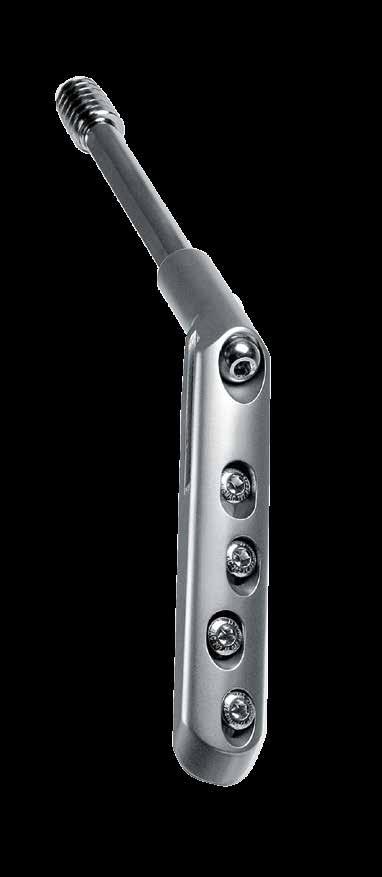
2 Swemac CHS Compression Hip Screw System This system provides a simple and easy-to-use solution for all surgeons facing hip fractures. Offering a wide choice of hip plates combined with a unique lag screw and innovative instrumentation. Implants are available sterile packaged for immediate use. All implants are made from stainless steel 316 LVM. Slimlined hip plates The Swemac Hip Plates has a smoothly curved outer profile and still maintains an extra ordinary mechanical strength. The hip plates are available with plate/barrel angles 130, 135 and 140 and in 2 to 16 holes. The most proximal hole in the hip plate will allow the use of a 6.5 mm selftapping cancellous bone screw. All plates have tracks for the Lateral Support Plate. The Swemac CHS is also available with holes that will allow the use of 5.1 mm locking screws as well as 4.5 mm cortical bone screws. The Swemac CHS Locking Plate is only available in 135 angle. Front-page X-ray by Dr. Johnard


3 Optimal Lag Screw fixation An adequate fixation in the femoral head is crucial for successful fixation of hip fractures. The Swemac Lag Screw has been designed to overcome the strength-reduction in the cancellous bone caused by osteoporosis. Swemac Lag Screw world patent: Patent no: Patent no: Strength of fixation is dependent upon both implant and bone properties. If too much bone trabeculae is removed, the interaction between bone and implant may be impared. To achieve a good fixation, it is vital to save sufficient bone trabeculae in the femoral head. The special design of the triple reamer and the Swemac Lag Screw minimize the disruption of the bone trabeculae. The Swemac Lag Screw has five unique features improving the holding power in the femoral head. l A conical core allows an increase of the total area of the thread. l Starting from the tip, a gradually decreasing distance between the threads compresses the bone and prevents forward rotation. l The threaded part has a consistent outer diameter over the whole length increasing the total area of the thread. l The Swemac Lag Screw has a blunt tip minimizing the risk of femoral head penetration. l A self-cutting compression thread does not destroy bone like traditional cutting flutes. 3


4 Prevents medial displacement The Swemac Lateral Support Plate prevents medial displacement of the femoral shaft relative to the neck and head fragment, while allowing dynamic axial compression to occur between the fractured lateral cortex and the femoral shaft. Adjustable l The Lateral Support Plate allows the surgeon to adjust the position of the support plate, depending on the distance between the plate barrel and the greater trochanter. This distance will vary depending on the plate angle. Low profile l The Lateral Support Plate has been designed to minimize soft tissue irritation. l The Lateral Support Plate has an anatomical curvature templating the greater trochanter. Sevenfold increase in complications if medial displacement is more than 30% Martyn Parker 1997 The Lateral Support Plate should always be used when the lateral cortex is fractured. 4


5 Innovative instrumentation Multi Angle guide with visualization windows l A correct placement of the Guide Wire is the most important step in the whole operative procedure. The Multi Angle Guide allows the surgeon to place the rigid 3.2 mm Guide Wire more accurately in the femoral head. The Guide Wire can be seen under image intensification in the visualisation windows in both AP and lateral views. Ensuring a correct placement of the Guide Wire. l The curvature of the handle reduces the skin incision by 20 mm. Direct length measurement Safe reading Rigid 3.2 mm Guide Wire Stainless steel instrument tray The required length of the 4.5 mm cortical bone screw can be read on the scale of the drill sleeve against the black mark on the 3.2 mm Drill. Less risk of misreading the adjusted length on the triple reamer. The rigid 3.2 mm Guide Wire ensures a quick, precise and safe lag screw placement. The threaded tip guarantees a secure seating in the subcondral bone. The instrument tray is made completely out of stainless steel wire, ensuring good penetration during steam autoclaving. 5

6 Indications Contraindications The physician s education, training and professional judgement must be relied upon to choose the most appropriate device and treatment. Conditions presenting an increased risk of implant failure include: l Any active or suspected latent infection, sepsis or marked local inflammation in or around the surgical area. l Severe osteoporosis, insufficient quantity or quality of bone/soft tissue. l Material sensitivity, documented or suspected. l Physical interference with other implants during implantation or use. l Compromised vascularity, inadequate skin or neurovascular status. l Compromised bone stock that cannot provide adequate support and/or fixation of the device due to disease, infection or prior implantation. l Femoral neck fractures l Stable trochanteric hip fractures. l Unstable trochanteric hip fractures. l Patients who are unwilling or incapable of following postoperative care instructions. l Other physical, medical or surgical conditions that would preclude the potential benefit of surgery. l Previously implanted or extracted osteosynthesis implants of the diaphyseal or proximal femur increases the risk of secondary fracture. l Obesity. An obese patient can produce loads on the implant that can lead to device/treatment failure. l The CHS System is not recommended for use with pediatric hip fractures. The surgeon must discuss all relevant risks, including the service life of the device and the need for postoperative protection of the implant with the patient, when necessary. 6


7 Surgical technique 1. Patient positioning 2. Reduction Place the patient in supine position on the fracture operating table. Position the leg on the healthy side with the hip in flexion and adequate abduction so that the C-arm can be adjusted intraoperatively for both the anterior/posterior view, and the axial view which is necessary to obtain a true axial view of the femoral neck and head. Reduction should be obtained by gentle manipulation according to the normal procedure for unstable fractures. The fracture position should be anatomical or with a slight valgus tilt and held by immobilization on a fracture operating table. The proximal femur should be positioned so that the head and neck are parallel to the floor. The foot should be rotated inwards and fixed between 15 and 30 of internal rotation. The patella should have an either horizontal or slightly inward position. The patient should then be prepared and draped. 7


 (the first Guide Wire can be used), is held under lateral view")


8 3. Locate the optimal point for skin incision B. 3. How to locate the optimal point for skin incision and entry point for the Guide Wire. A Guide Wire (1) and the Multi Angle Guide, are held under AP-view of the image intensifier, above the skin anterior to the hip joint and in line with the medial cortex of the femoral neck. A third Guide Wire, (3) (the first Guide Wire can be used), is held under lateral view of the image intensifier. It is placed along the midline of the axis of the femoral shaft. The point where the second and the third Guide Wires cross, (B), is the optimal starting point for the incision. B. A. A second Guide Wire, (2), is held in a vertical position to the femoral shaft and directed against the point where the Multi Angle Guide and the skin meet. (A). It is important to make the incision in the same angle as the chosen plate angle. 8


9 4. Make incision 5. Introduce the Multi Angle Guide A longitudinal incision is made, distal from this point through the skin. The deep fascia is divided in the direction of the fibres. The lateral cortex of the femur may be approached either directly or posterior-laterally by lifting the vastus lateralis muscle. The area of the femur where the plate is to be positioned is cleared with a raspatorium. Orientation and placement of the Guide Wire is the most critical step in the whole surgical procedure. In the frontal view the Guide Wire should run centrally in relation to the femoral neck and head. Length The length of the incision is determined by the length of the chosen plate. In the lateral view, the Guide Wire should be centered in relation to the femoral head and neck. NOTE: Introduce the Guide Wire through the Multi Angle Guide before starting to drill. The threaded tip of the Guide Wire may otherwise harm the Multi Angle Guide. 9

10 6. Guide Wire insertion The Multi Angle Guide is placed on the lateral cortex and the 3.2 mm Guide Wire is inserted in the desired angle (130, 135 or 140 ). Using image intensification, once the alignment of the Guide Wire is satisfactory, it is advanced to subchondral bone of the femoral head. The rigid 3,2 mm Guide Wire will allow the surgeon to adjust the position of the Guide Wire slightly while drilling. If the bone quality is good or if the fracture is a femoral neck fracture, a stabilizing Guide Wire should be used to prevent rotation of the femoral head during reaming, tapping and Lag Screw placement. NOTE: The Guide Wire is single use and shall not be re-used. 10

11 7. Length determination 8. Assemble the Step Reamer Reamer Locking Nut Reamer + Locking Nut Place the Measuring Guage over the Guide Wire and read the length at the end of the Guide Wire. The Locking Nut is pushed forward onto the Reamer and turned clockwise as far as it will go. The pre-assembled Reamer and Locking Nut is now ready to be slid onto the rear end of the Drill. Drill Reamer + Locking Nut Step Reamer Unlocked Step Reamer Locked Make sure that the Measuring Guage is in contact with the lateral cortex before reading the length. The measured value determines the length of the Lag Screw and the settings for the Step Reamer and the Tap. The correct depth for reaming, tapping and Lag Screw length will be 10 mm less than the measurement obtained from the measuring device. The Locking Nut of the Step Reamer is turned counterclockwise when the correct measurement is seen in the measurement window. The Reamer depth is adjustable in 5 mm increments. Example l Measuring device measurement: 115 mm l Step Reamer depth setting: 105 mm l Tap depth setting (if required): 105 mm l Lag Screw length selected: 105 mm 11

12 9. Reaming If the Guide Wire is withdrawn The Step Reamer is inserted over the Guide Wire and drilling is carried out to within 10 mm of the subchondral bone. In case the Guide Wire is accidentally withdrawn, it must be reinserted immediately. Otherwise there is a risk of placing the Lag Screw in a wrong direction, especially in osteoporotic bone. The hole which is made in one step has three different diameters: one for the Lag Screw, one for the plate barrel and one for the junction between the plate and the barrel. To reinsert the Guide Wire, push the Centering Sleeve into the hole and slide an inverted Lag Screw into the Centering Sleeve. 12

13 10. Assemble the implants and instruments 11. Lag Screw insertion Hip Plate Lag Screw Centering Sleeve Long Coupling Screw Wrench Assemble the Lag Screw, the Hip Plate and Insertion instruments. Insert the Long Coupling Screw into the Wrench, slide the appropriate plate onto the introducer assembly and connect the Lag Screw. The Centering Sleeve should be placed over the introducer assembly. Push the assembled instruments over the Guide Wire and slide the Centering Sleeve into the pre-drilled hole. Insert the Lag Screw until it reaches the desired position. The Lag Screw, the Hip Plate and Insertion instruments are assembled. 0 on the Centering Sleeve indicates that the end of the Lag Screw is positioned flush with the end of the plate on the Centering Sleeve indicates that the Lag Screw is positioned 5-15 mm within the end of the plate barrel. This will allow the surgeon to compress the fracture by turning a Compression Screw clockwise. The Lag Screw position should be 10 mm short from the joint. In osteoporotic bone, advance the Lag Screw 5 mm deeper. 13


14 If tapping is required If the lateral cortex is fractured In the case of hard cortical and cancellous bone, the reamed channel is tapped to prepare it for the Lag Screw threads. The Lateral Support Plate should always be used when the lateral cortex is fractured. The Lateral Support Plate is introduced together with the Hip Plate. The distal part of the Hip Plate can be used as a periost elevator in order to separate the muscles from the bone, creating a pocket proximal to the entrance hole of the plate barrel. The Centering Sleeve is placed over the Tap to ensure that the cutting flutes remain centered in the reamed hole. The Tap is usually not required in osteoporotic bone. The Lateral Support Plate allows the surgeon to adjust the position of the Lateral Support Plate depending on the distance between the plate barrel and the greater trochanter. The holes in the Lateral Support Plate will accept either 4.5 mm cortical bone screws or 6.5 mm cancellous bone screws. The Lateral Support Plate can be inserted without screw fixation. 14


15 12. Plate alignment 13. Plate Impaction The T-handle of the Wrench should be parallel to the femoral shaft at the end of the Lag Screw insertion. If not, the plate can not be placed in alignment with the femoral shaft and correctly onto the Lag Screw. Impaction of the plate and the fracture may be accomplished by using the Plate Impactor and a hammer. Remove the Centering Sleeve and push the plate over the introducer assembly onto the femoral shaft. Loosen the Long Coupling Screw and remove the introducer assembly. The Guide Wire is then removed, with the power drill in reverse. The Plate Impactor is inserted in the most proximal screw hole. 15

16 14. Drill for Cortical Bone Screw 15. Measure screw length For 4.5 mm screw placement, a pilot hole is drilled with a 3.2 mm Drill. Before any drilling is carried out, the surgeon may reduce traction from the fracture table. Care should be taken, not losing the fracture reduction. The screw length is read against the projecting part of the Drill. In this case, the selected cortical screw length is 40 mm. If the projecting part of the Drill is positioned between two screw sizes, always choose the longer one. Always start with the most distal screw hole in order to get the correct alignment along the femoral shaft. Compression may be accomplished by placing the screws distally in the oval slots, using the 3.2 mm Drill Sleeve. The most proximal slot will accommodate a 6.5 mm Self-tapping Cancellous Screw. For the 6.5 mm screw placement, a pilot hole is drilled with a 4.5 mm Drill through a 4.5 mm Drill Sleeve. NOTE: If the Swemac CHS Locking Plate is used, a Protective Measuring Sleeve and a 4.5 mm Drill should be used to drill and measure the drill depth of the 5.1 mm Locking Screws. This is the only difference in the surgical technique. Image intensification is used to determine the position of the Drill. 16


17 Measure with the Measuring Gauge 16. Place Cortical Bone Screw Measuring can also be done by using the traditional Measuring Gauge. The Drill is removed and the hook of the measuring gauge is introduced through the drill hole in the lateral cortex. The Hip Plate is attached to the femoral shaft with 4.5 mm self-tapping cortical screws. All screws are inserted with a 3.5 mm hexagonal Screw Driver. Capture the medial cortex with the hook and push the outer part of the Measuring Gauge forward against the lateral cortex. The length is read off the hook in the measurement window. The cutting flutes of the Cortical Bone Screw shall penetrate the medial cortex for maximal bone purchase. When all cortical bone screws have been inserted, the wound is closed in layers. According to the normal procedures for wound closure. 17


18 Self-drilling/self-tapping Unicortical Bone Screw 17. Compression of the fracture In order to minimize the risk of losing the alignment of the plate or the pre-drilled bone channel, a 28 mm self-drilling/selftapping Unicortical Bone Screw is available as an option to the insertion of the first bicortical bone screw. A Compression Screw can be used to compress the fracture. The Unicortical Bone Screw is attached to the quick coupling hex Screwdriver and inserted with the surgical power drill in the most distal hole of the plate. The Hip Plate is secured to the lateral cortex and the surgeon can move forward with the insertion of the bicortical bone screws. The risk of loosing the alignment of the plate or the pre-drilled bone channel have been minimized. This is only possible if the surgeon left the end of the Lag Screw inside (5-15 mm) the end of the plate barrel. NOTE: Too much compression might strip the bone thread. 18


19 18. Check implant position Implant extraction It is important to ensure that the Lag Screw is placed within the femoral head. This can be done by removing traction and rotating the hip under image intensification in both AP and lateral view. Should the need arise for implant removal, the Lag Screw is extracted after removal of the plate through the use of the introducer assembly. Post operative care The wound is closed in layers, according to the normal procedures for wound closure. Full weight-bearing as tolerated by the patient may be allowed in elderly patients. In younger patients, partial weight-bearing is preferable. 19
20 Product information CAT. NR. IMPLANTS SWEMAC HIP PLATES, S Swemac Hip Plate, holes S Swemac Hip Plate, holes S Swemac Hip Plate, holes S Swemac Hip Plate, holes S Swemac Hip Plate, holes S Swemac Hip Plate, holes S Swemac Hip Plate, holes S Swemac Hip Plate, holes SWEMAC HIP PLATES, S Swemac Hip Plate, holes S Swemac Hip Plate, holes S Swemac Hip Plate, holes S Swemac Hip Plate, holes S Swemac Hip Plate, holes S Swemac Hip Plate, holes S Swemac Hip Plate, holes S Swemac Hip Plate, holes SWEMAC HIP PLATES, S Swemac Hip Plate, holes S Swemac Hip Plate, holes S Swemac Hip Plate, holes S Swemac Hip Plate, holes S Swemac Hip Plate, holes S Swemac Hip Plate, holes S Swemac Hip Plate, holes S Swemac Hip Plate, holes 20
21 SWEMAC CHS LOCKING PLATES S CHS Locking Plate 2 holes S CHS Locking Plate 4 holes S CHS Locking Plate 6 holes S CHS Locking Plate 8 holes 135 SWEMAC LATERAL SUPPORT PLATES S Lateral Support Plate, short S Lateral Support Plate, standard SWEMAC LAG SCREWS S Lag Screw, 70 mm S Lag Screw, 75 mm S Lag Screw, 80 mm S Lag Screw, 85 mm S Lag Screw, 90 mm S Lag Screw, 95 mm S Lag Screw, 100 mm S Lag Screw, 105 mm S Lag Screw, 110 mm S Lag Screw, 115 mm S Lag Screw, 120 mm S Lag Screw, 125 mm S Lag Screw, 130 mm S Lag Screw, 135 mm COMPRESSION SCREW Compression Screw SELFTAPPING CORTICAL SCREWS Cortical Screw, 4,5 x 28 mm Cortical Screw, 4,5 x 30 mm Cortical Screw, 4,5 x 32 mm Cortical Screw, 4,5 x 34 mm Cortical Screw, 4,5 x 36 mm 21
22 Cortical Screw, 4,5 x 38 mm Cortical Screw, 4,5 x 40 mm Cortical Screw, 4,5 x 42 mm Cortical Screw, 4,5 x 44 mm Cortical Screw, 4,5 x 46 mm Cortical Screw, 4,5 x 48 mm Cortical Screw, 4,5 x 50 mm Cortical Screw, 4,5 x 52 mm LOCKING CORTICAL SCREW Locking cortical screw 5,1 x 28 mm Locking cortical screw 5,1 x 30 mm Locking cortical screw 5,1 x 32 mm Locking cortical screw 5,1 x 34 mm Locking cortical screw 5,1 x 36 mm Locking cortical screw 5,1 x 38 mm Locking cortical screw 5,1 x 40 mm Locking cortical screw 5,1 x 42 mm Locking cortical screw 5,1 x 44 mm Locking cortical screw 5,1 x 46 mm Locking cortical screw 5,1 x 48 mm Locking cortical screw 5,1 x 50 mm Locking cortical screw 5,1 x 52 mm SELFDRILLING / SELFTAPPING UNICORTICAL SCREW Unicortical Screw, 4,5 x 28 mm CAT. NR. INSTRUMENTS Sterilizing Tray Reamer Cannulated Drill for Lag Screw Cannulated Screw tap for Lag Screw Centering Sleeve for Lag Screw 22
23 Introducer for Lag Screw Inner Introducer for Lag Screw Impactor Impactor Tip Screwdriver, hex 3,5 mm Screwdriver, quick coupling, hex 3,5 mm Drill Guide & Tap Guide Depth Gauge, length 110 mm Measuring Gauge for Guide Wire Screw Forceps Drill 4,5 x 145 mm Threaded Guide Wire, 3,2 x 230 mm Multi Angle Guide Locking Nut Drill Guide with scale Drill, quick coupling, 3,2 x 195 mm Protective Measuring Sleeve Ø4.5 (For Locking Cortical Screw Ø5.1 mm) Drill Ø4.5 Length 195 mm (For Locking Cortical Screw Ø5.1 mm) COMPATIBILITY The Swemac CHS Hip Plate can be used in combination with a Swemac Lateral Support Plate The Swemac CHS Hip Plate can be used in combination with a Swemac Twin Hook. The Swemac CHS Lag Screw can be used in combination with a Medoff Sliding Plate The Swemac CHS Hip plate can be used in combination with Swemacs cortical and cancellous screws NOTE: The Guide Wire is single use and shall not be reused. IFU For the latest version of this Instruction For Use. Please visit: 23
24 Swemac develops and promotes innovative solutions for fracture treatment and joint replacement. We create outstanding value for our clients and their patients by being a very competent and reliable partner. Swemac CHS Compression Hip Screw System Manufacturer: Swemac Innovation AB 0413 Cobolgatan 1 SE Linköping Sweden Sales and distribution: Swemac Orthopaedics AB Cobolgatan 1 SE Linköping Sweden Phone Fax info@swemac.com P Print date:
Pre-Operative Planning. Positioning of the Patient
 Surgical Technique Pre-Operative Planning Decide upon the size and angle of the barrel plate to be used from measuring the x-rays. To maximise the sliding action when using shorter lag screws, the Short
Orthopedic Bone Nail System - Distal Femoral Nail Surgical Technique Manual
 Orthopedic Bone Nail System - Distal Femoral Nail Surgical Technique Manual Note: The surgical procedures should be performed under the guidance of qualified skilled orthopedic surgeons, and this surgical
Aesculap Targon FN. Head Preserving Solution for Medial Femoral Neck Fractures. Aesculap Orthopaedics
 Aesculap Targon FN Head Preserving Solution for Medial Femoral Neck Fractures Aesculap Orthopaedics Targon FN Operating Technique Indications for Targon FN AO 3 B. AO 3 B.2 AO 3 B.3 Undisplaced intracapsular
3. PATIENT POSITIONING & FRACTURE REDUCTION 3 8. DISTAL GUIDED LOCKING FOR PROXIMAL NAIL PROXIMAL LOCKING FOR LONG NAIL 13
 Contents IMPLANT FEATURES 2 1. INDICATIONS 3 2. PRE-OPERATIVE PLANNING 3 3. PATIENT POSITIONING & FRACTURE REDUCTION 3 4. INCISION 4 5. ENTRY POINT 4-6 6. PROXIMAL NAIL INSERTION 6-7 7. PROXIMAL LOCKING
System. Humeral Nail. Surgical Technique
 System Humeral Nail Surgical Technique Contents IMPLANT FEATURES 2 1. INDICATIONS 3 2. PRE-OPERATIVE PLANNING 3 3. PATIENT POSITIONING & FRACTURE REDUCTION 3 4. INCISION 4 5. ENTRY POINT 4-6 6. PROXIMAL
Technique Guide. LCP Proximal Femoral Hook Plate 4.5/5.0. Part of the LCP Periarticular Plating System.
 Technique Guide LCP Proximal Femoral Hook Plate 4.5/5.0. Part of the LCP Periarticular Plating System. Table of Contents Introduction Features and Benefits 2 AO ASIF Principles 4 Indications 5 Surgical
Motec. Wrist Joint Arthrodesis Straight Double Taper
 Motec Wrist Joint Arthrodesis Straight Double Taper Motec Wrist Joint Arthrodesis The Motec Wrist Joint Arthrodesis has been developed as a part of the Motec Wrist Prosthesis family to enable easy conversion
Biomet Large Cannulated Screw System
 Biomet Large Cannulated Screw System s u r g i c a l t e c h n i q u e A Complete System for Simplified Fracture Fixation 6.5mm & 7.3mm The Titanium, Self-drilling, Self-tapping Large Cannulated Screw
Technique Guide. DHS Blade. For osteoporotic bone.
 Technique Guide DHS Blade. For osteoporotic bone. Table of Contents Introduction Features and Benefits 2 Indications and Contraindications 4 Clinical Cases 5 Surgical Technique Implantation 6 Implant
Zimmer Small Fragment Universal Locking System. Surgical Technique
 Zimmer Small Fragment Universal Locking System Surgical Technique Zimmer Small Fragment Universal Locking System 1 Zimmer Small Fragment Universal Locking System Surgical Technique Table of Contents Introduction
3. Insert Tocar Sleeves Insert the NCB tissue protection sleeve assembly 1.6 to 10mm through a skin incision (Fig. 38).
.") NCB Proximal Humerus Plating System Surgical Technique 19 2. Temporary Plate Fixation The plate can be temporary fixed to the bone with 1.6mm K-wire through the proximal cannulated fixation screw of the
NCB Proximal Humerus Plating System Surgical Technique 19 2. Temporary Plate Fixation The plate can be temporary fixed to the bone with 1.6mm K-wire through the proximal cannulated fixation screw of the
Surgical Technique. CONQUEST FN Femoral Neck Fracture System
 Surgical Technique CONQUEST FN Femoral Neck Fracture System Table of Contents Introduction... 3 Indications... 3 Product Overview... 4 Surgical Technique... 5 Patient Positioning... 5 Reduce the Fracture...
LCP Medial Distal Tibia Plate, without Tab. The Low Profile Anatomic Fixation System with Angular Stability and Optimal Screw Orientation.
 LCP Medial Distal Tibia Plate, without Tab. The Low Profile Anatomic Fixation System with Angular Stability and Optimal Screw Orientation. Technique Guide LCP Small Fragment System Table of Contents Introduction
Surgical Technique. Fibula Rod System
 Surgical Technique Fibula Rod System Acumed is a global leader of innovative orthopaedic and medical solutions. We are dedicated to developing products, service methods, and approaches that improve patient
NeoGen Femoral Nail System
 NeoGen Femoral Nail System LESS IS MORE TE-2070-04 Surgical Technique BLE OF CONTENT Preface Standard Femoral Mode Recon Mode Post-Operative Management Appendix Products Information Indication Patient
Motec. Wrist Joint Arthrodesis Metacarpal Nail and Radius Connector
 Motec Wrist Joint Arthrodesis Metacarpal Nail and Radius Connector Motec Wrist Joint Arthrodesis The Motec Wrist Joint Arthrodesis has been developed as a part of the Motec Wrist Prosthesis family to enable
Technique Guide. 3.5 mm LCP Low Bend Medial Distal Tibia Plate Aiming Instruments. Part of the 3.5 mm LCP Percutaneous Instrument System.
 Technique Guide 3.5 mm LCP Low Bend Medial Distal Tibia Plate Aiming Instruments. Part of the 3.5 mm LCP Percutaneous Instrument System. Table of Contents Introduction 3.5 mm LCP Low Bend Medial Distal
Motec. Wrist Joint Arthrodesis Metacarpal Taper and Radius Connector
 Motec Wrist Joint Arthrodesis Metacarpal Taper and Radius Connector Motec Wrist Joint Arthrodesis The Motec Wrist Joint Arthrodesis has been developed as a part of the Motec Wrist Prosthesis family to
OPERATING MANUAL AND TECHNIQUE GUIDE FOR TITANIUM FEMORAL AND TIBIAL NAILING SYSTEMS
 OPERATING MANUAL AND TECHNIQUE GUIDE FOR TITANIUM FEMORAL AND TIBIAL NAILING SYSTEMS ORTHO-MEDICAL GMBH TITANIUM FEMORAL NAIL OPERATIVE TECHNIQUE Introduction: Why a new type of femoral nail? The latest
Fibula Rod System. Lateral Malleolus Fracture Indications:
 Fibula Rod System Fibula Rod System Since 1988, Acumed has been designing solutions for the demanding situations facing orthopaedic surgeons, hospitals and their patients. Our strategy has been to know
Cannulated Pediatric Osteotomy System (CAPOS). A single system of osteotomy blade plates and cannulated instrumentation.
. A single system of osteotomy blade plates and cannulated instrumentation.") Cannulated Pediatric Osteotomy System (CAPOS). A single system of osteotomy blade plates and cannulated instrumentation. Technique Guide This publication is not intended for distribution in the USA. Instruments
Cannulated Pediatric Osteotomy System (CAPOS). A single system of osteotomy blade plates and cannulated instrumentation. Technique Guide This publication is not intended for distribution in the USA. Instruments
Surgical Technique. Cannulated Angled Blade Plate 3.5 and 4.5, 90
 Surgical Technique Cannulated Angled Blade Plate 3.5 and 4.5, 90 Cannulated Angled Blade Plate 3.5 and 4.5, 90 Table of contents Indications/Contraindications 2 Implants 3 Surgical technique 5 Implant
Zimmer MIS Periarticular Distal Femoral Locking Plate
 For Clinical Evaluations Zimmer MIS Periarticular Distal Femoral Locking Plate Surgical Technique The Science of the Landscape Zimmer MIS Periarticular Distal Femoral Locking Plate Surgical Technique
Zimmer MIS Periarticular 3.5mm Proximal Tibial Locking Plate
 Zimmer MIS Periarticular 3.5mm Proximal Tibial Locking Plate Surgical Technique The Science of the Landscape Zimmer MIS Periarticular 3.5mm Proximal Tibial Locking Plate Surgical Technique 1 Zimmer MIS
NCB Distal Femur System. Surgical Technique
 NCB Distal Femur System Surgical Technique NCB Distal Femur System Surgical Technique 3 Surgical Technique NCB Distal Femur System Table of Contents Introduction 4 Indications 8 Preoperative Planning
WINSTA-C. Clavicle Plating System
 Clavicle Plating System Clinical Advisor Michael Kurer FRCS FRCS (Orth) Consultant Orthopaedic and Shoulder Surgeon North Middlesex University Hospital NHS Trust Table of Contents Introduction Indication
Technique Guide. 3.5 mm LCP Low Bend Medial Distal Tibia Plates. Part of the Synthes locking compression plate (LCP) system.
 system.") Technique Guide 3.5 mm LCP Low Bend Medial Distal Tibia Plates. Part of the Synthes locking compression plate (LCP) system. Table of Contents Introduction 3.5 mm LCP Low Bend Medial Distal Tibia Plates
Technique Guide 3.5 mm LCP Low Bend Medial Distal Tibia Plates. Part of the Synthes locking compression plate (LCP) system. Table of Contents Introduction 3.5 mm LCP Low Bend Medial Distal Tibia Plates
Technique Guide. Locking Attachment Plate. For treatment of periprosthetic fractures.
 Technique Guide Locking Attachment Plate. For treatment of periprosthetic fractures. Table of Contents Introduction Locking Attachment Plate 2 Indications 4 Surgical Technique Patient Positioning 5 Preparation
PediLoc 3.5mm and 4.5mm Contour Femur Plate Surgical Technique
 PediLoc 3.5mm and 4.5mm Contour Femur Plate Surgical Technique Surgical Technique Contour Femur Plate The technique description herein is made available to the healthcare professional to illustrate the
Surgical Technique. Forearm Fracture Solutions
 Surgical Technique Forearm Fracture Solutions Acumed is a global leader of innovative orthopaedic and medical solutions. We are dedicated to developing products, service methods, and approaches that improve
3.5 mm LCP Low Bend Medial Distal Tibia Plate Aiming Instruments
 Part of the 3.5 mm LCP 3.5 mm LCP Low Bend Medial Distal Tibia Plate Aiming Instruments Surgical Technique TABLE OF CONTENTS INTRODUCTION 3.5 mm LCP Low Bend Medial Distal Tibia Plate 2 Aiming Instruments
Locking Radial Head Plates
 Locking Radial Head Plates Locking Radial Head Plates Since 1988, Acumed has been designing solutions to the demanding situations facing orthopaedic surgeons, hospitals and their patients. Our strategy
Zimmer ITST Intertrochanteric/ Subtrochanteric Fixation System. Abbreviated Surgical Technique
 Zimmer ITST Intertrochanteric/ Subtrochanteric Fixation System Abbreviated Surgical Technique ITST System Abbreviated Surgical Technique Indications The ITST Intramedullary Nail is indicated for use in
A locking plate system that expands a surgeon s options in trauma surgery. Zimmer NCB Plating System
 A locking plate system that expands a surgeon s options in trauma surgery Zimmer NCB Plating System The Power of Choice The power of having true intraoperative options is at your fingertips. Using standard
operative technique Kent Hip
 operative technique Kent Hip The Kent Hip Operative Technique The Kent Hip was developed by Mr Cliff Stossel, FRCS in Maidstone, Kent, UK and first implanted in 1986. It was designed to deal with problems
X-BOLT. The Hip Fracture Fixation Company. X-BOLT Hip System (XHS ) Surgical Technique. X-BOLT Mini-Plate Surgical Technique
 Surgical Technique. X-BOLT Mini-Plate Surgical Technique") X-BOLT The Hip Fracture Fixation Company X-BOLT Hip System (XHS ) Surgical Technique X-BOLT Mini-Plate Surgical Technique X-BOLT SIMPLE AND SECURE The X-BOLT provides strong femoral head fixation and excellent
X-BOLT The Hip Fracture Fixation Company X-BOLT Hip System (XHS ) Surgical Technique X-BOLT Mini-Plate Surgical Technique X-BOLT SIMPLE AND SECURE The X-BOLT provides strong femoral head fixation and excellent
LCP Low Bend Medial Distal Tibia Plates 3.5 mm. Anatomic plates with low profile head for intra- and extraarticular fractures.
 LCP Low Bend Medial Distal Tibia Plates 3.5 mm. Anatomic plates with low profile head for intra- and extraarticular fractures. Surgical Technique This publication is not intended for distribution in the
Technique Guide. SureLock Distal Targeting Device. C-arm guided targeting for trochanteric fixation nail.
 Technique Guide SureLock Distal Targeting Device. C-arm guided targeting for trochanteric fixation nail. Table of Contents Introduction SureLock Distal Targeting Device 2 Surgical Technique Preoperative
Expert A2FN. Designed for small statured patients.
 Expert A2FN. Designed for small statured patients. Surgical Technique Expert Nailing System This publication is not intended for distribution in the USA. Instruments and implants approved by the AO Foundation.
NeoGen Tibia Nail System
 NeoGen Tibia Nail System LESS IS MORE TE-2070-03 Surgical Technique BLE OF CONTENT Preface Surgical Technique Appendix Products Information Patient Preparation Entry Portal Fracture Reduction Canal Preparation
humerus InSafeLOCK Nail
 humerus InSafeLOCK Nail Introduction Content Humerus InSafeLOCK Nail is an innovative intramedullary nailing system, developed for humerus problems. Humerus fractures have 5-6 % incidence of all bone fractures.
PediLoc 3.5mm and 4.5mm Bowed Femur Plate Surgical Technique
 PediLoc 3.5mm and 4.5mm Bowed Femur Plate Surgical Technique 2957 Bow Broch_REV_B.indd 1 2/10/11 12:47 PM Surgical Technique Bowed Femur Plate The technique description herein is made available to the
PROXIMAL TIBIAL PLATE
 SURGICAL NÁSTROJE TECHNIQUE PRO ARTROSKOPII PROXIMAL INSTRUMENTS TIBIAL FOR PLATE ARTHROSCOPY Proximal Tibial Plate Description of medical device The Proximal Tibial Plate is used in epyphyseal and metaphyseal
Technique Guide. 3.5 mm LCP Olecranon Plates. Part of the Synthes locking compression plate (LCP) system.
 system.") Technique Guide 3.5 mm LCP Olecranon Plates. Part of the Synthes locking compression plate (LCP) system. Table of Contents Introduction 3.5 mm LCP Olecranon Plates 2 AO Principles 3 Indications 3 Clinical
Technique Guide 3.5 mm LCP Olecranon Plates. Part of the Synthes locking compression plate (LCP) system. Table of Contents Introduction 3.5 mm LCP Olecranon Plates 2 AO Principles 3 Indications 3 Clinical
Distal Cut First Femoral Preparation
 Surgical Technique Distal Cut First Femoral Preparation Primary Total Knee Arthroplasty LEGION Total Knee System Femoral preparation Contents Introduction...3 DCF femoral highlights...4 Preoperative planning...6
Femur. Monoaxial Locking Plate System. Operative Technique. Distal Lateral Femur Universal Holes Targeting Instrumentation.
 Femur AxSOS 3 Titanium Monoaxial Locking Plate System Femur Fractures Operative Technique Distal Lateral Femur Universal Holes Targeting Instrumentation This publication sets forth detailed recommended
LCP Distal Humerus Plates
 The anatomic fixation system for the distal humerus with angular stability Surgical technique LCP Locking Compression Plate Contents Indications and contraindications 2 Implants 3 Instruments 5 Preparation
Sirus Antegrade Femoral Nail System Surgical Technique
 Sirus Antegrade Femoral Nail System Surgical Technique The Cannulated Titanium Nail with Anatomical Shape and Lateral Entry Point Disclaimer This document is intended exclusively for experts in the field,
Zimmer Periarticular Proximal Humeral Locking Plate
 Zimmer Periarticular Proximal Humeral Locking Plate Surgical Technique The Science of the Landscape Zimmer Periarticular Proximal Humeral Locking Plate 1 Surgical Technique Table of Contents Introduction
Olecranon Osteotomy Nail. For simple fractures and osteotomies of the olecranon.
 Olecranon Osteotomy Nail. For simple fractures and osteotomies of the olecranon. Technique Guide Discontinued June 2016; AVAILABLE FOR IMPLANT REMOVAL PURPOSES ONLY DSEM/TRM/0517/0843 Table of Contents
Technique Guide. TomoFix Osteotomy System. A comprehensive plating system for stable fixation of osteotomies around the knee.
 Technique Guide TomoFix Osteotomy System. A comprehensive plating system for stable fixation of osteotomies around the knee. Table of Contents Introduction TomoFix Osteotomy System 2 AO Principles 4 Indications
Low Bend Distal Tibia Plates
 Part of the DePuy Synthes Locking Compression Plate (LCP ) System 3.5 mm LCP Low Bend Medial Distal Tibia Plates Surgical Technique Table of Contents Introduction 3.5 mm LCP Low Bend Medial Distal Tibia
Expert A2FN. Designed for small statured patients.
 Expert A2FN. Designed for small statured patients. Technique Guide Expert Nailing System This publication is not intended for distribution in the USA. Instruments and implants approved by the AO Foundation
Surgical Technique.
 Surgical Technique www.biomet.co.uk INTRODUCTION design principals Recent advances in imaging technology have enabled orthopaedic surgeons to extend closed treatment of femoral fractures to include more
Cannulated Pediatric Osteotomy System (CAPOS)
") A Single System of Osteotomy Blade Plates and Cannulated Instrumentation Cannulated Pediatric Osteotomy System (CAPOS) Surgical Technique Table of Contents Introduction Cannulated Pediatric Osteotomy System
A Single System of Osteotomy Blade Plates and Cannulated Instrumentation Cannulated Pediatric Osteotomy System (CAPOS) Surgical Technique Table of Contents Introduction Cannulated Pediatric Osteotomy System
Surgical Technique. Targeter Systems Overview
 Surgical Technique Targeter Systems Overview PERI-LOC Locked Plating System Targeter Systems Overview Table of contents Product overview... 2 Introduction... 2 Indications... 2 Design features and benefits...
LCP Anterolateral Distal Tibia Plate 3.5. The low profile anatomic fixation system with optimal plate placement and angular stability.
 LCP Anterolateral Distal Tibia Plate 3.5. The low profile anatomic fixation system with optimal plate placement and angular stability. Technique Guide LCP Small Fragment System Table of Contents Introduction
Periarticular Aiming Arm Instruments for LCP Proximal Tibial Plate 4.5/5.0. Part of the LCP Periarticular Aiming Arm Instrument System (large).
.") Technique Guide Periarticular Aiming Arm Instruments for LCP Proximal Tibial Plate 4.5/5.0. Part of the LCP Periarticular Aiming Arm Instrument System (large). Image intensifier control Warning This description
Technique Guide Periarticular Aiming Arm Instruments for LCP Proximal Tibial Plate 4.5/5.0. Part of the LCP Periarticular Aiming Arm Instrument System (large). Image intensifier control Warning This description
Gamma3 TM Fragment Control Clip
 Osteosynthesis Gamma3 TM Fragment Control Clip Operative Technique Hip Fracture Introduction Indication The Fragment Control Clip is designed to stabilize rotationally unstable femoral head-neck fragments
For Distal Femur Fractures. 95º Condylar Plate. Quick Reference Chart
 For Distal Femur Fractures 95º Condylar Plate Quick Reference Chart 95 Condylar Plate. Quick reference chart for distal femur fractures. Insert guide wires Fix condylar fragments with 6.5 mm cancellous
Cannulated Angled Blade Plate 3.5 and 4.5, 90.
 Cannulated Angled Blade Plate 3.5 and 4.5, 90. Technique Guide This publication is not intended for distribution in the USA. Instruments and implants approved by the AO Foundation. Table of Contents Introduction
Technique Guide. DHS/DCS System. Including LCP DHS and DHS Blade.
 Technique Guide DHS/DCS System. Including LCP DHS and DHS Blade. Table of Contents Introduction System Overview 2 Features and Benefits 4 Indications and Contraindications 6 Clinical Cases 8 Surgical
Hansson Pin System. Strong stable fixation, through simple precise procedure with minimal surgical trauma
 Hansson Pin System Hansson Pin System Strong stable fixation, through simple precise procedure with minimal surgical trauma The Hansson Pin system was designed by Professor Lars Ingvar Hansson at the University
3.5 mm Locking Attachment Plate
 For Treatment of Periprosthetic Fractures 3.5 mm Locking Attachment Plate Surgical Technique Table of Contents Introduction 3.5 mm Locking Attachment Plate 2 Indications 4 Surgical Technique Preparation
PediNail Pediatric Femoral Nail
 PediNail Pediatric Femoral Nail Surgical Technique Table of Contents Indications...3 Patient Positioning...3 Approach...4 Reaming...5 Nail Placement...6 Proximal Interlocking...7 Distal Interlocking...8
AxSOS Locking Plate System
 AxSOS Locking Plate System Operative Technique Small Fragment Basic Fragment 1 2 Contents Page 1. Introduction 4 2. Features & Benefits 5 4 and 5mm Compression Plates 5 Reconstruction and 1/3 Tubular Locking
STRENGTH FROM WITHIN. Surgical Technique for Representatives
 STRENGTH FROM WITHIN Surgical Technique for Representatives Table of Contents PREOPERATIVE TEMPLATING... p.3 OUTRIGGER DRILL GUIDE OVERVIEW... p.4 PATIENT POSITIONING...p.5 STEP 1: REDUCE THE FRACTURE...p.5-6
Zimmer Natural Nail System
 Zimmer Natural Nail System Antegrade Femoral Nail Surgical Technique (Piriformis Fossa & Greater Trochanteric Approaches) Zimmer Natural Nail System Antegrade Femoral Surgical Technique 1 Zimmer Natural
LCP Medial Proximal Tibial Plate 4.5/5.0. Part of the Synthes LCP periarticular plating system.
 LCP Medial Proximal Tibial Plate 4.5/5.0. Part of the Synthes LCP periarticular plating system. Technique Guide This publication is not intended for distribution in the USA. Instruments and implants approved
Surgical Technique International Version
 Surgical Technique International Version PERI-LOC PFP 4.5mm Proximal Femur Locking Plate Surgical Technique Table of contents Product overview...2 Introduction...2 Indications...3 Case examples...4 Design
3.5 mm LCP Olecranon Plates
 Part of the DePuy Synthes Locking Compression Plate (LCP ) System 3.5 mm LCP Olecranon Plates Surgical Technique Table of Contents Introduction 3.5 mm LCP Olecranon Plates 2 AO Principles 3 Indications
Titanium Distal Femoral Nail System
 For Retrograde Insertion Titanium Distal Femoral Nail System Surgical Technique Table of Contents Introduction Titanium Distal Femoral Nail System 2 AO Principles 4 Indications 5 Clinical Cases 6 Surgical
OBSOLETED. LCP Medial Distal Tibia Plate, without Tab. The Low Profile Anatomic Fixation System with Angular Stability and Optimal Screw Orientation.
 LCP Medial Distal Tibia Plate, without Tab. The Low Profile Anatomic Fixation System with Angular Stability and Optimal Screw Orientation. Surgical Technique LCP Small Fragment System This publication
Cannulated Pediatric Osteotomy System (CAPOS). A single system of osteotomy blade plates and cannulated instrumentation.
. A single system of osteotomy blade plates and cannulated instrumentation.") Cannulated Pediatric Osteotomy System (CAPOS). A single system of osteotomy blade plates and cannulated instrumentation. Surgical Technique This publication is not intended for distribution in the USA.
Cannulated Pediatric Osteotomy System (CAPOS). A single system of osteotomy blade plates and cannulated instrumentation. Surgical Technique This publication is not intended for distribution in the USA.
TABLE OF CONTENTS. 2 (8144 Rev 2)
") 1 (8144 Rev 2) TABLE OF CONTENTS Introduction Conventus CAGE TM - Proximal Humerus...3 Indications and Contraindications...4 Surgical Summary...5 Patient Positioning & Approach...6 Surgical Technique Plate
1 (8144 Rev 2) TABLE OF CONTENTS Introduction Conventus CAGE TM - Proximal Humerus...3 Indications and Contraindications...4 Surgical Summary...5 Patient Positioning & Approach...6 Surgical Technique Plate
A locking plate system that expands a surgeon s options in trauma surgery. Zimmer NCB Plating System
 A locking plate system that expands a surgeon s options in trauma surgery Zimmer NCB Plating System The Power of Choice The power of having true intraoperative options is at your fingertips. Using standard
LCP DHHS. The Dynamic Helical Hip System for Proximal Femur Fractures.
 LCP DHHS. The Dynamic Helical Hip System for Proximal Femur Fractures. Surgical Technique This publication is not intended for distribution in the USA. Instruments and implants approved by the AO Foundation.
Surgical Technique. Olecranon Locking Plate
 Surgical Technique Olecranon Locking Plate PERI-LOC Locked Plating System Olecranon Locking Plate Surgical Techniquealog Infor Table of Contents Introduction...2 Indications...3 Plate Features...3 Patient
LCP Anterolateral Distal Tibia Plate 3.5. The low profile anatomic fixation system with optimal plate placement and angular stability.
 LCP Anterolateral Distal Tibia Plate 3.5. The low profile anatomic fixation system with optimal plate placement and angular stability. Technique Guide LCP Small Fragment System Table of Contents Introduction
PATENTED A-PFN. Antirotator Proximal Femoral Nail. Medical Devices
 PATENTED A-PFN Antirotator Proximal Femoral Nail Medical Devices Introductions Intertrochanteric femoral fractures constitute 0% of all the bone fractures. They are frequently seen in elderly patients
TITANIUM TIBIAL NAIL SySTEM
 TITANIUM TIBIAL NAIL SySTEM Solid and Cannulated Nails SURGICAL TEChNIqUE Table of contents Introduction Indications 2 Preoperative Implant Selection 6 Surgical Technique Instruments for Opening the Tibia
Surgical Technique. Proximal Humerus Locking Plate
 Surgical Technique Proximal Humerus Locking Plate PERI-LOC Upper Extremity Locked Plating System 3.5mm & 4.5mm Proximal Humerus Locking PlatesCatalog Infor Table of Contents Introduction.........................................................2
Surgical Technique. Clavicle Locking Plate
 Surgical Technique Clavicle Locking Plate PERI-LOC Locked Plating System Clavicle Locking Plate Surgical Technique Table of Contents Introduction...2 Indications...3 Plate Features...3 Patient Positioning...4
PROXIMAL FEMORAL NAIL REMOVAL SET
 PROXIMAL FEMORAL NAIL REMOVAL SET for PFN, TFN and PFNA/PFNA-II Instruments and Implants approved by the AO Foundation. This publication is not intended for distribution in the USA. SURGICAL TECHNIQUE
AcUMEDr. FoREARM ROD SYSTEM
 AcUMEDr FoREARM ROD SYSTEM FoREARM ROD SYSTEM Since 1988 Acumed has been designing solutions to the demanding situations facing orthopedic surgeons, hospitals and their patients. Our strategy has been
Arcos Interlocking Distal Stem. Surgical Technique Addendum to the Arcos Modular Femoral Revision System
 Arcos Interlocking Distal Stem Surgical Technique Addendum to the Arcos Modular Femoral Revision System One Surgeon. One Patient. Over 1 million times per year, Biomet helps one surgeon provide personalized
Distal Ulnar Locking Plate
 INDEX Indications Patient Position Surgical Technique - Step 1 Approach - Step 2 Plate Contouring - Step 3 Fracture Reduction - Step 4 Distal Plate Fixation - Step 5 Confirm Proper Reconstruction - Step
Types of Plates 1. New Dynamic Compression Plate: Diaphyseal fracture: Radius, Ulna, Humerus, Rarely tibia
 Types of Plates 1. New Dynamic Compression Plate: DCP Diaphyseal fracture: Radius, Ulna, Humerus, Rarely tibia 1. Undercut adjacent to the holes low contact: less stress shield 2. Undercut at the undersurface
A3.1 Simple, oblique A3.2 Simple, transverse A3.3 Comminuted
 Dynamic hip screw (DHS) - Indications A1 Fractures in the trochanter region, simple pertrochanteric A1.1 Along the intertrochanteric line A1.2 Through the greater trochanter A1.3 Extending distal to the
Zimmer ITST Intertrochanteric/ Subtrochanteric Fixation System
 Zimmer ITST Intertrochanteric/ Subtrochanteric Fixation System Surgical Technique The functional fit ITST Intramedullary Nail Surgical Technique 1 Surgical Technique for the ITST Intramedullary Nail System
TRAUMATOLOGY. Trochanter
 TRAUMATOLOGY Trochanter 1 References Prof. Dr. András Sárváry Head of Department Fővárosi Önkormányzat Péterfy Sándor úti Kórház Rendelőintézet és Baleseti Központ Budapest The following surgical description
The Vilex FUZETM. Dual Thread Screw & Intramedullary Nail in One Implant. The Ultimate TTC Arthrodesis Internal Fixator
 The Vilex FUZETM Dual Thread Screw & Intramedullary Nail in One Implant The Ultimate TTC Arthrodesis Internal Fixator Introduction The Vilex FUZE TM TTC Arthrodesis Compression Nail combines the attributes
Conventus CAGE PH Surgical Techniques
 Conventus CAGE PH Surgical Techniques Conventus Orthopaedics The Conventus CAGE PH (PH Cage) is a permanent implant comprised of an expandable scaffold, made from nitinol and titanium, which is deployed
Surgical Technique. Anterolateral and Medial Distal Tibia Locking Plates
 Surgical Technique Anterolateral and Medial Distal Tibia Locking Plates PERI-LOC Periarticular Locked Plating System Anterolateral and Medial Distal Tibia Locking Plates Surgical Technique Contents Product
PAL Pelvic Alignment Level
 PAL Pelvic Alignment Level Surgical Protocol For consistency during surgery Pelvic Alignment Level (PAL) Features Pelvic Alignment Level Surgical Protocol To Table To Floor 1. Patient Positioning & Preparation
LCP Distal Tibia Plate
 Surgical Technique LCP Locking Compression Plate Original Instruments and Implants of the Association for the Study of Internal Fixation AO/ASIF Table of contents Indications 3 Implants/Instruments 5 Surgical
Clinical Evaluation Surgical Technique
 Clinical Evaluation Surgical Technique Table of Contents EMPERION Specifications 3 EMPERION Surgical Technique 9 EMPERION Catalog 18 Nota Bene: This technique description herein is made available to the
Distal Radius Plate Instrument and Implant Set. Discontinued December 2017 DSUS/TRM/0916/1063(1)
") Distal Radius Plate Instrument and Implant Set Surgical Technique Discontinued December 2017 DSUS/TRM/0916/1063(1) The Distal Radius Plates Indications For fixation of fractures and osteotomies, including
Distal Radius Plate Instrument and Implant Set Surgical Technique Discontinued December 2017 DSUS/TRM/0916/1063(1) The Distal Radius Plates Indications For fixation of fractures and osteotomies, including