Characterization and optimization of antigenspecific T cell responses during ex vivo expansion of melanoma tumor-infiltrating lymphocytes
|
|
|
- Michael Griffith
- 5 years ago
- Views:
Transcription

. UT GSBS Dissertations and Theses (Open Access). 34. http://digitalcommons.library.tmc.")
1 Texas Medical Center Library UT GSBS Dissertations and Theses (Open Access) Graduate School of Biomedical Sciences Characterization and optimization of antigenspecific T cell responses during ex vivo expansion of melanoma tumor-infiltrating lymphocytes yufeng li Follow this and additional works at: Part of the Biological Factors Commons, Immunity Commons, and the Therapeutics Commons Recommended Citation li, yufeng, "Characterization and optimization of antigen-specific T cell responses during ex vivo expansion of melanoma tumorinfiltrating lymphocytes" (2010). UT GSBS Dissertations and Theses (Open Access) This Dissertation (PhD) is brought to you for free and open access by the Graduate School of Biomedical Sciences at DigitalCommons@TMC. It has been accepted for inclusion in UT GSBS Dissertations and Theses (Open Access) by an authorized administrator of DigitalCommons@TMC. For more information, please contact laurel.sanders@library.tmc.edu.
2 Characterization and optimization of antigenspecific T cell responses during ex vivo expansion of melanoma tumor-infiltrating lymphocytes A THESIS Presented to the Faculty of The University of Texas Health Science Center at Houston and The University of Texas M. D. Anderson Cancer Center Graduate School of Biomedical Sciences in Partial Fulfillment of the Requirements for the Degree of Doctor of Philosophy By Yufeng Li, MSc Houston, Texas May, 2010
3 Acknowledgements I would like to thank my advisors. Dr. Laszlo Radvanyi guided me during past years, with his leadership, patience, and day-to-day supervision, throughout this thesis work. Dr. Patrick Hwu taught me how to look at questions from thirty thousand feet view and spent much time on supervision for this thesis work. I especially thank both advisors for their help on my scientific presentation skills. I would also like to thank my committee members, Drs. Freedman, Ralph S; Gilliet, Michel; Komanduri, Krishna; Overwijk, Willem; Schluns, Kimberly; Watowich, Stephanie S; and Wilkins, Miles, for their support, ideas, and contributions they provided me throughout this thesis work. I would like to especially thank my family, friends, labmates and loved ones for their continuous support and encouragement. -iii-
4 Index Acknowledgements... iii Index... iv Tables and Figures... vi Abstract...viii Chapter General background Adoptive cell therapy T cell differentiation and anti-tumor T cell response Adoptive cell therapy is effective for metastatic melanoma Role of CD8 + CTL and CD4 + helper cells in anti-tumor responses Differentiation state of transferred T cells affects efficacy of ACT Optimal conditions for ACT T cell costimulation Overview of costimulatory signaling pathways in T cells: The yin-yang of costimulation CD28 is critical for T cell reactivation Common γ chain cytokine family Family members overview: functions on memory CD8 T cells and their applications for cancer therapy Rationale for combination of different cytokines Overall background picture of current studies...48 Chapter MART-1-specific melanoma tumor-infiltrating lymphocytes maintaining CD28 expression have improved survival and expansion capability following antigenic restimulation in vitro...52 Introduction...53 Results...55 Discussion...82 Chapter iv-
5 IL-15+IL-21 during the REP maintain TIL CD28 expression and their responsiveness to antigenic restimulation...87 Introduction...88 Results...90 Discussion Discussion Chapter IL-15 is superior to IL-2 in supporting antigen-specific expansion and maintenance of T cell memory in melanoma CD8 + tumor-infiltrating lymphocytes Introduction Results Discussion Discussion Chapter General discussions and Future directions Summary and discussion Further studies Chapter Materials and Methods References VITA v-
6 Tables and Figures Fig Early cell fate determination model for CD8 memory T cell development....4 Fig Current Adoptive T cell Therapy Protocol for Cancer (Melanoma) Table 1-I. Key findings in the history of costimulation...20 Fig pre-rep TIL have better proliferate ability than post-rep TIL in response to restimulation with antigen-pulsed DC...59 Fig Restimulated post-rep TIL exhibit stronger cytolytic activity than pre- REP TIL Table 2-I. Improved long-term survival and expansion of CD8 + MART-1 tetramer + pre-rep TIL over post-rep TIL...65 Fig Cell cycle entry is reduced and apoptosis increased in post-rep TIL after restimulation with antigen Fig Telomere length is shortened after the REP...70 Fig Cell-surface CD28 expression is lost after the REP...72 Fig Only CD28 + TIL remaining after the REP were capable of further cell division in response to restimulation with antigen Fig CD8 + CD28 + and CD8 + CD28 - post-rep TIL exhibit different gene expression profiles, as determined by cdna microarray analysis Table 2-II. Key immune-regulatory genes differentially and not differentially expressed in sorted post-rep CD8 + CD28 + versus CD8 + CD28 - melanoma TIL.81 Fig Large amount of TNF-α is present in REP and can be blocked by Remicade treatment...91 Fig Remicade treated REP can generate TIL with CD28 maintenance Fig IL-2, IL-7, or IL-15 does not revive CD28 expression on post-rep TIL Fig Effects of the REP done with IL-15+ IL-21 on TIL yield and CD8 + T cell phenotype vi-
7 Fig A combination of IL-15 and IL-21 in the REP improves the responses of post-rep CD8 + TIL to restimulation with MART Fig IL-15 stimulation preferentially expands the population of CD8 + CTLs and increases GrB expression Fig TIL gained enhanced cytolytic ability after IL-15 stimulation Fig IL-15 stimulation drives a better memory TIL response than IL Fig Memory TIL can be detected after stimulation and additional expansion Fig IL-15 generated a superior population of memory CD8 T cells Fig Rechallenge of memory TIL revealed their maintenance of antigen response Fig ACT: New approach with less differentiated TIL and DC vaccination. 140 Fig Proposed model that CD28 transcription is regulated through nucleolin phosphorylation mediated by CKII Fig PD1 + TIL cannot proliferate in responding to antigen stimulation vii-
8 Abstract Treatment of metastatic melanoma with tumor reactive T cells (adoptive T cell therapy, ACT) is a promising approach associated with a high clinical response rate. However, further optimization of this treatment modality is required to increase the clinical response after this therapy. ACT in melanoma involves an initial phase (pre-rep) of tumor-infiltrating lymphocyte (TIL) expansion ex vivo from tumor isolates followed by a second phase, rapid expansion protocol (REP) generating the billions of cells used as the TIL infusion product. The main question addressed in this thesis was how the currently used REP affected the responsiveness of the CD8 + T cells to defined melanoma antigens. We hypothesized that the REP drives the TIL to further differentiate and become hyporesponsive to antigen restimulation, therefore, proper cytokine treatment or other ways to expand TIL is required to improve upon this outcome. We evaluated the response of CD8 + TIL to melanoma antigen restimulation using MART-1 peptide-pulsed mature DC in vitro. Post-REP TILs were mostly hyporesponsive with poor proliferation and higher apoptosis. Phenotypic analysis revealed that the expression of CD28 was significantly reduced in post-rep TILs. By sorting experiment and microarray analysis, we confirmed that the few CD28 + post-rep TILs had superior survival capacity and proliferated after restimulation. We then went on to investigate methods to maintain CD28 expression during the REP and improve TIL responsiveness. Firstly, IL-15 and IL-21 were found to synergize in maintaining TIL CD28 expression and antigenic responsiveness during REP. Secondly, we found IL-15 was superior as compared to IL-2 in supporting the long-term expansion of antigen-specific CD8 + TIL after restimulation. -viii-
9 These results suggest that current expansion protocols used for adoptive T-cell therapy in melanoma yield largely hyporesponsive products containing CD8 + T cells unable to respond in vivo to re-stimulation with antigen. A modification of our current approaches by using IL-15+IL-21 as supporting cytokines in the REP, or/and administration of IL-15 instead of IL-2 after TIL infusion, may enhance the anti-tumor efficacy and long-term persistence of infused T cells in vivo. -ix-
10 Chapter 1 General background -1-
11 1. Adoptive cell therapy 1.1 T cell differentiation and anti-tumor T cell response Naïve T cells are activated upon encounter with cognate antigen and proper costimulation, in presence of different cytokines. For optimal T cell activation, three critical steps need to occur [1]: 1) TCR ligation with antigen presented by MHC class I or II molecules on APC; 2) Presence of costimulation on APC binding to their receptors on lymphocytes, 3) Permissive biochemical environment manifested as the proper signaling pathways. For example, T cell anergy, which occurs in the absence of proper costimulation, arises from the disproportional signaling events between highly activated calcium/nfat versus diminished or defective RAS/MAPK pathways [2, 3]. The anergized T cells also have upregulated diacylglycerol kinases (DGKs), early growth response 2 (EGR- 2), EGR-3, E3 ubiquitin ligases gene related to anergy in lymphocytes (GRAIL), and Casitas B-cell lymphoma-b (Cbl-b) [4, 5]. Any problems that occur at the above steps may result in either apoptosis or non-responsiveness of the T cells (also called anergy ) [1, 6]. Among these, the positive or negative costimulation (herein named costimulation or coinhibition) compose signal two, whereas antigen presentation by APCs provides signal one. Upon activation, for example with viral infection, antigen specific T cells can robustly expand and form a large pool of reactive T cell population. They migrate to tissue sites and exert their cytolytic (CD8 + ) or helper (CD4 + ) functions. After successfully battling with danger, the effector T cell pool contracts dramatically, up to 95% reduction. The remaining antigen specific T cells form a long-term memory population that can be reactivated more rapidly than the original naïve T cells. The maintenance of T cell memory has been shown to be antigenindependent but cytokine-dependent [7, 8]. -2-
12 It is intriguing, as well as important, for vaccine design, to discern how memory T cells differentiate, especially in the CD8 + T cell lineage. Most of the knowledge on phenotypic markers and factors regulating T cell activation and memory induction are from studies done on models of viral infection. Many of these concepts are applicable to human tumor immunology. As described early, naive CD8 + T cells experience three phases of responses [9-11]: initial activation and expansion, contraction and memory persistence. For the origin of memory T cells, there are mainly two models: the linear differentiation model and the signal strength differentiation model [12]. Specifically, Douglas Fearon and others propose memory T cells develop from effector T cells [13, 14]. Recently, a reporter mouse model was developed in which the Granzyme B promoter was controlled by Cremediated recombination [14]. Observations from this study showed that CD8 + T cells are activated and differentiated into effector T cells, following which a portion of the effector cells survive to form the memory T cell pool [14]. This result supports the linear differentiation model. On the other hand, Antonio Lanzavecchia, Leo Lefrançois and others, emphasized that the initial signal strength may dictate different fates of T cell differentiation immediately after initial activation [10, 15-17]. For example, in an in vivo study, the frequency of naive T cell precursor has a profound influence over the CD8 + memory T cells develop pathway [17]. The data suggested the limiting antigen availability could enhance memory T cell development. Leo Lefrançois and colleagues recently suggested a novel model, named early cell fate determination model [7], to reconcile the two models described above (Fig. 1-1). After initial activation, CD8 + T cells progress into early effector stage, phenotypically homogeneous and expressing Granzyme B and IFN-γ. As the development of immune response, these early effector T cells undergo significant heterogeneity based on the signal strength (antigen, cytokines, and others) they receive. IL-7Rα and KLRG-1 were two markers found to be useful to distinguish memory T cell precursors. -3-
13 Fig Early cell fate determination model for CD8 + memory T cell development. Naive CD8 + T cells get activated and become CD127 - KLRG1 - early effectors. This is reflecting the linear differentiation model and the corresponding supporting data. Upon further antigen experience, early effector cells could be differentiated along different pathways, depending on the antigen signal stimulation strength. This is in accordance with the signal strength differentiation model. High antigen load and/or inflammatory cytokines produce terminallydifferentiated CTL, that are CD127 - KLRG1 + CD62L -. This model predicts that memory-precursor effector cells are generated by weaker antigen load, including TCM (CD127 + KLRG1 - CD62L + ) and TEM (CD127 + KLRG1 - CD62L - ). Over longer periods of time, the memory population is either maintained or differentiated into CTL. -4-
14 Adapted from Ann N Y Acad Sci Jan;1183:
15 T cells in the memory stage have a higher frequency of antigen specificity and can self-renew independent from antigen stimulation [8]. More importantly, these persisting memory cells respond to antigen re-challenge quickly [18]. Further analysis of memory CD8 + T cells has sub-grouped these cells into central memory (TCM) and effector memory (TEM) cells [19-22] based on CD62L, CCR7. TCM are CD62L +, CCR7 +, and largely reside at lymph nodes, spleen and blood; Whereas TEM localize in peripheral non-lymphoid tissues, such as lung and liver. Viral clearance studies strongly suggest there are effector function differences between TCM and TEM. LCMV chronical clone (that replicates rather slowly), but not Vaccinia Virus (replicates rapidly), can be controlled by the adoptive transfer of TCM, whereas TEM can confer protection against both types of virus [12]. In general, it is recognized that TCM provide long-term protective immunity. There are plenty of barriers to overcome for successful cancer immunotherapy. Immune surveillance was not widely accepted until experimental evidence that IFN-γ and lymphocytes collaborate to suppress the development of tumors in mice [23, 24]. The dynamic interaction between tumors and the immune system in cancer has recently been hypothesized to occur in three stages: elimination, equilibrium, and escape [25]. To arrive at escape, many changes in the tumor cells, their microenvironment, and the anti-tumor immunity, collaborate in order to tip the balance favoring tumor outgrowth despite immune surveillance [26-29]. Tumor cells may become more resistant to apoptosis, insufficient nutrition, hypoxia, oxidative stress and many other negative factors. Costimulatory molecules (down), coinhibitory ligands (up) and MHC molecules (down) are regulated on tumor cells favoring their escape. The tumor microenvironment, including stromal cells, tumor associated immune cells, and other cell types, produce many inhibitory factors, such as, IL-10, TGF-β, and IDO. Inhibitory immune cells, including Tregs, Tr1 cells, immature DCs (idcs), pdcs, and MDSC can be found in the tumor microenvironment. Anti-tumor immune cells, especially T cells, are subject to chronic TCR triggering by tumor antigens and -6-
16 negative tumor microenvironmental factors. Therefore, T cells become anergic or hyporesponsive to stimulation. For example, the T cells can be driven to differentiate to end stage effector cells without a persisting reactive memory pool. CD8 + T cells can also abnormally differentiate into T cells with both end-stage CTL and early effector memory features (eg. CD27 + CD57 + phenotype), producing high amount of IFN-γ and IL-13, but having low Granzyme B expression and lacking perforin (Radvanyi L et al., unpublished observations). In addition, CTL can become exhausted and susceptible to apoptosis after upregulating CD57, KLRG-1, PD-1. Therefore, anti-tumor T cells have to be liberated from the negative microenvironment in order for their efficient anti-tumor response. 1.2 Adoptive cell therapy is effective for metastatic melanoma Surgery and chemotherapy are currently the key therapies for melanoma; however, the response rate remains low (about 10-12%). Throughout the years, immunotherapy has appeared to be very promising for cancer patients. In the case of melanoma, MART-1, gp100, TRP-2, along with other major melanoma associated antigens have been discovered. DNA vaccines, peptides vaccines, tumor lysate vaccines, and DC vaccines have been utilized as potential ways to boost human immunity against tumors [30]. Although there have been some successes in tumor control and prevention in murine tumor model studies, the actual benefit of those modalities for cancer patients remains poor. However, in contrast to these vaccination approaches, a strategy of combining adoptive T-cell therapy (ACT) with high-dose IL-2 therapy has become a promising treatment modality [31, 32]. The advantage of adoptive cell therapy relies on the ability of T cells to become activated ex vivo without any endogenous inhibitory factors, as well as being able -7-
17 to manipulate the host environment for the transferred cells (Fig. 1-2). Tumor infiltrating lymphocytes (TIL) cultures are initiated from freshly isolated tumor fragments and fed continuously with high dose IL-2 in vitro. TIL can grow out from tumor fragments, due to the presence of IL-2 in the culture. As soon as sufficient numbers of TIL and the tumor reactivity against melanoma are determined, cells are then processed by a rapid expansion protocol (REP). REP uses allogenic feeder cells, OKT3 (anti-cd3 antibody, 30 ng/ml) and IL-2 (6,000 IU/ml) to stimulate and expand TIL. During two weeks of REP culture, TIL can be expanded more than 1,000 folds. After the REP is complete, billions of TIL (between 2.5 x to 1 x ) are infused into patients, together with high dose IL-2 therapy. Adoptive cell therapy has been tested in metastatic melanoma patients since the 1980s. An overall objective response rate (RECIST: response evaluation criteria in solid tumors) of 34% in 86 patients was achieved with autologous TIL plus high dose IL-2 in advanced melanoma patients [33]. Overall, patients received two cycles of treatment in a period of two weeks, where mostly about 1 x cells were infused in the first cycle. Analysis showed that there was no significant difference between patient groups with or without prior IL-2 treatments, indicating autologous TIL played a critical role in the clinical response. The state-of-the-art ACT has achieved 51% of clinical response in metastatic melanoma patients [34]. A critical optimization of the ACT strategy was the addition of a preparative non-myeloablative regiment (2 days of 60mg/kg cyclophosphamide and following 5 days of 25mg/m 2 fludarabine) given to patients immediately before they received the infusion of ex vivo expanded TIL [34]. Lymphodepleting the patient using cyclophosphamide and fludarabine was shown to greatly improve the clinical efficacy. The benefit of prior lymphodepletion is due to three possible reasons: removal of regulatory T cells, elimination of endogenous lymphocytes acting as homeostatic cytokine sink, and initiation of TLR inflammatory pathways by released pathogens [34]. Recently, total body irradiation (TBI, 2 or 12 Gy) was tested together with lymphodepleting chemotherapy. The objective response by RECIST was improved with increased dose of TBI, with 49% response rate with no TBI, 52% -8-
18 with 2 Gy and 72% with 12 Gy [32]. This suggests increased lymphodepletion can enhance the clinical response of ACT. However, a random and large scale of clinical trial should be conducted to conclude about this. To broaden the application of ACT, PBMC have also been transduced with retroviral MART-1 specific TCR to confer tumor reactivity. An initial clinical trial in HLA-A2 + patients was reported in Objective regression of melanoma lesions was observed and sustained levels of engineered T cells could be detected up to one year post infusion [35]. As compared to non-specific ex vivo TIL expansion, clones of TIL were also obtained and expanded for ACT. Clones of gp100-specific T cells were generated from peptide-immunized patients, and expanded for adoptive transfer together with IL-2 treatment [36]. Although T-cell anti-tumor reactivity was confirmed in vitro, after infusion, clones of the T cells rapidly disappeared and no objective clinical response was achieved by RECIST criteria. Another study was done by Philip Greenberg [37] where MART-1- or gp100- specific clones were generated from patients PBMC by stimulation of dendritic cells (DC) pulsed with peptide. After establishment of the clone, T cells were expanded ex vivo and given to patients in four infusions. Although disease stabilization and some minor responses were induced in half of the patients (5/10), no objective response according to RECIST was achieved. Interestingly, a 2008 case report published by the same group (Philip Greenberg) showed the effectiveness of antigen specific T cell clones for ACT [38]. CD4 + T cell clones of NY-ESO-1 (an antigen highly expressed on melanoma) specificity were isolated and expanded for treatment. The patient had refractory metastatic melanoma and failed previous extensive treatments. After ACT, the patient achieved a durable clinical remission (26 months disease free) and other tumor antigenspecific T cells were induced as well. This encouraging case report showed how powerful ACT is for metastatic melanoma. Many well-established murine models have also been extensively studied [39], contributing to the understanding and improvement of ACT. Among them, a CD8 + TCR transgenic mouse with specificity for an MHC class I D b -restricted epitope from gp100 (called pmel ) -9-
19 has been extensively used to characterize the regulation of melanoma-specific T cell responses in vivo [39, 40]. -10-
20 Fig Current Adoptive T cell Therapy Protocol for Cancer (Melanoma). Schematic display of the procedure of adoptive cell therapy is provided. TIL isolation from freshly processed tumor fragment is shown on left up corner. Anticlockwise, in vitro culture of TIL, with high dose of IL-2, for 4-5 weeks can generate about 50 million of cells, which is sufficient for anti-tumor activity screening and set up of rapid expansion protocol (REP). OKT3 and allogenic feeder cells (irradiated) are used to stimulate TIL, in presence of high dose of IL- 2, for two weeks. At the same period, patients are pre-conditioned for lymphodepletion, as shown on center top. The infusion of expanded TIL (up to 100 billion cells) is followed by high dose IL-2 treatment. Typically, about half of the patient receiving ACT can have clinical benefit, either partial response or complete response. -11-
21 Lymphodepletion Tumor removed by surgery Tumor cut up into fragments T-cell infusion High-dose IL-2 Fragments put in culture dishes Tumor T-cells expanded and scaled up over a 7-week period Expanded T cells (10 to 100 billion) pooled into one bag for infusion -12-
22 1.3 Role of CD8 + CTL and CD4 + helper cells in anti-tumor responses The requirement for CD4 + T helper cells in the priming and maintenance of CD8 + anti-tumor CTL has been well accepted. However the existence of CD4 + Treg decreases CD8 + CTL response through cytokine deprivation, inhibitory costimulation, negatively affecting APC and/or cell-cell contact interaction. Retrospective analysis has shown that patients survival is positively correlated with CD8 + T cell infiltration of their tumors, but not other sites. Tumor specific CTL could be isolated from melanoma invaded lymph nodes, and exhibit ex vivo cytotoxicity and IFN-γ secretion. In mice bearing fibrosarcomas, CD8 + CD62L - effector T cells were found to migrate to and destroy tumor masses. However, CD8 + CTL alone do not necessarily lead to tumor rejection. In 1995, a mice tumor model M-3 was used to dissect the functions of the subpopulations of the immune cells in an anti-tumor effect [41]. Different subsets of cells were mixed with M-3 tumor cells and co-administered to sub-lethally irradiated DBA/2 hosts. Splenocytes (T cells enriched) from immunized mice conferred the protection of the recipient mice against M-3 tumor cell challenge. This protection was antigen specific because KLN 205 carcinoma cells formed tumors despite the presence of M-3 specific T cells. Transferring of T cell depleted splenocytes did not mediate mice protection. Splenocytes depleted of CD4 + or CD8 + subset conferred only partial protection from tumor growth. Ex vivo examination of T cells used for co-administration indicated that tumor rejection was directly mediated by T cells, not T cell-mediated stimulation of other cell types. This study demonstrated the importance of both CD4 + and CD8 + T cells in anti-tumor immunity. Riddell s study [42] showed that cytotoxic T cell activity was decreased in those patients with CMV specific CD4 + Th cell defects. This observation suggested the critical role of CD4 T cells help for the persistence and function of CD8 + CTL and therapeutic efficacy. To better understand the contribution of CD4 cells, Hu et al [43] used D5-G6 tumor cell line clone, a stable B16BL6 tumor -13-
23 transduced with GM-CSF. They showed that D5-G6 tumor primes therapeutic T cells in MHC-II KO mice, indicating that CD4 + T cells do not play a critical role during priming phase or acute effector phase in adoptive immunotherapy. However, CD4 + T cells appeared to be critical for maintaining long-term CD8 + effector function and the complete eradication of residual tumors. Restifo and colleagues have also done a series of adoptive transfer experiments using pmel mouse T cells and B16 tumors [44]. CD4 +, CD25 - Th cells and CD4 +, CD25 + Treg cells were fractionated and mixed with tumor-reactive CD8 + T cells. Combination adoptive transfers indicated that the Treg presence prevented effective immunotherapy, while T helper cells, dependent on IL-2 production, facilitated anti-tumor responses. Overall, the extent of the anti-tumor response varied according to the ratios of Th to Tregs transferred in this model. In conclusion, optimal anti-tumor responses have been found to depend on an interplay between activated CD8 + and CD4 + T cells, especially for long-term control of tumor growth and prevention of relapse. Thus, optimal ACT treatment should manipulate both arms of CD4 + T cells to boost CD8 + T cell function. As discussed later, another critical aspect in the use of T cells for ACT is the state of differentiation of the transferred CD8 + T cells. 1.4 Differentiation state of transferred T cells affects efficacy of ACT Strong evidence has shown that, in ACT for metastatic melanoma, clinical efficacy is associated with tumor-specific CD8 + T cell persistence in vivo. Retrospective analysis revealed that favorable clinical responses positively correlated with younger (less differentiated) TIL cultures, shorter doubling time (faster growers), longer telomere length, re-expression of CD27, and more circulating TIL with the markers CD27 and CD28. These observations [32] suggested that the state or extent of T cell differentiation likely plays a major role -14-
24 in determining the longevity of infused T cells and the success of the treatment. Thus, an understanding of T-cell differentiation and the specific markers associated with different states of CD8 + and CD4 + T-cell differentiation is critical in ACT. TCM has been shown to be superior at mediating anti-tumor responses upon adoptive transfer [45, 46]. In pre-clinical studies, a close examination of pmel CD8 + T cell differentiation stages in ACT of murine B16 melanoma model showed that terminal differentiated CD8 + cells (CD27 -, CD28 - ), possessing strong cytolytic ability against tumor, are less effective in triggering tumor regression in vivo [47]. Progressive stages of differentiation were achieved by multiple stimulations (0, 1, 2, or 3 times) of pmel T cells with antigen and IL-2 to get naïve, early effector, intermediate effector and late effector stages. Phenotypic and microarray analysis on these T cells confirmed that the greater number of times the T cells were stimulated, the more highly differentiated they became towards a CD62L -, CD127 -, CD27 -, Granzyme(A-K) hi, IFN-γ hi, KLRG1 + phenotype. T cells with stronger cytolytic ability showed impaired anti-tumor effect in vivo. Early effector cells subfractioned based on positive expression of CD62L identified ideal CD8 + populations with pronounced in vivo proliferation, effective priming from APC, and superior anti-tumor functions. To clarify the distinct functions of the subsets of effector-memory and central memory CD8 + T cells (TEM and TCM), Klebanoff et al used IL-2 and IL-15 as supportive cytokines during pmel stimulation with antigens, thus generating TEM and TCM pmel cells [46]. CD62L was again used as a marker to differentiate the two populations. In vivo lymphoid tissue trafficking, recall response, and tumor treatment all showed TCM produces robust anti-tumor responses. By adoptively transferring T cells between wild type and CD62L -/- mice, it was shown that CD62L + T cells specifically played a functional role in anti-tumor immunity [46]. The notion that TCM is superior to TEM in ACT was also supported by a study in -15-
25 macaques done by Riddell et al [48]. CMV specific T cells were expanded by CMV peptide pulsed monocytes from immunocompetent macaques with latent CMV infection, and CD62L + TCM and CD62L - TEM were sorted and cloned. Two populations of T cells were transferred to the same host, individually but separated at two time points by 42 days (TEM 42days TCM) or combined but with different markers (ΔCD19-TCM, ΔCD20-TEM). TCM CD8 + cells became the major persisting population in these experiments. Apoptosis analysis performed on day 1 following the adoptive transfer showed that TEM had 40% apoptotic cells while TCM had only 5% apoptotic cells. TCM derived effector T cell population was also able to reacquire the phenotypic and functional properties of memory cells upon adoptive transfer, for example the reexpression of CD62L and their ability to migrate to lymph nodes. However, there are other studies in murine models suggesting TEM may be more potent in mediating tumor protection. There was a study that showed adoptive transfer of CD62L - population enhanced tumor regression [49], however it was argued that the transferred population was not purified and therefore it was possible that there other tumor specific T cells were involved, such as contaminating CD62L + T cells. Alternatively, if CD62L + T cells were used for treatment, prolonged tumor control could be observed, as compared to CD62L - T cells. Recently, another study used the intradermal B16 tumor model treated with gp100 specific pmel T cells, following tumor excision and Treg depletion [50]. The authors showed that transferred T cells maintained CD62L - predominantly and protected mice against intradermal and i.v. secondary tumor challenges. However, observations in this study also found a small but distinct population of TCM residing in the mice. It is possible that the TCM provided sustained longterm protection by periodic expansion and differentiation into TEM cells. Also this study does not challenge the notion that TCM is superior to TEM in tumor immunity since there was not direct comparison between the two populations. -16-
26 1.5 Optimal conditions for ACT Based on literature studies [31, 32, 51-54] and the above discussion, development of improved ACT methods for cancer needs to consider the following critical parameters: Improved T cell generation techniques to increase the chance for patients to receive ACT, either by gene (TCR) transduction in PBMC or T cell cloning after vaccination Alternative ex vivo expansion of T cells in order to get less differentiated populations, using other cytokine, such as IL-15, instead of IL-2, and other alternative expansion strategies to keep T cells from exhaustion Use of immunomodulating agents (anti-ctla-4, anti-pd1, anti-4-1bb etc.) to boost the survival and activity of transferred T cells and create a more favorable host environment for T cells Use of DC vaccination or TLR ligands adjuvant to enhance antitumor immunity during ACT -17-
27 2. T cell costimulation In ACT for melanoma and other solid tumors, how the infused T cells respond to antigen reencounter in vivo is critical for their fate [55]. The cells can either get activated in vivo, further expand, and facilitate anti-tumor CTL activity, undergo activation-induced cell death (AICD), or be in an anergic state due to a lack of costimulation. Proper co-stimulation in the context of antigenic stimulation through the TCR can start and/or maintain robust T cell immunity against tumors. CD27, CD28, OX40, and 4-1BB are commonly used by T cells for second activation signals, of which, CD28 has been found to be the first and most important mediator of T cell costimulation. There are also other negative costimulatory molecules such as CTLA-4 and PD-1 [1]. Irregularity of these costimulatory molecules presented in cancer facilitates the immune-escape of tumor cells that eventually leads to loss of tumor control. 2.1 Overview of costimulatory signaling pathways in T cells: The yinyang of costimulation In 1970, Bretscher and Cohn observed B cell death in the absence of a helper cell signal, and introduced two signal models in lymphocyte regulation [56]. Four years later, Lafferty and Cunningham expanded the concept further to T cells and, importantly, suggested that signal one alone induces T cell tolerance [57]. Schwartz and colleagues [58, 59], as well as Janeway and others [60], found that APCs can provide signal two for T cell activation. CD28 was cloned. When ligated or cross-linked by a specific monoclonal antibody, it augmented T cell proliferation upon suboptimal TCR triggering [61]. June and others extensively studied the costimulatory role of CD28 in the enhancement of IL-2 secretion [62, 63]. Subsequently, CD80 (B7-1) and CD86 (B7-2) were described as CD28-18-
28 ligands. CTLA-4 was cloned on activated CD8 T cells the same year that CD28 was discovered, and B7-1 and B7-2 were also found to be a ligand for CTLA-4 [64]. Therefore in 1993, it became evident that B7-1, B7-2, CD28, and CTLA-4 pathways regulate T cell activation [65]. Then, in 1999, inducible costimulator (ICOS) and B7-h (ICOS-ligand, B7H2, B7RP-1) was found to be another critical pathway [66, 67]. B7-H1 had its name changed to PD-L1 upon the description of its receptor PD-1 (programmed cell death 1), and its sibling ligands that were also cloned with the name of PD-L2 (B7-DC). Thus, the PD-L1/2 and PD-1 pathway was established in 2001 [64, 68]. Over the past ten years, several other novel B7 family members were discovered, including B7-H3, B7-H4 (B7-S1, B7x), B7-S3, and BTNL2 (butyrophilin-like family member 2) [64, 69]. Most of these new ligands currently have their corresponding receptors unidentified. The close relationship between these B7 family members, or Ig superfamily, has facilitated the homology research and cloning of new members [69]. Additionally, lymphocyte activation gene 3 (LAG-3) [70], B and T lymphocytes attenuator (BTLA) [69] are also important Ig superfamily members. Table 1-I summarized these findings in the history. Another important family in costimulation and coinhibition is the TNF/TNFR family [71] that includes: CD27-CD70, 4-1BB-4-1BBL, OX40-OX40L, HVEM (Herpes Virus Entry Mediator)-LIGHT (lymphotoxin-like, exhibits inducible expression, and competes with HSV glycoprotein D for HVEM), CD30-C30L, GITR (glucocorticoid-induced TNFR family related gene)-gitrl, and CD40-CD40L. The TNF/TNFR family generally functions to sustain optimal T cell responses, after initial T cell activation. -19-
29 Table 1-I. Key findings in the history of costimulation Year Scientist Costimulation pairs 1970 Bretscher; Cohn 1974 Lafferty; Cunningham 1989 Schwartz; Janeway June 1993 B7-1, B7-2, and CD28, CTLA B7-h and ICOS 2001 PD-L1/2 and PD-1 B7-H3, B7-H4, B7- S3, and BTNL2 Conceptual development on costimulation B cell death in the absence of a helper cell signal; two signal models in lymphocyte regulation expanded the concept further to T cells; signal one alone induces T cell tolerance APCs can provide signal two for T cell activation costimulatory role of CD28 in the enhancement of IL-2 secretion -20-
30 CD28. CD28 expression on human T cells is different on CD4 (over 90% and constutively) and CD8 subset (average 50% and decreased over age). This is contrary to mouse T cells, where CD28 is not observed to be down-regulated [72]. The expression of CD80 (absent) and CD86 (low level) on resting APC is significantly upregulated upon activation, such as TLR ligation or cytokine treatment. Therefore, T cell costimulation provided by CD28 is enhanced upon danger or inflammation. The ligation of CD28 synergizes with the signaling from the TCR to enhance proliferation and survival of activated T cells, as well as increase cytokine production [62, 63, 73]. NFAT and AP-2 are two important transcription factors that are activated by CD28 signaling, which effectively reverses T cell anergy signaling. This synergistic effect was shown to lower the activation threshold by the presence of CD28 costimulation [74, 75]. Interestingly, Treg cells also utilize CD28 signaling to support their expansion and function [76]. Using an artificial APC system, it was shown that CD28 costimulation can maintain peripheral Tregs with their Foxp3 expression at increased levels [76]. CD28 costimulation also activates the PI-3 kinase (PI3K)-AKT/PKB pathway resulting in the induction of anti-apoptotic proteins of the bcl-2 family and protection of activated T cells from TCR-mediated apoptosis [77]. According to the two signal model used to activate T cells, tumor cells cannot deliver signal two because B7-1 and B7-2 are not found on them [64]. It is very reasonable to pursue the ectopic expression of B7 costimulatory molecules to render tumor cells ability to trigger antigen specific T cells, which should then lead to the eradication of tumor in vivo. Initial mouse experiments were conducted on melanoma and sarcoma tumors [78, 79], where the introduction into tumor of B7-1, and later B7-2, was found to mediate CD8 + T cell-mediated tumor rejection. The finding that APCs can cross-present tumor antigen to prime anti-tumor T cells, however further questioned the efficacy of ectopic expression of B7 members on tumor. The primary benefit of B7 expression on tumor cells is most likely through the improvement of the effector phase of anti tumor response, rather than on initial T cell priming [64, 70]. In one clinical trial with metastatic -21-
31 renal cell carcinoma where B7 vaccination was combined with systemic IL-2 administration, patients had over 70% reduction in lung or bone metastasis and several patients had stabilized disease (SD) [80]. The extended phase II clinical trial of this treatment had total 39 patients and the results were reported as one CR (complete response), two PR (partial response) and 24 SD. The clinical response is no better than immunotherapy of IL-2 alone[81]. In another trial of non small cell lung cancer treated by B7-1-expressing tumor cell vaccine [82], 1 out of 19 patients had a partial response and 5 had stable disease. B7-1 has also been combined with other vaccine strategies. For example, CEA was cointroduced with B7-1 by non replicating canarypox virus vector for vaccination [83]. In adeno-carcinoma patients, CEA specific immune response was induced and a few patients experienced stable disease. Recently, the modified ALVAC virus B7-1 and CEA delivery system, in combination with chemotherapy was tested on metastatic colorectal cancer [84]. However, the result was disappointing in that the objective clinical response rate (40%) was similar with the one where chemotherapy alone was used. TRICOM is another version of a combinatorial vaccine, where B7-1, ICAM-1, and LFA-3 are co-expressed by a fowlpox viral vector [85]. The combinations of CEA, MUC-1 or PSA with TRICOM gave very encouraging preclinical results and are undergoing early clinical trials [86, 87]. Preliminary data indicated the TRICOM vaccine is safe, and elicited significant immune response and some clinical benefit. Overall, the strategy utilizing B7 costimulation on tumor cells or in viral vaccine vectors to enhance tumor immunity has significant but limited efficacy. OX40 and 4-1BB. OX40 and 4-1BB are two interesting costimulatory receptors for T cells in augmenting T cell responses [71]. They are expressed on activated T cells, with respective ligands on professional APCs such as DC, B cells, and macrophages. OX40 and 4-1BB ligation aids in boosting T cell proliferation, cytokine production, and survival [70, 71, 88]. OX40 and 4-1BB act differently on T cell subsets. OX40 expression is upregulated more and stimulates more profoundly on CD4 + T cells [89]. On the contrary, 4-1BB signaling prefers CD8 + T -22-
32 cells [90, 91]. Blocking the 4-1BB pathway can impair alloantigen-specific CD8 + T cell priming, as well as the accumulation of recipient DC within the spleen [92]. This demonstrates that 4-1BB is critical for CD8 + T cell mediated allograft rejection. OX40 can effectively shut down Tregs. OX40 stimulation on Foxp3 + Tregs abrogates their suppressive function, and profoundly inhibits Foxp3 gene expression [93]. More importantly, OX40 stimulation prevented the induction of Foxp3 + Tregs (inducible Tregs) from T effector cells [94]. Furthermore, OX40L effectively inhibits the conversion of naïve and memory CD4 + T cells into IL-10- producing Tr1 cells in response to immunosuppressive drugs or immature DC [95]. Additionally, OX40L strongly inhibits suppressive function and IL-10 production by differentiated Tr1 cells. From the above observations, it seems that 4-1BB ligation can either promote or suppress immune response depends on different cellular context; whereas OX40 engagement mainly promotes immune responses. OX40 and 4-1BB seem very promising in treating cancer patients, due to their roles in promoting T cell function. OX40 ligation promotes T cell survival and memory generation, and more importantly, abrogates the suppressive function of Tregs. Forced expression of OX40L on tumor cells enhances their immunogenicity and induces tumor rejection [96]. Another study used a different approach where DCs were modified to increase OX40L expression. The intratumor injection of OX40L-DC effectively shrank tumors in this mice model, which was found to be dependent on a CD8 + T cell response. Moreover, this CD8 + T cell response is dependent on CD4 + T cells and NKT cells [97]. There have also been reports on the use of an agonist antibody against OX40 to improve the antitumor immunity [98]. The beauty of OX40 ligation is that it affects both the effector and regulatory sides of anti-tumor immunity for effective tumor eradication. However, a clinical grade, humanized, monoclonal antibody for OX40 has not been generated. 4-1BB is interesting due to its preferential effect on activated CD8 + T cells and that it induces the expression of anti-apoptotic genes protecting T cells from AICD. Furthermore, 4-1BB enhances CTL function -23-
33 by upregulating Granzyme B, perforin, and sometimes cytokines [71]. Reverse signaling through 4-1BBL generates an increased production of inflammatory mediators, enhances cell adhesion, and recruits immune effectors to inflammation sites [99]. Forced expression of 4-1BBL on tumor cells and agonistic monoclonal antibodies for 4-1BB have been examined in murine models [88]. Enhanced anti-tumor activity was observed in these studies, and this was found to be critically dependent on CD8 + T cells and NK cells. Bristol Myers Squibb (BMS) has generated a fully human anti-4-1bb monoclonal antibody (BMS ) for clinical testing in cancer patients [100]. Our lab has tested this antibody on melanoma TILs about the ability of 4-1BB to prevent T cells from undergoing AICD caused by TCR stimulation using anti-cd3. Clear survival advantage and enhanced CTL function were observed when anti-4-1bb was used (Hernandez JA et al., unpublished observations). However, an interesting phenomenon of hyperstimulation was also observed when higher doses of the 4-1BB antibody were used in the presence of TCR stimulation. An increased amount of IFN-γ secreted during stimulation and the potential induction of IDO were hypothesized to be the culprits in this hyperstimulation. The anti-4-1bb antibody has also been shown to ameliorate autoimmunity in several studies most likely through this hypercostimulation effect [101]. Therefore, developing 4-1BB antibody based cancer treatment requires extra caution due to above mentioned hyperstimulation effect and its suppressive function in the tumor immunity setting. CD27. The other five TNF/TNFR family members on T cells or APCs that have positive costimulatory functions are: CD27, GITR, LIGHT, CD30, and CD40 [71]. CD27 is mostly expressed on naïve CD4 and CD8 T cells, as well as NK cells, and B cells [71]. CD27 is upregulated by TCR ligation, peaks after limited cell division and is lost after end-stage effector cell (CTL) differentiation [102]. Its ligand, CD70, is expressed on medullary thymic epithelium and upregulated rapidly on activated APCs and T cells. CD27/CD70 blockade has minor affects -24-
34 on CD4 T cells, but a major impact on CD8 T cells [103]. CD8 + T cell-mediated allo-rejection, CD8 T cell proliferation and activation, as well as expansion of memory population, can be reduced upon CD27/CD70 blockade [103]. However, CD70 expression on DC promotes optimal T cell priming without adjuvant help in vivo [104]. These observations suggest that CD27 is a critical costimulatory molecule for adaptive immunity. PD-1 and its ligands. PD-1 is a coinhibitory receptor that was originally isolated on activated T cells and T cells undergoing apoptosis [105]. Activated T cells, as well as B, NKT cells, activated monocytes, express PD-1 [70, 88]. Upon ligation, PD-1 is phosphorylated on its cytoplasmic ITIM motif, which in turn recruits protein phosphatases (eg. SHP-2) to inhibit proximal TCR signals [106]. Knockout mice studies revealed the inhibitory function of the PD-1 pathway. PD-1 knock-out in C57BL/6 mice results in arthritis [107], while knock-out in BALB/c mice leads to cardiomyopathy [108]. The interaction of PD-1-PD-L1 may help prevent tissue damage caused by immune responses. PD-L2 expression, however, is restricted to APCs [70]. Regulation of PD-L1 and PD-L2 are through TLR4/STAT1 and IL-4Rα/STAT6, respectively, suggesting their possible distinct functions in type I and II T-cell responses [69]. A blocking antibody against PD-L1 on DC enhanced T cell proliferation and cytokine secretion [109]. Recent studies on chronic viral infection revealed that exhausted T cells upregulated PD-1 expression. These T cells failed to proliferate and kill target cells [110, 111]. Interestingly, applying PD-1 antibody in vivo effectively revived these exhausted T cells [112, 113]. The studies of PD-L2 have generated some controversial data. For example, the use of PD-L2-Ig to costimulate T cells was reported to be inhibitory or stimulatory in different studies [70]. However, PD-L2 -/- mice did give a more definitive answer regarding its role. Antigen specific CD4 + T cells were reduced in PD-L2 knock out mice following vaccination; and splenic injection of colon cancer grew much rapidly and mice survival decreased in PD-L2 -/- condition [114]. Therefore, PD-L2 seems to have a significant stimulatory role in -25-
35 vivo, which may be explained by the existing while unidentified distinct receptor for PD-L2. Many types of human cancers, including melanoma, lung, colon, kidney, and breast cancers, were reported to express PD-L1 [64, 70, 88], which may potentially ligate PD-1 on T cells to help tumor cells escape from immune surveillance. In renal cell cancer, higher PD-L1 expression correlates with tumor aggressiveness and shorter survival [115, 116]. The expression of PD-L1 on both tumor cells and lymphocytes serves as a strong indicator of bad patient outcome associated with regional lymph node involvement, metastasis at distant sites, and tumor necrosis. PD-L1 expression on ovarian epithelial tumor cells is negatively correlated with the local CD8 + T cell numbers and with patient survival [117]. Experiments in vitro showed that PD-L1-transfected P815 cells are much more resistant to cytolysis when compared to mock transfected P815 cells. Upon inoculation in vivo, PD-L1-P815 failed to be rejected, but anti-pd-l1 antibody could improve tumor control [118]. The naturally PD-L1 expressing myeloma cell line J558L can be well controlled upon transferred into PD-1 -/- recipient mice or wild type mice receiving anti-pd-l1 antibody treatment [118]. On the other hand, applying PD-1 blocking antibody greatly inhibited the metastasis of primary tumors, in both CT26 colon cancer and B16 melanoma models [119]. Furthermore, PD-1-deficient CD8 + transgenic T cells, but not wild type or even CTLA-4 deficient T cells, can cause tumor rejection upon adoptive transfer [120]. These studies strong suggested that blockade of the PD-1-PD-L1 coinhibitory pathway can improve anti-tumor response both in vitro and in vivo. Based on the abovementioned observations, two fully humanized anti-pd-1 monoclonal antibodies have been developed, by Medarex/BMS (MDX-1106) [121] and CureTech (CT-011) [122] respectively. PD-1 blockade using MDX-1106 during the stimulation melanoma tumor antigen-specific T cells (MART-1) increased the expansion of MART-1 MHC:peptide tetramer + CTLs [123]. This correlates with the increased IFN-γ secretion per antigen-specific cell and enhanced lysis of -26-
36 MART-1 + melanoma targets. The increased CTL expansion resulted from increased proliferation, but not decreased apoptosis [123]. Further, PD-1 blockade made these effector cells resistant to Treg inhibition [124]. This Treg inhibition reversed the increased expression of PD-1 and PD-L1 on CTL, rescued INF-γ and IL-2/TNF-α secretion. Therefore, the mechanisms of PD-1 blockade mediated immune enhancement included direct augmentation of antigen-specific and direct inhibition of the Treg suppression ability [123, 124]. Both MDX-1106 and CT-011 have been tested in phase I clinical trials and did not present serious toxicity. Their efficacy for various tumor types has to be formally tested through phase II clinical trials. Additional negative B7 family members, such as B7-H3, B7-H4, B7-S3, have been found on tumor cells or tumor associated macrophages [69, 70]. For example, B7-H4 mrna has been detected on various murine tumor cell lines, including colon, prostate, breast, and lung cancers [70]. Human specimens of ovarian and lung cancer have been found to express B7-H4 by IHC [125]. A novel ELISA technique revealed the existence of B7-H4 in ovarian cancer patient s serum, ascites and tumor lysates. In fact, B7-H4 and CA-125 have been combined to refine the sensitivity and specificity for ovarian cancer detection [126]. B7-H4 expression on tumor cells has been associated with advanced tumor stage. In ovarian cancer, tumor-associated macrophage (TAM) expression of B7-H4 can be upregualted by IL-6 and IL-10, and downregulated by GM-CSF and IL-4 [127]. These B7 family members present a novel population of molecules that may also need to be targeted in the future for cancer immunotherapy. 2.2 CD28 is critical for T cell reactivation CD28 is present on the surface of T cells, and interacts with B7 receptor family members (CD80 and CD86) on APCs to provide critical T-cell costimulation. -27-
37 Ligation of CD28 activates PI3K, AkT and Vav1 in T cells, leading to signaling pathways that facilitate cell division and cell survival [128]. CD28 knockout mice showed impaired anti-viral responses characterized by delayed kinetics of primary CD8 + T cell activation [129]. Naïve T cells, as well as memory CD8 + T cell reactivation require CD28 co-stimulation [130]. By assisting TCR signaling, CD28 signaling in T cells functions to lower the activation threshold [74, 75]. CD28 ligation to CD80/86 on DC makes DC undergo p38 MAPK dependent IL-6 secretion, which further potentates the immune response [131]. Loss of CD28 expression is associated with chronic activation in different autoimmune diseases (e.g., rheumatoid arthritis or RA) and T-cell senescence [132]. Accumulation of CD28 - CD4 + /CD8 + T cells also increases with age in human. Functionally, CD28 - T cells are highly differentiated effectors, having poor IL-2 secretion and proliferate potential, resulting in an anergic/senescent stage [132]. Studies have also suggested that CD28 - T cells can also function as regulatory (suppressor) cells, inhibiting immune responses [133, 134]. CD28 is a cell surface receptor that is highly glycosylated and homodimeric. From the nascent peptides of 202 amino acids, CD28 molecule has five glycosylation sites to form a 44 kda chain for dimerization. CD28 is in the Ig super family due to it s disulphide-linked immunoglobulin variable-like region on its extracellular domain [135]. The intracellular 41 amino acid tail of CD28 is linked to the extracellular domain through a single transmembrane region. In order to interact with CD80 or CD86, the Met-Tyr-Pro-Pro-Pro-Tyr motif on the CD28 extracellular domain is highly conserved in its CDR3-like region; this conserved motif is also found on CTLA-4. The intracellular tail of CD28 does not have any enzymatic function; instead, CD28 signals via recruiting other adaptor proteins. The consensus sequences of the CD28 cytoplasmic domain include: Tyr173-Met-Asn-Met, Pro178-Arg-Arg-Pro, and Pro190-Tyr-Ala-Pro [77, 135]. The Tyr173 motif renders CD28 to interact with SH2 domains on PI3K kinase p85 subunit and adaptor protein Growth-factor-Receptor Binding protein (Grb-2) [136, 137]. The proline rich motifs of Pro178 and Pro190 form two SH3-28-
38 consensus binding sequences, mediating CD28 signaling via other signaling proteins [138]. Additionally, CD28 cytoplasmic domain has other tyrosine residues (Tyr 170, 185, 188, 197), potentially mediating protein-protein interaction [77]. For example, a CD28 mutant mouse (Y170F) has some defects in T-cell signaling, such as the inability to activate PI3K and failure to upregulate Bcl-xL [139]. Therefore, T cells from the mutant mice retain CD28-dependent proliferation through IL-2 secretion, but exhibit a reduced anti-apoptosis phenotype. In general, common PI3K-Akt-NF-κB pathways mediate CD28 downstream signaling, to support T-cell proliferation, cytokine secretion, and antiapoptosis. CD28 engagement enhances cytokine production of T cells. IL-2 production is augmented through MAP kinase activation [136]. An inactivating phosphatase MAP kinase Phosphatase-6 (MKP) was discovered to interact with the CD28 cytoplasmic domain [140]. PI3K and GrB-2 are associated with the YMNM motif of CD28 through SH2 interaction [141]. Tec, a prototypical protein kinase, can translocate into the contact zone of T cells and APC, therefore co-localize with CD28 and mediate cytokine secretion [142]. CD28 can also collaborate with guanine nucleotide exchange factor VAV/SLP-76 adaptor to increase IL-2 and IL- 4 transcription. VAV/SLP76 interacts with the ribosome assembly chaperone (Rac) and MEKK1 to regulate NF-κB and AP-1 pathways [143]. PKC isotypes are also involved in CD28-regulated cytokine secretion. PKCζ is recruited into the immune synapse and enhances NF-AT and AP-1 translocation for IL-2 secretion [144]. PKCα is also important for IFN-γ secretion; its deficiency in T cells results in impaired IFN-γ secretion in response to CD3/CD28 stimulation [145]. In summary, CD28 signaling recruits PKCs, VAV/SLP76 complexes via NF-κB, and MAP/JNK pathways to regulate cytokine secretion. CD28 signaling also enhances T cell proliferation, which is mediated indirectly by the enhanced cytokine secretion and directly by other signaling proteins. -29-
39 Phosphatase and tensin homolog (PTEN) is a dual-specific phosphatase. CD28 signaling directly inhibits PTEN [146], through PI3K activation. The inhibition of PTEN results in an increase in cell proliferation. Bcl-xG, interestingly, does not function in cell survival, but is related with cell proliferation. Bcl-xG is an essential signaling component of CD28 ligation and its expression is restricted to activated T cells [147]. CD28 also controls aurora B kinase, which is involved in cytokinesis and chromosome architecture and can mediate cell proliferation [148]. Expression of aurora B in CD28-deficient T cells restores their proliferation via mtor, cyclina, Rb, and CDKs. Therefore, PTEN, Bcl-xG, and aurora B are potential mediators of CD28 signaling for cell proliferation. p27 (kip1) is a cell cycle progression inhibitor, by inhibiting CDK2 dependent G 1 -S progression. CD28 signaling induces degradation of p27[149], thereby removing the cell division block. It has also been shown that CD28 ligation negates p18 inhibition of CDK6/CyclinD, resulting in increased cell cycle progression [150]. CD28 ligation also promotes cell survival. Bcl-xL expression can be enhanced by CD28 signaling through RelA and p52 recruitment of the NK-kB pathway [151]. A novel protein, translationally controlled tumor protein (TCTP), was found to bind to CD28 cytoplasmic domain [152, 153]. TCTP regulates cell survival by interacting with Bcl-xL. Additionally, PI3K, Grb-2, and Gads (Grb2-related adaptor protein 2) also regulate Bcl-xL expression. CD28 also regulates bcl-2, bfl-1, and mcl-1 (myeloid cell leukemia sequence 1) to regulate cell survival [77]. CD28 transcriptional regulation. There are four distinct species of CD28 mrna products that share one common phorbol-sensitive promoter region [154], including two shorter (1.3Kb, 1.5Kb) mrna and two longer (3.5Kb, 3.7Kb) mrna. Studies have demonstrated that there are two non-overlapping motifs functioning together to initiate CD28 transcription [155, 156]. Nucleolin (Nuc) and hnrnp- D0A have been identified as major components of the CD28 transcription initiation complex. Interestingly, a key difference found between CD28 + and -30-
40 CD28 - T cells is not in the overall level of these transcription factors, but in the post-translational modification of Nuc [156]. Studies done on rheumatoid arthritis (RA) patients showed a strong correlation between high levels of TNF-α and CD28 loss [157]. TNF-α, together with IL-1, is major macrophage-derived cytokines present in the RA and contributes to the vicious cytokine milieu that results in pathological symptoms. TNF-α present in in vitro cultures of PBMC, induces down-regulation of CD28 cell surface expression [155, 158, 159]. RA patients receiving TNF-α blocking treatment (anti-tnf-α monoclonal antibody) had their CD28 expression normalized and symptoms ameliorated [160]. In contrast to TNF-α, IL-12 has been shown to upregulate CD28 expression [161]. However, there is still a lot to learn about how these cytokine signals regulate CD28 transcription initiation and whether there is any cross-talk between these signaling pathways. 3. Common γ chain cytokine family 3.1 Family members overview: functions on memory CD8 T cells and their applications for cancer therapy In the early 1990s, Noguchi and others described XSCID in humans, caused by mutations in IL2Rγ chain [162]. It was first noticed that the patients harboring the mutated IL2Rγ gene, lack T cell and NK cells, and their B cells are functionally deficient. However, in knock-out mice models, deletion of IL-2 or IL-2Rα (CD25) does not lead to abnormal T cell or NK cell development. The finding that XSCID patients have more severe immune defects than the patients or mice lacking IL-2 or CD25, suggested the IL2Rγ is also shared by other cytokines [162]. The whole common γ chain cytokine family, including IL-2, 4, 7, 9, 15, and 21, has been completely identified and cloned. These common γ chain cytokines play critical -31-
41 roles in immune homeostasis, and are especially important in CD8 memory T-cell formation, maintenance and reactivation [8, ]. IL-2. IL-2 is the prototype T cell growth factor. CD4 + helper T cells and CD8 + T cells can secret IL-2 in vivo [166]. Overall, IL-2 supports T cell survival and proliferation, enhances NK cell cytolytic ability and promotes B cell Ig production [8, 164, 165, 167, 168]. IL-2 plays important roles in generating CD4 + and CD8 + memory T cells [8, 164, 165, ]. WT and IL-2 -/- TCR transgenic T cells displayed similar effects in T-cell priming; however, the overall survival is enhanced in WT T cells, if they are transferred into cognate hosts after priming [170]. IL-2 supports Treg and peripheral tolerance [171, 172]. High doses of IL-2 stimulation during the expansion phase can promote apoptosis of activated lymphocytes as after TCR ligation, known as activation-induced cell death (AICD) [173]. It has to be noted that, in a pool of T cells without TCR stimulation, IL-2 will preferentially first activate Treg cells over other T lymphocytes due to the constitutive expression of CD25. IL-2 deficiency is characterized by the reduced numbers and decreased function of Tregs, lympho-proliferation and autoimmunity. The receptor of IL-2 includes three chains: IL-2Rα (CD25), IL-2Rβ (CD122) and IL-2Rγ (CD132, or common γ chain). On naive or basal level T cells, only receptor γ or γ β dimer is expressed. Upon T cell activation, CD25 is transiently upregulated and the α β γ trimer structure has the highest affinity for IL-2. CD25 does not contain a cytoplasmic tail; therefore, it cannot mediate IL-2 signaling. Treg cells (CD4 + CD25 hi CD127 low Foxp3 + ) constitutively express high levels of CD25. IL-2 in cancer immunotherapy. IL-2 was first described in 1965, and was first purified and characterized by Kendall Smith and his lab in 1983 [166]. IL-2 was -32-
42 the first interleukin to be cloned. Proleukin is a recombinant protein of IL-2 that is currently being used in the clinic. IL-2 has been used as an adjuvant for vaccines to boost CD4 + T cells in HIV patients [ ], and as an immunotherapy agent for cancer [177]. IL-2 was first used for renal cell carcinoma in 1992 and later for metastatic melanoma in For the treatment of melanoma patients, IL-2 is administrated at the dose of 720,000 IU/Kg, intra venous, every 8 hours and up to 5 days based on the tolerance of the patient due to toxicity [177]. The toxicity of IL-2 treatment is usually vascular leak syndrome, which causes organ edema, hypertension and other discomfort for patients [178, 179]. This side effect was attributed to the direct function of IL-2 on endothelial cells [180]. The clinical response rate of IL-2 treatment remains ~14%. It is generally accepted that IL-2 predominantly expands CD8 + T cells and NK cells in order to mediate tumor regression. Upon combination with peptide vaccination targeting a tumor antigen, IL-2 can increase the response rate to up to 22% [177]. A Phase III trial with more than 180 advanced metastatic melanoma patients has recently been completed. Study arms were high dose IL-2 alone, and IL-2 plus vaccine (gp-100 peptide vaccine with IFA adjuvant). It was found that patients in the gp100 vaccine plus IL-2 arm had a significantly higher response rate, and longer progression-free survival (2.9 months, compared to 1.6 months) [181]. A close monitoring of high-dose IL-2-treated melanoma patients has revealed several mechanisms on how this treatment works. The basal level of STAT1 and STAT5 phosphorylation on NK cells of certain patients is relatively high and can be further upregulated as soon as 10 minutes upon completion of the first infusion of IL-2 (Radvanyi L. et al., unpublished observations). In vitro studies on a subset of Tregs, which express ICOS, suggests they are a highly suppressive population secreting higher levels of IL-10 and exhibiting stronger suppression upon cell-cell contact. Interestingly, it was found that the IL-2 treatment responsive patients have a lower level of ICOS + Tregs as compared to the nonresponding patients (Radvanyi L. et al., unpublished observations). Strategies to reduce or eliminate ICOS + Treg population, such as OX40 ligation [95], may be -33-
43 an ideal combination candidate for high dose IL-2 treatment, to improve the clinical response rate. IL-2 is extensively used in adoptive cell therapy, due to its prominent role in the support of T cell survival and proliferation. The ex vivo expansion of tumor TIL for adoptive infusion, uses a very high dose of IL-2 (6,000 IU/ml) [182, 183]. Additionally, T cells also get stimulation from irradiated allogenic feeder cells (in 1:200 ratio) and anti-cd3 antibody. This expansion protocol induces high rates of T cell expansion (1,000-2,000 fold in a two week period) [182]. Upon adoptive transfer, melanoma patients also receive the high dose IL-2 cotherapy as cytokine support for the infused T cells. There are some disadvantages of IL-2 use in adoptive cell therapy. IL-2 is well known for its role in AICD and terminally-differentiating T cells [168]. The ex vivo expanded T cells therefore may not be optimal for adoptive infusion and high dose IL-2 may further push the cells into an exhausted state before encounter antigen in vivo. IL-2-expanded T cells may be very susceptible to apoptosis upon TCR ligation. IL-2-expanded T cells failed to maintain central memory phenotype cannot sustain in vivo, and this may lead to lose of long-term tumor control. It is not carefully studied in the ex vivo expansion of Tregs upon high dose IL-2 exposure, however, the in vivo Tregs are found to be preferentially supported by the IL-2 cotherapy after T cell adoptive transfer. Indeed, lymphodepletion prior to T cell infusion help increase the adoptive cell therapy clinical response rate from 34% to 51% [32]. One of the mechanisms is that Tregs were removed therefore following adoptive transferred T cells can function much better [52, 53, 184]. But there are still persisting and emerging of Tregs under IL-2 treatment in clinical observation [185]. The issue of Tregs potentially limits the anti-tumor function of IL-2. IL-7. IL-7 is the central cytokine for T-cell development in humans and mice [164, 165, 167]. IL-7 is primarily produced by the thymus and by bone marrow stromal -34-
44 cells. IL-7 mediates the survival of naïve and memory T cells, and is required for the development of B cells. There are two chains in the IL-7 receptor, IL-7 Rα (CD127) and CD132. In contrast to the IL-2Rα, CD127 is highly expressed on naïve T cells, but is quickly downregulated upon activation. This mechanism is thought to be used for controlling IL-7 responses and the effector-to-memory transition [165]. In the study of acute viral infection, the antigen experienced CD8 + T cells underwent asymmetric cell division in order to generate two daughter cells. The daughter cell proximal to the immune synapse has the phenotypic characteristics of CTL, while the other smaller daughter cell obtains most of the IL-7Rα mrna and therefore the memory potential [165]. The sustained IL-7Rα expression on the small subset of CD8 T cells thus define the memory precursor pool [186]. IL-7Rα hi KLRG1 low has been used as markers for the memory precursor effector cell population in the peak of the anti-viral CD8 T cell response [ ]. Studies have been conducted by Wojciechowski and colleagues in order to understand the mechanism of IL-7 mediated memory T cell survival [190, 191]. The regulation of Bcl-2 family members is associated with the survival benefit of antigen specific memory T cells mediated by IL-7. It has been observed that IL-7 is critical for the T cell memory generation against viral infection. This failure of memory is correlated with failure of upregulating Bcl-2 expression on T cells [192]. However, IL-7Rα Y449 signaling cannot reverse this memory defect despite its minute effect on Bcl-2 expression [193]. Therefore, it is reasoned that there are other apoptosis related molecules that play a role in this. Bim is a major pro-apoptotic protein that is able to permeabilize the mitochondria member, which then initiates the apoptosis cascade. Wojciechowski et al. used Bcl-2 -/- /Bim +/- and Bcl-2 -/- /Bim -/- mice to study the role of Bim in T cell memory responses upon antigen challenge [190]. It was shown that Bim/Bcl-2 balance is critical for memory T cell homeostasis maintenance. More interestingly, the IL-7Rα low shortlived memory cell population has a higher Bim/Bcl-2 ratio. In LCMV-specific T -35-
45 cells, IL-7Rα low memory cells cannot persist and generate a memory response [191]. Therefore, Bim and Bcl-2 are the two critical proteins targeted by IL-7 signaling to promote T cell survival. There is another cytokine that shares the same receptor α chain (CD127) with IL- 7, called Thymic Stromal Lymphopoietin (TSLP) [194]. With its distinct receptor chain TSLPR, that is highly homologous to the common γ receptor chain, TSLP can partially restore the defective T and B cell numbers caused by the absence of IL-7 in IL-7 -/- mice, but not in IL-7Rα -/- mice [195]. This suggests that TSLP has an overlapping role with IL-7 in lymphocytes homeostasis. Further, TSLP also promotes the survival of CD8 + T cells [196], both in normal conditions and under lymphopenia. IL-7 in cancer therapy. IL-7 of human was first cloned in 1989 and has been extensively studied for its role in T cell homeostasis for clinical application [197]. It is especially interesting that lymphopenia-induced proliferation (LIP) of T cells is partially induced by the increased availability of IL-7 in circulation and tissue. Naïve T cells are more responsive to the IL-7 signaling than effector T cells due to their higher expression level of IL-7Rα, whereas Tregs are generally less sensitive to IL-7 support given their CD127 low phenotype. Therefore, IL-7 has been investigated in the clinic for its effects on restoring of the T cell pool under lymphopenic conditions caused by viral infection (HIV), immune-deficiency disease, chemotherapy and other reasons. In cancer patients, IL-7 can benefit them by several ways. Firstly, as mentioned above, chemotherapy patients sometime undergo lymphopenia [198]. Aged patients especially have difficulty to deal with this because of the atrophic lymph nodes and low thymic output of new T cells. As evidenced by the data on mice models, IL-7 therapy maybe able to help cancer patients immune reconstitute and potentially prolong their survival [199, 200]. Secondly, there are reports on the topic of IL-7 augmenting immunization responses, preferentially against weak antigens. Administration of -36-
46 IL-7 with a vaccine against minor antigen H-Y helped expansion of antigenspecific T cells specific for sub-dominant epitope and the generation of memory cells [201]. Persistence of these memory cells lasted several months after IL-7 cessation. This suggests that IL-7 can enhance cancer vaccination weakly immunogenic self-antigens. Thirdly, systemic administration of IL-7 to cancer patients to enhance the anti tumor T cell survival therefore improve clinical response, is under active clinical investigation. Two clinical trials with recombinant human IL-7 have been reported by the NCI [202, 203]. One was a dose escalation study with IL-7 (subcutaneous doses of 3, 10, 30, or 60 μg/kg given every two days over two weeks). It was disappointing that no anti-tumor response was observed in 16 patients that were treated. However, in a dose dependent fashion, IL-7 effectively increased the levels of cycling CD8 + and CD4 + T cells in the blood for several weeks even after IL-7 administration was stopped. As expected, increases in Tregs were not observed. More interestingly, T cell repertoire diversity was substantially increased [203]. Maximal tolerated doses of IL-7 were not reached in the study and the toxicity was minimal without evidence of vascular leak syndrome or sign of undue inflammation as with IL-2. The second trial tested the role of IL-7 in supporting melanoma specific antigen peptide vaccination [202]. A very similar dose schedule was used again and no serious toxicity was observed. Although increased T cells numbers (but not Tregs), were found after administration of IL-7, no anti-tumor response was detected in 12 patients. However, it is premature to conclude anything, given the dosing of IL-7 and that the vaccines given may not have been optimal. Lastly, IL- 7 may be very helpful in expanding T cells ex vivo for ACT. Traditionally IL-2 is used for rapid expansion of T cells to generate ACT products for infusion. However, it generates mostly terminally-differentiated effector T cells and sensitizes T cells to AICD, as discussed earlier. IL-7 instead may sustain T cell expansion ex vivo with a higher output of memory T cells. For example, in one study CD4 + T cell specific for HBV surface antigen was extensively expanded in two weeks in presence of IL-7 versus. IL-2 [204]. Over 4,000-fold T cell -37-
47 expansion and upregulation of NKG2D were achieved with IL-7, as compared with IL-2 which induced less expansion and more sensitivity to apoptosis [204]. IL-15. IL-15 is a cytokine very similar to IL-2, in that 1) they share two receptor chains: CD122 and CD132, and 2) their functional support for T cell and NK cell proliferation and survival in vitro and in vivo [168]. IL-15 is produced mostly by activated monocytes, epithelial cells, DC and fibroblasts. The most striking difference between IL-15 and IL-2 is the distinct receptor alpha chain. Unlike CD25, IL-15Rα is not expressed on target cells, instead, it binds and presents IL- 15 to target cells (named trans-presentation) [205]. Experiments were conducted to transfer wild type CD8 + T cells to IL-15Rα deficient mice, or transfer IL-15Rα deficient CD8 + T cells to wild type host. CD8 T cells lost their proliferation ability in the first setting but not in the second [206]. These data elegantly proved the role of IL-15 trans-presented to CD8 + T cells from other cells. Usually, transpresenting cells, such as matured DC, express both IL-15 and IL-15Rα. The two proteins are synthesized and associated intracellularly and then transported to the cell surface, where the complexed IL-15 can signal to neighboring cells through their CD122/CD132 receptor complex. The trans-presentation of IL-15 has been shown to be important for the optimal activation of CD8 + T cells, as well as the homeostasis of memory CD8 + T cells [ ]. The high expression of IL-2/15 Rβ chain on memory CD8 + T cells and NK cells makes them particularly sensitive to IL-15 stimulation. IL-15 is tightly regulated. At the mrna level, IL-15 is constitutively expressed on various cell types and tissues. However, detection of IL-15 protein is mostly restricted to certain cells [211]. The translation of IL-15 has several checkpoints: its multiple AUG elements in the 5 -UTR, which attenuate the translation; its signal peptide sequences rendering IL-15 production very inefficient; and its C- terminal coding sequence. It is estimated that the removal of all three brakes can boost IL-15 production 250-fold [212, 213]. -38-
48 IL-15 is indispensable for NK cell development, and it has a critical role in memory CD4 + and CD8 + T cell homeostasis [ ]. Additionally, IL-15 enhances CTL response both in vitro and in vivo, by induction of key cytolytic molecules, eg. IFN-γ, Granzyme B and perforin [ ]. IL-15 also has a role in upregulating costimulatory molecules (eg. 4-1BB) on CD8 + T cells [217, 218]. IL-15 and IL-15Rα knock-out mice lack CD8 + memory T cells [219, 220]. However mice over-expressing IL-15 exhibit an increased CD8 + memory T cell population [221]. Further, the function of IL-15 is not only observed in the generation of memory CD8 + T cells, but also the maintenance of the memory pool. DC can regulate their expression of IL-15 and IL-15Rα, and CD4 + T helper cells promote the trans-presentation of IL-15 by DC [208, 210]. Oh et al. presented evidence that IL-15 signaling is critical for the generation of memory CD8 + T cells [222]. They found that during the T cell priming stage, the lack of CD4 + T cell help caused TRAIL mediated apoptosis of activated CD8 + T cells, and very ineffective CD8 + T cell memory. Provision of IL-15 at this stage did enhance memory CD8 + T cell generation and the following recall response. IL-15 can substitute CD4 + help, thus resulting in reduced TRAIL expression and apoptosis of CD8 + T cells. Studies on IL-7Rα low CD8 + memory T cells have shown that IL-15 is vital for their survival via the PI3K-Akt pathway activation in response to TCR restimulation [223]. Overall, IL-15 is important for CD8 + T cells during effector-to-memory transition, as well as the memory pool maintenance by helping cell proliferation and survival. There are several studies addressed the differential role of IL-2 versus IL-15 in driving T cell differentiation in some models in vivo [45, 46, 224]. It was consistently observed that IL-15 culture can generate superior memory T cells, and corresponding superior in vivo T cell persistence and response, than IL-2-39-
49 culture. For example, Hanspeter Pircher s group [224] used P14 TCR transgenic CD8 + T cells and polyclonal CD8 + T cells to compare the efficacy of IL-2 versus. IL-15 in stimulating T cells for adoptive immunotherapy. P14 T cells cultured with IL-15 conferred better in vivo protection against viral infection or melanoma growth, as compared with those cultured with IL-2. This improved protection was correlated with the longer survival of transferred T cells. Further, this observation was shown to be true when anti-cd3/cd28 activated polyclonal CD8 + T cells were tested for anti-viral activity in vivo. IL-15 in cancer therapy. The history of IL-15 dates back to 1994 when it was discovered as a new common γ chain cytokine signaling through the IL-2Rβ chain [225]. IL-15 has similar but distinct functions compared with IL-2, making it very attractive as a cancer treatment agent [164, 168]. In contrast to IL-2, IL-15 barely has no obvious effect on Tregs, and it does not cause effector T cell AICD, and instead has an anti-apoptotic effect most of the time [168, 211]. It has also been reported that IL-15 can reverse the anergy state of TIL in the tumor microenvironment [226]. The most exciting characteristic of IL-15 is, unlike IL-2, it is a homeostatic cytokine that promotes the survival turnover of memory CD8 + CD44 + T cells in the resting state [46, 224, 227]. These dramatic differences rely on their different α chain receptor and the role of IL-15 trans-presentation described earlier, as well as subtle differences in signaling downstream of their receptors on T cells (e.g., IL-15 induces T cell proliferation through FK506- binding protein 1A mediated p70 S6 kinase activation, whereas IL-2 signals involve FK506-binding protein 1B; TRAF2 and SYK recruited to the IL-15Rα chain) [168]. IL-15 seems emerging as a better candidate than IL-2 for cancer treatment [168]. IL-15 transgenic mice, upon syngeneic MC38 colon carcinoma cell challenge, did not develop lung metastasis and survived much longer than wild type mice [228]. In a further study, when IL-15Rα was transduced into MC38 colon carcinoma -40-
50 cells, mice challenged with these IL-15Rα-expressing cells did not develop tumors, whereas mice receiving unmodified tumor cells died within 40 days [228]. In ACT for melanoma in humans, prior lymphodepletion has improved the clinical response rate. One of the mechanisms of action of lymphodepletion is inducing an elevated level of circulating IL-15 and IL-7 in ACT patients, and this is thought to improve TIL survival and turnover [31, 184]. How endogenous IL-15 and IL-7 affect infused TIL persistence in ACT patients is an area that needs further work. These studies highly advocate the application of IL-15 in cancer treatment. However, the production of recombined human IL-15 and its availability for clinical use has been delayed due to issues with IL-15 stability in vivo. However, recently this stability issue has been solved by manufacturing a form of IL-15 bound to the sushi domain of IL-15Rα, a 65 amino acid motif, mimicking IL-15 bound to IL-15Rα and its trans-presentation structure stimulating IL-15 signaling in T cells [209]. Currently, the NCI (Bethesda, MD) is manufacturing this form of IL-15 under GMP conditions and beginning Phase I single agent clinical trials in metastatic cancer patients [168]. IL-21. IL-21 is most prominent for its role in promoting B cell differentiation into plasma cells and in initiation of CD4 + T-cell polarization towards Th17 cells [229, 230]. Upon BCR signaling, IL-21 enhances naïve B cell proliferation, as well as induces the master transcription factor Blimp-1 for IgG class switching [230, 231]. IL-21 also down-regulates IgE through the induction of Bmf (pro-apoptotic, bcl-2 modifying factor) on IgE-B cells [232] or Id2 (Inhibitor of Differentiation -2) in B cells for class switching to IgE [233]. During Th17 cell differentiation, IL-21 is induced by IL-6 and/or RORγt transcription factor, which in turn further amplifies Th17 cell differentiation. In addition, the up-regulation of IL-23R induced by IL-21, further stabilizes the Th17 lineage commitment [ ]. Although the production of IL-21 is mainly produced by CD4 + T cells (especially Th17 cells) and NKT cells, IL-21R is widely expressed on T cells, B cells, NK cells, DC, macrophages and keratinocytes [229, 230]. -41-
51 IL-21 deficiency in mice does not alter lymphocyte subsets; however, the overexpression of IL-21 does increase CD8 + memory T cells with the decrease of naïve T cells [237, 238]. In the LCMV clone 13 chronic viral infection model, IL-21 signaling on CD8 + T cells was essential for the maintenance of viral-specific memory pool and clearance of virus [239]. Without IL-21R signaling, the viral specific CD8 + T cells were exhausted, did not proliferate and lost effector function, therefore failing to clear the virus [240]. In a B16 tumor model, in vitro IL-21 primed tumor-specific CD8 + T cells induced tumor regression upon adoptive transfer, as compared to IL-2 generated T cells [241]. It was found that the T cell programming in the presence of IL-21 is dramatically different from the one under IL-2. For example, expression of Eomeseodermin upon antigen triggering and the upregulation of Granzyme B and CD44 are induced by IL-2, but suppressed by IL-21. Molecular profiling studies have shown that CD62L is upregulated on IL-21 primed T cells [241]. In vitro stimulation of melanoma antigen-specific CD8 + T cells showed that IL-21 induced T cell expansion, as well as increased CD28 expression [242]. IL-21 cytokine administration provided better tumor control, more persistent CD8 + memory T cells rejecting secondary tumor challenge, and enhanced CD8 + T cell infiltration into tumors [243]. Thus, IL-21 may support CD8 + T cells to develop into a super-memory phenotype and function. IL-21 will be very interesting for designing novel ACT and other immunotherapies against cancer and chronic viral infection (HIV, HBV, HCV and many others) by programming memory T cells. IL-21 in cancer therapy. IL-21 is the youngest member of the common γ chain cytokine family. It has begun to be studied extensively as a tumor immunotherapy agent [165, 229, 230]. The premise for these studies are the previous finding described above on the positive effects of IL-21 in anti-tumor immune responses through its maintenance of CD28 expression after CD8 + T cell activation and synergy with IL-7 or IL-15 in boosting CD8 + T cell stimulation [241, 242, ]. There are already two published Phase I/II clinical trials on -42-
52 IL-21 in metastatic melanoma and renal cell carcinoma patients [248, 249]. The highest dose of IL-21 given intravenously (100 μg/kg) induced some limited toxicity, including transient lymphopenia, flu like symptoms, and other side effects, but these were mostly mild. Both studies reported objective responses in metastatic cancer patients (around 4% in melanoma and 20% in renal cell carcinoma). More patients and more careful dosing of IL-21 should be done before an accurate and optimal objective response rate can be discerned from these clinical trials. The role of IL-21 is tumor immunosurveillance has also been studied recently using a number of approaches including the use of IL-21 -/- and IL-21R -/- mice [250]. Several tumor models were examined, including methylcholanthrene (MCA)-induced sarcomas, B16 melanomas, RMAS lymphomas, MC38 colon carcinoma, and EG7 lymphomas. NK cell activation and anti tumor effector function was normal in the absence of IL-21 or IL-21R. Using an OVA-expressing EG7 and MC38 tumor models, OVA-specific CD8 + T cell memory responses and anti-tumor CTL responses mediated by CD8 + T cells were intact, in both knockout mice [250]. The only observed function of endogenous IL-21 is that it restricts the CD8 + primary response against OVA antigen, suggesting a novel immunosuppressive function of IL-21. To reconcile these seeming contradictionary results, it has to be emphasized that the timing and context of IL-21 signaling is important for directing the CD8 + T cell response. At priming phase, endogenous low levels of IL-21 (in the range of pg/ml) may suppress antigen specific CD8 + T cell expansion. However, high levels of exogenous IL-21 (in the range of ng/ml) may sustain or boost the CD8 + T cell memory response after the initial priming. It is possible that the suppressive function of IL-21 is through its inhibition of DC maturation [251, 252] or its ability to induce immunosuppresive IL-10 producing T cells [253], or both. Furthermore, the presence or absence of antigen may also dictate the outcome of CD8 + T cell memory. These concepts -43-
53 should be rigorously tested and then used to guide the development of IL-21 in cancer therapy. The other aspect of IL-21 for cancer treatment is its potential for ex vivo priming or expansion of CD8 + T cells for ACT [241, 242, 254]. The super anti tumor response observed in B6 mice harboring B16 melanomas was achieved by generating phenotypically and functionally distinct memory CD8 + T cell, as compared to IL-2, for adoptive transfer. These IL-21-induced CD8 + T cells have higher expression of CD62L and mediated stronger anti-tumor responses [241]. The synergistic effect of IL-21 and IL-15 or IL-21 and IL-7 is of special interest due to its ability to expand better memory CD8 + T cells [244, 245, 247]. IL-21 may provide a very powerful alternative for IL-2 as a supportive cytokine added to the REP used to expand TIL for ACT in melanoma. 3.2 Rationale for combination of different cytokines Common γ chain cytokine family members work together, or collaborate with other cytokines and chemokines, to regulate lymphocyte development, activation, and homeostasis. Before antigen encounter, naïve T cells survive by IL-7 signaling. IL-R7α is also re-expressed in central memory T cells and supports long-term survival. For optimal activation, IL-2, IL-15 and/or IL-21 can provide proper survival and support for driving cell division. IL-2, IL-4, IL-9, and IL-21 also polarize different T cell lineages. Each individual cytokine works in a context dependent manner to fine tune T cell activity. The synergistic effect of combinations of these cytokines exists naturally in vivo, and therefore is under intensive investigation for disease treatment. -44-
54 IL-21 has been well studied for its synergy with IL-15 or IL-7 in regulating CD8 + T cell expansion and function [244, 245, 247]. TCR-independent expansion of lymphocytes, including NK cells, CD4 +, and CD8 + T cells, were examined under different cytokine conditions. IL-21 alone did not induce any expansion of the three subsets of cells in during 1 week of culture. However, IL-21 and IL-15 together expanded CD8 + T cells and NK cells, not CD4 + T cells, significantly better than IL-15 alone [247]. A relative weaker synergy was also observed when IL-21 and IL-7 were used together [245]. IL-2 did not present any synergistic effect in combination with IL-21. Because of the effect of IL-15 on memory CD8 + T cells, the synergy effect of IL-21 and IL-15 on this population has been tested. Both CD62L hi CD44 hi (central) and CD62L low CD44 hi (effector) memory T cells were expanded. Furthermore, IL-21 cooperated with IL-15 to greatly increase IFN-γ producing memory cells upon CD3 and CD28 stimulation. The expansion of memory, as well as naïve, CD8 + T cells resulted from accelerated cell division. To test the combination of IL-21 and IL-15 in vivo to control tumor growth, a mouse model using B16 melanoma (gp-100 expressing) and TCR transgenic pmel splenocytes (gp-100 specific) have been used [247]. Vaccination-primed pmel cells effectively shrank large established tumors and prolonged mice survival, only with exogenous IL-15 together with IL-21, but not either cytokine alone. Only the treated mice in the IL-15 and IL-21 group survived one month, and two out of five of them achieved complete regression with development of vitiligo (loss of normal melanocytes) indicating the breaking of tolerance against self melanocyte antigens. Gene array analysis on T cells under such synergistic conditions identified higher expression of Granzyme B and c-jun (for optimal proliferation) in the T cells than when IL-15 or IL-21 were used alone [247]. In another study, IL-21 and IL-15 expression plasmids were given to mice via hydrodynamic injection, followed by challenge of the mice with lymphoma cells intravenously [244]. Here, 80% of the IL-15- plus IL-21- treated mice achieved complete regression, whereas the IL-15 gene treated mice were only able to inhibit metastasis. It was found that the addition of IL-21 to IL-15 significantly elevated the spleen CTL activity and NK killing activity of tumor inoculated mice -45-
55 [244]. The synergistic effect of IL-7 with IL-21 was also examined in another later study [245]. CD8 + pmel T cells activated by IL-7 and IL-21 secreted increased levels of Th1 and inflammatory cytokines, including IL-2, IFN-γ, GM-CSF, TNF-α, IL-1β, and IL-6, but not IL-10. They also exhibited enhanced cytolytic killing ability against B16 melanoma or hgp peptide-pulsed EL-4 thymoma target cells. This improvement in CD8 + T cell expansion and function was associated with the maintenance of IL-7Rα and CD69 expression [245]. The prolonged IL-7 responsiveness may be one of the mechanisms of IL-7 and IL-21 synergy. CD69 is an integral membrane protein and its expression after antigen stimulation directly correlates with the functional maturation of CD8 + T cells [255]. It has also been reported that IL-21 synergizes with IL-15 or IL-18 to efficiently up-regulate IFN-γ production in both NK and T cells [244, 246]. Downstream signaling analysis showed IL-21 and IL-15 synergy induced STAT1 and STAT4 activation and their binding to the regulatory sites of the IFN-γ gene. IL-18 in synergy with IL-21, activated the binding of NF-κB to the IFN-γ promoter region. Since to IL-21 induces STAT3 activation, IL-6 has also been tested for its synergy with IL-7 or IL-15 given its ability to activate STAT3 as well [256]. In a study using p14 TCR transgenic CD8 + T cells IL-6 together with IL-7 showed stronger synergy than IL-6 together with IL-15 in stimulating naïve CD8 + T cell responses. Both combinations promoted the proliferation and cytolytic ability of CD8 + T cells upon antigen stimulation. The addition of IL-6 with IL-7 or IL-15 augmented STAT5 phosphorylation and the DNA-binding activity [256]. There are many studies evaluating the combination of IL-2 with other cytokines, in mice vaccination models and tumor models. IL-27 is mainly secreted by APC and functions to regulate B and T cells activity. IL-2 and IL-27 induced potent anti-neuroblastoma activity [257]. IL-27 was transfected into tumor cells (TBJ-IL- 27) in combination with IL-2 treatment. This strategy achieved 90% durable complete regression, as compared with 40% regression in mice challenged with -46-
56 TBJ-IL-27 receiving no IL-2 and 0% regression in of TBJ-FLAG-challenged mice receiving IL-2. Hydrodynamic delivery of IL-27 was also tested in combination with IL-2. Far more effective tumor control was repeatedly observed [257]. In the combined group, a potent memory CD8 + T cells response was generated. IL-27 seemed to potentate CTL reactivity during both initial priming and the effector phase, as well as to inhibit Foxp3 + Treg and Th17 cells. NK cells can be very effective in killing tumor cells. IFN-γ production by NK cells is increased by IL-4 and IL-2 [258]. This response is STAT6 dependent. IL-2 and stem cell factor (SCF) can generate CD56 bright NK cells from CD34 stem cells, and synergistically expanded NK cells [259]. This synergistic effect was further tested in a phase I study. MTD of IL-2 and SCF was identified and patients tolerated the treatment well. NK cells were expanded over 2 fold on therapy [259]. However, the NK cell expansion was not better than IL-2 alone in the same dose (650,000 IU/m 2 /d) [259]. Further study on the synergistic effect or IL-2 and SCF should be carefully conducted in animal models. IL-2 has also been combined with GM-CSF as an adjuvant for vaccination with HPV16 E7 peptide [260]. Specific immune responses, by ELISA and Cr-release assay, were enhanced by the combination of cytokines as compared with the individual cytokines alone or no cytokine. The combination induced higher CTL activity, better cytokine secretion, and no expansion of Tregs. The therapeutic effect of this combination is superior with complete regression of pre-established E7-expressing tumors. Mice were also protected against second tumor challenge in this study by an enhanced memory response induced by IL-2 and GM-CSF during E7 peptide vaccination [260]. -47-
57 4. Main theoretical question posed in this thesis Treatment of metastatic melanoma with tumor reactive T cells (adoptive T cell therapy) is a promising approach associated with a high clinical response rate. However, optimization is required to increase the number of complete responses and long-term survivors after this therapy. The current approach of ACT in melanoma involves an initial phase of TIL expansion in vitro from tumor isolates followed by a second phase of REP generating the billions of cells used as the TIL infusion product. The expanded TIL then are infused back to patients, with IL- 2 cotherapy, to drive further TIL expansion in vivo and facilitate an anti tumor response. The main question addressed in this thesis was how the currently used REP following initial TIL expansion from melanoma tumor fragments protocol affected the responsiveness of the CD8 + T cells to defined melanoma antigens. Expanded TIL come in contact with melanoma antigens in vivo after infusion into patients and, as a result, it is critical to understand how these cells respond to antigenic restimulation. For antigen experienced effector CD8 T cells, there are several possible fates following antigen rechallenge (Fig. 1-3). Given proper costimulation and cytokine stimulations, effector CD8 T cells may develop into long-term memory cells. Increased antigen exposure or presence of inflammatory cytokines (IL-12, TNF-α and others) can drive terminal CTL differentiation. It is this population of differentiated CTL that confers superior tumor lysis ability due to their highest expression of Granzyme B, perforin and IFN-γ. At the same time, the persisting memory CD8 T cell pool can provide sustained anti tumor T cells for long term. This fate of T cell stimulation is expected for effective tumor control upon adoptive cell therapy. However, the other possibilities of T cell reactivation also existing. Upon strong TCR ligation, coinhibition and/or inhibitive cytokines, such as IL-10, TGF-β, can induce T cell anergy, apoptosis (including AICD), or -48-
58 even senescence. These types of T cell reactivation fates will lead to failure of TIL therapy. Since most TIL clonotypes have been shown to rapidly disappear in the blood of patients within a few weeks after infusion, it was hypothesized that the REP drives most CD8 + TIL to differentiate into end-stage effector cells with loss of the effector-memory; these post-rep TIL then become hyporesponsive to antigen restimulation and become more prone to apoptosis. We also hypothesized that a subset of TIL may still be capable of survival and continued cell division after antigen restimulation explaining why some TIL clonotypes survive long-term in some patients. Thus, characterization of these stable responsive TIL subsets can help guide us in improving future clinical TIL expansion methods to enrich these specific T cell subsets for infusion. Further more, to correct the hypothetical terminal differentiation of post-rep TIL, certain cytokines may be tested to either modify the REP for more memory staged TIL or revive their response against antigen rechallenge. As described earlier, IL-7, IL-15, and IL-21 all are excellent candidates for maintain or improve TIL s memory phenotype and function. -49-
59 Fig Possible fates of TIL upon antigen rechallenge. Infused TIL may have two types of response against tumor antigen rechallenge in vivo. Upper group shows that proper stimulation of antigen, costimulation and cytokine can maintain the memory phenotype and function of TIL over long term. When further stimulated, memory TIL can differentiate into potent CTL to kill tumor cells. This pathway can support effective tumor control, which is expected by the ACT. However, very often, TIL will undergo apoptosis, anergy even senescent, due to lack of proper costimulation or inhibitive cytokine environment. This leads to the failure of tumor control in patients. -50-
60 Persistence / long-term turnover Ag contact Differentiation Tumor control Effectors Ag contact apoptosis quiescence Lack of persistence Loss of tumor control -51-
61 Chapter 2 MART-1-specific melanoma tumor-infiltrating lymphocytes maintaining CD28 expression have improved survival and expansion capability following antigenic re-stimulation in vitro -52-
62 Introduction Melanoma is a type of devastating disease, however a highly immunogenic form of cancer. The strategy of combining adoptive cell therapy (ACT) and IL-2 therapy continues to be the most effective treatment for metastatic melanoma [32, 52, 53]. One of the advantages of ACT is that the T cells that are used can be activated ex vivo, free of endogenous inhibitory factors. ACT for metastatic melanoma has demonstrated a rate of 51% for clinical responses in patients [32]. Moreover, transducing peripheral blood T cells with a MART-1 specific TCR has been shown to be promising as a method to bypass the restriction of expanding TIL from tumor fragments [35]. Historical data analysis showed that effective anti tumor activity and durable patient remission is highly correlated with the persistence of circulating reactive T cells that originate from the initially infused TIL population [261, 262]. One of the key questions in ACT in this respect is how do the infused TIL respond to tumor antigen restimulation in vivo and how can the transferred TIL persist for long periods in context of antigenic restimulation and treatment with IL-2. The state of differentiation of CD8 + T cells regulates how they respond to antigenic rechallenge, as discussed in early chapter. Retrospective analysis revealed that favorable clinical responses during ACT for melanoma have positively correlated with younger (less differentiated) TIL cultures, shorter doubling time, longer telomere length, and more circulating TIL with expression of CD27 and CD28 [32, 53, 261, 262]. These observations suggest that the different states of T cell differentiation play a major role in determining the longevity of infused T cells and the success of the treatment. T cells that are freshly isolated from tumor fragments are mainly TEM phenotype, characterized as being CD27 +, CD28 + and CD45RA - and CD45RO + able to secrete high levels of IFN-γ (Liu et al, unpublished data). Large-scale expansion of isolated TIL ex vivo is widely used in the current ACT protocols [182]. To stimulate the extensive -53-
63 expansion of TIL for therapy in the REP, high-dose IL-2 (6,000 IU/ml) and anti- CD3 stimulation are used in the presence of irradiated allogenic feeder cells (200:1 ratio to TIL). The two week expansion of TIL can induce a 1,000-2,000 fold expansion of T cells, thus, the name rapid expansion protocol (REP). A critical aspect for ACT used in melanoma and other solid tumors is how the infused T cells respond to antigen re-exposure after the T cells are infused back into the patients. The cells can either get activated in vivo, further expand, and facilitate anti-tumor CTL activity, or undergo activation-induced cell death (AICD), or be anergic due to a lack of costimulation. Only proper costimulation in context with antigenic stimulation through the TCR can start and/or maintain robust T cell immunity against tumors. It is still not clear how the ex vivo manipulation of TIL, especially CD8 + CTL, affects their response to further antigen encounter and their overall persistence. We hypothesized that the REP drives the TIL to further differentiate and become hyporesponsive to melanoma antigen restimulation. The purpose of our study in this chapter was to determine whether post-rep TIL have any changes in their ability to respond to antigen restimulation, and if so, what would be the possible molecular mechanism involved. -54-
64 Results Post expansion TIL are hyporesponsive to antigen restimulation We first investigated the ability of post-rep TIL to respond to antigen restimulation. HLA-A2 matched allogenic DC were generated using culture monocytes in the presence of GM-CSF and IL-4, followed by 1-2 days maturation using ITIP cocktail (IL-1β, TNF-α, IL-6, PGE2). CD80, CD83, CD86, HLA-A2 were all highly expressed on the matured DC that were used in our experiments for antigen presentation. MART-1 peptide was pulsed to mature DC. This DC and MART-1 stimulation can effectively expand the MART-1 tetramer positive TIL population, as compared to DC alone stimulation system (Figure 2-1 A). After coculture of the DC with post-rep TIL for 7 days, no extensive expansion of the MART-1 specific TIL was observed. MART-1 specific TIL were supposed to respond to the stimulation, however, tetramer staining did not show a robust increase of the MART-1 specific TIL (Figure 2-1 B). Indeed, the net numbers of MART-1 specific TIL were generally decreased in our one-week stimulation (Figure 2-1 D). To examine whether ex vivo expansion causes the TIL to become hyporesponsive, we tested pre rapid expansion (pre-rep) TIL using the same settings from the DC and MART-1 stimulation mentioned above. Interestingly, compared to the post-rep TIL, there was a significant increase in the MART-1 tetramer staining (~ 4 fold vs. ~ 0.5 fold, shown in Figure 2-1 B) and cell numbers (data not shown). This difference in cell expansion was also demonstrated by CFSE assay. MART-1 specific and MART-1 nonspecific pre-rep TIL actively divided up to 8 generations, while the majority of post-rep TIL failed to proliferate and resulted in a concentrated CFSE high population (Figure 2-1 C). To further validate our observation, seven more TIL lines were stimulated and examined. As shown in Figure 2-2, post-rep TIL always had decreased expansions, which are both antigen specific and antigen non-specific (due to presence of costimulation and IL-2 in culture). MART-1 specific TIL expansion is about 10 folds more effective in pre-rep TIL than in post-rep TIL (Figure 2-2). -55-
65 Thus, we conclude that TIL used in the clinic for patient infusion may not be in optimal condition as expected. -56-
66 Fig MART-1 specific TIL preferentially expand when restimulated with MART-1 peptide pulsed DC, and post-rep TIL are hyporesponsive to this form of restimulation. Mature DC were generated and pulsed with MART-1 peptide as described in Materials and Methods and then used to activate TIL (1:10 ratio). The cultures were incubated for 7 days. IL-2 (200 U/ml) was added to all cultures to maintain viability. (A) Preferential increase in the percentage of MART-1 tetramer + TIL (pre-rep) by flow cytometry analysis before and after the 7-day re-stimulation with MART-1 peptide pulsed normal donor HLA-A2.1 + DC. Changes in MART-1 tetramer staining (B) and CFSE dilution (C) in pre-rep versus post-rep TIL seven days after re-stimulation with MART-1 peptide pulsed DC are shown. (D) Total number of CD8 + MART-1 tetramer + T cells in post-rep melanoma TIL before and after re-stimulation with MART-1 peptide pulsed DC. -57-
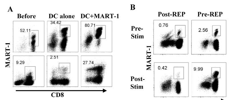
67 -58-
68 Fig pre-rep TIL have better proliferate ability than post-rep TIL in response to restimulation with antigen-pulsed DC. Multiple lines of TIL were expanded from tumors from different patients and restimulated at the pre-rep or post-rep stages with MART-1 peptide pulsed DC. The fold-increase in the bulk CD8 + TIL population (A) and the CD8 + MART-1 tetramer + subset (B) 7 days after re-stimulation are shown for seven different TIL lines. Each point represents a different TIL sample; the bars show the averages. The p-value was calculated using the Student t test. -59-
69 -60-
70 post-rep TIL exhibited stronger cytolytic ability Aside from cell number expansion, the acquisition of cytolytic ability is also crucial for effective T cell responses. CD8 T cells exert their CTL function by inflammatory cytokine secretion and direct killing, such as Granzyme B (GrB), perforin and IFN-γ. We therefore assessed whether post-rep TIL obtained enhanced cytolytic ability upon antigen restimulation. DC and MART-1 stimulation was used to activate the TIL for one week. After one-week stimulation, GrB intracellular expression was quantified. And IFN-γ secretion by TIL was also measured by cytokine intracellular staining. Upon antigenic challenge, post-rep TIL responded with elevated levels of GrB (MFI: 1884 vs. 1062) and increased secretion of IFN-γ (55.70% vs %), as compared to pre-rep TIL (Figure 2-3 B). This cytolytic ability was strictly MART-1 specific since MART-1 tetramer positive population were analyzed. In order to directly measure stimulated TIL s killing ability, T2 cells pulsed with MART-1 peptide were used as target cells. After co-incubation, MART-1 specific killing of post-rep TIL was almost two folds higher than pre-rep TIL (Figure 2-3 A). These data indicate that post-rep TIL, upon MART-1 restimulation, can differentiate into more potent CTL, as compared to pre-rep TIL. -61-
71 Fig Restimulated post-rep TIL exhibit stronger cytolytic activity than pre- REP TIL. (A) CTL activity of re-stimulated pre-rep and post-rep TIL against MART-1 peptide pulsed T2 cells using a caspase 3 cleavage CTL assay. The data shown are normalized by dividing the percentage caspase 3-cleavage in the targets by the percentage of CD8 + MART-1 tetramer + T cells added to the CTL assay (1:1 effector:target ratio) to calculate killing per added TIL. Statistical difference was calculated by using Student t test. (B) Granzyme B staining of a representative TIL line shows a higher level of staining (MFI for Granzyme B) in post-rep TIL. Post-REP TIL synthesized more IFN-γ in response to MART-1 peptide pulsed DC restimulation as determined by an intracellular cytokine staining assay. Results in panels B are representative of a minimum of three separate TIL lines tested. -62-
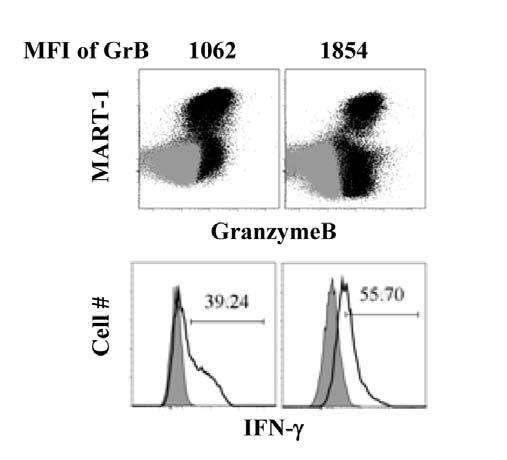
72 -63-
73 pre-rep TIL maintained better persistence after antigen restimulation Our in vitro stimulation system mimicked the in vivo TIL s response to weak tumor antigens; however, the long-term persistence of TIL has yet to be addressed. Pre- and post- REP TIL were stimulated for one week by DC and MART-1. After one week, the phenotypes and functional studies were addressed. The remaining TIL in each stimulation were then cultured in presence of only IL-2, without further antigen stimulation. We tracked the TIL for an additional three weeks and documented their cell numbers and phenotype. Pre-REP TIL showed increased number of persisting cells than post-rep TIL, through long-term culture (Table 2-I). This suggested that pre-rep TIL, as compared to post-rep TIL, have in vivo persisting advantage. Strikingly, in the TIL #2 and #3 lines, pre- REP TIL s cell numbers almost tripled. However, post-rep #2 and #3 TIL lines were unable to survive and expand over long term. These observations were all based on MART-1 specific TIL, and we observed a similar trend in the general, non-antigen specific TIL population. In short, pre-rep TIL had much better persistence in long-term upon antigen restimulation. -64-
74 Table 2-I. Improved long-term survival and expansion of CD8 + MART-1 tetramer + pre-rep TIL over post-rep TIL Pre-REP MART-1-specific Post-REP MART-specific TIL line T-cell recovery (%) a T-cell recovery (%) # Day 0 Day 7 Day 28 Day 0 Day 7 Day ND b ND ND ND ND ND TIL were stimulated with MART-1 peptide-pulsed DC on Day 0, and cultured for 7 days. Following the stimulation, there was an additional culture of 21 days with IL-2 (200 U/ml) added every 3-4 days. The cultures were stained for CD8 + MART- 1 tetramer + T cells on Day 0, 7, and 28. a Percentage of CD8 + MART-1 tetramer + T cells recovered at the time points are shown. b Not detected because cell recovery was too low after restimulation. -65-
75 post-rep TIL had a blockade in cell cycle progression and delayed apoptosis upon antigen restimulation In order to determine the mechanism of post-rep TIL s hyporesponsiveness, we looked at the proliferation and apoptosis of the post-rep TIL. Ki-67 is a molecule marker used to detect cell cycle entry. Pre- and post- REP TIL were stimulated by DC and MART-1 for 8 days. Post-REP TIL exhibited no expansion, while the pre-rep TIL expanded a lot. Increased Ki-67 positive staining was observed only on pre-rep TIL and the TIL were shown to be in G1 and S/M phase (Figure 2-4 A, B). However, post-rep TIL had a very low percentage of Ki-67 positive cells. In contrast, when apoptosis was examined by Annexin-V and 7-AAD staining, post-rep TIL manifested a delayed (from day 4) and increased level of apoptosis (Figure 2-4 C, D). 7-AAD can penetrate into dead cells and bind to nucleic acids indicating the degree of cell death. On day 7 of the stimulation, about 60% of post expansion TIL were dead, as compared to only 5-10% of the pre-rep TIL (Figure 2-4 C). This increased apoptosis and following cell death (7- AAD positive) may reflect the lack of cell cycle progression upon stimulation early on. Thus, our previous observations that post-rep TIL were hyporesponsive may be accountable by the diminished proliferation and increased apoptosis of this population upon antigen restimulation. -66-
76 Fig Cell cycle entry is reduced and apoptosis is increased in post-rep TIL after restimulation with antigen. Pre-REP and post-rep TIL including MART-1 tetramer + CD8 + T cells were stimulated with peptide-pulsed DC in the presence of IL-2 as already described. The cultures were assayed for cell cycle entry and progression using staining for Ki-67 expression and 7-AAD for DNA content after fixation and permeabilization on day 7 of stimulation (A and B). Panel A shows flow cytometry plots of Ki-67 versus 7-AAD performed on day 7 after restimulation of two representative TIL lines at the pre- and post-rep stages. Panel B shows the time course of changes in Ki-67 staining in these two pre-rep or post-rep CD8 + TIL after restimulation. (C) Post-REP CD8 + MART-1 tetramer + TIL underwent apoptosis after re-stimulation with peptide, as measured by uptake of 7-AAD and Annexin V staining of unfixed cells. (D) Time course of changes in 7-AAD + and Annexin V + CD8 + MART-1 tetramer + T cells in pre-rep and post-rep TIL after restimulation in two TIL lines. -67-
77 -68-
78 CD28 expression was downregulated after TIL expansion To delineate the molecular mechanism of the post-rep TIL hyporesponsiveness, several possibilities were hypothesized and tested. Our hypothesis was that this hyporesponsiveness was due to the severe telomere erosion, high inhibitory costimulatory molecules, lack of positive costimulation, or cytokine receptor downregulation. We first measured telomere length of pre- and post- REP TIL. Statistically shorter telomere lengths were observed in post-rep TIL (3.7 kb vs. 5.7 kb of pre-rep TIL) (Figure 2-5). However, the telomere lengths were still enough for at least 35 cell divisions ( bp per cell division [263, 264]). Further, OKT3 stimulation experiments supported that post-rep TIL were capable of extensive proliferation (data not shown). Memory T cell reactivation requires a proper combination of signals including antigen, costimulation and cytokine milieu. Therefore, we screened a serial of molecules such as: CTLA-4, PD-1, CD27, CD28, OX40, 4-1BB, CD25 (IL-2 Rα), CD122 (IL-2 Rβ), CD132 (IL- 2 Rγ) and others. We did not detect any significant differences between the preand post- REP TIL, other than CD28 expression (Figure 2-6 A). When combined with MART-1 tetramer staining, we found that CD28 expression was significantly downregulated during the rapid expansion (66% and 40% positive vs. 25% and 5% positive) (Figure 2-6 B). However, CD27, which is critical for memory T cell reactivation, did not get downregulated. CD28 is a key costimulatory molecule that quantitively lowers the T cell activation threshold and contributes to T cell survival, proliferation and differentiation. CD28 loss could account for the lack of responsiveness to antigen restimulation of post-rep TIL. Interestingly, we also discovered that GrB expression was increased in post-rep TIL, which may indicate that after expansion, TIL were more differentiated toward the effector stage. Thus, our data suggested that post-rep TIL lost CD28 expression, which in turn may lead to their hyporesponsiveness to antigen restimulation. -69-
79 Fig Telomere length is shortened after the REP. Telomere lengths in nine independent TIL lines were determined before and after the REP using flow cytometric FISH assay (flow-fish). Panel A shows how the telomere length was determined using flow-fish analysis on a representative TIL line. The telomere lengths were measured in gated G 1 -phase cells using 7- AAD staining. Flow-FISH assay of K652 cells (stable telomere length) was used to calculate the telomere lengths. Telomere length for nine independent pre-rep versus post-rep TIL lines, with p-value, is shown in panel B. Each point represents a different TIL sample; the heavy bar shows the average. -70-
80 -71-
81 Fig Cell-surface CD28 expression is lost after the REP. Pre-REP and post-rep TIL from over 15 different lines were analyzed for CD27, CD28, and CD57 cell-surface expression in the CD8 + subset using flow cytometry. (A) Percentage of positively staining CD8 + cells for each marker is shown for multiple matched pre- and post- REP TIL. Each point represents a different TIL sample; the heavy bar shows the average. The p-values were calculated by the Student t test. (B) Profound loss of cell-surface CD28 expression in the CD8 + MART-1 tetramer + TIL subset after the REP. Dot plots in B show analysis of CD27 versus CD28 staining in the gated CD8 + MART-1 tetramer + subset in two different matched pre- and post-rep TIL lines. -72-
82 -73-
83 CD28 as a marker for better TIL reactivation and expansion Recent studies have indicated that memory CD8 T cells require CD28 costimulation for reactivation and expansion. We discovered that there is a loss of CD28 expression in post-rep TIL, however, more direct evidence is needed to validate that CD28 loss causes TIL hyporesponsiveness. In our experiments, two types of APCs, DC and B cells were used to stimulate TIL. The difference between these two kinds of APCs is their CD70 expression level, with B cells expressing CD70 almost 100% while DC expressing little, if any CD70. To our surprise, the MART-1 stimulation presented by DC or B cells did not make different reactivations of TIL. A closer examination of post stimulation TIL showed their CD27 and CD28 expressions had no difference too (Figure 2-7 A). On the other hand, post-rep TIL were stimulated and their CD28 +/- subsets were analyzed. Interestingly, we found that CD28 + TIL fragments have more CFSE dilution than the CD28 - TIL fragments. This suggested that the CD28 + TIL has better antigen response. However, the effect was not observed when CD27 was used to segregate the TIL populations (Figure 2-7 B). Next, a longer period of stimulation was conducted and the CD28 expression kinetics of TIL was tracked. A gradually dimished CD28 - population was constantly observed, and at end of stimulation, almost all of the remaining TIL were CD28 + (Figure 2-7 C). We went on to sort the post-rep TIL into two fractions: CD4 - CD28 + and CD4 - CD28 -, using DC and MART-1 restimulation to test their responsiveness. After one-week stimulation, CD28 + population proliferated and MART-1 specific TIL population expanded up to 10 folds (Figure 2-7 D), however, CD28 - population did not respond well. CFSE dilution revealed that CD28 + post-rep TIL proliferated much better than CD28 - ones. This phenomenon is mirrored in part by the pre- and post- REP TIL upon antigen restimulation. Therefore, the CD28 + population was the major subset responding to antigen restimulation, while CD28 expression was lost during ex vivo TIL expansion. -74-
84 Next, we tried blocking CD28 signaling, either through CTLA-4-Ig or by CD80 and CD86 blocking. However, in both situations, the restimulation of pre-rep TIL, where ample CD28 expression could be found, was not affected at all (data not shown). This observation was very surprising; however, it may suggest the existence of a more dominant negative factor that is associated with CD28 loss to hamper TIL reactivation. -75-
85 Fig Only CD28 + TIL remaining after the REP were capable of further cell division in response to restimulation with antigen. Post-REP TIL from an HLA-A2.1 + patient were re-stimulated with either MART-1 peptide pulsed HLA-A2.1 + DC (CD70 low/- ) or MART-1 peptide pulsed CD40Lactivated HLA-A2.1 + B cells (CD70 + ). (A) FACS dot plots show the change in CD27 + versus CD28 + cells in the CD8 + MART-1 tetramer + population before and after restimulation with the DC or B cells. (B) Differential dilution of CFSE in the gated CD28 + versus CD28 - and CD27 + versus CD27 - MART-1 tetramer + T-cell subset 7 days after restimulation with peptide-pulsed B cells. (C) CFSE dilution versus CD28 staining in the TIL line on panel B was tracked on days 5, 7, and 10 after restimulation, showing the preferential outgrowth of CD28 + post-rep T cells during culture. (D) CD8 + CD28 + and CD8 + CD28 - T cell subsets were sorted from four independent HLA-A2.1 + MART-1 reactive patient TIL lines using FACS followed by restimulation with MART-1 peptide pulsed DC and measurement of the fold-change in CD8 + MART-1 tetramer + T cell numbers after 7 days using viable cell counting and FACS staining. -76-
86 -77-
87 Microarray analysis on CD28 +/- TIL revealed their gene expression differences To further understand why CD28 + TIL responded better against antigen restimulation and long-term survival advantage, post-rep TIL were sorted into CD8 + CD28 + and CD8 + CD28 - populations for cdna microarray analysis. The gene expression data based on Illumina human Ref6 chip has been stored at NCBI GEO database with Accession #GSE16517 ( Differences between CD28 + vs. CD28 - TIL were revealed by the analysis of the microarray data. Among the 12,430 genes detected (p-value <0.01), those with more than two folds change in expression were clustered in the heatmap (Figure 2-8 A). To extract the biological significance of this huge amount of data, we performed the pathway analysis. Overall, eight specific pathways were significantly different between CD28 + and CD28 - TIL, and these included genes involved in antigen processing and presentation (28/88), such as: Killer cell Immunoglobulin-like Receptors (KIR) pathway (47/131), TCR signaling (38/93), cell cycle (43/112), and p53 pathway (29/68). A list of differentially expressed genes, related to T cell memory, function, costimulation, cytokine receptors and others, were shown in Table 2-II. We found that CD28 - TIL express higher levels of cell cycle inhibitors (eg, p15, p16, p19), and cytolytic genes (such as granzymes, TNF, perforin, FasL). CD28 - TIL expressed relatively lower level of T cell memory markers (IL- 7Rα and CCR7). It was surprising to observe that CD28 + TIL have a higher expression of PD1, TP53BP2, IL-17Rβ, and IL-23A. CD28 expression level between the sorted two populations can be found to be much separated, and this serves as a perfect control for the whole gene expression analysis. The PD-1 flow cytometry staining has confirmed that CD28 + TIL have about two folds higher expression on their surface. Upon cell stimulation, PD-1 expression is still higher and may potentially inhibit the maximal proliferation of CD28 + TIL (Figure 2-8 C). In conclusion, the CD28 + TIL had distinct gene expression profiles in antigen processing, intracellular signaling, cell cycle regulation, and apoptosis. -78-
88 Fig CD8 + CD28 + and CD8 + CD28 - post-rep TIL exhibit different gene expression profiles, as determined by cdna microarray analysis. Post-REP TIL from three patients were sorted for CD8 + CD28 - and CD8 + CD28 - T cells and gene expression analyzed using cdna microarray as described in Materials and Methods. (A) Heatmap showing genes differentially expressed (>2- fold) and having statistically significant differences in expression between CD28 + and CD28 - samples (p<0.01). Onto-Tool software was used to determine differences in gene expression in major cellular signaling or functional pathways. (B) The signaling pathways or functions that had statistically significant differences between CD28 - and CD28 + cells (Impact Factor >20) are shown, together with the number of genes with each difference out of the total number of genes represented in each pathway. (C) Flow cytometry analysis of post-rep TIL for CD28 versus PD-1 staining in gated CD8 + MART-1 tetramer + T cells after the REP and 5 days after restimulation. The calculated percentage of PD-1 + and PD-1 - cells as a fraction of the CD28 + or CD28 - subset is depicted in the dot plots. -79-
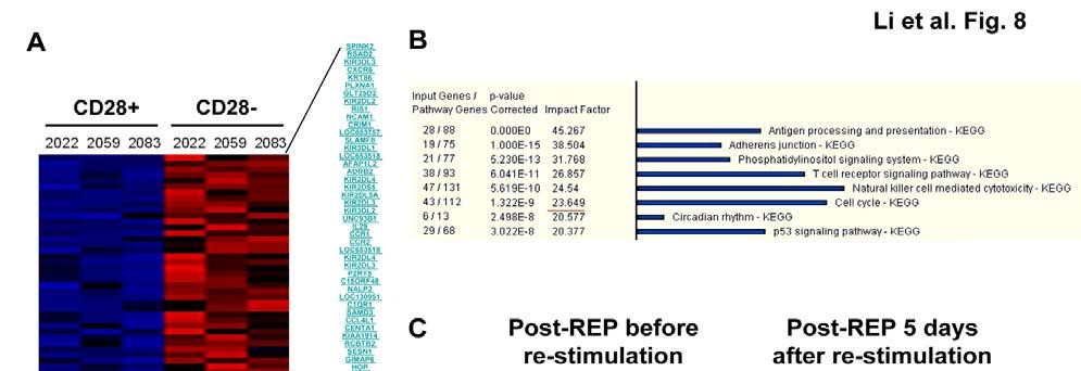
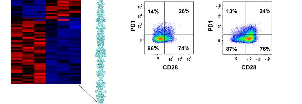
89 -80-
90 Table 2-II. Key immune-regulatory genes differentially and not differentially expressed in sorted post-rep CD8 + CD28 + versus CD8 + CD28 - melanoma TIL BeadStudio (Illumina) was used to perform microarray analysis. Gene expression was filtered Using a p<0.01 level of significance. A subset of relevant T cellrelated genes is shown. Real-time RT-PCR was used to validate changes in CD28, PD-1, IL-7Rα, and KIR2DL
91 Discussion One of the critical goals of ACT used for melanoma is to generate a large population of optimal antigen-reactive T cells. These T cells are expected not only to mediate immediate tumor killing after infusion, but also to persist to mediate a longer term control or continued eradication of metastatic tumors [32, 53]. One of the major problems of the current ACT protocol is that most of the infused TIL rapidly disappear within two weeks of infusion, and this is associated with a transient control of tumor growth. The reason for this transient persistence of infused TIL is still under investigation in the field. The TIL infused into melanoma patients contact tumor antigens and a number of microenvironmental factors, which in turn affect their effector function, cell division, and survival. These environmental factors, in context to the state of T cell differentiation, will ultimately regulate long-term TIL persistence. However, at present, very little is known about how classical REP generated TIL respond to antigen restimulation and what types of T cell phenotype are associated with TIL longer term persistence. It was our hypothesis that the REP drives the TIL to further differentiate and become hyporesponsive to antigen restimulation. In our study, we addressed the above questions with an in vitro stimulation assay to test REP expanded MART-1-specific TIL from several different melanoma patients. TIL were isolated and expanded in a standard REP procedure [32, 52, 182]. Paradoxically, it was found that CD8 + MART-1-specific post-rep TIL were hyporesponsive to antigen restimulation using mature peptide-pulsed DC. Stimulated TIL failed to enter the cell cycle, with a higher apoptosis after restimulation with antigen. In contrast, pre-rep TIL were able to enter cell cycle and proliferated much better against MART-1 peptide restimulation. Therefore, large-scale REP expansion potentially induces some function and phenotypic changes in TIL, which limits their optimal responsiveness to antigenic -82-
92 restimulation. This observation helps explaining the lack of T cell persistence in patients receiving ACT [261, 262]. One of the issues during T cell expansion is the induction of differentiation. Extensive cell divisions are usually associated with the differentiation towards end-stage effector cells and a decreased proliferate potential [6, 265, 266]. TIL isolated from the tumor microenvironments are pre-activated, mainly with an effector-memory CD27 + CD28 + CD57 - GB + Perf -/low phenotype (Liu et al, unpublished data). CD27 and CD28 are critical costimulatory molecules for T cell activation and survival [71, 128]. Viral-specific CD8 + T cells upon chronic viral exposure have down-modulated CD27 and CD28. This phenotype is associated with CTL terminal differentiation, exhibiting high cytotoxic and IFN-γ secretion, but loss of proliferate potential [267]. CD57 expression is increased upon CTL differentiation and is associated with T cells hypoproliferation. A classic case are CMV-reactive CD8 + CD27 - CD28 - CD57 + GrB ++ perforin + cells that are hypoproliferative, yet highly cytotoxic [267]. Therefore, the rapid expansion of T cells may mimic the long-term antigen exposure; hence generate reminiscence of such senescent viral specific T cells. We stained for CD27, CD28, and CD57, as well as other T cell phenotypic markers during CD8 + T cell differentiation. Interestingly, we discovered that the dramatic change on TIL upon REP is the profound loss of CD28 expression with a small increase of CD57 + cells. However, we did not observe a statistically significant change in CD27 expression, in both the MART-1-specific or general CD8 + subsets. Previous results have tracked the changes in post-rep in infused melanoma TIL. They reported a downregulation of CD27 expression after the REP. The reason for this difference is unclear, but it may reflect subtle differences of TIL REP protocol and feeders, as well as the timing of IL-2 addition to the REP cultures. High levels of IL-2 can downregulate CD27 cell surface expression on CD8 + TIL, through CD70 ligation, while lower IL- 2 doses leads to CD27 re-expression on the cell surface [268]. The relative low levels of CD70 expression on feeder cells, or the insufficient CD27-CD70 contact -83-
93 by T cells due to culture density, may explain the lack of CD27 downregulation in our system. Our sorting experiments and subsets analysis revealed that CD27 expression does not affect TIL response to MART-1 and matured DC stimulation. Further, the TIL s reactivation by MART-1 peptide presented by different APCs (mature DC and B cells) enhanced the above argument. High levels of CD70 on B cells and low levels of CD70 on DC did not generate different TIL responses. Instead, CD28 expression levels indicated the responsiveness of TIL upon tumor antigenic stimulation. Thus, sustained CD28 expression on TIL delineated TIL s ability of continued expansion and persistence. However, post-rep TIL greatly lost CD28 expression. This helps explain the clinical observations that very few long-term survival TIL clonotypes could be found in patients receiving ACT [261]. This is also demonstrated by the fact that long-term persistent TIL clonotypes tracked in ACT patients with durable remissions are associated with high CD28 expression [261]. We also found that CD28 does not play a functional role in cell responses to antigen restimulation. This was surprising given that there are several recent reports that suggest the role of CD28 ligation on the reactivation of effector-memory cells [129, 130]. CD28 - T cells have been reported to contain decreased telomere lengths. This has been associated with their lack of proliferation ability and persistence in vivo [269, 270]. Telomere erosion was observed in our experiments, with a significant shorter length on TIL after REP. This can be one of the explanations for post-rep TIL s loss of proliferation upon antigen restimulation. The fewer remaining CD28 + TIL could have kept sufficiently long telomere to support the continuous cell division. It will be important to study what signaling pathways and transcription factors regulate the expression of CD28, as well as other key markers of effector-memory T-cells. There will be a possibility to utilizing this knowledge for better maintenance of CD28 expression. A key question is whether the maintenance of CD28 expression using different cytokines or other immune modulators will result in cells with a less differentiated phenotype capable of better persistence and cell -84-
94 division after in vitro expansion in the REP. In other words, can we have the best of both worlds: extensive expansion of tumor-reactive TIL together with a favorable phenotype and continued cell division potential? The use of IL-15 or IL- 21, may offer an alternative approach in this context. IL-15 may be a good alternative to IL-2 as IL-15 has been shown to maintain memory CD8 + T-cell phenotype and facilitate the persistence of a central-memory (CD62L + and CCR7 + ) T-cell phenotype [46, 224, 227, 271]. In our study, microarray analysis on the gene expression profiles revealed some distinct characteristics between CD8 + CD28 + vs. CD8 + CD28 - TIL. This may provide an explanation for the observed responsive and persisting CD28 + TIL upon MART-1 antigen restimulation, as well as some potential targets to be supported or disrupted for improved TIL s antigenic response. Some significant differences on TIL, with or without CD28 expression, are in the expression of genes for key T cell functional pathways, such as cell cycle regulation, TCR signaling, p53, apoptosis, and KIRs (Table 2-II). CD127/IL-7Rα and CCR7 are found to be enriched in CD28 + TIL, together with the higher presence of PD-1 as well. IL-7 signaling can promote memory CD8 T cell survival, and CCR7 has long been described as a central memory T cell marker [8, 10]. The presence of these markers on CD28 + TIL indicates the memory potential of CD28 + TIL. However, PD-1 imposes a negative effect over T cell reactivation, such as inhibiting cell proliferation and cytokine secretion [88]. Blockade of PD-1 was proved to be able to enhance antigen specific T cell proliferation and, interestingly, reverse Treg inhibition [123, 124]. Therefore, administration of IL-7 in combination with anti- PD-1 antibody can greatly boost the CD28 + TIL response against antigen restimulation. The clinical availability of these reagents makes this hypothesis much more attractive [123, 124, 164]. The other highly striking difference we observed was the high degree of KIR expression in the CD28 - TIL subset. In general, the long intracellular tail version of KIR is inhibitory for T cell function, as opposed to the short tail s supportive role [272]. KIR2DL family (including -85-
95 members 1 to 5) was all highly expressed in CD28 - TIL, at different levels ranging from 6-fold (KIR2DL5A) to 19-fold (KIR2DL1). Interestingly enough, NKG2D, the activating NK receptor, was not found to be highly expressed on the CD28 - TIL. Given the functional inhibition of these KIR members, it is therefore reasoned that the hyporesponsiveness of post-rep TIL upon MART-1 restimulation may be the result of the ligation of these inhibitory KIRs by their ligands (MHC-B and MHC-C). The blocking of KIR ligation, through negating the receptor or masking their ligands should be interesting for further exploration to improve CD28 - TIL response against antigen restimulation. In conclusion, we have discovered that TIL upon REP have a profound loss of proliferate and survival potential upon MART-1 restimulation. The loss of CD28 expression on TIL is correlated, phenotypically as well as functionally, with the loss of TIL responsiveness. Thus, our data provide an explanation as to why CD28 + TIL are the dominant population with long-term persistence in patients with more durable clinical responses. Further, our microarray data suggested several therapeutically interventions, such as IL-7 administration, anti-pd-1 or anti-kir, in vivo after TIL infusion, should be able to further enhance the effect of current ACT therapy. On the other hand, investigating methods of maintaining CD28 expression during REP may help generate a superior memory population of TIL, which in turn can generate optimal clinical benefits upon adoptive transfer. -86-
96 Chapter 3 IL-15 plus IL-21 during the REP maintains TIL CD28 expression and their responsiveness to antigenic restimulation -87-
97 Introduction Metastatic melanoma patients benefit greatly from the combination of ACT and IL-2 therapy. The persistence of TIL after transfer has to be improved to enhance the clinical response. We have shown that ex vivo expansion of TIL renders the downregulation of CD28, which is associated with TIL s hyporesponsiveness to antigen restimulation. These observations suggest that generating less differentiated memory phenotype TIL for patients adoptive transfer is critical for the clinical response. We hypothesized that proper cytokine treatment should help achieve this goal. TNF-α was found to downregulate CD28 expression on T cells [157]. A higher level of TNF-α in rheumatoid arthritis (RA) patients has consistently been correlated with an abnormally higher frequency of CD28 - T cells. It is these more differentiated CD28 - T cells who contributed to the high amount of cytokine secretion and the uncontrolled auto-reactivity against self tissue [160]. In turn, the applications of anti-tnf-α antibodies, for example Remicade, have proved to be able to ameliorate patient symptoms. Interestingly enough, following the reduced TNF-α level in patients, their CD28 + T cell proportion was gradually restored [160]. In vitro experiments therefore were conducted and proved the direct regulation of CD28 expression by TNF-α [157, 158, 161]. In contrast to TNF-α, IL-12 was reported to upregulate CD28 expression on T cells [161]. Therefore, anti-tnf-α treatment, during ex vivo expansion of TIL, would be interesting for preserving CD28 expression. Cytokines play important roles in T cell homeostasis and, interestingly, sometimes interfere with the costimulatory molecules expressed on T cells. IL-15 has been shown to support optimal memory CD8 T cell reactivation, expansion -88-
98 and function development [208]. Upon IL-15 treatment, T cells upregulated 4-1BB expression. 4-1BB signaling promote CD8 T cell survival and cytokine secretion, which in turn enhance the memory response [217, 273]. IL-15 also helps to activate telomerase, therefore elongating telomere length of T cells [274]. Another interesting cytokine, a common gamma chain receptor family member, IL-7, has also been reported to help T cell survival and elongate telomere length [275]. IL-21 has been found to maintain CD28 expression in the presence of TCR ligation [242]. The transgenic T cells generated with IL-21, in contrast to IL-2, had a higher expression of c-jun and could elicit a potent anti-tumor response upon adoptive transfer [241]. The combinations of cytokines were also tested regarding their functions of supporting T cell expansion. IL-21 and IL-15 or IL-21 and IL-7 have been reported as great synergistic combinations [245, 247]. Both combinations promote stronger T cell proliferation and help develop superior T cell function than single cytokine alone. We therefore hypothesized that different cytokines (IL-7, IL-15) or combinations (IL-15+IL-21) can help maintain CD28 expression on T cells while expanding the cell number, either during or in shortterm post ex vivo expansion. In a current study, we tested 1) anti-tnf-α REP (by adding Remicade); 2) alternative cytokine for REP; and 3) alternative cytokine treatment of post-rep TIL. This should be instructive for designing novel ACT therapy. -89-
99 Results Large amounts of TNF-α in the REP culture can be blocked by Remicade We discovered that there is a loss in CD28 expression in post-rep TIL, and interestingly, TNF-α was shown to be able to downregulate CD28. To address whether TNF-α present in the REP culture negatively affects TIL phenotypic development, an examination of cytokine profiles of REP culture was conducted. As a control, IL-2 was first examined. IL-2 was not detected on very early stages of REP, day 0 and day 2 (culture supernatant sampled prior to exogenous IL-2 addition). However, since day 5, over 15 ng/ml of IL-2 could be detected and this high level sustained at least until day9. On the other hand, TNF-α level was elevated to about 750 pg/ml as early as day 2 of the REP. Further elevation of TNF-α was observed at day 5 of REP, but was not sustained to day 9. The TNFα peak probably happened around day 5 of the REP. To test the commercially available reagent Remicade for TNF-α blocking, 5 μg/ml of this antibody was added once at day 0 of the REP. Luminex assay results suggested Remicade could completely block the TNF-α presented in the REP, as plotted at Figure 3-1. However, IL-2 level in the REP was not affected by Remicade administration at all. Therefore, high levels of TNF-α present in the REP culture and they can be blocked successfully by the use of Remicade. -90-
100 Fig Large amount of TNF-α is present in REP and can be blocked by Remicade treatment. Three TIL lines (#2002, #2085, and #2105) were used to test the cytokine profile of REP culture, which takes 14 days. TNF-α and IL-2, as well as other cytokines (not shown), were detected at different time points of REP (labeled on X axis of each graph) by luminex multiple cytokine assay. As compared with regular REP (upper panels), anti-tnf-α REP (lower panels) contains Remicade (5 μg/ml) added once on day 0. Routine cell count and splitting, if necessary, was done in an identical manner for all treatments. -91-
101 Regular REP Anti-TNF-α REP TNF-α (pg/ml) TNF-α (pg/ml) TIL2002 TIL2085 TIL2105 Aveg Day Day IL-2 (pg/ml) IL-2 (pg/ml) Day Day -92-
102 Anti-TNF-α REP can help CD28 maintenance but decrease cell yield dramatically Next, we sought to address the hypothesis that removal of TNF-α in the REP can help TIL maintain their CD28 expression. TIL were harvested and phenotyped from anti-tnf-α REP, as well as regular REP. MART-1 reactive TIL were not affected or enhanced by the blocking of TNF-α. As expected, a moderate increase (about 25%) of CD28 expression on total T cells was observed with anti-tnf-α treatment (Figure 3-2 A). However, this observation was under the condition of Remicade addition at day 0 of the REP and at 5 μg/ml. It is possible that refinement of the dose and timing of Remicade addition may generate optimal CD28 maintenance on TIL during REP. Remicade titration was then tested, and adding time was set at day 0, day 2, and day 7 of the REP. Overall observation of Remicade addition was that the earlier it was added, the better maintenance of CD28 expression on TIL. In addition, a lower dose (0.2 μg/ml) of Remicade seemed to induce better CD28 maintenance. However, this increase of CD28 expression was moderate and complete recovery of CD28 expression was not induced (Figure 3-2 B). Blocking TNF-α greatly decreased the cell yield of the REP. Again, the earlier and the higher dose of Remicade administration, the lower of the cell yield (Figure 3-2 C). The poor cell yield prevented more investigation on this anti-tnf-α strategy, since sufficient cell expansion is the primary purpose of REP. -93-
103 Fig Remicade treated REP can generate TIL with CD28 maintenance. Anti-TNF-α treatment during REP by Remicade was used to evaluate the effect of CD28 maintenance. 5 μg/ml of Remicade was added on day 0 of REP and no more antibodies were used for the rest of the culture period. TIL were harvested on day 14 of the REPs; including traditional REP and anti-tnf-α REP. Flow cytometry staining of different cell surface molecules was conducted to phenotype different subsets and differentiation stages of TIL. CD28 expression and MART-1 tetramer staining comparison between two kinds of REPs was shown (A). To optimize the use of Remicade, for maintenance of CD28 expression and sufficient cell yield, the adding time and dose were tested under different parameters, as labeled on axis (B, C). After 14 days of REP, TIL were harvested, counted and phenotyped. CD8 T cells percentage out of total viable cells (open bar) and CD28 + cells out of CD8 T cells were plotted for each specific condition of REP, as compared with regular REP (B). Cell counts of final REP products, indicating the TIL yield, were also plotted (C). Data presented are representative for at least five different TIL lines. -94-
104 A B Pecentage of postive Day Day 2 CD8 CD28 in CD Day 7 Remicade (μg/ml) C 60 REP yield (*10e6) Remicade (μg/ml) 2 0 Day of REP -95-
105 Post-REP TIL rested with different cytokines did not regain CD28 expression Different cytokines drive distinct cellular signals and effects. During the REP, very high dose of IL-2 may drive T cells to go through cell cycle more rapidly. It is possible that a reduced concentration of IL-2 or alternative cytokines delivers a resting signal for post-rep TIL, reviving the CD28 expression. In order to investigate this possibility, IL-2, IL-7, or IL-15 were added at different doses for one week. IL-21 was not tested since its functions were reported effective only when in the presence of TCR ligation. IL-7 did not provide a dramatic survival benefit for TIL. The overall apoptosis rate of post-rep TIL was fairly low (about 5% of total). IL-2 or IL-15 maintained similar cell viabilities as IL-7. It is worthy noting that IL-15, in striking comparison to IL-2 or IL-7, did not induce a stronger cell cycle entry. IL-15 also up-regulated perforin and GrB expression in TIL, in a dose dependent manner. However, despite all these interesting observations, CD28 expression level on post-rep TIL was not revived by any condition investigated in the experiment. In the TIL line presented here, fresh post-rep TIL has 41.35% of CD28 + on CD8 + gated population. After one week of resting with different cytokines at different concentrations, the CD28 + remained about 40%. These data suggested CD28 expression on post-rep TIL could not be regained by alternative cytokine treatment. -96-
106 Fig IL-2, IL-7, or IL-15 does not revive CD28 expression on post-rep TIL. Post-REP TIL were harvested on day 14 of regular REP and rested one more week in presence or absence of different cytokines as indicated on the x-axis label. Flow cytometry staining of different cell surface or intracellular molecules was conducted after the rest period. The positive percentages of each molecule among in viable CD8 + T cells was plotted, as indicated on the y-axis. Data presented are representative for at least three different TIL lines. -97-
107 6% Annexin-V + 4% 2% CD8+ cells characteristics Ki-67 + Perf + GrB + CD28 + 0% 100% 80% 60% 40% 20% 0% 100% 80% 60% 40% 20% 0% 100% 80% 60% 40% 20% 0% IU/ml IL ng/ml IL ng/ml IL
108 IL-15 plus IL-21added into REP in place of IL-2 helps CD28 expression on TIL Although different cytokines did not upregulate the CD28 expression on post- REP TIL, there was still the possibility that, during the REP, alternative cytokine may drive a different pattern of TIL expansion. Given the widely reported role of IL-15 s support on CD8 memory T cells, novel REP with IL-15 (100 ng/ml) was tested. The TIL generated by the IL-15 REP did not change their phenotype as compared to IL-2 REP. The general cell yield was very similar in both IL-15 and IL-2 REP, about 1,000-fold expansion. The CD28 and CD27 expression levels on CD8 T cells, as well as total T cells, were not improved by the presence of IL-15 (Figure 3-4 A). IL-21 (100 ng/ml) was next tested for its potential role in preserving the memory phenotype of CD8 T cells. IL-15+IL-21 were also tested because of the report of the synergistic combination that can promote T cell proliferation. Interestingly, the presence of IL-21 in the REP greatly preserved CD28 expression (~90% vs only 20% in IL-2 REP). The combination of IL-15+IL- 21 also helped CD28 maintenance (~70%) (Figure 3-4 B). This observation is encouraging, since five different TIL lines all showed a dramatic increase in CD28 expression level by the addition of IL-21 or IL-15+IL-21. However, there is one shortcoming of the IL-21 REP despite its effect on the CD28 expression maintenance. It is very consistent that the IL-21 REP only has about 10-20% of cell yield of that of IL-2 REP. The IL-15+IL-21 REP actually achieved the best of both aspects: maintaining CD28 expression, as well maintaining adequate cell numbers (Figure 3-4 C). -99-
109 Fig Effects of the REP done with IL-15+ IL-21 on TIL yield and CD8 + T cell phenotype. TIL from the indicated patient lines were subjected to the REP using IL-2 (6,000 U/ml), IL-15 (100 ng/ml), IL-21 (100 ng/ml), or IL-15+IL-21 (both at 100 ng/ml). Media changes with the indicated cytokines were done as for the REP with IL-2. (A) TIL harvested after the REP using IL-2 versus IL-15 alone were analyzed for the expression of CD28 and CD27 in the CD8 + T cell subset by FACS. The percentage CD27 + and CD28 + CD8 + T cells isolated from six different TIL REPs are shown with the average percentage for each parameter shown as black bar. No statistical differences in CD27 or CD28 expression were found. (B) A representative experiment showing the extent of CD28 expression in CD8 + MART-1 tetramer + TIL after the REP using IL-2 alone, IL-15 alone, IL-21 alone, or a combination of IL-15 and IL-21. Data is representative of three experiments with similar results. (C) Five TIL lines were subjected to the REP with the different cytokines indicated. On day 14 of the REP the percentage of CD28 + cells in the CD8 + subset was determined in comparison to the fold expansion of the CD8 + T cells under the different conditions. In the case of IL-15 alone, only three TIL lines were tested with ND denoting not determined for the other two lines

110 Fold Expansion % of CD
111 Improved antigen-specific response in TIL after REP with IL-15 plus IL-21 The IL-15+IL-21 REP can help maintain CD28 expression on TIL; however, the functional competence of such TIL products remains to be analyzed. We subjected the TIL upon REPs using different cytokine cocktails to DC and MART- 1 restimulation. As shown in Figure 3-5 A, IL-15+IL-21 REPed TIL product presented an improved antigen-specific response to MART-1 restimulation, compared with those TIL from IL-2 REP or IL-15 REP. Two out of three TIL lines undergoing MART-1 restimulation showed IL-15+IL-21 REPed TIL had a markedly higher increase in MART-1 tetramer positive T cells one week after restimulation. IL-21 REP preserved CD28 expression the greatest, however, in most cases the post-rep MART-1 reactive TIL did not show an improved antigen specific response as compared with TIL from the IL-2 REP (Figure 3-5 A). Further analysis showed that the TIL generated from IL-21 REP had a significant higher expression levels of PD-1 than TIL generated from the REP with IL-2, IL-15, or IL-15+IL-21 (Figure 3-5 B)
112 Fig A combination of IL-15 and IL-21 in the REP improves the responses of post-rep CD8 + TIL to restimulation with MART-1. TIL from HLA-A2.1 + patients having a significant fraction of MART-1-reactive T cells were subjected to the REP under the cytokine conditions indicated. The isolated post-rep TIL were restimulated for 1 week with HLA-A2.1-matched DC pulsed with MART-1 peptide, as described before. (A) The fold expansion of CD8 + MART-1 tetramer + T cells is shown for three different TIL lines. (B) The levels of PD-1 expression in CD8 + TIL isolated after rapid expansion under the cytokine conditions indicated were for analyzed by FACS. A representative experiment from one TIL line is shown
113 Fold Expansion -104-
114 Discussion A key question that emerged from our work was how to maintain CD28 expression level on TIL during REP. This is interesting because of the ample evidence suggesting that CD28 + TIL are indeed the responding and persisting population against antigenic restimulation. The translational significance of such approaches, if discovered, can be very high. The ultimate purpose of our current study is to investigate novel methods of REP to generate a large amount of post- REP TIL with a less differentiated memory phenotype, capable of better persistence in vivo during ACT. Therefore, two possible methods were tested here: anti-tnf-α and alternative cytokines. TNF-α was attributed to be one of the key cytokines downregulating CD28 expression on the T cell surface [157]. This is experimentally proved and has therapeutic implication for management of RA disease [157, 160]. Therefore, we attempted to maintain the CD28 expression on TIL during REP by blocking TNFα. Interestingly, a high amount of TNF-α was detected during the REP from as early as day2 of the culture. The administration of Remicade effectively blocked the TNF-α and did not affect other key cytokines, e.g., IL-2 levels in the culture. Further, this TNF-α blockade helped maintain CD28 on TIL (Figure 3-2). However, the CD28 maintenance effect is not very consistent and, more importantly, is associated with devastating damage on TIL yield. Therefore, the strategy of anti-tnf-α REP cannot be effective for ACT protocol. On the other hand, the blocking of TNF-α in the culture was associated with poor expansion of TIL (Figure 3-2). We therefore hypothesized that TNF-α in the REP is critical for optimal T cell expansion. Addressing this hypothesis is not only biologically interesting, but also suggests a novel way of REP for ACT. For example, if TNFα is indeed critical for REP, an artificial APC (aapc) expressing TNF-α, on top of other costimulatory molecules, may be superior
115 Cytokines, especially the common γ chain cytokine family members, have been documented for their distinct roles on memory CD8 T cell homeostasis [8, ]. IL-7, IL-15, and IL-21 are very interesting candidates for preserving CD28 expression on TIL, as compared with IL-2. IL-2 used in the traditional REP has been shown to drive effector T cells differentiation much more rapidly, but the expanded T cells are more susceptible to AICD. We tested the possibility of these different cytokines to revive the CD28 expression of post-rep TIL. IL-7 is best known for its survival support in naïve and memory T cells. IL-15 could be a better alternative to IL-2, as IL-15 has been proven to maintain CD8 T cells of a central-memory (CD62L + CCR7 + ) phenotype [46, 224, 227, 271]. Additionally, it was reported that IL-15 can activate telomerase and prevent telomere length from shortening on T cells [274]. Rapid expanded TIL lose their CD28 expression, and short-term post-rep TIL with IL-7 or IL-15 as compared with IL-2, could not help restore CD28 expression (Figure 3-3). This observation indirectly proves that slowing down of rapid expansion could not maintain CD28, or less likely, once CD28 expression is lost, it cannot be regained. IL-15, in contrast to IL-2 or IL-7, greatly improved TIL cell cycle entrance in the absence of TCR stimulation, as indicated by the expression of Ki- 67 (Figure 3-3). This may suggest that IL-15 should be considered for optimal TIL culture in clinical. IL-15 cultured TIL presented increased expression levels of Granzyme B and perforin, two key cytolytic molecules for CTL function (Figure 3-3). Collectively, the lost of CD28 cannot be reverted by different cytokine treatment on post-rep TIL; however, IL-15 may be superior in support post-rep TIL expansion and function. IL-21 has been reported to exhibit an opposing role to IL-2 when driving CTL differentiation upon antigen contact [241, 242]. IL-21 has also been found to able to maintain CD28 expression on PBMC CD8 T cells after multiple rounds of -106-
116 MART-1 peptide stimulation [242]. We then tested the effects of IL-15 or IL-21 vs. IL-2 in the REP. IL-15 REP generated almost the same yield and phenotype of the TIL as the IL-2 REP. Although IL-21 was remarkably better at maintaining a high CD28 expression, it was very poor at post-rep TIL cell yields. Therefore, IL-21 REP is not optimal for the clinic since high number of post-rep TIL are required for infusion. Surprisingly, the combination of IL-15 and IL-21 in the REP provided both a significant maintenance of CD28 + expression on the TIL and a high level of TIL expansion. Therefore, IL-15 and IL-21 generated a synergistic effect on TIL during the REP. This is consistent with previous reports [247]. By ligating their different surface receptors, IL-15 signaling drives extensive cell division and has some effects on maintaining the memory CD8 population; IL-21 signaling can strongly preserve CD28 expression although IL-21 is a poor promoter of TIL proliferation. In addition, different post-rep TIL were tested on their functions in responding to MART-1 restimulation. In most cases, IL-21 REP generated TIL did not show an improved response, despite containing the highest proportion of CD28 + CD8 + TIL. The IL-15+IL-21 REP generated TIL showed the best reactivation upon MART-1 antigenic restimulation. A possibility is that the TIL derived from IL-21 REP express significantly higher levels of PD-1 (Figure 3-5). This may have an inhibitive effect on the activation and proliferation of the TIL, since the APCs used for stimulation have higher expressions of B7H1 and B7DC. In conclusion, we have evaluated two possibilities to improve the REP protocol used in the clinic to generate TIL infusion products. The ideal protocol should be able to maintain CD28 expression on TIL in order for an optimal TIL response upon antigenic encounter, as well as to achieve an optimal cell yield. Although TNF-α can downregulate CD28, the blockade of this cytokine during REP surprisingly decreases the cell expansion dramatically. Post-REP TIL cannot be revived of their CD28 expression with the different cytokines tested in the shortterm resting culture. Finally, the synergistic effect of IL-15 and IL-21 can generate -107-
117 phenoptypically less differentiated post-rep TIL and a good cell expansion. Our results suggested that IL-15+IL-21 may be superior to IL-2 in the REP for optimal ACT products
118 Chapter 4 IL-15 is superior to IL-2 in supporting antigen-specific expansion and maintenance of T cell memory in melanoma CD8 + tumorinfiltrating lymphocytes -109-
119 Introduction The success of adoptive cell therapy (ACT) using expanded TIL for metastatic melanoma relies on the ability of tumor antigen-specific T cells to persist and maintain their anti tumor effector function over long term. The advantage of ACT is that the T cells can be activated ex vivo, free of endogenous inhibitory factors, and also, the host environment can be manipulated to accommodate these transferred cells [32, 34, 38, 52, 53]. Several retrospective studies have indicated that strong anti-tumor activity and durable remission of metastatic melanoma is highly correlated with the presence of persistent, circulating reactive T cells upon infusion [261]. TIL with a memory phenotype (CD27 +, CD28 + ) have been found to persist in responding patients, while they were present in low numbers or were absent from non-responding patients [276]. Thus, a key issue in improving ACT is discovering how to enhance the infused T cells response to tumor antigen restimulation in vivo and how to effectively generate the anti-tumor memory response [32, 261, 262]. Currently IL-2 cotherapy is used for ACT. We hypothesized that alternative cytokine treatment maybe needed to improve the TIL function. Current research is examining more closely the culture conditions needed to generate TIL with improved persistence following adoptive transfer. One strategy for improving persistence currently being investigated is the cotransfer of cytokines, such as IL-2 and IL-15. IL-2 and IL-15 share a common receptor gamma chain (CD132) [8, 164, 165, 277]. When T cells are activated, CD122 and CD25 are upregulated and form a heterotrimeric high-affinity receptor for IL- 2 (IL-2Rαβγ). Conversely, IL-15 binds to its high-affinity receptor, IL-15Rα, and is transpresented to neighboring cells [211, 278]. Biophysical analysis has revealed that contact between IL-15 and IL-15Rα involves a large network of ionic interactions, whereas IL-2 and IL-2Rα contact is dominated by hydrophobic -110-
120 interactions [211, 271]. IL-2 is critical for T cell expansion and survival; however, the responders include both CD8 + effector CTLs and CD4 + regulatory T cells. IL- 15 plays a important role in the long-term expansion and homeostatic proliferation of CD8 + memory T cells, both in vitro and in vivo [46, 208, 227]. Several mouse studies, conducted to compare the supportive functions of IL-15 and IL-2, have concordantly suggested that IL-15 can generate central memory T cells (CD62L + ). IL-15 generated TCM can mediate a better immune response against tumor in vivo [224, 226]. Many studies have evaluate the effect of IL-15 from the perspective of cancer immunology (gp-100-specific pmel model) [46, 47] and viral immunity (LCMV-specific p14 model) [48], as well as from an analysis of polyclonal CD8 + T cells [227]. Recently it was proposed that dendritic cell (DC) vaccination should be combined with ACT and IL-2 therapy. This suggestion was based on evidence from a mouse study showing that the anti tumor effect of adoptive transferred T cells was enhanced by DC vaccination, in that DC vaccination after ACT greatly increased T cell persistence in vivo [279]. Thus, a clinical trial of the IL-2-ACT-DC strategy is now ongoing at M. D. Anderson Cancer Center. Given the distinct transpresentation mode, DC may be used to present IL-15 in vivo. However, to our knowledge, no one has examined whether cotherapy with an alternative cytokine, in particular IL-15, would improve this treatment. We hypothesized that IL-15 has better supportive effects for TIL, than IL-2. We developed an antigen-specific stimulation system (using mature DCs pulse with MART-1 peptide) to track TIL responses in an in vitro system that closely resembles in vivo physiological conditions. The results of the present study can be used to guide the immediate clinical evaluation of IL-15 in place of IL-2 with ACT in patients with metastatic melanoma
121 Results IL-15 drives stronger CD8 expansion and GrB expression upon T cell stimulation We first investigated the ability of TIL to respond to antigen restimulation in the presence of IL-2 or IL-15. To specifically stimulate and track antigen-specific TIL, mature DC were used to present the MART-1 peptide. Mature DC were examined for their surface expression of CD80 and CD86, as well as expression of IL-15Rα (Figure 4-1 A). DC used in our experiment were shown to be able to effectively transpresent IL-15 to T cells. Without cytokine support, DC pulsedpeptide stimulation was not sufficient to activate T cells to proliferate. After one week of stimulation, few live TIL were obtained. IL-2 and IL-15 were titrated to determine the optimal dose of each cytokine in supporting TIL restimulation (data not shown). Consistent with other reports, we found that 200 IU/ml of IL-2 and 10 ng/ml of IL-15 were able to support T cell stimulation, without any excessive effects, such as AICD. To examine whether IL-2 and IL-15 differed in their ability to support TIL restimulation, we subjected MART-1 + TIL lines to DC stimulation. Expansion of the TIL population was observed after coculture with DC and MART-1 supplemented by exogenous cytokines. At day 7 of the stimulation, TIL were harvested and stained. Flow cytometric analysis of cell subsets, as defined by their unique differentiation and activation markers, was then conducted. In several TIL lines derived from different patients, IL-15 stimulation always generated more CD8 + TIL than IL-2 (Figure 4-1 B). This is consistent with previous reports, and is potentially very important to ACT for melanoma because CD8 + TIL are the primary population that confer anti tumor responses in melanoma patients. The number of infused CD8 + T cell positively correlates with clinical efficacy. Intracellular staining of GrB, a cytolytic molecule that digests various substrates to facilitate cell killing, also differed between the IL-2 and IL-15 stimulations (Figure 4-1 C). IL-15 generated about 1.5 fold more GrB positive TIL -112-
122 than IL-2 (45% vs. 30%). These data suggest that one week of IL-15 stimulation drove a greater CD8 + TIL response and generated more GrB in activated cells. To test whether this enhanced GrB expression was due to direct cytokine support or indirectly through DC, we exposed TIL to cytokine alone in the absence of antigen stimulation for one week. Co-staining of GrB and perforin showed that IL- 15 upregulated both cytolytic molecules in a dose-dependent manner, while IL-2 barely changed their expression levels (Figure 4-1 D). This finding suggests that IL-15 alone was sufficient to enhance the cytolytic ability of TIL, independent of TCR ligation. However, we could not exclude the possibility that the effect was an indirect one and attributable to IL-15 s superior ability to stimulate DC
123 Fig IL-15 stimulation preferentially expands the population of CD8 + CTLs and increases GrB expression. Mature myeloid DC ere generated by culture with GM-CSF, IL-4 and ITIP maturation cocktail. Surface expression of IL-15Rα, as well as the co-stimulatory molecule CD80, CD86 and other antigens, was examined by flow cytometry staining (A). TIL derived from melanoma patients were stimulated by mature DC pulse-treated with MART-1 peptide for 1 week, in the presence of either IL-2 (200 IU/ml) or IL-15 (10 ng/ml). After the TIL were stimulated, flow staining was conducted to monitor TIL activation, differentiation, and cytolytic ability, along with tetramer staining. CD8 and GrB differed between the IL-2 and IL-15 stimulations. Summarized data from several TIL lines have been plotted in (C), where the heavy black bar indicates the mean percentage. Cytokine alone (as indicated) without antigen stimulation was used to support TIL for 1 week. GrB and perforin staining gated on MART-1 + TIL was shown, as compared to isotype staining (D)
124 -115-
125 IL-15-stimulated TIL portrayed enhanced antigen-specific killing ability The level of GrB staining does not directly correlate with TIL cytolytic activity. This may be due to weaker recognition of the target or a lack of cooperation from other cytolytic molecules, such as perforin. To help resolve this issue, we tested how TIL exert their CTL function by examining inflammatory cytokine secretion and direct killing of T2 target cells pulsed with MART-1. IL-15-stimulated TIL were hypothesized to possess enhanced cytolytic ability upon antigen restimulation. TIL were challenged with the MART-1 peptide pulsed T2 target cells for 18 hours, after which IFN-γ secretion by TIL was measured by ELISA of the supernatant. Indeed, we found that IL-15-stimulated TIL secreted more IFN-γ than IL-2-stimulated cells. Combined with the MART-1 tetramer staining, per cell antigen-specific IFN-γ secretion was calculated and plotted (Figure 4-2 A). IL-15- stimulated TIL presented at least 5 times higher levels of IFN-γ upon target contact. To directly measure the TIL killing ability, we used a highly sensitive flow cytometry-based CTL assay measuring the cleavage of caspase 3 in target cells [280]. T2 cells pulsed with MART-1 peptide were the targets, and were mixed with stimulated TIL at different effector-to-target ratios (Figure 4-2 B). For a fair comparison, MART-1 staining was also taken into consideration so that per cell MART-1-specific killing could be calculated. As shown in Figure 4-2 C, IL-15- stimulated TIL generally have two to three folds better killing ability. These data indicate that IL-15-stimulated TIL, in response to MART-1 restimulation, can differentiate into potent CTLs compared with those stimulated by IL
126 Fig TIL gained enhanced cytolytic ability after IL-15 stimulation. After one week of stimulation, TIL were harvested, and their direct target-killing effect was tested. T2 cells pulsed with MART-1 peptide or HIV peptide as control were mixed with TIL at different effector-to-target ratios, as indicated. IFN-γ was measured by ELISA on the culture supernatant, which was collected at 18 hours after the CTL reaction was initiated. Net IFN-γ per MART-1 + TIL was calculated considering the MART-1 tetramer-positive T cell percentage in the TIL (A). A flow cytometry-based sensitive CTL assay to measure cleavage of caspase 3 (B) was also used to test post-stimulation TIL function. In 10 TIL lines, the cytotoxicity of MART-1 + TIL was calculated similar to the IFN-γ secretion ability described above, and the results are plotted in (C)
127 -118-
128 Long-term persistence of memory T cells is maintained better by IL-15 than IL-2 Our in vitro stimulation mimicked the TIL in vivo response to weak tumor antigens; however, long-term TIL persistence had yet to be addressed. Either IL- 2 or IL-15 was added to DC and MART-1 restimulation for one week. We determined the MART-1 specific TIL expansion at end of stimulation, by cell count and MART-1 tetramer staining. After the phenotyping and functional studies, the remaining TIL in each case were returned to culture and provided with only cytokine support, without further antigen stimulation. We tracked the TIL for three additional weeks and documented their cell numbers and phenotype. As seen in Figure 4-3, short-term (one-week) stimulation of the TIL yielded a strong antigen-specific cell expansion. TIL expanded on average 8.2-fold in the IL-2 setting, and 13-fold in the IL-15 setting. Conversely, in long-term (28 day) expansions, the populations of TIL expanded only 1.9-fold in response to IL-2 but 4.8-fold in response to IL-15. This result revealed IL-15 s potential advantage for in vivo stimulation and expansion of the TIL population. In multiple TIL lines, IL- 15 had a statistically significant benefit only for long-term expansion. Phenotyping of tetramer and memory T cell marker staining also revealed the preferential maintenance of memory antigen-specific TIL by IL-15. For example, in one TIL line, after 4 weeks, IL-2 support maintained only 3.26% of MART-1 + TIL, whereas IL-15 support maintained 36.77% (Figure 4-4 A). Over the long term, IL-15 also expanded more CD8 + TIL than IL-2 (87% vs. 74%). By comparing the MART-1 + TIL phenotypes, we found that CD27 + and CD28 + TIL were preferentially expanded. The enrichment of CD27 + TIL during the four-week period was very striking: increasing from 35% to 67% (Figure 4-4 B). The trend toward CD27 + and CD28 + enrichment remained true in both IL-2 and IL-15 settings, but IL-15 always maintained the higher level of cells with such markers (Figure 4-4 C)
129 Fig IL-15 stimulation drives a better memory TIL response than IL-2. TIL were initially stimulated for one week (until day 7) in the presence of IL-2 or IL-15, followed by an additional three weeks of long-term expansion (until day28) without further re-stimulation. In each case, TIL were harvested, counted, and phenotyped by flow cytomtery staining at the end of the treatment. The plots show the MART-1-specific TIL expansions. The two-tail Student t test was used to determine the statistical significance of the difference between the IL-2 and IL- 15-supported expansions at day
130 -121-
131 Fig Memory TIL can be detected after stimulation and additional expansion. In tests of TIL stimulation and expansion, at end of day 28 of culture, MART-1 tetramer staining was done, as was flow cytometry staining using the differentiation markers CD27 and CD28. MART-1 + TIL were quantified and compared between the IL-2 and IL-15 settings (A). In addition, tetramer-positive TIL were analyzed for CD27 and CD28 expression by comparing expression levels on day 0 and day 28 of MART-1-specific stimulation and expansion (B). Subtle differences in these markers of memory cells were also found by comparing the IL-2- and IL-15-supported TIL cultures (C), gated on the number of total CD8 + TIL
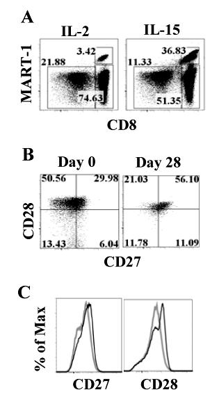
132 -123-
133 IL-15 generated more-persistent memory CD8 + TIL To investigate whether IL-15-generated memory CD8 + T cells differed from those generated by IL-2, we compared the proliferation and apoptosis in the two resulting TIL populations. After being stimulated for one week, TIL were cultured for an additional three weeks. During the additional culture period, the cells were closely monitored for their proliferation by staining for Ki-67, which is only detectable during active phases of the cell cycle [281]. Higher levels of Ki-67 were consistently observed in IL-15-cultured TIL, both in the general CD8 + population and the antigen-specific MART-1 + population (Figure 4-5 A). Apoptosis of TIL was examined at the end of the long-term culture (that is, on day28) by quantitative real-time PCR. As shown in Figure 4-5 B, although the anti-apoptosis gene Bcl-Xl was expressed at similar levels by the two populations, expression of the pro-apoptotic gene Bim was almost two fold higher in IL-2- cultured TIL than in IL-15-cultured TIL, indicating that the IL-15 rendered TIL less vulnerable to apoptosis. Additionally, we examined two genes related to memory CD8 + T cells. Eomesodermin has been reported to be highly expressed on memory T cells [282], and we observed a higher level of expression of this gene in IL-15-cultured TIL. LEF1 was originally reported to be a transcription factor differentiating between naïve and antigen-experienced T cells. It was reported that Wnt pathway maintains hematopoietic stem cells by promoting quiescence and inhibiting proliferation [283]. LEF1 is the downstream effector of the Wnt pathway and is downregulated upon TCR or IL-15 receptor engagement [283]. To our surprise, we observed a much lower level of LEF1 in IL-15-cultured TIL than in IL-2-cultured TIL; suggesting IL-15 drives less activation of Wnt-βcatenin-LEF1/TCF-1 pathway. Overall, the evidence showed that IL-15, as compared with IL-2, helps generate a superior memory status of CD8 + TIL
134 Fig IL-15 generated a superior population of memory CD8 T cells. After TIL stimulation and expansion, MART-1 tetramer staining as well as Ki-67 intracellular staining was performed to monitor cell proliferation during the 3-week long-term culture. As shown in (A), representative flow data revealed the difference in proliferation between IL-2- and IL-15-cultured TIL. At the end of day 28, TIL were harvested and their RNA was converted to cdna for quantitative real-time PCR. The apoptosis-related genes Bcl-Xl and Bim and the memory T cell markers Eomesodermin and LEF1 were examined (B). β-actin was also included as an internal control and reference for calculations
135 -126-
136 Stimulation of memory T cells indicated their non-anergic status Next, we investigated whether TIL cultured long-term could respond to antigen restimulation. As shown in Figure 4-6, IL-2- and IL-15-stimulated TIL were still able to expand upon DC and MART-1 restimulation, as shown by tetramer staining and cell counts. This result indicated that the persisting memory TIL were still reactive upon encountering antigen, instead of being anergic due to some expression of an inhibitory signal, such as PD-1 or BTLA. Interestingly, TIL stimulated with IL-15 had better viability, were larger, and contained more granules than those stimulated by IL-2 (Figure 4-6 A). IL-15 support also generated higher CD27, CD28 and GrB expression on MART-1 + TIL (Figure 4-6 B) and on other two TIL lines tested. Therefore, we specifically demonstrated that IL-15 could better support TIL stimulation and long-term memory development than IL-2. When the expansions of MART-1-specific TIL populations were quantified and compared, we found that IL-15-supported TIL expanded 20- to 60- fold after restimulation, while IL-2-supported TIL expanded no more than 20-fold (Figure 4-6 C). These data are concordant with those from long-term cultures, showing that TIL cultured in the presence IL-15 presented superior memory characteristics. This observation reflected their early proliferation advantage, reduced apoptosis, and higher expression of Eomesodermin
137 Fig Rechallenge of memory TIL revealed their maintenance of antigen response. At day 28 of TIL culture, a second round of mdcs pulse-treated with MART-1 were applied to test the TIL memory to an antigen rechallenge. The TIL were harvested and examined 1 week later (day 35). IL-2 and IL-15 supported TIL activation differently, as indicated by morphologic properties such as the size and granularity of the cells (A). MART-1 tetramer staining could still be detected after stimulation, although the two populations differentiation markers and cytolytic ability were not the same (B). The tests were repeated and similar results obtained in independent TIL lines. Absolute numbers of MART-1-specific TIL were calculated, and their expansion over 4 weeks was plotted in (C)
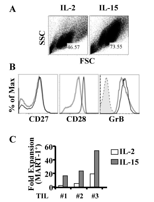
138 -129-
DEVELOPMENT OF CELLULAR IMMUNOLOGY
 DEVELOPMENT OF CELLULAR IMMUNOLOGY 1880 s: Antibodies described (dominated studies of immunology until 1960 s) 1958: Journal of Immunology (137 papers) lymphocyte not listed in index Two papers on transfer
General Overview of Immunology. Kimberly S. Schluns, Ph.D. Associate Professor Department of Immunology UT MD Anderson Cancer Center
 General Overview of Immunology Kimberly S. Schluns, Ph.D. Associate Professor Department of Immunology UT MD Anderson Cancer Center Objectives Describe differences between innate and adaptive immune responses
T Cell Activation, Costimulation and Regulation
 1 T Cell Activation, Costimulation and Regulation Abul K. Abbas, MD University of California San Francisco 2 Lecture outline T cell antigen recognition and activation Costimulation, the B7:CD28 family
FOCiS. Lecture outline. The immunological equilibrium: balancing lymphocyte activation and control. Immunological tolerance and immune regulation -- 1
 1 Immunological tolerance and immune regulation -- 1 Abul K. Abbas UCSF FOCiS 2 Lecture outline Principles of immune regulation Self-tolerance; mechanisms of central and peripheral tolerance Inhibitory
Antigen Presentation and T Lymphocyte Activation. Abul K. Abbas UCSF. FOCiS
 1 Antigen Presentation and T Lymphocyte Activation Abul K. Abbas UCSF FOCiS 2 Lecture outline Dendritic cells and antigen presentation The role of the MHC T cell activation Costimulation, the B7:CD28 family
Tumors arise from accumulated genetic mutations. Tumor Immunology (Cancer)
") Tumor Immunology (Cancer) Tumors arise from accumulated genetic mutations Robert Beatty MCB150 Mutations Usually have >6 mutations in both activation/growth factors and tumor suppressor genes. Types of
Tumor Immunology (Cancer) Tumors arise from accumulated genetic mutations Robert Beatty MCB150 Mutations Usually have >6 mutations in both activation/growth factors and tumor suppressor genes. Types of
Determinants of Immunogenicity and Tolerance. Abul K. Abbas, MD Department of Pathology University of California San Francisco
 Determinants of Immunogenicity and Tolerance Abul K. Abbas, MD Department of Pathology University of California San Francisco EIP Symposium Feb 2016 Why do some people respond to therapeutic proteins?
Immune Checkpoints. PD Dr med. Alessandra Curioni-Fontecedro Department of Hematology and Oncology Cancer Center Zurich University Hospital Zurich
 Immune Checkpoints PD Dr med. Alessandra Curioni-Fontecedro Department of Hematology and Oncology Cancer Center Zurich University Hospital Zurich Activation of T cells requires co-stimulation Science 3
Darwinian selection and Newtonian physics wrapped up in systems biology
 Darwinian selection and Newtonian physics wrapped up in systems biology Concept published in 1957* by Macfarland Burnet (1960 Nobel Laureate for the theory of induced immune tolerance, leading to solid
T-cell activation T cells migrate to secondary lymphoid tissues where they interact with antigen, antigen-presenting cells, and other lymphocytes:
 Interactions between innate immunity & adaptive immunity What happens to T cells after they leave the thymus? Naïve T cells exit the thymus and enter the bloodstream. If they remain in the bloodstream,
Therapeutic efficacy of MUC1- specific CTL and CD137 costimulation. mammary cancer model. Pinku Mukherjee & Sandra Gendler
 Therapeutic efficacy of MUC1- specific CTL and CD137 costimulation in a spontaneous mammary cancer model Pinku Mukherjee & Sandra Gendler Goal of Immunotherapy Boosting the low level anti-tumor immune
COURSE: Medical Microbiology, PAMB 650/720 - Fall 2008 Lecture 16
 COURSE: Medical Microbiology, PAMB 650/720 - Fall 2008 Lecture 16 Tumor Immunology M. Nagarkatti Teaching Objectives: Introduction to Cancer Immunology Know the antigens expressed by cancer cells Understand
T Lymphocyte Activation and Costimulation. FOCiS. Lecture outline
 1 T Lymphocyte Activation and Costimulation Abul K. Abbas, MD UCSF FOCiS 2 Lecture outline T cell activation Costimulation, the B7:CD28 family Inhibitory receptors of T cells Targeting costimulators for
The Immune System. Innate. Adaptive. - skin, mucosal barriers - complement - neutrophils, NK cells, mast cells, basophils, eosinophils
 Objectives - explain the rationale behind cellular adoptive immunotherapy - describe methods of improving cellular adoptive immunotherapy - identify mechanisms of tumor escape from cellular adoptive immunotherapy
ACTIVATION AND EFFECTOR FUNCTIONS OF CELL-MEDIATED IMMUNITY AND NK CELLS. Choompone Sakonwasun, MD (Hons), FRCPT
, FRCPT") ACTIVATION AND EFFECTOR FUNCTIONS OF CELL-MEDIATED IMMUNITY AND NK CELLS Choompone Sakonwasun, MD (Hons), FRCPT Types of Adaptive Immunity Types of T Cell-mediated Immune Reactions CTLs = cytotoxic T lymphocytes
ACTIVATION AND EFFECTOR FUNCTIONS OF CELL-MEDIATED IMMUNITY AND NK CELLS Choompone Sakonwasun, MD (Hons), FRCPT Types of Adaptive Immunity Types of T Cell-mediated Immune Reactions CTLs = cytotoxic T lymphocytes
IMMUNOTHERAPY FOR CANCER A NEW HORIZON. Ekaterini Boleti MD, PhD, FRCP Consultant in Medical Oncology Royal Free London NHS Foundation Trust
 IMMUNOTHERAPY FOR CANCER A NEW HORIZON Ekaterini Boleti MD, PhD, FRCP Consultant in Medical Oncology Royal Free London NHS Foundation Trust ASCO Names Advance of the Year: Cancer Immunotherapy No recent
The Adaptive Immune Responses
 The Adaptive Immune Responses The two arms of the immune responses are; 1) the cell mediated, and 2) the humoral responses. In this chapter we will discuss the two responses in detail and we will start
Central tolerance. Mechanisms of Immune Tolerance. Regulation of the T cell response
 Immunoregulation: A balance between activation and suppression that achieves an efficient immune response without damaging the host. Mechanisms of Immune Tolerance ACTIVATION (immunity) SUPPRESSION (tolerance)
Mechanisms of Immune Tolerance
 Immunoregulation: A balance between activation and suppression that achieves an efficient immune response without damaging the host. ACTIVATION (immunity) SUPPRESSION (tolerance) Autoimmunity Immunodeficiency
New insights into CD8+ T cell function and regulation. Pam Ohashi Princess Margaret Cancer Centre
 New insights into CD8+ T cell function and regulation Pam Ohashi Princess Margaret Cancer Centre New insights into CD8+ T cell function and regulation Pam Ohashi Princess Margaret Cancer Centre No Disclosures
Micro 204. Cytotoxic T Lymphocytes (CTL) Lewis Lanier
 Lewis Lanier") Micro 204 Cytotoxic T Lymphocytes (CTL) Lewis Lanier Lewis.Lanier@ucsf.edu Lymphocyte-mediated Cytotoxicity CD8 + αβ-tcr + T cells CD4 + αβ-tcr + T cells γδ-tcr + T cells Natural Killer cells CD8 + αβ-tcr
Micro 204 Cytotoxic T Lymphocytes (CTL) Lewis Lanier Lewis.Lanier@ucsf.edu Lymphocyte-mediated Cytotoxicity CD8 + αβ-tcr + T cells CD4 + αβ-tcr + T cells γδ-tcr + T cells Natural Killer cells CD8 + αβ-tcr
Adaptive immune responses: T cell-mediated immunity
 MICR2209 Adaptive immune responses: T cell-mediated immunity Dr Allison Imrie allison.imrie@uwa.edu.au 1 Synopsis: In this lecture we will discuss the T-cell mediated immune response, how it is activated,
Effector mechanisms of cell-mediated immunity: Properties of effector, memory and regulatory T cells
 ICI Basic Immunology course Effector mechanisms of cell-mediated immunity: Properties of effector, memory and regulatory T cells Abul K. Abbas, MD UCSF Stages in the development of T cell responses: induction
ACTIVATION OF T LYMPHOCYTES AND CELL MEDIATED IMMUNITY
 ACTIVATION OF T LYMPHOCYTES AND CELL MEDIATED IMMUNITY The recognition of specific antigen by naïve T cell induces its own activation and effector phases. T helper cells recognize peptide antigens through
NKTR-255: Accessing IL-15 Therapeutic Potential through Robust and Sustained Engagement of Innate and Adaptive Immunity
 NKTR-255: Accessing IL-15 Therapeutic Potential through Robust and Sustained Engagement of Innate and Adaptive Immunity Peiwen Kuo Scientist, Research Biology Nektar Therapeutics August 31 st, 2018 Emerging
T cell maturation. T-cell Maturation. What allows T cell maturation?
 T-cell Maturation What allows T cell maturation? Direct contact with thymic epithelial cells Influence of thymic hormones Growth factors (cytokines, CSF) T cell maturation T cell progenitor DN DP SP 2ry
Tumor Microenvironment and Immune Suppression
 Tumor Microenvironment and Immune Suppression Hassane M. Zarour,, MD Department of Medicine, Division of Hematology-Oncology, University of Pittsburgh Cancer Institute Hallmarks of Cancer: The Next Generation
08/02/59. Tumor Immunotherapy. Development of Tumor Vaccines. Types of Tumor Vaccines. Immunotherapy w/ Cytokine Gene-Transfected Tumor Cells
 Tumor Immunotherapy Autologous virus Inactivation Inactivated virus Lymphopheresis Culture? Monocyte s Dendritic cells Immunization Autologous vaccine Development of Tumor Vaccines Types of Tumor Vaccines
Immunology Basics Relevant to Cancer Immunotherapy: T Cell Activation, Costimulation, and Effector T Cells
 Immunology Basics Relevant to Cancer Immunotherapy: T Cell Activation, Costimulation, and Effector T Cells Andrew H. Lichtman, M.D. Ph.D. Department of Pathology Brigham and Women s Hospital and Harvard
NKTR-255: Accessing The Immunotherapeutic Potential Of IL-15 for NK Cell Therapies
 NKTR-255: Accessing The Immunotherapeutic Potential Of IL-15 for NK Cell Therapies Saul Kivimäe Senior Scientist, Research Biology Nektar Therapeutics NK Cell-Based Cancer Immunotherapy, September 26-27,
Objectives. Abbas Chapter 11: Immunological Tolerance. Question 1. Question 2. Question 3. Definitions
 Objectives Abbas Chapter 11: Immunological Tolerance Christina Ciaccio, MD Children s Mercy Hospitals and Clinics February 1, 2010 To introduce the concept of immunologic tolerance To understand what factors
TCR, MHC and coreceptors
 Cooperation In Immune Responses Antigen processing how peptides get into MHC Antigen processing involves the intracellular proteolytic generation of MHC binding proteins Protein antigens may be processed
SEVENTH EDITION CHAPTER
 Judy Owen Jenni Punt Sharon Stranford Kuby Immunology SEVENTH EDITION CHAPTER 16 Tolerance, Autoimmunity, and Transplantation Copyright 2013 by W. H. Freeman and Company Immune tolerance: history * Some
Immune Regulation and Tolerance
 Immune Regulation and Tolerance Immunoregulation: A balance between activation and suppression of effector cells to achieve an efficient immune response without damaging the host. Activation (immunity)
Scott Abrams, Ph.D. Professor of Oncology, x4375 Kuby Immunology SEVENTH EDITION
 Scott Abrams, Ph.D. Professor of Oncology, x4375 scott.abrams@roswellpark.org Kuby Immunology SEVENTH EDITION CHAPTER 13 Effector Responses: Cell- and Antibody-Mediated Immunity Copyright 2013 by W. H.
Scott Abrams, Ph.D. Professor of Oncology, x4375 Kuby Immunology SEVENTH EDITION
 Scott Abrams, Ph.D. Professor of Oncology, x4375 scott.abrams@roswellpark.org Kuby Immunology SEVENTH EDITION CHAPTER 11 T-Cell Activation, Differentiation, and Memory Copyright 2013 by W. H. Freeman and
Immune Tolerance. Kyeong Cheon Jung. Department of Pathology Seoul National University College of Medicine
 Immune Tolerance Kyeong Cheon Jung Department of Pathology Seoul National University College of Medicine Immune tolerance Unresponsiveness to an antigen that is induced by previous exposure to that antigen
Regulation of anti-tumor immunity through migration of immune cell subsets within the tumor microenvironment Thomas F. Gajewski, M.D., Ph.D.
 Regulation of anti-tumor immunity through migration of immune cell subsets within the tumor microenvironment Thomas F. Gajewski, M.D., Ph.D. Professor, Departments of Pathology and Medicine Program Leader,
CELLULAR AND MOLECULAR REQUIREMENTS FOR REJECTION OF B16 MELANOMA IN THE SETTING OF REGULATORY T CELL DEPLETION AND HOMEOSTATIC PROLIFERATION
 CELLULAR AND MOLECULAR REQUIREMENTS FOR REJECTION OF B16 MELANOMA IN THE SETTING OF REGULATORY T CELL DEPLETION AND HOMEOSTATIC PROLIFERATION Justin Kline 1, Long Zhang 1, and Thomas F. Gajewski 1,2 Departments
5/1/13. The proportion of thymus that produces T cells decreases with age. The cellular organization of the thymus
 T cell precursors migrate from the bone marrow via the blood to the thymus to mature 1 2 The cellular organization of the thymus The proportion of thymus that produces T cells decreases with age 3 4 1
Lecture outline. Immunological tolerance and immune regulation. Central and peripheral tolerance. Inhibitory receptors of T cells. Regulatory T cells
 1 Immunological tolerance and immune regulation Abul K. Abbas UCSF 2 Lecture outline Central and peripheral tolerance Inhibitory receptors of T cells Regulatory T cells 1 The immunological equilibrium:
T Cell Activation. Patricia Fitzgerald-Bocarsly March 18, 2009
 T Cell Activation Patricia Fitzgerald-Bocarsly March 18, 2009 Phases of Adaptive Immune Responses Phases of T cell responses IL-2 acts as an autocrine growth factor Fig. 11-11 Clonal Expansion of T cells
LAMPvax DNA Vaccines as Immunotherapy for Cancer - Three Case Studies
 LAMPvax DNA Vaccines as Immunotherapy for Cancer - Three Case Studies Cancer immunotherapy has emerged as a clinically validated tool for fighting certain kinds of cancers. These therapeutic cancer vaccines
TITLE: Development of Antigen Presenting Cells for adoptive immunotherapy in prostate cancer
 AD Award Number: W8-XWH-5-- TITLE: Development of Antigen Presenting Cells for adoptive immunotherapy in prostate cancer PRINCIPAL INVESTIGATOR: Mathias Oelke, Ph.D. CONTRACTING ORGANIZATION: Johns Hopkins
Cancer immunity and immunotherapy. General principles
 1 Cancer immunity and immunotherapy Abul K. Abbas UCSF General principles 2 The immune system recognizes and reacts against cancers The immune response against tumors is often dominated by regulation or
Chapter 10 (pages ): Differentiation and Functions of CD4+ Effector T Cells Prepared by Kristen Dazy, MD, Scripps Clinic Medical Group
: Differentiation and Functions of CD4+ Effector T Cells Prepared by Kristen Dazy, MD, Scripps Clinic Medical Group") FIT Board Review Corner September 2015 Welcome to the FIT Board Review Corner, prepared by Andrew Nickels, MD, and Sarah Spriet, DO, senior and junior representatives of ACAAI's Fellows-In-Training (FITs)
FIT Board Review Corner September 2015 Welcome to the FIT Board Review Corner, prepared by Andrew Nickels, MD, and Sarah Spriet, DO, senior and junior representatives of ACAAI's Fellows-In-Training (FITs)
Effector T Cells and
 1 Effector T Cells and Cytokines Andrew Lichtman, MD PhD Brigham and Women's Hospital Harvard Medical School 2 Lecture outline Cytokines Subsets of CD4+ T cells: definitions, functions, development New
Immunotherapy on the Horizon: Adoptive Cell Therapy
 Immunotherapy on the Horizon: Adoptive Cell Therapy Joseph I. Clark, MD, FACP Professor of Medicine Loyola University Chicago Stritch School of Medicine Maywood, IL June 23, 2016 Conflicts of Interest
Third line of Defense
 Chapter 15 Specific Immunity and Immunization Topics -3 rd of Defense - B cells - T cells - Specific Immunities Third line of Defense Specific immunity is a complex interaction of immune cells (leukocytes)
Follicular Lymphoma. ced3 APOPTOSIS. *In the nematode Caenorhabditis elegans 131 of the organism's 1031 cells die during development.
 Harvard-MIT Division of Health Sciences and Technology HST.176: Cellular and Molecular Immunology Course Director: Dr. Shiv Pillai Follicular Lymphoma 1. Characterized by t(14:18) translocation 2. Ig heavy
Cellular Immune response. Jianzhong Chen, Ph.D Institute of immunology, ZJU
 Cellular Immune response Jianzhong Chen, Ph.D Institute of immunology, ZJU Concept of adaptive immune response T cell-mediated adaptive immune response I. Concept of immune response A collective and coordinated
International Society of Breast Pathology. Immune Targeting in Breast Cancer. USCAP 2017 Annual Meeting
 International Society of Breast Pathology USCAP 2017 Annual Meeting Immune Targeting in Breast Cancer Ashley Cimino-Mathews, MD Assistant Professor of Pathology and Oncology The Johns Hopkins Hospital
Emerging Concepts of Cancer Immunotherapy
 Emerging Concepts of Cancer Immunotherapy Jeffrey Schlom, Ph.D. Laboratory of Tumor Immunology and Biology (LTIB) Center for Cancer Research National Cancer Institute, NIH Immune Cell Infiltrate in Primary
Novel RCC Targets from Immuno-Oncology and Antibody-Drug Conjugates
 Novel RCC Targets from Immuno-Oncology and Antibody-Drug Conjugates Christopher Turner, MD Vice President, Clinical Science 04 November 2016 Uveal Melanoma Celldex Pipeline CANDIDATE INDICATION Preclinical
T cell and Cell-mediated immunity
 T cell and Cell-mediated immunity ( 第十章 第十二章第十二章 ) Lu Linrong ( 鲁林荣 ) PhD Laboratory of Immune Regulation Institute of Immunology Zhejiang University, School of Medicine Medical Research Building B815-819
CONTRACTING ORGANIZATION: Johns Hopkins University School of Medicine Baltimore, MD 21205
 AD Award Number: DAMD7---7 TITLE: Development of Artificial Antigen Presenting Cells for Prostate Cancer Immunotherapy PRINCIPAL INVESTIGATOR: Jonathan P. Schneck, M.D., Ph.D. Mathias Oelke, Ph.D. CONTRACTING
Endogenous and Exogenous Vaccination in the Context of Immunologic Checkpoint Blockade
 Endogenous and Exogenous Vaccination in the Context of Immunologic Checkpoint Blockade Jedd Wolchok Ludwig Center for Cancer Immunotherapy Memorial Sloan-Kettering Cancer Center MEMORIAL SLOAN- KETTERING
Tumor Immunology: A Primer
 Transcript Details This is a transcript of a continuing medical education (CME) activity accessible on the ReachMD network. Additional media formats for the activity and full activity details (including
Immune surveillance hypothesis (Macfarlane Burnet, 1950s)
") TUMOR-IMMUNITÄT A.K. Abbas, A.H. Lichtman, S. Pillai (6th edition, 2007) Cellular and Molecular Immunology Saunders Elsevier Chapter 17, immunity to tumors Immune surveillance hypothesis (Macfarlane Burnet,
TUMOR-IMMUNITÄT A.K. Abbas, A.H. Lichtman, S. Pillai (6th edition, 2007) Cellular and Molecular Immunology Saunders Elsevier Chapter 17, immunity to tumors Immune surveillance hypothesis (Macfarlane Burnet,
T Cell Receptor & T Cell Development
 T Cell Receptor & T Cell Development Questions for the next 2 lectures: How do you generate a diverse T cell population with functional TCR rearrangements? How do you generate a T cell population that
VISTA, a novel immune checkpoint protein ligand that suppresses anti-tumor tumor T cell responses. Li Wang. Dartmouth Medical School
 VISTA, a novel immune checkpoint protein ligand that suppresses anti-tumor tumor T cell responses Li Wang Dartmouth Medical School The B7 Immunoglobulin Super-Family immune regulators APC T cell Co-stimulatory:
Immune response. This overview figure summarizes simply how our body responds to foreign molecules that enter to it.
 Immune response This overview figure summarizes simply how our body responds to foreign molecules that enter to it. It s highly recommended to watch Dr Najeeb s lecture that s titled T Helper cells and
Basic Principles of Tumor Immunotherapy and Mechanisms of Tumor Immune Suppression. Bryon Johnson, PhD
 Basic Principles of Tumor Immunotherapy and Mechanisms of Tumor Immune Suppression Bryon Johnson, PhD Disclosures There will be discussion about the use of products for non-fda indications in this presentation.
TITLE: MODULATION OF T CELL TOLERANCE IN A MURINE MODEL FOR IMMUNOTHERAPY OF PROSTATIC ADENOCARCINOMA
 AD Award Number: DAMD17-01-1-0085 TITLE: MODULATION OF T CELL TOLERANCE IN A MURINE MODEL FOR IMMUNOTHERAPY OF PROSTATIC ADENOCARCINOMA PRINCIPAL INVESTIGATOR: ARTHUR A HURWITZ, Ph.d. CONTRACTING ORGANIZATION:
2/16/2018. The Immune System and Cancer. Fatal Melanoma Transferred in a Donated Kidney 16 years after Melanoma Surgery
 C007: Immunology of Melanoma: Mechanisms of Immune Therapies Delphine J. Lee, MD, PhD Chief and Program Director, Dermatology, Harbor UCLA Medical Center Principal Investigator, Los Angeles Biomedical
Immuno-Oncology Clinical Trials Update: Therapeutic Anti-Cancer Vaccines Issue 7 April 2017
 Delivering a Competitive Intelligence Advantage Immuno-Oncology Clinical Trials Update: Therapeutic Anti-Cancer Vaccines Issue 7 April 2017 Immuno-Oncology CLINICAL TRIALS UPDATE The goal of this MONTHLY
The chronic inflammatory nature of atherosclerotic disease
 T-Cell Costimulation and Coinhibition in Atherosclerosis Israel Gotsman, Arlene H. Sharpe, Andrew H. Lichtman Abstract Evidence from many human and rodent studies has established that T lymphocytes enhance
The development of T cells in the thymus
 T cells rearrange their receptors in the thymus whereas B cells do so in the bone marrow. The development of T cells in the thymus The lobular/cellular organization of the thymus Immature cells are called
Supplemental Table I.
 Supplemental Table I Male / Mean ± SEM n Mean ± SEM n Body weight, g 29.2±0.4 17 29.7±0.5 17 Total cholesterol, mg/dl 534.0±30.8 17 561.6±26.1 17 HDL-cholesterol, mg/dl 9.6±0.8 17 10.1±0.7 17 Triglycerides,
5% of patients with genetic immunodeficiency develop a cancer during their lifetime (200x)
") Immune surveillance 5% of patients with genetic immunodeficiency develop a cancer during their lifetime (200x) Transplanted patients following an immunosuppressor therapy are 80 times more likely to develop
Immune surveillance 5% of patients with genetic immunodeficiency develop a cancer during their lifetime (200x) Transplanted patients following an immunosuppressor therapy are 80 times more likely to develop
Immunological Tolerance
 Immunological Tolerance Introduction Definition: Unresponsiveness to an antigen that is induced by exposure to that antigen Tolerogen = tolerogenic antigen = antigen that induces tolerance Important for
T cell and Cell-mediated immunity
 T cell and Cell-mediated immunity Lu Linrong ( 鲁林荣 ) PhD Laboratory of Immune Regulation Institute of Immunology Zhejiang University, it School of Medicine i Medical Research Building B815-819 Email: Lu.Linrong@gmail.com
Tolerance, autoimmunity and the pathogenesis of immunemediated inflammatory diseases. Abul K. Abbas UCSF
 Tolerance, autoimmunity and the pathogenesis of immunemediated inflammatory diseases Abul K. Abbas UCSF Balancing lymphocyte activation and control Activation Effector T cells Tolerance Regulatory T cells
Molecular mechanisms of the T cellinflamed tumor microenvironment: Implications for cancer immunotherapy
 Molecular mechanisms of the T cellinflamed tumor microenvironment: Implications for cancer immunotherapy Thomas F. Gajewski, M.D., Ph.D. Professor, Departments of Pathology and Medicine Program Leader,
Cancer Immunotherapy: Active Immunization Approaches
 Cancer Immunotherapy: Active Immunization Approaches Willem W. Overwijk, PhD Department of Melanoma Medical Oncology MD Anderson Cancer Center Houston, TX, USA Disclosures No relevant financial relationships
Adaptive (acquired) immunity. Professor Peter Delves University College London
 immunity. Professor Peter Delves University College London") Adaptive (acquired) immunity Professor Peter Delves University College London p.delves@ucl.ac.uk Haematopoiesis Haematopoiesis Lymphocytes = adaptive response Recognition of pathogens by adaptive cells,
Adaptive (acquired) immunity Professor Peter Delves University College London p.delves@ucl.ac.uk Haematopoiesis Haematopoiesis Lymphocytes = adaptive response Recognition of pathogens by adaptive cells,
STATE OF THE ART 4: Combination Immune Therapy-Chemotherapy. Elizabeth M. Jaffee (JHU) James Yang (NCI) Jared Gollob (Duke) John Kirkwood (UPMI)
 James Yang (NCI) Jared Gollob (Duke) John Kirkwood (UPMI)") STATE OF THE ART 4: Combination Immune Therapy-Chemotherapy Elizabeth M. Jaffee (JHU) James Yang (NCI) Jared Gollob (Duke) John Kirkwood (UPMI) Topics for Consideration What are the rules for integrating
STATE OF THE ART 4: Combination Immune Therapy-Chemotherapy Elizabeth M. Jaffee (JHU) James Yang (NCI) Jared Gollob (Duke) John Kirkwood (UPMI) Topics for Consideration What are the rules for integrating
Radiation Therapy as an Immunomodulator
 Radiation Therapy as an Immunomodulator Yvonne Mowery, MD, PhD February 20, 2017 Tumor/Immune System Balance Kalbasi, JCI 2013 UNC-Duke-NC State-Wake Forest Spring 2017 2 RT Can Shift Balance Toward Elimination
Bases for Immunotherapy in Multiple Myeloma
 Bases for Immunotherapy in Multiple Myeloma Paola Neri, MD, PhD Associate Professor of Medicine University of Calgary, Arnie Charbonneau Cancer Institute Disclosures Paola Neri MD, PhD Grants/research
Allergy and Immunology Review Corner: Chapter 19 of Immunology IV: Clinical Applications in Health and Disease, by Joseph A. Bellanti, MD.
 Allergy and Immunology Review Corner: Chapter 19 of Immunology IV: Clinical Applications in Health and Disease, by Joseph A. Bellanti, MD. Chapter 19: Tolerance, Autoimmunity, and Autoinflammation Prepared
Belatacept: An Opportunity to Personalize Immunosuppression? Andrew Adams MD/PhD Emory Transplant Center
 Belatacept: An Opportunity to Personalize Immunosuppression? Andrew Adams MD/PhD Emory Transplant Center Disclosure Research Funding from BMS. Learning Objectives -Define belatacept-resistant rejection
Immunology for the Rheumatologist
 Immunology for the Rheumatologist Rheumatologists frequently deal with the immune system gone awry, rarely studying normal immunology. This program is an overview and discussion of the function of the
Synergistic combinations of targeted immunotherapy to combat cancer
 Synergistic combinations of targeted immunotherapy to combat cancer Myung Ah Lee, M.D., Ph. D Division of Medical Oncology, Hepato-biliary pancreatic cancer center Seoul St. Mary s hospital, The Catholic
The T cell receptor for MHC-associated peptide antigens
 1 The T cell receptor for MHC-associated peptide antigens T lymphocytes have a dual specificity: they recognize polymporphic residues of self MHC molecules, and they also recognize residues of peptide
ACTIVATING THE 4-1BB PATHWAY FOR THE EXPANSION OF TUMOR- INFILTRATING LYMPHOCYTES FOR ADOPTIVE T-CELL THERAPY FOR METASTATIC MELANOMA PATIENTS
 Texas Medical Center Library DigitalCommons@TMC UT GSBS Dissertations and Theses (Open Access) Graduate School of Biomedical Sciences 5-2014 ACTIVATING THE 4-1BB PATHWAY FOR THE EXPANSION OF TUMOR- INFILTRATING
Tolerance 2. Regulatory T cells; why tolerance fails. FOCiS. Lecture outline. Regulatory T cells. Regulatory T cells: functions and clinical relevance
 1 Tolerance 2. Regulatory T cells; why tolerance fails Abul K. Abbas UCSF FOCiS 2 Lecture outline Regulatory T cells: functions and clinical relevance Pathogenesis of autoimmunity: why selftolerance fails
Medical Virology Immunology. Dr. Sameer Naji, MB, BCh, PhD (UK) Head of Basic Medical Sciences Dept. Faculty of Medicine The Hashemite University
 Head of Basic Medical Sciences Dept. Faculty of Medicine The Hashemite University") Medical Virology Immunology Dr. Sameer Naji, MB, BCh, PhD (UK) Head of Basic Medical Sciences Dept. Faculty of Medicine The Hashemite University Human blood cells Phases of immune responses Microbe Naïve
Medical Virology Immunology Dr. Sameer Naji, MB, BCh, PhD (UK) Head of Basic Medical Sciences Dept. Faculty of Medicine The Hashemite University Human blood cells Phases of immune responses Microbe Naïve
M.Sc. III Semester Biotechnology End Semester Examination, 2013 Model Answer LBTM: 302 Advanced Immunology
 Code : AS-2246 M.Sc. III Semester Biotechnology End Semester Examination, 2013 Model Answer LBTM: 302 Advanced Immunology A. Select one correct option for each of the following questions:- 2X10=10 1. (b)
Immunotherapy of HNC: immune mechanisms and therapeutic targets
 Immunotherapy of HNC: immune mechanisms and therapeutic targets Ourania Tsitsilonis, MD, PhD Department of Biology National & Kapodistrian University of Athens What does the Immune System see in Cancer?
CANCER IMMUNOPATHOLOGY. Eryati Darwin Faculty of Medicine Andalas University
 CANCER IMMUNOPATHOLOGY Eryati Darwin Faculty of Medicine Andalas University Padang 18 Mei 2013 INTRODUCTION Tumor: cells that continue to replicate, fail to differentiate into specialized cells, and become
1. Overview of Adaptive Immunity
 Chapter 17A: Adaptive Immunity Part I 1. Overview of Adaptive Immunity 2. T and B Cell Production 3. Antigens & Antigen Presentation 4. Helper T cells 1. Overview of Adaptive Immunity The Nature of Adaptive
Understanding Checkpoint Inhibitors: Approved Agents, Drugs in Development and Combination Strategies. Michael A. Curran, Ph.D.
 Understanding Checkpoint Inhibitors: Approved Agents, Drugs in Development and Combination Strategies Michael A. Curran, Ph.D. MD Anderson Cancer Center Department of Immunology Disclosures I have research
Identification of novel immune regulators of tumor growth using highthroughput
 Identification of novel immune regulators of tumor growth using highthroughput screening in vivo Tom Brennan 1 Five Prime s Unique Platform Tests Nearly Every Extracellular Protein to Identify Protein
Oncolytic Immunotherapy: A Local and Systemic Antitumor Approach
 Oncolytic Immunotherapy: A Local and Systemic Antitumor Approach Oncolytic immunotherapy Oncolytic immunotherapy the use of a genetically modified virus to attack tumors and induce a systemic immune response
RAISON D ETRE OF THE IMMUNE SYSTEM:
 RAISON D ETRE OF THE IMMUNE SYSTEM: To Distinguish Self from Non-Self Thereby Protecting Us From Our Hostile Environment. Innate Immunity Acquired Immunity Innate immunity: (Antigen nonspecific) defense
Tumor Immunology. Wirsma Arif Harahap Surgical Oncology Consultant
 Tumor Immunology Wirsma Arif Harahap Surgical Oncology Consultant 1) Immune responses that develop to cancer cells 2) Escape of cancer cells 3) Therapies: clinical and experimental Cancer cells can be
Immune response to infection
 Immune response to infection Dr. Sandra Nitsche (Sandra.Nitsche@rub.de ) 20.06.2018 1 Course of acute infection Typical acute infection that is cleared by an adaptive immune reaction 1. invasion of pathogen
Advances in Adoptive Cellular Therapy of Cancer. Melanoma Bridge Meeting December 5, 2014
 Advances in Adoptive Cellular Therapy of Cancer Melanoma Bridge Meeting December 5, 2014 David Stroncek, MD Chief, Cell Processing Section, DTM, CC, NIH Bethesda, Maryland, USA Disclosures None Focus
Lecture 9: T-cell Mediated Immunity
 Lecture 9: T-cell Mediated Immunity Questions to Consider How do T cells know where to go? Questions to Consider How do T cells know where to go? How does antigen get targeted to a T cell expressing the