Carpal Instability. Caroline Yang
|
|
|
- Sharlene Osborne
- 6 years ago
- Views:
Transcription
1 Carpal Instability Caroline Yang
2
3 Diagnosis of carpal instability Radiographs PA (neutral, ulnar and radial deviation) AP (tightly clenched fist) Oblique Lateral assess alignment on radiographs Articular bones have opposing surfaces 2 mm or less apart. Check Gilulas lines- 3 smooth arcs. Disruption in the continuity suggests abnormality at site of broken arc.
4 Diagnosis of carpal instability Fluroscopy can be used in transient subluxations in the wrist, especially when combined with applied stress CT arthrography- increasing popularity for internal derangement evaluation. Intrinsic ligaments (S/L and L/T) Extrinsic ligament evaluation is difficult
 MDCT athrography has been recently shown to be an excellent technique for the assessment of scapholunate and lunotriquetral interosseous ligament")
5 CT arthrography Dorsal scaphotriquetral lig Radiotriquetral lig. Radioscaphocapitate (part of palmar arcuate ligament) MDCT athrography has been recently shown to be an excellent technique for the assessment of scapholunate and lunotriquetral interosseous ligament tears with superior accuracy when compared to MRI according to Moser and Schmid Moser et al. Wrist ligament tears: evaluation of MRI and combined MDCT and MR arthrography. Am J Roentgenol. 2007;188(5):1278 Schmid et al. Interosseous ligament tears of the wrist: comparison of multi-detector row CT arthrography and MR imaging.
6 MR and MRA MR and MRA to evaluate intrinsic ligaments and TFCC. MR arthrography is useful for extrinsic ligaments as well. VISI and DISI are well seen on sagittal MR images with the wrist in neutral position. Must be sure wrist is not in ulnar deviation when evaluating for DISI since this position can produce some dorsal tilt of the lunate. Evaluate for sequelae of malalignment degenerative arthritis, crowding of the carpal tunnel.

7 Most important stabilzers of the wrist Silly bands

8 Most important stabilzers of the wrist Schmitt et al. Eur Radiol. (2006 ) 16:
9 Many ways to describe instability M. Garcias Elias. The Treatment of Wrist Instability. JBJS. Vol 79-B, No.4, July 1997
10 Patterns of Carpal Instability Dissociative (CID) Scapholunate dissociation Lunotriquetral dissociation Scaphoid fractures that are unstable, ununited, or malunited Kienbock s disease Non-dissociative (CIND) Dissociative is more common Radiocarpal than nondissociative Midcarpal Complex carpal instability (CIC) Perilunate dislocations Adaptive carpal instability (CIA)
11 CID - Scapholunate dissociation A frequent cause of proximal CID. Clinically: tenderness in the anatomic snuffbox. Scapholunate interosseous ligament is the strongest and stiffest of the interosseous ligaments Rupture site most often at scaphoid attachment sites because fibers less dense. Occurs as an isolated injury or with distal radius or scaphoid fractures. Tears are traumatic or degenerative.
12 CID-Scapholunate dissociation (SLD) **A spectrum of rotational abnormalities that is dependent on the severity of the injury and the nature and location of the resulting ligamentous abnormalities. dynamic instability (requiring stress) fixed or static instability patterns readily evident on radiography. Extreme example: Rotatory subluxation of the scaphoid.
13 Normal Carpal Kinematics At the carpus, OPPOSING dynamic torques are always acting: Under axial load or radial deviation: scaphoid flexes triquetrum extends With ulnar deviation: scaphoid extends triquetrum flexes Lunate is the intercalated segment between these opposing forces of the scaphoid and triquetrum It is highly unstable due to minor ligamentous insertions only Dorsal intercalated segmental instability
14 Normal Carpal Kinematics When the dynamic balance is interrupted, the lunate will tend to: flex with the loss of ulnar support from the triquetrum via the LTL (VISI) extend if there is loss of radial stability via the SLL (DISI) Forces are balanced by a ligamentous ring. Dorsal intercalated segmental instability
15 DISI Pattern With complete disruption of the SL, a static carpal collapse may develop. Scaphoid: Volar flexion, ulnar deviation, and pronation Lunate and triquetrum: Extension, supination, and radial deviation
16 Normal DISI VISI MR imaging of the major carpal stabilizing ligaments: normal anatomy and clinical examples..radiographics May;15(3):575-87
17 Things to remember: Dorsal tilting of the lunate round distal contour of the lunate Volar tilting angular distal margin Palmar tilting of the scaphoid on the PA view a ring produced by the cortex of the distal pole of the scaphoid Note: false + ring sign may be seen with wrist deviation in the coronal plane.
18 EXAMPLES
19 Degenerative Scapholunate Tear with OA
20 Degenerative Scapholunate Tear with OA Arthrosis at the lunate capitate space occurs in combination with scapholunate separation and narrowing of the radioscaphoid space. Disruption of the scapholunate ligament by trauma or crystal deposition

 16:")
21 SLD Schmitt et al. Eur Radiol. (2006 ) 16:


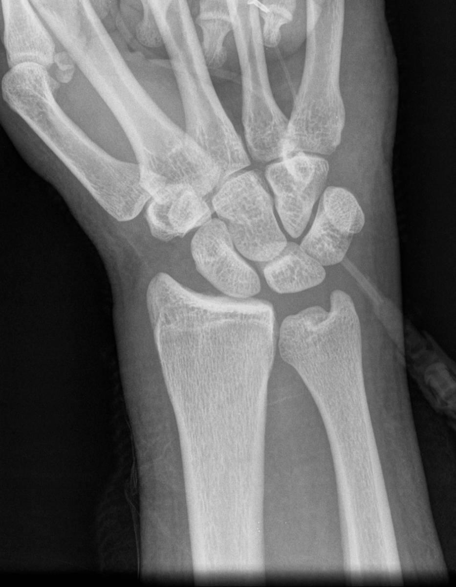
22 Hx: 35 y/o male with wrist injury 1 week ago. SLD Hx: 35 y/o male with wrist injury 1 week ago. Widening of the Scapholunate distance Disruption of the SL ligament at scaphoid attachment Foreshortened appearance of the scaphoid


23 Partial disruption of radioscaphoid portion of RCL at scaphoid attachment site Flexion of distal pole of the scaphoid Dorsal subluxation of the proximal pole with respect to radius

 will allow it to rotate around radioscaphocaptitate ligament leading to dorsal rotary")
24 Rotary Subluxation of the Scaphoid Mechanism: stress loading of extended carpus, usually in ulnar deviation Associated injuries: radiocapitate ligaments, radiotriquetral ligaments, dorsal radiocarpal ligaments, DISI deformity, radial styloid fx and non-displaced scaphoid fx. Why does it rotate? Scaphoid inherently tends to palmar flex because of its oblique position and the loading applied thru scaphotrapezium joint. Lack of ligament restraint (primary by SL, secondary by RSCL and the FCR tendon) will allow it to rotate around radioscaphocaptitate ligament leading to dorsal rotary subluxation of the proximal pole. Figure: Schmitt et al. Eur Radiol. (2006 ) 16:


25 Rotary Subluxation of the Scaphoid and DISI
.")
26 SLAC wrist SLAC (scapholunate advanced collapse) - a specific pattern of osteoarthritis and subluxation results from untreated chronic scapholunate dissociation or from chronic scaphoid nonunion (SNAC). The degenerative changes occur in areas of abnormal loading: radial-scaphoid joint, lunatocapitate joint (as capitate subluxes dorsally on lunate.)
27 SLAC WRIST Characterized by Narrowing of both the radioscaphoid and capitolunate spaces Common pattern of degenerative joint disease of the wrist Seen with CPPD Post Traumatic
28 SLAC causes Traumatic Rotatory subluxation of scaphoid Scaphoid fx w/ delayed/non-union IA fx s violating the radioscaphoid or lunoatocapitate joint Kienbock s disease Midcarpal instability Non traumatic. CPPD: compromises intrinsic/extrinsic lig.
29 examples

30 Scapholunate dissociation with SLAC wrist

31 Scapholunate dissociation with SLAC wrist

32 Scapholunate dissociation with SLAC wrist


33 SLAC wrist another example

34 SLAC stages Stage 1: Arthrosis limited to radial styloid-scaphoid articulation Stage 2 : Arthrosis of entire radioscaphoid articulation Stage 3 : Capitolunate arthrosis Additionally: destruction of scaphocapitate articulation with proximal migration of capitate on radius

35 SLAC wrist in 78 y.o with 3 months dorsal wrist pain r/o mass


36

37
38
39
40 High Grade PT of 2 nd digit extensor Tendon
41 Treatment SLAC 1: Radiostyloidectomy. (may impair RC and part of RL lig- can re-attach to radius w/ anchor). SLAC 2: Proximal row carpectomy if cartilage of head of capitate preserved. SLAC 3: Scaphoidectomy + reconstruction around normal RL jnt + stabilisation w/ ltd arthrodesis ( CL arthrodesis or 4 corner).
42 Patterns of Carpal Instability Dissociative (CID) Scapholunate dissociation (SLD) Lunotriquetral dissociation (LTD) Scaphoid fractures that are unstable, ununited, or malunited Kienbock s disease Non-dissociative (CIND) Radiocarpal Midcarpal Complex carpal instability (CIC) Perilunate dislocations Adaptive carpal instability (CIA)
43 CID: Lunotriquetral Dissociation (LTD) Progressive destruction of the LTL Mostly described in the context as a later stage of perilunate instability In this progression of instability, the disruption progresses from scapholunate lunocapitate lunotriquetral. Alternatively, isolated lunotriquetral abnormalities may relate to reverse perilunate injury that begins on the triquetral side of the lunate and proceeds in the radial direction. Ulnocarpal impaction is another cause a fixed VISI (volar intercalated segmental instability) deformity may occur (but may require failure of other ligaments (ex. Dorsal radiolunotriquetral ligament as well)

44 CID-LT dissociation The capitatolunate angle is over 30 degrees. Lunate is not trapezoid, but moon shaped- VISI. The lunotriquetral joint has nonparallel articulating surfaces
45 Normal DISI VISI MR imaging of the major carpal stabilizing ligaments: normal anatomy and clinical examples..radiographics May;15(3):575-87
46

47 16 y/o with wrist injury, triquetral fx with VISI Triquetral fractures may be associated with transcaphoid perilunate dislocations of the wrist
48

49
50 Normal DISI VISI MR imaging of the major carpal stabilizing ligaments: normal anatomy and clinical examples..radiographics May;15(3):575-87
51 Patterns of Carpal Instability Dissociative (CID) Scapholunate dissociation Lunotriquetral dissociation Scaphoid fractures that are unstable, ununited, or malunited Kienbock s disease Non-dissociative (CIND) Radiocarpal Midcarpal Complex carpal instability (CIC) Perilunate dislocations Adaptive carpal instability (CIA)
52 CID: Scaphoid Fracture When 2 (or more) unstable fragements result, the distal fragment rotates with the distal carpal row and the proximal fragment(s) with the proximal carpal row Radiographics May;15(3):
53 CID: Scaphoid Fracture With an intact SLIL, the proximal scaphoid fragment may rotate dorsally (extend) and the distal sccaphoid fragment may flex Radiographics May;15(3):

54 Humpback deformity Over time, an unstable fracture nonunion or malunion may appear, with a humpback deformity


55 CID: Scaphoid Fracture- 28M with diffuse pain after fall one month ago 28M with diffuse pain after fall one month ago
56 Scaphoid Fracture:Clinical Presentation Mechanism of Injury Hyperextension of the wrist Compressive force (FOOSH) Snuff-box pain, LROM, Weak grip Age 15 to 40
57 Scaphoid Fracture: Clinical Presentation 65% of carpal fractures 5-12% of scaphoid fractures are associated with other fractures Waist fx : 70% Proximal pole fx : 20%
58 Scaphoid Fracture:Imaging Evaluation Radiographs PA, lateral, external oblique, and scaphoid views CT MR Gado may help evaluate proximal pole blood supply Bone scintigraphy
 wrist Malunion Osteonecrosis More common in proximal pole")
59 Complications Nonunion Occurs in 50% of unstable fractures after nonsurgical /inadequate treatment If delay in treatment of > 4wks Long-standing scaphoid non-union leads to carpal collapse, known as scaphoid nonunion advanced collapse (SNAC) wrist Malunion Osteonecrosis More common in proximal pole Arthritis
60 Treatment of scaphoid fx Stable fractures (nondisplaced) Thumb-spica cast 95% healing rate Unstable fractures Surgical treatment


61 CID: Scaphoid nonunion 35 year old male with direct trauma with a board 3 weeks ago.


62 Cystic change in distal pole


63 Scapholunate intact


64 Humpback deformity
65 Humpback deformity- volar tilting distal pole of scaphoid
66 Dorsal tilting of proximal portion scaphoid
67 Dorsal tilting of lunate. Bony fragment
68

69 Scaphoid Nonunion - DISI Humpback- another example Sag T1 Sag PDFS
70 Scaphoid Nonunion - DISI - Humpback Sag T1 Sag PDFS
71 Scaphoid Nonunion - DISI - Humpback Sag T1 Sag PDFS
72 Scaphoid Nonunion - DISI - Humpback Sag T1 Sag PDFS
73 Scaphoid Nonunion - DISI - Humpback Sag T1 Sag PDFS

74 Scaphoid Nonunion - DISI - Humpback Sag T1 Sag PDFS

75

76

77

78

79
80
81
82
83
84
85 SCAPHOID FRACTURE: RISK FACTORS FOR INSTABILITY vertical fracture line orientation fragment displacement >1mm ligamentous instability [DISI characterized by scapholunate angle > 60 degrees (normal = degrees), or radiolunate or capitolunate angle > 15 degrees (normal = 0 +/- 15 degrees)], humpback deformity

86 SCAPHOID NON-UNION Often due to undiagnosed or undertreated nondisplaced scaphoid fractures. Even as late as 6 mo, the fx may heal Findings: Sclerosis at fx site Cysit cavitation Displacement more than 1 mm Local tenderness Persistent lucent line >2 mm. All nonunions are considered unstable Late complications of osteonecrosis of the scaphoid or inadequately treated scaphoid nonunion include a peculiar pattern of osteoarthritis of the wrist.scaphoid nonunion advanced collapse.
87 SNAC wrist Scaphoid Non-Union Advanced Collapse Un-united scaphoid fracture Osteonecrosis of the proximal pole Secondary osteoarthritis between distal scaphoid fragment and radial styloid (+/- DISI) Most patients who have SNAC wrist with nonunion of the middle or distal third also have DISI. Resnick, D. Diagnosis of Bone and Joint Disorders. 4 th Ed. Vol. 2. W.B. Saunders , (Vol 3: 2847, Vol. 4: 3645)
88 SNAC AND SNAC + Radiographic features of SNAC wrist include: non-united scaphoid fracture scaphoid collapse marked osteoarthritis of the radioscaphoid joint the radiolunate joint is usually spared from degenerative involvement. SNAC+ is longstanding scaphoid non-union with osteonecrosis of the proximal pole and development of secondary radiocarpal osteoarthritis. The radiocarpal joint narrowing is between the radial styloid and distal pole of the fractured scaphoid
 Remote non-united ulnar styloid fracture (blue arrowhead) 48 y.o. male with a remote fall on an outstretched hand.")
89 SNAC : PLAIN FILM FINDINGS Non-united scaphoid fracture Cystic changes in the distal pole scaphoid (green arrow) Small sclerotic proximal pole (curved yellow arrow) Scapholunate injury with small bone fragment noted (black wavy arrow) Remote non-united ulnar styloid fracture (blue arrowhead) 48 y.o. male with a remote fall on an outstretched hand. Chronic wrist pain which has been progressive over the past 10 years.
 of scaphoid with chondral loss at radioscaphoid articulation Cyst in lunate (blue arrowhead) with adjacent thickening of scapholunate ligament with bone")
90 SNAC: MR FINDINGS Non-united scaphoid fracture with high signal between the fragments suggestive of pseudoarthrosis (yellow curved arrow) Cyst in distal and proximal pole of scaphoid (green arrow) Small sclerotic proximal pole (black arrow) of scaphoid with chondral loss at radioscaphoid articulation Cyst in lunate (blue arrowhead) with adjacent thickening of scapholunate ligament with bone marrow edema at lunate attachment site

91


92 scaphoid nonunion advanced collapse Wrist pain distant trauma

93 COR T2 COR PD
94 scaphoid nonunion advanced collapse Wrist pain distant trauma


95 scaphoid nonunion advanced collapse Wrist pain distant trauma
96 scaphoid nonunion advanced collapse Wrist pain distant trauma
97 scaphoid nonunion advanced collapse Wrist pain distant trauma
98 Wrist pain distant trauma

99 Wrist pain distant trauma
100 Wrist pain distant trauma
101 TREATMENT Surgical treatment is usually required for scaphoid non-union.- debridement,reduction, bone gaft If more severe pseudoarthrosis, AVN or osteoarthritis is present, the rate of union is progressively decreased. In severe or late cases, on nonunion, factors, such as the patient s age, occupation, symptoms and needs, are considered when determining whether treatment should involve conservative measures, scaphoid reconstruction or a salvage procedure.
102 SNAC
103 SNAC
104 SNAC
105 SNAC
106 SNAC
107 SNAC
108 SNAC
109 SNAC

110 SNAC
111 SNAC
112 SNAC
113 SNAC
114 SNAC
115 SNAC
116 SNAC
117 SNAC
118 SNAC
119 SNAC
120 SNAC
121 SNAC
122 SNAC
123 SNAC
124 SNAC
125 SNAC
126 SNAC
127 SNAC
128 SNAC
129 SNAC
130 SNAC
131 SNAC
132 SNAC

133 SNAC
134 SNAC
135 Patterns of Carpal Instability Dissociative Scapholunate dissociation Lunotriquetral dissociation Scaphoid fractures that are unstable, ununited, or malunited Kienbock s disease Non-dissociative Radiocarpal Midcarpal Complex carpal instability Perilunate dislocations Adaptive carpal instability
136 Kienbock s disease Fragementation and progressive collapse of the lunate bone Disruption of the scapholunate and lunotriquetral interosseous ligaments. Lunate flexes with proximal migration of the triquetrum Either VISI or DISI pattern of instability may develop
137 Patterns of Carpal Instability Dissociative (CID) Scapholunate dissociation Lunotriquetral dissociation Scaphoid fractures that are unstable, ununited, or malunited Kienbock s disease Non-dissociative (CIND) Radiocarpal Midcarpal Complex carpal instability (CIC) Perilunate dislocations Adaptive carpal instability (CIA)
138 Patterns of Carpal Instability Dissociative (CID) Scapholunate dissociation Lunotriquetral dissociation Scaphoid fractures that are unstable, ununited, or malunited Kienbock s disease Non-dissociative (CIND) Radiocarpal Ulnar translocation Dorsal Volar Combination of the above Midcarpal Complex carpal instability Perilunate dislocations Adaptive carpal instability
139 CIND: radiocarpal Can be asstd with articular disorders, injuries, developmental anomalies or prior surgeries (resection of the distal portion of the ulna. Instability can be ulnar (ulnar translocation), dorsal volar combinations of some of these There are 2 types of ulnar translocation
140 CIND: radiocarpal Type I ulnar translocation Type II ulnar translocation Resnick, Kang, Pretterklieber : Internal Derrangements of Joints. 2nd ed. P 1289
141 CIND: radiocarpal Note: Normally, the radiolunate joint area covers more than 50% of the proximal lunate joint surface. Radiolunate contact is decresed with ulnar translocation. Resnick, Kang, Pretterklieber : Internal Derrangements of Joints. 2nd ed. P 1289

142 Radiocarpal CIND: ulnar translocation, type I Ligaments disrupted Radioscaphoid (RSL) Radioscaphocapitate (RSCL) radiolunotriquetral (RLTL) These ligaments resist the tendency to slide down the articular tilt of the radius. Resnick, Kang, Pretterklieber : Internal Derrangements of Joints. 2nd ed. P 1289

143 Radiocarpal CIND: ulnar translocation, type I Most common causes are rheumatoid arthritis and Medelung s deformity Traumatic dislocation is rare Iatrogenic (excessive resection of the radial styloid process or of the ulnar head)


144 Injur23yo M s/p MVA with wrist pain, Injury films no ulnar translocation initially

145 2 weeks later

146 2 weeks later later
147 Extrinsic ligament disruption Major radiocarpal stabilizers: Radioscaphocapitate Radiolunotriquetral (aka-long radiolunate) Short radiolunate All 3 are intracapsular and extrasynovial RLT is disrupted in Type II ulnar translocation RSC and RLT disrupted in Type I ulnar translocation
148 UT I: Extrinsic ligament disruption Radioscaphocapitate (RSC) ligament Courses from volar radial styloid over waist of scaphoid (without attaching) and attaches to center of capitate

149 Extrinsic ligament disruption Radiolunotriquetral (RLT) ligament Largest ligament of the wrist Courses from volar radial styloid to attach to the volar lunate and then the triquetrum
150 Extrinsic ligament disruption Dorsal Extrinsic Wrist Ligaments Dorsal Radiocarpal Ligament 3 parts: Radioscaphoid Radiolunate Radiotriquetral
151 CIND: ulnar translocation Treatment: Ligament reconstruction or radiocarpal arthrodesis


152 BCIND: ulnar translocation I Post opulnar translocation
153 CIND: ulnar translocation, type II Scaphoid remains in place, lunate and triquetrum slide in an ulnar direction SLL tear The radiolunotriquetral ligament (RLTL) is disrupted (not shown) Resnick, Kang, Pretterklieber : Internal Derrangements of Joints. 2nd ed. P 1289
154 CIND: ulnar translocation, type II Although this is classified as CIND, it has features of BOTH CIND and CID (and therefore could be classified as carpal instability complex (CIC) CIC= CIND + CID Aside : CIC includes perilunate dislocations and their sequelae Resnick, Kang, Pretterklieber : Internal Derrangements of Joints. 2nd ed. P 1289
155 Tele case no history
156 CIND/CIC: ulnar translocation, type II (extrinsic) Dorsal radiotriquetral ligament- intact
157 CIND/CIC: ulnar translocation, type II
158 CIND/CIC: ulnar translocation, type II


159 CIND/CIC: ulnar translocation, type II Torn SLL
160 CIND/CIC: ulnar translocation, type II Mild uncovering of the lunate
161 CIND/CIC: ulnar translocation, type II
162 CIND/CIC: ulnar translocation, type II
163
164

165
166
167 avulsion fragment of the scaphoid in the dorsal SLL (torn)
168

169 Dorsal scaphotriquetral ligament is torn Donor site of avulsion fracture
170 Dorsal scaphotriquetral ligament
171 ulnar translocation, type II Dorsal scaphotriquetral ligament
172
173 Dorsal subluxation of scaphoid
174
175
176
177
178
179 Summary of Findings: Complete tear of the SLIL with scaphoid avulsion fragment Marked widening of the scapholunate interosseous space ulnar translocation of the lunate and triquetrum at the radiocarpal joint (radiocarpal instability) Dorsal subluxation of the scaphoid with respect to the distal radius Lunate is mildly dorsally tilted
180 CIND: radiocarpal, other Other patterns are rare Volar dorsal Those associated with displaced or badly malunited fx s of the distal radius. Ex. Dorsal tilt of distal radius extension of the entire proximal row. The distal row shifts into flexion to compensate A kind of caral instability complex (CIA)
181 CIND: radiocarpal, other
182 CIND: radiocarpal, other

183 CIND: radiocarpal, other

184 CIND: radiocarpal, other

185 CIND: radiocarpal, other


186
187 Patterns of Carpal Instability Dissociative (CID) Scapholunate dissociation Lunotriquetral dissociation Scaphoid fractures that are unstable, ununited, or malunited Kienbock s disease Non-dissociative (CIND) Radiocarpal Midcarpal Complex carpal instability (CIC) Perilunate dislocations Adaptive carpal instability (CIA)
188 CIND: midcarpal instability (MCI) MCI is a group of conditions that is a source of ongoing debate in terms of its etiology, terminology, classification, and treatment. Instability of the proximal carpal row with both radiocarpal and midcarpal joint alterations are charachteristic Andoni et al. Skeletal Radiol. May14,2010.

189 CIND: midcarpal instability (MCI) Major ligaments involved: Triquetro-hamatecapitate Andoni et al. Skeletal Radiol. May14,2010.
 ligaments have a role too.")
190 Main extrinsic ligaments involved MCI Ex vivo and in vivo evidence shows that deficiency of these 2 ligaments results in Palmar MCI- Andoni et al. Anteromedial scaphocapitate and scaphotrapeziotrapezoid (STT) ligaments have a role too. The palmar ligaments are demonstrated at the front of this transparent diagram Dorsal radiotriquetral ligament Palmar arcuate ligament Andoni et al. Skeletal Radiol. May14,2010.
191 Principle extrinsic ligaments prone to dysfunction in midcarpal instability Palmar arcuate ligament Dorsal radiotriquetral ligament The palmar ligaments are demonstrated at the front of this transparent diagram Andoni et al. Skeletal Radiol. May14,2010.
192 MCI: palmar arcuate ligament Resists the tendency during axial loading for the distal carpal row to rotate into extension and the proximal carpal row into flexion. Has two limbs: Triquetro-hamatecapitate scaphocapitate Andoni et al. Skeletal Radiol. May14,2010.
193 Insert fig of midcarpal instability types IDJ p1289 Resnick et al. IDJ. 2 nd ed. Type I: palmar midcarpal instability (PMCI) volar flexion of the entire proximal row VISI deformity. Most common type c/o painful clunking wrist
194 Insert fig of midcarpal instability types IDJ p1289 Resnick et al. IDJ. 2 nd ed. Type II: Dorsal midcarpal instability Dorsal subluxation of the capitate and dorsal tilting of the scaphoid and lunate Includes: Capitolunate instability pattern (CLIP) Chronic capitolunate instability (CCI)
195 Insert fig of midcarpal instability types IDJ p1289 Type III: Dorsal and Palmar midcarpal instability exaggeration of the dorsal MCI with additional dorsal subluxation of the scaphoid and lunate Resnick et al. IDJ. 2 nd ed.
 Resnick et al. IDJ.")
196 Insert fig of midcarpal instability types IDJ p1289 Type IV: Extrinsic midcarpal instability asstd with prior radial fx s with persistent dorsal angulation of the radius Dorsal displacement of carpal bones is related to stretching of the dorsal ligaments. Thus, this could also be classified as (CIA) Resnick et al. IDJ. 2 nd ed.

197 Proximal Midcarpal Instability (PMCI) Capitolunate angle 30 (nl <30) MCI demonstrating flexion of the proximal carpal row. This is sometimes confusingly referred to as a mild VISI deformity because of the flexed lunate. However, a full VISI deformity, with rupture of the lunotriquetral ligament, typically results in a reduction of the scapholunate angle However, the entire proximal carpal row ( including the scaphoid) is flexed evidenced here by the ring sign(dashed outline, arrow) and the posterior apex of the lunate is rotated distally (arrowhead) AND the scaphlolunate angle is normal Scapholunate angle 50 (normal)
198 Catch up clunk in PMCI Radial deviation Ulnar deviation During radial to ulnar deviation, the proximal row sags towards the palm without extending until the triquetrohamate joint engages and forces theproximal carpal row into extension
 ligament Normal")

199 CIND: midcarpal Norma ulnar limb of palmar arcuate (triquetrohamatecapitate) ligament Normal Torn Torn triquetrohamatecapitate ligament in a patient with MCI Andoni et al. Skeletal Radiol. May14,2010.
200 CIND: midcarpal Conclusions of Andoni et al.: Association of abnormalities demonstrated on MRI with the diagnosis of MCI has yet to be demonstrated. Pattern of injury is probably more complex than simple isolated injuries. Defects in the extrinsic carpal ligaments that cause MCI, and the resulting dysfunctional carpal mechanics can be demonstrated with US and MR arthrography But the accuracy of these techniques, and therefore the role they may play in the management of patients with MCI, has yet to be determined.
201 Patterns of Carpal Instability Dissociative Scapholunate dissociation Lunotriquetral dissociation Scaphoid fractures that are unstable, ununited, or malunited Kienbock s disease Non-dissociative Radiocarpal Midcarpal Complex carpal instability Perilunate dislocations Adaptive carpal instability
202 CIC: Includes CID+ CIND cases 5 patterns of dislocation: Dorsal perilunate dislocation (lesser arc injury) Dorsal perilunate fracture-dislocations (greater arc injury) Volar perilunate dislocations Axial dislocations Isolated carpal bone dislocations
203 CIC: Most typical example is the result of failed treatment of a perilunate dislocation. Ex. Perilunate injuries create both radiocarpal and intercarpal injuries which if not treated properly chronic SLD and LTD (CID patterns), AND ulnar translation of the lunate (CIND pattern) Usually involves both intrinsic and extrinsic radiocarpal ligaments Failure to obtain stability of the joints of the proximal row may result in chronic CID type carpal collapse
204 CIC: If the Extrinsic lig s do not heal (after perilunate dislocation) or are ineffective, there is a tendency to develop radiocarpal CIND (ulnar translocation) Need surgical stabilization of both causes of instability ie. midcarpal fusion with tightening of the dorsal and volar radiocarpal ligaments If there is substantial cartilage wear, then formal wrist fusion is better. Typically, a DISI deformity appears.

")
205 Greater and Lesser Arc Injuries lesser arc injury : pure ligamentous perilunar injury Some disagreement exists regarding some of the stages of perilunate instability greater arc injury: transosseous variants (transscaphoid,transcapitate, transhamate, transtriquetral fracture-dislocation) Various combinations are seen clinically


206 Space of Poirer Volar intrinsic ligaments
207 Perilunate Instability Dorsal side Stage I Scapholunate dissociation -Injury to radial side of wrist leads to injury to scaphoid lunate interosseous ligament -scaphoid is pulled into extension * Space of Poirer ( pear ) is opened- a weak triangular region in which the volar capsule is not reinforced by ligaments. Resnick IDJ, p.1304.
208 Perilunate Instability Dorsal side Stage II Lunocapitate dislocation (Perilunate) - Dorsal translocation or dislocation of distal row relative to capitate Resnick IDJ, p.1304.

209 Perilunate Instability Dorsal side Stage III Lunotriquetral disruption (Midcarpal) - Triquetrum separates from the lunate owing to displacement of the capitate causing disruption of the lunotriquetral interosseous ligament Resnick IDJ, p.1304.
210 Perilunate Instability Dorsal side Stage IV Lunate dislocation (Final Step) - Dorsally dislocated capitate contacts lunate, causing palmar dislocation in a rotary fashion Resnick IDJ, p.1304.

211 41 year old female with wrist pain

212
213 Greater arc injury

214 30 year-old male, Fall 1 month ago with wrist dislocation. c/o Pain and edema

215

216

217 Torn radiolunotriquetral
218
219

220
221
222
223
224
225
226
227 Triquetral fracture
228
229 CIC: Findings: Disruption of Scapholunate interosseous ligament with DISI Volar Radioscaphocapitate ligament (CIND- like) Volar Radiolunotriquetral ligament (CIND -like) Fractures of Radial styloid Triquetrium (CID like) Ulnar styloid
230 Diagnosis Complex wrist injury Injury involving Greater and Lesser Arcs Trans-radial styloid, trans-triquetral perilunate instability with ulnar styloid fracture and residual DISI soft tissue injuries = S-L, R-S-C, R-L-T disruption
231 Patterns of Carpal Instability Dissociative Scapholunate dissociation Lunotriquetral dissociation Scaphoid fractures that are unstable, ununited, or malunited Kienbock s disease Non-dissociative Radiocarpal Midcarpal Complex carpal instability Perilunate dislocations Adaptive carpal instability
232 Adaptive Carpal Instability Change in carpal alignment adapting to the pathologic change outside of the carpal bones and their ligaments. Ex. malunion of the a distal radial fx results in tilting of the carpal bones to maintain a straight light between the distal radius and the metacarpals. Typical pattern is a DISI type alignment.
233 Adaptive Carpal Instability Consequences of adaptive carpal instability Altered carpal kinetics, overloading the cartilage, arthritis Tx: Osteotomy. However, if there is associated radiocarpal ligament disruption or stretched after injury, ligament reconstruction or radiolunate or radioscapholunate arthrodesis is needed in addition. The effects of dorsally angulated distal radius fractures on carpal kinematics. Park MJ. J Hand Surg [Am] Mar;27(2):


234 CIA Top: malunited Colles fracture with dorsal angulation of the distal radius. The axes of the lunate, capitate and metacarpals is not paralled. Bottom: corrected malunion with osteotomy. The carpal bones are now collinear.
235 Adaptive Carpal Instability
236
237
238
239 Treatment of carpal instabilites No single treatment Must meet needs of individuals by considering: Chronicity (healing potnetial of the lig. Involved) Constancy (dynamic or static) Etiology (traumtic, congenital, or inflammatory) Location (site of the major dysfunction radiocarpal, midcarpal, intercapral, carpometacarpal)
240 Thank you! Special thanks to Tudor Hughes.
241 REFERENCES Goldfarb CA, Yin Y, Gilula LA et al. Wrist Fractures: What the Clinician Wants to Know. Radiology 2001; 219: Mann FA, Wilson AJ, Gilula LA. Radiographic Evaluation of the Wrist: What does the Hand Surgeon Want to Know. Radiology 1992; 184: Resnick, D. Diagnosis of Bone and Joint Disorders. 4 th Ed. Vol. 2. W.B. Saunders , (Vol 3: 2847, Vol. 4: 3645)
Carpal Instability: Clarification of the Most Common Etiologies and Imaging Findings
 Carpal Instability: Clarification of the Most Common Etiologies and Imaging Findings Corey Matthews DO, Nicholas Strle DO, Donald von Borstel DO Oklahoma State University Medical Center, Department of
Sean Walsh Orthopaedic Surgeon Dorset County Hospital
 Sean Walsh Orthopaedic Surgeon Dorset County Hospital Shapes and orientation of articular surfaces Ligaments Oblique positioning of scaphoid Tendons surrounding the joints Other soft tissues Peripheral
3. Ulno lunate, Ulno triquetral ligament. Poirier: Between RSC &LRL. 5. Dorsal intercarpal ligament
 CARPAL INSTABILITY Ligaments Intrinsic Scapho lunate ligament: Dorsal component stronger than volar ligament Luno triquetral ligament: Volar component stronger than dorsal ligament Extrinsic Palmar 1 Radio
MR IMAGING OF THE WRIST
 MR IMAGING OF THE WRIST Wrist Instability Dissociative Pattern apparent on routine radiographs Non-dissociative Stress / positional radiographs Dynamic fluoroscopy during stress Arthrography MRI / MR arthrography
SCAHPO-LUNATE DISSOCIATION
 SCAHPO-LUNATE DISSOCIATION Introduction Scapho-lunate dissociation is the most common significant ligamentous injury of the wrist. The condition is also sometimes referred to as rotary subluxation of the
Introduction. The wrist contains eight small carpal bones, which as a group act as a flexible spacer between the forearm and hand.
 Wrist Introduction The wrist contains eight small carpal bones, which as a group act as a flexible spacer between the forearm and hand. Distal forearm Distal forearm 4 Distal end of the radius A. anterior
Abstract Submission Form
 Abstract Submission Form All abstracts must be submitted to the AOCR by September 15 th. All information included must be the original work of the author(s) and be in typed form. Incomplete or handwritten
Scapholunate Ligament Lesions Imaging Which and when?
 Scapholunate Ligament Lesions Imaging Which and when? Kolo Frank Lesions to scapholunate ligament(sl) Most frequent cause of carpal instability Traumatic tears of SL ligament = most common ligament injury
Scaphoid Fractures. Mohammed Alasmari. Orthopaedic Surgery Demonstrator Majmaah University
 Scaphoid Fractures Mohammed Alasmari Orthopaedic Surgery Demonstrator Majmaah University 1 2 Scaphoid Fractures Introduction Anatomy History Clinical examination Radiographic evaluation Classification
SCAPHOID FRACTURE. Relevant antomy
 SCAPHOID FRACTURE Relevant antomy The proximal row consists of the scaphoid, the lunate, and the triquetrum. The proximal carpal row is regarded as an intercalated segment The keystone in the coordination
Carpal rows injuries!
 Carpal rows injuries! Michael Papaloïzos! Center for Hand Surgery and Therapy Geneva, Switzerland no conflict of interest to declare Fractures of carpal bones! The fractured scaphoid! Fracture-dislocations
Wrist Arthritis & Partial Wrist Fusion
 Wrist Arthritis & Partial Wrist Fusion Mr Jason N Harvey MB.BS. FRACS (Orth) Hand,Wrist & Elbow Surgeon Clinical Symptoms Outline Physical Examination Diagnosis Differential Diagnosis Outline Non-operative
COMMON CARPAL INJURIES IN ATHLETES Nicholas A. Bontempo, MD Orthopedic Associates of Hartford I HAVE NO CONFLICTS OR DISCLOSURES TO REPORT OUTLINE
 COMMON CARPAL INJURIES IN ATHLETES Nicholas A. Bontempo, MD Orthopedic Associates of Hartford I HAVE NO CONFLICTS OR DISCLOSURES TO REPORT OUTLINE The carpus Scaphoid fracture Scapholunate ligament tear
Hand and wrist emergencies
 Chapter1 Hand and wrist emergencies Carl A. Germann Distal radius and ulnar injuries PEARL: Fractures of the distal radius and ulna are the most common type of fractures in patients younger than 75 years.
Union rate: Union: Stable 94% All fracture 90% Union after surgery for nonunion with surgery 80% OA in healed scaphoid: 9%
 Complications Incidence of Non-union 1 cm displacement of fracture caused 55% Non-union It takes 5-20 yrs to develop SNAC. SNAC appears to be more common with waist fracture than a proximal pole. However
Acute Wrist Injuries OUCH!
 Acute Wrist Injuries OUCH! Case the athlete FOOSH from sporting event 2 days ago C/O wrist swelling, pain, worse with movement Hmmm Wrist pain Exam of the wrist - basics Appearance Swelling, bruising,
The Kienböck disease and scaphoid fractures. Mariusz Bonczar
 The Kienböck disease and scaphoid fractures Mariusz Bonczar The Kienböck disease and scaphoid fractures Mariusz Bonczar Kienböck disease personal experience My special interest for almost 25 years Thesis
Mayo Clinic Disorders of the Wrist
 Mayo Clinic Disorders of the Wrist Thursday, May 19, 2016 Pre-Conference Laboratory Workshop Anatomy of the Wrist & Wrist Arthroscopy 6:30 a.m. Registration and Breakfast 7:30 a.m. Welcome and Introduction
C L I N I C A L A RT I C L E
 Page 32 / SA ORTHOPAEDIC JOURNAL Winter 2008 C L I N I C A L A RT I C L E Treatment of lunate and perilunate dislocations with a combined approach and anchor repair of the dorsal scapholunate interosseous
SYMPOSIUM ON ADVANCES IN THE MANAGEMENT OF SCAPHOID PROBLEMS Scaphoid malunion
 Hong HKJOS Kong Journal of Orthopaedic Surgery 2002;6(2):104-108. SYMPOSIUM ON ADVANCES IN THE MANAGEMENT OF SCAPHOID PROBLEMS Scaphoid malunion Department of Orthopaedics and Traumatology, Prince of Wales
PREVIEW ONLY 27/10/2014. Instabilities in the Wrist
 Be sure to convert to your own time zone at Andrew Ellis BSc (Ex. Sci), M. Phty Instabilities in the Wrist Presented by: Ben Cunningham Be sure to convert to your own time zone at Ben Cunningham Member
TRIQUETRUM FRACTURE. The triquetrum bone is one of the small bones that make up the carpus.
 TRIQUETRUM FRACTURE Introduction The triquetrum bone is one of the small bones that make up the carpus. It is also known as the triquetral bone, (and in the past the pyramidal or triangular bone) Triquetrum
EXAMINATION OF THE WRIST BEYOND THE BASICS OMA SPORT MED Janice Harvey MD CCFP CFFP Dip. Sp Med.
 EXAMINATION OF THE WRIST BEYOND THE BASICS OMA SPORT MED 2019 Janice Harvey MD CCFP CFFP Dip. Sp Med. CFPC CoI Templates: Slide 1 used in Faculty presentation only. FACULTY/PRESENTER DISCLOSURE Faculty:
SPORTS INJURIES IN HAND
 Grundkurs SGSM-SSMS Sion 2015 SPORTS INJURIES IN HAND Dr S. KŠmpfen EPIDEMIOLOGY Incidence of hand, finger and wrist injuries in sports : 3% Ð 9 % RADIAL-SIDED WRIST PAIN 1)! Distal Radius Fractures 2)!
SURGICAL/APPLIED ANATOMY
 Página 1 de 11 Copyright 2001 Lippincott Williams & Wilkins Bucholz, Robert W., Heckman, James D. Rockwood & Green's Fractures in Adults, 5th Edition SURGICAL/APPLIED ANATOMY Part of "19 - FRACTURES AND
Index. Note: Page numbers of article titles are in boldface type. Hand Clin 21 (2005)
") Hand Clin 21 (2005) 501 505 Index Note: Page numbers of article titles are in boldface type. A Antibiotics, following distal radius fracture treatment, 295, 296 Arthritis, following malunion of distal
Hand Clin 21 (2005) 501 505 Index Note: Page numbers of article titles are in boldface type. A Antibiotics, following distal radius fracture treatment, 295, 296 Arthritis, following malunion of distal
Alvin S. Chen, Harvard Medical School Year III Gillian Lieberman, MD Radiology Core Clerkship
 Alvin S. Chen, Harvard Medical School Year III Gillian Lieberman, MD Radiology Core Clerkship Overview Wrist: Normal Anatomy & Biomechanics Approach to Wrist Imaging: Menu of Tests & Efficacious Use Index
Scapholunate Advanced Collapse and Scaphoid Nonunion Advanced Collapse: MDCT Arthrography Features
 Musculoskeletal Imaging Pictorial Essay Crema et al. MDCT rthrography of SLC and SNC Musculoskeletal Imaging Pictorial Essay Michel D. Crema 1,2 Joachim Zentner 3 li Guermazi 1 Nabil Jomaah 4 Monica D.
Carpal Injuries. AO Advanced Principles of Fracture Management Middelfart, april 2016
 Carpal Injuries AO Advanced Principles of Fracture Management Middelfart, 11.-14. april 2016 Overlæge Marianne Vestergaard Lind Traumesektionen Ortopædkirurgisk Klinik Rigshospitalet AOT Advanced Principles
Interesting Case Series. Perilunate Dislocation
 Interesting Case Series Perilunate Dislocation Tom Reisler, BSc (Hons), MB ChB, MRCS (Ed), Paul J. Therattil, MD, and Edward S. Lee, MD Division of Plastic and Reconstructive Surgery, Department of Surgery,
Neglected trans-scaphoid trans-styloid volar dislocation of the lunate
 CASE REPORT Neglected trans-scaphoid trans-styloid volar dislocation of the lunate LATE RESULT FOLLOWING OPEN REDUCTION AND K-WIRE FIXATION P. Givissis, A. Christodoulou, B. Chalidis, J. Pournaras From
CLINICAL PRESENTATION AND RADIOLOGY QUIZ QUESTION
 Donald L. Renfrew, MD Radiology Associates of the Fox Valley, 333 N. Commercial Street, Suite 100, Neenah, WI 54956 10/13/2012 Radiology Quiz of the Week # 94 Page 1 CLINICAL PRESENTATION AND RADIOLOGY
Carpal ligaments evaluation with ultrasound and MRI: A pictorial review
 Carpal ligaments evaluation with ultrasound and MRI: A pictorial review Poster No.: C-2246 Congress: ECR 2010 Type: Educational Exhibit Topic: Musculoskeletal Authors: A. Kraus, C. Barwick, S. Wenham,
FOOSH It sounded like a fun thing at the time!
 FOOSH It sounded like a fun thing at the time! Evaluating acute hand and wrist injuries Larry Collins, MPAS, PA-C, ATC, DFAAPA Assistant Professor, Physician Assistant Program Assistant Professor, Department
Carpal Bone Fractures Excluding the Scaphoid
 CHAPTER 9 Carpal Bone Fractures Excluding the Scaphoid Khemarin R. Seng * and Philip E. Blazar * MD, Resident, Department of Orthopaedic Surgery, Massachusetts General Hospital, Boston, MA MD, Assistant
Integra. Spider and Mini Spider Limited Wrist Fusion System SURGICAL TECHNIQUE
 Integra Spider and Mini Spider Limited Wrist Fusion System SURGICAL TECHNIQUE Table of contents Description... 02 Indications... 02 Contraindications... 02 Surgical Technique... 03 Spider Introduction-Four
Chapter 51 Wrist and Forearm Episode Overview
 Chapter 51 Wrist and Forearm Episode Overview 1) Describe normal radiographic relationships: a. Radial length measurement b. Radial inclination c. Volar Tilt d. Scapholunate angle e. Capitolunate angle
7/16/2015. Imaging in the Diagnosis of Ulnar Sided Wrist Pain. Sources of Ulnar Sided Wrist Pain. Triangular Fibrocartilage Anatomy
 7/16/2015 Imaging in the Diagnosis of Ulnar Sided Wrist Pain Kimberly K. Amrami, MD Professor of Radiology Chair, Division of Musculoskeletal Radiology Mayo Clinic Rochester, Minnesota Sources of Ulnar
Surgical Technique. SLIC Screw System
 Surgical Technique SLIC Screw System Acumed is a global leader of innovative orthopaedic and medical solutions. We are dedicated to developing products, service methods, and approaches that improve patient
FOOSH It sounded like a fun thing at the time!
 FOOSH It sounded like a fun thing at the time! Evaluating acute hand and wrist injuries Larry Collins, MPAS, PA-C, ATC, DFAAPA Assistant Professor, Physician Assistant Program Assistant Professor, Department
Hand & Wrist Injuries. DR MA Manjra
 Hand & Wrist Injuries DR MA Manjra 1 Background Up to 25% of all athletic injuries General population Sport people Sport specific Position specific Multifaceted Time of season Level of athlete Parents
Spectrum of Carpal Dislocations and Fracture-Dislocations: Imaging and Management
 Musculoskeletal Imaging Review Scalcione et al. Carpal Dislocations and Fracture- Dislocations Musculoskeletal Imaging Review FOCUS ON: Luke R. Scalcione 1 Lana H. Gimber 1 nnette M. Ho 1 Stephen S. Johnston
8/25/2014. Radiocarpal Joint. Midcarpal Joint. Osteology of the Wrist
 Structure and Function of the Wrist 2 joints and 10 different bones Combine to create wrist motion Anatomical Terms: Wrist/Hand Palmar = anterior aspect of the wrist and hand Dorsal = posterior aspect
Surgical Technique. DISCLOSURE: This device is not approved for sale in the U.S.A. Customer Service:
 DISCLOSURE: This device is not approved for sale in the U.S.A. INDICATIONS FOR USE The KinematX Modular Wrist Arthroplasty System is indicated for the replacement of a wrist joints disabled by pain, deformity,
Link to related CJSM article: ts Frequency_and.5.
 Link to related CJSM article: https://journals.lww.com/cjsportsmed/abstract/2002/11000/wrist_pain_in_young_gymnas ts Frequency_and.5.aspx Link to related case: https://www.amssm.org/when_a_quot%3bsimple_fractur-csa-437.html?startpos=0&part=
SPEED TM Hand & Wrist System. Procedure Manual
 SPEED TM Hand & Wrist System Procedure Manual Table of Contents Limited intercarpal joint arthrodesis (including isolated capitolunate, 3 two-column and four-corner fusions) Thumb carpometacarpal (CMC)
10/15/2014. Wrist. Clarification of Terms. Clarification of Terms cont
 Wrist Clarification of Terms Palmar is synonymous with anterior aspect of the wrist and hand Ventral is also synonymous with anterior aspect of the wrist and hand Dorsal refers to the posterior aspect
journal ORIGINAL RESEARCH
 texas orthopaedic journal ORIGINAL RESEARCH Assessment of Volar Tilt Measurements with Variations in X-Ray Beam Centralization Along the Longitudinal Axis of the Radius Russell A. Wagner, MD; Will Junius,
Interesting Case Series. Ulnolunate Impaction Syndrome
 Interesting Case Series Ulnolunate Impaction Syndrome Saptarshi Biswas, MD, FRCS Westchester University Medical Center, Valhalla, NY Keywords: ulnar impaction, ulnar impaction syndrome, ulnar wrist pain,
Wrist movements, apart from the distal radioulnar joint, take place in two planes:
 The wrist consists of eight bones in two rows: the proximal and distal. The proximal row includes (starting from the radial bone): the scaphoid bone, the lunate bone, the triangular bone and the postulnar
Physical therapy of the wrist and hand
 Physical therapy of the wrist and hand Functional anatomy wrist and hand The wrist includes distal radius, scaphoid, lunate, triquetrum, pisiform, trapezium, trapezoid, capitate, and hamate. The hand includes
MUSCULOSKELETAL. Carpal instability
 Eur Radiol (2006) 16: 2161 2178 DOI 10.1007/s00330-006-0161-1 MUSCULOSKELETAL R. Schmitt S. Froehner G. Coblenz G. Christopoulos Carpal instability Received: 8 August 2005 Revised: 29 December 2005 Accepted:
Hand & Wrist Casey G. Batten MD Assistant Clinical Professor UCSF Sports Medicine
 Hand & Wrist Casey G. Batten MD Assistant Clinical Professor UCSF Sports Medicine Topics: Scaphoid Fracture Scapholunate Separation TFCC Injury Thumb Ulnar Collateral Lig (UCL) Injury Extensor Injury /
Technique Guide. VA-Locking Intercarpal Fusion System. Variable angle locking technology for mediocarpal partial arthrodesis.
 Technique Guide VA-Locking Intercarpal Fusion System. Variable angle locking technology for mediocarpal partial arthrodesis. Table of Contents Introduction VA-Locking Intercarpal Fusion System 2 Indications
Pathogenesis and evolution of carpal instability: imaging and topography
 ACTA BIOMED 2006; 77: 168-180 Mattioli 1885 U P T O D A T E Pathogenesis and evolution of carpal instability: imaging and topography Massimo De Filippo, Jonathan J. Sudberry 1, Eugenio Lombardo, Maurizio
Common Limb Fractures. Mr Sheraz Malik MB BS MRCS Instructor Mr Paul Ofori-Atta Mb ChB FRCS President Motc Life UK April 2009
 Common Limb Fractures Mr Sheraz Malik MB BS MRCS Instructor Mr Paul Ofori-Atta Mb ChB FRCS President Motc Life UK April 2009 Objectives To be able to describe all characteristics of a fracture Describe
Arhtroscopy of the wrist joint: Setup, instrumentation, anatomy & indications
 Arhtroscopy of the wrist joint: Setup, instrumentation, anatomy & indications Andreas Panagopoulos, MD, PhD Upper Limb and Sports Medicine Surgeon Assistant Professor in Orthopaedics Patras University
WRIST TERMINOLOGY AS DEFINED BY THE INTERNATIONAL WRIST INVESTIGATORS WORKSHOP (IWIW)
") 1 WRIST TERMINOLOGY AS DEFINED BY THE INTERNATIONAL WRIST INVESTIGATORS WORKSHOP (IWIW) GILULA LA, MANN FA, DOBYNS JH, YIN Y, AND IWIW TERMINOLOGY COMMITTEE* the shape or size of the carpal bones even
1 WRIST TERMINOLOGY AS DEFINED BY THE INTERNATIONAL WRIST INVESTIGATORS WORKSHOP (IWIW) GILULA LA, MANN FA, DOBYNS JH, YIN Y, AND IWIW TERMINOLOGY COMMITTEE* the shape or size of the carpal bones even
TABLE OF CONTENTS. Limited intercarpal joint arthrodesis (including isolated capitolunate, two-column and four-corner fusions)
") PROCEDURE MANUAL TABLE OF CONTENTS 3 4 5 6 7 8 9 10 11 12 13 Limited intercarpal joint arthrodesis (including isolated capitolunate, two-column and four-corner fusions) Thumb carpometacarpal (CMC) joint
PROCEDURE MANUAL TABLE OF CONTENTS 3 4 5 6 7 8 9 10 11 12 13 Limited intercarpal joint arthrodesis (including isolated capitolunate, two-column and four-corner fusions) Thumb carpometacarpal (CMC) joint
journal REVIEW Midcarpal Instability: Unlocking the Secrets of the Wrist
 texas orthopaedic journal REVIEW Midcarpal Instability: Unlocking the Secrets of the Wrist David M. Lichtman, MD 1 ; William F. Pientka II, MD 2 1 Department of Orthopaedic Surgery, University of North
Forearm and Wrist Regions Neumann Chapter 7
 Forearm and Wrist Regions Neumann Chapter 7 REVIEW AND HIGHLIGHTS OF OSTEOLOGY & ARTHROLOGY Radius dorsal radial tubercle radial styloid process Ulna ulnar styloid process ulnar head Carpals Proximal Row
Characterizing scaphoid nonunion deformity using 2-D and 3-D imaging techniques ten Berg, P.W.L.
 UvA-DARE (Digital Academic Repository) Characterizing scaphoid nonunion deformity using 2-D and 3-D imaging techniques ten Berg, P.W.L. Link to publication Citation for published version (APA): ten Berg,
The Role of Lunate Morphology on Scapholunate Instability and Fracture Location in Patients Treated for Scaphoid Nonunion
 Original Article Clinics in Orthopedic Surgery 2016;8:175-180 http://dx.doi.org/10.4055/cios.2016.8.2.175 The Role of Lunate Morphology on Scapholunate Instability and Fracture Location in Patients Treated
Ligaments of Elbow hinge: sagittal plane so need lateral and medial ligaments
 Ligaments of Elbow hinge: sagittal plane so need lateral and medial ligaments Ulnar Collateral ligament on medial side; arising from medial epicondyle and stops excess valgus movement (lateral movement)
Radiographic observation of the scaphoid shift test
 Radiographic observation of the scaphoid shift test Min Jong Park From Sungkyunkwan University School of Medicine, Seoul, Korea T he movements of the carpal bones during the scaphoid shift test were evaluated
The Wrist Fusion Set. Stainless Steel and Titanium TECHNIQUE GUIDE. Instruments and implants approved by the AO Foundation
 The Wrist Fusion Set Stainless Steel and Titanium TECHNIQUE GUIDE Instruments and implants approved by the AO Foundation Three Plate Options Stainless Steel or Titanium* Standard Bend Stainless Steel [242.510]
Concurrent scaphoid fracture with scapholunate ligament rupture
 Acta Orthop. Belg., 2004, 70, 485-491 CASE REPORT Concurrent scaphoid fracture with scapholunate ligament rupture Chun-Ying CHENG, Kuo-Yao HSU, I-Chuan TSENG, Hsin-Nung SHIH From Chang Gung University
Wrist and Hand Complaints
 Wrist and Hand Complaints Charles S. Day, M.D., M.B.A. Chief, Hand & Upper Extremity Surgery St. Elizabeth s Medical Center Tufts University School of Medicine Primary Care Internal Medicine 2018 Outline
Department of Surgery, Medical Centre Haaglanden, The Hague, the Netherlands 2. Department of Surgery, Gelre Hospitals, Apeldoorn, the Netherlands 3
 Chapter 1 F.J.P. Beeres 1 S.J. Rhemrev 1 M. Hogervorst 2 P. den Hollander 3 G.N. Jukema 4 1 Department of Surgery, Medical Centre Haaglanden, The Hague, the Netherlands 2 Department of Surgery, Gelre Hospitals,
Kristin Kelley, DPT, OCS, FAAOMPT Orthopaedic Manual Physical Therapy Series Charlottesville Trauma/Fractures
 WRIST/HAND PATHOLOGY Kristin Kelley, DPT, OCS, FAAOMPT Orthopaedic Manual Physical Therapy Series Charlottesville 2017-2018 Trauma/Fractures Hook of Hamate Fractures Triangular Fibrocartilage Complex (TFCC)
Trauma/Fractures WRIST/HAND PATHOLOGY. TFCC Injury. Hook of Hamate Fracture. Property of VOMPTI, LLC
 WRIST/HAND PATHOLOGY Kristin Kelley, DPT, OCS, FAAOMPT Orthopaedic Manual Physical Therapy Series Charlottesville 2017-2018 Trauma/Fractures Hook of Hamate Fractures Triangular Fibrocartilage Complex (TFCC)
Trans-scaphoid Perilunate Fracture-dislocation with Concomitant Lunotriquetral Ligament Disruption: A Case Report
 Case Reports Trans-scaphoid Perilunate Fracture-dislocation with Concomitant Lunotriquetral Ligament Disruption: A Case Report Kentaro Sonoki, Yuji Tomori, Yoshinori Obara, Mitsuhiko Nanno, Norie Kodera
Traumatic Instability of the Wrist
 Traumatic Instability of the Wrist From the Mayo Clinic and Mayo Foundation, Rochester Post-traumatic instability of the carpus and the zigzag or sink deformity of the intercarpal joint in rheumatoid arthritis
)451( COPYRIGHT 2017 BY THE ARCHIVES OF BONE AND JOINT SURGERY. Evaluation of Normal Ranges of Wrist Radiologic Indexes in Mashhad Population
451( COPYRIGHT 2017 BY THE ARCHIVES OF BONE AND JOINT SURGERY. Evaluation of Normal Ranges of Wrist Radiologic Indexes in Mashhad Population") )451( COPYRIGHT 2017 BY THE ARCHIVES OF BONE AND JOINT SURGERY SHORT COMMUNICATION Evaluation of Normal Ranges of Wrist Radiologic Indexes in Mashhad Population Tohid Vaezi, MD; Golnaz Ghayyem Hassankhani,
)451( COPYRIGHT 2017 BY THE ARCHIVES OF BONE AND JOINT SURGERY SHORT COMMUNICATION Evaluation of Normal Ranges of Wrist Radiologic Indexes in Mashhad Population Tohid Vaezi, MD; Golnaz Ghayyem Hassankhani,
Scaphoid Fractures- Anatomy And Diagnosis: A Systemic Review Of Literature
 Article ID: WMC001290 2046-1690 Scaphoid Fractures- Anatomy And Diagnosis: A Systemic Review Of Literature Corresponding Author: Dr. Dharm Meena, Junior Resident, Orthopaedics, PGIMER, E 402, MDH,PGIMER,Chandigarh,
A Stepwise Approach to Management of Open Radiocarpal Fracture-Dislocations: A Case Report
 Case Report The Journal of Hand Surgery (Asian-Pacific Volume) 2017;22(3):366-370 DOI: 10.1142/S021881041772025X A Stepwise Approach to Management of Open Radiocarpal Fracture-Dislocations: A Case Report
Capitolunate Arthrodesis With Compression Screws
 11(1):24 28, 2007 T E C H N I Q U E Capitolunate Arthrodesis With Compression Screws Jean-Noël Goubier and Frédéric Teboul Centre International de Chirurgie de la Main (CICM) clinique du parc Monceau,
University of Groningen. Fracture of the distal radius Oskam, Jacob
 University of Groningen Fracture of the distal radius Oskam, Jacob IMPORTANT NOTE: You are advised to consult the publisher's version (publisher's PDF) if you wish to cite from it. Please check the document
Chapter 13. Arthroscopic Lunotriquetral Arthrodesis and Head of the Hamate Resection. Introduction. Operative Technique (Fontes) Midcarpal Exploration
 Midcarpal Exploration") Chapter 13 Arthroscopic Lunotriquetral Arthrodesis and Head of the Hamate Resection Introduction Lunotriquetral arthrodesis is a controversial procedure but is sometimes proposed as a last resort for lunotriquetral
Chapter 13 Arthroscopic Lunotriquetral Arthrodesis and Head of the Hamate Resection Introduction Lunotriquetral arthrodesis is a controversial procedure but is sometimes proposed as a last resort for lunotriquetral
Wrist and Hand Anatomy
 Wrist and Hand Anatomy Bone Anatomy Scapoid Lunate Triquetrium Pisiform Trapeziod Trapezium Capitate Hamate Wrist Articulations Radiocarpal Joint Proximal portion Distal portion Most surface contact found
Exam of the Injured Hand and Wrist. Christina M. Ward, MD Regions Hospital TRIA Woodbury
 Exam of the Injured Hand and Wrist Christina M. Ward, MD Regions Hospital TRIA Woodbury Disclosures We have no disclosures that are pertinent to this presentation Terminology Ring Long Index Small Thumb
Carpal instability in rheumatoid arthritis and
 Annals of the Rheumatic Diseases, 1977, 36, 311-318 Carpal instability in rheumatoid arthritis and calcium pyrophosphate deposition disease Pathogenesis and roentgen appearance D. RESNICK AND G. NIWAYAMA
Research Article Osteoarthritis of the Wrist STT Joint and Radiocarpal Joint
 Arthritis Volume 2012, Article ID 242159, 5 pages doi:10.1155/2012/242159 Research Article Osteoarthritis of the Wrist STT Joint and Radiocarpal Joint Ronit Wollstein, 1 Julio Clavijo, 1 and Louis A. Gilula
Mayo Clinic Disorders of the Wrist
 Mayo Clinic Disorders of the Wrist Thursday, May 16, 2019 Pre-Conference Laboratory Workshop Anatomy of the Wrist 6:45 a.m. Pre-Conference Registration and Breakfast 7:00 a.m. Welcome and Introduction
Scaphoid Non-Union Current Treatment Options
 Scaphoid Non-Union Current Treatment Options Peter J. Evans, MD, PhD, FRCSC Director Upper Extremity Center, Peripheral Nerve Center, Cleveland Combined Hand Fellowship Cleveland Clinic Outline The Basics
Isolated dislocation of the carpal scaphoid without the
 )332( COPYRIGHT 2017 Y THE RCHIVES OF ONE ND JOINT SURGERY CSE REPORT Unusual Complete Isolated Scaphoid Dislocation, Report of a Case Efstathios G. allas, MD; Konstantinos Raptis, MD; Ioannis P. Stathopoulos,
FRCS orth course Important papers in Orthopaedics
 FRCS orth course Important papers in Orthopaedics Scaphoid, Distal radius Scaphoid fracture JBJS Am 2005 oct Should acute scaphoid fractures be fixed? A randomized controlled trial. Dias JJ, Wildin CJ,
CARPAL ANATOMY. 2 carpal rows:
 biomechanics CARPAL ANATOMY 2 carpal rows: 1. Distal Trapezium, trapezoid, capitate, hamate bound together by strong interosseous (intrinsic) ligaments to form distal row, which moves together as a single
A Slightly Dorsally Tilted Lunate on MRI can be Considered Normal
 )48( COPYRIGHT 016 BY THE ARCHIVES OF BONE AND JOINT SURGERY RESEARCH ARTICLE A Slightly Dorsally Tilted Lunate on MRI can be Considered Normal Anne-Carolin Döring, MD; Celeste L. Overbeek, MD; Teun Teunis,
triquetrum in rheumatoid arthritis
 Ann. rheum. Dis. (1976), 35, 46 Early abnormalities of pisiform and triquetrum in rheumatoid arthritis DONALD RESNICK From the Department of Radiology, Veterans Administration Hospital, San Diego, and
Technique Guide. 2.4 mm Variable Angle LCP Distal Radius System. For fragment-specific fracture fixation with variable angle locking technology.
 Technique Guide 2.4 mm Variable Angle LCP Distal Radius System. For fragment-specific fracture fixation with variable angle locking technology. Table of Contents Introduction 2.4 mm Variable Angle LCP
Vascular Pedicle Pisiform Bone Grafting for Kienbocks Disease : A Case Report
 Case Report Vascular Pedicle Pisiform Bone Grafting for Kienbocks Disease : A Case Report Nagamuneendrudu K 1, Valya B 2, Vishnu Vardhan M 3 1 Associate Professor Department of Orthopaedics Osmania Medical
Chapter 7. Anatomy of the Triangular Fibrocartilage Complex: Current Concepts. Introduction. Anatomy. Histology
 Chapter 7 Anatomy of the Triangular Fibrocartilage Complex: Current Concepts Introduction The triangular fibrocartilage complex (TFCC) is one of the intrinsic ligaments of the wrist. It is often injured
Bilateral Trans-Scaphoid Perilunate Fracture Dislocation
 CASE REPORT pissn 1598-3889 eissn 2234-0998 J Korean Soc Surg Hand 2015;20(3):127-132. http://dx.doi.org/10.12790/jkssh.2015.20.3.127 JOURNAL OF THE KOREAN SOCIETY FOR SURGERY OF THE HAND Bilateral Trans-Scaphoid
Dynamic high-resolution ultrasound of intrinsic and extrinsic ligaments of the wrist: how to make it simple
 Dynamic high-resolution ultrasound of intrinsic and extrinsic ligaments of the wrist: how to make it simple Poster No.: P-0092 Congress: ESSR 2016 Type: Educational Poster Authors: S. Gitto, C. Messina,
4/20/2015. SL Reconstruction: Long-Term Results of RASL Reconstruction. No Disclosure relevant to this subject
 SL Reconstruction: Long-Term Results of RASL Reconstruction No Disclosure relevant to this subject Case: 4028969 55yo M Chronic Static SL diastasis (DISI) Persistent pain, weakness, functional limitation
Common. Common Hand Problems in Elite Athletes
 Common Hand Problems in Elite Athletes Fred Corley M.D. Dept. of Orthopaedic Surgery UTHSCSA I have no disclosures concerning this talk. The University of Texas Health Science Center @ San Antonio - Orthopaedics
An Anusual Varıety Of Sımultaneous Fracture Pattern: Fracture Of Radıus(Colles) Wıth Scaphoıdeum And Capıtatum
 Wıth Scaphoıdeum And Capıtatum") ISPUB.COM The Internet Journal of Orthopedic Surgery Volume 14 Number 1 An Anusual Varıety Of Sımultaneous Fracture Pattern: Fracture Of Radıus(Colles) Wıth Scaphoıdeum And S Inal, F Celikyay, S Turan,
ISPUB.COM The Internet Journal of Orthopedic Surgery Volume 14 Number 1 An Anusual Varıety Of Sımultaneous Fracture Pattern: Fracture Of Radıus(Colles) Wıth Scaphoıdeum And S Inal, F Celikyay, S Turan,
Chapter 12 Distal Ulnar Resection
 Chapter 12 Distal Ulnar Resection Introduction Ulnar impaction syndrome is a common but often unrecognized cause of pain on the ulnar side of the wrist. Although it can be congenital (due to a long ulna),
Wrist and Hand Anatomy/Biomechanics
 Wrist and Hand Anatomy/Biomechanics Kristin Kelley, DPT, OCS, FAAOMPT Orthopaedic Manual Physical Therapy Series Charlottesville 2017-2018 Orthopaedic Manual Physical Therapy Series 2017-2018 Anatomy -