Differential diagnosis of posterior uveitis
|
|
|
- Angela McKenzie
- 5 years ago
- Views:
Transcription


1 Differential diagnosis of posterior uveitis

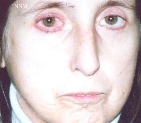
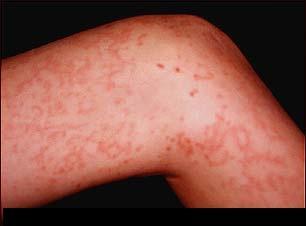
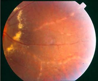
2 Diagnostic approach 45-year old male. Floaters and decreased vision since 1 week Fever, lymphadenopathy, myalgias, night sweats, two months ago Oral ulcer sporadically BCVA: 0.2 OD, 1 OS IOP: 12mmHg OU 1+ cells in AC 3+ haze in vitreous White fluffy lesion OS within normal limits 2.5 disc diameters

3 Development of a differential diagnosis Even in cases with typical picture, we should generate a list of possible diagnoses Adamantiades-Behcet s Sarcoidosis SLE Birdshot chorioretinopathy Lymphoma Cat scratch disease Acute retinal necrosis Toxoplasmosis Syphilis Fungal endophthalmitis Tuberculosis CMV


4 Differential diagnosis Autoimmune? Infectious? Malignancy? Systematic? Isolated?

5 Posterior uveitis Ocular Inflammation Institute of Athens: patients % 43% % Autoimmune Infectious Masquerade


6 Infectious posterior uveitis CMV HSV-VZV TB HIV CSD Fungi Syphilis Other Toxoplasmosis Ocular inflammation Institute of Athens: 481 patients


7 Masquerade syndromes Vascular pseudotumor Paraneoplastic Metastatic Blood malignancies Other Lymphoma Ocular inflammation Institute of Athens: 76 patients


8 Autoimmune posterior uveitis Serpiginous Birdshot Systemic vasculitis Sarcoid Other Idiopathic VKH Adamantiades- Bechcet Disease Choriocapillaritis Ocular inflammation Institute of Athens: 555 patients

9 Systemic diseases Ocular inflammation Institute of Athens: 555 patients % Ophtalmic 28% Systematic


10 Scleroderma Relapsing polychondritis SLE Churg-Straus VKH ABD Sarcoidosis VZV ABD ABD ABD Wegener Syphilis ABD PAN JIA ABD Cat Scratch VZV


11 Diagnosis Evaluation of extraocular findings is not an easy matter Ocular manifestations may precede Assessment exceeding our knowledge Sarcoidosis Fungi Tuberculosis Sarcoidosis

12 Differential diagnosis Where is the inflammation located? Is the uveitis acute or chronic? Is the disease focal or multifocal? What is the condition of the vitreous? Are the retinal vessels involved? Is the optic nerve involved?

13 Where is the inflammation located?


14 Retinitis or Retinochoroiditis Toxoplasmosis CMV HSV or VZV Bartonellosis Adamantiades-Behçet


15 Choroiditis or chorioretinitis VKH Serpiginous Birdshot Sympathetic APMPPE MEWDS MCP PIC Vitritis Undetermined Neuritis Retinal vasculitis Retinitis Tuberculosis Fungi Choroiditis


16 Retinitis or choroiditis Sarcoidosis Syphilis Lymphoma


17 Is the posterior uveitis acute or chronic? Explosive onset Adamantiades-Behçet disease Acute retinal necrosis Toxoplasmosis VKH disease APMPPE Insidious onset Chronic course Birdshot Sarcoidosis Tuberculus uveitis Serpiginous Intraocular lymphoma


18 Is the disease focal or multifocal? Toxoplasmosis Cysticercosis Retinoblastoma Melanoma ARN CMV Disseminated TB APMPPE Birdshot VKH Metastatic carcinoma


19 What is the size of the lesions? ABD Acquired Toxoplasmosis MEWDS APMPPE PORN Lymphoma Serpiginous


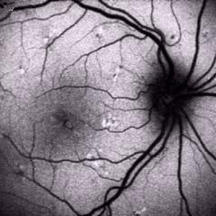
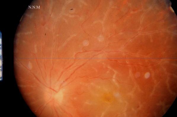
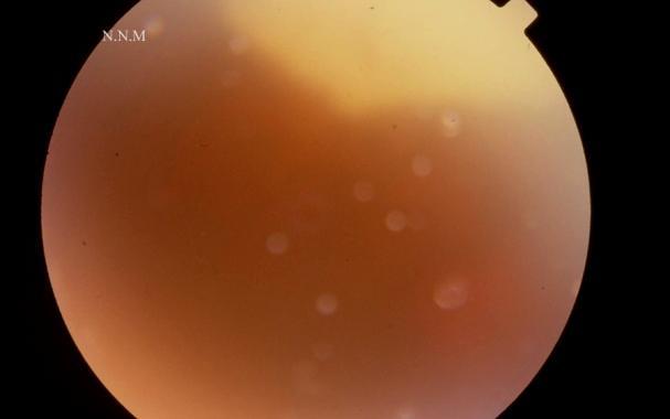
20 What is the condition of the vitreous? N.N.M N.N.M N.N.M N.N.M N.N.M


21 Without vitreous cells N.N.M N.N.M Cat scratch POR Toxoplasmosis Serpiginous Histoplasmosis APMPPE MEWDS ARPE Immunocompromised patients PIC


22 Vitreous inflammation Sarcoidosis Candida Multiple Sclerosis

23 Are the retinal vessels involved? The only sign A sign of uveitis % 20% 40% 60% 80% 100%


24 Retinal vasculitis? unclassifiable uveitis 3% neoplasia primary vasculitis inflamatory ocular syndrome ocular infection systemic diseases Retinal vasculitis frequently associated with a systemic disease


25 Which vessels are affected? Both Veins Arteries


26 Arteritis and Phlebitis Adamantiades-Behçet Wegener Antiphospholipid Non-necrotic herpetic diseases


27 Phlebitis Tuberculosis Sarcoidosis Multiple Sclerosis Idiopathic vasculitis Eales disease HIV


28 Arteritis N.N.M Polyarteritis nodosa SLE Churg-Strauss Syphilis IRVAN ARN (VZV HSV)

29 Retinal vasculitis


30 Retinal vasculitis Candle wax drippings Kyrileis arteriolitis Sarcoidosis Retinal hemorrhages Toxoplasmosis Cracked mude Frosted branch angiitis CMV VZV

31 Is the optic nerve involved? A sign of uveitis The only sign % 20% 40% 60% 80% 100%


32 Neuroretinitis Toxoplasmosis Syphilis Cat scratch disease


33 Papillitis ABD VKH Sympathetic ophthalmia VZV encephalitis Giant cell arteritis


34 Optic nerve infiltration Sarcoidosis CNS lymphoma Leukemia


35 Serous retinal detachment Vogt-Koyanagi-Harada Uveal effusion Malignant tumors Metastatic carcinoma


36 Work-up Laboratory investigations FAF, FA and ICG Ocular Coherence Tomography Other Imaging modalities Diagnostic surgery


37 Ocular imaging modalities


38 Ocular imaging modalities FA should not be overrated


39 Stereotypic findings on FA or ICG VKH


40 Stereotypic findings on FA or ICG APMPPE


41 Stereotypic findings on FA or ICG Serpiginous choriodopathy

42 Identification of the involved retinal vessels Active or inactive? Perivascular sheathing
43 Identification of the involved retinal vessels Active or inactive? Perivascular sheathing


44 Identification of the involved retinal vessels Inactive Perivascular fibrosis


45 Identification of the involved retinal vessels


46 Is the vasculitis focal or diffuse? Sarcoidosis Multiple sclerosis Adamantiades-Behcet Birdshot


47 Is the vasculitis occlusive or not?


48 Is the vasculitis occlusive or not? Sarcoidosis Multiple sclerosis Adamantiades-Behcet Eales TB Viral infections SLE PAN Wegener Crohn Susac

49 Identification of complications


50 Identification of complications


51 Identification of complications


52 Identification of the involved retinal vessels a photo montage or a wide-field lens angiogram is needed to assess the peripheral fundus


53 Fundus Auto Fluorescence


54 Ocular Coherence Tomography


55 Ocular Coherence Tomography


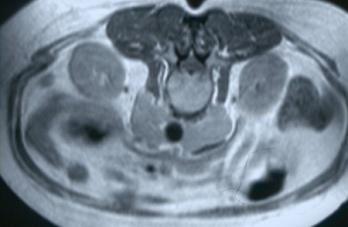
56 Systemic imaging modalities Plain Films Computed Tomography Magnetic Resonance Imaging Nuclear Medicine Ultrasound


57 Systemic imaging modalities MRI CT scan Ga-scan Suspected disease GAD + GAD - Contr + Contr - Sarcoidosis Wegener s MS CNS lymphoma
 Flow- cytometry Retinal or Chorioretinal biopsy Microbiology (Culture PCR) Immunopathology Microscopy (light")
58 Diagnostic surgery Anterior chamber paracentesis Staining and microscopy Goldmann-Witmer coefficient PCR Vitrectomy Microbiology (Culture PCR) Cytology (Immunohistohemical staining) Flow- cytometry Retinal or Chorioretinal biopsy Microbiology (Culture PCR) Immunopathology Microscopy (light electron)

59 , 45-year old. Floaters and decreased vision since 1 week OS within normal limits Visual acuity: 0.2 OD 1+ cells in AC 3+ haze in vitreous White fluffy lesion 2.5 disc diameters Fever, lymphadenopathy, myalgias, night sweats two months ago Two episodes of oral ulcers
60 Differential diagnosis Adamantiades-Behcet s Sarcoidosis SLE Wegener s granulomatosis VKH Lymphoma Metastatic carcinoma Cat scratch disease Tuberculosis CMV Acute retinal necrosis Syphilis Toxoplasmosis Fungal endophthalmitis
61 Differential diagnosis 1. Toxoplasmosis 2. Sarcoidosis 3. Adamantiades-Behcet s 4. SLE 5. Tuberculosis 6. CMV 7. Acute retinal necrosis 8. VKH 9. Syphilis 10. Wegener s granulomatosis 11. Lymphoma 12. Cat scratch disease 13. Fungal endophthalmitis 14. Metastatic carcinoma
62 Differential diagnosis 1. Toxoplasmosis 2. Sarcoidosis 3. Adamantiades-Behcet s 4. SLE 5. Tuberculosis 6. CMV 7. Acute retinal necrosis 8. VKH 9. Syphilis 10. Wegener s granulomatosis 11. Lymphoma 12. Cat scratch disease 13. Fungal endophthalmitis 14. Metastatic carcinoma
63 Differential diagnosis 1. Toxoplasmosis 2. Sarcoidosis 3. Adamantiades-Behcet s 4. SLE 5. Tuberculosis 6. CMV 7. Acute retinal necrosis 8. VKH 9. Syphilis 10. Wegener s granulomatosis 11. Lymphoma 12. Cat scratch disease 13. Fungal endophthalmitis 14. Metastatic carcinoma
64 Differential diagnosis 1. Toxoplasmosis 2. Sarcoidosis 3. Adamantiades-Behcet s 4. SLE 5. Tuberculosis 6. CMV 7. Acute retinal necrosis 8. VKH 9. Syphilis 10. Wegener s granulomatosis 11. Lymphoma 12. Cat scratch disease 13. Fungal endophthalmitis 14. Metastatic carcinoma
65 Differential diagnosis 1. Toxoplasmosis 2. Sarcoidosis 3. Adamantiades-Behcet s 4. SLE 5. Tuberculosis 6. CMV 7. Acute retinal necrosis 8. VKH 9. Syphilis 10. Wegener s granulomatosis 11. Lymphoma 12. Cat scratch disease 13. Fungal endophthalmitis 14. Metastatic carcinoma

66 Differential diagnosis 1. Toxoplasmosis 2. Sarcoidosis 3. Adamantiades-Behcet s 4. SLE 5. Tuberculosis 6. CMV 7. Acute retinal necrosis 8. VKH 9. Syphilis 10. Wegener s granulomatosis 11. Lymphoma 12. Cat scratch disease 13. Fungal endophthalmitis 14. Metastatic carcinoma

67 Differential diagnosis 1. Toxoplasmosis 2. Adamantiades-Behcet s 3. Acute retinal necrosis 4. Syphilis 5. Cat scratch disease
68 Differential diagnosis 1. Toxoplasmosis 2. Acute retinal necrosis 3. Syphilis 4. Cat scratch disease Anti-Toxo IgM: 2IU, IgG:3200 IU Anti-HSV: IgM: 1IU, IgG: 22 IU Anti-VZV: IgM: 2IU, IgG: 10 IU FTA-ABS: negative Anti-Bartonella: IgM, IgG : 1/32


69 Diagnosis: Acquired Toxoplasmosis After anti-toxoplasmic therapy


70 Differential diagnosis of posterior uveitis The small differences that make a big difference


71 Simi Island, Dodecanese
Retinal Manifestations of Systemic Disease Part 1
 The Retina and Systemic diseases Retinal Manifestations of Systemic Disease Part 1 Sundeep Dev, MD VRSF Retinal Update 2019 VitreoRetinal Surgery, PA 1 Retinitis/Vasculitis Vitreous cells Serous detachments
Diagnosis of uveitis, how to proceed?
 EOS meeting Cairo, 2018 Diagnosis of uveitis, how to proceed? Mohamed G.A Saleh Lecturer of Ophthalmology Assiut University Size of the problem 15/100000 in US every year. 10% of blindness Prevalence varies
o White dot syndromes pattern recognition o Activity and damage o Quality of life o Key points o Idiopathic o Sarcoidosis o Multiple sclerosis
 Introduction Clinical Assessment of Posterior Uveitis Philip I. Murray Centre for Translational Inflammation Research University of Birmingham Birmingham and Midland Eye Centre o Classification of uveitis
Moncef Khairallah, MD
 Moncef Khairallah, MD Department of Ophthalmology, Fattouma Bourguiba University Hospital Faculty of Medicine, University of Monastir Monastir, Tunisia INTRODUCTION IU: anatomic form of uveitis involving
UVEITIS. Dr. Yılmaz ÖZYAZGAN
 UVEITIS Dr. Yılmaz ÖZYAZGAN UVEITIS DEFINITION BY STRICT DEFINITION, UVEITIS IS AN INFLAMMATION OF UVEAL TRACT. BUT IN PRACTICAL, IT IS GENERALLY NOT RESTRICTED TO THE UVEA AND INVOLVES OTHER ADJACENT
Rare Presentation of Ocular Toxoplasmosis
 Case Report Rare Presentation of Ocular Toxoplasmosis Rakhshandeh Alipanahi MD From Department of Ophthalmology, Nikookari Eye Hospital, Tabriz University of Medical Sciences, Tabriz, Iran. Correspondence:
Acute Retinal Necrosis Secondary to Varicella Zoster Virus in an Immunosuppressed Post-Kidney Transplant Patient
 CM&R Rapid Release. Published online ahead of print September 20, 2012 as Aperture Acute Retinal Necrosis Secondary to Varicella Zoster Virus in an Immunosuppressed Post-Kidney Transplant Patient Elizabeth
Ophthalmology. Juliette Stenz, MD
 Ophthalmology Juliette Stenz, MD Required Slide Disclosures NO SIGNIFICANT FINANCIAL, GENERAL, OR OBLIGATION INTERESTS TO REPORT Required Slide At the end of this session, students will be able to: 1.
Various presentations of herpes simplex retinochoroiditis A case series
 Various presentations of herpes simplex retinochoroidits 47 Various presentations of herpes simplex retinochoroiditis A case series M. T. K. Perera 1, T. S. Keragala 1, M. Gamage 2 The Journal of the College
Slide 4. Slide 5. Slide 6
 Slide 1 Slide 4 Demographics El Paso Eye Care Border Healthcare-Based Grand Rounds Derek N. Cunningham, O.D. 80-90% Mexican-Americans Diabetes Hypertension Hyperlipidemia Obesity 70% uninsured High poverty
What do you need to know about posterior uveitis
 What do you need to know about posterior uveitis Dr. Anthony Hall MD FRANZCO Director of Ophthalmology Alfred Hospital, Melbourne, Australia Alfred Hospital Disclosures Off label treatments Paid advisory
Misdiagnosed Vogt-Koyanagi-Harada (VKH) disease and atypical central serous chorioretinopathy (CSC)
 disease and atypical central serous chorioretinopathy (CSC)") HPTER 12 Misdiagnosed Vogt-Koyanagi-Harada (VKH) disease and atypical central serous chorioretinopathy (S) linical Features VKH disease is a bilateral granulomatous panuveitis often associated with exudative
HPTER 12 Misdiagnosed Vogt-Koyanagi-Harada (VKH) disease and atypical central serous chorioretinopathy (S) linical Features VKH disease is a bilateral granulomatous panuveitis often associated with exudative
Necrotizing retinitis of multifactorial etiology
 Romanian Journal of Ophthalmology, Volume 61, Issue 1, January-March 2017. pp:49-53 CASE REPORT Necrotizing retinitis of multifactorial etiology Pirvulescu Ruxandra Angela* **, Popa Cherecheanu Alina*
Vasculitides in Surgical Neuropathology Practice
 Vasculitides in Surgical Neuropathology Practice USCAP requires that all faculty in a position to influence or control the content of CME disclose any relevant financial relationship WITH COMMERCIAL INTERESTS
Management of uveitis
 Management of uveitis DR. ANUPAMA KARANTH Anti-inflammatory agents -itis = inflammation Treatment : stop inflammation Use anti-inflammatory drugs Most potent of such agents : Corticosteroids Corticosteroids
Head prof. MUDr. E. Vlková, CSc.
 MUDr. Karkanová Michala, Oční klinika LF MU a FN Brno Head prof. MUDr. E. Vlková, CSc. 3 parts: iris (iris) ciliary body (corpus ciliare) choroid (choroidea) Function: regulating the entry of light into
Optical coherence tomography findings in a child with posterior scleritis
 European Journal of Ophthalmology / Vol. 18 no. 6, 2008 / pp. 1007-1010 SHORT OMMUNITIONS & SE REPORTS Optical coherence tomography findings in a child with posterior scleritis H. ERDÖL, M. KOL,. TÜRK
Retina Grand Rounds. Stephen Huddleston MD Charles Retina Institute University of Tennessee Hamilton Eye Institute
 Retina Grand Rounds Stephen Huddleston MD Charles Retina Institute University of Tennessee Hamilton Eye Institute Fundus Autoflourescence 2013 2016 Plaquenil Toxicity Excellent treatment for a variety
Uveitis. What is Uveitis?
 Uveitis What is Uveitis? Uveitis [u-vee-i-tis] is a term for inflammation of the eye. It can occur in one eye or both eyes and affects the layer of the eye called the uvea [u-vee-uh]. It also can be associated
Why Is Imaging Critical in My Uveitis Practice?
 Why Is Imaging Critical in My Uveitis Practice? Dilraj S. Grewal, MD Developed in collaboration Imaging Is the Backbone of Uveitis Workup and Monitoring Treatment Response FP FAF B- scan Multimodal Imaging
Rhegmatogenous retinal detachment in uveitis
 De Hoog et al. Journal of Ophthalmic Inflammation and Infection (2017) 7:22 DOI 10.1186/s12348-017-0140-5 Journal of Ophthalmic Inflammation and Infection ORIGINAL ARTICLE Open Access Rhegmatogenous retinal
Atlas of the Vasculitic Syndromes
 CHAPTER e40 Atlas of the Vasculitic Syndromes Carol A. Langford Anthony S. Fauci Diagnosis of the vasculitic syndromes is usually based upon characteristic histologic or arteriographic findings in a patient
Vasculitis and Vasculitides. OMONDI OYOO Physician/Rheumatologist; Senior Lecturer, Department of Medicine University of Nairobi
 Vasculitis and Vasculitides OMONDI OYOO Physician/Rheumatologist; Senior Lecturer, Department of Medicine University of Nairobi Definition Presence of leucocytes in the vessel wall with reactive damage
Dr Rodney Itaki Lecturer Anatomical Pathology Discipline. University of Papua New Guinea School of Medicine & Health Sciences Division of Pathology
 Vasculitis Dr Rodney Itaki Lecturer Anatomical Pathology Discipline University of Papua New Guinea School of Medicine & Health Sciences Division of Pathology Disease Spectrum Hypersensitivity vasculitis/microscopic
Factors that lead to progression. Evaluation of Diabetics. Ocular Circulation. Ischemic Optic Neuropathy. Proliferative Diabetic Retinopathy
 Non proliferative Diabetic Retinopathy Proliferative Diabetic Retinopathy Factors that lead to progression Puberty and pregnancy Systolic and diastolic blood pressure Hyperlipidemia : hard exudates in
Nausheen Khuddus, MD Melissa Elder, MD, PhD
 Nausheen Khuddus, MD Melissa Elder, MD, PhD Nausheen Khuddus, MD Pediatric Ophthalmologist and Strabismus Specialist Accent Physicians Gainesville, Florida What Is Uveitis? Uveitis is caused by inflammatory
WHAT IS YOUR DIAGNOSIS? By ADREA R. BENKOFF M.D.
 WHAT IS YOUR DIAGNOSIS? By ADREA R. BENKOFF M.D. Anterior Chamber Inflammation and Iris Depigmentation Noted 25 Years After Cataract Extraction Decreasing Vision Over a 5- Year Period 64 year old white
Interesting, unusual and eclectic cases from 2017 Robert A. Mittra, MD VitreoRetinal Surgery, P.A. Minneapolis, MN
 Fundus, SG Interesting, unusual and eclectic cases from 2017 Robert A. Mittra, MD VitreoRetinal Surgery, P.A. Minneapolis, MN Which is most likely? A) Age > 65, history of HTN B) Age 40 65, history of
Interesting, unusual, eclectic cases from 2017 Robert A. Mittra, MD VitreoRetinal Surgery, P.A. Minneapolis, MN
 56 yo female, EW Presented to outside Ophthalmologist Diagnosed with viral conjunctivitis, but viral testing was negative. Also had pain around the eye and on the right side of her face Interesting, unusual,
Re-emerging infections: Syphilis & Tuberculosis
 Re-emerging infections: Syphilis & Tuberculosis Nicholas Jones Manchester Royal Eye Hospital Syphilis and TB - historical plagues? Syphilis incidence over 40yrs Manchester: Manchester: The Syphilis Capital
Solitary idiopathic choroiditis
 Optometry (2007) 78, 176-180 Solitary idiopathic choroiditis Kimberly D. Kohne, O.D., a Victor E. Malinovsky, O.D., a and Hua Gao, M.D., Ph.D. b a School of Optometry and b Department of Ophthalmology,
Index. Note: Page numbers of article titles are in boldface type.
 Note: Page numbers of article titles are in boldface type. A ANCA vasculitis. See Antineutrophil cytoplasmic antibody associated (ANCA) vasculitis Angiography 54 Antineutrophil cytoplasmic antibody correlation
Choroidal Neovascularization in Sympathetic Ophthalmia
 Choroidal Neovascularization in Sympathetic Ophthalmia Lucia Sobrin, Miguel Cordero Coma, C. Stephen Foster Case Report A 49-year-old man presented after a ruptured globe repair of his left eye status
Herpesviruses. Tools of diagnosis : what to use and when. Corinne Liesnard Laboratory of Virology Erasme Hospital - ULB
 Herpesviruses Tools of diagnosis : what to use and when Corinne Liesnard Laboratory of Virology Erasme Hospital - ULB Evolution of the techniques in the virology lab Techniques : "Classic" methods Ag detection
Uveitis Review for the OKAPS
 Uveitis Review for the OKAPS What you should and should not know! By: Armando L. Oliver, MD Ocular Immunology and Uveitis Specialist Vitreoretinal Surgeon Assistant Professor, UPR Disclaimer... The information
Infectious Retina. The Fight Against HIV/AIDS?? HAART. Common Posterior Segment Manifestations 1/8/2019. Raman Bhakhri, OD, FAAO Assistant Professor
 Infectious Retina Raman Bhakhri, OD, FAAO Assistant Professor No Disclosures Disclosure Statement: Nothing to disclose Common Posterior Segment Manifestations o HIV Retinopathy o Cytomegalovirus (CMV)
OPTIC NERVE DISORDERS
 OPTIC NERVE DISORDERS OPTIC NEUROPATHIES INFLAMMATORY OPTIC NEUROPATHIES Cat scratch disease. Lyme disease. Viral infections of childhood (measles, mumps, chicken pox) with or without encephalitis Immun-
Infectious Retina Raman Bhakhri, OD, FAAO Assistant Professor SCCO/MBKU
 Infectious Retina Raman Bhakhri, OD, FAAO Assistant Professor SCCO/MBKU rbhakhri@ketchum.edu Please silence all mobile devices and remove items from chairs so others can sit. Unauthorized recording of
Uveitis: Classification, Etiologies and Clinical Signs
 E-ISSN 2454-2784 Recent Advances Uveitis: Classification, Etiologies and Clinical Signs Aditya Shreekant Kelkar 1, Ekta Rakesh Arora 1, B.Sowkath 2, Jyotirmay Biswas 3 1 National Institute of Ophthalmology,Pune
Neuro-Ocular Grand Rounds Anthony B. Litwak,OD, FAAO VA Medical Center Baltimore, Maryland
 Neuro-Ocular Grand Rounds Anthony B. Litwak,OD, FAAO VA Medical Center Baltimore, Maryland Dr. Litwak is on the speaker and advisory boards for Alcon and Zeiss Meditek COMMON OPTIC NEUROPATHIES THAT CAN
Patterns of Uveitis at a Tertiary Referral Center in Southern Iran
 Original Article Patterns of Uveitis at a Tertiary Referral Center in Southern Iran Mansour Rahimi, MD; Ghazaleh Mirmansouri, MD Poustchi Eye Research Center and Ophthalmology Department, Shiraz University
Outline. Brief history and principles of ophthalmic ultrasound. Types of ocular ultrasound. Examination techniques. Types of Ultrasound
 Ultrasound and Intraocular Tumors 2015 Ophthalmic Photographers' Society Mid-Year Program Cagri G. Besirli MD, PhD Kellogg Eye Center University of Michigan Outline Brief history and principles of ophthalmic
UNDERSTAND MORE ABOUT UVEITIS UVEITIS
 UNDERSTAND MORE ABOUT UVEITIS UVEITIS Uveitis What is uveitis? Uveitis is inflammation of the uvea, the middle layer of your eye. The eye is shaped much like a tennis ball, with three different layers
Neuro-Ocular Grand Rounds
 Neuro-Ocular Grand Rounds Anthony B. Litwak,OD, FAAO VA Medical Center Baltimore, Maryland Dr. Litwak is on the speaker and advisory boards for Alcon and Zeiss Meditek COMMON OPTIC NEUROPATHIES THAT CAN
The White Re)na. Joseph Alsberge, MD January 20, 2018
na. Joseph Alsberge, MD January 20, 2018") The White Re)na Joseph Alsberge, MD January 20, 2018 58 y/o man with floaters and pain OD x 2 weeks PMH: oral and genital herpes Va OD 20/50 Anterior OD: KP and 3+ AC cell Posterior: Vitri)s, occlusive
The White Re)na Joseph Alsberge, MD January 20, 2018 58 y/o man with floaters and pain OD x 2 weeks PMH: oral and genital herpes Va OD 20/50 Anterior OD: KP and 3+ AC cell Posterior: Vitri)s, occlusive
Bilateral acute retinal necrosis in a patient with multiple sclerosis on natalizumab
 Bilateral acute retinal necrosis in a patient with multiple sclerosis on natalizumab Arjun B. Sood, Emory University Gokul Kumar, Emory University Joshua Robinson, Emory University Journal Title: Journal
Ocular Pathology. I. Congenital and/or developmental. A. Trisomy 21. Hypertelorism (widely spaced eyes) Keratoconus (cone shaped cornea)
 Keratoconus (cone shaped cornea)") I. Congenital and/or developmental Robbins Pathologic Basis of Disease, 6 th Ed. A. Trisomy 21 Hypertelorism (widely spaced eyes) Keratoconus (cone shaped cornea) Focal hypoplasia of iris Cataracts frequently
I. Congenital and/or developmental Robbins Pathologic Basis of Disease, 6 th Ed. A. Trisomy 21 Hypertelorism (widely spaced eyes) Keratoconus (cone shaped cornea) Focal hypoplasia of iris Cataracts frequently
REFRESHER: ANTERIOR UVEITIS
 REFRESHER: ANTERIOR UVEITIS 2. SAoO Kongress 28.2.2018 Messe Luzern Dr. med. Christian Böni Augenklinik Universitätsspital Zürich Christian Böni Seite 1 Anterior Uveitis: Clinical Issues Diagnostics: yes
Content. Polyarteritis nodosa. Vasculitis. Giant cell arteritis. Primary cerebral angiitis. Other autoimmune CNS disease.
 Content Other autoimmune CNS disease Philippe Demaerel Vasculitis Systemic lupus erythematosus Wegener granulomatosis Behçet disease Rhombencephalitis - CLIPPERS Neurosarcoidosis Langerhans cell histiocytosis
Development and validation of new diagnostic criteria for acute retinal necrosis
 Jpn J Ophthalmol DOI 10.1007/s10384-014-0362-0 CLINICAL INVESTIGATION Development and validation of new diagnostic criteria for acute retinal necrosis Hiroshi Takase Annabelle A. Okada Hiroshi Goto Nobuhisa
OCULAR MANIFESTATIONS OF SYSTEMIC DISEASES THUCANH MULTERER, MD
 OCULAR MANIFESTATIONS OF SYSTEMIC DISEASES THUCANH MULTERER, MD UNDERGRADUATE: Philadelphia College of Pharmacy and Science 1996 MEDICAL SCHOOL: MCP Hahnemann School of Medicine, Philadelphia PA 2000 RESIDENCY:
11/29/2016 MACULAR MALADIES: TYPICAL & ATYPICAL CASES
 MACULAR MALADIES: TYPICAL & ATYPICAL CASES Dawn Pewitt, OD, FAAO Triad Eye Institute, Grove, OK Dpewitt@triadeye.com Disclosure Statement: No financial disclosures COPE 51218-PS Please silence all mobile
CLINICALCASE PROVOST J, SEKFALI R, AMOROSO F, ZAMBROWSKI O, MIERE A
 CLINICALCASE PROVOST J, SEKFALI R, AMOROSO F, ZAMBROWSKI O, MIERE A Department of ophthalmology, Souied E. (MD,PhD) Centre Hospitalier Intercommunal de Créteil Université Paris Est HISTORY 13 years old
Evaluation of Neck Mass. Disclosure. Learning Objectives 3/24/2014. Karen T. Pitman MD, FACS Banner MDACC, Gilbert AZ. Nothing to disclose
 Evaluation of Neck Mass Karen T. Pitman MD, FACS Banner MDACC, Gilbert AZ Nothing to disclose Disclosure Learning Objectives 1. Describe a systematic method to evaluate a patient with a neck mass 2. Select
2009 REIMBURSEMENT GUIDE, VISUCAM and VISUCAM NM/FA
 2009 REIMBURSEMENT GUIDE FF 450 PLUS PRO NM, VISUCAM and VISUCAM NM/FA Zeiss Fundus Cameras INTRODUCTION The following guide provides an overview of billing and reimbursement for procedures performed with
Macular Hole Associated with Vogt-Koyanagi-Harada Disease at the Acute Uveitic Stage
 Published online: September 15, 2015 2015 The Author(s) Published by S. Karger AG, Basel 1663 2699/15/0063 0328$39.50/0 This article is licensed under the Creative Commons Attribution-NonCommercial 4.0
Uveitis in patients with Multiple Sclerosis
 Uveitis in patients with Multiple Sclerosis An observational descriptive clinical study submitted in partial fulfillment of Master Degree in Ophthalmology By Dr. Ahmed Mohamed Karara M.B, B Ch. Supervised
10/18/2018. Unraveling Uveitis
 Unraveling Uveitis Trenton Cleghern, OD, FAAO VisionAmerica UAB School of Optometry 11/10/2018 1 Disclosure Statement: Nothing to disclose 2 Objectives Classify uveitis Current and new therapeutic options
Algorithm of Choroiditis
 10.5005/jp-journals-10020-1027 REVIEW ARTICLE Ovi Sofia, Sudharshan Sridharan, Jyotirmay Biswas ABSTRACT Choroiditis may present as focal or multifocal lesions, and could be due to infectious or noninfectious
White-Spot Syndromes of the Retina Lee Jampol, M.D. Chicago, IL
 Objectives At the conclusion of the program, the attendees will be able to: 1. recognize the various white-spot syndromes of the retina 2. initiate appropriate diagnostic tests of patients with the white-spot
Multimodal imaging of retinal metastasis masquerading as an acute retinal necrosis
 https://doi.org/10.1186/s40942-018-0149-4 International Journal of Retina and Vitreous CASE REPORT Multimodal imaging of retinal metastasis masquerading as an acute retinal necrosis Fukutaro Mano 1*, Stephen
Convergence in. Introduction. Case Report: Dr. Piyali SenM.B.B.S, Dr. Abhipsha Saha M.B.B.S, Dr. Anuradha Chandra M.S,FAICO
 Convergence in Dr. Piyali SenM.B.B.S, Dr. Abhipsha Saha M.B.B.S, Dr. Anuradha Chandra M.S,FAICO Introduction non-progressive ophthalmoplegia with or without ptosis affecting part or all of the occulomotor
The Royal College of Ophthalmologists
 Clinical Dataset Uveitis Dataset The Royal College of Ophthalmologists Authorship Group: Alastair K. Denniston, Richard W. Lee, Carlos Pavesio, Miles R Stanford, Philip I Murray, Annabelle Okada, H. Nida
Evolving therapies for posterior uveitis. Infliximab (Remicade) Infliximab: pharmacology. FDA-approved monoclonal antibody therapy Target
 Infliximab: pharmacology. FDA-approved monoclonal antibody therapy Target") Evolving therapies for posterior uveitis Sam Dahr, M.D. September 17, 2005 Midwest Ophthalmology Conference Infliximab (Remicade) FDA approved for Crohn s disease, rheumatoid arthritis, and psoriatic arthritis
Evolving therapies for posterior uveitis Sam Dahr, M.D. September 17, 2005 Midwest Ophthalmology Conference Infliximab (Remicade) FDA approved for Crohn s disease, rheumatoid arthritis, and psoriatic arthritis
D JO. Bilateral Shallow Anterior Chamber And Transient Myopia As A Presenting Feature Of Vogt Koyanagi Harada Syndrome
 46 Bilateral Shallow Anterior Chamber And Transient Myopia As A Presenting Feature Of Vogt Koyanagi Harada Syndrome Abstract Rahul Kumar Sharma, Abhishek Dagar, Vivek Kumar Vitreo-Retina Department, Venu
Patterns of uveitis in a Philippine eye clinic
 VOL. NO. PHILIPPINE JOURNAL OF Ophthalmology JANUARY ORIGINAL ARTICLE - MARCH 5 Harvey S. Uy, MD Irene W. Tam, OD Asian Eye Institute Makati, Philippines Patterns of uveitis in a Philippine eye clinic
Approach to Pediatric Uveitis. Paris Tranos PhD,ICO,FRCS OPHTHALMICA Vitreoretinal & Uveitis Service
 Approach to Pediatric Uveitis Paris Tranos PhD,ICO,FRCS OPHTHALMICA Vitreoretinal & Uveitis Service Epidemiology Uveitis is the 3 rd leading cause of blindness in USA 5-10% of uveitis cases involve children
CLINICAL PEARLS IN OCULAR ONCOLOGY
 CLINICAL PEARLS IN OCULAR ONCOLOGY IRIS NEVUS - Two kinds circumscribed and diffuse - Photodocumentation important to monitor growth - Risk Factors for iris nevus growth to melanoma (ABCDEF) A Age (young),
How and Why Should we Care? Muge R. Kesen, MD Retina Specialty Institute (RSI)
") How and Why Should we Care? Muge R. Kesen, MD Retina Specialty Institute (RSI) Disclosure No financial interest or relationships to disclose. Objectives Overview of different types of uveitis Diagnostic
How and Why Should we Care? Muge R. Kesen, MD Retina Specialty Institute (RSI) Disclosure No financial interest or relationships to disclose. Objectives Overview of different types of uveitis Diagnostic
I ntraocular inflammation affecting the uvea (uveitis)
") 444 EXTENDED REPORT Uveitis in children and adolescents D BenEzra, E Cohen, G Maftzir... See end of article for authors affiliations... Correspondence to: David BenEzra, MD, PhD, Department of Ophthalmology,
444 EXTENDED REPORT Uveitis in children and adolescents D BenEzra, E Cohen, G Maftzir... See end of article for authors affiliations... Correspondence to: David BenEzra, MD, PhD, Department of Ophthalmology,
Optic Nerve Disorders
 Optic Nerve Disorders Optic Nerve Disorders Diagnosis and Management, MD Associate Professor of Ophthalmology and Neurology, University of Kentucky College of Medicine, Lexington, Kentucky, USA , MD Associate
Scleritis LEN V KOH OD
 Scleritis LEN V KOH OD 2014 PUCO 1 Introduction A painful, destructive, and potentially blinding disorder Highly symptomatic High association with systemic disease Immunosuppresssive agents 2014 PUCO 2
Table of Contents 1 Orbit 3 2 Eyelids 7
 Table of Contents Preface, x List of abbreviations xi Glossary xii Section I Atlas 1 1 Orbit 3 Clinical signs associated with orbital neoplasia 3 Clinical signs associated with orbital cellulitis 3 Enophthalmos
Mohammad Reza Shakibi M.D Kerman university of medical sciences (KMU) Shafa Hospital, Rheumatology ward
 Shafa Hospital, Rheumatology ward") VASCULITIS SYNDROMES Mohammad Reza Shakibi M.D Kerman university of medical sciences (KMU) Shafa Hospital, Rheumatology ward ILLUSTRATED CASE 1 A 56 years old lady refered me for prolonged fever, arthritis
VASCULITIS SYNDROMES Mohammad Reza Shakibi M.D Kerman university of medical sciences (KMU) Shafa Hospital, Rheumatology ward ILLUSTRATED CASE 1 A 56 years old lady refered me for prolonged fever, arthritis
Management of Immune Reconstitution Inflammatory Syndrome (IRIS)
") Management of Immune Reconstitution Inflammatory Syndrome (IRIS) Adult Clinical Guideline from the New York State Department of Health AIDS Institute www.hivguidelines.org Purpose of the IRIS Guideline
Management of Immune Reconstitution Inflammatory Syndrome (IRIS) Adult Clinical Guideline from the New York State Department of Health AIDS Institute www.hivguidelines.org Purpose of the IRIS Guideline
Pattern of Uveitis in Saudi Female Patients in Western Region of Saudi Arabia
 JKAU: Med. Sci., Vol. 19 No. 3, pp: 73-83 (2012 A.D. / 1433 A.H.) DOI: 10.4197/Med. 19-3.6 Pattern of Uveitis in Saudi Female Patients in Western Region of Saudi Arabia Nizamuddin Shaik Hakim Mohammad
Optic Nerve Disorders: Structure and Function and Causes
 Optic Nerve Disorders: Structure and Function and Causes Using Visual Fields, OCT and B-scan Ultrasound to Diagnose and Follow Optic Nerve Visual Losses Ohio Ophthalmological Society and Ophthalmic Tech
Eye Examination Techniques in Horses
 Eye Examination Techniques in Horses Dennis E. Brooks DVM, PhD Dip ACVO University of Florida brooksd@mail.vetmed.ufl.edu Basic Instruments How to tell the potential of vision? PLRs (retina, CN 2, chiasm,
Mycobacterial Ocular Inflammation. Akbar Shakoor, M.D. John A. Moran Eye Center, University of Utah
 Mycobacterial Ocular Inflammation Akbar Shakoor, M.D. John A. Moran Eye Center, University of Utah Financial Disclosure I have no financial interests or relationships to disclose. Applied anatomy What
Dr Keerti Gedela. Chelsea and Westminster Hospital NHS Foundation Trust, London. 19 th Annual Conference of the British HIV Association (BHIVA)
") 19 th Annual Conference of the British HIV Association (BHIVA) Dr Keerti Gedela Chelsea and Westminster Hospital NHS Foundation Trust, London 16-19 April 2013, Manchester Central Convention Complex Value
19 th Annual Conference of the British HIV Association (BHIVA) Dr Keerti Gedela Chelsea and Westminster Hospital NHS Foundation Trust, London 16-19 April 2013, Manchester Central Convention Complex Value
Note: This is an outcome measure and can be calculated solely using registry data.
 Measure #191 (NQF 0565): Cataracts: 20/40 or Better Visual Acuity within 90 Days Following Cataract Surgery -- National Quality Strategy Domain: Effective Clinical Care DESCRIPTION: Percentage of patients
Imaging in uveitis. Anthony Hall
 Imaging in uveitis Anthony Hall Causes of Vision Loss in Uveitis 1. Cystoid macular oedema 26% 2. Cataract 19% 3. Glaucoma 11% 4. Permanent macular damage 5% Rothova et al BJO 1996; 80: 332-336 Macular
Optical Coherence Tomograpic Features in Idiopathic Retinitis, Vasculitis, Aneurysms and Neuroretinitis (IRVAN)
") Columbia International Publishing Journal of Ophthalmic Research (2014) Research Article Optical Coherence Tomograpic Features in Idiopathic Retinitis, Vasculitis, Aneurysms and Neuroretinitis (IRVAN)
Columbia International Publishing Journal of Ophthalmic Research (2014) Research Article Optical Coherence Tomograpic Features in Idiopathic Retinitis, Vasculitis, Aneurysms and Neuroretinitis (IRVAN)
Alan G. Kabat, OD, FAAO (901)
") THE SWOLLEN OPTIC DISC: EMERGENCY OR ANOMALY? Alan G. Kabat, OD, FAAO (901) 252-3691 Memphis, Tennessee alan.kabat@alankabat.com Course description: The swollen disc presents a diagnostic dilemma. While
THE SWOLLEN OPTIC DISC: EMERGENCY OR ANOMALY? Alan G. Kabat, OD, FAAO (901) 252-3691 Memphis, Tennessee alan.kabat@alankabat.com Course description: The swollen disc presents a diagnostic dilemma. While
Frosted branch angiitis with undiagnosed Hodgkin lymphoma
 European Journal of Ophthalmology / Vol. 19 no. 2, 2009 / pp. 310-313 SHORT COMMUNICTIONS & CSE REPORTS Frosted branch angiitis with undiagnosed Hodgkin lymphoma MINH-TRI HU 1, PIERRE LISE 1, LURENCE DE
2/23/18. Disclosures. Rheumatic Diseases of Childhood. Making Room for Rheumatology. I have nothing to disclose. James J.
 Making Room for Rheumatology James J. Nocton, MD Disclosures I have nothing to disclose Rheumatic Diseases of Childhood Juvenile Idiopathic Arthritis (JIA) Systemic Lupus Erythematosus (SLE) Juvenile Dermatomyositis
Regional vs. Systemic Therapy. Corticosteroids. Regional vs. Systemic Therapy for Uveitis. Considerations
 Regional vs. Systemic Therapy for Uveitis Nisha Acharya,, M.D., M.S. Director, Uveitis Service F.I. Proctor Foundation University of California, San Francisco December 4, 2010 No financial disclosures
2008 Gross Ocular Pathology. Gross Pathology 2
 2008 Gross Ocular Pathology Gross Pathology 2 08rd1281 Feline T-Cell Lymphoma 08rd1300 Canine Iridociliary Adenoma Foam Cell Variant 08rd1331 Feline Feline Iridociliary Adenoma 08rd1340 Canine Retinal
Doc, I See a Donut in My Vision : An Optometrist s Guide to a Rare Cause of Choroidal Neovascular Membrane
 Doc, I See a Donut in My Vision : An Optometrist s Guide to a Rare Cause of Choroidal Neovascular Membrane Linda Pham, OD, Tobin Ansel, OD, Nancy Shenouda-Awad, OD, FAAO, West Haven VA Medical Center Abstract
Early detection of Retinoblastoma in children. Max Mantik
 Early detection of Retinoblastoma in children Max Mantik Introduction The most common primary intraocular malignancy of childhood 10 to 15 % of cancers that occur within the first year of life Typical
Double trouble: a patient with both HLA-B27 anterior uveitis and HLA-A29 birdshot chorioretinitis
 Haddad and Reddy Journal of Ophthalmic Inflammation and Infection 2014, 4:28 BRIEF REPORT Open Access Double trouble: a patient with both HLA-B27 anterior uveitis and HLA-A29 birdshot chorioretinitis Zeina
Deep Trouble. Thomas Stone, MD Retina Associates of Kentucky River City Retina Conference May 15, 2014
 Deep Trouble Thomas Stone, MD Retina Associates of Kentucky River City Retina Conference May 15, 2014 History 20 yo WM Decreased vision OU, OD>OS Sudden onset blurred central vision 12 days prior 4 days
Neuro-Ophthalmic Masqueraders
 Neuro-Ophthalmic Masqueraders Leonid Skorin, Jr., OD, DO, MS, FAAO, FAOCO Mayo Clinic Health System in Albert Lea Denise Goodwin, OD, FAAO Pacific University College of Optometry Please silence all mobile
Relevance of erythrocyte sedimentation rate and C-reactive protein in patients with active uveitis
 Graefe's Archive for Clinical and Experimental Ophthalmology (2019) 257:175 180 https://doi.org/10.1007/s00417-018-4174-7 INFLAMMATORY DISORDERS Relevance of erythrocyte sedimentation rate and C-reactive
Sudden Vision Loss. Brendan Girschek, MD, FRCSC, FACS Vitreoretinal Surgery Cedar Valley Medical Specialists
 Sudden Vision Loss Brendan Girschek, MD, FRCSC, FACS Vitreoretinal Surgery Cedar Valley Medical Specialists My Credentials -Residency in Ophthalmology at the LSU Eye Center in New Orleans, LA -Fellowship
Complicated Cataract to Intraocular Tumors, Beware of the unexpected
 Complicated Cataract to Intraocular Tumors, Beware of the unexpected Ihab Saad Othman, MD, FRCS Professor of Ophthalmology Cairo University In this part of the world: We Master Phakoemulsification 1 Intraoperative/Second
Patterns of Macular Edema in Uveitis as Diagnosed by Optical Coherence Tomography in Tertiary Eye Center
 Original Article Patterns of Macular Edema in Uveitis as Diagnosed by Optical Coherence Tomography in Tertiary Eye Center Sharad Gupta 1, Dev Narayan Shah 2, Sagun Narayan Joshi 3, Manoj Aryal 4, Lila
Local Coverage Determination (LCD): Scanning Computerized Ophthalmic Diagnostic Imaging (SCODI) (L34431)
: Scanning Computerized Ophthalmic Diagnostic Imaging (SCODI) (L34431)") Local Coverage Determination (LCD): Scanning Computerized Ophthalmic Diagnostic Imaging (SCODI) (L34431) Links in PDF documents are not guaranteed to work. To follow a web link, please use the MCD Website.
Local Coverage Determination (LCD): Scanning Computerized Ophthalmic Diagnostic Imaging (SCODI) (L34431) Links in PDF documents are not guaranteed to work. To follow a web link, please use the MCD Website.
LUCY HOWE, MILES STANFORD, ELIZABETH GRAHAM, JOHN MARSHALL
 Indocyanine green angiography in inflammatory eye LUCY HOWE, MILES STANFORD, ELIZABETH GRAHAM, JOHN MARSHALL disease Abstract Purpose The choroid plays an integral role in the evolution of a number of
White Dot Syndromes Noninfectious Chorioretinopathies Update 2019
 White Dot Syndromes Noninfectious Chorioretinopathies Update 2019 Kelly T. Mitchell, MD Retina Service TTUHSC Definition Noninfectious disease Inflammation of choroid, choriocapillaris, RPE, and Retina