Computed tomography. Dr. Csébi Péter SZIE, ÁOTK, Surgery. Development of the CT scanners 1. Fourth generation CT. Third generation CT.
|
|
|
- Neil Ball
- 5 years ago
- Views:
Transcription


1 Computed tomography Dr. Csébi Péter SZIE, ÁOTK, Surgery The main parts of the CT GE 9000 Controll panel Gantry and table High voltage generator Multiformat kamera Shimadzu 4500 TE GE Development of the CT scanners 1. Third generation CT Fourth generation CT 1


 -water Possible complications with iv.")
2 Digital imaging CT window Hounsfield scale: The CT windows Different windows Density measuring Distance, size Magnification High resolution CT Reconstructions 3D CT Postprocessing Anaesthesia! Symmetrical positioning Scout view DV Scout view LL Transversal slices Contrast procedures Postprocessing Hard copy The CT examination Contrast materials Iv. contrast -f.i.:.: iohexol Intrathecal contrast material - f.i.:.: iohexol Per os c.: -positiv contrast m. (f.i( f.i.:.: Gastrografin) -water Possible complications with iv. contrast materials: - allergy-anaphylaxy anaphylaxy! 2


 Generally over 6 years of age Most often glioma in")


3 CT in veterinarian medicine CT MRI Brain and skull Bulla tympani Orbita Nasal cavity and sinuses Temporomandibular joint Spine Orthopedy (elbow, tarsus, hip, pelvis etc.) Thorax, endocrin glands, abdomen Neoplastic diseases Etc. Brain tumors - Incidentia: Stünzi et al 5300 dogs / 61 brain tumor (1.2 %) Generally over 6 years of age Most often glioma in dogs, and meingeoma in cats More than 50 % in brachycephal dogs Diagnostic imaging in brain tumors CT and MRI Iv. contrast material! Thick bones, thick muscles, small brain Generally severe progrediating cases CT is effective in most of the cases, but not allways! Imaging is not equal with a histopat. result- brain biopsy! 3


4 Plexus chorioideus papilloma Chorioid plexus papilloma Nationale: 9 y, mastiff, male dog Anamnesis: chronic, progrediating vestibular signs Neurology: apathy, head tilt, inbalance, anorexy Lokalisation: brainstem, central vestibular syndrome Nativ Postcontast Morphology dilatatio ventriculi 4


5 Morphology Brain tumor Brain tumors Hydrocephalus CT is very sensitive and specific in case of ventriculomegalia The clinical signs are not linearly connected to the ventriculomegalia The result of the neuro. exam is very important! Hydrocephalus Traumatic injuries of the skull and brain Consequences: fracture, epidural, subdural, subarachnoideal haemorrhage, contusion, diffuse axonal injuries, cerebral edema Rtg: fracture of the skull CT: fracture, intracranial bleeding, cerebral edema, planning of surgical treatment 5


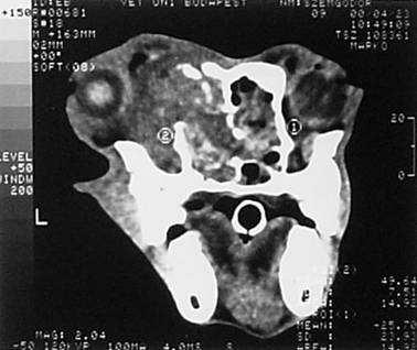
6 Traumatic injuries of the skull and brain Car accident Bulla tympani CT and conv. radiology Remedios: the result of the x-ray exam. was neg. In 25% of the surgically treated otitis media cases. Griffiths: by experimental examinations the radiographs show 80% sensitivity and 65% specificity in fluid accumulation in the bulla cavity Love: : CT is more sensitive than radiographs Horner-syndrome Bulla tympani Bulla tympani 6


")

: soft")


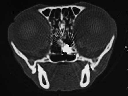
7 Eye and orbital region Exophtalmus and 3rd eyelid protrusion in most of the cases retrobulbar mass! Tumor, abscess, cyst, haematoma Diag. imaging: : UH, CT and MR (generally( radiograph is not helpful) UH: eyeglobe and retrobulbar soft tissues. CT, (MR): retrobulbar soft tisues + bones Osteosarcoma of the eyeglobe Retrobulbar abscess Retrobulbar foreign body 7


8 Retrobulbar masses Neoplasma Neoplasma Nasal cavity and paranasal sinuses Tumor or chronic rhinitis? CT is more sensitive and specific than conv. rad. Easier planning of the radio and surdgical therapy Nasal cavity and sinuses Coronal view Transversal view 8


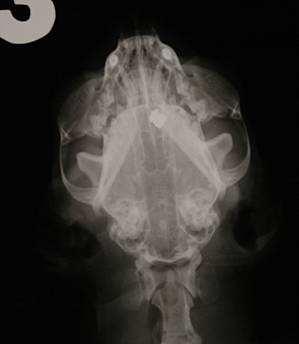
9 Nasal cavity and sinuses Adenocarcinoma adenocarcinoma Nationale: 9 y, female, shephard dog History: chronic nasal discharge, epistaxis on the left side, later epileptic seizures and lethargy Neurology: semicoma,, no menace reflex, epistaxis, exophtalmus Lokalisation: forebrain, nasal cavity, retrobulbar region CT CT Natív Postcontrast 9


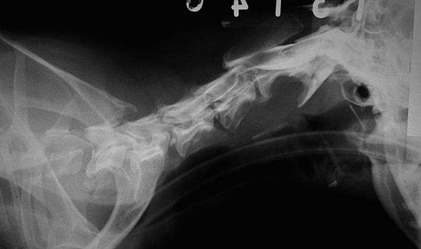
10 Macromorphology Macromorphology Diabolo bullet osteoma osteoma 10


")


11 2 months control Spine Trauma CT, MRI, (RTG) compression MRI, (myelography (myelography,, CT, CTCT-myelo) myelo) Myelon Preop CT 2 months control 11


12 Spine compressive lesions Intramedullar compression neoplasma Spine Extramedullar compression - neoplasma osteosarcoma osteosarcoma 12


13 osteosarcoma osteosarcoma Discus hernia 13


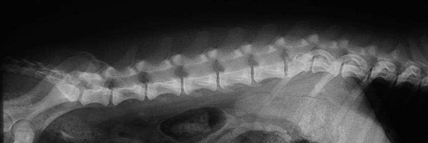


14 Discus hernia Discus protrusio subarachnoideal cyst 14


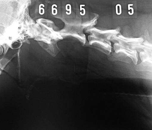

15 Fracture of the atlas Post op. C5 fracture Post op. Tumor 15


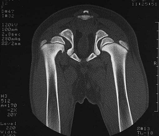


16 fibrosarcoma nativ postcontrast Orthopedy Thyroid gland follicular adenocarcinoma FCP j.o. 16


17 Thorax acropachia neoplasma Development of CT scanners Helical CT (volumen CT) Hernia diaphragmatica 17


18 Multislice CT Thanks for your attention! 18
HEAD AND NECK IMAGING. James Chen (MS IV)
") HEAD AND NECK IMAGING James Chen (MS IV) Anatomy Course Johns Hopkins School of Medicine Sept. 27, 2011 OBJECTIVES Introduce cross sectional imaging of head and neck Computed tomography (CT) Review head
HEAD AND NECK IMAGING James Chen (MS IV) Anatomy Course Johns Hopkins School of Medicine Sept. 27, 2011 OBJECTIVES Introduce cross sectional imaging of head and neck Computed tomography (CT) Review head
Head CT Scan Interpretation: A Five-Step Approach to Seeing Inside the Head Lawrence B. Stack, MD
 Head CT Scan Interpretation: A Five-Step Approach to Seeing Inside the Head Lawrence B. Stack, MD Five Step Approach 1. Adequate study 2. Bone windows 3. Ventricles 4. Quadrigeminal cistern 5. Parenchyma
Supplementary Table 1. ICD-9/-10 codes used to identify cycling injury hospitalizations. Railway accidents injured pedal cyclist
 Supplementary Table 1. ICD-9/-10 codes used to identify cycling injury hospitalizations. ICD Code ICD-9 E800-E807(.3) E810-E816, E818-E819(.6) E820-E825(.6) E826-E829(.1) ICD-10-CA V10-V19 (including all
CT - Brain Examination
 CT - Brain Examination Submitted by: Felemban 1 CT - Brain Examination The clinical indication of CT brain are: a) Chronic cases (e.g. headache - tumor - abscess) b) ER cases (e.g. trauma - RTA - child
Advanced Animal Imaging Ryan Harrell BS, BS, CNMT
 Advanced Animal Imaging Ryan Harrell BS, BS, CNMT It is a specialized form of x -ray that helps veterinarians determine damage to th e spinal cord. A myelogram can help determine if there is a serious
Diagnostic modalities of the Central Nervous System
 NEURORADIOLOGY Kinga Karlinger, MD, PhD Associate Professor Semmelweis University, Budapest Diagnostic modalities of the Central Nervous System X-ray: screening is not used any more, x-ray images instead
TIME: 3 Hours Marks: 100. I. FILL IN THE BLANKS - ANSWER FOR ANY 10 QUESTIONS 1 x 10 = 10
 1 BHARAT SEVAK SAMAJ NATIONAL DEVELOPMENT AGENCY, PROMOTED BY GOVERNMENT OF INDIA CENTRAL BOARD OF EXAMINATIONS BSS NATIONAL VOCATIONAL EDUCATION MISSION AHE009-CT SCAN TECHNICIAN ONE YEAR EXAMINATION
Practical CT and MRI Anthony J. Fischetti, DVM, MS, DACVR Department Head of Diagnostic Imaging The Animal Medical Center, New York OBJECTIVE:
 Practical CT and MRI Anthony J. Fischetti, DVM, MS, DACVR Department Head of Diagnostic Imaging The Animal Medical Center, New York OBJECTIVE: This lecture describes the most common indications for referred
Head injuries. Severity of head injuries
 Head injuries ED Teaching day 23 rd October Severity of head injuries Minor GCS 14-15 Must not have any of the following: Amnesia 10min Neurological sign or symptom Skull fracture (clinically or radiologically)
The diagnostic value of Computed Tomography in evaluation of maxillofacial Trauma
 The diagnostic value of Computed Tomography in evaluation of maxillofacial Trauma Qais H. Muassa FICMS College of Dentistry, Babylon University Ibrahim S. Gataa, BDS, FICMS College of Dentistry, Sulaimania
X-Ray & CT Physics / Clinical CT
 Computed Tomography-Basic Principles and Good Practice X-Ray & CT Physics / Clinical CT INSTRUCTORS: Dane Franklin, MBA, RT (R) (CT) Office hours will be Tuesdays from 5pm to 6pm CLASSROOM: TIME: REQUIRED
Contributors. Thanks to Peter Miller, MD; LCDR Kevin Preston, MD; and Keith Newbrough, MD for their generous contribution of images:
 Contributors Thanks to Peter Miller, MD; LCDR Kevin Preston, MD; and Keith Newbrough, MD for their generous contribution of images: Peter Miller, MD, Indiana University School of Medicine Chapter 1: Figure
Surgical Care at the District Hospital. EMERGENCY & ESSENTIAL SURGICAL CARE
 Surgical Care at the District Hospital 1 17 Orthopedic Techniques Key Points 2 17.1 Traction Use an appropriate method of traction to treat fractures of the extremities and cervical spine Apply extremity
Neurological examination of the neurosurgical patient. Dániel Bereczki SU Department of Neurology
 Neurological examination of the neurosurgical patient Dániel Bereczki SU Department of Neurology E-learning Indivudual study Interactive learning Self assessment at the end of chapters E-learning Indivudual
Imaging of the Paranasal Sinuses
 14. Sommerschule Imaging of the Paranasal Sinuses Bettlach 24.08.2018 Christoph Schlegel Conventional Radiology NNH-Status: okzipito-frontal: frontal sinus, anterior ethmoid okzipito-nasal : maxillary
Applicable Neuroradiology
 For the Clinical Neurology Clerkship LSU Medical School New Orleans Amy W Voigt, MD Clerkship Director Introduction The field of Radiology first developed following the discovery of X-Rays by Wilhelm Roentgen
Radiotherapy in feline and canine head and neck cancer
 Bettina Kandel Like surgery radiotherapy is usually a localized type of treatment. Today it is more readily available for the treatment of cancer in companion animals and many clients are well informed
CT Guided Procedures And Interesting Cases. Stephen Kim, MD Diagnostic and Interventional Radiology
 CT Guided Procedures And Interesting Cases Stephen Kim, MD Diagnostic and Interventional Radiology CT guided procedure benefits Precise lesion targeting Clear image guidance for needle placement Immediate
Brain Injuries. Presented By Dr. Said Said Elshama
 Brain Injuries Presented By Dr. Said Said Elshama Types of head injuries 1- Scalp injuries 2- Skull injuries 3- Intra Cranial injuries ( Brain ) Anatomical structure of meninges Intra- Cranial Injuries
Clinical Assessment of the Diagnostic Value of Facial Radiography in Facial Trauma Patients at the Emergency Department
 Chin J Radiol 2005; 30: 327-333 327 Clinical Assessment of the Diagnostic Value of Facial Radiography in Facial Trauma Patients at the Emergency Department PI-YUN CHIU 1 JEN-DAR CHEN 1,2 PING-YI KO 1 CHENG-YEN
Brain Tumors. What is a brain tumor?
 Scan for mobile link. Brain Tumors A brain tumor is a collection of abnormal cells that grows in or around the brain. It poses a risk to the healthy brain by either invading or destroying normal brain
8/29/2011. Brain Injury Incidence: 200/100,000. Prehospital Brain Injury Mortality Incidence: 20/100,000
 Traumatic Brain Injury Almario G. Jabson MD Section Of Neurosurgery Asian Hospital And Medical Center Brain Injury Incidence: 200/100,000 Prehospital Brain Injury Mortality Incidence: 20/100,000 Hospital
2012 CPT Radiology Codes Requiring Review Blue Cross and Blue Shield of Louisiana
 2012 CPT Radiology Codes Requiring Review Blue Cross and Blue Shield of Louisiana CT Head 70480 CT orbit, sella or posterior fossa; w/o CT Head 70481 CT orbit, sella or posterior fossa; with CT Head 70482
Pediatric CT Protocols (18 years old or less)
") Pediatric CT Protocols (18 years old or less) Ped1: Head CT Ped2: Cervical spine CT Ped3: Sinus CT Ped4: Neck CT Ped5: Chest CT Ped6: Abdomen and pelvis CT Ped7: Thoracic or lumbar spine CT Ped8: Extremity
Pediatric CT Protocols (18 years old or less) Ped1: Head CT Ped2: Cervical spine CT Ped3: Sinus CT Ped4: Neck CT Ped5: Chest CT Ped6: Abdomen and pelvis CT Ped7: Thoracic or lumbar spine CT Ped8: Extremity
RADPrimer Curriculum Breast Topics Covered Basic Intermediate 225
 Breast Anatomy & Normal Variants 11 Breast Imaging Modalities 13 BI RADS Lexicon 3 Mammography: Masses 9 Mammography: Calcifications 17 Mammography: Additional Findings 8 Ultrasound Features 10 Ultrasound
 www.oralradiologists.com CONE BEAM CT REPORT CASE ---- Case Information Referring Doctor: - Patient Name: - Scan Date: December 1, 2015 Patient DOB: - Reason for Exam: - Study Details: icat Flex, 160x160x112
Class Time: 9/12,10-10,11-21 Days: Listed Saturdays 1p-4p Room:HS208. Office Phone: Home Phone:
 Semester/Year: FALL 2015 CASPER COLLEGE COURSE SYLLABUS RDTK 1920 H1 Computed Tomography Procedures I Lecture Hours: 3 Lab Hours: 0 Credit Hours: 3 Class Time: 9/12,10-10,11-21 Days: Listed Saturdays 1p-4p
NEURO PROTOCOLS MRI NEURO PROTOCOLS (SIEMENS SCANNERS)
") Page 1 NEURO PROTOCOLS Brain Stroke Brain Brain with contrast Brain for seizures Brain for MS Brain for Pineal gland Sella FAST Scan for hydrocephalus MRA/MRV Brain MRA carotids 8 th nerve Cranial nerves
Page 1 NEURO PROTOCOLS Brain Stroke Brain Brain with contrast Brain for seizures Brain for MS Brain for Pineal gland Sella FAST Scan for hydrocephalus MRA/MRV Brain MRA carotids 8 th nerve Cranial nerves
Neuroimaging. spine / spinal cord
 Neuroimaging spine / spinal cord Spine & spinal cord imaging methodology Plain x-ray of spine Computed tomography CT - traditional ( normal CT) - reconstructions - myelo-ct Magnetic resonance MR - standard
Evaluation of Craniocerebral Trauma Using Computed Tomography
 IOSR Journal of Dental and Medical Sciences (IOSR-JDMS) e-issn: 2279-0853, p-issn: 2279-0861.Volume 13, Issue 9 Ver. IV (Sep. 2014), PP 57-62 Evaluation of Craniocerebral Trauma Using Computed Tomography
CT of the Head, Spine, and Cerebral Vessels
 CT of the Head, Spine, and Cerebral Vessels Objectives Determine specific imaging plane used to acquire or reformat CT scan, i.e. sagittal, coronal, transverse, and offaxis or oblique. Assess and evaluate
CT AND MRI CT COMPUTED (AXIAL) TOMOGRAPHY. CT and MRI Clinical applications in Veterinary Medicine MRI CT VS. MRI THE DIFFERENCES
 TOMOGRAPHY. CT and MRI Clinical applications in Veterinary Medicine MRI CT VS. MRI THE DIFFERENCES") AND MRI CLINICAL APPLICATIONS IN VETERINARY MEDICINE BVM, MVM, DipECVN AND MRI Cross sectional images Digital processing Sedation / anesthesia required in animals Comparison to radiography / ultrasonography
AND MRI CLINICAL APPLICATIONS IN VETERINARY MEDICINE BVM, MVM, DipECVN AND MRI Cross sectional images Digital processing Sedation / anesthesia required in animals Comparison to radiography / ultrasonography
Computed Tomography (CT) - Head
 - Head") Scan for mobile link. Computed Tomography (CT) - Head Computed tomography (CT) of the head uses special x-ray equipment to help assess head injuries, severe headaches, dizziness, and other symptoms of
Scan for mobile link. Computed Tomography (CT) - Head Computed tomography (CT) of the head uses special x-ray equipment to help assess head injuries, severe headaches, dizziness, and other symptoms of
STRUCTURED EDUCATION REQUIREMENTS EFFECTIVE: JANUARY 1, 2016
 Computed Tomography The purpose of structured education is to provide the opportunity for individuals to develop mastery of discipline-specific knowledge that, when coupled with selected clinical experiences,
Spine. Neuroradiology. Spine. Spine Pathology. Distribution of fractures. Radiological algorithm. Role of radiology 18/11/2015
 Spine Neuroradiology Spine Prof.Dr.Nail Bulakbaşı X Ray: AP/L/Oblique Vertebra & disc spaces CT & CTA Vertebra, discs, vessels MRI & MRA Vertebra, disc, vessels, meninges Spinal cord & nerves Myelography
LOSS OF CONSCIOUSNESS & ASSESSMENT. Sheba Medical Center Acute Medicine Department MATTHEW WRIGHT
 LOSS OF CONSCIOUSNESS & ASSESSMENT Sheba Medical Center Acute Medicine Department MATTHEW WRIGHT OUTLINE Causes Head Injury Clinical Features Complications Rapid Assessment Glasgow Coma Scale Classification
Radiology. General radiology department. X-ray
 The radiology directorate provides a diagnostic, interventional and therapeutic service for its local population, and a tertiary service for the region. It also provides support to some national work such
RADIATION PROTECTION OF THE PATIENT IN PAEDIATRIC RADIOLOGY. Bahnarel Ion, Dimov Nicolae, Coretchi Liuba, Cujba Natalia
 RADIATION PROTECTION OF THE PATIENT IN PAEDIATRIC RADIOLOGY Bahnarel Ion, Dimov Nicolae, Coretchi Liuba, Cujba Natalia Medical Diagnostic Centre Magnific Chisinau, Republic of Moldova, e-mail: Ndimov@mail.ru
Klinikleitung: Dr. Kessler Dr. Kosfeld Dr. Tassani-Prell Dr. Bessmann. Radiotherapy in feline and canine head and neck cancer.
 Radiotherapy in feline and canine head and neck cancer Bettina Kandel Like surgery radiotherapy is usually a localized type of treatment. Today it is more readily available for the treatment of cancer
Block 3 VIRGINIA CAMPUS Neurological System and Special Senses 2017
 WEEK 1 Time/Date Monday, January 30 Tuesday, January 31 Wednesday, February 1 Thursday, February 2 Friday, February 3 1. PATHOLOGY 2-3. NEUROSCIENCE 6. NEUROSCIENCE 9-10. NEUROSCIENCE 11. NEUROSCIENCE
JUSTIFICATION PROTOCOLS FOR CT SCANNING ALBURY WODONGA HEALTH WODONGA CAMPUS
 JUSTIFICATION PROTOCOLS FOR CT SCANNING ALBURY WODONGA HEALTH WODONGA CAMPUS JUSTIFICATION PROTOCOLS FOR CT SCANNING INTRODUCTION: In accordance with the Victorian Radiation Act 2005 Wodonga Medical Imaging,
Note the high activity in the venous sinuses on all images
 6. AGE 3 4 YEARS 65 66 Blood Pool Images 6: Age 3 4 Years Fig. 1. Posterior view of skull and thorax Fig. 2. Right lateral view of skull and of thorax Fig. 5. Left lateral view of skull and anterior view
Pre-hospital Response to Trauma and Brain Injury. Hans Notenboom, M.D. Asst. Medical Director Sacred Heart Medical Center
 Pre-hospital Response to Trauma and Brain Injury Hans Notenboom, M.D. Asst. Medical Director Sacred Heart Medical Center Traumatic Brain Injury is Common 235,000 Americans hospitalized for non-fatal TBI
Is there a role of CT in the evaluation of Proptosis
 International Journal of scientific research and management (IJSRM) Volume 3 Issue 4 Pages 2662-2666 2015 \ Website: www.ijsrm.in ISSN (e): 2321-3418 Is there a role of CT in the evaluation of Proptosis
RADIOLOGY (Management)
") ULTRASOUND BETA SCAN/ U/S ORBITAL 1600 Daily U/S WHOLE ABDOMEN (Abd + Pelvis) 1200 Daily U/S PELVIS 1200 Daily U/S ABDOMEN 1200 Daily U/S BREAST 1800 Daily U/S FOLLICULAR STUDY 3000 Daily U/S FOLLICULAR
ULTRASOUND BETA SCAN/ U/S ORBITAL 1600 Daily U/S WHOLE ABDOMEN (Abd + Pelvis) 1200 Daily U/S PELVIS 1200 Daily U/S ABDOMEN 1200 Daily U/S BREAST 1800 Daily U/S FOLLICULAR STUDY 3000 Daily U/S FOLLICULAR
Index. Note: Page numbers of article titles are in bold face type.
 Neurosurg Clin N Am 13 (2002) 259 264 Index Note: Page numbers of article titles are in bold face type. A Abdominal injuries, in child abuse, 150, 159 Abrasions, in child abuse, 157 Abuse, child. See Child
BRAIN TRAUMA THERAPEUTIC RECOMMENDATIONS
 1 BRAIN TRAUMA THERAPEUTIC RECOMMENDATIONS Richard A. LeCouteur, BVSc, PhD, Dip ACVIM (Neurology), Dip ECVN Professor Emeritus, University of California, Davis, California, USA Definitions Hemorrhage:
Diagnostic Tools: Equine Dentistry. Dr. Chris Blevins Equine Field Service Clinician
 Diagnostic Tools: Equine Dentistry Dr. Chris Blevins Equine Field Service Clinician Objectives Know 3 useful diagnostic tools. What is most important aspect about dental radiology? Know 3 standard radiographs
2
 1 2 3 4 5 6 7 8 9 10 11 12 13 Cine loop of tomosynthesis slice images through the chest. 14 Standard PA chest radiograph (left) and single slice from the tomosynthesis image dataset (right) of a patient
Head & Neck Clinical Sub Group. Network Agreed Imaging Guidelines for UAT and Thyroid Cancer. Measure Nos: 11-1C-105i & 11-1C-106i
 Greater Manchester, Lancashire & South Cumbria Strategic Clinical Network & Senate Head & Neck Clinical Sub Group Network Agreed Imaging Guidelines for UAT and Thyroid Cancer Measure Nos: 11-1C-105i &
Introduction to Neuroimaging Aaron S. Field, MD, PhD Assistant Professor of Radiology Neuroradiology Section University of Wisconsin Madison
 Introduction to Neuroimaging Aaron S. Field, MD, PhD Assistant Professor of Radiology Neuroradiology Section University of Wisconsin Madison Updated 7/17/07 Neuroimaging Modalities Radiography (X-Ray)
Proceedings of the World Small Animal Veterinary Association Sydney, Australia 2007
 Proceedings of the World Small Animal Veterinary Association Sydney, Australia 2007 Hosted by: Australian Small Animal Veterinary Association (ASAVA) Australian Small Animal Veterinary Association (ASAVA)
The Computed Tomography Examination
 CONTENT SPECIFICATIONS The Computed Tomography Examination The purpose of The American Registry of Radiologic Technologists (ARRT ) Computed Tomography Examination is to assess the knowledge and cognitive
Radiologic Imaging Magnetic Resonance Imaging (MRI)
") Radiologic Imaging X-ray has always been the golden rule in diagnosing and treating podiatric patients. Unfortunately, for some patients the diagnosis is not as evident. That is when we need to utilize
Radiologic Imaging X-ray has always been the golden rule in diagnosing and treating podiatric patients. Unfortunately, for some patients the diagnosis is not as evident. That is when we need to utilize
AIM 2014 CPT Radiology & Cardiac Codes Requiring Review
 AIM 2014 CPT Radiology & Cardiac Codes Requiring Review Modality Body Part CT Head 1 70480 CT orbit, sella or posterior fossa; w/o contrast 1 CT Head 1 70481 CT orbit, sella or posterior fossa; with CT
Index. Note: Page numbers of article titles are in boldface type.
 Index Note: Page numbers of article titles are in boldface type. A Abdominal injuries, abdominal wall muscle injury, 212 213 diaphragmatic spasm, 212 liver injury, 213 214 pancreatic injury, 216 rectus
OP-14: SIMULTANEOUS USE OF BRAIN COMPUTED TOMOGRAPHY (CT) AND SINUS COMPUTED TOMOGRAPHY (CT)
 AND SINUS COMPUTED TOMOGRAPHY (CT)") OP-14: SIMULTANEOUS USE OF BRAIN COMPUTED TOMOGRAPHY (CT) AND SINUS COMPUTED TOMOGRAPHY (CT) Description of Measure This measure calculates the percentage of Brain CT studies with a simultaneous Sinus
OP-14: SIMULTANEOUS USE OF BRAIN COMPUTED TOMOGRAPHY (CT) AND SINUS COMPUTED TOMOGRAPHY (CT) Description of Measure This measure calculates the percentage of Brain CT studies with a simultaneous Sinus
Case Presentation 主治醫師 : 宋文鑫日期 :
 Case Presentation 主治醫師 : 宋文鑫日期 : 2015-2-28 General Data Name:OOO Chart Number:OOOOOOO Date of Admission:2014 年 08 月 04 日 Age: 33 y/o Sex:female Occupation : 會計 Chief Complaint Palpable soft tissue mass
APPROACHES TO NEUROLOGICAL EXAMINATION IN RABBITS
 Vet Times The website for the veterinary profession https://www.vettimes.co.uk APPROACHES TO NEUROLOGICAL EXAMINATION IN RABBITS Author : ELISABETTA MANCINELLI Categories : Vets Date : October 6, 2014
Abdomen and Pelvis CT (1) By the end of the lecture students should be able to:
 By the end of the lecture students should be able to:") RAD 451 Abdomen and Pelvis CT (1) By the end of the lecture students should be able to: State the common indications for Abdomen and pelvis CT exams Identify possible contra indications for Abdomen and
RAD 451 Abdomen and Pelvis CT (1) By the end of the lecture students should be able to: State the common indications for Abdomen and pelvis CT exams Identify possible contra indications for Abdomen and
CSF Leaks. Abnormal communication between the subarachnoid space and the tympanomastoid space or nasal cavity. Presenting symptoms:
 CSF Leaks Steven Wright, M.D. Faculty Advisor: Matthew Ryan, M.D. The University of Texas Medical Branch Department of Otolaryngology Grand Rounds Presentation January 5, 2005 CSF Leaks Abnormal communication
Semiotics in Radiology
 Adelino Santos Health Technology College Coimbra, Portugal Collaboration of António Agudo Student of Radiology College of Health Technology Coimbra, Portugal What are the most important points to evaluate
Radiotherapy in small animals. How is it realized? How does it work? For which patient is it indicated?
 Radiotherapy in small animals How is it realized? How does it work? For which patient is it indicated? Radiotherapy Our linear accelerator can generate photons or electrons. The electron beam is used to
Pediatric Subdural Hematoma and Traumatic Brain Injury J. Charles Mace MD FACS Springfield Neurological Institute CoxHealth. Objectives 11/7/2017
 Pediatric Subdural Hematoma and Traumatic Brain Injury J. Charles Mace MD FACS Springfield Neurological Institute CoxHealth Objectives 1. Be able to discuss brain anatomy and physiology as it applies to
PA SYLLABUS. Syllabus for students of the FACULTY OF MEDICINE No.2
 Approved At the meeting of the Faculty Council Medicine No. of Approved At the meeting of the chair of Neurosurgery No. of Dean of the Faculty Medicine No.2 PhD, associate professor M. Betiu Head of the
How to interpret an unenhanced CT brain scan. Part 2: Clinical cases
 How to interpret an unenhanced CT brain scan. Part 2: Clinical cases Thomas Osborne a, Christine Tang a, Kivraj Sabarwal b and Vineet Prakash c a Radiology Registrar; b Radiology Foundation Year 1 Doctor;
Imaging of Acute Cerebral Trauma
 July, 2005 Imaging of Acute Cerebral Trauma Louis Rivera, Harvard Medical School, Year III 46 y/o Female s/p Trauma - Unrestrained? MVC requiring Med Flight - Facial bruising/swelling - DEEP COMA - SEIZURES
An Introduction to Imaging the Brain. Dr Amy Davis
 An Introduction to Imaging the Brain Dr Amy Davis Common reasons for imaging: Clinical scenarios: - Trauma (NICE guidelines) - Stroke - Tumours - Seizure - Neurological degeneration memory, motor dysfunction,
Advances in Emergency Imaging
 Hampton Symposium,, October 16 th, 2010 Advances in Emergency Imaging Robert A. Novelline, MD Professor of Radiology, Harvard Medical School Director of Emergency Radiology, Massachusetts General Hospital
Contrast Guidelines for Common CT/CTA & MRI/MRA
 Contrast Guidelines for Common /A & /MRA Body Imaging Gastrointestinal CLINICAL GUIDELINES EXAM DESCRIPTION /A CPT CODES EXAM DESCRIPTION /MRA CPT CODES Abdominal mass Abdomen & Pelvis w 74177 Abdomen
Diagnostic Tools: Equine Dentistry. Dr. Chris Blevins Equine Field Service Clinician
 Diagnostic Tools: Equine Dentistry Dr. Chris Blevins Equine Field Service Clinician Objectives Know 3 useful diagnostic tools. What is most important aspect about dental radiology? Know 3 standard radiographs
Computed tomography of the chest: I. Basic principles
 BJA Education, 15 (6): 299 304 (2015) doi: 10.1093/bjaceaccp/mku063 Advance Access Publication Date: 2 February 2015 Matrix reference 1A03, 2A12 Computed tomography of the chest: I. Basic principles P
SPECIFIC PRINCIPLES FOR DOSE REDUCTION IN HEAD CT IMAGING. Rajiv Gupta, MD, PhD Neuroradiology, Massachusetts General Hospital Harvard Medical School
 SPECIFIC PRINCIPLES FOR DOSE REDUCTION IN HEAD CT IMAGING Rajiv Gupta, MD, PhD Neuroradiology, Massachusetts General Hospital Harvard Medical School OUTLINE 1 st Presentation: Dose optimization strategies
PRACTICE GUIDELINE. DEFINITIONS: Mild head injury: Glasgow Coma Scale* (GCS) score Moderate head injury: GCS 9-12 Severe head injury: GCS 3-8
 score Moderate head injury: GCS 9-12 Severe head injury: GCS 3-8") PRACTICE GUIDELINE Effective Date: 9-1-2012 Manual Reference: Deaconess Trauma Services TITLE: TRAUMATIC BRAIN INJURY GUIDELINE OBJECTIVE: To provide practice management guidelines for traumatic brain
PRACTICE GUIDELINE Effective Date: 9-1-2012 Manual Reference: Deaconess Trauma Services TITLE: TRAUMATIC BRAIN INJURY GUIDELINE OBJECTIVE: To provide practice management guidelines for traumatic brain
CNS Imaging. Dr Amir Monir, MD. Lecturer of radiodiagnosis.
 CNS Imaging Dr Amir Monir, MD Lecturer of radiodiagnosis www.dramir.net Types of radiological examinations you know Plain X ray X ray with contrast GIT : barium (swallow, meal, follow through, enema) ERCP
Special Instructions
 FDA and ACR guidelines are as follows: Special Instructions Safety concerning NSF and gadolinium-based contrast agents (GBCA) Prior to administering MRI contrast (GBCA), any patient who answers yes to
Lecture 1. Lecture 1: The Different Modalities
 Lecture 1 Lecture 1: The Different Modalities In this Lecture Understanding the difference between the different modalities available Learn when to chose the appropriate modality Trust me, during the next
Regional Human Anatomy (HBA 461/561/540): Course Objectives
: Course Objectives") Regional Human Anatomy (HBA 461/561/540): Course Objectives This is a 5-credit course that consists of 1-hour lectures followed by 3-hour labs. It is organized into three modules (see syllabus): Module
Regional Human Anatomy (HBA 461/561/540): Course Objectives This is a 5-credit course that consists of 1-hour lectures followed by 3-hour labs. It is organized into three modules (see syllabus): Module
Proceedings of the World Small Animal Veterinary Association Sydney, Australia 2007
 Proceedings of the World Small Animal Sydney, Australia 2007 Hosted by: Next WSAVA Congress THE LAST GASP II: LUNGS AND THORAX David Holt, BVSc, Diplomate ACVS University of Pennsylvania School of Veterinary
CT FINDINGS OF THORACOLUMBAR SPINE LESIONS IN DOGS
 CT FINDINGS OF THORACOLUMBAR SPINE LESIONS IN DOGS C. DARABAN 1, V. VULPE 1, FLORENTINA BOCĂNEŢI 1, GIUSEPPINA MENNONNA 2, M. SACCONE 2, G. FATONE 2, L. MEOMARTINO 2 1 University of Agriculture Science
Request Card Task ANSWERS
 Request Card Task ANSWERS Medical Student Workbook Author: Dr Sam Leach, SpR Case 1 What differential diagnoses are most likely? Which investigation is most appropriate? Case 1 The most likely diagnosis
V. CENTRAL NERVOUS SYSTEM TRAUMA
 V. CENTRAL NERVOUS SYSTEM TRAUMA I. Concussion - Is a clinical syndrome of altered consiousness secondary to head injury - Brought by a change in the momentum of the head when a moving head suddenly arrested
Introduction to Neurosurgical Subspecialties:
 Introduction to Neurosurgical Subspecialties: Trauma and Critical Care Neurosurgery Brian L. Hoh, MD 1, Gregory J. Zipfel, MD 2 and Stacey Q. Wolfe, MD 3 1 University of Florida, 2 Washington University,
General Imaging. Imaging modalities. Incremental CT. Multislice CT Multislice CT [ MDCT ]
![General Imaging. Imaging modalities. Incremental CT. Multislice CT Multislice CT [ MDCT ]](/thumbs/76/74079340.jpg "General Imaging. Imaging modalities. Incremental CT. Multislice CT Multislice CT [ MDCT ]") General Imaging Imaging modalities Conventional X-rays Ultrasonography [ US ] Computed tomography [ CT ] Radionuclide imaging Magnetic resonance imaging [ MRI ] Angiography conventional, CT,MRI Interventional
General Imaging Imaging modalities Conventional X-rays Ultrasonography [ US ] Computed tomography [ CT ] Radionuclide imaging Magnetic resonance imaging [ MRI ] Angiography conventional, CT,MRI Interventional
Computed Tomography (CT) - Spine
 - Spine") Scan for mobile link. Computed Tomography (CT) - Spine Computed tomography (CT) of the spine is a diagnostic imaging test used to help diagnose or rule out spinal column damage in injured patients. CT
Scan for mobile link. Computed Tomography (CT) - Spine Computed tomography (CT) of the spine is a diagnostic imaging test used to help diagnose or rule out spinal column damage in injured patients. CT
Med 536 Communicating About Prognosis Workshop. Case 1
 Med 536 Communicating About Prognosis Workshop Case 1 ID / CC: 39 year-old woman status-post motor-vehicle collision History of the Presenting Illness Previously healthy 39 year-old woman was found in
Catalog Addendum
 Catalog Addendum - 2018 Day Class RADIOLOGIC TECHNOLOGY (Levittown) HEGIS CODE: 5207.00 Radiologic Technologies (X-Ray) Day Program - 2005 Hours (16 mos./67 wks.) Diploma Program Hunter Business School
Radiography. 1. Introduction. 2. Documentation of Compliance. 3. Didactic Competency Requirements. 4. Clinical Competency Requirements
 PRIMARY CERTIFICATION AND REGISTRATION Radiography 1. Introduction Candidates for certification and registration are required to meet the Professional Education Requirements specified in the ARRT Rules
Correlation of Computed Tomography findings with Glassgow Coma Scale in patients with acute traumatic brain injury
 Journal of College of Medical Sciences-Nepal, 2014, Vol-10, No-2 ABSTRACT OBJECTIVE To correlate Computed Tomography (CT) findings with Glasgow Coma Scale (GCS) in patients with acute traumatic brain injury
A Guide to the Radiologic Evaluation of Extra-Axial Hemorrhage
 July 2013 A Guide to the Radiologic Evaluation of Extra-Axial Hemorrhage John Dickson, Harvard Medical School Year III Agenda 1. Define extra-axial hemorrhage and introduce its subtypes 2. Review coup
Overview. Imaging Indications. Paediatric Radiation Safety 2015/03/12. Paediatric radiation safety General guidelines Protocols
 Overview Paediatric radiation safety General guidelines Protocols Paediatric Radiation Safety Paediatric patients are unique Children are more susceptible to radiation induced cancer than adults Younger
B. CT protocols for the spine
 B. CT protocols for the spine Poster No.: A-003 Congress: ECR 2010 Type: Invited Speaker Topic: Neuro Authors: B. Tins; Oswestry/UK Keywords: CT, spine, diagnostic imaging protocol DOI: 10.1594/ecr2010/A-003
Date of Admission: [DATE]. Date of Discharge:
![Date of Admission: [DATE]. Date of Discharge:](/thumbs/74/71277118.jpg "Date of Admission: [DATE]. Date of Discharge:") Date of Admission: [DATE]. Date of Discharge: History of Present Illness: Mr. [NAME] AKA [NAME] is a 31-year-old male who presents to the [PLACE] Trauma Surgery Service as a moderate trauma on [DATE] following
Date of Admission: [DATE]. Date of Discharge: History of Present Illness: Mr. [NAME] AKA [NAME] is a 31-year-old male who presents to the [PLACE] Trauma Surgery Service as a moderate trauma on [DATE] following
Computed Tomography (CT) - Sinuses
 - Sinuses") Scan for mobile link. Computed Tomography (CT) - Sinuses Computed tomography (CT) of the sinuses uses special x-ray equipment to evaluate the paranasal sinus cavities hollow, air-filled spaces within the
Scan for mobile link. Computed Tomography (CT) - Sinuses Computed tomography (CT) of the sinuses uses special x-ray equipment to evaluate the paranasal sinus cavities hollow, air-filled spaces within the
Traumatic Brain Injury TBI Presented by Bill Masten
 1 2 Cerebrum two hemispheres and four lobes. Cerebellum (little brain) coordinates the back and forth ballet of motion. It judges the timing of every movement precisely. Brainstem coordinates the bodies
Pediatric Abusive Head Trauma
 Pediatric Abusive Head Trauma Rebecca Girardet Associate Professor of Pediatrics Director, Division of Child Protection Pediatrics McGovern Medical School at The University of Texas Health Science Center
Dental Cone Beam CT. What is Dental Cone Beam CT?
 Scan for mobile link. Dental Cone Beam CT Dental cone beam computed tomography (CT) is a special type of x-ray equipment used when regular dental or facial x-rays are not sufficient. Your doctor may use
The central nervous system
 Sectc.qxd 29/06/99 09:42 Page 81 Section C The central nervous system CNS haemorrhage Subarachnoid haemorrhage Cerebral infarction Brain atrophy Ring enhancing lesions MRI of the pituitary Multiple sclerosis
BHARAT SEVAK SAMAJ NATIONAL DEVELOPMENT AGENCY, PROMOTED BY GOVERNMENT OF INDIA CT SCAN TECHNOLOGY COURSE ONE YEAR EXAMINATION
 BHARAT SEVAK SAMAJ NATIONAL DEVELOPMENT AGENCY, PROMOTED BY GOVERNMENT OF INDIA CENTRAL BOARD OF EXAMINATIONS BSS NATION TIONAL VOCA OCATION TIONAL EDUCATION MISSION CT SCAN TECHNOLOGY COURSE ONE YEAR
Meninges and Ventricles
 Meninges and Ventricles Irene Yu, class of 2019 LEARNING OBJECTIVES Describe the meningeal layers, the dural infolds, and the spaces they create. Name the contents of the subarachnoid space. Describe the