Spine. Neuroradiology. Spine. Spine Pathology. Distribution of fractures. Radiological algorithm. Role of radiology 18/11/2015
|
|
|
- Caroline Chambers
- 5 years ago
- Views:
Transcription
1 Spine Neuroradiology Spine Prof.Dr.Nail Bulakbaşı X Ray: AP/L/Oblique Vertebra & disc spaces CT & CTA Vertebra, discs, vessels MRI & MRA Vertebra, disc, vessels, meninges Spinal cord & nerves Myelography Spinal nerves, discs Spine Pathology Trauma Degenerative disease Tumors and other masses Inflammation and infection Vascular disorders Congenital anomalies Distribution of fractures Upper cervical (atlas and axis) Lower cervical (C5-C7) Upper thoracic (T4-T6) Thoracolumbar and lumbar Role of radiology Diagnose the lesion Classify the lesion Detect stability / instability Decide on further investigations when the radiological diagnosis is incompatible with neurological signs Radiological algorithm Imaging is not necessary in asymptomatic patients Imaging in symptomatic patients According to clinical and neurological findings According to the technical possibilities A high rate of symptomatic cases are diagnosed in proper direct radiography 2-way, oblique, functional (flexion and extension) radiographs 1


 Contusion (hematomyelia) Edema Denis three column")


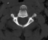
2 Radiological algorithm CT is performed when Fracture on X-ray Suspected fracture on X-ray Normal X-ray in a symptomatic pt MRI is performed when Positive neurological sign Suspected ligament, cord or disk damage Suspected epidural / paravertebral soft tissue lesion What we are looking for? Bone fractures Ligamentous tear Cord / nerve root compression due to bone fragments Disc herniation Epidural hematoma Cord avulsion without fracture (0.7%) Contusion (hematomyelia) Edema Denis three column theory Stable: One column involvement Two non-adjacent column involvement Unstable: 3 column involvement Involvement of two adjacent columns The middle column involvement Jefferson burst fracture Result of vertical compression Bilateral fracture of both anterior and posterior arch of C1 Concomitant fractures in 50% of cases Axis fracture in 33% cases Neurological deficit (-) Transverse atlantal ligament is intact or damaged Unstable Fracture Hangman fracture Bilateral fracture of the pars interarticularis due to hyperextension strain 2


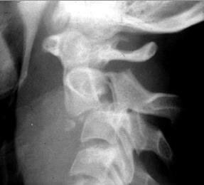
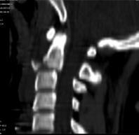
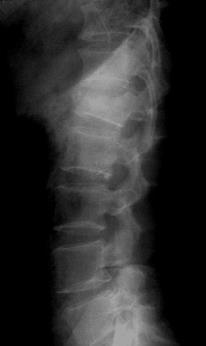

3 Hangman fracture Type I Stable Hangman fracture Type II İnstabile Hangman fracture Type III İnstabile Teardrop fracture Unstable burst fx Translocation 3


: Focal, anterosuperior end plate, in")
: Widespread and settles")

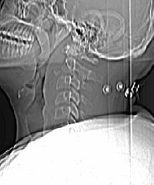
4 Anterolisthesis Fractures of C6 left pedicle and lamina Vertebral Artery Dissection Occlusion due to C6 Fracture Vertebral degeneration Modic 1: T1 hypo / T2 hyper / C + Subchondral edema due to increased vascularity Modic 2: T1/T2 hyper Fatty degeneration due to chronic bone marrow ischemia Modic 3: T1/T2 hypo End plate sclerosis Type 1 changes correlated with low back pain but 10-25% of patients may be asymptomatic * Symptom (-): Focal, anterosuperior end plate, in the middle lumbar spine, normal adjacent discs Symptom (+): Widespread and settles in end plates adjacent to the degenerated disc *Chung CB, et al. Skeletal Radiol 2004;33(7):


 Spondylolysis = chronic")


")
5 Spondylolysis / Spondylolisthesis Confusing Spondy- Terminology Spondylosis = spondylosis deformans = degenerative spine Spondylitis = inflamed spine (e.g. ankylosing, pyogenic, etc.) Spondylolysis = chronic fracture of pars interarticularis with nonunion ( pars defect ) Spondylolisthesis = anterior slippage of vertebra typically resulting from bilateral pars defects Pseudospondylolisthesis = degenerative spondylolisthesis (spondylolisthesis resulting from degenerative disease rather than pars defects) Degenerative Disc Disease Degenerative disc disease 5
6 Degenerative Disc Disease Schmorl s Nodes 6


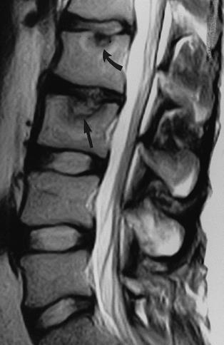
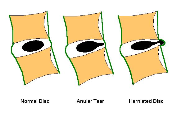
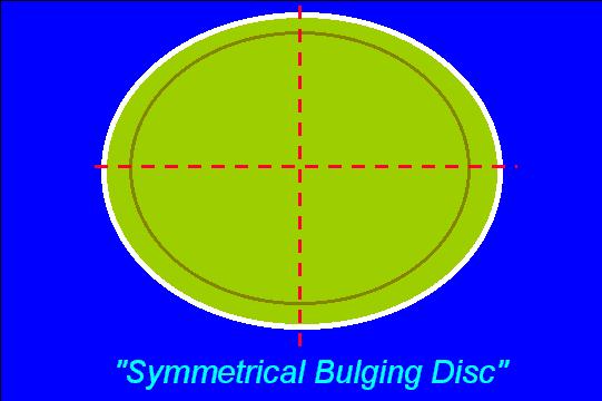
7 Bulging Bulging 7
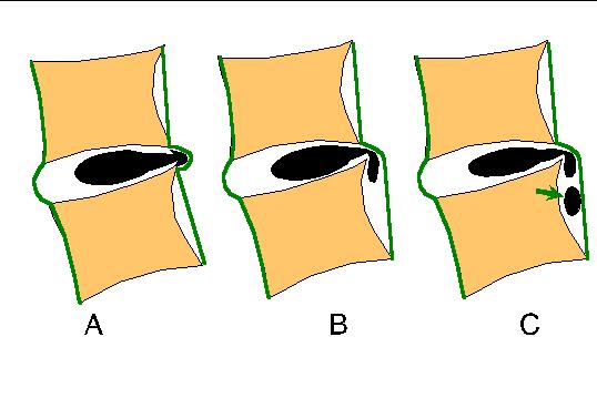
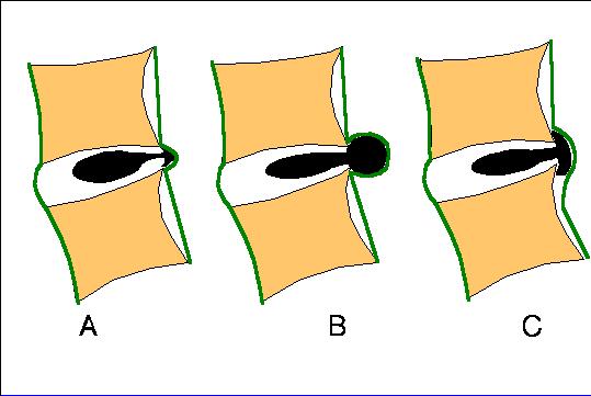
8 Protrusion Protrusion Extrusion Extrusion 8


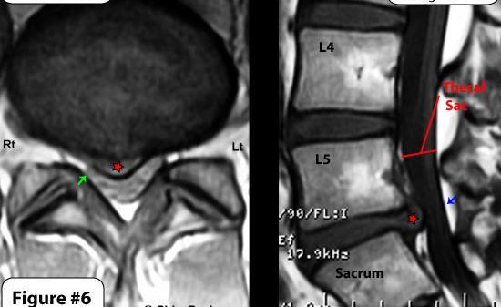
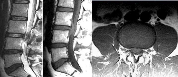
9 Lumbar Spinal Stenosis Sequestration Lumbar Spinal Stenosis Lumbar Spinal Stenosis Disc bulge, facet hypertrophy and flaval ligament thickening frequently combine to cause central spinal stenosis Classification of Spinal Lesions Location of Spinal Lesions Extradural outside the thecal sac (including vertebral bone lesions) Intradural extramedullary within thecal sac but outside cord Intramedullary within cord 9


.")

10 Intramedullary Astrocytoma Ganglioglioma Ependymoma Hemangioblastoma AVM Metastasis Abscess Intradural extramedullary Myxopapillary ependymoma Nerve sheath tumors Meningioma Metastasis ARTT PNET Dermoid Epidermoid Arachnoid cyst Neuroenteric cyst Extradural Benign bone tumors Hemangiomas Osteoid osteoma Osteoblastoma Aneurysmal bone cyst Eosinophilic granuloma Teratoma Malignant bone tumors Ewing's sarcoma Osteosarcoma Lymphoma / leukemia Epidural space tumors Bone sarcomas off Lymphoma / leukemia Germ cell tumors Extradural tumors Neuroblastoma Nerve sheath tumors EM hematopoiesis Extradural: Epidural Abscess Intradural Extramedullary Meningioma Intramedullary: Astrocytoma Intramedullary: Syringohydromyelia Confusing Syrinx Terminology Hydromyelia: Fluid accumulation/dilatation within central canal, therefore lined by ependyma Syringomyelia: Cavitary lesion within cord parenchyma, of any cause (there are many). Located adjacent to central canal, therefore not lined by ependyma Syringohydromyelia: Term used for either of the above, since the two may overlap and cannot be discriminated on imaging Hydrosyringomyelia: Same as syringohydromyelia Syrinx: Common term for the cavity in all of the above 10


 venous hypertension")
11 Infectious Spondylitis / Diskitis Spinal TB (Pott s Disease) T2 T1 T1+C T1+C Transverse Myelitis Inflamed cord of uncertain cause Viral infections Immune reactions Idiopathic Myelopathy progressing over hours to weeks DDX: MS, glioma, infarction Multiple Sclerosis As in the brain, may be secondary to ischemia (e.g. embolus to spinal artery) venous hypertension (e.g. AV fistula) Cord Edema 11
Introduction to Neuroimaging spine. John J. McCormick MD
 Introduction to Neuroimaging spine John J. McCormick MD Neuroanatomy Netter drawings Radiographic Anatomy Cervical Spine Cervical Spine Oblique View Cervical Spine Dens View Thoracic Spine Lumbar Spine
NEURORADIOLOGY. Part III. Angela Csomor University of Szeged Department of Radiology
 NEURORADIOLOGY Part III Angela Csomor University of Szeged Department of Radiology DISEASES OF SPINE AND SPINAL CORD I. Non-tumourous diseases developmental anomalies vascular disorders inflammatory processes
Index. Note: Page numbers of article titles are in boldface type.
 Index Note: Page numbers of article titles are in boldface type. A Adolescent athlete, anatomy and biomechanics of spine of, 424 425 back pain in. See Back pain, in pediatric and adolescent athlete. injury
Spinal Neoplasms. First Things First!! Localize the Lesion!! Ependymomas. Common Intramedullary Lesions
 Acta Radiológica Portuguesa, Vol.XXIII, nº 90, pág. 101-114, Abr.-Jun., 2011 Spinal Neoplasms Bruno A Policeni University of Iowa Hospitals and Clinics Assistant Professor of Radiology Disclosure of Commercial
A Journey Down The Canal
 A Journey Down The Canal Radiological Assessment of Spinal Cord Masses John Berry-Candelario HMS III Gillian Lieberman, MD BIDMC Objectives Patient review Anatomy of the spine Imaging techniques Classification
Imaging the Spinal Cord & Intradural Disease
 Department of Radiology University of California San Diego Imaging the Spinal Cord & Intradural Disease John R. Hesselink, M.D. Spinal Cord Diseases Tumors Syringohydromyelia Trauma Ischemia / Infarction
Objectives. Comprehension of the common spine disorder
 Objectives Comprehension of the common spine disorder Disc degeneration/hernia Spinal stenosis Common spinal deformity (Spondylolisthesis, Scoliosis) Osteoporotic fracture Destructive spinal lesions Anatomy
Imaging of Cervical Spine Trauma Tudor H Hughes, M.D.
 Imaging of Cervical Spine Trauma Tudor H Hughes, M.D. General Considerations Most spinal fractures are due to a single episode of major trauma. Fatigue fractures of the spine are unusual except in the
Neuroimaging. spine / spinal cord
 Neuroimaging spine / spinal cord Spine & spinal cord imaging methodology Plain x-ray of spine Computed tomography CT - traditional ( normal CT) - reconstructions - myelo-ct Magnetic resonance MR - standard
Epidemiology of Low back pain
 Low Back Pain Definition Pain felt in your lower back may come from the spine, muscles, nerves, or other structures in that region. It may also radiate from other areas like the mid or upper back, a inguinal
AMERICAN ACADEMY OF NEUROLOGY SPINE FELLOWSHIP CORE CURRICULUM
 AMERICAN ACADEMY OF NEUROLOGY SPINE FELLOWSHIP CORE CURRICULUM Introduction Spine conditions affect virtually everyone at some time during their life. Surveys indicate a yearly prevalence of spine-related
SUBAXIAL CERVICAL SPINE TRAUMA- DIAGNOSIS AND MANAGEMENT
 SUBAXIAL CERVICAL SPINE TRAUMA- DIAGNOSIS AND MANAGEMENT 1 Anatomy 3 columns- Anterior, middle and Posterior Anterior- ALL, Anterior 2/3 rd body & disc. Middle- Posterior 1/3 rd of body & disc, PLL Posterior-
Comprehension of the common spine disorder.
 Objectives Comprehension of the common spine disorder. Disc degeneration/hernia. Spinal stenosis. Common spinal deformity (Spondylolisthesis, Scoliosis). Osteoporotic fracture. Anatomy Anatomy Anatomy
SPINAL CORD DISEASE IN DOGS PART TWO: MOST LIKELY CAUSES
 Vet Times The website for the veterinary profession https://www.vettimes.co.uk SPINAL CORD DISEASE IN DOGS PART TWO: MOST LIKELY CAUSES Author : RITA GONÇALVES Categories : Vets Date : April 7, 2014 RITA
Outline. Epidemiology Indications for C-spine imaging Modalities Interpretation Types of fractures
 C-Spine Plain Films Outline Epidemiology Indications for C-spine imaging Modalities Interpretation Types of fractures Epidemiology 7000-10000 c-spine injuries treated each year Additional 5000 die at the
Hidayatullah Hamidi. MD Consultant Radiologist. Lumbar Spine MR Imaging Interpretation
 Hidayatullah Hamidi. MD Consultant Radiologist Lumbar Spine MR Imaging Interpretation 13/12/2018 Presenter Hidayatullah Hamidi Consultant Radiologist, Radiology PGME program director, FMIC, Kabul, Afghanistan
ESSENTIALS OF PLAIN FILM INTERPRETATION: SPINE DR ASIF SAIFUDDIN
 ESSENTIALS OF PLAIN FILM INTERPRETATION: SPINE DR ASIF SAIFUDDIN Consultant Musculoskeletal Radiologist Royal National Orthopaedic Hospital Stanmore,UK. INTRODUCTION 2 INTRODUCTION 3 INTRODUCTION Spinal
Spine and spinal cord
 NEURORADIOLOGY Spine and spinal cord Erika Vörös University of Szeged Department of Radiology SZEGED DISEASES OF SPINE AND SPINAL CORD I. Non-tumourous diseases developmental anomalies vascular disorders
Pediatric back pain and diagnostic strategies
 ANDREA ROSSI, MD Department of Pediatric Neuroradiology G. Gaslini Children s Research Hospital Genoa Italy Pediatric back pain and diagnostic strategies Pediatric back pain: an underestimated problem
Degenerative Disc Disease. Nafi Aygun, MD. Associate Professor of Radiology
 Degenerative Disc Disease Nafi Aygun, MD. Associate Professor of Radiology Big Problem Great majority of adults suffer from at least one episode of acute low back pain during life time Disc degeneration
Imaging of Trauma to the Spine. Orthopedic Diplomate Program University of Bridgeport College of Chiropractic
 Imaging of Trauma to the Spine Orthopedic Diplomate Program University of Bridgeport College of Chiropractic Jefferson Fracture Yee, LL: The Jefferson Fracture, Radiology Cases in Pediatric Emergency Medicine.
Spinal Imaging. ssregypt.com. Mamdouh Mahfouz MD
 Spinal Imaging Degenerative diseases ssregypt.com Mamdouh Mahfouz MD mamdouh.m5@gmail.com MRI Open MRI Closed Extremity MRI Dynamic MRI Dynamic MRI The bed rotates from Upright to Recumbent, stopping at
Spinal canal stenosis Degenerative diseases F 06
 What is spinal canal stenosis? The condition known as spinal canal stenosis is a narrowing (stenosis) of the spinal canal that in most cases develops due to the degenerative (wear-induced) deformation
Common fracture & dislocation of the cervical spine. Theerachai Apivatthakakul Department of Orthopaedic Chiangmai University
 Common fracture & dislocation of the cervical spine Theerachai Apivatthakakul Department of Orthopaedic Chiangmai University Objective Anatomy Mechanism and type of injury PE.and radiographic evaluation
Pediatric Spine Tumors (and other masses)
") Pediatric Spine Tumors (and other masses) Francisco A Perez, MD, PhD Assistant Professor Neuroradiology and Pediatric Radiology Seattle Children s Hospital University of Washington, Seattle Commercial
Pediatric Spine Tumors (and other masses) Francisco A Perez, MD, PhD Assistant Professor Neuroradiology and Pediatric Radiology Seattle Children s Hospital University of Washington, Seattle Commercial
102 Results RESULTS. Age Mean=S.D Range 42= years -84 years Number % <30 years years >50 years
 102 Results RESULTS A total of 50 cases were studied 39 males and 11females.Their age ranged between 16 years and 84 years (mean 42years). T1 and T2WI were acquired for all cases in sagittal and axial
Degenerative Disease of the Spine
 Degenerative Disease of the Spine Introduction: I. Anatomy Talk Overview II. Overview of Disease Processes: A. Spondylosis B. Intervertebral Disc Disease III. Diagnosis IV. Therapy Introduction: Myelopathy
ORIGINAL ARTICLE. Abstract. Aim. Materials and methods. Introduction. Results
 Is anatomical distribution helpful for differentiating TB spondylitis from neoplastic causes of extradural spinal cord compression in children? A pilot study Reena George, MD, MMed Rad, FRCR (UK) Savvas
1/9/2013 EXTRAMEDULLARY TUMORS OF THE PEDIATRIC SPINE. Introduction. Classification for Extramedullary Tumors
 EXTRAMEDULLARY TUMORS OF THE PEDIATRIC SPINE Eugene Wang 1/20/12 Dent Neurologic Institute Introduction 2/3 of all intraspinal tumors of childhood are extramedullary 50% Extradural 10-15% Intradural Back
Essentials of Clinical MR, 2 nd edition. 51. Primary Neoplasms
 51. Primary Neoplasms As with spinal central canal neoplasms in other regions, those of the lumbar spine may be classified as extradural, intradural extramedullary, and medullary. If an extradural lesion
Spinal Cord Injuries: The Basics. Kadre Sneddon POS Rounds October 1, 2003
 Spinal Cord Injuries: The Basics Kadre Sneddon POS Rounds October 1, 2003 Anatomy Dorsal columntouch, vibration Corticospinal tract- UMN Anterior horn-lmn Spinothalamic tractpain, temperature (contralateral)
Contrast Guidelines for Common CT/CTA & MRI/MRA
 Contrast Guidelines for Common /A & /MRA Body Imaging Gastrointestinal CLINICAL GUIDELINES EXAM DESCRIPTION /A CPT CODES EXAM DESCRIPTION /MRA CPT CODES Abdominal mass Abdomen & Pelvis w 74177 Abdomen
The ABC s of LUMBAR SPINE DISEASE
 The ABC s of LUMBAR SPINE DISEASE Susan O. Smith ANP-BC University of Rochester Department of Neurological Surgery Diagnosis/Imaging/Surgery of Lumbar Spine Disorders Objectives Identify the most common
Original Date: October 2015 LUMBAR SPINAL FUSION FOR
 National Imaging Associates, Inc. Clinical guidelines Original Date: October 2015 LUMBAR SPINAL FUSION FOR Page 1 of 9 INSTABILITY AND DEGENERATIVE DISC CONDITIONS FOR CMS (MEDICARE) MEMBERS ONLY CPT4
Subaxial Cervical Spine Trauma. Introduction. Anatomic Considerations 7/23/2018
 Subaxial Cervical Spine Trauma Sheyan J. Armaghani, MD Florida Orthopedic Institute Assistant Professor USF Dept of Orthopedics Introduction Trauma to the cervical spine accounts for 5 of all spine injuries
Imaging of Cervical Spine Trauma
 Imaging of Cervical Spine Trauma C Craig Blackmore, MD, MPH Professor of Radiology and Adjunct Professor of Health Services University of Washington, Harborview Medical Center Salary support: AHRQ grant
Imaging the Degenerative Diseases of the Lumbar Spine
 221 Imaging the Degenerative Diseases of the Lumbar Spine David Malfair, MD a, Douglas P. Beall, MD b,c, * MAGNETIC RESONANCE IMAGING CLINICS Magn Reson Imaging Clin N Am 15 (2007) 221 238 - Degenerative
Spondylolysis. Lysis (Greek λύσις, lýsis from lýein "to separate") refers to the breaking down.
 refers to the breaking down.") Spondylolysis Lysis (Greek λύσις, lýsis from lýein "to separate") refers to the breaking down. Thomas J Kishen Spine Surgeon Sparsh Hospital for Advanced Surgeries Bangalore Spondylolysis Defect in the
Spondylolysis Lysis (Greek λύσις, lýsis from lýein "to separate") refers to the breaking down. Thomas J Kishen Spine Surgeon Sparsh Hospital for Advanced Surgeries Bangalore Spondylolysis Defect in the
SpineFAQs. Lumbar Spondylolisthesis
 SpineFAQs Lumbar Spondylolisthesis Normally, the bones of the spine (the vertebrae) stand neatly stacked on top of one another. The ligaments and joints support the spine. Spondylolisthesis alters the
VERTEBRAL COLUMN ANATOMY IN CNS COURSE
 VERTEBRAL COLUMN ANATOMY IN CNS COURSE Vertebral body Sections of the spine Atlas (C1) Axis (C2) What type of joint is formed between atlas and axis? Pivot joint What name is given to a fracture of both
Subaxial Cervical Spine Trauma
 Subaxial Cervical Spine Trauma Pooria Salari, MD Assistant Professor Of Orthopaedics Department of Orthopaedic Surgery St. Louis University School of Medicine St. Louis, Missouri, USA Initial Evaluation
AO CLASSIFICATIONS THORACO-LUMBAR SPINAL INJURIES
 AO CLASSIFICATIONS THORACO-LUMBAR SPINAL INJURIES T H E A O / A S I F ( A R B E I T S G E M E I N S C H A F T F Ü R O S T E O S Y N T H E S E F R A G E N / A S S O C I A T I O N F O R T H E S T U D Y O
CERVICAL SPINE: Radiographs and MRI Cases
 www.jprad.com Radiology reports with recommendations & clinical information - $30 per region, x-ray - $50 per MRI - Medpay Monthly Newsletter 700 East Redlands Blvd, Redlands CA 92373 909.353.9348 jpedley299@yahoo.com
Pediatric Spinal Anomalies
 Department of Radiology University of California San Diego Pediatric Spinal Anomalies John R. Hesselink, M.D. Spine Embryogenesis 1. Primitive streak 2. Proliferation of cells at primitive pit (Hensen's
MRI findings in proven Mycobacterium tuberculosis (TB) spondylitis
 spondylitis") CASE ORIGINAL REPORT ARTICLE MRI findings in proven Mycobacterium tuberculosis (TB) spondylitis D J Kotzé, MB ChB L J Erasmus, MB ChB Department of Diagnostic Radiology, University of the Free State, Bloemfontein
CASE ORIGINAL REPORT ARTICLE MRI findings in proven Mycobacterium tuberculosis (TB) spondylitis D J Kotzé, MB ChB L J Erasmus, MB ChB Department of Diagnostic Radiology, University of the Free State, Bloemfontein
Module 1: Basic Comprehensive Course
 The Hellenic Spine Society organize 5 modules according to the following program, which is based on the Eurospine program Module 1: Basic Comprehensive Course SESSION1: SPINE THE BIGGER PICTURE Evidence
RETROLISTHESIS. Retrolisthesis. is found mainly in the cervical spine and lumbar region but can also be often seen in the thoracic spine
 RETROLISTHESIS A retrolisthesis is a posterior displacement of one vertebral body with respect to adjacent vertebrae Typically a vertebra is to be in retrolisthesis position when it translates backward
Index. aneurysm, 92 carotid occlusion, 94 ICA stenosis, 95 intracranial, 92 MCA, 94
 A ADC. See Apparent diffusion coefficient (ADC) Aneurysm cerebral artery aneurysm, 93 CT scan, 93 gadolinium, 93 Angiography, 13 Anoxic brain injury, 25 Apparent diffusion coefficient (ADC), 7 Arachnoid
Magnetic Resonance Imaging of the Cervical, Thoracic, and Lumbar Spine in Children: Spinal Incidental Findings in Pediatric Patients
 Global Spine Journal Original Article 223 Magnetic Resonance Imaging of the Cervical, Thoracic, and Lumbar Spine in Children: Spinal Incidental Findings in Pediatric Patients Uma E. Ramadorai 1 Justin
SpineFAQs. Neck Pain Diagnosis and Treatment
 SpineFAQs Neck Pain Diagnosis and Treatment Neck pain is a common reason people visit their doctor. Neck pain typically doesn't start from a single injury. Instead, the problem usually develops over time
Spinal Trauma. Dr T G Kruger
 Spinal Trauma Dr T G Kruger Epidemiology Spine injury in 6% of trauma patients Multiple levels involved in 20% of cases 80% of spinal cord injury patients have concurrent other system injuries 41% have
Spinal Vascular Lesions
 Spinal Vascular Lesions Spinal Vascular Lesions Spinal cord infarction Hemangioblastoma Cavernous malformation Vascular malformations (Type 1-4) Spinal artery aneurysm Troy Hutchins, MD Assistant Professor
MDCT and MRI evaluation of cervical spine trauma
 Insights Imaging (2014) 5:67 75 DOI 10.1007/s13244-013-0304-2 PICTORIAL REVIEW MDCT and MRI evaluation of cervical spine trauma Michael Utz & Shadab Khan & Daniel O Connor & Stephen Meyers Received: 10
Fractures of the thoracic and lumbar spine and thoracolumbar transition
 Most spinal column injuries occur in the thoracolumbar transition, the area between the lower thoracic spine and the upper lumbar spine; over half of all vertebral fractures involve the 12 th thoracic
DEGENERATIVE SPINAL DISEASE PRABIN SHRESTHA ANISH M SINGH B&B HOSPITAL
 SPINAL CHAPTER, NESON DEGENERATIVE SPINAL DISEASE PRABIN SHRESTHA ANISH M SINGH B&B HOSPITAL INTRODUCTION DEGENERATIVE SPINAL DISEASE Gradual loss of normal structure and function of spine with time Also
Ependymoma of the spine
 Ependymoma of the spine Tenny Zhang, MS-3 Harvard Medical School 1 Case presentation: history and exam HPI: A 30-year-old man with no significant past medical history presents with one week of bilateral
Thoracolumbar Spine Fractures
 Thoracolumbar Spine Fractures C. Craig Blackmore, MD, MPH Professor of Radiology Adjunct Professor of Health Services Harborview Injury Prevention and Research Center University of Washington Outline Who
Back pain in children presents less frequently than in
 Published November 19, 2009 as 10.3174/ajnr.A1832 REVIEW ARTICLE D.P. Rodriguez T.Y. Poussaint Imaging of Back Pain in Children SUMMARY: While back pain presents less frequently in children than in adults,
Subaxial Cervical Spine Trauma Dr Hesarikia BUMS
 Subaxial Cervical Spine Trauma Dr. Hesarikia BUMS Subaxial Cervical Spine From C3-C7 ROM Majority of cervical flexion Lateral bending Approximately 50% rotation Ligamentous Anatomy Anterior ALL, PLL, intervertebral
SPINAL MAGNETIC RESONANCE IMAGING INTERPRETATION
 CLINICAL VIGNETTE 2017; 3:2 SPINAL MAGNETIC RESONANCE IMAGING INTERPRETATION Editor-in-Chief: Idowu, Olufemi E. Neurological surgery Division, Department of Surgery, LASUCOM/LASUTH, Ikeja, Lagos, Nigeria.
Role of Magnetic Resonance Imaging in the Evaluation of Compressive Myelopathy in Rohilkhand Region, India
 Mohit Agarwal et al Original article 10.5005/jp-journals-10050-10091 Role of Magnetic Resonance Imaging in the Evaluation of Compressive Myelopathy in Rohilkhand Region, India 1 Mohit Agarwal, 2 Pramod
Kathleen R. Fink, MD Virginia Mason Medical Center. 6 th Nordic Emergency Radiology Course 2017
 Kathleen R. Fink, MD Virginia Mason Medical Center 6 th Nordic Emergency Radiology Course 2017 Disclosure My spouse receives research salary support from: Guerbet Outline Acute neck and back pain Acute
ASJ. A Rare Hyperextension Injury in Thoracic Spine Presenting with Delayed Paraplegia. Asian Spine Journal. Introduction
 sian Spine Journal 126 Dong-Eun Case Shin Report et al. http://dx.doi.org/10.4184/asj.2013.7.2.126 Rare Hyperextension Injury in Thoracic Spine Presenting with Delayed Paraplegia Dong-Eun Shin, Ki-Sik
Vertebral and Paravertebral Diseases
 Department of Radiology University of California San Diego Vertebral and Paravertebral Diseases John R. Hesselink, M.D. Vertebral / Paravertebral Disease (Extradural) Metastatic disease Primary bone tumors
Occupational Occupational low low back back pain pain Dr mehdi habibollahi
 Occupational low back pain Occupational low back pain Dr mehdi habibollahi LBP definition Low back pain was defined as pain and discomfort, localized below the costal margin and above the inferior gluteal
Case SCIWORA in patient with congenital block vertebra
 Case 15428 SCIWORA in patient with congenital block vertebra Lucas Walgrave 1, Charlotte Vanhoenacker 1-2, Thomas Golinvaux 3, Filip Vanhoenacker3-5 1: Leuven University Hospital, Department of Radiology,
Spinal infection. Outline ANATOMY 6/2/2017. Anatomy Pathogen
 Outline Spinal infection Pramot Tanutit, M.D. Department of Radiology, Songklanagarind Hospital Faculty of Medicine, Prince of Songkla University Anatomy Pathogen Pyogenic spondylodiscitis Tuberculous
Cervical Spine Trauma 2016 Nordic Trauma Society
 Cervical Spine Trauma 2016 Nordic Trauma Society Stuart E. Mirvis. M.D., FACR Department of Radiology and Maryland Shock-Trauma Center University of Maryland School of Medicine Topics to Review Definition
In-Training Examination for Diagnostic Radiology Residents Rationales
 28th Annual In-Training Examination for Diagnostic Radiology Residents Rationales Sponsored by: Commission on Education Committee on Residency Training in Diagnostic Radiology February 3, 2005 The American
Signs of Nature in Spine Radiology
 Open Access Review Article DOI: 10.7759/cureus.2456 Signs of Nature in Spine Radiology MN Baig 1, Fergus Byrne 2, A Devitt 3, J P. McCabe 3 1. Trauma & Orthopaedic Surgery, Galway University Hospital,
11 May Disclosure. + Outline: Acute Spine Emergencies
 Kathleen R. Fink, MD University of Washington 5 th Nordic Emergency Radiology Course May 21, 2015 Disclosure My spouse receives research salary support from: Bracco BayerHealthcare Guerbet K Fink Nordic
Key Primary CPT Codes: Refer to pages: 7-9 Last Review Date: October 2016 Medical Coverage Guideline Number:
 National Imaging Associates, Inc. Clinical guidelines CERVICAL SPINE SURGERY: ANTERI CERVICAL DECOMPRESSION WITH FUSION CERVICAL POSTERI DECOMPRESSION WITH FUSION CERVICAL ARTIFICIAL DISC CERVICAL POSTERI
CERVICAL SPONDYLOSIS & CERVICAL DISC DISEASE
 CERVICAL SPONDYLOSIS & CERVICAL DISC DISEASE Cervical spondylosis l Cervical osteophytosis l Most common progressive disease in the aging cervical spine l Seen in 95% of the people by 65 years Pathophysiology
University of Jordan. Professor Freih Abuhassan -
 Freih Odeh Abu Hassan F.R.C.S.(Eng.), F.R.C.S.(Tr.& Orth.). Professor of Orthopedics University of Jordan 1 A. Sacroiliitis History Trauma is very common Repetitive LS motion--lumbar rotation or axial
8/4/2012. Causes and Cures. Nucleus pulposus. Annulus fibrosis. Vertebral end plate % water. Deforms under pressure
 Causes and Cures Intervertebral discs Facet (zygopophyseal) joints Inter body joints Spinal nerve roots Nerve compression Pathological conditions Video Causes of back pain Nucleus pulposus Annulus fibrosis
22110 vertebral segment; cervical vertebral segment; thoracic vertebral segment; lumbar
 The following codes are authorized by Palladian Health for applicable product lines. Visit palladianhealth.com to request authorization and to access guidelines. Palladian Musculoskeletal Program Codes
A Patient s Guide to Back Pain in Children
 A Patient s Guide to Back Pain in Children 2350 Royal Boulevard Suite 200 Elgin, IL 60123 Phone: 847.931.5300 Fax: 847.931.9072 DISCLAIMER: The information in this booklet is compiled from a variety of
Clinical Sciences Department. Notes on reserve in the library
 SYLLABUS Name of Course: Practical Radiology ACS 407 Length of Course: 2 units, 33 hours Course Description: This course is directed to the understanding of Magnetic Resonance Imaging of the spine and
CERVICAL PROCEDURES PHYSICIAN CODING
 CERVICAL PROCEDURES PHYSICIAN CODING Anterior Cervical Discectomy with Interbody Fusion (ACDF) Anterior interbody fusion, with discectomy and decompression; cervical below C2 22551 first interspace 22552
Orthopadic cors. Topic : -Cervical spondylitis. -Development disorders(spondylolysis and Spodylolsithesis)
") Orthopadic cors Topic : -Cervical spondylitis. -Development disorders(spondylolysis and Spodylolsithesis) Cervical spondylitis. Definition : - a painful condition of the cervical spine resulting from the
Orthopadic cors Topic : -Cervical spondylitis. -Development disorders(spondylolysis and Spodylolsithesis) Cervical spondylitis. Definition : - a painful condition of the cervical spine resulting from the
CHAPTER 13 SKELETAL SYSTEM
 CHAPTER 13 SKELETAL SYSTEM Structure and Function Functions of the skeletal system Provides shape and support Protects internal organs Stores minerals and fat Produces blood cells and platelets Assists
Magnetic resonance imaging in acute spinal trauma: Pictorial essay
 Magnetic resonance imaging in acute spinal trauma: Pictorial essay Poster No.: C-1463 Congress: ECR 2013 Type: Educational Exhibit Authors: S. Khurana 1, S. Manchanda 1, N. Rajpal 1, S. Agrawal 1, S. Gupta
Clinician s Guide To Ordering NeuroImaging Studies
 Clinician s Guide To Ordering NeuroImaging Studies MRI CT South Jersey Radiology Associates The purpose of this general guide is to assist you in choosing the appropriate imaging test to best help your
1/15/2012. Cervical Spine Trauma. Who to Image. Who to Image. Who to Image. Who to Image. Trauma Cx Spine Protocols NEXUS. CCR and Nexus CCR CCR
 Trauma Cx Spine Protocols Cervical Spine Trauma Issues The clinically negative Cx-spine Does everyone need a CT Dr. Tudor H. Hughes M.D., FRCR Department of Radiology University of California School of
MR Imaging of the Degenerative Lumbar Spine. Acknowledgements 3/3/2016 MRI
 MR Imaging of the Degenerative Lumbar Spine Gina A. Ciavarra Assistant Professor of Radiology NYU-Langone Medical Center 4/1/2016 Acknowledgements Thank you to Leon Rybak, M.D. and Michael Mechlin, M.D.,
River North Pain Management Consultants, S.C., Axel Vargas, M.D., Regional Anesthesiology and Interventional Pain Management.
 River North Pain Management Consultants, S.C., Axel Vargas, M.D., Regional Anesthesiology and Interventional Pain Management. Chicago, Illinois, 60611 Phone: (888) 951-6471 Fax: (888) 961-6471 Clinical
Peggers Super Summaries: The Aging Spine
 Aging Spine: AGING PROCESS Osteopenia 10% of 50 year old males and 25% of 50 year females Disc dehydration Facet degeneration Soft tissue hypertrophy 2 0 deformity Leg pain worse than back pain from nerve
Non-Traumatic Neuro Emergencies
 Department of Radiology University of California San Diego Non-Traumatic Neuro Emergencies John R. Hesselink, M.D. Nontraumatic Neuroemergencies 1. Acute focal neurological deficit 2. Worst headache of
1 Normal Anatomy and Variants
 1 Normal Anatomy and Variants 1.1 Normal Anatomy MR Technique. e standard MR protocol for a routine evaluation of the spine always comprises imaging in sagittal and axial planes, while coronal images are
AXIAL SKELETON FORM THE VERTICAL AXIS OF THE BODY CONSISTS OF 80 BONES INCLUDES BONES OF HEAD, VERTEBRAL COLUMN, RIBS,STERNUM
 AXIAL SKELETON FORM THE VERTICAL AXIS OF THE BODY CONSISTS OF 80 BONES INCLUDES BONES OF HEAD, VERTEBRAL COLUMN, RIBS,STERNUM APPENDICULAR SKELETON BONES OF THE FREE APPENDAGES & THEIR POINTS OF ATTACHMENTS
Original Policy Date
 MP 6.01.39 Positional Magnetic Resonance Imaging Medical Policy Section Radiology Issue 12:2013 Original Policy Date 12:2013 Last Review Status/Date Reviewed with literature search/12:2013 Return to Medical
Clinical Policy: Evoked Potential Testing
 Clinical Policy: Evoked Potential Testing Reference Number: PA.CP.MP.134 Last Review Date: 09/18 Effective Date: 09/18 Coding Implications Revision Log Description Evoked potentials evaluate electrical
BACK PAIN. Disclaimer. Integrated web marketing. Multimedia Health Education
 BACK PAIN Disclaimer This movie is an educational resource only and should not be used to make a decision on. All decisions about surgery must be made in conjunction with your surgeon or a licensed healthcare
THE VERTEBRAL COLUMN. Average adult length: In male: about 70 cms. In female: about 65 cms.
 THE VERTEBRAL COLUMN Average adult length: In male: about 70 cms. In female: about 65 cms. 1 Vertebral Column (Regions and Curvatures) Curvatures of the vertebral column: A. Primary curvature: C-shaped;
ACDF. Anterior Cervical Discectomy and Fusion. An introduction to
 An introduction to ACDF Anterior Cervical Discectomy and Fusion This booklet provides general information on ACDF. It is not meant to replace any personal conversations that you might wish to have with
Spine Trauma- Part B
 Spine Trauma- Part B Cervical Spine Injuries Atlanto- Occipital Dislocation Hyperextension and distraction mechanism Down s syndrome, RA more susceptible Asymmetric lateral masses on odontoid view Widened
Diplomate, American Board of Chiropractic Orthopedists, 1990, Registry #2120
 Curriculum Vitae Gary Kurtz, DC, DABCO 1804 North Ash Street, PO Box 412 Nevada, MO 64772 Phone: 417-667-3456 Fax: 417-667-4654 Email: gkurtzdc@yahoo.com SELECTED OCCUPATIONAL HISTORY Full time Chiropractic
Revised Dec Spine MR Protocols
 Spine MR Protocols Sp 1: Cervical spine MRI without contrast Sp 2: Pre- and post-contrast cervical spine MRI Sp 3: Pre- and post-contrast cervical spine MRI (multiple sclerosis protocol) Sp 4: Thoracic
Chapter 35 Back Pain. Episode overview: Wisecracks: Crack Cast Show Notes Back Pain July 2016
 Chapter 35 Back Pain Episode overview: 1) List 10 historical red flags for back pain 2) List 6 Emergent Diagnosis for back pain Wisecracks: 1) Describe the most common sites of disc protrusion with their
eck and Low ack pain: ddressing he Surgical valuation
 eck and Low ack pain: ddressing he Surgical valuation KI FOX, DO T WORTH BRAIN & SPINE Goals Review anatomy Identify sources of pain Imaging: the good, the bad, and the ugly PE: findings to determine source