Proton-Radiotherapy for Tumors of the Skull Base: Indications, Advantages, Limitations
|
|
|
- Matthew Stafford
- 5 years ago
- Views:
Transcription
1 Proton-Radiotherapy for Tumors of the Skull Base: Indications, Advantages, Limitations Eugen B. Hug, MD Director, Center for Proton-Radiotherapy, Paul Scherrer Institut, Villigen and Chair of Proton-Radiotherapy. University of Zürich, Switzerland


2 Proton Radiation Therapy for Skull Base Chordomas and Chondrosarcomas The Clivus

3 Proton Radiation Therapy for Skull Base Chordomas and Chondrosarcomas: Available Results: Massachusetts General Hospital / Harvard Cyclotron Laboratory: The World s largest, long-term experience. The largest data base Loma Linda Univ. Med Center Paul Scherrer Institute Centre de Protontherapie d Orsay


4 Chordomas: Midline, soft, gelatinous, Chondrosarcomas: Midline or lateral, can be calcified, hard
5 Courtesy: John Munzenrider, MGH/HCL
6 Proton Radiotherapy for Chondrosarcomas
7 SURGERY for Chondrosarcomas Chondrosacroma: Adequately, completely resected lesions: local control: 94% survival: 78% (Gitellis, 1991) Inadequate resection local RECURRENCE 75-92% survival 55 60% (Dahlin, Evans) Most adequately resected lesions are in the extremities The challenge: to obtain complete resection in the skull base

8 Mass. General Hospital: Long term results Courtesy: John Munzenrider, MGH/HCL
9 Courtesy: John Munzenrider, MGH/HCL
10 Proton and Carbon-Ion-Radiotherapy for Chondrosarcomas: Summary of Data Orsay / France: (Noel, Neurosurg, (6)). 26 pts., median dose 67 CGE, median F/U 34 mos. LC: 91.6% at 3 years PSI / Switzerland: (Weber, IJROBP2005, 63(2)). 11 pts., median dose 68 CGE, median F/U 29 mos. LC: 100% at 3 years Loma Linda / USA: (Hug, J Neurosurg, 1999, 91(3)).25 pts., mean dose 70.7 CGE, mean F/U 33 mos. LC: 92% (23/25) Heidelberg/GSI: (Schulz-Ertner, IJROBP, 2007, 67(1)). 54 pts., median dose 60 GCGE (7X3.0), median F/U 33 mos. LC: 96.2% and 89.8% (at 3 and 4 years)
11 Outcome after modern microsurgical resection of Skull Base Chondrosarcomas Patient outcome at long term follow up after aggressive microsurgical resection of cranial base chondrosarcomas Tzortzidis, Wright, Sekhar et al. Neurosurgery, 2006, 58(6), M&M: 47 patients with cranial base chondrosarcoma over a 20 year period 72 operative procedures Gross total Res. (GTR) 62% Subtotal Res. (STR) 38%. Postop. RT after Subtotal Resections: Protons (16%), SRS (68%), fract. RT (16%) Average F/U 86 months
12 Outcome after modern microsurgical resection of Skull Base Chondrosarcomas Patient outcome at long term follow up after aggressive microsurgical resection of cranial base chondrosarcomas Tzortzidis, Wright, Sekhar et al. Neurosurgery, 2006, 58(6), RESULTS: At conclusion of study : 36 pts.(76.6%) alive, 21 pts. (45%) without disease Recurrence-Free Survival: 32% at 10 years. 42% primary versus 14% recurrent disease COMPLICATIONS: No operative deaths. 18% postoperative complications.
13 Outcome after modern microsurgical resection of Skull Base Chondrosarcomas Patient outcome at long term follow up after aggressive microsurgical resection of cranial base chondrosarcomas Tzortzidis, Wright, Sekhar et al. Neurosurgery, 2006, 58(6), CONCLUSION by the authors: Approximately half of the patients survived without recurrence at long-term follow-up (>132 mo). Cranial base chondrosarcomas can be managed well by complete surgical resection or by a combination of surgery and radiotherapy. The study cannot comment about the efficacy of radiotherapy.
14 Neoplasms of the Skull Base: The present state of Tx for Chondrosarcomas Long-term outcome data suggest possible CURE following subtotal surgical resection and high-dose radiation therapy (protons) to approx Gy. This represents a dramatic improvement of prognosis in a skull base tumor considered universally fatal 20 years ago
15 Proton Radiotherapy for Chordomas
16 Mass. General Hospital: Long term results Courtesy: John Munzenrider, MGH/HCL
17 Proton Radiotherapy of the Skull Base at the Paul Scherrer Institute: The Center for Proton-Radiotherapy

18 Proton treatment delivery Spot ScanningTechnique: Developed at PSI and in Clinical Practice since 1996 Magnetic scanner Proton pencil beam Target Range shifter plate Patient Spot scanning speed on Gantry 1: Spots/min


19 Proton treatment delivery Intensity Modulated Proton Therapy: The simultaneous optimisation of all Bragg peaks from all incident beams. E.g.. F 1 F 3 Combined distribution F 4 F 2 Lomax, Phys. Med. Biol. 44: , 1999
 4 field IMPT plan with constraints on brainstem and optic")
20 Chordoma case using IMPT for 2 nd series (2004) 1st series (0-40CGE) 3 field hand plan to PTV Patient P = Full treatment 2nd series (40-74CGE) 4 field IMPT plan with constraints on brainstem and optic structures
21 Normal Tissue constraints at PSI (2007) Structures at risk and dose limitations OAR Brainstem surface Brainstem center Brainstem opposite side Optic Chiasma Optic Nerve(s) Cochlea left Cochlea right Pituitary gland R_TMJ L_TMJ Dmax 64 CGE 53 CGE 48 CGE 60 CGE 60 CGE 62 CGE 62 CGE no constraint 60 CGE 60 CGE
22 Clinical results for skull-base lesions at PSI. Weber DC et al. IJROBP 2005, 63(2): patients ( ) Median age 39 ( ) Chordomas: n = 18 Prescription dose 74CGE Median GTV volume 16.4ml ( ml) Chondrosarcomas: n = 11 Prescription dose 68CGE Median GTV volume 15.2ml ( ml) Median follow-up 28.7 months ( months)
23 Clinical results for skull-base lesions at PSI. Weber DC et al. IJROBP 2005, 63(2): year local control rate Chordomas: 87.5% Chondrosarcoma: 100% 3 year progression free survival 90% (whole patient cohort) Overall 3 year survival: 93.8% (whole patient cohort) 3 year complication free survival: 82.2% (whole patient cohort) Radiation induced pituitary dysfunction (grade 2) 4/29 (14%) Brainstem/optical pathway dysfunction 0% Summary: Early data indicate safety and efficacy of Proton- Radiotherapy based on Spot-Scanning Technique
24 PRELIMINARY UPDATE of skull base results at PSI. Ares, personal communication / not ready for publication 65 patients ( ) Chordomas: n = 43 Prescription dose 74CGE, 2 CGE / frct., 4x per week Chondrosarcomas: n = 22 Prescription dose 68CGE, 2 CGE 7 frct., 4x per week Mean follow-up 33.2 months ( months)
25 PRELIMINARY UPDATE for skull-base results at PSI. Ares, personal communication Local Failures: 7 patients Chordomas: 5 Chondrosarcoma: 2 (with brainstem compression) Local Control (not actuarial): 88% Chordomas, 91% Chondrosarcomas Deaths: 5 patients 3 DOD, 2 unrelated Overall survival (not actuarial): 92% (patient cohort) Radiation induced high Grade Toxicity: 3 in 3 patients

26 Proton Radiotherapy for Chordomas: Prognostic Factors
27 The MGH-Data suggest a strong correlation of local control with Gender Courtesy: John Munzenrider, MGH/HCL
28 Female Gender as negative Prognostic Factor: MGH data need to be confirmed by other centers
29 Proton-RT for Skull Base Chordomas Prognostic Factors: Influence of (residual) tumor size on the ability to achieve local control: Improved LC for smaller size < 70 ml vs. > 70 ml (MGH) <20cc vs cc vs. > 35 cc (LBL) (80% vs. 33%) < 25 ml vs. > 25 ml (LLUMC) (100% vs. 56%) Loma Linda UMC Analysis < 25ml GTV Hug, Laredo, et al. J Neurosurg. 91: , p = ml GTV
30 RT for Skull Base Chordomas Prognostic Factors: Influence of ability to deliver dose vs. limitations of dose delivery 2 nd to normal structure constraints: 100 No brainstem compression Hug, Laredo, et al. J Neurosurg. 91: , 1999 Local control brainstem compression 20 p = Noel, Habrand, et al. Neurosurg. 55:1252, 2004 Factors predicting Local Control: High minimum dose (p=0.02) High GTV encompassed by 90% Isodose (p=0.01) Small GTV excluded from 90% isodose (p=0.04)
31 Importance of interdisciplinary collaboration between the Surgeons and Radiation Oncologists: 1) Accomplish tumor reduction 2) Remove tumor component compressing or close to critical structures 3) However: no need to attempt gross total resection if high-risk of permanent neurologic deficits for patient
32 CAVEAT for Proton Therapy in the Skull Base or Spine (..for spot scanning?): Multiple Surgical Implants and subsequent distortion of CT images
33 Range uncertainty - CT artifacts Chordomas and chondrosarcomas of the spinal axis 1 Local control 3-yr, 5-yr 100% Without implant (n = 13) yr 69% 5-yr 46% Implants (n =13) Months Rutz et al, To be submitted to IJROBP

34 Range uncertainty - CT artifacts Chordomas and chondrosarcomas of the spinal axis More advanced initial tumour at diagnosis? Difficulties defining CTV? Difficulties in dose calculation? Difficulties in range calculations? Overall significance of finding presently not clear
35 Proton-Radiotherapy for CHORDOMAS and (CHONDROSARCOMAS) of the Skull Base and Axial Skeleton Prognostic factors: (+++) (+++) (++) (+) (+) (++) (+) (+) (+++) (+++) (+++) Chondrosarcomas versus Chordomas Tumor Size Skull Base versus Spine Primary versus recurrent disease Chondroid versus Non-Chondroid Pathology Gender Age Pediatric versus Adult Ability versus Inability to deliver dose: Optimal/suboptimal Dose Distribution by involvement or abutment of critical structures Radiation Dose Protons versus Photons
36 Long-term Side Effects of high-dose Skull Base Irradiation including Protons The risks of severe side effects following high dose,precision RT depend on several variables: Tumor size, tumor compression of normal brain, critical structure involvement, dose to normal tissues, number of prior surgeries, general medical risk factors (diabetes, HTN, smoking,), KPS Low-risk group: < 5% High-risk group: > 10 % -?? * * RT as last modality after multiple failures
37 RT for Skull Base Chordomas GOAL: Develop a risk-classification low - intermediate - high to correlate with recommendations for adjuvant Tx observation - aggressive Tx - palliative Tx Rather than stating all skull base chordoma patients should / should not undergo adjuvant Tx - the question should be WHICH patient will likely benefit from adjuvant Tx
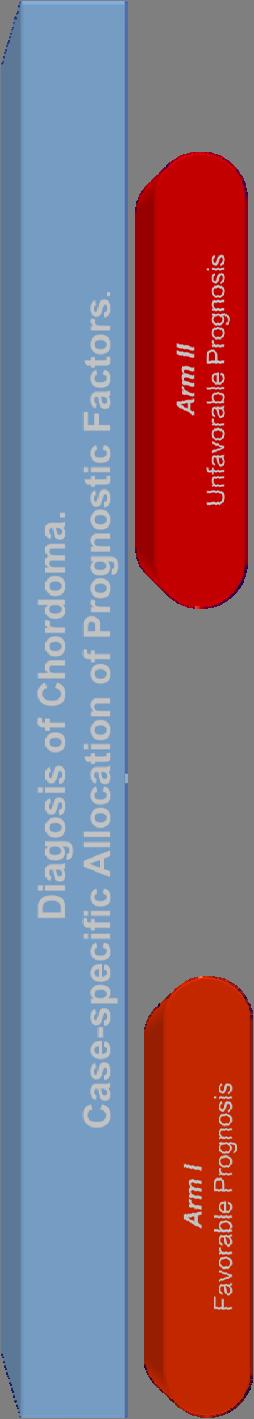

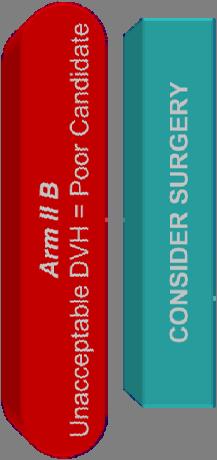

38 Draft-Proposal for a Chordoma Protocol
39 Management of atypical and malignant meningiomas: role of high-dose, 3D-conformal radiation therapy Hug, De Vries et al. J Neurooncol. 2000, 48(2) patients treated at Massachusetts General Hospital: 15 Atypical, 16 Malignant Meningioma Primary Dx: 16 pts., Recurrent: 15 pts. 8 total, 21 pts. subtotal resection, 2 biopsy RT: 15 photons, 16 protons/photons mean F/U time 59 months (range: months) Actuarial local control rates at 5- and 8-years were similar for both histologies: 38% and 19% for Atypical Meningioma 52% and 17% for Malignant Meningioma
40 Management of atypical and malignant meningiomas: role of high-dose, 3D-conformal radiation therapy Hug, De Vries et al. J Neurooncol. 2000, 48(2) Improved Local Control at 5 years: Proton versus Photon RT: 80% versus 17% (p = 0.003) Target doses > or = 60 Gy for both, atypical (p = 0.025) and malignant meningioma (p = ). Actuarial 5- and 8-year survival rates for Malignant Meningioma were significantly improved by use of proton over photon RT and radiation doses > 60 CGE.
41 Proton Radiotherapy for Pediatric Neoplasms involving the Base of Skull
42 HIGH DOSE Proton RT for Pediatric Neoplasms PEDIATRIC Skull Base and Paraspinal Sarcomas Base of Skull Chordomas (Benk, IJROBP, 1995) Base of Skull and Head & Neck mesenchymal tumors ( Willers, Hug et al, IJROBP, 1997) Paraspinal Sarcomas (Hug, IJROBP, ) Pediatric BOS Chordomas (Liebsch, NASBSmeeting, 2001) Pediatric BOS Tumors (Hug et al, IJROBP, 2002)
43 Hug E et al. Proton Radiation Therapy in the management of pediatric base of skull tumors. Int J Radiat Oncol Biol Phys, 52: , 2002 No. of Patients TOTAL 29 Malignant Histology 20 Chordoma 10 Chondrosarcoma 3 Epithelioid Sarcoma 1 Malignant Fibrous Histiocytoma 1 Myxoid Sarcoma 1 Rhabdomyosarcoma 4 Median dose: 70 CGE ( ) No. of Patients TOTAL 29 Benign Histology 9 Giant Cell Tumor 6 Angiofibroma 2 Chondroblastoma 1 Median dose: 60.4 CGE ( )

44 Example: 13 y.o. M with Malignant Fibrous Histiocytoma 20 pts. with Malignant Histology 5-yr LC: 72% 5-Yr OS: 56% 9 pts. Benign Histology LC: 8/9, OS 100%
45 Applications of Proton-Therapy in the Skull Base: Skull Base: Chordomas, Chondrosarcomas Giant Cell Tumors Soft Tissue- and Osteogenic Sarcomas other mesenchymal tumors Nasopharynx CA Paransal Sinus Carcinomas Olfactory Neuroblastoma Adenoid Cystic Carcinoma Pediatric Tumors There is sufficient evidence to recommend Protons for Skull Base Tumors irrespective of Histology
46 Proton-Radiotherapy for Skull Base Tumors: Referral Centers for rare disease Accumulation of large series of patients treated homogeneously Add to understanding of natural history of disease Foster multidisciplinary approach Accomplishes previously unknown CURE in some patients/tumors Understand Prognostic Factors for others Develop new treatment algorithms.
47 EXTERNAL Beam Radiation Therapy for Skull Base Tumors Single Fraction RT (photons) Multiple Fraction RT (photons) Particles RADIOSURGERY (RS) 2D- standard RT 3D-standard RT Stereotactic RT Intensity Modulated RT (IMRT) (IGRT Electrons Neutrons Protons Carbon Ions
48 Gamma-Knife for Skull BaseTumors the Mayo Clinic Experience Radiosurgery for cranial base chordomas and chondrosarcomas Krishnan, Foote et al. Neurosurgery, 2005, 56(4), patients with cranial base chordoma (n = 25) or chondrosarcoma (n = 4) SRS between September 1990 and December Median tumor volume: 14.4 cm3. Median tumor margin dose: 15 Gy (range, Gy); Median maximum radiation dose was 30 Gy (range, Gy). 19 patients had SRS combined with fractionated RT either before or in conjunction (median dose 50 Gy) Median clinical and imaging follow-up periods: 4.8 and 4.5 years
49 Gamma-Knife for Skull BaseTumors the Mayo Clinic Experience Radiosurgery for cranial base chordomas and chondrosarcomas Krishnan, Foote et al. Neurosurgery, 2005, 56(4), RESULTS: CHORDOMAS: Tumor progression: 7 patients (28%) (in-field, n = 3; out-of-field, n = 4),Actuarial tumor control rates: 89% at 2 yrs., 32% at 5 years CHONDROSARCOMAS: Tumor control: All 4 patients COMPLICATIONS: Ten patients (34%) had radiation-related complications. Cranial nerve deficits (n = 6), radiation necrosis (n = 5), and pituitary dysfunction (n = 3). NO complications if SRS alone (10 patients).
50 Gamma-Knife for Skull BaseTumors the Mayo Clinic Experience Radiosurgery for cranial base chordomas and chondrosarcomas Krishnan, Foote et al. Neurosurgery, 2005, 56(4), CONCLUSION by the authors: Cranial base chordomas and chondrosarcomas remain a formidable management challenge. Radiosurgery as an adjunct to surgical resection provides in-field tumor control for some patients, but radiation-related complications are relatively high, especially when radiosurgery is combined with fractionated radiation therapy.
51 Limitations of Radiosurgery: Target Size/ Volume (max. diameter 3-4 cm, risk of side effects increases with size) Location a) proximity to critical structures, i.e. brainstem, chiasm relative indication if abutting or compressing brainstem, no RS for tumors compressing optic chiasm or inside the brainstem) b) inferior limitation of skull base coverage by frame Need to deliver RT to microscopic target BEYOND visible target (malignant tumors) Age (pediatric patient = relative contraindication) These limitations largely do not apply for Protons

52 Proton Radiotherapy of the Skull Base at the Paul Scherrer Institute: The Center for Proton-Radiotherapy
 Horizontal beam line for OPTIS 2 (start 2008) Medical cyclotron Exp.")
53 Layout of the new Center of Proton Radiation Therapy Installation of a dedicated cyclotron (completed) Beam for Gantry 1 for year-round operation (starting fall 2007) patiens treatments restarted in February Second generation compact gantry for scanning: Gantry 2 (start 2008) Horizontal beam line for OPTIS 2 (start 2008) Medical cyclotron Exp. Area (PIF) OPTIS 2 Gantry 2 Disconnect from Ring cyclotron Gantry 1
54 Treatment planning for advanced scanning Work of Alexander Tourovsky PSI-mulate Dose and optimisation module Optimisation options Generalised pencil beam structures (Initial) Re-calculation options Optimisation modes Dose calculation engines Recalculation modes Generalised pencil beam structures (Optimised) Optimised dose distributions Re-calculated dose distributions Generalised pencil beam structures (Recalculated)

 1690 spots/field")
55 Spot weight degeneracy in IMPT Work of Mathias Bosshardt. 2 field IMPT plan (1) 2 field IMPT plan (2) 1690 spots/field 770 spots/field
56 Spot weight degeneracy in IMPT Spot reduction optimisation over 7 cases Work of Mathias Bosshardt. Proportion of spots eliminated % spots eliminated Meningioma Skull base chor. Thoracic chor. Prostate Skull base chor. Prostate Sacral chor. Tumour type Possible advantages: Reduced dynamic range Reduced dead time Reduced utilisation of low energy Bragg peaks
57 Thank you!
Proton Radiation Therapy for Osteosarcomas, Chondrogenic Tumors and Soft Tissue Sarcomas
 Proton Radiation Therapy for Osteosarcomas, Chondrogenic Tumors and Soft Tissue Sarcomas Eugen B. Hug Center for Proton Radiation Therapy Paul Scherrer Institute Histologies Osteogenic Tumors Osteogenic
Proton Radiation Therapy for Osteosarcomas, Chondrogenic Tumors and Soft Tissue Sarcomas
 Proton Radiation Therapy for Osteosarcomas, Chondrogenic Tumors and Soft Tissue Sarcomas Eugen B. Hug Center for Proton Radiation Therapy Paul Scherrer Institute Is there a place for Proton/Particle Radiotherapy
Proton Therapy for tumors of the skull base - RESULTS. Eugen B. Hug, MD Medical Director, ProCure Proton Therapy Centers, NY
 Proton Therapy for tumors of the skull base - RESULTS Eugen B. Hug, MD Medical Director, ProCure Proton Therapy Centers, NY Petroclival Chondrosarcoma: 68 72 Gy(RBE) at 1.8 or 2.0 Gy(RBE) GTV: 70.2 Gy(RBE)
Proton Radiotherapy for Skull Base and Para-spinal Tumors
 Proton Radiotherapy for Skull Base and Para-spinal Tumors Carmen Ares Primary tumors Tumors of the Skull Base - Chordomas, Chondrosarcomas Secondary infiltration or involvement by intracranial tumors -
Effectiveness and Safety of Spot Scanning Proton Radiation Therapy for Skull Base Tumors: First Long Term Report of the PSI Experience
 Effectiveness and Safety of Spot Scanning Proton Radiation Therapy for Skull Base Tumors: First Long Term Report of the PSI Experience Carmen Ares, Antony J Lomax, Eugen B Hug, Alessandra Bolsi, Beate
Particle Therapy for Tumors of the Skull Base. Eugen B. Hug, MD Medical Director, ProCure Proton Therapy Centers, NY
 Particle Therapy for Tumors of the Skull Base Eugen B. Hug, MD Medical Director, ProCure Proton Therapy Centers, NY Particle Radiation Therapy for Tumors of the Skull Base Primary skull base tumors: Chordoma,
Sacral Chordoma: The Loma Linda University Radiation Medicine Experience. Kevin Yiee MD, MPH Resident Physician
 Sacral Chordoma: The Loma Linda University Radiation Medicine Experience Kevin Yiee MD, MPH Resident Physician What is a chordoma? 1 st chordoma discovered in clivus by Virchow and Luschka 1856 Rare tumor
Carbon Ion Radiotherapy for Skull Base and Paracervical Chordomas
 Carbon Ion Radiotherapy for Skull Base and Paracervical Chordomas Azusa Hasegawa, Jun-etsu Mizoe and Hirohiko Tsujii Research Center Hospital for Charged Particle Therapy National Institute of Radiological
Specifics of treatment planning for active scanning and IMPT
 Specifics of treatment planning for active scanning and IMPT SFUD IMPT Tony Lomax, Centre for Proton Radiotherapy, Paul Scherrer Institute, Switzerland Treatment planning for scanning 1. Single Field,
State-of-the-art proton therapy: The physicist s perspective
 State-of-the-art proton therapy: Tony Lomax, Centre for Proton Radiotherapy, Paul Scherrer Institute, Switzerland Overview of presentation 1. State-of-the-art proton delivery 2. Current challenges 3. New
Particle Radiation Therapy: CurrentStatus Indications -Results
 Particle Radiation Therapy: CurrentStatus Indications -Results Eugen B. Hug Center for Proton Therapy Paul Scherrer Institute and University of Zürich Switzerland Particle Radiation Therapy: Selection
PEDIATRIC ORBITAL TUMORS RADIOTHERAPY PLANNING
 PEDIATRIC ORBITAL TUMORS RADIOTHERAPY PLANNING ANATOMY ANATOMY CONT ANATOMY CONT. ANATOMY CONT. EYE OF A CHILD Normal tissue tolerance doses (in conventional #) TD 5/5 TD 50/5 Endpoint Gy Gy Optic nerve
Collection of Recorded Radiotherapy Seminars
 IAEA Human Health Campus Collection of Recorded Radiotherapy Seminars http://humanhealth.iaea.org The Role of Radiosurgery in the Treatment of Gliomas Luis Souhami, MD Professor Department of Radiation
HEAVY PARTICLE THERAPY
 HEAVY PARTICLE THERAPY DR. G.V. GIRI KIDWAI MEMORIAL INSTITUTE OF ONCOLOGY ICRO 2012 BHATINDA HEAVY PARTICLES USED IN A EFFORT TO IMPROVE TUMOR CONTROL, THAT DO NOT RESPOND TO PHOTONS OR ELECTRONS BETTER
Clinical Results of Carbon Ion Radiotherapy: The Heidelberg Experience
 Clinical Results of Carbon Ion Radiotherapy: The Heidelberg Experience Stephanie E. Combs, MD Department of Radiation Oncology University of Heidelberg, Germany Carbon ion RT at GSI Active beam delivery
NCCN GUIDELINES ON PROTON THERAPY (AS OF 4/23/18) BONE (Version , 03/28/18)
 BONE (Version , 03/28/18)") BONE (Version 2.2018, 03/28/18) NCCN GUIDELINES ON PROTON THERAPY (AS OF 4/23/18) Radiation Therapy Specialized techniques such as intensity-modulated RT (IMRT); particle beam RT with protons, carbon ions,
BONE (Version 2.2018, 03/28/18) NCCN GUIDELINES ON PROTON THERAPY (AS OF 4/23/18) Radiation Therapy Specialized techniques such as intensity-modulated RT (IMRT); particle beam RT with protons, carbon ions,
25 Years of Proton Radiation Therapy at PSI an Overview
 25 Years of Proton Radiation Therapy at PSI an Overview Gudrun Goitein for the Team of the Center for Proton Therapy Center for Proton Therapy Paul Scherrer Institut (www.psi.ch) 5232 Villigen PSI Switzerland
Protons for Head and Neck Cancer. William M Mendenhall, M.D.
 Protons for Head and Neck Cancer William M Mendenhall, M.D. Protons for Head and Neck Cancer Potential Advantages: Reduce late complications via more conformal dose distributions Likely to be the major
Treatment Planning (Protons vs. Photons)
") Treatment Planning Treatment Planning (Protons vs. Photons) Acquisition of imaging data Delineation of regions of interest Selection of beam directions Dose calculation Optimization of the plan Hounsfield
Treatment Planning Treatment Planning (Protons vs. Photons) Acquisition of imaging data Delineation of regions of interest Selection of beam directions Dose calculation Optimization of the plan Hounsfield
Proton- Radiotherapy:
 Proton- Radiotherapy: Future of Medical Indications and Treatment Concepts Eugen B. Hug and Ralf A. Schneider HUG 11/07 The emerging role of Proton Radiotherapy in the framework of modern Photon-RT 2000
Clinical Report for Wanjie Proton Therapy Center. Li Jiamin, MD Wanjie Proton Therapy Center
 Clinical Report for Wanjie Proton Therapy Center Li Jiamin, MD Wanjie Proton Therapy Center General Information Wanjie Proton Therapy Center was founded in June 2001 The first patient was treated in Dec.
Proton radiation therapy for chordomas and chondrosarcomas of the skull base
 J Neurosurg 91:432 439, 1999 Proton radiation therapy for chordomas and chondrosarcomas of the skull base EUGEN B. HUG, M.D., LILIA N. LOREDO, M.D., JERRY D. SLATER, M.D., ALEXANDER DEVRIES, M.D., ROGER
Role of protons, heavy ions and BNCT in brain tumors
 Role of protons, heavy ions and BNCT in brain tumors Prof G K Rath Head, NCI (AIIMS-2) Chief, Dr. BRA IRCH, Professor Radiation Oncology All India Institute of Medical Sciences, New Delhi 1 Overview of
Proton- Radiotherapy: Overview of Clinical Indications
 Proton- Radiotherapy: Overview of Clinical Indications Eugen B. Hug (with emphasis on indications treated at PSI For comprehensive clinical reviews: ESTRO or PTCOG seminars) HUG 11/07 Complication Free
PRINCESS MARGARET CANCER CENTRE CLINICAL PRACTICE GUIDELINES
 PRINCESS MARGARET CANCER CENTRE CLINICAL PRACTICE GUIDELINES CENTRAL NERVOUS SYSTEM MENINGIOMA CNS Site Group Meningioma Author: Dr. Norm Laperriere Date: February 20, 2018 1. INTRODUCTION 3 2. PREVENTION
Status of H 1 and C 12
 Status of H 1 and C 12 Herman Suit No Conflict of Interest 1 Goal of a New Treatment Modality Tumor Control Probability or No in Complication Rate 2 Truism No Advantage to: any Patient for any Radiation
IMPT with Carbon Ions
 IMPT with Carbon Ions PTCOG 48, Heidelberg, 28.09.-03.10.2009 Malte Ellerbrock Medical Physics Expert Heidelberg Ion-Beam Therapy Center HIT Betriebs GmbH am Universitätsklinikum Heidelberg http://www.hit-centrum.de
-Proton Beam Therapy in Paediatric Radiation Oncology -
 -Proton Beam Therapy in Paediatric Radiation Oncology - Beate Timmermann, M.D. West German Proton Therapy Centre Essen Germany Preview Survival Toxicity Why protons? (theoretically) Experiences so far
Feasibility of Proton Beam Therapy for Chordoma and Chondrosarcoma of the Skull Base
 ORIGINAL ARTICLE Feasibility of Proton Beam Therapy for Chordoma and Chondrosarcoma of the Skull Base Hiroshi Fuji, M.D., Ph.D., 1 Yoko Nakasu, M.D., Ph.D., 2 Yuji Ishida, M.D., Ph.D., 3 Satoshi Horiguchi,
Radiation Technology, Hyogo Ion Beam Medical Center, Tatsuno, Hyogo, JAPAN
 Analysis of Visual Loss Due to Radiation- Induced Optic Neuropathy After Particle Therapy for Head and Neck and Skull Base Tumors Adjacent to Optic Nerves Y. Demizu 1, M. Murakami 1, D. Miyawaki 1, Y.
Neutron Radiotherapy: Past, Present, and Future Directions
 Neutron Radiotherapy: Past, Present, and Future Directions Theodore L. Phillips Lecture -- 2014 George E. Laramore Ph.D., M.D. NONE Conflicts of Interest Peter Wootton Professor of Radiation Oncology University
Proton Therapy for Ependymoma and Craniopharyngioma at the University of Florida: Lessons Learned About Efficacy and Toxicity
 Proton Therapy for Ependymoma and Craniopharyngioma at the University of Florida: Lessons Learned About Efficacy and Toxicity Danny Indelicato, MD Mendenhall Chair of Pediatric Radiotherapy University
Advances in external beam radiotherapy
 International Conference on Modern Radiotherapy: Advances and Challenges in Radiation Protection of Patients Advances in external beam radiotherapy New techniques, new benefits and new risks Michael Brada
Current Status and Future Medical Perspectives at MedAustron. U. Mock EBG MedAustron GmbH
 Current Status and Future Medical Perspectives at MedAustron U. Mock EBG MedAustron GmbH Cancer treatment facility Ion beam therapy with protons and carbon ions Research facility Medical physics Radiobiology
Chapter 5 Section 3.1
 Radiology Chapter 5 Section 3.1 Issue Date: March 27, 1991 Authority: 32 CFR 199.4(b)(2), (b)(2)(x), (c)(2)(viii), and (g)(15) 1.0 CPT 1 PROCEDURE CODES 37243, 61793, 61795, 77261-77421, 77427-77799, 0073T
Disclosures. Proton therapy advantages. Why are comparing therapies difficult? Proton Therapy for Low Risk Prostate Cancer
 Proton Therapy for Low Risk Prostate Cancer Disclosures No relevant financial disclosures This presentation will not discuss off-label or investigational treatments Andrew K. Lee, MD, MPH Associate Professor
Summary Talk of the Workshop
 Medical experience History of IBT 1954 1975 1976 1977 1982 1987 1992 Pituitary 1st He pt Treatment 1st C, Ne pt Eye treatment Phase-1 He Phase I-II Ne Phase I-II Ne & He 1st Comp Tx Plan 3D planning LBNL
8/3/2016. Clinical Significance of RBE Variations in Proton Therapy. Why RBE (relative biological effectiveness)?
?") 8//06 Clinical Significance of Variations in Proton Therapy H. Paganetti PhD Professor, Harvard Medical School Director of Physics Research, Massachusetts General Hospital, Radiation Oncology Introduction
8//06 Clinical Significance of Variations in Proton Therapy H. Paganetti PhD Professor, Harvard Medical School Director of Physics Research, Massachusetts General Hospital, Radiation Oncology Introduction
Potential benefits of intensity-modulated proton therapy in head and neck cancer van de Water, Tara Arpana
 University of Groningen Potential benefits of intensity-modulated proton therapy in head and neck cancer van de Water, Tara Arpana IMPORTANT NOTE: You are advised to consult the publisher's version (publisher's
Corporate Medical Policy
 Corporate Medical Policy File Name: Origination: Last CAP Review: Next CAP Review: Last Review: charged_particle_radiotherapy 3/12/96 5/2017 5/2018 5/2017 Description of Procedure or Service Charged-particle
Clinical Concept and History of Protons. Relevance and Limitations of Conformality. Gudrun Goitein
 Clinical Concept and History of Protons Relevance and Limitations of Conformality Gudrun Goitein PSI Winter School January 2010 Bad Zurzach and PSI, Villigen Switzerland P + Who came first: The Clinical
Case Conference: SBRT for spinal metastases D A N I E L S I M P S O N M D 3 / 2 7 / 1 2
 Case Conference: SBRT for spinal metastases D A N I E L S I M P S O N M D 3 / 2 7 / 1 2 Case 79 yo M with hx of T3N0 colon cancer diagnosed in 2008 metastatic liver disease s/p liver segmentectomy 2009
Proton Stereotactic Radiotherapy: Clinical Overview. Brian Winey, Ph.D. Physicist, MGH Assistant Professor, HMS
 Proton Stereotactic Radiotherapy: Clinical Overview Brian Winey, Ph.D. Physicist, MGH Assistant Professor, HMS Acknowledgements Radiation Oncologists and Physicists at various institutions (MGH, MDACC,
Image-guided, intensity-modulated radiation therapy (IG-IMRT) for skull base chordoma and chondrosarcoma: preliminary outcomes
 for skull base chordoma and chondrosarcoma: preliminary outcomes") Neuro-Oncology Neuro-Oncology 17(6), 889 894, 2015 doi:10.1093/neuonc/nou347 Advance Access date 27 December 2014 Image-guided, intensity-modulated radiation therapy (IG-IMRT) for skull base chordoma and
Neuro-Oncology Neuro-Oncology 17(6), 889 894, 2015 doi:10.1093/neuonc/nou347 Advance Access date 27 December 2014 Image-guided, intensity-modulated radiation therapy (IG-IMRT) for skull base chordoma and
The Heidelberg Ion Therapy Center and PARTNER. Thomas Haberer Heidelberg Ion Therapy Center
 The Heidelberg Ion Therapy Center and PARTNER Thomas Haberer Heidelberg Ion Therapy Center Goal The key element to improve the clinical outcome is local control! entrance channel: low physical dose low
Characterization and implementation of Pencil Beam Scanning proton therapy techniques: from spot scanning to continuous scanning
 Characterization and implementation of Pencil Beam Scanning proton therapy techniques: from spot scanning to continuous scanning Supervisors Prof. V. Patera PhD R. Van Roermund Candidate Annalisa Patriarca
PTCOG 46. Educational Workshop Session IV. Head & Neck CLINICAL. J. Mizoe (NIRS, Japan)
") PTCOG 46 Educational Workshop Session IV CLINICAL Head & Neck J. Mizoe (NIRS, Japan) Photon X-Ray γ-ray Fast Neutron Non-Charged Radiation Electron Proton Helium Light Ion Heavy Particle Carbon Neon Argon
PTCOG 46 Educational Workshop Session IV CLINICAL Head & Neck J. Mizoe (NIRS, Japan) Photon X-Ray γ-ray Fast Neutron Non-Charged Radiation Electron Proton Helium Light Ion Heavy Particle Carbon Neon Argon
Radiotherapy and tumours in veterinary practice: part one
 Vet Times The website for the veterinary profession https://www.vettimes.co.uk Radiotherapy and tumours in veterinary practice: part one Author : Aleksandra Marcinowska, Jane Dobson Categories : Companion
Treatment Planning for Skull Base Tumors PTCOG 52, June 2013
 Treatment Planning for Skull Base Tumors PTCOG 52, June 2013 Judy Adams CMD Hanne Kooy Ph.D, Norbert Liebsch MD Department of Radiation Oncology Massachusetts General Hospital Chordomas and Chondrosarcomas
PRINCESS MARGARET CANCER CENTRE CLINICAL PRACTICE GUIDELINES
 PRINCESS MARGARET CANCER CENTRE CLINICAL PRACTICE GUIDELINES CENTRAL NERVOUS SYSTEM GERM CELL TUMOURS CNS Site Group Germ Cell Tumours Author: Dr. Norm Laperriere Date: February 20, 2018 1. INTRODUCTION
Clinical considerations of RBE in proton therapy
 Clinical considerations of RBE in proton therapy H. Paganetti PhD Professor, Harvard Medical School Director of Physics Research, Massachusetts General Hospital, Radiation Oncology Why do we need the RBE
Sarcoma and Radiation Therapy. Gabrielle M Kane MB BCh EdD FRCPC Muir Professorship in Radiation Oncology University of Washington
 Sarcoma and Radiation Therapy Gabrielle M Kane MB BCh EdD FRCPC Muir Professorship in Radiation Oncology University of Washington Objective: Helping you make informed decisions Introduction Process Radiation
Future Directions in Prostate Cancer: The Case for Protons. John J. Coen, MD Helen & Harry Gray Cancer Center
 Future Directions in Prostate Cancer: The Case for Protons John J. Coen, MD Helen & Harry Gray Cancer Center November 14, 2012 Protons and prostate cancer Early proton experience at the MGH The case for
The role of Radiation Oncologist: Hi-tech treatments for liver metastases
 The role of Radiation Oncologist: Hi-tech treatments for liver metastases Icro Meattini, MD Radiotherapy-Oncology Unit AOU Careggi Hospital Florence University, Italy Liver Metastases - Background The
Radiotherapy for intracranial meningiomas SAMO Interdisciplinary Workshop on Brain Tumors and Metastases November 2016
 WIR SCHAFFEN WISSEN HEUTE FÜR MORGEN PD Dr Alessia Pica, Pr Damien Charles Weber: Paul Scherrer Institut Radiotherapy for intracranial meningiomas SAMO Interdisciplinary Workshop on Brain Tumors and Metastases
PRINCESS MARGARET CANCER CENTRE CLINICAL PRACTICE GUIDELINES
 PRINCESS MARGARET CANCER CENTRE CLINICAL PRACTICE GUIDELINES CENTRAL NERVOUS SYSTEM EPENDYMOMA Last Revision Date July 2015 1 CNS Site Group Ependymoma Author: Dr. Norm Laperriere 1. INTRODUCTION 3 2.
TOMOTERAPIA in Italia: Esperienze a confronto
 TOMOTERAPIA in Italia: Esperienze a confronto BARD 20 novembre 2010 L esperienza di Reggio Emilia Testa collo Alessandro Muraglia Reasons for the use of tomotherapy: - Complex tumor geometry and proximity
Page 1. Helical (Spiral) Tomotherapy. UW Helical Tomotherapy Unit. Helical (Spiral) Tomotherapy. MVCT of an Anesthetized Dog with a Sinus Tumor
 Tomotherapy. UW Helical Tomotherapy Unit. Helical (Spiral) Tomotherapy. MVCT of an Anesthetized Dog with a Sinus Tumor") Helical (Spiral) Tomotherapy Novel Clinical Applications of IMRT Linac Ring Gantry CT Detector X-Ray Fan Beam Binary Multileaf Collimator Binary MLC Leaves James S Welsh, MS, MD Department of Human Oncology
Helical (Spiral) Tomotherapy Novel Clinical Applications of IMRT Linac Ring Gantry CT Detector X-Ray Fan Beam Binary Multileaf Collimator Binary MLC Leaves James S Welsh, MS, MD Department of Human Oncology
Application of Implanted Markers in Proton Therapy. Course Outline. McLaren Proton Therapy Center Karmanos Cancer Institute McLaren - Flint
 Application of Implanted Markers in Proton Therapy Sung Yong Park, Ph.D. McLaren Proton Therapy Center Karmanos Cancer Institute McLaren - Flint AAPM 2016, SAM Therapy Educational Course, 2016.08.04. Course
Pediatrics -Proton Beam Therapy in Children -
 Pediatrics -Proton Beam Therapy in Children - Beate Timmermann, M.D. West German Proton Therapy Centre Essen Germany Preview Survival Toxicity Why protons? (theoretically) Experiences so far (clinically)
Proton Therapy in Neurosurgery: A Historical Review and Future Perspective Based on Currently Available New Generation Systems
 Review Article Proton Therapy in Neurosurgery: A Historical Review and Future Perspective Based on Currently Available New Generation Systems Parisa Azimi 1, Amir Movafeghi 2 1 Functional Neurosurgery
Overview. Proton Therapy in lung cancer 8/3/2016 IMPLEMENTATION OF PBS PROTON THERAPY TREATMENT FOR FREE BREATHING LUNG CANCER PATIENTS
 IMPLEMENTATION OF PBS PROTON THERAPY TREATMENT FOR FREE BREATHING LUNG CANCER PATIENTS Heng Li, PhD Assistant Professor, Department of Radiation Physics, UT MD Anderson Cancer Center, Houston, TX, 773
This LCD recognizes these two distinct treatment approaches and is specific to treatment delivery:
 National Imaging Associates, Inc. Clinical guidelines STEREOTACTIC RADIOSURGERY (SRS) AND STEREOTACTIC BODY RADIATION THERAPY (SBRT) CPT4 Codes: 77371, 77372, 77373 LCD ID Number: L33410 J-N FL Responsible
FROM ICARO1 TO ICARO2: THE MEDICAL PHYSICS PERSPECTIVE. Geoffrey S. Ibbott, Ph.D. June 20, 2017
 FROM ICARO1 TO ICARO2: THE MEDICAL PHYSICS PERSPECTIVE Geoffrey S. Ibbott, Ph.D. June 20, 2017 1 DISCLOSURES My institution holds Strategic Partnership Research Agreements with Varian, Elekta, and Philips
National Horizon Scanning Unit Horizon scanning prioritising summary
 National Horizon Scanning Unit Horizon scanning prioritising summary Volume 13, Number 7: Proton Beam Therapy for the treatment of cancer June 2006 Commonwealth of Australia 2006 [add ISSN] [add Publications
Stereotactic radiotherapy
 Stereotactic radiotherapy Influence of patient positioning and fixation on treatment planning - clinical results Frank Zimmermann Institut für Radioonkologie Universitätsspital Basel Petersgraben 4 CH
PRINCESS MARGARET CANCER CENTRE CLINICAL PRACTICE GUIDELINES
 PRINCESS MARGARET CANCER CENTRE CLINICAL PRACTICE GUIDELINES CENTRAL NERVOUS SYSTEM LOW GRADE GLIOMAS CNS Site Group Low Grade Gliomas Author: Dr. Norm Laperriere 1. INTRODUCTION 3 2. PREVENTION 3 3. SCREENING
Evaluation of Monaco treatment planning system for hypofractionated stereotactic volumetric arc radiotherapy of multiple brain metastases
 Evaluation of Monaco treatment planning system for hypofractionated stereotactic volumetric arc radiotherapy of multiple brain metastases CASE STUDY Institution: Odette Cancer Centre Location: Sunnybrook
Case Report Carbon Ion Beam Radiotherapy for Sinonasal Malignant Tumors Invading Skull Base
 Case Reports in Otolaryngology, Article ID 241856, 4 pages http://dx.doi.org/10.1155/2014/241856 Case Report Carbon Ion Beam Radiotherapy for Sinonasal Malignant Tumors Invading Skull Base Nobuo Ohta,
11/27/2017. Modern Treatment of Meningiomas. Disclosures. Modern is Better? No disclosures relevant to this presentation
 Modern Treatment of Meningiomas Michael A. Vogelbaum MD, PhD Professor of Neurosurgery Cleveland Clinic Disclosures No disclosures relevant to this presentation IP and royalties related to drug and device
PRINCESS MARGARET CANCER CENTRE CLINICAL PRACTICE GUIDELINES
 PRINCESS MARGARET CANCER CENTRE CLINICAL PRACTICE GUIDELINES CENTRAL NERVOUS SYSTEM BRAIN METASTASES CNS Site Group Brain Metastases Author: Dr. Norm Laperriere Date: February 20, 2018 1. INTRODUCTION
ORIGINAL ARTICLE GAMMA KNIFE STEREOTACTIC RADIOSURGERY FOR SALIVARY GLAND NEOPLASMS WITH BASE OF SKULL INVASION FOLLOWING NEUTRON RADIOTHERAPY
 ORIGINAL ARTICLE GAMMA KNIFE STEREOTACTIC RADIOSURGERY FOR SALIVARY GLAND NEOPLASMS WITH BASE OF SKULL INVASION FOLLOWING NEUTRON RADIOTHERAPY James G. Douglas, MD, MS, 1,2 Robert Goodkin, MD, 1,2 George
Stereotactic Radiosurgery. Extracranial Stereotactic Radiosurgery. Linear accelerators. Basic technique. Indications of SRS
 Stereotactic Radiosurgery Extracranial Stereotactic Radiosurgery Annette Quinn, MSN, RN Program Manager, University of Pittsburgh Medical Center Using stereotactic techniques, give a lethal dose of ionizing
Overview of MLC-based Linac Radiosurgery
 SRT I: Comparison of SRT Techniques 1 Overview of MLC-based Linac Radiosurgery Grace Gwe-Ya Kim, Ph.D. DABR 2 MLC based Linac SRS Better conformity for irregular target Improved dose homogeneity inside
DOES RADIOTHERAPY TECHNIQUE / DOSE / FRACTIONATION REALLY MATTER? YES
 DOES RADIOTHERAPY TECHNIQUE / DOSE / FRACTIONATION REALLY MATTER? YES Marco Krengli Radiotherapy, Department of Translational Medicine, University of Piemonte Orientale A. Avogadro THE STANDARD OF CARE
Otolaryngologist s Perspective of Stereotactic Radiosurgery
 Otolaryngologist s Perspective of Stereotactic Radiosurgery Douglas E. Mattox, M.D. 25 th Alexandria International Combined ORL Conference April 18-20, 2007 Acoustic Neuroma Benign tumor of the schwann
Reirradiazione. La radioterapia stereotassica ablativa: torace. Pierluigi Bonomo Firenze
 Reirradiazione La radioterapia stereotassica ablativa: torace Pierluigi Bonomo Firenze Background Stage III NSCLC isolated locoregional recurrence in 25% of pts mostly unresectable; low RR with 2 nd line
Insights into Thymic Epithelial Tumors: Radiation Therapy
 Insights into Thymic Epithelial Tumors: Radiation Therapy Charles R. Thomas, MD Professor and Chairman, Department of Radiation Medicine Professor, Department of Medicine, Division of Hematology/Medical
Evaluation of Whole-Field and Split-Field Intensity Modulation Radiation Therapy (IMRT) Techniques in Head and Neck Cancer
 Techniques in Head and Neck Cancer") 1 Charles Poole April Case Study April 30, 2012 Evaluation of Whole-Field and Split-Field Intensity Modulation Radiation Therapy (IMRT) Techniques in Head and Neck Cancer Abstract: Introduction: This study
1 Charles Poole April Case Study April 30, 2012 Evaluation of Whole-Field and Split-Field Intensity Modulation Radiation Therapy (IMRT) Techniques in Head and Neck Cancer Abstract: Introduction: This study
PRINCESS MARGARET CANCER CENTRE CLINICAL PRACTICE GUIDELINES
 PRINCESS MARGARET CANCER CENTRE CLINICAL PRACTICE GUIDELINES CENTRAL NERVOUS SYSTEM ANAPLASTIC GLIOMAS CNS Site Group Anaplastic Gliomas Author: Dr. Norm Laperriere Date: February 20, 2018 1. INTRODUCTION
Radiotherapy approaches to pituitary tumors
 Disclosures No relevant disclosures Radiotherapy approaches to pituitary tumors Pituitary Disorders: Advances in Diagnosis and Management Steve Braunstein, MD, PhD UCSF Department of Radiation Oncology
SBRT in early stage NSCLC
 SBRT in early stage NSCLC Optimal technique and tumor dose Frank Zimmermann Clinic of Radiotherapy and Radiation Oncology University Hospital Basel Petersgraben 4 CH 4031 Basel radioonkologiebasel.ch Techniques
Charged-Particle (Proton or Helium Ion) Radiation Therapy. Original Policy Date
 Radiation Therapy. Original Policy Date") MP 8.01.08 Charged-Particle (Proton or Helium Ion) Radiation Therapy Medical Policy Section Therapy Issue 12/2013 Original Policy Date 12/2013 Last Review Status/Date Reviewed with literature search/12/2013
MP 8.01.08 Charged-Particle (Proton or Helium Ion) Radiation Therapy Medical Policy Section Therapy Issue 12/2013 Original Policy Date 12/2013 Last Review Status/Date Reviewed with literature search/12/2013
Refresher Course EAR TUMOR. Sasikarn Chamchod, MD Chulabhorn Hospital
 Refresher Course EAR TUMOR Sasikarn Chamchod, MD Chulabhorn Hospital Reference: Perez and Brady s Principles and Practice of radiation oncology sixth edition Outlines Anatomy Epidemiology Clinical presentations
Radiosurgery by Leksell gamma knife. Josef Novotny, Na Homolce Hospital, Prague
 Radiosurgery by Leksell gamma knife Josef Novotny, Na Homolce Hospital, Prague Radiosurgery - Definition Professor Lars Leksell The tools used by the surgeon must be adapted to the task and where the human
Protocol of Radiotherapy for Head and Neck Cancer
 106 年 12 月修訂 Protocol of Radiotherapy for Head and Neck Cancer Indication of radiotherapy Indication of definitive radiotherapy with or without chemotherapy (1) Resectable, but medically unfit, or high
Changing Paradigms in Radiotherapy
 Changing Paradigms in Radiotherapy Marco van Vulpen, MD, PhD Mouldroomdag-2015 Towards the elimination of invasion 1 NIH opinion on the future of oncology Twenty-five years from now,i hope that we won
State of the Art Radiotherapy for Pediatric Tumors. Suzanne L. Wolden, MD Memorial Sloan-Kettering Cancer Center
 State of the Art Radiotherapy for Pediatric Tumors Suzanne L. Wolden, MD Memorial Sloan-Kettering Cancer Center Introduction Progress and success in pediatric oncology Examples of low-tech and high-tech
Impact of Gamma Knife Radiosurgery on the neurosurgical management of skull-base lesions: The Combined Approach
 Radiosurgery as part of the neurosurgical armamentarium: Educational Symposium November 24 th 2011 Impact of Gamma Knife Radiosurgery on the neurosurgical management of skull-base lesions: The Combined
IMRT - the physician s eye-view. Cinzia Iotti Department of Radiation Oncology S.Maria Nuova Hospital Reggio Emilia
 IMRT - the physician s eye-view Cinzia Iotti Department of Radiation Oncology S.Maria Nuova Hospital Reggio Emilia The goals of cancer therapy Local control Survival Functional status Quality of life Causes
Treatment Planning Evaluation of Volumetric Modulated Arc Therapy (VMAT) for Craniospinal Irradiation (CSI)
 for Craniospinal Irradiation (CSI)") Treatment Planning Evaluation of Volumetric Modulated Arc Therapy (VMAT) for Craniospinal Irradiation (CSI) Tagreed AL-ALAWI Medical Physicist King Abdullah Medical City- Jeddah Aim 1. Simplify and standardize
Treatment Planning Evaluation of Volumetric Modulated Arc Therapy (VMAT) for Craniospinal Irradiation (CSI) Tagreed AL-ALAWI Medical Physicist King Abdullah Medical City- Jeddah Aim 1. Simplify and standardize
Helical Tomotherapy Experience. TomoTherapy Whole Brain Head & Neck Prostate Lung Summary. HI-ART TomoTherapy System. HI-ART TomoTherapy System
 The Challenges Associated with Differential Dose Delivery using IMRT Chester Ramsey, Ph.D. Director of Medical Physics Thompson Cancer Center Knoxville, Tennessee, U.S.A Collaborators Chester Ramsey, Ph.D.
Proton and heavy ion radiotherapy: Effect of LET
 Proton and heavy ion radiotherapy: Effect of LET As a low LET particle traverses a DNA molecule, ionizations are far apart and double strand breaks are rare With high LET particles, ionizations are closer
A Comparison of IMRT and VMAT Technique for the Treatment of Rectal Cancer
 A Comparison of IMRT and VMAT Technique for the Treatment of Rectal Cancer Tony Kin Ming Lam Radiation Planner Dr Patricia Lindsay, Radiation Physicist Dr John Kim, Radiation Oncologist Dr Kim Ann Ung,
Potential benefits of intensity-modulated proton therapy in head and neck cancer van de Water, Tara Arpana
 University of Groningen Potential benefits of intensity-modulated proton therapy in head and neck cancer van de Water, Tara Arpana IMPORTANT NOTE: You are advised to consult the publisher's version (publisher's
Outline. WBRT field. Brain Metastases. Whole Brain RT Prophylactic WBRT Stereotactic radiosurgery (SRS) 1 fraction Stereotactic frame
 1 fraction Stereotactic frame") Radiation Therapy for Advanced NSC Lung Ca Alexander Gottschalk, M.D., Ph.D. Associate Professor Director of CyberKnife Radiosurgery Department of Radiation Oncology University of California San Francisco
Radiation Therapy for Advanced NSC Lung Ca Alexander Gottschalk, M.D., Ph.D. Associate Professor Director of CyberKnife Radiosurgery Department of Radiation Oncology University of California San Francisco
Original Date: April 2016 Page 1 of 7 FOR CMS (MEDICARE) MEMBERS ONLY
 MEMBERS ONLY") National Imaging Associates, Inc. Clinical guidelines STEREOTACTIC RADIATION THERAPY: STEREO RADIOSURGERY (SRS) AND STEREOTACTIC BODY RADIATION THERAPY (SBRT) CPT4 Codes: Please refer to pages 5-6 LCD
National Imaging Associates, Inc. Clinical guidelines STEREOTACTIC RADIATION THERAPY: STEREO RADIOSURGERY (SRS) AND STEREOTACTIC BODY RADIATION THERAPY (SBRT) CPT4 Codes: Please refer to pages 5-6 LCD
UCSF Uveal Melanoma Program: Outcomes with Proton Beam Radiation Therapy Kavita K. Mishra, M.D., M.P.H. UCSF Comprehensive Cancer Center
 Disclosures UCSF Uveal Melanoma Program: Outcomes with Proton Beam Radiation Therapy No disclosures Kavita K. Mishra, M.D., M.P.H. UCSF Comprehensive Cancer Center UCSF Uveal Melanoma Program: Ocular Melanoma
R. Harding, P. Trnková, S. J. Weston, J. Lilley, C. M. Thompson, S. C. Short, C. Loughrey, V. P. Cosgrove, A. J. Lomax, and D. I.
 Benchmarking of a treatment planning system for spot scanning proton therapy: Comparison and analysis of robustness to setup errors of photon IMRT and proton SFUD treatment plans of base of skull meningioma
Disclosures. Overview 8/3/2016. SRS: Cranial and Spine
 SRS: Cranial and Spine Brian Winey, Ph.D. Department of Radiation Oncology Massachusetts General Hospital Harvard Medical School Disclosures Travel and research funds from Elekta Travel funds from IBA