Advances in Ocular Imaging
|
|
|
- Kristian Howard
- 5 years ago
- Views:
Transcription


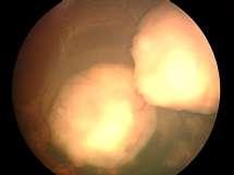
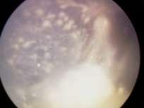
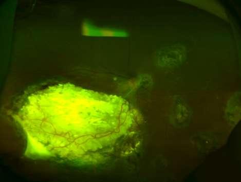
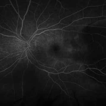
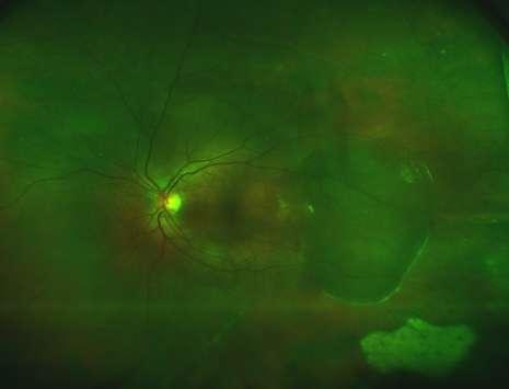
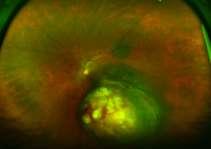
1 Wide angle fundus imaging and Fuorescein angiography in evaluation and management of intraocular tumors Ihab Saad Othman, MD, FRCS Professor of Ophthalmology Cairo University Cairo, Egypt Advances in Ocular Imaging Kowa Topcon Retcam 1

.")


2 Wide-Field (Digital) Imaging. Field-of-view: The instantaneous field-of -view (FOV). Field-of-regard (FOR): Total field of imaging available with camera. Ora serrata Equator ORA SERRATA EQUATOR Standard fundus camera field-of-view (FOV) What would we do differently if we could remotely image the entire eye? Wide Angle Imaging Retcam Panoret Optos 2


3 Wide angle viewing: Staging of Retinoblastoma Reese-Ellsworth Staging system (1963): Predicts visual prognosis with external beam radiotherapy Group I: Very favorable prognosis: Single tumor, 4 DD at or behind the equator. Multiple tumors, 4 DD at or behind the equator Group II: favorable prognosis: Solitary tumor, 4-10 DD at or behind the equator. Multiple tumors, 4-10 DD at or behind the equator. Reese-Ellsworth Staging system Group III: doubtful prognosis: Any lesion anterior to the equator Solitary tumors larger than 10 disc diameters behind the equator Group IV: unfavorable prognosis: Multiple tumors, some >10 DD Any lesion extending anteriorly to ora serrata. 3


4 Reese-Ellsworth Staging system Group V: very unfavorable prognosis: Massive tumor involving over half the retina. Vitreous seeding. Retinoblastoma Staging: International classification of retinoblastoma (2003) is based on the rate of ocular salvage with systemic chemotherapy A B Very favorable. Tumor confined to C the retina D Unfavorable. Tumor dispersed to adjacent spaces E Unsalvageable 4


5 Retinoblastoma Staging: International classification of retinoblastoma Group A: Tumor Not more than 3mm Located more than 1.5 mm from optic disc and 3 mm from fovea Retinoblastoma International classification of retinoblastoma Group B: Tumors confined to the retina 10mm Any location No subretinal fluid beyond 3 mm from the base of the tumor 5


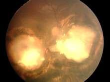
6 Retinoblastoma Group C: Any size tumor < ½ the eye Subtle dispersion Fine vitreous seeds No dispersed tumor masses Subretinal seeds less than 3 mm from the tumor Retinoblastoma Group D: Diffuse disease with significant vitreous or subretinal seeding Subretinal fluid in more than one quadrant of the retina Subretinal fluid, present or past, without seeding, involving up to total retinal detachment 6


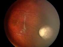
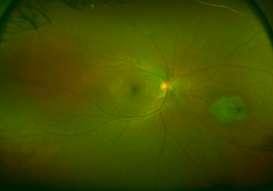
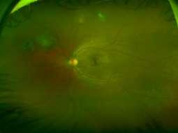
7 Group D: Tumor(s) may be massive or diffuse Diffuse subretinal seeding, present or past, may include subretinal plaques or tumor nodules Diffuse or massive vitreous disease may include "greasy" seeds or avascular tumor masses Wide Angle Viewing Extremely valuable in: Treatment monitoring 7
8 8

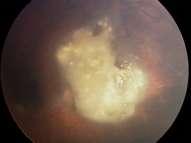
9 Documents Plaque Centration IOAM, 3 cycles 9

10 IOAM, 3 cycles 10


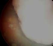
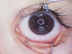
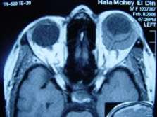
11 5 months follow up: Subretinal seeds recurrence/ Enucleation Direct Gonioscopy: Case: 1.5 year-old male Ciliochoroidal mass: PLSU 11


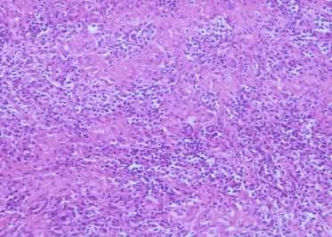
12 Case: 1.5 year-old male Ciliochoroidal mass: PLSU Pathology: Ectopic lacrimal gland Direct Gonioscopy: Medulloepithelioma 12


13 CB masses 18 YOM CB mass followed for the last 4 years Growing on UBM UBM: AP UBM: Transverse 13


14 Differential Diagnosis Adenoma (non-pigmented ciliary epithelium) Medulloepithelioma Metastasis Melanoma Transcleral Resection 14

15 Specimen Specimen 15

16 Granulaomatous inflammation Non-caseating ZN-negative?? Sarcoidosis, chest X-ray normal, ACE normal Opto Tx


17 Follow up of Pediatric age group > 3 years vs GA 8 YOF VA: 6/24 17


18 4 YOF VA: 6/6 10 YOF VA: 6/24 18
19 5 YOM Monophthalmic VA: 6/36 32 YOF Mom 19


20 Choroidal Tumors Diagnosis and Management Choroidal Melanoma: Most common primary intraocular malignancy in adults Dendritic melanocytes (neural crest origin) 20


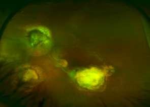
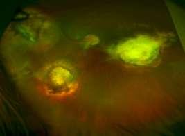
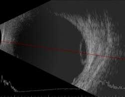
21 Opaque media Initial evaluation of thickness Follow up of treatment response A-scan: Low to medium reflectivity B-scan: Acoustic hollowness, choroidal excavation, + angle kappa 21


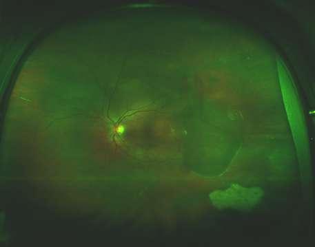
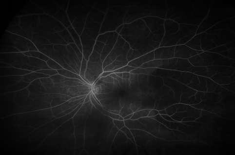
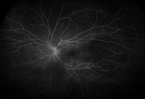
22 45 year-old-female with choroidal melanoma 16 YOF Peripheral choroidal mass Wide Angle FF/A 22

23 23


24 63 YOF: Melanoma Post-Ru 106 / TTT 24


25 Tumor Extent 55 YOF Enucleation Autofluorescence 25


26 48 YOM Tumor control post PLSU/TTT 2 months VA: 6/36 26


27 18 month VA: 6/9 28 YOF PLSU/TTT 3 months VA: 6/36 27


28 1 year VA: 6/9 28


29 Choroidal Metastases 4 7 year-old female with multiple breast metastases FNAB: Highly reliable Uncertain diagnosis Difficult technique Experienced cytopathologist 29


30 72 year-old female with choroidal mass: unknown primary Classically: FNAB: indirect ophthalmoscopy: Inverted image Laterally rotated image Mastering and meticulous manipulations Hundreds of cases with a steep learning curve 30


31 Interventional Photo-guided choroidal biopsy Retcam Interventional Photo-guided choroidal biopsy Adenocarcinoma 31


32 57 YOF DVA OS for 3 months 32

33 Lymphoma: 60 YOF FNAB Plasmacytoma in a 65 YOF 33
34 Hemorrhagic Lesions Simulating Melanoma 58 year-old female Choroidal Mass OS x 1 month VA: 6/18 34
35 35

36 36
37 37

38 Vertical Transverse 38

39 Revise History: Discussion New Investigative tools: Wide Angle FF/A is a primordial tool in management of intraocular tumors Paradigm shift with a whole chapter in panoramic view and peripheral fundus changes In tumors: Early diagnosis and timely intervention is imperative in SURVIVAL 39

40 Remember: WHEN DEALING WITH CANCER YOU ARE DELAING WITH LETHAL DISEASE YOU ARE DELAING WITH HUMAN LIFE NO ROOM FOR TRIAL AND ERROR EYE SALVAGE WILL DEPEND ON PROPER DIAGNOSIS AND SOUND MANAGEMENT EVERY EFFORT IS JUSTIFIED TO SAVE AN EYE WITHOUT COMPROMISING LIFE Wide angle fundus Digital Screening: Retcam Ihab S Othman, Consultant Ophthalmic Oncologist and VR Surgeon 40


41 Opto Tx 200 Opto Tx
42 42
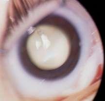
Complicated Cataract to Intraocular Tumors, Beware of the unexpected
 Complicated Cataract to Intraocular Tumors, Beware of the unexpected Ihab Saad Othman, MD, FRCS Professor of Ophthalmology Cairo University In this part of the world: We Master Phakoemulsification 1 Intraoperative/Second
Retina Center of Oklahoma Sam S. Dahr, M.D. Adult Intraocular Tumors
 Adult Intraocular Tumors Sam S. Dahr, M.D. Retina Center of Oklahoma www.retinacenteroklahoma.com www.rcoklahoma.com Table of Contents Posterior uveal malignant melanoma Uveal metastasis Uveal melanoma
Outline. Brief history and principles of ophthalmic ultrasound. Types of ocular ultrasound. Examination techniques. Types of Ultrasound
 Ultrasound and Intraocular Tumors 2015 Ophthalmic Photographers' Society Mid-Year Program Cagri G. Besirli MD, PhD Kellogg Eye Center University of Michigan Outline Brief history and principles of ophthalmic
INDIAN COUNCIL OF MEDICAL RESEARCH INDIAN RETINOBLASTOMA GROUP
 NATIONAL RETINOBLASTOMA REGISTRY INDIAN COUNCIL OF MEDICAL RESEARCH INDIAN RETINOBLASTOMA GROUP Centre Code 01. Dr. Rajendra Parasad Centre for Ophthalmic Sciences 02. L.V. Prasad Eye Institute, Hyderabad
J of Evolution of Med and Dent Sci/ eissn , pissn / Vol. 4/ Issue 55/ July 09, 2015 Page 9665
 RARE PRESENTATION OF BILATERAL CHOROIDAL METASTASIS FROM PRIMARY MUCO-EPIDERMOID CARCINOMA OF THE PAROTID GLAND: A G. Premalatha 1, Ramya Seetamraju 2 HOW TO CITE THIS ARTICLE: G. Premalatha, Ramya Seetamraju.
The Egyptian Journal of Hospital Medicine (October 2018) Vol. 73 (9), Page
 Vol. 73 (9), Page") The Egyptian Journal of Hospital Medicine (October 2018) Vol. 73 (9), Page 7412-7417 Mohammad Ahmad Wahdan 1, Abd Allah El Hussainy Shaleel 1, Hossam El Dein Ahmed El Zomor 2, Hossam El Din Hassan El Sayed
The Egyptian Journal of Hospital Medicine (October 2018) Vol. 73 (9), Page 7412-7417 Mohammad Ahmad Wahdan 1, Abd Allah El Hussainy Shaleel 1, Hossam El Dein Ahmed El Zomor 2, Hossam El Din Hassan El Sayed
Tiffany L. Kruger, D.O. Children s Hospital of Michigan Wayne State University/Kresge Eye Institute
 Pediatric Cases Nt Not To Be Missed Tiffany L. Kruger, D.O. Pediatric Ophthalmology Fellow Children s Hospital of Michigan Wayne State University/Kresge Eye Institute Case Presentation CC: Left eye turns
Vitreoretinal surgical management In ocular oncology
 www.ophtalmique.ch Vitreoretinal surgical management In ocular oncology Pournaras Jean-Antoine C Vitreoretinal Surgery Unit 1. Surgical resection after proton beam therapy 2. Ocular Biopsy 3. RD in advanced
Retinoblastoma. Protocol applies to retinoblastoma only.
 Retinoblastoma Protocol applies to retinoblastoma only. Protocol revision date: January 2005 Based on AJCC/UICC TNM, 6 th edition Procedures Cytology (No Accompanying Checklist) Biopsy (No Accompanying
Proton Radiation Therapy of Ocular Melanoma at PSI
 Proton Radiation Therapy of Ocular Melanoma at PSI G. Goitein*, A. Schalenbourg, J. Verwey*, A. Bolsi*, C. Ares*, L. Chamot, E. Hug*, L. Zografos *Paul Scherrer Institut, 5232 Villigen PSI; Hôpital Ophtalmique,
Role of Ultrasound Biomicroscopy in the Management of Retinoblastoma
 Med. J. Cairo Univ., Vol. 82, No. 1, March: 233-237, 2014 www.medicaljournalofcairouniversity.net Role of Ultrasound Biomicroscopy in the Management of Retinoblastoma SAFWAT K. ELKADY, M.D. The Department
Uveal Melanoma. Protocol applies to malignant melanoma of the uvea.
 Uveal Melanoma Protocol applies to malignant melanoma of the uvea. Protocol revision date: January 2005 Based on AJCC/UICC TNM, 6 th edition Procedures Cytology (No Accompanying Checklist) Biopsy (No Accompanying
Pediatric Ocular Sonography
 Pediatric Ocular Sonography Cicero J Torres A Silva, MD Associate Professor of Radiology 2016 SPR Pediatric Ultrasound Course Yale University School of Medicine None Disclosures Objectives of Presentation
Characteristic Ultrasonographic Findings of Choroidal Tumors
 Characteristic Ultrasonographic Findings of Choroidal Tumors Tsung-Jen Wang, Chang-Hao Yang, Shu-Lang Liao, Tzyy-Chang Ho, Jen-Shang Huang, Chang-Ping Lin, Chung-May Yang, Muh-Shy Chen and Luke Long-Kuang
ACTIVATED OR NOT? RETINAL CASE PRESENTATION Shorye Payne, MD Medical Retinal Specialist Robley Rex VA Eye Clinic
 ACTIVATED OR NOT? RETINAL CASE PRESENTATION Shorye Payne, MD Medical Retinal Specialist Robley Rex VA Eye Clinic C We anticipate that the future management of posterior uveal melanoma (PUM) will focus
Technique. 92i. M. M. J. McNicholas, 2 D. P. Brophy,1 W. J. Power,3 4 and J. F. Griffin1 3
 92i Ocular Sonography M. M. J. McNicholas, 2 D. P. Brophy,1 W. J. Power,3 4 and J. F. Griffin1 3 High-frequency ocular sonography is the ideal method for imaging the eye and intraocular structures. In
A RESOURCE MANUAL MANAGEMENT RETINOBLASTOMA LOW & MIDDLE RESOURCE SETTINGS
 A RESOURCE MANUAL FOR THE MANAGEMENT OF RETINOBLASTOMA IN LOW & MIDDLE RESOURCE SETTINGS UPDATED SEPTEMBER 2017 1 CONTENTS PAGE INTRODUCTION 3 SERVICE LEVEL for Rb MANAGEMENT 4 SCREENING 5 EARLY DIAGNOSIS
Coagulative necrosis in a malignant melanoma of the choroid at the macula with extensive subretinal hemorrhage
 Coagulative necrosis in a malignant melanoma of the choroid at the macula with extensive subretinal hemorrhage Robert D. Yee, Robert Y. Foos, and Bradley R. Straatsma The authors present a case report
Ultrasound Evaluation of the Posterior Segment of the Eye A Ready Reckoner
 180 Kerala Journal of Ophthalmology Vol. XX, No. 2 OPHTHALMIC INSTRUMENTATION Ultrasound Evaluation of the Posterior Segment of the Eye A Ready Reckoner Dr. Mahesh G. MS DO DNB FRCSEd., Dr. A. Giridhar
EVIDENCE BASED MANAGEMENT FOR Retinoblastoma
 CLINICAL EVALUATION & STAGING EVIDENCE BASED MANAGEMENT FOR Retinoblastoma Symptoms & Signs : White eye reflex, squint, diminished vision, red eye, proptosis. History - Family history of retinoblastoma
V.M.L. COHEN, S. DINAKARAN, M.A. PARSONS, I.G. RENNIE
 Transvitreal fine needle aspiration biopsy: the V.M.L. COHEN, S. DINAKARAN, M.A. PARSONS, I.G. RENNIE influence of intraocular lesion size on diagnostic biopsy resu It Abstract Purpose To detennine the
Financial Disclosures
 Retinoblastoma Management: Update Jesse L. Berry, MD Associate Director, Ocular Oncology Service Associate Program Director USC/CHLA, Keck School of Medicine Financial Disclosures Research Support: Bright
CLINICAL PEARLS IN OCULAR ONCOLOGY
 CLINICAL PEARLS IN OCULAR ONCOLOGY IRIS NEVUS - Two kinds circumscribed and diffuse - Photodocumentation important to monitor growth - Risk Factors for iris nevus growth to melanoma (ABCDEF) A Age (young),
Early detection of Retinoblastoma in children. Max Mantik
 Early detection of Retinoblastoma in children Max Mantik Introduction The most common primary intraocular malignancy of childhood 10 to 15 % of cancers that occur within the first year of life Typical
Carlo Mosci. Ocular Oncology Service Galliera Hospital Genova Italy (www.galliera.it)
") RADIATION INDUCED Carlo Mosci Ocular Oncology Service Galliera Hospital Genova Italy (www.galliera.it) "Working Day - Radiation Side Effects" carlo.mosci@galliera.it RADIATION INDUCED Different treatment
RADIATION INDUCED Carlo Mosci Ocular Oncology Service Galliera Hospital Genova Italy (www.galliera.it) "Working Day - Radiation Side Effects" carlo.mosci@galliera.it RADIATION INDUCED Different treatment
Fluorescein and Indocyanine Green Videoangiography of Choroidal Melanomas
 luorescein and Indocyanine Green Videoangiography of Choroidal Melanomas Leyla S. Atmaca, igen Batioğlu and Pelin Atmaca Eye Clinic, Ankara University Medical School, Ankara, Turkey Purpose: This study
Bilateral Retinoblastoma Joseph Junewick, MD FACR
 Bilateral Retinoblastoma Joseph Junewick, MD FACR 06/11/2010 History 17 month old adopted female with proptosis. Diagnosis Bilateral Retinoblastoma Discussion Retinoblastoma is the most common pediatric
Indocyanine Green-Enhanced Transpupillary Thermotherapy for Retinoblastoma: Analysis of 42 Tumors
 Indocyanine Green-Enhanced Transpupillary Thermotherapy for Retinoblastoma: Analysis of 42 Tumors Murat Hasanreisoglu, MD; Jarin Saktanasate, MD; Rachel Schwendeman, NR-CMA; Jerry A. Shields, MD; Carol
Financial Disclosures. The Eye in Neoplastic Disease. Course Goal. We wish to acknowledge and thank: Tumor Definition
 The Eye in Neoplastic Disease Carlo J. Pelino, OD, FAAO Joseph J. Pizzimenti, OD, FAAO cpelino@salus.edu pizzimen@uiwtx.edu Financial Disclosures! Speakers have no relevant financial relationships to declare.
Transvitreal Fine Needle Aspiration Biopsy of Choroidal Melanoma via Pars Plana Vitrectomy
 Surgical Technique Is pars plana vitrectomy a safe method for performing fine needle aspiration biopsy of choroidal melanoma? What are the rates of complications? Clinical Characteristics Do tumor thickness
Ultrasound B-Scan for Posterior Segment Evaluation
 Retina Ultrasound B-Scan for Posterior Segment Evaluation Shalini Singh MS Shalini Singh MS, Manisha Agarwal MS, Aditya Bansal DNB Dr. Shroff s Charity Eye Hospital, New Delhi B reproducible investigation
Gene Expression Profiling has been proposed as a method of risk stratification for uveal melanoma.
 Last Review Status/Date: September 2014 Description Page: 1 of 5 Gene Expression Profiling has been proposed as a method of risk stratification for uveal melanoma. Background Uveal melanoma Uveal melanoma,
Dispelling Rumors about Tumors. Case
 Dispelling Rumors about Tumors Jesse L. Berry, MD Arizona Ophthalmology Society 2017 Associate Director, Ocular Oncology Service Associate Program Director USC/CHLA, Keck School of Medicine Case 65 year
Probe Selection A high frequency (7-12 MHz) linear array transducer should be used to visualize superficial structures (Image 1).
 linear array transducer should be used to visualize superficial structures (Image 1).") ! Teresa S. Wu, MD, FACEP Director, Emergency Ultrasound Program & Fellowships Co-Director, Women s Imaging Fellowship Maricopa Medical Center Associate Professor, Emergency Medicine Director, Simulation
! Teresa S. Wu, MD, FACEP Director, Emergency Ultrasound Program & Fellowships Co-Director, Women s Imaging Fellowship Maricopa Medical Center Associate Professor, Emergency Medicine Director, Simulation
Adenocarcinorna of the Ciliary Body A Report of 2 Cases in Dogs
 Path. vet. 5: 122-126 (1968) From the Ophthalmic Pathology Laboratory, Department of Ophthalmology, New York University School of Medicine, New York Adenocarcinorna of the Ciliary Body A Report of 2 Cases
Asadi-Amoli et al Adenocarcinoma of RPE Iranian Journal of Ophthalmology - Volume 19, Number 4, 2007
 Adenocarcinoma of Retinal Pigment Epithelium Clinically Diagnosed as Malignant Melanoma; A Case Report with Unsystematic Review of Literature Fahimeh Asadi-Amoli, MD, 1 Hedyeh Moradi, MD 2 Mohammad-Taher
DIAGNOSTIC TRANSVITREAL FINE-NEEDLE ASPIRATION BIOPSY OF SMALL MELANOCYTIC CHOROIDAL TUMORS IN NEVUS VERSUS MELANOMA CATEGORY
 DIAGNOSTIC TRANSVITREAL FINE-NEEDLE ASPIRATION BIOPSY OF SMALL MELANOCYTIC CHOROIDAL TUMORS IN NEVUS VERSUS MELANOMA CATEGORY BY James J. Augsburger, MD, Zélia M. Corrêa, MD (BY INVITATION), Susan Schneider,
Diffuse infiltrating retinoblastoma
 Brit. 1. Ophthal. (I 971) 55, 6oo Diffuse infiltrating retinoblastoma GWYN MORGAN Department of Pathology, Institute of Ophthalmology, University of London The term "diffuse infiltrating retinoblastoma"
Clinical Evaluation of a Three-Dimensional Ultrasonography System in the Ophthalmic Field
 Yonago Acta medica 2005;48:57 62 Clinical Evaluation of a Three-Dimensional Ultrasonography System in the Ophthalmic Field Takeshi Kumagami*, Yuji Sasaki, Atsushi Yamasaki, Takashi Baba, Jiro Hasegawa,
Retinoblastoma, the most common intraocular. Utility of Pupillary Dilation for Detecting Leukocoria in Patients With Retinoblastoma
 Utility of Pupillary Dilation for Detecting Leukocoria in Patients With Retinoblastoma John C. Canzano, MD, and James T. Handa, MD ABSTRACT. In the United States, 50% of all retinoblastoma cases are diagnosed
Pigmented lesions of the
 Pigmented lesions of the choroid and retina are commonly encountered by optometrists in everyday practice. The increasing use of retinal imaging and indirect ophthalmoscopy among community optometrists
Ruthenium-106 plaque brachytherapy in the primary management of ocular medulloepithelioma
 Poon, DS; Reich, E; Smith, VM; Kingston, J; Reddy, MA; Hungerford, JL; Sagoo, MS; (2015) Ruthenium-106 Plaque Brachytherapy in the Primary Management of Ocular Medulloepithelioma. Ophthalmology, 122 (9)
Retinoblastoma. At-A-Glance
 5 2 Retinoblastoma At-A-Glance SUMMARY OF CHANGES Clinical Classification The definitions of T1 T4 were modified The definitions for M1 were modified Pathologic Classification Minor modifications were
2/26/2017. Sameh Galal. M.D, FRCS Glasgow. Lecturer of Ophthalmology Research Institute of Ophthalmology
 Sameh Galal M.D, FRCS Glasgow Lecturer of Ophthalmology Research Institute of Ophthalmology No financial interest in the subject presented 1 Managing cataracts in children remains a challenge. Treatment
Fundus Autofluorescence. Jonathan A. Micieli, MD Valérie Biousse, MD
 Fundus Autofluorescence Jonathan A. Micieli, MD Valérie Biousse, MD The retinal pigment epithelium (RPE) has many important functions including phagocytosis of the photoreceptor outer segments Cone Rod
Bilateral Simultaneous Central Serous Retinopathy As A Presenting Sign Of Metastatic Lung Adenocarcinoma
 ISPUB.COM The Internet Journal of Ophthalmology and Visual Science Volume 6 Number 1 Bilateral Simultaneous Central Serous Retinopathy As A Presenting Sign Of Metastatic Lung P Massaoutis, C Pavesio Citation
CLINICAL SCIENCES. Acquired Tumors Arising From Congenital Hypertrophy of the Retinal Pigment Epithelium. well-known fundus condition
 CLINICAL SCIENCES Acquired Tumors Arising From Congenital Hypertrophy of the Retinal Pigment Epithelium Jerry A. Shields, MD; Carol L. Shields, MD; Arun D. Singh, MD Background: Congenital hypertrophy
Retinoblastoma: A Review of Current Treatment Strategies
 Retinoblastoma: A Review of Current Treatment Strategies ABSTRACT: Since the last review of retinoblastoma therapies in the 15 years ago, there has been a significant shift in the approach to treating
Intraoperative Visualization of Peripheral Retina with Wide-Angle Viewing Systems
 Intraoperative Visualization of Peripheral Retina with Wide-Angle Viewing Systems Homayoun Tabandeh, M.D., MS, Francesco Boscia, M.D. 1. Retina -Vitreous Associates Medical Group, Los Angeles, California,
Metastasis of choroidal melanoma to the contralateral
 British Journal of Ophthalmology, 1988, 72, 456-460 Metastasis of choroidal melanoma to the contralateral choroid, orbit, and eyelid* JERRY A SHIELDS,' CAROL L SHIELDS,' ERIC P SHAKIN,' AND LARRY E KOBETZ2
Factory loaded, sterilized, ready to implant plaques:!
 in partnership with Factory loaded, sterilized, ready to implant plaques: Eye Physics plaques. 2 nd generation plaques (cast in 18K gold from hand carved wax prototypes). 3 rd generation plaques (cast
FELINE DIFFUSE IRIDAL MELANOMA
 Vet Times The website for the veterinary profession https://www.vettimes.co.uk FELINE DIFFUSE IRIDAL MELANOMA Author : JAMES OLIVER Categories : Vets Date : June 3, 2013 JAMES OLIVER looks at several clinical
and at the same patient encounter. Code has been deleted. For scanning computerized ophthalmic diagnostic imaging of optic nerve and retin
 92227: Remote imaging for detection of retinal disease (eg, retinopathy in a patient with diabetes) with analysis and report under physician supervision, unilateral or bilateral. For Medicare, bill only
Patient AB. Born in 1961 PED
 Clinical Atlas Patient AB Born in 1961 PED Autofluorescence Dilated 45 EasyScan Zero-dilation IR 45 Fundus Dilated 45 In the fundus photos (Canon CX1) the PED is not able to be seen. However, the extent
Pseudohypopyon in Retinoblastoma. Choroidal Nevus. Masquerade Syndromes. Vision pathways. Flat with uniform color
 Primary Intraocular Tumors Thomas F. Freddo, O.D., Ph.D., F.A.A.O. Professor and Former Director School of Optometry University of Waterloo Masquerade Syndromes
Clinically Significant Macular Edema (CSME)
") Clinically Significant Macular Edema (CSME) 1 Clinically Significant Macular Edema (CSME) Sadrina T. Shaw OMT I Student July 26, 2014 Advisor: Dr. Uwaydat Clinically Significant Macular Edema (CSME) 2
Clinically Significant Macular Edema (CSME) 1 Clinically Significant Macular Edema (CSME) Sadrina T. Shaw OMT I Student July 26, 2014 Advisor: Dr. Uwaydat Clinically Significant Macular Edema (CSME) 2
Contents. Chapter 1 Common Pitfalls in the Use of Optical Coherence Tomography for Macular Diseases Lihteh Wu and Teodoro Evans
 Contents Chapter 1 Common Pitfalls in the Use of Optical Coherence Tomography for Macular Diseases Lihteh Wu and Teodoro Evans 1.1 Introduction... 1 1.2 Limitations of Time-Domain OCT... 2 1.2.1 Acquisition
Rare Presentation of Ocular Toxoplasmosis
 Case Report Rare Presentation of Ocular Toxoplasmosis Rakhshandeh Alipanahi MD From Department of Ophthalmology, Nikookari Eye Hospital, Tabriz University of Medical Sciences, Tabriz, Iran. Correspondence:
Ocular imaging in acquired retinopathy with multiple myeloma
 Ocular imaging in acquired retinopathy with multiple myeloma ABDELRAHMAN GABER SALMAN MD- FRCS (GLASG)- MRCS (ED) ASSOCIATE PROFESSOR AIN SHAMS UNIVERSITY EVRS 2015 Immunogammopathies Immunogammopathies
What is so special about Retinoblastoma?
 Definition Retinoblastoma is a primary malignant neoplasm of the retina that arises from immature retinal cells. It is the most common primary intraocular malignancy of childhood. What is so special about
UCSF Uveal Melanoma Program: Outcomes with Proton Beam Radiation Therapy Kavita K. Mishra, M.D., M.P.H. UCSF Comprehensive Cancer Center
 Disclosures UCSF Uveal Melanoma Program: Outcomes with Proton Beam Radiation Therapy No disclosures Kavita K. Mishra, M.D., M.P.H. UCSF Comprehensive Cancer Center UCSF Uveal Melanoma Program: Ocular Melanoma
Case Report Complete Disappearance of Choroidal Metastasis from Lung Adenocarcinoma Treated with Bevacizumab and Chemotherapy
 Case Reports in Ophthalmological Medicine Volume 2015, Article ID 142408, 4 pages http://dx.doi.org/10.1155/2015/142408 Case Report Complete Disappearance of Choroidal Metastasis from Lung Adenocarcinoma
Dr. Lim, maybe we should start by you telling us a little about yourself and what exactly you do.
 Support for Yale Cancer Answers comes from AstraZeneca, dedicated to providing innovative treatment options for people living with cancer. Learn more at astrazeneca-us.com Welcome to Yale Cancer Answers
COEXISTENCE OF OPTIC NERVE HEAD DRUSEN
 COEXISTENCE OF OPTIC NERVE HEAD DRUSEN AND COMBINED HAMARTOMA OF THE RETINA AND RETINAL PIGMENT EPITHELIUM IN A TAIWANESE MALE Yo-Chen Chang 1 and Rong-Kung Tsai 2,3 1 Department of Ophthalmology, Kaohsiung
CONFLUENCE - EYE AND BEYOND
 Day 1, December 13, 2013 OCULOPLASTY Session 1, Instruction courses 1. Entropion and Ectropion - Refinements in Evaluation and Management 2. Enucleation, Evisceration and Exenteration - Practice Patterns
Funduscopic Interpretation Understanding the Fundus: is that normal?
 Funduscopic Interpretation Understanding the Fundus: is that normal? Gillian McLellan BVMS PhD DVOphthal DECVO DACVO MRCVS With thanks to Christine Heinrich and all who contributed images Fundus Retina
Tall, dark and.. Uh oh
 Tall, dark and.. Uh oh Jesse L. Berry, MD Arizona Ophthalmology Society 2017 Ocular Oncology Service USC Eye Institute Financial Disclosures Research Support: Bright Eyes Nautica Foundation Knights Templar
Case Study. Monocular Malignant Melanoma
 Case Study Monocular Malignant Melanoma Case History A 52 year old Caucasian female presented with a number of naevi on the skin and a right ciliary body malignant melanoma twelve years ago and had an
Although photocoagulation and photodynamic PROCEEDINGS PEGAPTANIB SODIUM FOR THE TREATMENT OF AGE-RELATED MACULAR DEGENERATION *
 PEGAPTANIB SODIUM FOR THE TREATMENT OF AGE-RELATED MACULAR DEGENERATION Evangelos S. Gragoudas, MD ABSTRACT In December 24, the US Food and Drug Administration (FDA) approved pegaptanib sodium. Pegaptanib
Metastatic tapioca iris melanoma
 British Journal of Ophthalmology, 1979, 63, 744-749 Metastatic tapioca iris melanoma KAMAL A. ZAKKA, ROBERT Y. FOOS, AND HECTOR SULIT From the Departments of Ophthalmology and Pathology, Jules Stein Eye
Mesectodermal suprauveal iridociliary leiomyoma: Transscleral excision without postoperative iris defect
 4 Chapter Mesectodermal suprauveal iridociliary leiomyoma: Transscleral excision without postoperative iris defect Lubna Razzaq 1, Ekaterina A Semenova 2, Marina Marinkovic 1, Rob JW de Keizer 1, Sjoerd
Maximizing your Retinal Exam with Slit Lamp Fundus Lenses Caroline B. Pate, OD, FAAO and Elizabeth A. Steele, OD, FAAO
 Maximizing your Retinal Exam with Slit Lamp Fundus Lenses (2 Hour Workshop) Caroline B. Pate, OD, FAAO and Elizabeth The University of Alabama at Birmingham School of Optometry 1716 University Boulevard
Michael P. Blair, MD Retina Consultants, Ltd Libertyville/Des Plaines, Illinois Clinical Associate University of Chicago 17 October 2015
 Michael P. Blair, MD Retina Consultants, Ltd Libertyville/Des Plaines, Illinois Clinical Associate University of Chicago 17 October 2015 So What Parts of the Eye Retina are Affected by VHL Neural tissue
Optical Coherence Tomography: Pearls for the Anterior Segment Surgeon Basic Science Michael Stewart, M.D.
 Optical Coherence Tomography: Pearls for the Anterior Segment Surgeon Basic Science Michael Stewart, M.D. Disclosure OCT Optical Coherence Tomography No relevant financial relationships I will refer to
CATARACT IN RETINOBLASTOMA
 CATARACT IN RETINOBLASTOMA P R O F. D R A D E L A L E I E L D I N, M D R E S E A E R C H I N S T I T U T E O F O P H T H A L M O L O G Y H E A D O F P E D I A T R I C O P H T H A L M O L O G Y A N D O
Eye and Ocular Adnexa, Auditory Systems
 Eye and Ocular Adnexa, Auditory Systems CPT copyright 2011 American Medical Association. All rights reserved. Fee schedules, relative value units, conversion factors and/or related components are not assigned
Ocular Anatomy for the Paraoptometric
 Ocular Anatomy for the Paraoptometric Minnesota Optometric Association Paraoptometric CE Friday September 30, 2016 Lindsay A. Sicks, OD, FAAO Assistant Professor, Illinois College of Optometry lsicks@ico.edu
The Orbit. The Orbit OCULAR ANATOMY AND DISSECTION 9/25/2014. The eye is a 23 mm organ...how difficult can this be? Openings in the orbit
 The eye is a 23 mm organ...how difficult can this be? OCULAR ANATOMY AND DISSECTION JEFFREY M. GAMBLE, OD COLUMBIA EYE CONSULTANTS OPTOMETRY & UNIVERSITY OF MISSOURI DEPARTMENT OF OPHTHALMOLOGY CLINICAL
Optical Coherence Tomography in Diabetic Retinopathy. Mrs Samantha Mann Consultant Ophthalmologist Clinical Lead of SEL-DESP
 Optical Coherence Tomography in Diabetic Retinopathy Mrs Samantha Mann Consultant Ophthalmologist Clinical Lead of SEL-DESP Content OCT imaging Retinal layers OCT features in Diabetes Some NON DR features
CLINICAL SCIENCES. Neoplasms of the Retinal Pigment Epithelium. The 1998 Albert Ruedemann, Sr, Memorial Lecture, Part 2
 CLINICAL SCIENCES Neoplasms of the Retinal Pigment Epithelium The 1998 Albert Ruedemann, Sr, Memorial Lecture, Part 2 Jerry A. Shields, MD; Carol L. Shields, MD; Kaan Gündüz, MD; Ralph C. Eagle, Jr, MD
Financial Disclosure. I have nothing to disclose. Lifetime risk of developing cancer in U.S.*
 Brian P. Mahoney, OD, FAAO Department of Veterans Affairs Wilmington, DE bcktmahoney@msn.com Financial Disclosure I have nothing to disclose Top 10 cancers in the US 1. Skin 2. Lung 3. Prostate 4. Breast
PEDIATRIC ORBITAL TUMORS RADIOTHERAPY PLANNING
 PEDIATRIC ORBITAL TUMORS RADIOTHERAPY PLANNING ANATOMY ANATOMY CONT ANATOMY CONT. ANATOMY CONT. EYE OF A CHILD Normal tissue tolerance doses (in conventional #) TD 5/5 TD 50/5 Endpoint Gy Gy Optic nerve
Why Is Imaging Critical in My Uveitis Practice?
 Why Is Imaging Critical in My Uveitis Practice? Dilraj S. Grewal, MD Developed in collaboration Imaging Is the Backbone of Uveitis Workup and Monitoring Treatment Response FP FAF B- scan Multimodal Imaging
OPHTHALMOLOGY AND ULTRASOUND
 Vet Times The website for the veterinary profession https://www.vettimes.co.uk OPHTHALMOLOGY AND ULTRASOUND Author : JAMES OLIVER Categories : Vets Date : April 28, 2008 JAMES OLIVER discusses why ultrasound
Scleral buckling. Surgical Treatment
 Dr. Ayman M. Khattab MD, FRCS professor of Ophthalmology Cairo University Surgical Treatment Pneumatic retinopexy. Primary pars plana vitrectomy. 1 Indications for scleral buckling. SB is used to treat
Moncef Khairallah, MD
 Moncef Khairallah, MD Department of Ophthalmology, Fattouma Bourguiba University Hospital Faculty of Medicine, University of Monastir Monastir, Tunisia INTRODUCTION IU: anatomic form of uveitis involving
SITE OF ELECTIVE: OCULAR ONCOLOGY DEPARTMENT, WILLS EYE INSTITUTE, PHILADELPHIA, PENNSYLVANNIA, USA
 TEJAL MAGAN FINAL YEAR MEDICAL ELECTIVE REPORT 2015 SITE OF ELECTIVE: OCULAR ONCOLOGY DEPARTMENT, WILLS EYE INSTITUTE, PHILADELPHIA, PENNSYLVANNIA, USA FUNDING: ROYAL COLLEGE OF OPHTHALMOLOGISTS PATRICK
IJHOSCR. Difficult Diagnosis of Colon Adenocarcinoma Metastasis to Retina: A Case Report and Literature Review. Case Report
 IJHOSCR International Journal of Hematology-Oncology and Stem Cell Research Case Report Difficult Diagnosis of Colon Adenocarcinoma Metastasis to Retina: A Case Report and Literature Review Ratnam Nookala
Disease-Specific Fluorescein Angiography
 Ruth E. Picchiottino, CRA Disease-Specific Fluorescein Angiography 15 Disease-Specific Fluorescein Angiography Recommendations for tailoring retinal fluorescein angiography to diabetic retinopathy, macular
Unit VIII Problem 8 Anatomy: Orbit and Eyeball
 Unit VIII Problem 8 Anatomy: Orbit and Eyeball - The bony orbit: it is protecting our eyeball and resembling a pyramid: With a base directed: anterolaterally. And an apex directed: posteromedially. Notes:
FACING YOUR FUNDIC FEARS: EXAMINATION OF THE OCULAR FUNDUS J. Seth Eaton, VMD, DACVO Cornell University Veterinary Specialists
 FACING YOUR FUNDIC FEARS: EXAMINATION OF THE OCULAR FUNDUS J. Seth Eaton, VMD, DACVO Cornell University Veterinary Specialists The goal of a thorough fundus examination is to clinically evaluate the structures
Role of B-scan ocular ultrasound as adjuvant for the clinical assessment of eyeball diseases
 Role of B-scan ocular ultrasound as adjuvant for the clinical assessment of eyeball diseases Poster No.: C-1323 Congress: ECR 2013 Type: Educational Exhibit Authors: E. ferrer, L. H. ROS MENDOZA, G. DESSI,
DRI OCT Triton Series A Multimodal Swept Source OCT
 DRI OCT Triton Series A Multimodal Swept Source OCT Color Red-Free FA FAF Posterior Anterior See what others can t see. A Multimodal Swept Source OCT DEEP RANGE IMAGING Swept Source OCT imaging massively
Amber Priority. Image Library
 Amber Priority Image Library Amber flag Diabetic Maculopathy (M1) Pre-proliferative Diabetic Retinopathy (R2) Old, treated and now inactive DR (R1/M0/P1or R0/M0/P1) Where only partial or incomplete images
Objectives. Myth 1: Weiss ring = PVD. Myths 1/23/2018. To review misconceptions and clinical pearls regarding common vitreoretinal presentations.
 Objectives David RP Almeida MD MBA PhD VitreoRetinal Surgery, PA To review misconceptions and clinical pearls regarding common vitreoretinal presentations. Retina Update Minneapolis MN January 2018 Myths
ISPUB.COM. An Atypical Presentation of Posterior Scleritis. A Ramanathan, A Gaur CASE REPORT
 ISPUB.COM The Internet Journal of Ophthalmology and Visual Science Volume 8 Number 2 A Ramanathan, A Gaur Citation A Ramanathan, A Gaur.. The Internet Journal of Ophthalmology and Visual Science. 2009
EXAMINATIONN WITH B SCAN ULTRASONOGRAPHY
 CLINICAL OPHTHALMIC ULTRASOUND PROFESSOR OF OPTHALMOLGY FACULTY OF MEDICINE TANTA UNIVERSITY MEMBER OF INTERNATIONAL SOCIETY FOR OPHTHALMIC ULTRASOUND (SIDUO) EXAMINATIONN WITH B SCAN ULTRASONOGRAPHY THREE
Choroidal Melanoma: from diagnosis to treatment
 Choroidal Melanoma: from diagnosis to treatment Poster No.: R-025 Congress: RANZCR-AOCR 2012 Type: Educational Exhibit Authors: C. Mandel, N. Bergen, C. Phillips Keywords: Eyes, Oncology, Neuroradiology
Recei in f reatmentfforfretino lastoma atfst.fjudefchildren'sfresearchfhospital
 W it r nnietinoblastoma? e n ttnnrr AfGuidefforfParentsfoffChildren tinob nin nairrnro P ea n rrer Recei in f reatmentfforfretino lastoma atfst.fjudefchildren'sfresearchfhospital Welcome We created this
Advances in OCT Murray Fingeret, OD
 Disclosures Advances in OCT Murray Fingeret, OD Consultant Alcon, Allergan, Bausch & Lomb, Carl Zeiss Meditec, Diopsys, Heidelberg Engineering, Reichert, Topcon Currently Approved OCT Devices OCT Devices