DRI OCT Triton Series A Multimodal Swept Source OCT
|
|
|
- Susanna Ellis
- 5 years ago
- Views:
Transcription

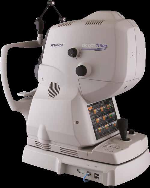

1 DRI OCT Triton Series A Multimodal Swept Source OCT Color Red-Free FA FAF Posterior Anterior


2 See what others can t see. A Multimodal Swept Source OCT DEEP RANGE IMAGING Swept Source OCT imaging massively increases my diagnostic capabilities in practice. The Topcon DRI OCT Triton is simple to operate and provides uniform detailed information from the vitreous through to the sclera, and beyond. The ability of the Topcon Triton to provide so many imaging modalities in one machine is a great advantage to future system wide diagnostic approaches and directly enables multimodal imaging approaches. Richard Spaide, MD Vitreous Retina Macula Consultants of New York

3 Welcome to the New Frontier in OCT Imaging The DRI OCT Triton combines Swept Source OCT and eye tracking with multimodal fundus imaging in an all in-one state of the-art imaging tool. The Triton brings the next level of diagnostic capability to you and your patients. Unprecedented Image Quality Triton s Swept Source OCT, with a scanning speed of 100,000 A scans/sec and 1,050nm wavelength light source, results in stunningly clear and detailed images. You will not only see the retina and vitreous, but also the choroid and the sclera like never before! Remarkable Diagnostic Capability Seeing deeper makes it possible to have a better understanding of many ocular pathologies. Combined with unique features such as Spaide autofluorescence filters, Fluorescein Angiography and en face imaging, 1 Triton empowers you to take proactive steps to preserve your patients eye health. A Trusted Brand The Triton has become a trusted brand and recognized leader in Swept Source OCT around the globe. With thousands of units in place, doctors are choosing the Triton for its unprecedented image quality, remarkable diagnostic capabilities, and clinical efficiencies. Triton Product Lineup The Triton is available in the standard model, the DRI OCT Triton, which includes Swept Source OCT, color fundus imaging, red-free, and optional anterior segment OCT imaging. There is also a DRI OCT Triton Plus model, which incorporates all of the above plus fluorescein angiography (FA) and fundus autofluorescence (FAF) imaging. Color Digital Red-free FA FAF Optional Anterior OCT Triton Triton plus 1. Requires IMAGEnet 6 software.


 and Fundus Autofluorescence (FAF) are also available.")
/ Color / Red-Free FA FAF Exclusive Spaide autofluorescence filters 1 The Triton Plus comes with built-in Spaide")

4 DRI meets Multimodal Fundus Imaging: see the whole picture Swept Source OCT incorporates multimodal fundus imaging DRI OCT Triton acquires the OCT and fundus image in a single capture. Pin-Point Registration identifies the location of the B-scan on the fundus image. A clear comparison between the B-scan and fundus image supports clinical efficiency. 4 High-quality fundus images The DRI OCT Triton offers non-mydriatic color fundus imaging. Fluorescein Angiography (FA) and Fundus Autofluorescence (FAF) are also available.* Color Red free *DRI OCT Triton plus: OCT / Anterior OCT (Option) /Color / Red-Free / FA / FAF DRI OCT Triton: OCT /Anterior OCT (Option)/ Color / Red-Free FA FAF Exclusive Spaide autofluorescence filters 1 The Triton Plus comes with built-in Spaide autofluorescence filters. They were developed by Richard Spaide, MD of Vitreous Macula Retina Consultants of New York and are exclusive to Topcon. The Spaide filters allow for a much more vivid and detailed image of the Lipofuscin that accumulates in the RPE of the retina, which can be a key in the early detection of eye disease. The Spaide filters do not stimulate fluorescein or ICG so images can be taken post angiography without any wavelength overlap. 1. Available on DRI OCT Triton Plus model only.


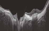
5 Optimized wavelength: 1,050nm The longer wavelength light source provides better tissue penetration and more OCT data deeper in the retina 1 than conventional Spectral Domain OCT technology, allowing visualization into the deepest layers of the eye even through cataracts, hemorrhages, and gas bubbles. Courtesy: Professor Jose Maria Ruiz Moreno, University of Albacete, Spain. OCT images through media opacities 2 The 1,050nm light source on the Triton allows the OCT scan to penetrate through media opacities, including cataracts and hemorrhages, making it possible for more patients to be imaged. SS OCT imaging through hemorrhage 5 SS OCT imaging through cataract Courtesy: Dr. Netan Choudhry, Vitreous Retina Macula Specialists of Toronto, Canada 2. Huang et al. Signal-to-Noise Ratio Comparisons Between Spectral Domain and Swept-Source OCTs. Association for Research in Vision and Ophthalmology (ARVO) 2016.


6 Swept Source OCT Imaging Superior visualization Invisible OCT Capture The 1,050nm light source is not visible to the human eye, enabling patients to concentrate on the fixation target during capture, which can reduce involuntary eye movement, eye fatigue and increase workflow. Conventional OCT DRI OCT Triton Tracing the visible scan line Concentrate on the fixation target 6 En face OCT imaging 1 En face imaging allows for independent dissection of the vitreoretinal interface, retina, retinal pigment epithelium (RPE), and choroid by flattening the B scan image. Pathology throughout the posterior pole can be studied and correlated with a patient s symptoms and disease progression. en face image Projection image en face image en face image Courtesy: Prof. T. Nakazawa, Tohoku University, Japan Courtesy: Prof. T. Nakazawa, Tohoku University, Japan Visualize the vitreous Utilizing a 1,050nm light source, the DRI OCT Triton provides uniform scanning sensitivity allowing superior visualization of the vitreous and choroid in the same scan. Uniform image quality 1. Requires IMAGEnet 6 software.


7 Eye Tracking Eye Tracking comes standard with the Triton. During capture of selected scans, Triton s eye tracking system ensures that you image the exact location of the retina that you want every time. Widefield OCT The Triton incorporates a 12mm x 9mm widefield scan providing measurement of the optic nerve and macula in a single scan. Besides significantly reducing patient exam time, the widefield scan provides a comprehensive assessment with reference database in a single easy to read report. 7 High Density HD OCT Scanning 512 x 256 OCT scan patterns capture twice the OCT data than conventional 512 x 128 scanning patterns, significantly increasing the available data for diagnosis.


8 Discover from Anterior through the Choroid Panoramic widefield photography 1 Preset fixation targets enable you to easily acquire panoramic peripheral views of the retina. 8 Reference database with Swept Source OCT DRI OCT Triton includes an FDA-cleared reference database for statistical comparison of the thickness maps and optic disc parameters. By comparing individual measurement values with the corresponding reference database, the DRI OCT Triton provides you with a powerful tool. 1. Requires IMAGEnet 6 software.


9 Automatic layer segmentation Retinal layers are automatically segmented by the Topcon Advanced Boundary Software (TABS ), enabling the quantification of layer thickness for change analysis. Retina RNFL GCL++ GCL+ Anterior segment imaging 2 Optional anterior imaging capabilities enhance the view of the anterior chamber and ciliary body. The unique anterior segment attachment ensures sharp images, even in the extreme periphery of the retina and anterior chamber. 9 OCT image B-scan length 16mm 2. Optional accessory.

10 Transform Your Ophthalmic Data and Images with IMAGEnet and Synergy 10 Synergy Efficient, Scalable, Flexible SYNERGY provides eye care professionals with the ability to efficiently collect, store and manage digitized ophthalmic data captured with both today s most advanced instruments and legacy devices. Data can be accessed securely and remotely at any time without the need for a VPN. As you add more devices, more clinics, more data, SYNERGY will grow with your demands. What s Unique About SYNERGY?» Offers both a cloud-based and on-premise solution.» Store, manage and review diagnostic data from over 200 different devices.» Review and share information in real-time with your colleagues» Multi-modal display with a single click» Enhanced OCT viewing PinPoint TM Registration of OCT image with fundus photo Advanced Comparison registers and aligns thickness maps to review change between visits Reference database for Macula disease and Glaucoma diagnosis Glaucoma prognostic management view» New workflow for ease of use and access to your data with fewer clicks» Seamless integration with your EMR and practice management systems» Bidirectional communication between EMR/PMS and ophthalmic devices» DICOM and Non-DICOM Compliance LEGACY Non-DICOM DEVICE HL7 EMR PATIENT RECORDS PATIENT RECORDS DICOM PACS ODM DICOM PATIENT RECORDS MWL DICOM COMPLIANT DEVICE BI-DIRECTIONAL BI-DIRECTIONAL UNI-DIRECTIONAL UNI-DIRECTIONAL RAW DATA RAW DATA

 supports a deeper understanding of your")
11 Operating Room Imaging Room Examination Room Laser Treatment Room Doctors Room Consultation Room Reception 11 IMAGEnet 6 Universally Connected IMAGEnet 6 is a browser based application, Operating System and PC independent, that can access Topcon ophthalmic data, images and OCT data from Topcon devices 1 connected to your practice or hospital network. Comprehensive Data Management Now you can review all data captured by any TOPCON device with one software application without the need to download and maintain review software. 2 Multimodal display Dynamic viewing of OCT B Scans, 3D images, thickness maps, enface data along with registered fundus photos (color, red-free, FA and FAF) supports a deeper understanding of your patient s condition. Remarkably Easy The data you need is just a click away. IMAGEnet 6 was developed to give you a simple and efficient way to review data. With an informative one-page Graphical User Interface (GUI), this browser-based application requires no installation. IMAGEnet Applications and Image Management Tools IMAGEnet 6 includes a wide array of standard image management tools and application programs including:» Stereo Viewer» Disc and Cup measurement» Patient Education module» Auto-mosaic program» Quick Draw tool with image annotation» Brightness/Contrast» Area Enhancement» Image Sharpness» Magnifier» Image flip 1. Reference for specific connections on file. 2. Capture software is required.
 Lab at N IHR/ Welcome Trust Manchester CRF &")

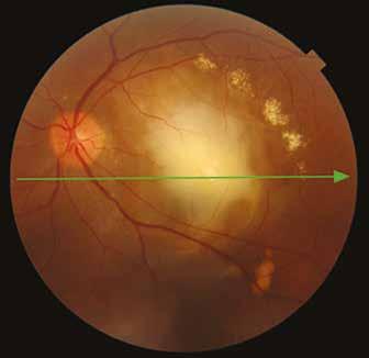
12 Case Reports Proliferative diabetic retinopathy Lateral: 12mm 12 Courtesy: Prof. P. E. Stanga, Manchester Royal Eye Hospital, Manchester Vision Regeneration (MVR) Lab at N IHR/ Welcome Trust Manchester CRF & University of Manchester *FA photography and FAF photography can only be performed on the DRI OCT Triton plus. Color FA* FAF* Courtesy: Prof. P. E. Stanga, Manchester Royal Eye Hospital, Manchester Vision Regeneration (MVR) Lab at N IHR/ Welcome Trust Manchester CRF & University of Manchester
 Lab at N IHR/ Welcome Trust Manchester CRF")

13 Central serous retinopathy Lateral: 12mm 13 Courtesy: Prof. P. E. Stanga, Manchester Royal Eye Hospital, Manchester Vision Regeneration (MVR) Lab at N IHR/ Welcome Trust Manchester CRF & University of Manchester *FA photography and FAF photography can only be performed on the DRI OCT Triton plus. Color FA* FAF* Courtesy: Prof. P. E. Stanga, Manchester Royal Eye Hospital, Manchester Vision Regeneration (MVR) Lab at N IHR/ Welcome Trust Manchester CRF & University of Manchester


14 Case Reports Myopia 14 Macular fibrosis
 15")
 Japan)")
15 Image through cataract Lateral: 12mm Courtesy: Kazuya Yamagishi, MD (Hirakata Yamagishi Eye Clinic, Japan) 15 Courtesy: Kazuya Yamagishi, MD (Hirakata Yamagishi Eye Clinic, Japan) Courtesy: Kazuya Yamagishi, MD (Hirakata Yamagishi Eye Clinic, Japan)
 Operating Distance 34.8mm Minimum Pupil Diameter Ø2.")
 Horizontal Within 3 to 12mm Vertical Within 3 to 12mm Scan Patterns 3D scan (12x9mm, 7x7mm, 3x3mm) Linear scan")
16 Specifications OCT Imaging Methodology Swept Source OCT Optical Light Source Swept Source tunable laser at 1,050nm Scan Speed 100,000 A-Scans per second Lateral Resolution20 μm In-depth Resolution Optical resolution: 8 μm, 2.6 μm digital resolution Photography Type Color, FA,* FAF,* Red-free** Picture Angle45 Equivalent 30 (Digital Zoom) Operating Distance 34.8mm Minimum Pupil Diameter Ø2.5mm OCT, 3.3mm fundus photo Observation & Photography of Fundus Tomogram Scanning Range (on fundus) Horizontal Within 3 to 12mm Vertical Within 3 to 12mm Scan Patterns 3D scan (12x9mm, 7x7mm, 3x3mm) Linear scan (Line-scan/Cross-scan/Radial-scan) Fixation target Internal fixation target: Dot matrix type organic EL The display position can be changed and adjusted. The displaying method can be changed. Peripheral fixation target: This is displayed according to the internal fixation target displayed position. External fixation target Observation & photography of anterior segment*** Photography type IR Operating distance17mm Scan range (on cornea) Horizontal Within 3 to 16mm Vertical Within 3 to 16mm Scan pattern3d scan Linear scan (Line-scan/Radial-scan) Fixation target Internal fixation target External fixation target Electrical Rating Power Source Voltage: V Frequency: 50-60Hz Power input250va Dimensions mm(W) X mm(D) X mm(H) Weight 21.8 kg (DRI OCT Triton) 23.8 kg (DRI OCT Triton plus) * FA photography and FAF photography can only be performed on the DRI OCT Triton plus. ** The color image is processed and is displayed as a pseudo red-free photographed image. *** Observation & photography of anterior segment can be performed only when the anterior segment attachment kit is used. All trademarks are the property of their respective owners. TOPCON MEDICAL SYSTEMS, INC. 111 Bauer Drive, Oakland, NJ Phone: Fax: topconmedical.com 2018 Topcon Medical Systems, Inc. MCA# 2296 IMPORTANT Subject to change in design and/or specifications without advanced notice. In order to obtain the best results with this instrument, please be sure to review all user instructions prior to operation.
DRI OCT Triton series Swept Source Optical Coherence Tomography
 DRI OCT Triton series Swept Source Optical Coherence Tomography See. Discover. Explore. The diagnostic power of Swept Source OCT Deep Range Imaging. Swept Source adds a new dimension to OCT. The Topcon
DRI OCT Triton series. Swept Source Optical Coherence Tomography
 DRI OCT Triton series Swept Source Optical Coherence Tomography Welcome to the New Frontier in OCT Imaging The DRI OCT Triton combines the world s first swept source OCT technology with multimodal fundus
The World s fastest OCT. As simple as pressing. the start button
 The World s fastest OCT As simple as pressing the start button lution continues Optopol engineering team, designers of the first commercially available Spectral Domain OCT in the world, are proud to present
SOCT Copernicus REVO. * - Currently import and overlay are avaibale in manual mode only
 SOCT Copernicus REVO Easy Operation (Full auto & Auto mode) Auto alignment (Z-position, C-gate, Focus, Tomogram) Voice guide (support patient through examination) Powerful analysis tools Enhanced tomograms
PRIMUS 200 from ZEISS The essential OCT
 PRIMUS 200 from ZEISS The essential OCT Seeing beyond the surface. ZEISS PRIMUS 200 // INNOVATION MADE BY ZEISS Clear Visualization. Advanced Technology. Reliability. Essential elements of your first OCT.
PRIMUS 200 from ZEISS The essential OCT
 EN 00_00I The contents of the brochure may differ from the current status of approval of the product in your country. Please contact your regional representative for more information. Subject to change
LEE EYE CENTRE. YOUR VISION, OUR PASSION LEC EyeNews
 LEE EYE CENTRE YOUR VISION, OUR PASSION LEC EyeNews FOR INTERNAL CIRCULATION ONLY www.lec.com.my ISSUE 51/003 SEPT OCT 2017 The American Society of Cataract and Refractive Surgery is one of the leading
DRI OCT Triton series
 DRI OCT Triton series Swept Source Optical Coherence Tomography PERFORMANCE YOU CAN COUNT ON See. Discover. Explore. The diagnostic power of Swept Source OCT Deep Range Imaging. Swept source adds a new
PLEX Elite 9000 from ZEISS Swept-Source OCT
 PLEX Elite 9000 from ZEISS Swept-Source OCT Uncovering the undiscovered. ZEISS PLEX Elite 9000 // INNOVATION MADE BY ZEISS 2 Ultra-wide angiography En face montage Image courtesy of Prof. G. Querques,
Widefield Retinal Imaging with Auto Fluorescence Technology in the Optometric Practice
 Widefield Retinal Imaging with Auto Fluorescence Technology in the Optometric Practice This course will define ultra-widefield retinal imaging and autofluorescence for the attendee. Will show how it is
Optical Coherence Tomography (OCT) in Uveitis Piergiorgio Neri, BMedSc, MD, PhD Head Ocular Immunology Unit
 in Uveitis Piergiorgio Neri, BMedSc, MD, PhD Head Ocular Immunology Unit") The Eye Clinic Polytechnic University of Marche Head: Prof Alfonso Giovannini November, 1991 Optical Coherence Tomography (OCT) in Uveitis Piergiorgio Neri, BMedSc, MD, PhD Head Ocular Immunology Unit
The Eye Clinic Polytechnic University of Marche Head: Prof Alfonso Giovannini November, 1991 Optical Coherence Tomography (OCT) in Uveitis Piergiorgio Neri, BMedSc, MD, PhD Head Ocular Immunology Unit
OCT Image Analysis System for Grading and Diagnosis of Retinal Diseases and its Integration in i-hospital
 Progress Report for1 st Quarter, May-July 2017 OCT Image Analysis System for Grading and Diagnosis of Retinal Diseases and its Integration in i-hospital Milestone 1: Designing Annotation tool extraction
Optical Coherence Tomography 3D OCT-1 Maestro
 Optical Coherence Tomography 3D OCT-1 Maestro The OCT World at Your Fingertips 3D OCT-1 Maestro» 50,000 A Scan/sec SD OCT with color fundus camera» Auto alignment, focus and capture of OCT and color fundus
Visualize. Analyze. Personalize. OCT + OCTA
 Visualize. Analyze. Personalize. OCT + OCTA A New Approach to Protecting Vision AngioVue OCT Angiography brings valuable new information to clinical practice. Non-invasive visualization of retinal vasculature.
Visualize. Analyze. Personalize. OCT + OCTA. with
 Visualize. Analyze. Personalize. OCT + OCTA with Avanti Widefield OCT with AngioVue OCTA Imaging Comprehensive Structural and Functional Imaging in a Single Imaging Platform Comprehensive OCT Imaging The
Moving forward with a different perspective
 Moving forward with a different perspective The Leader In Vision Diagnostics Offers A New Perspective Marco has served the eyecare community by offering exceptional lane products and automated high tech
The ideal tool for early detection and monitoring of AMD.
 The ideal tool for early detection and monitoring of AMD. presenting maia 1 MAIA, the new frontier of Fundus Perimetry (microperimetry) assesses the function of the macula representing an effective clinical
Experience Spectacular Retinal Imaging with the new NIDEK F-10 Digital Ophthalmoscope
 Experience Spectacular Retinal Imaging with the new NIDEK F-10 Digital Ophthalmoscope The F-10 was developed to give Ophthalmologists a high definition (HD) diagnostic imaging system. Designed to provide
EasyScan: Smart Retinal Imaging
 easyscan EasyScan: Smart Retinal Imaging Superior Imaging Enjoy the benefits of SLO technology and capture high-quality images easily for accurate diagnosis. Never Dilate Reduce examination time, capture
Swept-Source OCT Angiography: SS OCT Angio TM
 Swept-Source OCT Angiography: SS OCT Angio TM Not available in all countries, please check with your distributor. 2015.09 Swept-Source OCT Angiography: SS OCT Angio TM Introduction Optical coherence tomography
Introducing ANGIOVUE ESSENTIAL. Built on the Avanti Widefield OCT Platform. OCT Angiography for Primary Eye Care
 Introducing ANGIOVUE ESSENTIAL Built on the Avanti Widefield OCT Platform OCT Angiography for Primary Eye Care Transform Your View of the Retina OCT Angiography (OCTA) is a quick non-invasive test that
Cirrus TM HD-OCT. Details define your decisions
 Cirrus TM HD-OCT Details define your decisions 2 With high-definition OCT Carl Zeiss Meditec takes you beyond standard spectral domain Built on 10 years experience at the vanguard of innovation, Carl Zeiss
HOCT-1I 1F All-in-One Optical Coherence Tomography with Fundus
 HOCT-1I 1F All-in-One Optical Coherence Tomography with Fundus Specification Type Resolution(in Tissue) A scan Rate Scan Range SD-OCT / Fundus Z :6~7um, XY:20um 68,000 A-scan/sec. [Fundus] X:6-12mm, Y:6-9mm,
Cirrus TM HD-OCT. Details defi ne your decisions
 Cirrus TM HD-OCT Details defi ne your decisions 2 With high-defi nition OCT Carl Zeiss Meditec takes you beyond standard spectral domain Built on 10 years experience at the vanguard of innovation, Carl
On Different Wavelengths: The Spectrum of Retinal Imaging. On Different Wavelengths: The Spectrum of Retinal Imaging. Wavelength Specific Imaging
 On Different Wavelengths: The Spectrum of Retinal Imaging Timothy J. Bennett, CRA, FOPS, OCT-C Penn State Hershey Eye Center Hershey, PA On Different Wavelengths: The Spectrum of Retinal Imaging Wavelengths
The Evolution of Fundus Perimetry
 The Evolution of Fundus Perimetry Company Profile CenterVue designs and manufactures highly automated medical devices for the diagnosis and management of ocular pathologies, including those that represent
Evolving glaucoma management True diagnostic integration for the preservation of vision
 Evolving glaucoma management True diagnostic integration for the preservation of vision // GLAUCOMA MANAGEMENT MADE BY ZEISS The moment you are certain it is glaucoma. This is the moment we work for. There
4/19/2018 FUNDUS AUTOFLUORESCENCE. Fluorescence Imaging. Fundus Autofluorescence (FAF) Fluorescence. Fluorescence
 Fluorescence. Fluorescence") I have no financial or proprietary interest in the subject matter of this presentation. FUNDUS AUTOFLUORESCENCE Timothy J. Bennett, CRA, OCT-C, FOPS Penn State Eye Center Hershey, PA Fluorescence Imaging
I have no financial or proprietary interest in the subject matter of this presentation. FUNDUS AUTOFLUORESCENCE Timothy J. Bennett, CRA, OCT-C, FOPS Penn State Eye Center Hershey, PA Fluorescence Imaging
Five Things You re Missing with Your Fundus Camera
 ebook Five Things You re Missing with Your Fundus Camera By Donald J. Siegel, OD, Sun City West Eye Care Sponsored by: Before I began incorporating EIDON true-color imaging into my practice, my retinal
Introducing the future of DXA. Powerful images. Clear answers. Horizon DXA System
 Introducing the future of DXA Powerful images. Clear answers. Horizon DXA System Hologic turns ideas into innovation. Again. Hologic cares about you and your patients about keeping their bones healthy,
The New Frontier of Microperimetry
 Macular Integrity Assessment The New Frontier of Microperimetry Microperimetry is attracting our attention more and more as a method that is superior to standard automated perimetry for visual function
The New Frontier of Microperimetry
 Macular Integrity Assessment The New Frontier of Microperimetry Index 4 Company Profile Microperimetry is attracting our attention more and more as a method that is superior to standard automated perimetry
Mark Dunbar: Disclosure
 Important Things to Understand About OCT Mark T. Dunbar, O.D., F.A.A.O. Bascom Palmer Eye Institute University of Miami, School of Medicine Mark Dunbar: Disclosure Optometry Advisory Board for: Allergan
Do You See What I See!!! Shane R. Kannarr, OD
 Do You See What I See!!! Shane R. Kannarr, OD skannarr@kannarreyecare.com Define Specialty Testing Additional Test to: Prove/Disprove Diagnosis To monitor progression of a condition To document a condition
Fundus Autofluorescence
 Brittany Bateman, BS Fundus autofluorescence imaging is used to record fluorescence that may occur naturally in ocular structures or as a byproduct of a disease process. This technique allows the topographic
Observation of Posterior Precortical Vitreous Pocket Using Swept-Source Optical Coherence Tomography
 Anatomy and Pathology Observation of Posterior Precortical Vitreous Pocket Using Swept-Source Optical Coherence Tomography Hirotaka Itakura, Shoji Kishi, Danjie Li, and Hideo Akiyama Department of Ophthalmology,
OCT Angiography in Primary Eye Care
 OCT Angiography in Primary Eye Care An Image Interpretation Primer Julie Rodman, OD, MS, FAAO and Nadia Waheed, MD, MPH Table of Contents Diabetic Retinopathy 3-6 Choroidal Neovascularization 7-9 Central
3/23/2016. Diagnostic Services Taylor Pannell CRA, OCT-C. Services Available. Important info for the Tech to know. Visual Fields
 Services Available Diagnostic Services Taylor Pannell CRA, OCT-C Static and Kinetic Visual Fields Pachymetry Anterior and Posterior Segment OCT Fundus Photos FAF,FA,ICG Slit Lamp Photography Confocal HRT
THE OPHTHALMOLOGIST S NEEDS FOR THE ANALYSIS OF THE RETINA
 biophotonics end-users needs THE OPHTHALMOLOGIST S NEEDS FOR THE ANALYSIS OF THE RETINA Dr Matonti Frédéric CHU Nord / INT AMU Marseille ANATOMY OF THE RETINA ANATOMY OF THE RETINA ANATOMY OF THE RETINA
Navigated Laser Therapy. A New Era in Retinal Disease Management
 Navigated Laser Therapy A New Era in Retinal Disease Management Bringing Navigation to Retina Treatment Navilas Laser System To unleash the full potential of Retina Navigation, the Navilas Laser System
Perspectives on Screening for Diabetic Retinopathy. Dr. Dan Samaha, Optometrist, MSc Clinical Lecturer School of Optometry, Université de Montréal
 Perspectives on Screening for Diabetic Retinopathy 1 Dr. Dan Samaha, Optometrist, MSc Clinical Lecturer School of Optometry, Université de Montréal Current standards 2 According to the Canadian Diabetes
FA vs. OCTA? The status of OCTA, today. Fukuoka, JSOS 2016 Gerd Klose. Korobelnik J Fr Ophthalmol (2015)
") FA vs. OCTA? The status of OCTA, today Korobelnik J Fr Ophthalmol (2015) Fukuoka, JSOS 2016 Gerd Klose 1 2 FA / ICGA a well-founded Gold standard! Benefits Useful for many pathologies High contrast, detailed
FA vs. OCTA? The status of OCTA, today Korobelnik J Fr Ophthalmol (2015) Fukuoka, JSOS 2016 Gerd Klose 1 2 FA / ICGA a well-founded Gold standard! Benefits Useful for many pathologies High contrast, detailed
WORKSHOP B Ophthalmic Imaging: All Hands on Tech! COPE Course PS
 WORKSHOP B Ophthalmic Imaging: All Hands on Tech! COPE Course 44334-PS Ophthalmic Imaging: All Hands on Tech! Southern College of Optometry April 17, 2015 COPE #44334-PS Faculty Dr. Michael Gerstner Dr.
Advances in OCT Murray Fingeret, OD
 Disclosures Advances in OCT Murray Fingeret, OD Consultant Alcon, Allergan, Bausch & Lomb, Carl Zeiss Meditec, Diopsys, Heidelberg Engineering, Reichert, Topcon Currently Approved OCT Devices OCT Devices
Yasser R. Serag, MD Tamer Wasfi, MD El- Saied El-Dessoukey, MD Magdi S. Moussa, MD Anselm Kampik, MD
 Microperimetric Evaluation of Brilliant Blue G- assisted Internal Limiting Membrane Peeling By Yasser R. Serag, MD Tamer Wasfi, MD El- Saied El-Dessoukey, MD Magdi S. Moussa, MD Anselm Kampik, MD The internal
Ultrahigh Speed Imaging of the Rat Retina Using Ultrahigh Resolution Spectral/Fourier Domain OCT
 Ultrahigh Speed Imaging of the Rat Retina Using Ultrahigh Resolution Spectral/Fourier Domain OCT The MIT Faculty has made this article openly available. Please share how this access benefits you. Your
Fundus Autofluorescence. Jonathan A. Micieli, MD Valérie Biousse, MD
 Fundus Autofluorescence Jonathan A. Micieli, MD Valérie Biousse, MD The retinal pigment epithelium (RPE) has many important functions including phagocytosis of the photoreceptor outer segments Cone Rod
What is the Value of Swept Source oct Technology in Biometry? Experts discussed the IOLMaster 700 at the ESCRS ebook. Content provided by:
 ebook Content provided by: What is the Value of Swept Source oct Technology in Biometry? Experts discussed the IOLMaster 700 at the ESCRS 2016 Participating experts: G. Barrett, MD, Australia; D. Chang,
A COMPLETE GUIDE TO THE CLARUS 500 ULTRA-WIDEFIELD RETINAL CAMERA
 A COMPLETE GUIDE TO THE CLARUS 500 ULTRA-WIDEFIELD RETINAL CAMERA BY DANIEL EPSHTIEN, OD Originally published on newgradoptometry.com Ultra-widefield retinal imaging has had a contentious path to acceptance
OCT Interpretation in Retinal Disease
 OCT Interpretation in Retinal Disease Jay M. Haynie, OD, FAAO Financial Disclosure I have received honoraria or am on the advisory board for the following companies: Carl Zeiss Meditec Advanced Ocular
and at the same patient encounter. Code has been deleted. For scanning computerized ophthalmic diagnostic imaging of optic nerve and retin
 92227: Remote imaging for detection of retinal disease (eg, retinopathy in a patient with diabetes) with analysis and report under physician supervision, unilateral or bilateral. For Medicare, bill only
Optical Coherence Tomography: Pearls for the Anterior Segment Surgeon Basic Science Michael Stewart, M.D.
 Optical Coherence Tomography: Pearls for the Anterior Segment Surgeon Basic Science Michael Stewart, M.D. Disclosure OCT Optical Coherence Tomography No relevant financial relationships I will refer to
Abstracts DRI OCT-1. DRI OCT-1 See, Discover, Explore. Invest Ophthalmol Vis Sci Jul 1;52(8): Print 2011 Jul.
: Print 2011 Jul.") Abstracts Invest Ophthalmol Vis Sci. 2011 Jul 1;52(8):4971-8. Print 2011 Jul. Macular choroidal thickness and volume in normal subjects measured by swept-source optical coherence tomography. Hirata M,
Abstracts Invest Ophthalmol Vis Sci. 2011 Jul 1;52(8):4971-8. Print 2011 Jul. Macular choroidal thickness and volume in normal subjects measured by swept-source optical coherence tomography. Hirata M,
Cornea/Anterior Segment OCT. User Experience
 Cornea/Anterior Segment OCT User Experience User Experience Case#1 Post Penetrating Keratoplasty Tokyo Medical University / Kohsei Chuo General Hospital Hideki Mori MD, PhD Almost eight years have passed
Clinically Significant Macular Edema (CSME)
") Clinically Significant Macular Edema (CSME) 1 Clinically Significant Macular Edema (CSME) Sadrina T. Shaw OMT I Student July 26, 2014 Advisor: Dr. Uwaydat Clinically Significant Macular Edema (CSME) 2
Clinically Significant Macular Edema (CSME) 1 Clinically Significant Macular Edema (CSME) Sadrina T. Shaw OMT I Student July 26, 2014 Advisor: Dr. Uwaydat Clinically Significant Macular Edema (CSME) 2
Il contributo dell'angio-oct: valutazione integrata della componente nervosa e vascolare della malattia glaucomatosa
 SIMPOSIO G.O.A.L. - LE NUOVE FRONTIERE DIAGNOSTICHE E LE LINEE DI INDIRIZZO AMBULATORIALI DEL GLAUCOMA Coordinatore e moderatore: D. Mazzacane Presidente: L. Rossetti Il contributo dell'angio-oct: valutazione
Structural examina.on: Imaging
 ManaMa: Glaucoma Structural examina.on: Imaging Luís Abegão Pinto, MD, PhD Department of Ophthalmology CHLC Lisbon Faculty of Medicine, Lisbon University 1 11-10- 2013 Structural changes Qualitative changes
Technologies and Methods for Visualizing the Retina
 Transcript Details This is a transcript of an educational program accessible on the ReachMD network. Details about the program and additional media formats for the program are accessible by visiting: https://reachmd.com/programs/revealing-retina/technologies-and-methods-for-visualizing-theretina/3663/
Focus on your needs. Ultrasound system HS60 SAMSUNG MEDISON CO., LTD. CT-HS60 V1.0-GI-FT EN
 Samsung Medison, an affiliate of Samsung Electronics, is a global medical company founded in 1985. With a mission to bring health and well-being to people's lives, the company manufactures diagnostic ultrasound
tracking progression we can better manage our patients. Like any tool, any instrument you ve got to
 EIYESS ALBEINUTI, MD 1 As we know OCT has become very instrumental in taking care of glaucoma patients whether we have the ability to objectively image the RNFL and therefore pickup earlier signs of damage
Reliable versatility. Philips HD5 ultrasound system
 Reliable versatility Philips HD5 ultrasound system Affordability and features in one Designed to perform as you need it Every day, your patients come to you for high-quality care. Now, there s an ultrasound
R&M Solutions
 Mohamed Hosny El-Bradey, MD., Assistant Professor of Ophthalmology, Tanta University. Wael El Haig, MD., Professor of Ophthalmology. Zagazeeg University. 1 Myopic CNV is considered the most common vision
A Patient s Guide to Diabetic Retinopathy
 Diabetic Retinopathy A Patient s Guide to Diabetic Retinopathy 840 Walnut Street, Philadelphia PA 19107 www.willseye.org Diabetic Retinopathy 1. Definition Diabetic retinopathy is a complication of diabetes
International Journal of Advance Engineering and Research Development EARLY DETECTION OF GLAUCOMA USING EMPIRICAL WAVELET TRANSFORM
 Scientific Journal of Impact Factor (SJIF): 4.72 International Journal of Advance Engineering and Research Development Volume 5, Issue 1, January -218 e-issn (O): 2348-447 p-issn (P): 2348-646 EARLY DETECTION
Optical Coherence Tomography in Diabetic Retinopathy. Mrs Samantha Mann Consultant Ophthalmologist Clinical Lead of SEL-DESP
 Optical Coherence Tomography in Diabetic Retinopathy Mrs Samantha Mann Consultant Ophthalmologist Clinical Lead of SEL-DESP Content OCT imaging Retinal layers OCT features in Diabetes Some NON DR features
The College of Optometrists - Learning outcomes for the Professional Certificate in Medical Retina
 Learning outcomes for the Professional Certificate in Medical Retina, incorporating diabetic retinopathy screening and age related macular degeneration The professional certificate is a prerequisite to
 measure of your overall performance. An isolated glucose test is helpful to let you know what your sugar level is at one moment, but it doesn t tell you whether or not your diabetes is under adequate control
Why Is Imaging Critical in My Uveitis Practice?
 Why Is Imaging Critical in My Uveitis Practice? Dilraj S. Grewal, MD Developed in collaboration Imaging Is the Backbone of Uveitis Workup and Monitoring Treatment Response FP FAF B- scan Multimodal Imaging
The Measure of Confidence
 Heidelberg_936357.qxd:Layout 1 5/9/08 12:01 PM 12:02 Page 1 (Cyan (Magenta (Yellow (Black (UV Five Powerful Solutions to Fit Your Practice PowerCheck Glaucoma FastCheck+ GPS Software and Retina Edema Index
Measurement of Choroidal Thickness in Normal Eyes Using 3D OCT-1000 Spectral Domain Optical Coherence Tomography
 pissn: 111-8942 eissn: 292-9382 Korean J Ophthalmol 212;26(4):255-259 http://dx.doi.org/1.3341/kjo.212.26.4.255 Original Article Measurement of Choroidal Thickness in Normal Eyes Using 3D OCT-1 Spectral
Look differently. Invenia ABUS. Automated Breast Ultrasound
 Look differently. Invenia ABUS Automated Breast Ultrasound InveniaTM ABUS from GE Healthcare offers a view beyond mammography, with breast screening technology that looks differently. 40 % The unseen risk.
Clinical Study Choroidal Thickness in Eyes with Unilateral Ocular Ischemic Syndrome
 Hindawi Publishing Corporation Journal of Ophthalmology Volume 215, Article ID 62372, 5 pages http://dx.doi.org/1.1155/215/62372 Clinical Study Choroidal Thickness in Eyes with Unilateral Ocular Ischemic
Diabetic retinopathy damage to the blood vessels in the retina. Cataract clouding of the eye s lens. Cataracts develop at an earlier age in people
 Diabetic Retinopathy What is diabetic eye disease? Diabetic eye disease refers to a group of eye problems that people with diabetes may face as a complication of diabetes. All can cause severe vision loss
Ophthalmology. Caring For Your Eyes. Jurong Medical Centre
 Ophthalmology Caring For Your Eyes Jurong Medical Centre Your eyes and you At Jurong Medical Centre, we have a dedicated team of ophthalmologists that specialise in treating a wide range of acute and chronic
RETINAL CONDITIONS RETINAL CONDITIONS
 GENERAL INFORMATION RETINAL CONDITIONS RETINAL CONDITIONS WHAT ARE RETINAL CONDITIONS? Retinal conditions affect the light-sensitive tissue at the back of eye known as the retina. They include diseases
Exceptional versatility without compromise
 Introducing the VICTUS femtosecond laser platform Exceptional versatility without compromise FEMTOSECOND TECHNOLOGY that empowers Introducing VICTUS the first femtosecond laser capable of exceptional performance
Diabetes & Your Eyes
 Diabetes & Your Eyes Diabetes is a disease that occurs when the pancreas does not secrete enough insulin or the body is unable to process it properly. Insulin is the hormone that regulates the level of
CONFERENCE-AT-A-GLANCE
 CONFERENCE-AT-A-GLANCE Friday November 13, 2015 FR-1-A Pediatric Retina: What Your Peds Doc Needs FR-1-B Wild & Wacky World of Neuro- Ophthal FR-1-C Correlation/Discrep between FA & OCT FR-1-D (8:30 am
Lasers and Imaging PHOTOCOAGULATION PHOTODISRUPTION SLT PHOTOREGENERATION DIAGNOSTIC ULTRASOUND
 Lasers and Imaging PHOTOCOAGULATION PHOTODISRUPTION SLT PHOTOREGENERATION DIAGNOSTIC ULTRASOUND One Powerful Vision Photodisruption The tough demands of today s new-generation intraocular lenses (IOLs)
Case Report Peripapillary Intrachoroidal Cavitation in Myopia Evaluated with Multimodal Imaging Comprising (En-Face) Technique
 Technique") Case Reports in Ophthalmological Medicine Volume 2015, Article ID 890876, 5 pages http://dx.doi.org/10.1155/2015/890876 Case Report Peripapillary Intrachoroidal Cavitation in Myopia Evaluated with Multimodal
Case Reports in Ophthalmological Medicine Volume 2015, Article ID 890876, 5 pages http://dx.doi.org/10.1155/2015/890876 Case Report Peripapillary Intrachoroidal Cavitation in Myopia Evaluated with Multimodal
RETINA REVEALED. Dynamic Developments in AMD Diagnosis and Treatment (2014) The Dawn of: Pharmaco-Genetics (aka Nutrigenomics) Jerome Sherman, OD
 The Dawn of: Pharmaco-Genetics (aka Nutrigenomics) Jerome Sherman, OD") RETINA REVEALED Dynamic Developments in AMD Diagnosis and Treatment (2014) The Dawn of: Pharmaco-Genetics (aka Nutrigenomics) Jerome Sherman, OD Disclosures: Jerome Sherman Dr. Sherman has lectured, received
RETINA REVEALED Dynamic Developments in AMD Diagnosis and Treatment (2014) The Dawn of: Pharmaco-Genetics (aka Nutrigenomics) Jerome Sherman, OD Disclosures: Jerome Sherman Dr. Sherman has lectured, received
OCT Fundal Angiography Initial Experience The new era in Medical Retina Imaging Based on Cirrus 5000 AngioPlex 2016 Model Sheena George & Nicholas
 OCT Fundal Angiography Initial Experience The new era in Medical Retina Imaging Based on Cirrus 5000 AngioPlex 2016 Model Sheena George & Nicholas Lee Consultants Ophthalmologist at The Hillingdon Hospital
optic disc neovascularisation
 British Journal of Ophthalmology, 1979, 63, 412-417 A comparative study of argon laser and krypton laser in the treatment of diabetic optic disc neovascularisation W. E. SCHULENBURG, A. M. HAMILTON, AND
Note: This is an outcome measure and can be calculated solely using registry data.
 Measure #191 (NQF 0565): Cataracts: 20/40 or Better Visual Acuity within 90 Days Following Cataract Surgery -- National Quality Strategy Domain: Effective Clinical Care DESCRIPTION: Percentage of patients
Choroidal Mapping; a Novel Approach for Evaluating Choroidal Thickness and Volume
 Imaging Technique Choroidal Mapping; a Novel Approach for Evaluating Choroidal Thickness and Volume Jila Noori 1, MD; Mohammad Riazi Esfahani 1,2, MD Fedra Hajizadeh 2, MD; Mohammad-Mehdi Zaferani 1, MD
ZEISS presents innovations that support ophthalmologists in their work
 ZEISS presents innovations that support ophthalmologists in their work At the 2015 Annual Meeting of the American Academy of Ophthalmology (AAO) in Las Vegas, ZEISS announces innovations for clinical excellence
The MP-1 Microperimeter Clinical Applications in Retinal Pathologies
 The MP-1 Microperimeter Clinical Applications in Retinal Pathologies Nelson R. Sabates, MD Director, Retina/Vitreous Service Vice-Chairman Department of Ophthalmology University of Missouri Kansas City
CONFERENCE-AT-A-GLANCE
 CONFERENCE-AT-A-GLANCE REGISTRATION STRUCTURE Attendees selecting the Unlimited Lecture Package will be able to attend courses during each timeline of every day of the program. Attendees selecting the
GE Healthcare 9900 Innovation Drive Wauwatosa, WI U.S.A.
 2011 General Electric Company All rights reserved. General Electric Company reserves the right to make changes in specifications and features shown herein, or discontinue the product described at any time
Optical Coherence Tomograpic Features in Idiopathic Retinitis, Vasculitis, Aneurysms and Neuroretinitis (IRVAN)
") Columbia International Publishing Journal of Ophthalmic Research (2014) Research Article Optical Coherence Tomograpic Features in Idiopathic Retinitis, Vasculitis, Aneurysms and Neuroretinitis (IRVAN)
Columbia International Publishing Journal of Ophthalmic Research (2014) Research Article Optical Coherence Tomograpic Features in Idiopathic Retinitis, Vasculitis, Aneurysms and Neuroretinitis (IRVAN)
Incorporating OCT Angiography Into Patient Care
 Incorporating OCT Angiography Into Patient Care Beth A. Steele, OD, FAAO OCT A: Introduction Isolates microvascular circulation from OCT image data Axial resolution = 5 microns (i.e. fine capillaries visible)
CONFERENCE-AT-A-GLANCE
 CONFERENCE-AT-A-GLANCE Friday November 15, 2013 Room Pelican I LaSalle Blrm C LaSalle Blrm B LaSalle Blrm A 9:30 am- 10:30 am FR-1-A Pediatric OCT in the OR FR-1-B Hi-Tech Spanish for Ophthal Photographers
Digital mammography imaging from Carestream Health solutions for great workflow, productivity, and patient care.
 Digital Mammography Imaging on KODAK CR Systems Digital mammography imaging from Carestream Health solutions for great workflow, productivity, and patient care. Commercial distribution of the CR Mammography
Pushing the boundaries
 Samsung Medison is a global leading medical devices company. Founded in 1985, the company now sells cutting-edge medical devices including diagnostic ultrasound, digital X-ray and blood analyzer, around
OUR EYES & HOW WE SEE
 OUR EYES & HOW WE SEE UNDERSTAND MORE ABOUT OUR EYES & HOW WE SEE Our Eyes & How We See The eye is our visual gateway to the world. Within it, an array of delicate components labour away to give us the
NEW AUTOMATED PERIMETERS NEW. Fast and precise perimetry at your fingertips. ZETA strategy EyeSee recording DPA analysis
 NEW AUTOMATED PERIMETERS Fast and precise perimetry at your fingertips NEW ZETA strategy EyeSee recording DPA analysis PTS 920 PTS 925W I PTS 2000 PTS AUTOMATED PERIMETER SERIES THRESHOLD IN 3 MINUTES**
Tips and Tactics for Retinal Imaging
 Tips and Tactics for Retinal Imaging Retinal Imaging Fundus camera Scanning Laser Ophthalmoscope (cslo) SD-Optical Coherence Tomography Timothy J. Bennett, CRA, OCT-C, FOPS Penn State Eye Center Hershey,
Clinical Trial Endpoints for Macular Diseases
 Clinical Trial Endpoints for Macular Diseases Developed in collaboration Learning Objective Upon completion, participants should be able to: Summarize types of biomarkers of progression and treatment response
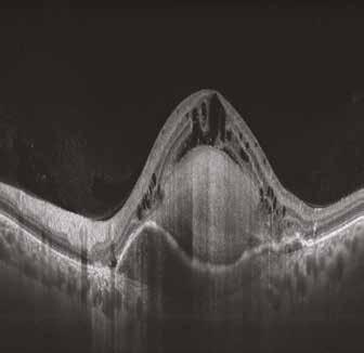
DOME SHAPED MACULOPATHY. Ιωάννης Ν. Βαγγελόπουλος Χειρ. Οφθαλμίατρος - Βόλος
 DOME SHAPED MACULOPATHY Ιωάννης Ν. Βαγγελόπουλος Χειρ. Οφθαλμίατρος - Βόλος DOME SHAPED MACULOPATHY-DEFINITIONS The entity Dome Shaped Macula ( DSM ) was first described by Gaucher and associates in 2008
Overview. Macular OCT Artifact Study
 Imaging Artifacts Sarah Moyer, CRA, OCT-C Director, Ophthalmic Imaging Kittner Eye Center University of North Carolina Chapel Hill, NC Disclose financial interest now Overview Sarah s Thoughts on Artifacts