Peripheral T-cell Lymphomas. Current Classification and Differential Diagnosis. Elaine S Jaffe, M.D.
|
|
|
- Patrick Malone
- 5 years ago
- Views:
Transcription

1 Peripheral T-cell Lymphomas Current Classification and Differential Diagnosis Elaine S Jaffe, M.D.

2 International T-cell Lymphoma. Study: Frequency of Subtypes Study limited to adults JCO 2008;26: by American Society of Clinical Oncology
3 ALK+ ALK- Vose et al. JCO 2008
4 Innate Immune System Adaptive Immune System γδ T-cells, NK-like T- cells, NK-cells B-cell T-cell Toll like receptors Not MHC restricted Cytokines Chemokines Complement Apoptotic & necrotic cell death pathways Ag specific receptors on B + T-cells APC Antigen presentation to T-cells in the context of MHC First line of defense with a major role in barrier immunity Immunological defense characterized by specificity & memory
5 Innate Immune System Adaptive Immune System γδ T-cells, NK-like T- cells, NK-cells T-cell Often cutaneous, mucosal, spleen & BM Cytotoxic Activated cells show frequent apoptosis, necrosis Includes most extranodal PTCLs, EBV+ T/NK cell lymphomas Lymphomas may relate to specific effector T-cells T FH, Treg Functional consequences may be clinically apparent Includes most nodal PTCLs in adults
6 Peripheral T-cell Lymphoma, NOS A diagnosis of exclusion, by definition a heterogeneous category Characterized by a broad morphologic spectrum New approaches include segregation of tumors of TFH origin (follicular variant) Gene expression profiling recognizes tumors of TH1 and TH2 origin The diffuse large B-cell lymphoma of the PTLs


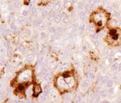
7 Lymphoepithelioid variant


8


9


10 CD 3
 Unclassifiable")
11 Subclassification of PTCL, NOS by gene expression TBX21 / TBET (Th1) Unclassifiable GATA3 (Th2)
12 Subclassification of PTCL, NOS: GATA3 & TBX21 Median OS (yrs) GATA3 Unclassifiable TBX21 /TBET TBX21/TBET (Th1 cells) - GATA3 (Th2 cells)
 CD3+")

13 Angioimmunoblastic T-cell Lymphoma is a disease of germinal center derived T-cells (T FH cell) CD3+ CD10+ BCL6 +/- CD279/PD-1+ CXCL13 + Increased B-cells - both EBV pos and neg CD21 B-cells clonal in up to 50%

14 CD10 Perifollicular Localization of AITL T-cells


15 PD1-CD279

16 What is the utility of PD-1 immunostaining in differential diagnosis of AITL vs Reactive Hyperplasia? Exercise caution Intensity is key! PD-1+ T-cells are invariably present in reactive paracortical hyperplasia

17 Reactive paracortical hyperplasia 18 yo drug hypersensitivity

18 Strong PD-1 + cells in germinal center Weak PD-1 in reactive paracortical T-cells
19 Nodal Peripheral T-cell Lymphomas of TFH origin Angioimmunoblastic T-cell lymphoma Follicular T-cell lymphoma Nodal peripheral T-cell lymphoma with TFH phenotype T-zone variant TFH phenotype requires 2 or more TFH markers Suggested panel: PD1 ICOS CD10 CXCL13
20 B-cell proliferations in AITL & TFH-PTCL EBV-positive Variable numbers of EBV+ blasts, may be dominant picture Hodgkin/Reed-Sternberg like cells EBV-negative B-immunoblasts Polyclonal plasma cells Monotypic/ Monoclonal plasma calls Hodgkin/Reed-Sternberg like cells


21 EBER CD20


22 EBV neg B-cell proliferations in AITL and PTCL-TFH Plasma cells Often Abundant May be monoclonal & atypical Balague et al. Am J Surg Path 2007 CD20 + B-immunoblasts

23 Huppmann et al JCO 2012 T T T


24 CD279/ PD-1 CD3


25 CD138 κ λ
26 TCR IGH FR2 IGH FR3 Huppmann et al JCO 2012


27 Peripheral T-cell lymphoma with EBV+ HRS cells Quintanilla-Martinez et al. Am J Surg Pathol 1999 T-cell population clonal, Cytologically atypical Usually has a TFH phenotype HRS-cells are B-lineage CD30 CD15 EBER
28 PTCL with HRS-like cells An Update (57 cases) Nicolae et al AJSP 2013 PTCL classified as AITL, PTCL, often with T FH markers Intimate relationship between the HRS-like cells & neoplastic T-cells HRS-like cells EBV-positive (52 cases) EBV-negative (5 cases) Progression to classical Hodgkin s lymphoma not observed


29 CD3 CD10 CD15 CD30


30 CD15 PAX5 EBER CD10 PD1 EBV-negative HRS like cells are also rosetted by neoplastic TFH cells
31 Angioimmunoblastic T-cell Lymphoma & PTCL TFH Take Home Points & Remaining Questions AITL is characterized by proliferation and sometimes clonal expansion of B-cells, as well as neoplastic T FH -cells Recent data indicate that B-cells may carry the same mutations as T-cells? TET2 mutations identified in B-cells of patients with AITL (Schwartz FH J Pathol, 2017) AITL may be a stem cell disease
32 Lymphomas of the Innate Immune System γδ T-cells, NK-like T- cells, NK-cells Often cutaneous, mucosal, spleen & BM Cytotoxic Activated cells show frequent apoptosis, necrosis Includes most pediatric T/NK neoplasms
33 Hepatosplenic T-cell lymphoma Most common in young males May be seen with chronic immune suppression Crohn s disease, pts treated with anti-tnf, 6-MP, Azathioprine late occuring PTLD Hepatosplenomegaly, cytopenias, systemic symptoms; lack LN and PB involvement Aggressive behavior and dismal prognosis (<2y survival) Differential Dx: T-LGL in bone marrow, spleen


34

35


36 TCRγ TIA-1 CD5 CD56


37 BM TCRγ CD56
38 Recurrent Mutations in HSTCL HSTCL STAT5B (33%); STAT3 (10%) SETD2 (71%) T-LGL STAT3 (40%); STAT5B (2%) T-ALL JAK1, JAK3, STAT5B (subset) T-PLL STAT5B (36%)
39 Subcutaneous Panniculitis-Like T-cell Lymphoma CLINICAL FEATURES: Broad age range (1 yr to 57 yrs) Median age - 30 Males = Females Deep subcutaneous nodules primarily affecting extremities, trunk Overall survival > 80% 5 years Absence of nodal involvement
40 Subcutaneous Panniculitis-Like T-cell Lymphoma MORPHOLOGY: Usually confined to subcutis, lobular distribution Absence of dermal, epidermal involvement Helpful in distinction from cutaneous γδ T-cell lymphoma Necrosis and karyorrhexis prominent May show vascular invasion


41 CCD8 TIA-1
42 Subcutaneous Panniculitis-like T-cell Lymphoma Immunophenotype & Genotype Activated αβ T-cytotoxic phenotype CD3+, CD8+ TIA-1+, Granzyme B+, Perforin + CD56 negative - in contrast to γδ EBV-negative TCR γ/β genes rearranged Germline mutations in HAVCR2 encoding TIM-3 in both Asian and European patients with HLH & SPTCL (Gayden et al. 2018)
43 Differential Diagnosis of SPTCL Lupus profundus Mixture of T-cells, B-cells, plasma cells Lobular pattern with preserved septa Fibrinoid change in connective tissue Interstitial infiltration, but infrequent rimming of fat spaces Mixture of CD4+/CD8+ cells Scattered gamma-delta T-cells Increased PDC s (CD123+)


44


45 CD4 CD8


46 TCR gamma CD123
47 Subcutaneous Panniculitis-like TCL vs. Lupus profundus Not always easy Oligoclonal T-cell populations can be seen in some patients with lupus, inclusive of the cutaneous lesions Correlate clinical, histological, and genetic features One should be cautious about making the diagnosis of SPTCL in a patient with lupus

48 Enteropathy Associated T-cell Lymphoma (EATL) Broad morphological spectrum Adjacent mucosa shows villous atrophy CD3+, CD103+, Cytotoxic markers, TCR αβ often double negative for CD4/CD8 Often presents with intestinal perforation aggressive clinical course with poor prognosis
49 Enteropathy-associated T-Cell Lymphoma [Classical form or Type I] Associated with celiac disease 95% of patients have HLA-DQ2 and HLA-DQ8 Autoantibodies against tissue transglutaminase Antibodies against gliadin Gluten-free diet may reduce risk of lymphoma

50


51


52 CD30


53 Enteropathy Associated T-cell Lymphoma, Types I & II are distinct EATL I Usually αβ Celiac disease N European EATL II Usually γδ Epitheliotropic Asian, Hispanic γδ

54 Monomorphic epitheliotropic intestinal T-cell lymphoma (EATL II) Medium sized cells with clear cytoplasm CD56 +, CD8+, CD4- Usually gamma delta + MAT kinase + Mutations in STAT5B, JAK3 SETD2

55 JAK/STAT Pathway is an attractive target for therapy of Cytotoxic T-cell Lymphomas and Leukemias
56 T-cell & NK cell Lymphomas of Gastrointestinal Tract EATL Classical αβ > γδ MEITL γδ > αβ Extranodal NK/T EBV+ NK or T Mainly Asian All clinically aggressive All cytotoxic
57 Indolent T-cell lymphoproliferative disease of the GI tract (10 cases) (Perry et al. 2013) Ages (median 48), M:F 6:4 Oral cavity, stomach, small intestine, colon, esophagus Diarrhea, pain, rectal bleeding Crohn s disease (2 patients), Colitis Follow-up: months; Median 38 months 2 pts followed >10 yrs, without progression 6 patients received chemotherapy for PTCL, with no response, but no progression Optimal therapy uncertain

58 Colon

59

60

61 CD3


62 CD8 TIA-1 TCR γ TCR β


63 Superficial infiltrate Confined to mucosa No invasion of the wall Very low proliferation rate No destruction of the glands No cytological atypia Very bland infiltrate? Optimal management Do not respond to chemorx Ki-67
64 Indolent T-cell LPD of GI tract Subset of cases are CD4+ These my ay be more likely to progress to overt T-cell lymphoma Recurrent STAT3-JAK2 fusions in CD4+ cases but not in CD8+ cases (Sharma et al Blood 2018) Prior reports published as Low grade intestinal T-cell lymphoma Lymphomatous polyposis of T-cell type Carbonnel 1994; Egawa 1995; Hirakawa 1996 Margolskee 2013
65 NK-cell Enteropathy An atypical proliferative lesion mimicking lymphoma Mansoor, A., S. et al. Blood 117: , cases: M:F 1:3; Median age 49 (27-70) Vague GI symptoms, but negative for celiac disease Superficial lesions with hemorrhage, edema, ulceration Lesions in stomach, small intestine, and colon Indolent, relapsing clinical course without dissemination Do not mistake for aggressive NK-cell lymphoma


66 Colon biopsy CD56


67 CD3 Colon: Positive for cytoplasmic CD3, CD56, CD7; CD2+/- Negative for CD5, CD4, CD8, EBER

68 Let s switch gears.
 NPM;ALK Hallmark cells")
69 EVOLUTION OF ANAPLASTIC LARGE CELL LYMPHOMA, ALK+ Initial Description Immunophenotypic Studies Molecular Pathogenesis Definition of Entity HD PTCL MH Ki-1+ Sinusoidal lymphoma CD30+ EMA+ LCA+ CD15- CD3 -/+ t(2;5) NPM;ALK Hallmark cells ALK+

70


71


72 ALK CD30


73 CD30 EMA ALK Granzyme B


74 ALCL Bone marrow Involvement Scattered single cells in biopsy & smear Adverse prognostic factor best diagnosed with IHC of bone marrow

75 ALK
76 Translocations and fusion proteins involving ALK Nucleophosmin Anaplastic Lymphoma Kinase Staining Frequency t(2;5) t(1;2) NPM Tropomyosin 3 TPM3 ALK ALK cytoplasmic/ nuclear/ nucleolar cytoplasmic 70-80% 10-20% t(2;3) Trk Fusion Gene TFG ALK cytoplasmic 2-5% Inv2 ATIC (Pur H gene) ATIC ALK cytoplasmic 2-5% t(2;17) Clathrin heavy chain CLTC ALK cytoplasmic granular 2-5% t(2;19) TPM4 ALK cytoplasmic 1-2%
77 Histological Spectrum of ALCL, ALK+ Anaplastic large cell lymphoma Common Lymphohistiocytic Small cell Other histological patterns Sarcomatoid appearance with myxoid stroma Hypocellular with edematous background


78 Lymphohistiocytic variant of ALCL CD30


79 CD30


80 PC ALCL Small Cell Variant of ALCL

81 CD 30
82 Time to treatment failure curve according to the presence of a small-cell (SC) and/or lymphohistiocytic (LH) component (n = 361 patients). Lamant L et al. JCO 2011;29: by American Society of Clinical Oncology
83 Genetic correlates with survival in ALCL, ALK+/ ALK- Feldman et al. Blood 2014 Percent Survival p< Months After ALCL Diagnosis Subset with DUSP22 R Comparable to ALK+ DUSP22 (# 22) ALK+ (# 32) P63 (# 6) ALK neg, no aberrations (#45)


84 DUSP 22 Mutated ALCL has distinctive morphology and phenotype King RL et al. Am J Surg Pathol 2016 Classical Hallmark cells Granzyme B negative
85 WHO Classification of T/NK cell neoplasms Leukemic/ Systemic T-cell prolymphocytic leukaemia T-cell large granular lymphocytic leukaemia Chronic lymphoproliferative disorder of NK cells Aggressive NK cell leukaemia Systemic EBV+ T-cell Lymphoma of childhood Hydroa vacciniforme-like lymphoproliferative disorder Adult T-cell leukaemia/lymphoma Hepatosplenic T-cell lymphoma Cutaneous Subcutaneous panniculitis- like T-cell lymphoma Mycosis fungoides/ Sézary syndrome Primary cutaneous CD30 positive T-cell lymphoproliferative disorders Lymphomatoid papulosis Primary cutaneous anaplastic large cell lymphoma Primary cutaneous gamma-delta T-cell lymphoma Primary cutaneous CD8 positive aggressive epidermotropic cytotoxic T-cell lymphoma Primary cutaneous acral CD8+ T-cell lymphoma Primary cutaneous CD4 positive small/medium T-cell lymphoproliferative disorder Extranodal Extranodal NK/T-cell lymphoma, nasal type Enteropathy-associated T-cell lymphoma Monomorphic epitheliotropic intestinal T-cell lymphoma Indolent T-cell lymphoproliferative disorder of the GI tract Breast implant-associated anaplastic large cell lymphoma Nodal/ Extranodal Peripheral T-cell lymphoma, NOS Angioimmunoblastic T-cell lymphoma Follicular T-cell lymphoma Nodal peripheral T-cell lymphoma with TFH phenotype Anaplastic large cell lymphoma, ALK positive Anaplastic large cell lymphoma, ALK negative
What s new on the horizon in T-cell lymphoma Elaine S Jaffe National Cancer Institute, Bethesda MD
 What s new on the horizon in T-cell lymphoma Elaine S Jaffe National Cancer Institute, Bethesda MD WHO classification: where are we today? Of 12 monographs planned for 4 th Edition Bluebook series, only
Peripheral T-cell lymphomas
 XXXVI REUNIÓN ANUAL Peripheral T-cell lymphomas Dr. Antonio Martinez Hospital Clinic, University ofbarcelona antonmar@clinic.ub.es Madrid, February 8th, 2013 T-NHL vs B-NHL: the T-cell paradigm lambda
Hepatic Lymphoma Diagnosis An Algorithmic Approach
 Hepatic Lymphoma Diagnosis An Algorithmic Approach Ryan M. Gill, M.D., Ph.D. University of California, San Francisco PLEASE TURN OFF YOUR CELL PHONES Disclosure of Relevant Financial Relationships USCAP
T cell lymphoma diagnostics and differential diagnosis to Hodgkin lymphoma
 T cell lymphoma diagnostics and differential diagnosis to Hodgkin lymphoma Sylvia Hartmann Dr. Senckenberg Institute of Pathology Goethe University Frankfurt Overview Borderline ALCL classical HL Borderline
Anaplastic Large Cell Lymphoma (of T cell lineage)
") Anaplastic Large Cell Lymphoma (of T cell lineage) Definition T-cell lymphoma comprised of large cells with abundant cytoplasm and pleomorphic, often horseshoe-shaped nuclei CD30+ Most express cytotoxic
Anaplastic Large Cell Lymphoma (of T cell lineage) Definition T-cell lymphoma comprised of large cells with abundant cytoplasm and pleomorphic, often horseshoe-shaped nuclei CD30+ Most express cytotoxic
SH/EAHP Workshop 2011 Los Angeles, California, USA
 SH/EAHP Workshop 2011 Los Angeles, California, USA October 27-29, 2011 Session 3 Non-Mycosis Fungoides CTCL Patty Jansen & Rein Willemze Introduction Submitted: 101 cases + 7 cases group 1: 108 Deactivated
Commentary on the 2008 WHO classification of mature T- and NK-cell neoplasms
 J Hematopathol (2009) 2:65 73 DOI 10.1007/s12308-009-0034-z COMMENT Commentary on the 2008 WHO classification of mature T- and NK-cell neoplasms Megan S. Lim & Laurence de Leval & Leticia Quintanilla-Martinez
Primary Cutaneous CD30-Positive T-cell Lymphoproliferative Disorders
 Primary Cutaneous CD30-Positive T-cell Lymphoproliferative Disorders Definition A spectrum of related conditions originating from transformed or activated CD30-positive T-lymphocytes May coexist in individual
T-cell Lymphomas: Diagnosis and New Agents. Mary Jo Lechowicz Thursday, July 27 Debates and Didactics in Hematology and Oncology
 T-cell Lymphomas: Diagnosis and New Agents Mary Jo Lechowicz Thursday, July 27 Debates and Didactics in Hematology and Oncology 1 Mature T and NK-cell neoplasms in the WHO Classification 2016 revision
Lymphoma/CLL 101: Know your Subtype. Dr. David Macdonald Hematologist, The Ottawa Hospital
 Lymphoma/CLL 101: Know your Subtype Dr. David Macdonald Hematologist, The Ottawa Hospital Function of the Lymph System Lymph Node Lymphocytes B-cells develop in the bone marrow and influence the immune
88-year-old Female with Lymphadenopathy. Faizi Ali, MD
 88-year-old Female with Lymphadenopathy Faizi Ali, MD Clinical History A 88-year-old caucasian female presented to our hospital with the complaints of nausea, vomiting,diarrhea, shortness of breath and
Lymphoma Update: Lymphoma Update: What s Likely to be New in the New WHO. Patrick Treseler, MD, PhD University of California San Francisco
 Lymphoma Update: What s Likely to be New in the New WHO Blood 127:2375; 2016 Patrick Treseler, MD, PhD University of California San Francisco Lymphoma Update: What IS New in the New WHO! Patrick Treseler,
Non-Hodgkin s Lymphomas Version
 NCCN Clinical Practice Guidelines in Oncology (NCCN Guidelines ) Non-Hodgkin s Lymphomas Version 2.2015 NCCN.org Continue Use of Immunophenotyping/ Genetic Testing in Differential Diagnosis of Mature B-Cell
Unusual cutaneous presentation of a T-cell lymphoproliferation
 Department of Pathology and Cytology University Hospital Centre Zagreb, Croatia Unusual cutaneous presentation of a T-cell lymphoproliferation Snjezana Dotlic, Stefan Dojcinov, Leticia Quintanilla-Fend
WHO UPDATE ON LYMPHOMAS. Dr Priya Mary Jacob Asst Professor, Pathology.
 WHO UPDATE ON LYMPHOMAS Dr Priya Mary Jacob Asst Professor, Pathology 3 rd 4 th 4 th revised 2001 2008 2017 The Change The Significance of the Change- Diagnostic, Prognostic The Rationale behind the change.
Common Problem Areas. WHO Classification. Defines separate diseases (entities) with their CLINICAL AGGRESSIVENESS LOW GRADE / HIGH GRADE
 with their CLINICAL AGGRESSIVENESS LOW GRADE / HIGH GRADE") WHO Classification Defines separate diseases (entities) with their CLINICAL AGGRESSIVENESS REVIEW OF MOST COMMON LYMPHOMA ENTITIES Dr Stefan Dojcinov LOW GRADE / HIGH GRADE (June 2014) The Non-Hodgkin
WHO Classification Defines separate diseases (entities) with their CLINICAL AGGRESSIVENESS REVIEW OF MOST COMMON LYMPHOMA ENTITIES Dr Stefan Dojcinov LOW GRADE / HIGH GRADE (June 2014) The Non-Hodgkin
Recent diagnostic and therapeutic innovations of T-cell-lymphoma. Prof. Nossrat Firusian, Recklinghausen, Germany
 Recent diagnostic and therapeutic innovations of T-cell-lymphoma Prof. Nossrat Firusian, Recklinghausen, Germany NODAL Angioimmunoblastic T-cell Lymphoma Peripheral T-cell-Lymphoma Anaplastic Large-cell-Lymphoma
10/31/2017. Immunodeficiencies. Outline. Discuss EBV. Non-destructive Polymorphic Monomorphic Therapies Challenges
 I have no financial disclosures Joo Y. Song, MD Assistant Professor of Clinical Pathology City of Hope National Medical Center Immunodeficiencies Outline Transplant Congenital Autoimmunity T-cell/immune
Methods used to diagnose lymphomas
 Institut für Pathologie Institut für Pathologie Methods used to diagnose lymphomas Prof. Dr.Med. Leticia Quintanilla-Fend Molecular techniques NGS histology Cytology AS-PCR Sanger seq. MYC Immunohistochemistry
ECP meeting, Lisbon, september 2012 Slide seminar New and old challenges in the diagnosis of peripheral T-cell lymphomas
 ECP meeting, Lisbon, september 2012 Slide seminar New and old challenges in the diagnosis of peripheral T-cell lymphomas Philippe Gaulard, Dept of Pathology, INSERM U955, Hôpital Henri Mondor, 94010 -
11/2/2017. Immunodeficiencies. Joo Y. Song, MD Assistant Professor of Clinical Pathology. I have no financial disclosures.
 I have no financial disclosures Joo Y. Song, MD Assistant Professor of Clinical Pathology City of Hope National Medical Center Immunodeficiencies Transplant Autoimmunity Drugs T-cell dysfunction (Age,
Immunopathology of Lymphoma
 Immunopathology of Lymphoma Noraidah Masir MBBCh, M.Med (Pathology), D.Phil. Department of Pathology Faculty of Medicine Universiti Kebangsaan Malaysia Lymphoma classification has been challenging to pathologists.
Lymphadenopathies mimicking lymphoma and vice versa. Session 3
 Lymphadenopathies mimicking lymphoma and vice versa Session T/ NK Cell System The human and rodent T and NK cell system is characterized by many highly specialized cell suopulations and functions, that
Monoclonal B-cell Lymphocytosis
 Entity Centred Approach Lymphoma Classification: WHO and Beyond Clinically meaningful categories Dr Stefan Dojcinov University Hospital of Wales, Cardiff WHO UPDATE - NEW ENTITIES Early lesions lymphoma
WHO Classification. B-cell chronic lymphocytic leukemia/small T-cell granular lymphocytic leukemia
 Blood Malignancies-II Prof. Dr. Herman Hariman, a Ph.D, SpPK (KH). Prof. Dr. Adikoesoema Aman, SpPK (KH) Dept. of Clinical Pathology, School of Medicine, University of North Sumatra WHO classification
Differential diagnosis of hematolymphoid tumors composed of medium-sized cells. Brian Skinnider B.C. Cancer Agency, Vancouver General Hospital
 Differential diagnosis of hematolymphoid tumors composed of medium-sized cells Brian Skinnider B.C. Cancer Agency, Vancouver General Hospital Lymphoma classification Lymphoma diagnosis starts with morphologic
Overview of Cutaneous Lymphomas: Diagnosis and Staging. Lauren C. Pinter-Brown MD, FACP Health Sciences Professor of Medicine and Dermatology
 Overview of Cutaneous Lymphomas: Diagnosis and Staging Lauren C. Pinter-Brown MD, FACP Health Sciences Professor of Medicine and Dermatology Definition of Lymphoma A cancer or malignancy that comes from
Changing the landscape of treatment in Peripheral T-cell Lymphoma
 Changing the landscape of treatment in Peripheral T-cell Lymphoma Luis Fayad Associate Professor MD Anderson Cancer Center Department of Lymphoma and Myeloma 1 6 What is peripheral 2008 WHO CLASSIFICATION
Contents. vii. Preface... Acknowledgments... v xiii
 Contents Preface... Acknowledgments... v xiii SECTION I 1. Introduction... 3 Knowledge-Based Diagnosis... 4 Systematic Examination of the Lymph Node... 7 Cell Type Identification... 9 Cell Size and Cellularity...
Incidence. Bimodal age incidence 15-40, >55 years Childhood form (0-14) more common in developing countries M:F=1.5:1; in all subtypes except NS
 more common in developing countries M:F=1.5:1; in all subtypes except NS") Hodgkin Lymphoma Hodgkin Lymphoma 30% of all lymphomas Absolute incidence unchanged Arise in lymph node, cervical region Neoplastic tissues usually contain a small number of tumor cells Incidence Bimodal
Hodgkin Lymphoma Hodgkin Lymphoma 30% of all lymphomas Absolute incidence unchanged Arise in lymph node, cervical region Neoplastic tissues usually contain a small number of tumor cells Incidence Bimodal
Michi Shinohara MD Associate Professor University of Washington/Seattle Cancer Care Alliance Dermatology, Dermatopathology
 Michi Shinohara MD Associate Professor University of Washington/Seattle Cancer Care Alliance Dermatology, Dermatopathology Agenda Overview of cutaneous T and B- cell lymphomas Diagnosis, Staging, Prognosis
Pearls and pitfalls in interpretation of lymphoid lesions in needle biopsies
 Pearls and pitfalls in interpretation of lymphoid lesions in needle biopsies Megan S. Lim MD PhD University of Pennsylvania October 8, 2018 Objectives To understand how the trend toward less invasive lymph
The 2016 updated WHO classification of lymphoid neoplasias
 DOI: 10.1002/hon.2399 SUPPLEMENT ARTICLE The 2016 updated WHO classification of lymphoid neoplasias Leticia Quintanilla Martinez Institute of Pathology, University Hospital Tübingen, Eberhard Karls University
3/23/2017. Disclosure of Relevant Financial Relationships. Pitfalls in Immunohistochemistry in Hematopathology: CD20 and CD3 Can Let Me Down?!
 Pitfalls in Immunohistochemistry in Hematopathology: CD20 and CD3 Can Let Me Down?! Judith A. Ferry Massachusetts General Hospital Disclosure of Relevant Financial Relationships USCAP requires that all
2010 Hematopoietic and Lymphoid ICD-O Codes - Alphabetical List THIS TABLE REPLACES ALL ICD-O-3 Codes
 Acute basophilic leukemia 9870/3 Acute biphenotypic leukemia [OBS] 9805/3 Acute erythroid leukemia 9840/3 Acute megakaryoblastic leukemia 9910/3 Acute monoblastic and monocytic leukemia 9891/3 Acute myeloid
2012 Hematopoietic and Lymphoid ICD-O Codes - Numerical List THIS TABLE REPLACES ALL ICD-O-3 Codes
 Malignant lymphoma, NOS 9590/3 Non-Hodgkin lymphoma, NOS 9591/3 B-cell lymphoma, unclassifiable, with features intermediate between diffuse large B-cell lymphoma and classical Hodgkin lymphoma 9596/3 Primary
Bone Marrow. Procedures Blood Film Aspirate, Cell Block Trephine Biopsy, Touch Imprint
 Bone Marrow Protocol applies to acute leukemias, myelodysplastic syndromes, myeloproliferative disorders, chronic lymphoproliferative disorders, malignant lymphomas, plasma cell dyscrasias, histiocytic
Classification of Cutaneous T cell Lymphomas (CTCLs) Hernani Cualing, MD
 Hernani Cualing, MD") Classification of Cutaneous T cell Lymphomas (CTCLs) Hernani Cualing, MD Pathology and Cell Biology, USF IFLOW, Inc. CTCL, MF, and Sézary syndrome In 1806, mycosis fungoides (MF) was first described 1
Classification of Cutaneous T cell Lymphomas (CTCLs) Hernani Cualing, MD Pathology and Cell Biology, USF IFLOW, Inc. CTCL, MF, and Sézary syndrome In 1806, mycosis fungoides (MF) was first described 1
Immunohistochemical classification of haematolymphoid tumours. Stephen Hamilton-Dutoit Institute of Pathology Aarhus University Hospital
 Immunohistochemical classification of haematolymphoid tumours Stephen Hamilton-Dutoit Institute of Pathology Aarhus University Hospital Malignant lymphoproliferative diseases What are they? Haematolymphoid
Corrigenda. WHO Classification of Tumours of Haematopoietic and Lymphoid Tissues (revised 4th edition): corrections made in second print run
: corrections made in second print run") Corrigenda WHO Classification of Tumours of Haematopoietic and Lymphoid Tissues (revised 4th edition): corrections made in second print run In addition to corrections of minor typographical errors, corrections
Corrigenda WHO Classification of Tumours of Haematopoietic and Lymphoid Tissues (revised 4th edition): corrections made in second print run In addition to corrections of minor typographical errors, corrections
Combinations of morphology codes of haematological malignancies (HM) referring to the same tumour or to a potential transformation
 referring to the same tumour or to a potential transformation") Major subgroups according to the World Health Organisation (WHO) Classification Myeloproliferative neoplasms (MPN) Myeloid and lymphoid neoplasms with eosinophilia and abnormalities of PDGFRA, PDGFRB or
Major subgroups according to the World Health Organisation (WHO) Classification Myeloproliferative neoplasms (MPN) Myeloid and lymphoid neoplasms with eosinophilia and abnormalities of PDGFRA, PDGFRB or
Small B-cell (Histologically Low Grade) Lymphoma
 Lymphoma") Frequency of Lymphoid Neoplasms Small B-cell (Histologically Low Grade) Lymphoma Stephen Hamilton-Dutoit Institute of Pathology Aarhus University Hospital B-cell neoplasms 88% Diffuse large B-cell lymphoma
Frequency of Lymphoid Neoplasms Small B-cell (Histologically Low Grade) Lymphoma Stephen Hamilton-Dutoit Institute of Pathology Aarhus University Hospital B-cell neoplasms 88% Diffuse large B-cell lymphoma
Non-Hodgkin lymphomas (NHLs) Hodgkin lymphoma )HL)
 Hodgkin lymphoma )HL)") Non-Hodgkin lymphomas (NHLs) Hodgkin lymphoma )HL) Lymphoid Neoplasms: 1- non-hodgkin lymphomas (NHLs) 2- Hodgkin lymphoma 3- plasma cell neoplasms Non-Hodgkin lymphomas (NHLs) Acute Lymphoblastic Leukemia/Lymphoma
Non-Hodgkin lymphomas (NHLs) Hodgkin lymphoma )HL) Lymphoid Neoplasms: 1- non-hodgkin lymphomas (NHLs) 2- Hodgkin lymphoma 3- plasma cell neoplasms Non-Hodgkin lymphomas (NHLs) Acute Lymphoblastic Leukemia/Lymphoma
Prognostic Factors for PTCL. Julie M. Vose, M.D., M.B.A. University of Nebraska Medical Center
 Prognostic Factors for PTCL Julie M. Vose, M.D., M.B.A. University of Nebraska Medical Center jmvose@unmc.edu Distribution of 1314 Cases by Consensus Diagnosis International T-Cell Lymphoma Project Vose
Defined lymphoma entities in the current WHO classification
 Defined lymphoma entities in the current WHO classification Luca Mazzucchelli Istituto cantonale di patologia, Locarno Bellinzona, January 29-31, 2016 Evolution of lymphoma classification Rappaport Lukes
Lymphoma and Pseudolymphoma
 Lymphoma and Pseudolymphoma Laura B. Pincus, MD Co-Director, Cutaneous Lymphoma Clinic Associate Professor Dermatology and Pathology University of California, San Francisco I HAVE NO RELEVANT RELATIONSHIPS
Integrated Hematopathology. Morphology and FCI with IHC
 Integrated Hematopathology Morphology and FCI with IHC FrontMatter.indd i 9/6/2009 9:30:12 PM FrontMatter.indd ii 9/6/2009 9:30:18 PM Integrated Hematopathology Morphology and FCI with IHC Cherie H Dunphy,
ACCME/Disclosures 4/13/2016. Clinical History
 ACCME/Disclosures The USCAP requires that anyone in a position to influence or control the content of CME disclose any relevant financial relationship WITH COMMERCIAL INTERESTS which they or their spouse/partner
Exploring the Borderlands between Diffuse Large B-cell Lymphoma and Classical Hodgkin s Lymphoma
 Exploring the Borderlands between Diffuse Large B-cell Lymphoma and Classical Hodgkin s Lymphoma Elaine S. Jaffe National Cancer Institute Bethesda, MD, USA On the Pathological Changes In Hodgkin s Disease
T-cell Lymphomas Biology and Management
 T-cell Lymphomas Biology and Management March-27-2017 Outline Epidemiology Initial Work-up International Prognostic Index Treatment of Diffuse Large B-cell Lymphoma: -Limited Stage -Advanced Stage Frontline:
HODGKIN LYMPHOMA DR. ALEJANDRA ZARATE OSORNO HOSPITAL ESPAÑOL DE MEXICO
 HODGKIN LYMPHOMA DR. ALEJANDRA ZARATE OSORNO HOSPITAL ESPAÑOL DE MEXICO HODGKIN LYMPHOMA CLASSIFICATION Lukes & Butler Rye WHO-2016 Linphocytic and/or histiocytic Nodular & diffuse Nodular Sclerosis Lymphocyte
Methotrexate-associated Lymphoproliferative Disorders
 Methotrexate-associated Lymphoproliferative Disorders Definition A lymphoid proliferation or lymphoma in a patient immunosuppressed with methotrexate, typically for treatment of autoimmune disease (rheumatoid
Non-Hodgkin Lymphoma. Protocol applies to non-hodgkin lymphoma involving any organ system except the gastrointestinal tract.
 Non-Hodgkin Lymphoma Protocol applies to non-hodgkin lymphoma involving any organ system except the gastrointestinal tract. Protocol revision date: January 2005 No AJCC/UICC staging system Procedures Cytology
FOLLICULARITY in LYMPHOMA
 FOLLICULARITY in LYMPHOMA Reactive Follicular Hyperplasia Follicular Hyperplasia irregular follicles Follicular Hyperplasia dark and light zones Light Zone Dark Zone Follicular hyperplasia MIB1 Follicular
The spectrum of flow cytometry of the bone marrow
 The spectrum of flow cytometry of the bone marrow Anna Porwit Lund University Faculty of Medicine Dept. of Clinical Sciences Div. Oncology and Pathology anna.porwit@med.lu.se Disclosure of speaker s interests
Classification of Hematologic Malignancies. Patricia Aoun MD MPH
 Classification of Hematologic Malignancies Patricia Aoun MD MPH Objectives Know the basic principles of the current classification system for hematopoietic and lymphoid malignancies Understand the differences
Aggressive B-Cell Lymphomas
 Aggressive B-cell Lymphomas Aggressive B-Cell Lymphomas Stephen Hamilton Dutoit Institute of Pathology Aarhus Kommunehospital B-lymphoblastic lymphoma Diffuse large cell lymphoma, NOS T-cell / histiocyte-rich;
The History of Lymphoma Classification and the 2017 Revision
 The History of Lymphoma Classification and the 2017 Revision ESMO Perceptorship on Lymphoma, Lugano 2018 German Ott Department of Clinical Pathology, Robert-Bosch-Krankenhaus and Dr. Margarete Fischer-Bosch
Cutaneous Lymphoid Proliferations: A Comprehensive Textbook of Lymphocytic Infiltrates of the Skin
 Cutaneous Lymphoid Proliferations: A Comprehensive Textbook of Lymphocytic Infiltrates of the Skin Magro, Cynthia M., MD ISBN-13: 9780471695981 Table of Contents Chapter One: Introduction to the Classification
Disclosures. Diagnostic Issues. Sinusoidal Infiltrates in the Liver. Ryan M. Gill, M.D., Ph.D. Assistant Professor Department of Pathology.
 Disclosures I have nothing to disclose Sinusoidal Infiltrates in the Liver Ryan M. Gill, M.D., Ph.D. Assistant Professor Department of Pathology Diagnostic Issues Reactive infiltrates are common and may
LYMPHOMAS an overview of some subtypes of NHLs
 One of the confusing aspects of the lymphoid neoplasms concerns the use of the descriptive terms "leukemia" and "lymphoma." LYMPHOMAS an overview of some subtypes of NHLs Leukemia is used for lymphoid
Molecular Pathology of Lymphoma (Part 1) Rex K.H. Au-Yeung Department of Pathology, HKU
 Rex K.H. Au-Yeung Department of Pathology, HKU") Molecular Pathology of Lymphoma (Part 1) Rex K.H. Au-Yeung Department of Pathology, HKU Lecture outline Time 10:00 11:00 11:15 12:10 12:20 13:15 Content Introduction to lymphoma Review of lymphocyte biology
Molecular Pathology of Lymphoma (Part 1) Rex K.H. Au-Yeung Department of Pathology, HKU Lecture outline Time 10:00 11:00 11:15 12:10 12:20 13:15 Content Introduction to lymphoma Review of lymphocyte biology
ACCME/Disclosures ALK FUSION-POSITIVE MESENCHYMAL TUMORS. Tumor types with ALK rearrangements. Anaplastic Lymphoma Kinase. Jason L.
 Companion Meeting of the International Society of Bone and Soft Tissue Pathology The Evolving Concept of Mesenchymal Tumors ALK FUSION-POSITIVE MESENCHYMAL TUMORS Jason L. Hornick, MD, PhD March 13, 2016
Gene expression profiling in peripheral T-cell lymphoma. Wing C Chan City of Hope Medical Center Wing C Chan City of Hope National Medical Center
 Gene expression profiling in peripheral T-cell lymphoma Wing C Chan City of Hope Medical Center Wing C Chan City of Hope National Medical Center Rationale for GEP studies on PTCL The diagnosis of PTCL
Change Summary - Form 2018 (R3) 1 of 12
 1 of 12") Summary - Form 2018 (R3) 1 of 12 Form Question Number (r3) Type Description New Text Previous Text Today's date was removed 2018 N/A Today's Date Removed from Key Fields 2018 N/A HCT Type 2018 N/A Product
Summary - Form 2018 (R3) 1 of 12 Form Question Number (r3) Type Description New Text Previous Text Today's date was removed 2018 N/A Today's Date Removed from Key Fields 2018 N/A HCT Type 2018 N/A Product
Disclosures. Advisory Board. Consultant. Investigator. MiRagen, Actelion, Celgene, Therakos. Mindera
 Cutaneous Lymphomas Christiane Querfeld, MD, PhD Director, Cutaneous Lymphoma Program City of Hope ~ How the Experts Treat Hematologic Malignancies Symposium March 10 13, 2017 Disclosures Advisory Board
Peripheral T-cell Lymphomas
 Peripheral T-cell Lymphomas Teresa Palomero, PhD Institute for Cancer Genetics Department of Pathology and Cell Biology Herbert Irving Comprehensive Cancer Center Columbia University Medical Center T-lymphocytes
High grade B-cell lymphomas (HGBL): Altered terminology in the 2016 WHO Classification (Update of the 4 th Edition) and practical issues Xiao-Qiu Li,
: Altered terminology in the 2016 WHO Classification (Update of the 4 th Edition) and practical issues Xiao-Qiu Li,") High grade B-cell lymphomas (HGBL): Altered terminology in the 2016 WHO Classification (Update of the 4 th Edition) and practical issues Xiao-Qiu Li, M.D., Ph.D. Fudan University Shanghai Cancer Center
High grade B-cell lymphomas (HGBL): Altered terminology in the 2016 WHO Classification (Update of the 4 th Edition) and practical issues Xiao-Qiu Li, M.D., Ph.D. Fudan University Shanghai Cancer Center
Classification! Immunohistochemical classification of haematolymphoid tumours. Malignant lymphoproliferative diseases
 Immunohistochemical classification of haematolymphoid tumours Haematolymphoid Neoplasias: Leukaemia vs Lymphoma C L O N A L M A L I G N A N C I E S Stephen Hamilton-Dutoit Institute of Pathology Aarhus
Lymphoma: What You Need to Know. Richard van der Jagt MD, FRCPC
 Lymphoma: What You Need to Know Richard van der Jagt MD, FRCPC Overview Concepts, classification, biology Epidemiology Clinical presentation Diagnosis Staging Three important types of lymphoma Conceptualizing
Case year old male with abdominal lymphadenopathy Treated with 8 cycles of R-CHOP One year later B-symptoms and progressive disease
 Codirectors Tsieh Sun, M.D., FASCP Francisco Vega, M.D., Ph.D. Department of Hematopathology UT MD Anderson Cancer Center Houston Texas There is no conflict of interest involved in the content and presentation
Lymphoma: The Basics. Dr. Douglas Stewart
 Lymphoma: The Basics Dr. Douglas Stewart Objectives What is lymphoma? How common is it? Why does it occur? How do you diagnose it? How do you manage it? How do you follow patients after treatment? What
From Morphology to Molecular Pathology: A Practical Approach for Cytopathologists Part 1-Cytomorphology. Songlin Zhang, MD, PhD LSUHSC-Shreveport
 From Morphology to Molecular Pathology: A Practical Approach for Cytopathologists Part 1-Cytomorphology Songlin Zhang, MD, PhD LSUHSC-Shreveport I have no Conflict of Interest. FNA on Lymphoproliferative
Thomas Hodgkin and Hodgkin lymphoma
 J Hematopathol (2014) 7:123 138 DOI 10.1007/s12308-014-0214-3 REVIEW ARTICLE Thomas Hodgkin and Hodgkin lymphoma Judith A. Ferry Received: 26 June 2014 /Accepted: 31 July 2014 /Published online: 12 August
New Haven, Connecticut
 New Haven, Connecticut Yale University Main Campus Yale mascot: Handsome Dan Cutaneous Lymphomas Tony Subtil, MD, MBA Associate Professor Yale University Cutaneous Lymphomas: 1. Intro 2. CTCL/NK 3. CBCL
Chronic Lymphocytic Leukemia Mantle Cell Lymphoma Elias Campo
 Chronic Lymphocytic Leukemia Mantle Cell Lymphoma Elias Campo Hospital Clinic, University of Barcelona Small B-cell lymphomas NAIVE -B LYMPHOCYTE MEMORY CELL CLL MCL FL MZL Small cell size Low proliferation
184 Cutaneous Lymphomas: Morphology, Immunohistochemistry and Molecular Testing. David Cassarino MD, PhD Aaron Auerbach MD
 184 Cutaneous Lymphomas: Morphology, Immunohistochemistry and Molecular Testing David Cassarino MD, PhD Aaron Auerbach MD 2011 Annual Meeting Las Vegas, NV AMERICAN SOCIETY FOR CLINICAL PATHOLOGY 33 W.
Aggressive B-cell Lymphoma 2013
 Aggressive B-cell Lymphoma 2013 Diffuse Large B-Cell Lymphoma Burkitt Lymphoblastic lymphoma Gray zone Intermediate DLBCL/HL Intermediate BL/DLBCL Diffuse Large B-cell lymphoma Common morphology: diffuse
WHO 2016 update lymphoid neoplasms. Dr Sue Morgan Alfred Hospital, Melbourne
 WHO 2016 update lymphoid neoplasms Dr Sue Morgan Alfred Hospital, Melbourne WHO 4 th edition 2008 Established guideline worldwide for diagnosis of haematological malignancy for the last 9 years Significant
Supportive Care in the Management of T-cell Lymphomas
 Supportive Care in the Management of T-cell Lymphomas Erin Kopp, ACNP-BC City of Hope Comprehensive Cancer Center NCCN.org For Clinicians NCCN.org/patients For Patients Objectives Discuss the role of supportive
Classifications of lymphomas
 Classifications of lymphomas Lukes and Collins Kiel classification Working formulation REAL classification (1994) WHO classification (2000) WHO CLASSIFICATIONF OF NEOPLASMS HAEMATOPETIC AND LYMPHOID TISSUES
7 Omar Abu Reesh. Dr. Ahmad Mansour Dr. Ahmad Mansour
 7 Omar Abu Reesh Dr. Ahmad Mansour Dr. Ahmad Mansour -Leukemia: neoplastic leukocytes circulating in the peripheral bloodstream. -Lymphoma: a neoplastic process in the lymph nodes, spleen or other lymphatic
Interesting case in lymphoma. Kitsada Wudhikarn, MD Division of Hematology, Department of Medicine Faculty of Medicine, Chulalongkorn University
 Interesting case in lymphoma Kitsada Wudhikarn, MD Division of Hematology, Department of Medicine Faculty of Medicine, Chulalongkorn University Eosinophilia Benign reactive etiologies 1. Allergy 2. Infection
Large cell immunoblastic Diffuse histiocytic (DHL) Lymphoblastic lymphoma Diffuse lymphoblastic Small non cleaved cell Burkitt s Non- Burkitt s
 Lymphoblastic lymphoma Diffuse lymphoblastic Small non cleaved cell Burkitt s Non- Burkitt s") Non Hodgkin s Lymphoma Introduction 6th most common cause of cancer death in United States. Increasing in incidence and mortality. Since 1970, the incidence of has almost doubled. Overview The types of
Non Hodgkin s Lymphoma Introduction 6th most common cause of cancer death in United States. Increasing in incidence and mortality. Since 1970, the incidence of has almost doubled. Overview The types of
SH Comprehensive Molecular Profiling of an ALK-Negative, Anaplastic Large Cell Lymphoma with DUSP22 rearrangement
 SH2017-0277 Comprehensive Molecular Profiling of an ALK-Negative, Anaplastic Large Cell Lymphoma with DUSP22 rearrangement Caleb Ho, M.D.; Alexander Chan, M.D., Yanming Zhang, M.D.; Lu Wang, M.D., Ph.D;
Case Presentation. Maha Akkawi, MD, Fatima Obeidat, MD, Tariq Aladily, MD. Department of Pathology Jordan University Hospital Amman, Jordan
 Case Presentation Maha Akkawi, MD, Fatima Obeidat, MD, Tariq Aladily, MD Department of Pathology Jordan University Hospital Amman, Jordan The 25th Annual Congress of the ADIAP The 8/11/2013 1 5th International
Peripheral T-cell lymphomas (PTCL) Specified and Unspecified. Eric Van Den Neste Cliniques universitaires Saint-Luc Bruxelles
 Specified and Unspecified. Eric Van Den Neste Cliniques universitaires Saint-Luc Bruxelles") Peripheral T-cell lymphomas (PTCL) Specified and Unspecified Eric Van Den Neste Cliniques universitaires Saint-Luc Bruxelles BHS seminar 12, 07 March 2015 Peripheral T-cell lymphomas (PTCL) Specified and
Peripheral T-cell lymphomas (PTCL) Specified and Unspecified Eric Van Den Neste Cliniques universitaires Saint-Luc Bruxelles BHS seminar 12, 07 March 2015 Peripheral T-cell lymphomas (PTCL) Specified and
T CELL LYMPHOMA ANALYSIS
 T CELL LYMPHOMA ANALYSIS Charles Goolsby, Ph.D. Floyd E. Patterson Research Professor of Pathology Northwestern Feinberg School of Medicine c-goolsby@northwestern.edu 1 T CELL LYMPHOMA ANALYSIS Diverse
CASE year old male with a PET avid nodule in the left adrenal gland
 CASE 1 55 year old male with a PET avid nodule in the left adrenal gland Case 1 Adrenal gland parenchyma partly replaced by a spindle cell tumour with mild nuclear pleomorphism Atypical mitoses present
EQA SCHEME CIRCULATION 33 EDUCATIONAL SLIDES DR GRAEME SMITH MONKLANDS DGH
 EQA SCHEME CIRCULATION 33 EDUCATIONAL SLIDES DR GRAEME SMITH MONKLANDS DGH CASE E1 M: 68 yrs Left destructive sinonasal lesion.?lymphoma?adenocarcinoma CD20 CD10 BCL6 MIB1 Answers Diffuse large B cell
Dermatopathology. Dr. Rafael Botella Estrada. Hospital La Fe de Valencia
 Dermatopathology Dr. Rafael Botella Estrada. Hospital La Fe de Valencia Melanoma and mimics Dr. Martin Mihm Malignant lesions result from the accumulation of mutations Class I lesions (benign) Class II
A Unique Case of Nasal NK/T Cell Lymphoma with Frequent Remission and Relapse Showing Different Histological Features During 12 Years of Follow Up
 J Clin Exp Hematopathol Vol. 50, No. 1, May 2010 Case Study A Unique Case of Nasal NK/T Cell Lymphoma with Frequent Remission and Relapse Showing Different Histological Features During 12 Years of Follow
Low-grade B-cell lymphoma
 Low-grade B-cell lymphoma Patho-Basic 11. September 2018 Stephan Dirnhofer Pathology Outline Definition LPL, MBL/CLL/SLL, MCL FL Subtypes & variants Diagnosis including Grading Transformation Summary Be
Aggressive B-cell Lymphomas
 Neoplastic Hematopathology Update 2018 Aggressive B-cell Lymphomas Raju K. Pillai City of Hope National Medical Center I do not have any disclosures Disclosures Outline New entities and changes in WHO
CASE 35 CLINICAL HISTORY
 Female, 24 Painful ulcerated lesion Left buttock Developed over a few weeks?abscess Excision CASE 35 CLINICAL HISTORY Two months later developed a similar lesion on right buttock CD30 CD3 CD4
Aggressive B-cell Lymphomas Updated WHO classification Elias Campo
 Aggressive B-cell Lymphomas Updated WHO classification Elias Campo Hospital Clinic, University of Barcelona Diffuse Large B-cell Lymphoma A Heterogeneous Category Subtypes with differing: Histology and
Patient underwent hemicolectomy: 7 x 4.5 cm intusscepted segment of ileum in colon - mucosal
 Extranodal Lymphomas Rena Buckstein Odette Cancer Center Case: JT 69 yo male COO software company PMHx: basal cell back, cholesterol Presents to ER with severe abdominal pain, bloody diarrhea x 2d In ER
The gastrointestinal (GI) tract is a site of continual
 tract is a site of continual") Lymphoproliferative Disorders of the Gastrointestinal Tract Brian F. Skinnider, MD, FRCPC Context. The diagnosis of gastrointestinal lymphoproliferative disorders can be challenging because of the small
Lymphoproliferative Disorders of the Gastrointestinal Tract Brian F. Skinnider, MD, FRCPC Context. The diagnosis of gastrointestinal lymphoproliferative disorders can be challenging because of the small