CINtec PLUS Cytology. Interpretation training
|
|
|
- Donna Heath
- 6 years ago
- Views:
Transcription

1 CINtec PLUS Cytology Interpretation training
2 Objectives After reviewing this learning module, you will have a basic understanding of how to interpret CINtec PLUS Cytology, including: The mechanism of action for p16 in cervical disease The functional utility of CINtec PLUS Cytology Principles of interpretation Troubleshooting pitfalls and artifacts 2
3 Agenda Interpretation training for CINtec PLUS Cytology CINtec PLUS Cytology: Principle Interpretation of CINtec PLUS Cytology Isolated cells Cell cluster Troubleshooting Practical slide review training May be included after completing the module Microscope session (multihead and/or single microscopes) Quiz slides (single microscopes) 3
4 The mechanism of action for p16 in cervical disease The functional utility of CINtec PLUS Cytology Principles of interpretation Troubleshooting pitfalls and artifacts 4
, as HPV viral E7 oncoproteins inactivate the tumour suppressor prb upstream from p16 p16 over-expression in CIN Cervical intraepithelial")
5 p16 protein expression p16, negative cell cycle regulator Anti-proliferative effect in physiologically normal cells In cervical dysplasia, p16 cannot block cell cycle progression (proliferation), as HPV viral E7 oncoproteins inactivate the tumour suppressor prb upstream from p16 p16 over-expression in CIN Cervical intraepithelial neoplasia 5
6 Normal cell cycle arrest (not dividing) No mitosis Retinoblastoma protein (prb) binds to the transcription factor E2F The prb-e2f protein complex blocks transcription of genes that promote cell cycle progression and proliferation 6


7 Normal cell cycle arrest (dividing) Progression Arrest Feedback control mechanism maintaining cell cycle proliferation and arrest The release of E2F from prb results in cell cycle progression, mitotic replication and activation of the p16 gene. This enables p16 protein production and facilitates the re-binding of prb to E2F, leading to cell cycle arrest. 7


8 HPV infection The onset of HPV-mediated cervical disease occurs when HR-HPV types infect the basal cells of the epithelium The vast majority of HPV infections are transient and clear within 6-12 months 8


9 Transient HPV infection Progression Arrest Although transient HPV infection may result in increased cell proliferation, these infections do not disrupt the balance between prb and E2F or the control of p16 expression. 9


10 Transforming HPV infection Some HR-HPV infections persist and produce levels of viral E6 and E7 oncoproteins that can mediate oncogenic transformation by disrupting the cell cycle regulatory mechanism. 10

11 Transforming HPV infection Fully transformed cells are characterized by unregulated cell-cycle progression, disrupted maturation and the ability to invade underlying cervical stroma, resulting in cervical cancer. 11


12 Transforming infection: oncogenesis In cells with transforming HPV infections, HPV viral oncoprotein E7 impairs the function of prb, disrupting its ability to bind to E2F This leads to deregulated cell proliferation, genetic instability and p16 over-expression Detectible by immunohistochemistry and immunocytochemistry staining 12
13 Knowledge check The normal function of p16 is to place a dividing cell into. a. proliferation b. cell arrest c. a malignant state d. None of the above Over-expression of p16 in a cervical squamous cells may indicate the following? a. normal cell division process b. cell cycle de-regulation c. The binding of E7 onco-protein to prb d. B and C 13
14 Knowledge check The normal function of p16 is to place a dividing cell into. a. proliferation b. cell arrest c. a malignant state d. None of the above Over-expression of p16 in a cervical squamous cells may indicate the following? a. normal cell division process b. cell cycle de-regulation c. The binding of E7 onco-protein to prb d. B and C 14
15 Knowledge check The normal function of p16 is to place a dividing cell into. a. proliferation b. cell arrest c. a malignant state d. None of the above Over-expression of p16 in a cervical squamous cells may indicate the following? a. normal cell division process b. cell cycle de-regulation c. The binding of E7 onco-protein to prb d. B and C 15
16 The mechanism of action for p16 in cervical disease The functional utility of CINtec PLUS Cytology Principles of interpretation Troubleshooting pitfalls and artifacts 16
 have been shown to over-express p16 Therefore, p16 over-expression can be used as a marker for hr-hpv related cervical")
17 p16 mechanism principle summary An objective biomarker for cervical disease p16 plays a key role in cell cycle regulation, and as a result, has potential applications in other cancers Cells where HPV oncoproteins have started cellular transformation processes (oncogenesis) have been shown to over-express p16 Therefore, p16 over-expression can be used as a marker for hr-hpv related cervical disease, measuring the carcinogenic activity at the cellular level, independent of hr-hpv subtype or patient age Data produced through clinical trials and KOL-lead studies demonstrates a marked increase in specificity for high-grade disease 17

18 Proliferation marker Ki-67 Nuclear protein can be detected in proliferating cells with Ki-67 Expression restricted to the G1-, S-, G2 and M-phase of the cell cycle Marker of cell proliferation No expression in non-dividing cells Absent in G0-phase of the cell cycle Proliferating cells in normal squamous cervical epithelium show nuclear Ki-67 staining Normal squamous epithelium 18
19 p16 P16 expression in normal cells Cell cycle arrest 19
20 p16, Ki-67 P16 expression in normal cells Ki-67 expression in normal cells Cell cycle arrest Cell cycle progression/ proliferation 20
21 p16, Ki-67 and co-expression CINtec PLUS Cytology P16 expression in normal cells Ki-67 expression in normal cells Simultaneous co-expression Cell cycle arrest Cell cycle progression/ proliferation Cell cycle deregulation 21
22 p16, Ki-67 and co-expression CINtec PLUS Cytology Simultaneous expression of p16 and Ki-67 is mutually exclusive of each other in cells with intact cell cycle control p16/ki-67 dual staining in the same cell indicates cell cycle deregulation Identification of double-immunoreactive cells in cervical cytology preparations can be an indicator for the presence of highgrade cervical dysplastic lesions p16 Ki-67 22

23 CINtec PLUS Cytology Immunocytochemistry procedure p16 Ki-67 23
24 Knowledge check Ki-67 protein can be detected in cells that are. a. Proliferating b. At cell arrest c. Dying d. A and C The co-localization of p16 and Ki-67 in a Cervical Squamous cell can indicate. a. The presence of high-grade cervical dysplastic lesions b. Cell cycle de-regulation c. The binding of E7 HPV Oncoprotein to prb d. All of the above 24
25 Knowledge check Ki-67 protein can be detected in cells that are. a. Proliferating b. At cell arrest c. Dying d. A and C The co-localization of p16 and Ki-67 in a Cervical Squamous cell can indicate. a. The presence of high-grade cervical dysplastic lesions b. Cell cycle de-regulation c. The binding of E7 HPV Oncoprotein to prb d. All of the above 25
26 Knowledge check Ki-67 protein can be detected in cells that are. a. Proliferating b. At cell arrest c. Dying d. A and C The co-localization of p16 and Ki-67 in a Cervical Squamous cell can indicate. a. The presence of high-grade cervical dysplastic lesions b. Cell cycle de-regulation c. The binding of E7 HPV Oncoprotein to prb d. All of the above 26
27 The mechanism of action for p16 in cervical disease The functional utility of CINtec PLUS Cytology Principles of interpretation Troubleshooting pitfalls and artifacts 27


28 Interpretation of CINtec PLUS Cytology p16/ki-67 Dual stain Locator function: Ki-67 Screen slide for p16/ki-67 Dual-stained cells Interpreter function: p16 At least one Dual-stained cell present? Yes No CINtec PLUS Positive CINtec PLUS Negative 28
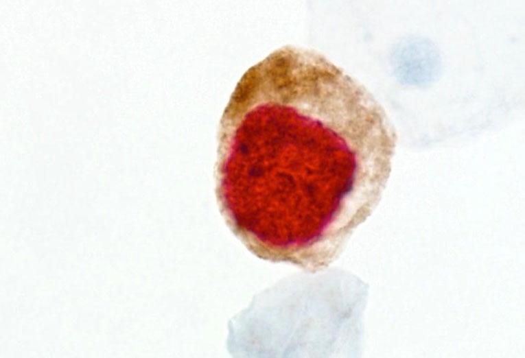
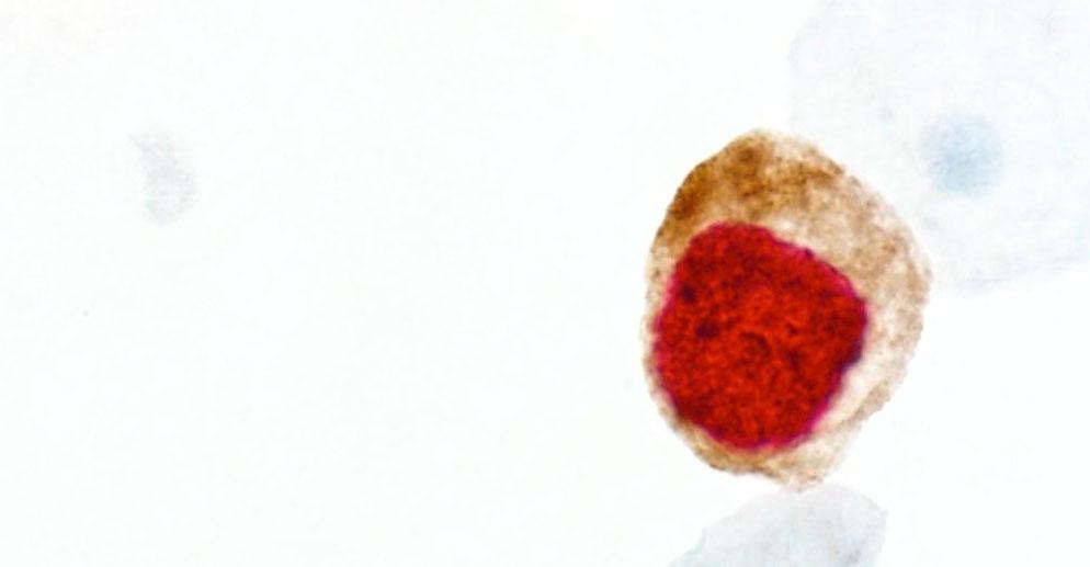
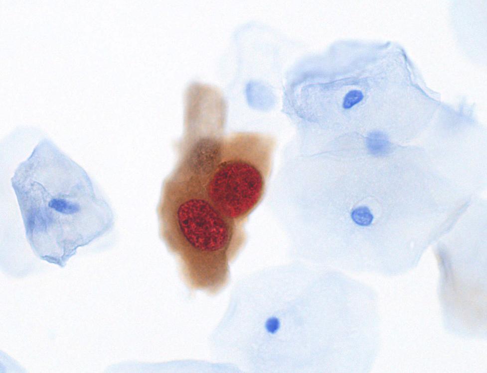
29 Definition of a dual-stained cell The cytoplasm is stained brown and the nucleus stained red p16 signal (brown) and Ki-67 signal (red) within the same cell Any level of expression, weak or strong The red stained nucleus and the brown stained cytoplasm must be within the same plane of focus (level) Interpretation is independent from morphologic criteria One dual-stained cell indicates a positive test result 29


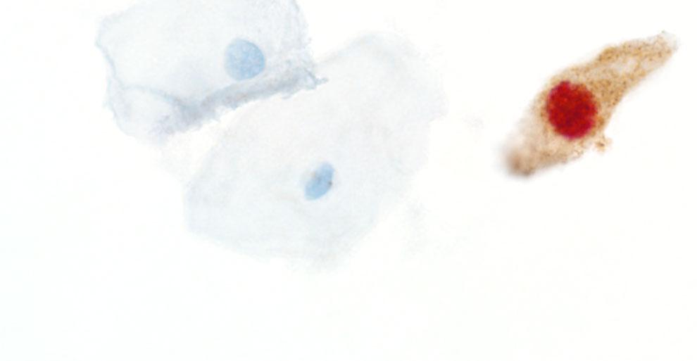
30 Single and dual-stained cells Example from the same case Single p16 Single Ki-67 Dual-stained cell 30
31 Only p16 stained cells 31

32 Only p16 stained cells 32

33 Only Ki-67 stained cells 33

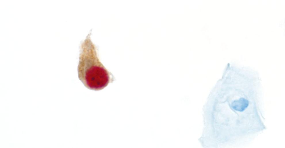
34 Single dual-stained cells 34
35 Dual-stained cells 35

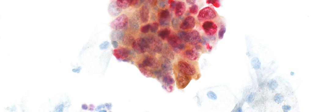
36 Group of dual-stained cells 36
37 Group with dual-stained and p16 only cell 37


38 Dual-stained cells, low level expression Weak brown or red staining, is positive with co-expression 38

39 Cluster with dual-stained cells 20x 39

40 Cluster with dual-stained cells 60x 40
41 Cluster with dual-stained cells 40x 41
42 CINtec PLUS Cytology staining results Negative Result No dual-stained cells present Positive Result Detection of at least one dual-stained cell Isolated cell(s) or within cell cluster 42
")
43 Recommended screening procedure CINtec PLUS Cytology Systematic overlapping fields of view Initially search for isolated dual-stained cells Easiest to evaluate If an isolated dual-stained cell has been identified, the stained nucleus and cytoplasm must be clearly allocated to the same cell Verify the cytoplasm and nucleus are in the same plane of focus (no overlying or underlying red nucleus) When evaluating cell clusters with red and brown staining, an interpretation algorithm must be followed 43
44 Interpretation algorithm for cell clusters Interpretation of red and brown stained cell clusters 1. Look for dual-stained cells at the edge of the cluster If yes test result is positive, dual-stained cells present If no proceed with step 2 2. Check if the p16 staining of the cluster can be defined as diffuse or focal Focal p16 staining: no further evaluation of this negative cluster Diffuse p16 staining: proceed with step 3 3. Check if the p16 staining is specific to the cytoplasm p16 staining is not specific (mucus or non-specific background staining) no further evaluation, a negative cluster Specific p16 staining: proceed with step 4 4. Are the Ki-67 staining, red nuclei over/underlying or embedded? (use the fine focus knob) Ki-67 staining, red nuclei are over/underlying no further evaluation, a negative cluster Ki-67 staining, red nuclei are embedded test result is positive dual-stained cells present 44
45 Interpretation algorithm for cell clusters Cell cluster checklist Separated cells at the edge of the cluster yes / no P16 staining pattern focal / diffuse Specific staining yes / no Ki-67-positive nuclei embedded in the cluster (same plane of focus) yes /no 45
46 Knowledge check The 2 cell group in the image indicates the following: a. A positive CINtec PLUS Cytology result b. A negative CINtec PLUS Cytology result c. Is difficult to evaluate d. None of the above True or False: p16 staining reaction is red and Ki- 67 staining reaction is brown True False 46
47 Knowledge check The 2 cell group in the image indicates the following: a. A positive CINtec PLUS Cytology result b. A negative CINtec PLUS Cytology result c. Is difficult to evaluate d. None of the above True or False: p16 staining reaction is red and Ki- 67 staining reaction is brown True False 47
48 Knowledge check The 2 cell group in the image indicates the following: a. A positive CINtec PLUS Cytology result b. A negative CINtec PLUS Cytology result c. Is difficult to evaluate d. None of the above True or False: p16 staining reaction is red and Ki- 67 staining reaction is brown True False 48
49 Cluster examples 49

50 Cluster with dual-stained cells 50

51 Cluster with dual-stained cells 51

52 One isolated dual-stained cell at the edge 52

53 Cluster with dual-stained cells 53

54 No dual stain in individual cells 54

55 Cluster with no dual-stained cells 55
56 The mechanism of action for p16 in cervical disease The functional utility of CINtec PLUS Cytology Principles of interpretation Troubleshooting pitfalls and artifacts 56
57 Non-specific background staining Detection chemistry chromogen can become trapped within cells, mucus or other cellular debris and could cause: Various intensities: low/medium/high Various distribution: few cells to all cells Homogeneous/heterogeneous 57

58 Non-specific brown background staining 20X Dual stained cells (+) 58

59 Non-specific brown background staining 40X Dual stained cells (+) 59

60 Non-specific brown background staining 60X 60


61 Specific staining Ki-67 (red) only Proliferating cells show only ki-67 signal (red) without background without specific p16 signal negative test result 61


 in")


62 Non-specific brown background staining Be aware to avoid false positive results Proliferating cells (Ki-67 positive) in combination with non-specific background staining could cause a false positive result 62


63 Interpretation with non-specific brown background Key recommendation for interpretation The brown p16 stain must be more intense than the nonspecific surrounding brown background to evaluate the cell(s) as positive for p16 & Ki-67 Do not evaluate cells with nonspecific brown background, regardless of the Ki-67 staining 63
64 Non-specific brown background Rule To avoid false-positive dual-stained cell evaluation follow this rule: A true dual-stained cell can only be called when the intensity of the brown (p16) in the cytoplasm of the cell is more intense than the surrounding brown background intensity in other cells (reference cells) 64
65 Dual-stained cell with non-specific background Example #1 Positive cell for CINtec PLUS Cytology: The brown p16 stain in the cytoplasm of this cell with a red Ki-67 stained nucleus is more intense than the non-specific brown surrounding background Non-specific brown background staining in reference cell 65
66 Dual-stained cell with non-specific background Example #2 Positive cell for CINtec PLUS Cytology: The brown p16 stain in the cytoplasm of this cell with a red Ki-67 stained nucleus is more intense than the non-specific brown surrounding background Non-specific brown background staining 66


67 Dual-stained cells, low level expression Example #3 Weak brown or red staining is positive with coexpression Must always compare the p16 (brown) staining to your reference cells Both negative or cells with non-specific brown background staining 67

 to avoid false positive")
68 Non-specific red background staining Red speckling /particles Non-specific red background staining will typically not interfere with interpretation If red speckling is present and is directly over or under the nucleus of a cell with p16 (brown) staining cytoplasm, use the fine focus to determine if it is true nuclear staining, or a red particle is in a different focal plane (not nuclear staining) to avoid false positive results 68
69 Knowledge check Non-specific background staining can be caused by detection particles (chromagen) becoming trapped within: a. Cells b. Mucus c. Cellular debris d. All of the above True or False: To successfully evaluate a dual-stained positive cell or cluster of cells, the brown background must be less than the p16 signal in the dual-stained cell or cluster? a. True b. False 69
70 Knowledge check Non-specific background staining can be caused by detection particles (chromagen) becoming trapped within: a. Cells b. Mucus c. Cellular debris d. All of the above True or False: To successfully evaluate a dual-stained positive cell or cluster of cells, the brown background must be less than the p16 signal in the dual-stained cell or cluster? a. True b. False 70
71 Knowledge check Non-specific background staining can be caused by detection particles (chromagen) becoming trapped within: a. Cells b. Mucus c. Cellular debris d. All of the above True or False: To successfully evaluate a dual-stained positive cell or cluster of cells, the brown background must be less than the p16 signal in the dual-stained cell or cluster? a. True b. False 71

72 Brown mucus background staining Artifact 72

73 Red bleeding into cytoplasm Artifact 73

74 Neutrophils staining red Artifact 74

75 Cornflaking Drying artifact Air drying that occurs before applying aqueous mounting media 75
 Any contact with")


76 Fading of the fast red stain Artifact from improper post processing procedure (mounting/coverslipping) Any contact with alcohol leads to Fading of Fast Red chromogen Aqueous mounting procedure to prevent fading The use Roche Hematoxylin, do not use alcoholic Hematoxylin Exposed to ETOH one day later: Cause: contact with alcohol during mounting/coverslipping procedure or counterstaining 76

77 Aqueous mounting procedure Cracking Artifact Cracks: A result of incomplete drying of aqueous mounting media 77
78 CINtec PLUS Cytology Virtual quiz Knowledge check 78
79 Endocervical glandular cells 1. Isolated cell Cluster 2. Dual stained cells at the edge of the cluster 3. p16 staining pattern focal diffuse 4. Specific p16 staining yes no 5. Ki-67-positive nuclei are embedded in the cluster (in same plane) yes no 6. Result with CINtec PLUS positive negative 79
80 Endocervical glandular cells 1. Isolated cell Cluster 2. Dual stained cells at the edge of the cluster 3. p16 staining pattern focal diffuse 4. Specific p16 staining yes no 5. Ki-67-positive nuclei are embedded in the cluster (in same plane) yes no 6. Result with CINtec PLUS positive negative 80
81 Endometrial glandular cells 1. Isolated cell Cluster 2. Dual stained cells at the edge of the cluster 3. p16 staining pattern focal diffuse 4. Specific p16 staining yes no 5. Ki-67-positive nuclei are embedded in the cluster (in same plane) yes no 6. Result with CINtec PLUS positive negative 81
 yes no 6.")
82 Endometrial glandular cells 1. Isolated cell Cluster 2. Dual stained cells at the edge of the cluster 3. p16 staining pattern focal diffuse 4. Specific p16 staining yes no 5. Ki-67-positive nuclei are embedded in the cluster (in same plane) yes no 6. Result with CINtec PLUS positive negative 82
83 Tissue repair 1. Isolated cell Cluster 2. Dual stained cells at the edge of the cluster 3. p16 staining pattern focal diffuse 4. Specific p16 staining yes no 5. Ki-67-positive nuclei are embedded in the cluster (in same plane) yes no 6. Result with CINtec PLUS positive negative 83
 yes no 6.")
84 Tissue repair 1. Isolated cell Cluster 2. Dual stained cells at the edge of the cluster 3. p16 staining pattern focal diffuse 4. Specific p16 staining yes no 5. Ki-67-positive nuclei are embedded in the cluster (in same plane) yes no 6. Result with CINtec PLUS positive negative 84
85 Immature metaplastic cells 1. Isolated cell Cluster 2. Result with CINtec PLUS positive negative 85
86 Immature metaplastic cells 1. Isolated cell Cluster 2. Result with CINtec PLUS positive negative 86
87 Maturing metaplastic cells 1. Isolated cell Cluster 2. Dual stained cells at the edge of the cluster 3. p16 staining pattern focal diffuse 4. Specific p16 staining yes no 5. Ki-67-positive nuclei are embedded in the cluster (in same plane) yes no 6. Result with CINtec PLUS positive negative 87
88 Maturing metaplastic cells 1. Isolated cell Cluster 2. Dual stained cells at the edge of the cluster 3. p16 staining pattern focal diffuse 4. Specific p16 staining yes no 5. Ki-67-positive nuclei are embedded in the cluster (in same plane) yes no 6. Result with CINtec PLUS positive negative 88
89 LSIL 1. Result with CINtec PLUS positive negative 89

90 LSIL 1. Result with CINtec PLUS positive negative 90
91 1. Isolated cell Cluster 2. p16 signal (brown) and Ki-67 signal (red) within the same cell yes no 3. Result with CINtec PLUS positive negative 91
 and Ki-67 signal (red) within the same cell yes no 3.")
92 1. Isolated cell Cluster 2. p16 signal (brown) and Ki-67 signal (red) within the same cell yes no 3. Result with CINtec PLUS positive negative 92
93 1. Isolated cell Cluster 2. p16 signal (brown) and Ki-67 signal (red) within the same cell yes no 3. The Cytoplasm is stained brown and the Nucleus appears red yes no 4. Result with CINtec PLUS positive negative 93
94 1. Isolated cell Cluster 2. p16 signal (brown) and Ki-67 signal (red) within the same cell yes no 3. The Cytoplasm is stained brown and the Nucleus appears red yes no 4. Result with CINtec PLUS positive negative 94
95 1. Isolated cell Cluster 2. Dual stained cells at the edge of the cluster 3. p16 staining pattern focal diffuse 4. Specific p16 staining yes no 5. Ki-67-positive nuclei are embedded in the cluster (in same plane) yes no 6. Result with CINtec PLUS positive negative 95
 yes no 6.")
96 1. Isolated cell Cluster 2. Dual stained cells at the edge of the cluster 3. p16 staining pattern focal diffuse 4. Specific p16 staining yes no 5. Ki-67-positive nuclei are embedded in the cluster (in same plane) yes no 6. Result with CINtec PLUS positive negative 96
97 1. Isolated cell Cluster 2. Dual stained cells at the edge of the cluster 3. p16 staining pattern focal diffuse 4. Specific p16 staining yes no 5. Ki-67-positive nuclei are embedded in the cluster (in same plane) yes no 6. Result with CINtec PLUS positive negative 97
 yes no 6.")
98 1. Isolated cell Cluster 2. Dual stained cells at the edge of the cluster 3. p16 staining pattern focal diffuse 4. Specific p16 staining yes no 5. Ki-67-positive nuclei are embedded in the cluster (in same plane) yes no 6. Result with CINtec PLUS positive negative 98
99 1. Isolated cell Cluster 2. Dual stained cells at the edge of the cluster 3. p16 staining pattern focal diffuse 4. Specific p16 staining yes no 5. Ki-67-positive nuclei are embedded in the cluster (in same plane) yes no 6. Result with CINtec PLUS positive negative 99
 yes no 6.")
100 1. Isolated cell Cluster 2. Dual stained cells at the edge of the cluster 3. p16 staining pattern focal diffuse 4. Specific p16 staining yes no 5. Ki-67-positive nuclei are embedded in the cluster (in same plane) yes no 6. Result with CINtec PLUS positive negative 100
101 1. Isolated cell Cluster 2. p16 signal (brown) and Ki-67 signal (red) within the same cell yes no 3. The Cytoplasm is stained brown and the Nucleus appears red yes no 4. Result with CINtec PLUS positive negative 101

102 1. Isolated cell Cluster 2. p16 signal (brown) and Ki-67 signal (red) within the same cell yes no 3. The Cytoplasm is stained brown and the Nucleus appears red yes no 4. Result with CINtec PLUS positive negative 102
103 1. Isolated cell Cluster 2. Dual stained cells at the edge of the cluster 3. p16 staining pattern focal diffuse 4. Specific p16 staining yes no 5. Ki-67-positive nuclei are embedded in the cluster (in same plane) yes no 6. Result with CINtec PLUS positive negative 103
 yes no 6.")
104 1. Isolated cell Cluster 2. Dual stained cells at the edge of the cluster 3. p16 staining pattern focal diffuse 4. Specific p16 staining yes no 5. Ki-67-positive nuclei are embedded in the cluster (in same plane) yes no 6. Result with CINtec PLUS positive negative 104
105 VENTANA Empowering Cancer Diagnostics Ventana Medical Systems, Inc. VENTANA and the VENTANA logo are trademarks of Roche. All other trademarks are the property of their respective owners. Web A
106 Doing now what patients need next
Interpretation guide. Abnormal cytology can t hide anymore
 Interpretation guide Abnormal cytology can t hide anymore Unique dual-biomarker technology makes you certain about the presence of transforming HPV infection. The science that creates certainty. Table
Table of Contents. 1. Overview. 2. Interpretation Guide. 3. Staining Gallery Cases Negative for CINtec PLUS
 Staining Atlas Table of Contents 1. Overview 1.1 Introduction 1.2 Role of p16 INK4a 1.3 Role of Ki-67 1.4 Molecular Pathogenesis 1.5 p16 INK4a Expression in Cervical Dysplasia 1.6 The Concept of CINtec
P16 et Ki67 Biomarkers: new tool for risk management and low grade intraepithelial lesions (LGSIL): be ready for the future.
: be ready for the future.") P16 et Ki67 Biomarkers: new tool for risk management and low grade intraepithelial lesions (LGSIL): be ready for the future. Mark H Stoler, MD University of Virginia Health System, Charlottesville, VA,
P16 et Ki67 Biomarkers: new tool for risk management and low grade intraepithelial lesions (LGSIL): be ready for the future. Mark H Stoler, MD University of Virginia Health System, Charlottesville, VA,
CINtec p16 INK4a Staining Atlas
 CINtec p16 INK4a Staining Atlas Rating Rating Positive The rating positive will be assigned if the p16 INK4a -stained slide shows a continuous staining of cells of the basal and parabasal cell layers of
p16 Cervical HISTOLOGY Histology Compendium & Staining Atlas
 p16 Cervical HISTOLOGY Histology Compendium & Staining Atlas Chapter 1: An Introduction to p16...... 3 Normal Cervical Epithelium and the Cell Cycle....4 HPV Infection and Cervical Disease......................................
chapter 4. The effect of oncogenic HPV on transformation zone epithelium
 chapter 4. The effect of oncogenic HPV on transformation zone epithelium CHAPTER 1 All squamous cervical cancer (and probably all cervical adenocarcinoma) is associated with oncogenic HPV, and the absence
Histopathology: Cervical HPV and neoplasia
 Histopathology: Cervical HPV and neoplasia These presentations are to help you identify basic histopathological features. They do not contain the additional factual information that you need to learn about
New Diagnoses Need New Approaches: A Glimpse into the Near Future of Gynecologic Pathology
 New Diagnoses Need New Approaches: A Glimpse into the Near Future of Gynecologic Pathology United States and Canadian Academy of Pathology 102 nd Annual Meeting Baltimore, Maryland Christina S. Kong, M.D.
Cervical cancer prevention: Advances in primary screening and triage system
 Cervical cancer prevention: Advances in primary screening and triage system Dr Farid Hadi Regional Medical and Scientific Affairs Roche Diagnostics Asia-Pacific, Singapore Cervical cancer is highly preventable
SQUAMOUS CELLS: Atypical squamous cells (ASC) - of undetermined significance (ASC-US) - cannot exclude HSIL (ASC-H)
 - of undetermined significance (ASC-US) - cannot exclude HSIL (ASC-H)") SQUAMOUS CELLS: Atypical squamous cells (ASC) - of undetermined significance (ASC-US) - cannot exclude HSIL (ASC-H) ASC refers to cytologic changes suggestive of SIL, which are qualitativley or quantitatively
SQUAMOUS CELLS: Atypical squamous cells (ASC) - of undetermined significance (ASC-US) - cannot exclude HSIL (ASC-H) ASC refers to cytologic changes suggestive of SIL, which are qualitativley or quantitatively
Pushing the Boundaries of the Lab Diagnosis in Asia
 Pushing the Boundaries of the Lab Diagnosis in Asia Diana Lim MBBS, FRCPA, FRCPath (UK) Senior Consultant National University Health System and National University of Singapore Department of Pathology
p16ink4a expression in cervical intraepithelial neoplasia and cervical cancer
 Original Article Brunei Int Med J. 2013; 9 (3): 165-171 p16ink4a expression in cervical intraepithelial neoplasia and cervical cancer Kalpana KUMARI 1 and Akhila Arcot VADIVELAN 2 1 Department of Pathology,
P16 INK4A EXPRESSION AS A POTENTIAL PROGNOSTIC MARKER IN CERVICAL PRECANCEROUS AND CANCEROUS LESIONS IN MOROCCO
 P16 INK4A EXPRESSION AS A POTENTIAL PROGNOSTIC MARKER IN CERVICAL PRECANCEROUS AND CANCEROUS LESIONS IN MOROCCO Yassine Zouheir Laboratory of histo-cytopathology of Institut Pasteur of Morocco, Casablanca,
Cervical Cancer : Pap smear
 Taking a PAP SMEAR Cervical Cancer : Pap smear George N Papanicolaou introduced cervical cytology in clinical practice in 1940 In 1945, PAP smear was endorsed by American cancer society as an effective
Colposcopy. Attila L Major, MD, PhD
 Colposcopy Attila L Major, MD, PhD Histology Colposcopy Cytology It has been estimated that annual Pap smear testing reduces a woman s chance of dying of cervical cancer from 4 in 1000 to about 5 in 10,000
Workshop for O& G trainees and paramedics 17 Dec 2011 Cytological Interpretation
 Workshop for O& G trainees and paramedics 17 Dec 2011 Cytological Interpretation May Yu Director of Cytology Laboratory Service Department of Anatomical & Cellular Pathology Prince of Wales Hospital Cervical
Understanding Your Pap Test Results
 Understanding Your Pap Test Results Most laboratories in the United States use a standard set of terms called the Bethesda System to report pap test results. Normal: Pap samples that have no cell abnormalities
Digital Pathology and CAP Guidelines
 Digital Pathology and CAP Guidelines Frequently asked questions The VENTANA family of digital pathology products empowers you with the convenience of a comprehensive image and workflow solution. When used
Prepared By Jocelyn Palao and Layla Faqih
 Prepared By Jocelyn Palao and Layla Faqih The structure of the suspected atypical cell should always be compared to the structure of other similar, benign, cells which are present in the smears. The diagnosis
Done by khozama jehad. Neoplasia of the cervix
 Done by khozama jehad Neoplasia of the cervix An overview of cervical neoplasia very import. Most tumors of the cervix are of epithelial origin and are caused by oncogenic strains of human papillomavirus
CK17 and p16 expression patterns distinguish (atypical) immature squamous metaplasia from high-grade cervical intraepithelial neoplasia (CIN III)
 immature squamous metaplasia from high-grade cervical intraepithelial neoplasia (CIN III)") Histopathology 2007, 50, 629 635. DOI: 10.1111/j.1365-2559.2007.02652.x CK17 and p16 expression patterns distinguish (atypical) immature squamous metaplasia from high-grade cervical intraepithelial neoplasia
Histopathology 2007, 50, 629 635. DOI: 10.1111/j.1365-2559.2007.02652.x CK17 and p16 expression patterns distinguish (atypical) immature squamous metaplasia from high-grade cervical intraepithelial neoplasia
CINtec PLUS and the Pap smear: a co-testing alternative
 CINtec PLUS and the Pap smear: a co-testing alternative Rosemary Tambouret MD p16/ki67 (CINtec PLUS) and the Pap smear Rosemary Tambouret MD CINtec PLUS dual stain: p16 and Ki67 p16 is anti-proliferative
Biomarkers for cervical cancer screening: the role of p16 INK4a to highlight transforming HPV infections
 Review For reprint orders, please contact reprints@expert-reviews.com Biomarkers for cervical cancer screening: the role of p16 INK4a to highlight transforming HPV infections Expert Rev. Proteomics 9(2),
HPV and Lower Genital Tract Disease. Simon Herrington University of Edinburgh, UK Royal Infirmary of Edinburgh, UK
 HPV and Lower Genital Tract Disease Simon Herrington University of Edinburgh, UK Royal Infirmary of Edinburgh, UK Conflict of interest/funding X None Company: Product royalties Paid consultant Research
Hyperchromatic Crowded Groups: What is Your Diagnosis? Session 3000
 Hyperchromatic Crowded Groups: What is Your Diagnosis? Session 3000 Thomas A. Bonfiglio, M.D. Professor Emeritus, Pathology and Laboratory Medicine University of Rochester Disclosures In the past 12 months,
Objectives. Atypical Glandular Cells. Atypical Endocervical Cells. Reactive Endocervical Cells
 2013 California Society of Pathologists 66 th Annual Meeting San Francisco, CA Atypical Glandular Cells to Early Invasive Adenocarcinoma: Cervical Cytology and Histology Christina S. Kong, MD Associate
Cytyc Corporation - Case Presentation Archive - July 2002
 ThinPrep Pap Test History: 34 Year Old Female LMP: Day 20 Specimen Type: Cervical/Vaginal Case provided by Mark Tulecke, M.D. and Gabrielle Trawinski CT (ASCP), Mount Auburn Hospital, Cambridge, Massachusetts.
VENTANA MMR IHC Panel Interpretation Guide for Staining of Colorectal Tissue
 VENTANA MMR IHC Panel Interpretation Guide for Staining of Colorectal Tissue VENTANA anti-mlh1 (M1) Mouse Monoclonal Primary Antibody VENTANA anti-pms2 (A16-4) Mouse Monoclonal Primary Antibody VENTANA
Cervical Dysplasia and HPV
 Cervical Dysplasia and HPV J. Anthony Rakowski D.O., F.A.C.O.O.G. MSU SCS Board Review Coarse HPV Double stranded DNA virus The HPV infect epithelial cells of the skin and mucous membranes Highest risk
When Immunostains Can Get You in Trouble: Gynecologic Pathology p16: Panacea or Pandora s Box?
 When Immunostains Can Get You in Trouble: Gynecologic Pathology p16: Panacea or Pandora s Box? Teri A. Longacre, MD Stanford Medicine Stanford California pi6 in Gynecologic Pathology: Panacea or Pandora
To further assess abnormalities detected on cervical cytological sample. To guide colposcopically directed biopsy
 1 To further assess abnormalities detected on cervical cytological sample To guide colposcopically directed biopsy To exclude invasive disease To aid in outpatient management and treatment of precancerous
Cytology Report Format
 Squamous Precursor Lesions and Malignancies In Pap Test Dina R. Mody, MD, FCAP Director of Cytology The Methodist Hospital, Houston, TX Professor of Pathology and Laboratory Medicine Weill Medical College
CASE 4 21/07/2017. Ectopic Prostatic Tissue in Cervix. Female 31. LLETZ for borderline nuclear abnormalities
 Female 31 CASE 4 LLETZ for borderline nuclear abnormalities PSA Ectopic Prostatic Tissue in Cervix AJSP 2006;30;209-215 usually incidental microscopic finding usually in ectocervical stroma? developmental
TISSUE TUMOR MARKER EXPRESSION IN
 TISSUE TUMOR MARKER EXPRESSION IN NORMAL CERVICAL TISSUE AND IN CERVICAL INTRAEPITHELIAL NEOPLASIA, FOR WOMEN WHO ARE AT HIGH RISK OF HPV (HUMAN PAPILLOMA VIRUS INFECTION). Raghad Samir MD PhD Verksamhet
1.Acute and Chronic Cervicitis - At the onset of menarche, the production of estrogens by the ovary stimulates maturation of the cervical and vaginal
 Diseases of cervix I. Inflammations 1.Acute and Chronic Cervicitis - At the onset of menarche, the production of estrogens by the ovary stimulates maturation of the cervical and vaginal squamous mucosa
Efficacy evaluation of a test CINtec p16ink4a in screening for cervical HPV infection
 Vol.1, No.3, 154-163 (2011) doi:10.4236/ojpm.2011.13020 Open Journal of Preventive Medicine Efficacy evaluation of a test CINtec p16ink4a in screening for cervical HPV infection Pafumi Carlo, Leanza Vito,
Neoplasia 2018 Lecture 2. Dr Heyam Awad MD, FRCPath
 Neoplasia 2018 Lecture 2 Dr Heyam Awad MD, FRCPath ILOS 1. List the differences between benign and malignant tumors. 2. Recognize the histological features of malignancy. 3. Define dysplasia and understand
CYTOMORPHOLOGY MODULE 28.1 INTRODUCTION OBJECTIVES 28.2 GENERAL GUIDELINES. Notes
 28 CYTOMORPHOLOGY 28.1 INTRODUCTION Light microscopic examination of stained cells in smears is the method of choice of diagnostic cytology. It allows classification of most normal cells as to type and
It depends on the site: In Cervix 99%, in Anus ~ 85-90% and in Vulva, Penis ~ 40-50%. True.
 Are all high grade lesions caused by HPV, or are there other etiologies? The issue is not if you are infected with HPV high risk, but which of the patients infected with HR hpv would go into progressive
BOSNIAN-TURKISH CYTOPATHOLOGY SCHOOL June 18-19, 2016 Sarajevo. Case Discussions. 60 year old woman Routine gynecologic control LBC
 BOSNIAN-TURKISH CYTOPATHOLOGY SCHOOL June 18-19, 2016 Sarajevo Case Discussions Prof Dr Sıtkı Tuzlalı Tuzlalı Pathology Laboratory 60 year old woman Routine gynecologic control LBC 1 2 Endometrial thickening
EU guidelines for reporting gynaecological cytology
 EU guidelines for reporting gynaecological cytology Amanda Herbert Guy s & St Thomas Foundation NHS Trust 5th EFCS Annual Tutorial, Trondheim, Norway 28 th May 1 st June 2012 EU guidelines aim to harmonize
Cervical Cancer Screening. David Quinlan December 2013
 Cervical Cancer Screening David Quinlan December 2013 Cervix Cervical Cancer Screening Modest variation provincially WHO and UK begin at 25 stop at 60 Finland begin at 30 stop at 60 Rationale for
A Study on Diagnostic Accuracy of Cervical Pap Smear by Correlating with Histopathology in a Tertiary Care Centre
 Original Article DOI: 10.21276/APALM.1878 A Study on Diagnostic Accuracy of Cervical Pap Smear by Correlating with Histopathology in a Tertiary Care Centre Rachana L Y, S.S. Hiremath*, Prabhu M H, S.S
Mody. AIS vs. Invasive Adenocarcinoma of the Cervix
 Common Problems in Gynecologic Pathology Michael T. Deavers, M.D. Houston Methodist Hospital, Houston, Texas Common Problems in Gynecologic Pathology Adenocarcinoma in-situ (AIS) of the Cervix vs. Invasive
Molecular Probes Introducing 14 new probes
 Molecular Probes Introducing 14 new probes Gene and Chromosome Probes Dual Colour ISH INFORM HER2 Dual ISH DNA Probe Cocktail Assay Product Part Number INFORM HER2 Dual ISH DNA Probe Cocktail 800-4422
Cervical Screening for Dysplasia and Cancer in Patients with HIV
 Cervical Screening for Dysplasia and Cancer in Patients with HIV Adult Clinical Guideline from the New York State Department of Health AIDS Institute w w w.hivg uidelines.org Purpose of the Guideline Increase
In situ and Invasive Endocervical Carcinoma: Problems and Pitfalls in Diagnosis
 In situ and Invasive Endocervical Carcinoma: Problems and Pitfalls in Diagnosis Rouba Ali-Fehmi,MD The Karmanos Cancer Institute, Wayne State University School of Medicine Global incidence of cervical
Human Papillomavirus
 Human Papillomavirus Dawn Palaszewski, MD Assistant Professor of Obstetrics and Gynecology University of February 18, 2018 9:40 am Dawn Palaszewski, MD Assistant Professor Department of Obstetrics and
Chapter 10: Pap Test Results
 Chapter 10: Pap Test Results On completion of this section, the learner will be able to: 1. Identify how Pap test results are interpreted and the reasons for normal and abnormal results. 2. Describe the
Gynecologic Cytopathology: Glandular lesions
 Gynecologic Cytopathology: Glandular lesions Lin Wai Fung (MSc, MPH, CMIAC) 17/4/2014 Glandular lesions of the uterus Endocervix Endometrium Normal endocervical cells Sheets, strips well-preserved architecture:
Lessons From Cases of Screened Women Who Developed Cervical Carcinoma
 Lessons From Cases of Screened Women Who Developed Cervical Carcinoma R. Marshall Austin MD,PhD Magee-Womens Hospital of University of Pittsburgh Medical Center raustin@magee.edu Why Focus Study On Cases
76 Arch Pathol Lab Med Vol 138, January 2014 p16 Utilization in Cervical Biopsies Singh et al
 Variability of Pathologists Utilization of p16 and Ki-67 Immunostaining in the Diagnosis of Cervical Biopsies in Routine Pathology Practice and Its Impact on the Frequencies of Cervical Intraepithelial
CERVIX. MLS Basic histological diagnosis MLS HIST 422 Semester 8- batch 7 L12 : Dr. Ali Eltayb.
 CERVIX MLS Basic histological diagnosis MLS HIST 422 Semester 8- batch 7 L12 : Dr. Ali Eltayb. CERVIX Most cervical lesions are: Most are Cervicitis. cancers ( common in women worldwide). CERVICITIS Extremely
JMSCR Vol 05 Issue 01 Page January 2017
 www.jmscr.igmpublication.org Impact Factor 5.244 Index Copernicus Value: 83.27 ISSN (e)-2347-176x ISSN (p) 2455-0450 DOI: https://dx.doi.org/10.18535/jmscr/v5i1.37 Immuno-Histochemical Study of P16INK4A
Immunohistochemical Expression of Cell Proliferating Nuclear Antigen (PCNA) and p53 Protein in Cervical Cancer
 and p53 Protein in Cervical Cancer") DOI 10.1007/s13224-012-0180-6 ORIGINAL ARTICLE Immunohistochemical Expression of Cell Proliferating Nuclear Antigen (PCNA) and p53 Protein in Cervical Cancer Madhumati Goel Kavita Somani Anju Mehrotra
DOI 10.1007/s13224-012-0180-6 ORIGINAL ARTICLE Immunohistochemical Expression of Cell Proliferating Nuclear Antigen (PCNA) and p53 Protein in Cervical Cancer Madhumati Goel Kavita Somani Anju Mehrotra
South Afr J Gynaecol Oncol RESEARCH
 Southern African Journal of Gynaecological Oncology 2017; 9(2):25 29 https://doi.org/10.1080/20742835.2017.1370841 Open Access article distributed under the terms of the Creative Commons License [CC BY-NC
Appropriate Use of Cytology and HPV Testing in the New Cervical Cancer Screening Guidelines
 Appropriate Use of Cytology and HPV Testing in the New Cervical Cancer Screening Guidelines Tim Kremer, MD Ralph Anderson, MD 1 Objectives Describe the natural history of HPV particularly as it relates
Beyond Pap Morphological Triage: p16/ki67 Dual Staining
 Moving away from Beyond Pap Morphological Triage: p16/ki67 Dual Staining Nicolas Wentzensen MD, PhD, MS Deputy Chief, Clinical Genetics Branch; Head, Clinical Epidemiology Unit National Cancer Institute
Key Words: SurePath. CINtec; p16; Ki-67, MIB1; sensitivity; specificity;
 Evaluation of CINtec PLUS 1 Testing as an Adjunctive Test in ASC-US Diagnosed Surepath 1 Preparations Neil Edgerton, M.D., Cynthia Cohen, M.D., andmomint. Siddiqui,M.D., F.I.A.C.* The CINtec PLUS 1 system
Clinical Practice Guidelines June 2013
 Clinical Practice Guidelines June 2013 General Principles: The Papanicolaou (Pap) smear is widely credited with reducing mortality from cervical cancer, and remains the single best method for the early
HPV: cytology and molecular testing
 HPV: cytology and molecular testing Human Papillomavirus and how we test for it at Medlab Central Palmerston North for Cervical Cancer prevention and management. Developed by Reem Mustafa Cytology and
The LAST Guidelines in Clinical Practice. Implementing Recommendations for p16 Use
 AJCP / Original Article The LAST Guidelines in Clinical Practice Implementing Recommendations for p16 Use Lani K. Clinton, MD, PhD, 1,2 Kyle Miyazaki, 1 Asia Ayabe, 1 James Davis, PhD, 2 Pamela Tauchi-Nishi,
3 CHAPTER 3: RESULTS
 3 CHAPTER 3: RESULTS 3.1 Histopathology 3.1.1 Normal Squamous Epithelium The squamous epithelium that covers the ectocervix of the uterus is composed of different layers starting at the basement membrane
Ph.D. THESIS ENDOMETRIAL HYPERPLASIAS IN PERIMENOPAUSE SUMMARY
 UNIVERSITY OF MEDICINE AND PHARMACY OF CRAIOVA FACULTY OF MEDICINE Ph.D. THESIS ENDOMETRIAL HYPERPLASIAS IN PERIMENOPAUSE SUMMARY SCIENTIFIC COORDINATOR: PROF. DR. MIHAI B. BRĂILA, Ph.D. Ph.D. Graduand:
Chapter 8. Summary. Chapter 8. Summary
 Chapter 8 Chapter 8 Summary Summary Summary In CHAPTER 1 an introduction to several aspects of cervical cancer, HPV and their relation with immunology is given. It took almost one-and-a-half centuries
Is cervical cancer a part of the Lynch Spectrum?
 Is cervical cancer a part of the Lynch Spectrum? HNPCC annual meeting October 12, 2017 Karina Rønlund MD, Ph.D. Department of Clinical Genetics OUH and Vejle Hospital Lynch Syndrom Inherited in an autosomal
Detecting High-Grade Cervical Disease on ASC-H Cytology. Role of BD ProEx C and Digene Hybrid Capture II HPV DNA Testing
 Anatomic Pathology / BD ProEx C Use in ASC-H Cy t o l o g y Detecting High-Grade Cervical Disease on ASC-H Cytology Role of BD ProEx C and Digene Hybrid Capture II HPV DNA Testing Momin T. Siddiqui, MD,
New Developments in Immunohistochemistry for Gynecologic Pathology
 New Developments in Immunohistochemistry for Gynecologic Pathology Michael T. Deavers, M.D. Professor, Departments of Pathology and Gynecologic Oncology Immunohistochemistry in Gynecologic Pathology Majority
THE ROLE OF HUMAN PAPILLOMA VIRUS IN THE DEVELOPMENT OF ORAL LEUKOPLAKIA
 Oral biology THE ROLE OF HUMAN PAPILLOMA VIRUS IN THE DEVELOPMENT OF ORAL LEUKOPLAKIA Yu. G. KOLENKO 1 1 Associate Prof., PhD, Dept. Operative Dentistry, Faculty of Medical Dentistry, Bogomolets National
Prevalence of human papillomavirus and Chlamydia trachomatis infection in paired urine and cervical smear samples of Palestinian young women.
 Prevalence of human papillomavirus and Chlamydia trachomatis infection in paired urine and cervical smear samples of Palestinian young women. Walid Salim Basha, PhD Faculty of Human Medicine An-Najah National
Part II The Cell Cell Division, Chapter 2 Outline of class notes
 Part II The Cell Cell Division, Chapter 2 Outline of class notes 1 Cellular Division Overview Types of Cell Division Chromosomal Number The Cell Cycle Mitoses Cancer Cells In Vitro Fertilization Infertility
Thin HSIL of the Cervix: Detecting a Variant of High-grade Squamous Intraepithelial Lesions With a p16 INK4a Antibody
 International Journal of Gynecological Pathology 00:1 5, Lippincott Williams & Wilkins, Baltimore r 2016 International Society of Gynecological Pathologists Original Article Thin HSIL of the Cervix: Detecting
Dr Rodney Itaki Lecturer Anatomical Pathology Discipline. University of Papua New Guinea School of Medicine & Health Sciences Division of Pathology
 Neoplasia Dr Rodney Itaki Lecturer Anatomical Pathology Discipline University of Papua New Guinea School of Medicine & Health Sciences Division of Pathology General Considerations Overview: Neoplasia uncontrolled,
CELL BIOLOGY - CLUTCH CH CANCER.
 !! www.clutchprep.com CONCEPT: OVERVIEW OF CANCER Cancer is a disease which is primarily caused from misregulated cell division, which form There are two types of tumors - Benign tumors remain confined
number Done by Corrected by Doctor Maha Shomaf
 number 16 Done by Waseem Abo-Obeida Corrected by Zeina Assaf Doctor Maha Shomaf MALIGNANT NEOPLASMS The four fundamental features by which benign and malignant tumors can be distinguished are: 1- differentiation
Cervical FISH Testing for Triage and Support of Challenging Diagnoses: A Case Study of 2 Patients
 Cervical FISH Testing for Triage and Support of Challenging Diagnoses: A Case Study of 2 Patients Richard Hopley, MD, Alexandra Gillespie, MD* Laboratory Medicine 47:1:52-56 CLINICAL HISTORY Patients:
Cancer Cells. It would take another 20 years and a revolution in the techniques of biological research to answer these questions.
 Cancer Cells Cancer, then, is a disease in which a single normal body cell undergoes a genetic transformation into a cancer cell. This cell and its descendants, proliferating across many years, produce
Utilization of the Biomarkers to Improve Cervical Cancer Screening
 Utilization of the Biomarkers to Improve Cervical Cancer Screening Elena BERNAD Victor Babes University of Medicine and Pharmacy Timisoara, Romania Cervical cancer is at the second most common cancer in
Early Embryonic Development
 Early Embryonic Development Maternal effect gene products set the stage by controlling the expression of the first embryonic genes. 1. Transcription factors 2. Receptors 3. Regulatory proteins Maternal
Eradicating Mortality from Cervical Cancer
 Eradicating Mortality from Cervical Cancer Michelle Berlin, MD, MPH Vice Chair, Obstetrics & Gynecology Associate Director, Center for Women s Health June 2, 2009 Overview Prevention Human Papilloma Virus
Making Sense of Cervical Cancer Screening
 Making Sense of Cervical Cancer Screening New Guidelines published November 2012 Tammie Koehler DO, FACOG The incidence of cervical cancer in the US has decreased more than 50% in the past 30 years because
Over-diagnoses in Cytopathology: Is histology the gold standard?
 Over-diagnoses in Cytopathology: Is histology the gold standard? Teresa M. Darragh, MD UCSF Departments of Pathology and Obstetrics, Gynecology & Reproductive Sciences Faculty Disclosures: Teresa M. Darragh,
Morphology I Slide: 1
 Morphology I Slide: 1 Morphology I Slide: 2 ThinPrep Morphology Normal Cytology Morphology I Slide: 3 CT & Pathologist Training Training program begins with ThinPrep morphology presentation Microscopic
Case year female. Routine Pap smear
 Case 1 57 year female Routine Pap smear Diagnosis? 1. Atypical glandular cells of unknown significance (AGUS) 2. Endocervical AIS 3. Endocervical adenocarcinoma 4. Endometrial adenocarcinoma 5. Adenocarcinoma
Cervical Cancer Screening for the Primary Care Physician for Average Risk Individuals Clinical Practice Guidelines. June 2013
 Cervical Cancer Screening for the Primary Care Physician for Average Risk Individuals Clinical Practice Guidelines General Principles: Since its introduction in 1943, Papanicolaou (Pap) smear is widely
Histopathology: skin pathology
 Histopathology: skin pathology These presentations are to help you identify, and to test yourself on identifying, basic histopathological features. They do not contain the additional factual information
Cell Cycle Phase. Interphase (G 1, S, G 2 ) Mitotic Phase (M phase) Prophase. Metaphase. Anaphase. Telophase
 Mitotic Phase (M phase) Prophase. Metaphase. Anaphase. Telophase") Part I: The Cell Cycle Use your resources at hand and the Explore Student Guide to outline what occurs within the cell during each stage of the cell cycle. Record this information in Table 1 below. Cell
Part I: The Cell Cycle Use your resources at hand and the Explore Student Guide to outline what occurs within the cell during each stage of the cell cycle. Record this information in Table 1 below. Cell
Management Algorithms for Abnormal Cervical Cytology and Colposcopy
 Management Algorithms for Abnormal Cervical Cytology and Colposcopy Table of Contents Standard Colposcopic Definitions... 1 Guidelines for the Assessment of Abnormal Cervical Cytology... 2 Ia: Persistent
Colposcopic Principles. Simon Leeson Consultant Obstetrician/ Gynaecologist Betsi Cadwaladr University Health Board UK
 Colposcopic Principles Simon Leeson Consultant Obstetrician/ Gynaecologist Betsi Cadwaladr University Health Board UK The cervix topics discussed are: original squamous epithelium endocervical epithelium
Dr. Issraa Ali Hussein
 CLINICAL 09888888;rCYTOLOGY Dr. Issraa Ali Hussein objectives Define diagnostic cytology (clinical cytology). Explain the differences between histopathology and cytopathology. Recognize the methods for
Endometrial Metaplasia, Hyperplasia & Other Cancer Mimics: a Consultant s Experience
 Endometrial Metaplasia, Hyperplasia & Other Cancer Mimics: a Consultant s Experience Pacific Northwest Society of Pathologists Vancouver, B.C. September 26, 2015 Teri A. Longacre, M.D. longacre@stanford.edu
Studying The Role Of DNA Mismatch Repair In Brain Cancer Malignancy
 Kavya Puchhalapalli CALS Honors Project Report Spring 2017 Studying The Role Of DNA Mismatch Repair In Brain Cancer Malignancy Abstract Malignant brain tumors including medulloblastomas and primitive neuroectodermal
I have no financial interests in any product I will discuss today.
 Cervical Cancer Screening Update and Implications for Annual Exams George F. Sawaya, MD Professor Department of Obstetrics, Gynecology and Reproductive Sciences Department of Epidemiology and Biostatistics
NEOPLASIA! Terminology and Classification of Neoplastic cells! Objectives: Asst. Prof. Prasit Suwannalert, Ph.D. Leading Questions
 NEOPLASIA! Asst. Prof. Prasit Suwannalert, Ph.D. (Email: prasit.suw@mahidol.ac.th)! Department of Pathobiology Faculty of Science, Mahidol University! Objectives: After learning, students should be able
MOLECULAR BASIS OF ONCOGENESIS
 MOLECULAR BASIS OF ONCOGENESIS MUDr. Jiří Vachtenheim, CSc. 1 Cell processes which result also in cell cycle effects. Differentiation. Differentiated cells are usually in the G0 phase of the cell cycle.
HPV and Oral Cancer. Oral Cancer
 HPV and Oral Cancer How do we prevent oral cancer? Dr. Peter Angeletti Oral Cancer 3% of all cancers in the US are in oral compartment 8 th most common in men 30,000 new cases each year 8,000 deaths each
Control of Cell Cycle. Unit 2 Part f III
 Control of Cell Cycle Unit 2 Part f III How often do cells divide and why? The timing and rate of cell division in different parts of the plant or animals are crucial to normal growth, development and
Cytyc Corporation - Case Presentation Archive - June 2003
 ThinPrep General Cytology History: Asymptomatic 35 Year Old Male Specimen type: Anal Cytology - This specimen was collected using a Dacron swab under proctoscopic visualization. This case was provided
Cell Cycle and Cancer
 142 8. Cell Cycle and Cancer NOTES CELL CYCLE G 0 state o Resting cells may re-enter the cell cycle Nondividing cells (skeletal and cardiac muscle, neurons) o Have left the cell cycle and cannot undergo