Gynecologic Cytopathology: Glandular lesions
|
|
|
- Nickolas Wade
- 5 years ago
- Views:
Transcription
1 Gynecologic Cytopathology: Glandular lesions Lin Wai Fung (MSc, MPH, CMIAC) 17/4/2014


2
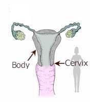
3 Glandular lesions of the uterus Endocervix Endometrium
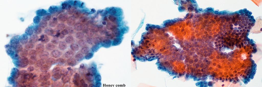
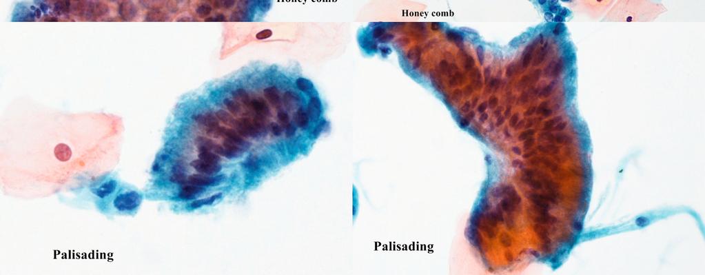
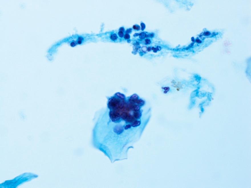
4 Normal endocervical cells Sheets, strips well-preserved architecture: honeycomb or palisading Nuclei: may show variation in size (2 x enlarged) and shape Ovulation: secretory and with naked nuclei
5

6 Normal endocervical cells: denuded nuclei
7 Benign glandular lesions of cervix
8 Cytopathology of Benign glandular lesions of the cervix Reparative changes Endocervical polyp Tubal Metaplasia Microglandular hyperplasia Cells of Lower Uterine Segment
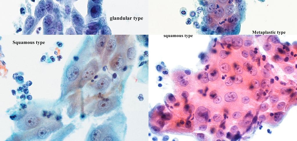
9 Reparative changes May involve: squamous, metaplastic, columnar epithelium Cytology: nuclear size, prominent nucleoli, monolayer sheet with polymorph infiltration, nuclei oriented in same direction (streaming), occasion mitotic figures, no single cells Marked nuclear anisonucleosis + irregular chromatin distribution: atypical endocervcial cell, atypical squamous cell

10 Repair cells
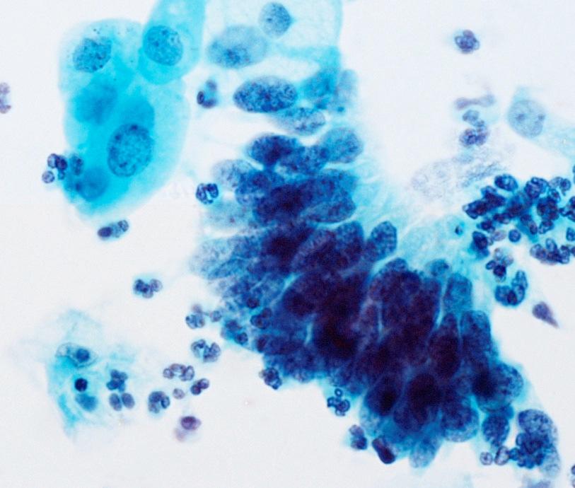
11 Cervical polyps Common Asymptomatic cause intermittent or post-coital bleeding Histology: central connective tissue stalk linked by endocervical, metaplastic cells No specific cytology pattern except large sheets of endocervical cells Sometimes show atypical or reactive nuclei (AGC)

12 Cervical Polyp with atypical cells / repair cells. F/52 inter-menstrual breeding
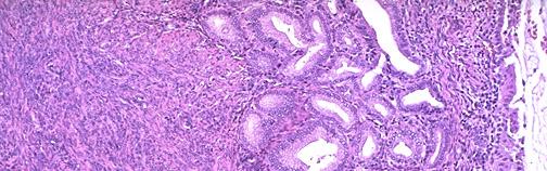
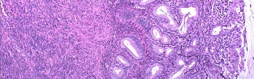
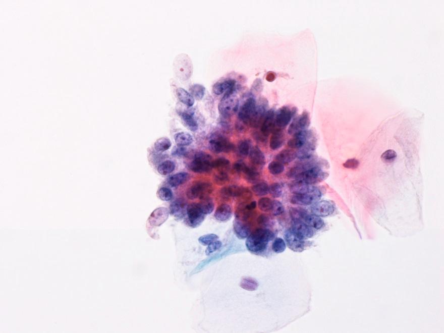
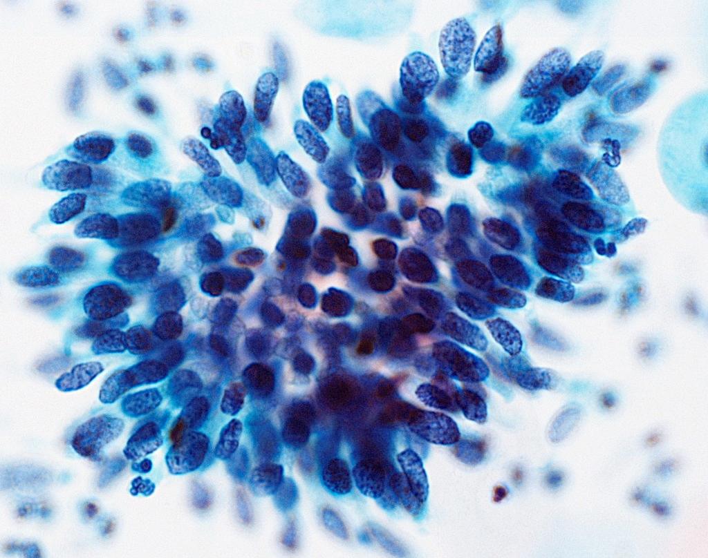
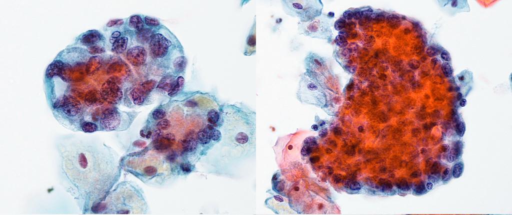
13 Tubal Metaplasia Benign, non-neoplastic replacement of normal endocervical (or endometrial) epithelium with cells characteristic of the fallopian tube: ciliated, clear cell, non-ciliated secretory cells, and intercalated cells common, prominent in upper third of endocervical canal. Endocervical brush: increase detection in cervical smears flat sheet, cohesive 3-D aggregates, columnar, apical terminal bar with cilia Nuclei, regular, oval, elongated, hyperchromatic, pseudo-stratification: may mimic adenocarcinoma in situ (AIS)

14 Tubal metaplasia with mild nuclear atypia, F/44
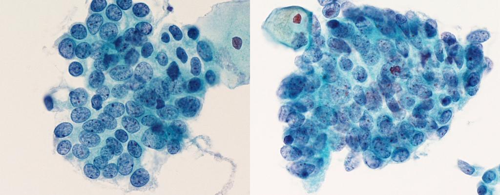
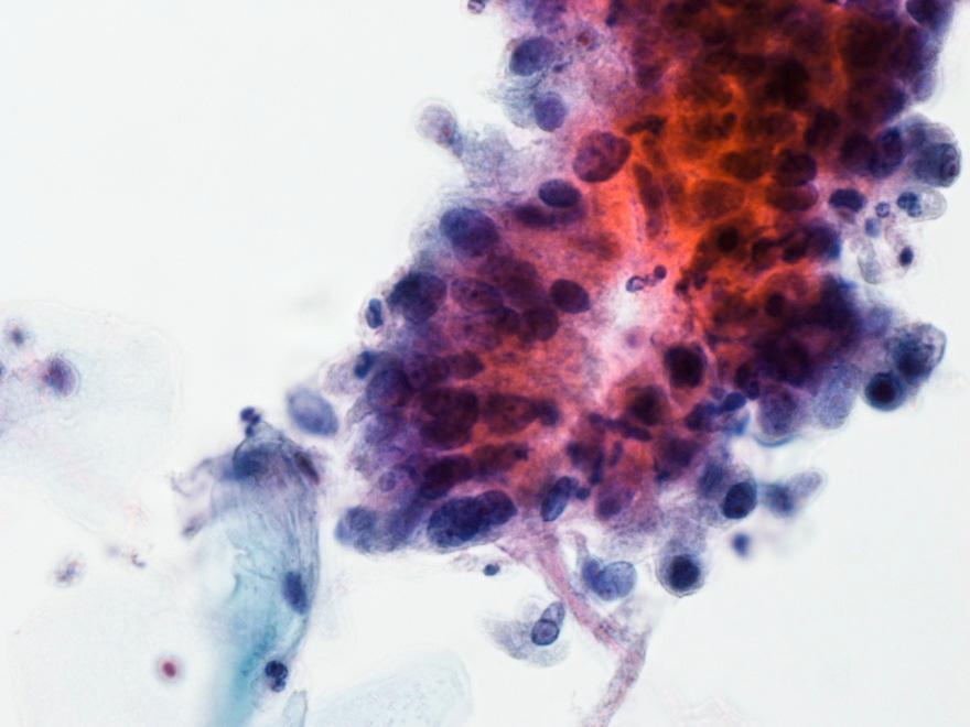
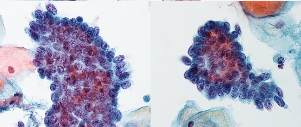
15 Microglandular hyperplasia Benign localized proliferation of endocervical glands Incidental finding or associated with polyp Young women associated with pregnancy and contraceptive use Histology: closely packed irregular glands, lined by benign endocervical cells Cytology features: non-specific 2D or 3D sheets of cuboidal and columnar glandular cells with finely vacuolated cytoplasm May have cytologic atypia due to hyperchromatic crowded groups, pseudostratified strip, nuclear enlargement, hyperchromasia (not to overdiagnosis as adenoca or AIS)


16 Microglandular hyperplasia, F/26, Uterus cervix; mild to moderate glandular hyperplasia

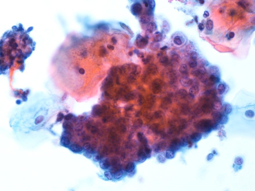
17 Cells of the Lower Uterine Segment (LUS) Isthmus of cervix: short transistional zone between endocervical and endometrium Cone biopsy shortens the endocervcial canal: easier access to LUS Cells: mainly endometrial less responsive to hormonal stimulation Endocervical brushes detection, No need to report LUS do not shed spontaneously Cytology: glandular + stromal element, large irregular branched groups, round nuclei, fine chromatin, nuclear crowding, May be mistaken for AIS, adenca Source:


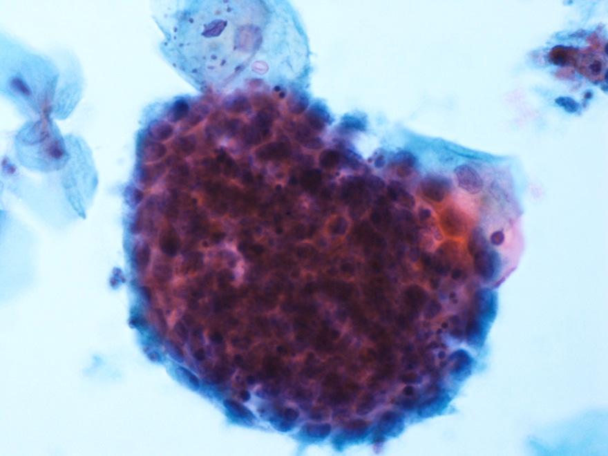
")
18 Cytology of the Lower Uterine Segment (LUS)
19 Glandular Abnormalities
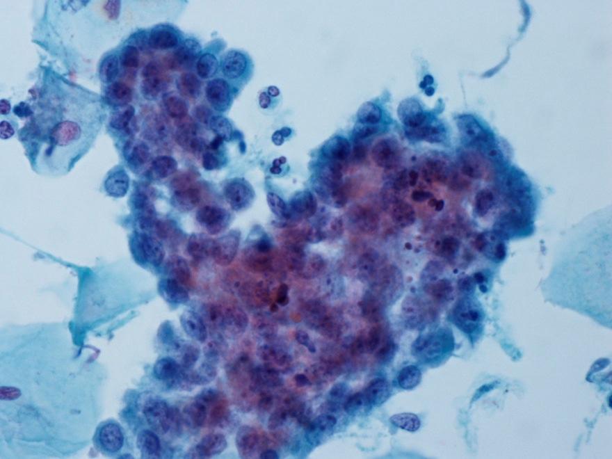
20 Glandular Abnormalities Cervical cytology screening test for Squamous intraepithelial lesion (SIL), low sensitivity for glandular lesions because of sampling & interpretation
21 Bethesda system 2001 classified 3 types of atypical endocervical cells: 1. Atypical glandular cells, not otherwise specified (AGC, NOS) 2. Atypical glandular cells, favour neoplastic (AGC, favour neoplastic) (If the endocervical origin of glandular cells is sure, specific atypical endocervical cells (NOS, or neoplasic) 3. Endocervical adenocarcinoma in situ (AIS)

22 Atypical endocervical cells vs reactive Reactive endocervical cells may show 2 x in nuclear size and conspicuous nucleoli The Bethesda 2001 (TBS 2001) defined atypical endocervical cells as endocervical-type cells that display nuclear atypia that exceed obvious reactive / reparative changes, but lack unequivocal features of endocervical adenocarcinoma. Reactive endocervical cells
23 Criteria of Atypical Glandular Cells-NOS (AGC, NOS) Architecture Loss of orderly architecture with minimal nuclei overlapping and crowding Cytology Nuclear enlargement 3 to 5 times the size of normal endocervical nuclei. (2 times nuclear enlargement: reactive) Increase N/C ratio smooth nuclear membrane Uniformly distributed granular chromatin Nucleoli may be presence Mild hyperchromasia Some variation in nuclear size and shape


24 AGC (NOS) F/51 Follow up: CxBx: Acute and chronic inflammation with focal erosion
25 Criteria of Atypical glandular cells, favour neoplastic (AGC, favour neoplastic) Architecture Hyperchromatic crowded groups Sheets, strips, irregular clusters, rosette, papillary Atypical single cells Cytology Increased N/C ratio, Nucleoli usually absent Hyperchromasia Even chromatin with coarse granularity Irregular nuclear membranes (Differentiate from Adenocarcinoma in situ (AIS): e.g. lack feathering or rosette)

26 AGC (favour neoplastic) F/47 Follow up: AIS
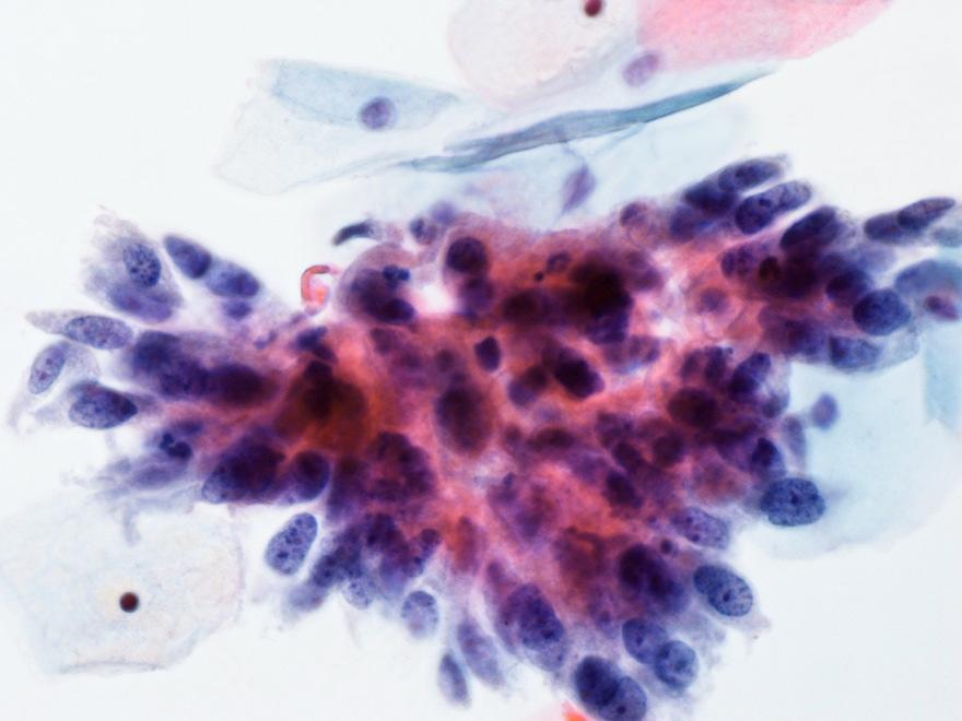
27 Cytology of Adenocarcinoma in situ (AIS) Architecture Sheets, clusters, strips, and rosettes Nuclear crowding: hyperchromatic crowded group Loss of honeycomb pattern Palisading, feathering, pseudo-stratification Cytology ( feathering best criterion for predicting glandular neoplasia, differentiation from squamous neoplasm and non-neoplastic diagnosis) Nuclei: enlarged hyperchromatic, variation in size, elongated, stratified Nucleoli: may be present N/C ratio mitosis, apoptotic bodies (may be present) Background: clean or inflammatory

28 Adenocarcinoma in situ: F/48
29 Adenocarcinoma in situ F/33
30 Cytological of Endocervical adenocarcinoma Architecture 3-D clusters with vacuolated cytoplasm 2-D sheets, strips or strands, papillary form Isolated cells may be present Cytology Dominant cancer cell: columnar shape Nuclei appearance: hyperchromiasia, anisokaryosis, clearing of chromatin, loss of polarity, macronucleoli, N/C ratio Background Tumor diathesis may present
31 Endocervical Adenocarcinoma F/52
32 Cytology of endometrial lesions
33 Morphology of Benign endometrial cells Include both the glandular and stromal cells Exfoliate in ball or gland-like clusters, single rare 1st half of menstrual cycle: glandular cells surrounding a core of stromal cells ( exodus ) Nuclei: small, round or bean-shaped, regular, degenerated (nuclei detail not clear) Nucleoli: inconspicuous Scant cytoplasm, cell borders not well defined LBP: 3-D cell ball, better chromatin detail, apoptosis


34 Endocervical Endometrial Cell size ++ + Cytoplasm Abundant ++ Scanty Nucleus Oval / elongated Round /bean shaped


35 Benign endometrial cells from menstruating epithelium exodus Key features bloody background in Conventional smear, less blood in LBP exit ball: glandular cells + stromal cell histiocytes + stromal cells in background

36 Benign endometrial cells Day 6
37 Benign endometrial cells Day 4
38 TBS 2001 describes 3 types of Endometrial lesions Benign endometrial cells in women over 40 years of age Atypical endometrial cells, NOS (not further classified as favour neoplastic because of difficulty and not reproducible) Endometrial adenocarcinoma


39 Benign Endometrial Cells in a woman >=40 years (F/47 prolonged mense, FU: Simple endometrial hyperplasia, no cytological atypia)
40 Cytology of atypical endometrial cells, NOS Architecture Small groups: 5 to 10 cells per group Cytology Nuclei slightly / relatively enlarged Mild hyperchromasia Small nucleoli Occasionally vacuolated cytoplasm Cell borders ill-defined Clean background
 (F/62.")

41 Atypical endometrial cells (NOS) (F/62. PMB Follow up endometrial biopsy: at least complex hyperplasia with atypia)
42 Cytology of Endometrial adenocarcinoma Architecture Irregular aggregates: usually small tight clusters Isolated cells usually seen Compared with endocervical adenoca (direct scrapping), fewer abnormal cells (exfoliated) Cytology Size varies (best differentiated: smallest) Small to prominent nucleoli Nuclei enlarged and irregular shape,eccentrically placed Granular, reticular, clearing Cytoplasm: scant, often vacuolated, may have intracytoplasmic neutrophils Background Finely granular or watery tumor diathesis may be present


43 Endometrial adenocarcinoma, low grade F/52 Follow up : Uterus: endometrioid adenocarcinoma FIGO grade 1

44 Endometrial adenocaricnma, low grade, F/50, perimenopausal bleeding, Uterus: Endometrioid adenocaricnoma, FIGO grade 1
45 Morphologic features for differentiating endocervical from endometrial adenocarcinoma (modified from Ayala MJ, 2011) Cytological features Microarchitecture Endocervical AdenoCA Palisading, sheets, papillary, strips, single cells (less) Endometrial AdenoCA Acini, small, 3-D clusters, single cells (frequent) Shape of cells columnar Cuboidal, rounded Cell size larger smaller Cytoplasm Granular Vacuolated with occasional polymorph infiltration Nuclear size larger smaller Nuclear chromatin coarse fine Macronucleoli common Rare in low grade No. of abnormal cells more less Tumor diathesis Usually present Less prominent, watery or granular
46 Extrauterine adenocarcinoma
47 Extrauterine adenocarcinoma CA metastatic to cervix: unusual Most frequent extragenital origin: ovary, breast, GI tract Clinical correlation and ancillary tests are needed to reach a correct diagnosis Cytology clean background morphology unusual to that of endocervical or endometrial degenerative changes


48 Adenocarcinoma, extrauterine F/56 PMB FU: Endometrial sampling: Adenocarcinoma, suggestive of metastatic from rectal primary
")

49 Adenocarcinoma (extrauterine, in keeping with metastasis) F/49 Cervical biopsy: metastatic carcinoma, c/w breast primary
50 The End
Objectives. Atypical Glandular Cells. Atypical Endocervical Cells. Reactive Endocervical Cells
 2013 California Society of Pathologists 66 th Annual Meeting San Francisco, CA Atypical Glandular Cells to Early Invasive Adenocarcinoma: Cervical Cytology and Histology Christina S. Kong, MD Associate
BOSNIAN-TURKISH CYTOPATHOLOGY SCHOOL June 18-19, 2016 Sarajevo. Case Discussions. 60 year old woman Routine gynecologic control LBC
 BOSNIAN-TURKISH CYTOPATHOLOGY SCHOOL June 18-19, 2016 Sarajevo Case Discussions Prof Dr Sıtkı Tuzlalı Tuzlalı Pathology Laboratory 60 year old woman Routine gynecologic control LBC 1 2 Endometrial thickening
Hyperchromatic Crowded Groups: What is Your Diagnosis? Session 3000
 Hyperchromatic Crowded Groups: What is Your Diagnosis? Session 3000 Thomas A. Bonfiglio, M.D. Professor Emeritus, Pathology and Laboratory Medicine University of Rochester Disclosures In the past 12 months,
CINtec p16 INK4a Staining Atlas
 CINtec p16 INK4a Staining Atlas Rating Rating Positive The rating positive will be assigned if the p16 INK4a -stained slide shows a continuous staining of cells of the basal and parabasal cell layers of
Case 3 - GYN. History: 66 year old, routine Pap test. Dr. Stelow
 Case 3 - GYN History: 66 year old, routine Pap test Dr. Stelow Case 3 66 year year old woman Routine Pap Test Cytologic Features 3 dimensional clusters of cells with small to moderate amount of
Prepared By Jocelyn Palao and Layla Faqih
 Prepared By Jocelyn Palao and Layla Faqih The structure of the suspected atypical cell should always be compared to the structure of other similar, benign, cells which are present in the smears. The diagnosis
Glandular lesions in cervical cytology. Margareta Strojan Fležar Institute of Pathology Faculty of Medicine University of Ljubljana Slovenia
 Glandular lesions in cervical cytology Margareta Strojan Fležar Institute of Pathology Faculty of Medicine University of Ljubljana Slovenia 2nd PANNONIA CONGRESS OF PATHOLOGY, SIÓFOK, HUNGARY, 17-19 MAY
Workshop for O& G trainees and paramedics 17 Dec 2011 Cytological Interpretation
 Workshop for O& G trainees and paramedics 17 Dec 2011 Cytological Interpretation May Yu Director of Cytology Laboratory Service Department of Anatomical & Cellular Pathology Prince of Wales Hospital Cervical
Morphologic Clues and Pitfalls for High Grade Lesions in Cervical Cytology
 Morphologic Clues and Pitfalls for High Grade Lesions in Cervical Cytology Ritu Nayar, MD Northwestern University, Feinberg School of Medicine Chicago, IL, USA Disclosures Editor, Cervical Cytology Bethesda
SQUAMOUS CELLS: Atypical squamous cells (ASC) - of undetermined significance (ASC-US) - cannot exclude HSIL (ASC-H)
 - of undetermined significance (ASC-US) - cannot exclude HSIL (ASC-H)") SQUAMOUS CELLS: Atypical squamous cells (ASC) - of undetermined significance (ASC-US) - cannot exclude HSIL (ASC-H) ASC refers to cytologic changes suggestive of SIL, which are qualitativley or quantitatively
SQUAMOUS CELLS: Atypical squamous cells (ASC) - of undetermined significance (ASC-US) - cannot exclude HSIL (ASC-H) ASC refers to cytologic changes suggestive of SIL, which are qualitativley or quantitatively
Conflict of Interest 9/7/2018. Dr. Mody 1. None with vendors of cytology equipment/testing/vaccines Amirsys (now Elsevier)
") Glandular Lesions in Cervicovaginal Cytology: Patterns, Pitfalls and Bethesda Updates Dina R Mody, MD Director of Cytology Laboratories Houston s Methodist Hospital and Bioreference Laboratory The Ibrahim
Glandular Lesions in Cervicovaginal Cytology: Patterns, Pitfalls and Bethesda Updates Dina R Mody, MD Director of Cytology Laboratories Houston s Methodist Hospital and Bioreference Laboratory The Ibrahim
Mody. Atypical Glandular Cells(TBS 2001) Adenocarcinoma In Situ(TBS 2001)
 Adenocarcinoma In Situ(TBS 2001)") Glandular Lesions in Cervicovaginal Cytology Dina R. Mody, MD, FCAP Director of Cytology The Methodist Hospital, Houston, TX Professor of Pathology and Laboratory Medicine Weill Medical College of Cornell
Glandular Lesions in Cervicovaginal Cytology Dina R. Mody, MD, FCAP Director of Cytology The Methodist Hospital, Houston, TX Professor of Pathology and Laboratory Medicine Weill Medical College of Cornell
Mody. AIS vs. Invasive Adenocarcinoma of the Cervix
 Common Problems in Gynecologic Pathology Michael T. Deavers, M.D. Houston Methodist Hospital, Houston, Texas Common Problems in Gynecologic Pathology Adenocarcinoma in-situ (AIS) of the Cervix vs. Invasive
Cytyc Corporation - Case Presentation Archive - July 2002
 ThinPrep Pap Test History: 34 Year Old Female LMP: Day 20 Specimen Type: Cervical/Vaginal Case provided by Mark Tulecke, M.D. and Gabrielle Trawinski CT (ASCP), Mount Auburn Hospital, Cambridge, Massachusetts.
Demystifying Endometrial Hyperplasia
 Demystifying Endometrial Hyperplasia A review from Diagnostic Histopathology 19:7 Dr R Hadden ST5 Histopathology Derriford Hospital Plymouth Endometrium Target for sex-steroid hormones Glands Stroma Proliferate
Case year female. Routine Pap smear
 Case 1 57 year female Routine Pap smear Diagnosis? 1. Atypical glandular cells of unknown significance (AGUS) 2. Endocervical AIS 3. Endocervical adenocarcinoma 4. Endometrial adenocarcinoma 5. Adenocarcinoma
Page # 1. Endometrium. Cellular Components. Anatomical Regions. Management of SIL Thomas C. Wright, Jr. Most common diseases:
 Endometrium Pathology of the Endometrium Thomas C. Wright Columbia University, New York, NY Most common diseases: Abnormal uterine bleeding Inflammatory conditions Benign neoplasms Endometrial cancer Anatomical
6/5/2010. Outline of Talk. Endometrial Alterations That Mimic Cancer & Vice Versa: Metaplastic / reactive changes. Problems in Biopsies/Curettages
 Outline of Talk Endometrial Alterations That Mimic Cancer & Vice Versa: Problems in Biopsies/Curettages Metaplastic / reactive changes Mucinous change Microglandular hyperplasia-like change Squamous metaplasia
New Diagnoses Need New Approaches: A Glimpse into the Near Future of Gynecologic Pathology
 New Diagnoses Need New Approaches: A Glimpse into the Near Future of Gynecologic Pathology United States and Canadian Academy of Pathology 102 nd Annual Meeting Baltimore, Maryland Christina S. Kong, M.D.
Maturation Index 3/29/2017. Disclosure of Relevant Financial Relationships. Gynecologic Cytology. Normal Maturation of Squamous Epithelium : :
 Gynecologic Cytology Fadi W. Abdul Karim, MD MEd Department of Anatomic Pathology Vice Chair Education RT PLMI Professor of Pathology Cleveland Clinic. Cleveland Ohio Disclosure of Relevant Financial Relationships
GYN (Glandulars) Still Difficult After All These Years! Dina R Mody, MD Director of Cytology Laboratories and fellowship Program Methodist Hospital
 Still Difficult After All These Years! Dina R Mody, MD Director of Cytology Laboratories and fellowship Program Methodist Hospital") GYN (Glandulars) Still Difficult After All These Years! Dina R Mody, MD Director of Cytology Laboratories and fellowship Program Methodist Hospital and Bioreference Labs (Houston) Department of Pathology
GYN (Glandulars) Still Difficult After All These Years! Dina R Mody, MD Director of Cytology Laboratories and fellowship Program Methodist Hospital and Bioreference Labs (Houston) Department of Pathology
LGM International, Inc.
 Liqui-PREP TM Cytology Atlas Preface The following pictures are examples with descriptions of cytology slides processed with the Liqui-PREP TM System.. The descriptions are reviewed by Pathologists. It
EU guidelines for reporting gynaecological cytology
 EU guidelines for reporting gynaecological cytology Amanda Herbert Guy s & St Thomas Foundation NHS Trust 5th EFCS Annual Tutorial, Trondheim, Norway 28 th May 1 st June 2012 EU guidelines aim to harmonize
Pancreatitis: A Potential Pitfall in Endoscopic Ultrasound Guided Pancreatic FNA
 Pancreatitis: A Potential Pitfall in Endoscopic Ultrasound Guided Pancreatic FNA Jack Yang, MD Department of Pathology, Medical University of South Carolina Objectives Understand the indication of EUS
Endometrial Metaplasia, Hyperplasia & Other Cancer Mimics: a Consultant s Experience
 Endometrial Metaplasia, Hyperplasia & Other Cancer Mimics: a Consultant s Experience Pacific Northwest Society of Pathologists Vancouver, B.C. September 26, 2015 Teri A. Longacre, M.D. longacre@stanford.edu
In situ and Invasive Endocervical Carcinoma: Problems and Pitfalls in Diagnosis
 In situ and Invasive Endocervical Carcinoma: Problems and Pitfalls in Diagnosis Rouba Ali-Fehmi,MD The Karmanos Cancer Institute, Wayne State University School of Medicine Global incidence of cervical
Index. Cytoplasm, nonepithelial malignant tumor features 70
 Accurette device 23 Adenosarcoma, differential diagnosis 80, 81 Arias-Stella reaction 65 Atypical endocervical cells 8 Atypical endometrial cells 8 Atypical glandular cells (AGC) 8, 9 Atypical glandular
Outline 11/2/2017. Pancreatic EUS-FNA general aspects. Cytomorphologic features of solid neoplasms/lesions of the pancreas
 ENDOSCOPIC ULTRASOUND GUIDED-FINE NEEDLE ASPIRATION CYTOLOGY OF PANCREAS Khalid Amin M.D. Assistant Professor Department of Laboratory Medicine and Pathology University of Minnesota Outline Pancreatic
Cervical Cancer : Pap smear
 Taking a PAP SMEAR Cervical Cancer : Pap smear George N Papanicolaou introduced cervical cytology in clinical practice in 1940 In 1945, PAP smear was endorsed by American cancer society as an effective
Villoglandular adenocarcinoma of cervix a tumour with bland cytological features: report of a case missed on cytology
 Malaysian J Pathol 2003; 25(2) : CERVICAL 139 143 VILLOGLANDULAR ADENOCARCINOMA CYTOLOGY CASE REPORT Villoglandular adenocarcinoma of cervix a tumour with bland cytological features: report of a case missed
Cytyc Corporation - Case Presentation Archive - March 2002
 FirstCyte Ductal Lavage History: 68 Year Old Female Gail Index: Unknown Clinical History: Negative Mammogram in 1995 6 yrs. later presents with bloody nipple discharge Subsequent suspicious mammogram Suspicious
Lessons From Cases of Screened Women Who Developed Cervical Carcinoma
 Lessons From Cases of Screened Women Who Developed Cervical Carcinoma R. Marshall Austin MD,PhD Magee-Womens Hospital of University of Pittsburgh Medical Center raustin@magee.edu Why Focus Study On Cases
Cytyc Corporation - Case Presentation Archive - October 2001
 ThinPrep Pap Test History: 82 Year Old Female Specimen Type: Peritoneal Washings Case provided by Dr. Berle Stratton, Southwest Washington Medical Center, Vancouver, Washington. *The images, analysis and
Received, June 29, 1904; accepted for publication
 THE AMEBICAN JOURNAL OF CLINICAL PATHOLOGY Copyright 1964 by The Williams & Wilkins Co. Vol. 42, No. 0 Printed in U.S.A. CARCINOMA IN SITU OF THE ENDOMETRIUM ISABELLE A. BUEHL, M.D., PRANK VELLIOS, M.D.,
Normal endometrium: A, proliferative. B, secretory.
 Normal endometrium: A, proliferative. B, secretory. Nội mạc tử cung Nội mạc tử cung Cyclic changes in endometrium.. Approximate relationship of useful microscopic changes. Arias-Stella reaction in endometrial
Morphology I Slide: 1
 Morphology I Slide: 1 Morphology I Slide: 2 ThinPrep Morphology Normal Cytology Morphology I Slide: 3 CT & Pathologist Training Training program begins with ThinPrep morphology presentation Microscopic
3/28/2017. Disclosure of Relevant Financial Relationships. GU Evening Subspecialty Case Conference. Differential Diagnosis:
 GU Evening Subspecialty Case Conference Rajal B. Shah, M.D. VP, Medical Director, Urologic Pathology Miraca Life Sciences, Irving, Texas Clinical Associate Professor of Pathology Baylor College of Medicine,
Normal Morphology. Anatomic Considerations. Normal Urothelial Histology and Cytology
 1 Normal Morphology Anatomic Considerations The urinary tract can be divided into three regions: the kidney; the calyces, pelves and ureters (upper collecting system or upper tract); and the bladder and
Endometrial hyperplasia vs. Intraepithelial neoplasia. Martin Chang, MD PhD FRCPC Pathology Update Friday November 9, 2012
 Endometrial hyperplasia vs. Intraepithelial neoplasia Martin Chang, MD PhD FRCPC Pathology Update Friday November 9, 2012 Disclosure No relevant financial conflicts to declare. Case 1 Gland crowding Gland
Salivary Gland Cytology
 Salivary Gland Cytology Diagnostic challenges and potential pitfalls Tarik M. Elsheikh, MD Professor and Medical Director Anatomic Pathology Cleveland Clinic FNA Salivary Gland Lesions Indications Distinguish
Index 179. Genital tract contaminants, 17, 20, 22, 150 papilloma virus-infected cells, 47 squamous cells, sources of, 7
 Index Accuracy of urinary cytology, 166 Acute inflammatory cells, 38 catheter sample, 39 herpes simplex infections, 44 carcinomas, 104, 105 non-viral inclusions, 52, 53 voided urine, 17 Adenocarcinoma
Comparison of Cytologic Characteristics between Adenoid Cystic Carcinoma and Adenoid Basal Carcinoma in the Uterine Cervix
 Journal of Pathology and Translational Medicine 2015; 49: 396-402 ORIGINAL ARTICLE Comparison of Cytologic Characteristics between Adenoid Cystic Carcinoma and Adenoid Basal Carcinoma in the Uterine Cervix
PRESENTATION PLAN. Aim: Bethesda System 2001
 REACTIVE CELLULAR CHANGES AND INFECTIONS OF FEMALE GENITAL TRACT Aysun Uğuz, Prof, MD, FIAC Çukurova Üniv. Tıp Fak. Pathology Department-Cytology Division 18.Nisan.2015 Aim: The aim of the presentation
Introduction. 23 rd Annual Seminar in Pathology. FLUIDS, Part 1. Pittsburgh, PA Gladwyn Leiman UVMMC, VT
 23 rd Annual Seminar in Pathology Pittsburgh, PA Gladwyn Leiman UVMMC, VT FLUIDS, Part 1 "Blue walls", Claudia Hansen, 2009 Introduction o Challenging to everyone o Almost any benign or malignant process
PAP SMEAR by Dr.Shantha Krishnamurthy MD Senior Consultant Pathology Fortis Hospitals
 PAP SMEAR by Dr.Shantha Krishnamurthy MD Senior Consultant Pathology Fortis Hospitals Historical Named after George Papanicolaou, a Greek American Studied cervical epithelium in menstrual cycle of guinea
Thyroid follicular neoplasms in cytology. Ulrika Klopčič Institute of Oncology, Department of Cytopathology, Ljubljana, Slovenia
 Thyroid follicular neoplasms in cytology Ulrika Klopčič Institute of Oncology, Department of Cytopathology, Ljubljana, Slovenia Lecture overview importance of FNAB in assessing thyroid lesions follicular
Biliary Tract Neoplasia: A Cyto-histologic Review. Michelle Reid, MD, MSc Professor of Pathology Director of Cytopathology Emory University Hospital
 Biliary Tract Neoplasia: A Cyto-histologic Review Michelle Reid, MD, MSc Professor of Pathology Director of Cytopathology Emory University Hospital Bile Duct Brushings (BDB) BDBs are the initial diagnostic
QUALITY ASSURANCE PROGRAM CYTOLOGY CYCLE 01/2018 (TRIAL)
") [Pick the Date] FINAL REPORT QUALITY ASSURANCE PROGRAM CYTOLOGY CYCLE 01/2018 (TRIAL) NOTES FROM THE COORDINATOR 1. For this cycle 01/2018, a total of 32 pen drives had been circulated. Twenty-eight institutions
[Pick the Date] FINAL REPORT QUALITY ASSURANCE PROGRAM CYTOLOGY CYCLE 01/2018 (TRIAL) NOTES FROM THE COORDINATOR 1. For this cycle 01/2018, a total of 32 pen drives had been circulated. Twenty-eight institutions
International Society of Gynecological Pathologists Symposium 2007
 International Society of Gynecological Pathologists Symposium 2007 Anais Malpica, M.D. Department of Pathology The University of Texas M.D. Anderson Cancer Center Grading of Ovarian Cancer Histologic grade
Liquid-Based Cytology of Villoglandular Adenocarcinoma of the Cervix: A Report of 3 Cases
 The Korean Journal of Pathology 2012; 46: 215-220 CSE REPORT Liquid-ased Cytology of Villoglandular denocarcinoma of the Cervix: Report of 3 Cases Younghwa Choi Haeryoung Kim Haiyoung Choi Daehyun Hwang
Cytology Report Format
 Squamous Precursor Lesions and Malignancies In Pap Test Dina R. Mody, MD, FCAP Director of Cytology The Methodist Hospital, Houston, TX Professor of Pathology and Laboratory Medicine Weill Medical College
Intravascular Endometrium Mimicking Vascular Invasion
 ISPUB.COM The Internet Journal of Pathology Volume 12 Number 1 A Papanicolau, G Lin Citation A Papanicolau, G Lin.. The Internet Journal of Pathology. 2010 Volume 12 Number 1. Abstract Intravascular endometrium
Respiratory Tract Cytology
 Respiratory Tract Cytology 40 th European Congress of Cytology Liverpool, UK Momin T. Siddiqui M.D. Professor of Pathology and Laboratory Medicine Director of Cytopathology Emory University Hospital, Atlanta,
When Immunostains Can Get You in Trouble: Gynecologic Pathology p16: Panacea or Pandora s Box?
 When Immunostains Can Get You in Trouble: Gynecologic Pathology p16: Panacea or Pandora s Box? Teri A. Longacre, MD Stanford Medicine Stanford California pi6 in Gynecologic Pathology: Panacea or Pandora
Atypical Hyperplasia/EIN
 EIN Atypical Hyperplasia/EIN Based on scientific and diagnostic advances, in 2014 the WHO moved that the precursor lesion for endometrioid carcinoma be atypical hyperplasia/ein, rather than what was previously
Neoplasia 2018 Lecture 2. Dr Heyam Awad MD, FRCPath
 Neoplasia 2018 Lecture 2 Dr Heyam Awad MD, FRCPath ILOS 1. List the differences between benign and malignant tumors. 2. Recognize the histological features of malignancy. 3. Define dysplasia and understand
Colposcopy. Attila L Major, MD, PhD
 Colposcopy Attila L Major, MD, PhD Histology Colposcopy Cytology It has been estimated that annual Pap smear testing reduces a woman s chance of dying of cervical cancer from 4 in 1000 to about 5 in 10,000
A Study on Diagnostic Accuracy of Cervical Pap Smear by Correlating with Histopathology in a Tertiary Care Centre
 Original Article DOI: 10.21276/APALM.1878 A Study on Diagnostic Accuracy of Cervical Pap Smear by Correlating with Histopathology in a Tertiary Care Centre Rachana L Y, S.S. Hiremath*, Prabhu M H, S.S
PRM Associated Endometrial Change Introduction & Illustrations 12-Feb-2012
 Introductory Remarks: These images are from clinical trial endometrial samples collected by catheter biopsy. They are presented with a low power section view with selected higher power images to show detailed
Ductal Proliferations of the Breast: The Good, the Bad, and the Ugly
 Ductal Proliferations of the Breast: The Good, the Bad, and the Ugly Melinda F. Lerwill, MD CRITERIA FOR DISTINGUISHING LOW-GRADE DUCTAL CARCINOMA IN SITU FROM USUAL DUCTAL HYPERPLASIA CYTOLOGY Low-grade
Endometrial line thickness in different conditions.
 Endometrial line thickness in different conditions 1 Endometrial thickens in response to Rising estrogen levels during the menstrual cycle and then shedding endometrial at the times of menses 2 The thickens
We are IntechOpen, the world s leading publisher of Open Access books Built by scientists, for scientists. International authors and editors
 We are IntechOpen, the world s leading publisher of Open Access books Built by scientists, for scientists 4,000 116,000 120M Open access books available International authors and editors Downloads Our
intraepithelial neoplasia: a premalignant lesion?
 J Clin Pathol 1986;39:22-28 Cervical glandular atypia associated with squamous intraepithelial neoplasia: a premalignant lesion? LJR BROWN, M WELLS From the Department of Pathology, University ofleeds,
FNA of Thyroid. Toward a Uniform Terminology With Management Guidelines. NCI NCI Thyroid FNA State of the Science Conference
 FNA of Thyroid NCI NCI Thyroid FNA State of the Science Conference Toward a Uniform Terminology With Management Guidelines Thyroid Thyroid FNA Cytomorphology NCI Thyroid FNA State of the Science Conference
ENODMETRIAL CARCINOMA: SPECIAL & NOT SO SPECIAL VARIANTS
 ENODMETRIAL CARCINOMA: SPECIAL & NOT SO SPECIAL VARIANTS Pacific Northwest Society of Pathologists Vancouver, B.C. September 26, 2015 Teri A. Longacre, M.D. longacre@stanford.edu Stanford University, Stanford,
05/07/2018. Types of challenges. Challenging cases in uterine pathology. Case 1 ` 65 year old female Post menopausal bleeding Uterine Polyp
 Types of challenges Challenging cases in uterine pathology Nafisa Wilkinson Gynaecological Pathologist UCLH London Lack of complete history often, NO clinical history at all! Cases from other centres often
The incidence of cervical adenocarcinoma (ADC) has
 has") Cervical Adenocarcinoma of Human Papillomavirus Positive and Human Papillomavirus Negative Tumors Edyta C. Pirog, MD, PhD Context. Cervical adenocarcinomas span a diverse group of tumors with several distinct
Cervical Adenocarcinoma of Human Papillomavirus Positive and Human Papillomavirus Negative Tumors Edyta C. Pirog, MD, PhD Context. Cervical adenocarcinomas span a diverse group of tumors with several distinct
Histopathology: Cervical HPV and neoplasia
 Histopathology: Cervical HPV and neoplasia These presentations are to help you identify basic histopathological features. They do not contain the additional factual information that you need to learn about
1.Acute and Chronic Cervicitis - At the onset of menarche, the production of estrogens by the ovary stimulates maturation of the cervical and vaginal
 Diseases of cervix I. Inflammations 1.Acute and Chronic Cervicitis - At the onset of menarche, the production of estrogens by the ovary stimulates maturation of the cervical and vaginal squamous mucosa
Salivary Glands 3/7/2017
 Salivary Glands 3/7/2017 Goals and objectives Focus on the entities unique to H&N Common board type facts Information for your future practice Salivary Glands Salivary Glands Major gland. Paratid. Submandibular.
The ABCs of TBS. A Novice's Guide to the Bethesda System
 CE U P D A T E W O M E N ' S HEALTH III Julia Woodruff Wildes, MD The ABCs of TBS A Novice's Guide to the Bethesda System This is the third and final article in a three-part series on women's health. The
Diagnostically Challenging Cases in Gynecologic Pathology
 Diagnostically Challenging Cases in Gynecologic Pathology Eric C. Huang, M.D., Ph.D. Department of Pathology and Laboratory Medicine University of California, Davis Medical Center Case 1 Presentation 38
Chapter 10: Pap Test Results
 Chapter 10: Pap Test Results On completion of this section, the learner will be able to: 1. Identify how Pap test results are interpreted and the reasons for normal and abnormal results. 2. Describe the
ACCME/Disclosures. Cribriform Lesions of the Prostate. Case
 Cribriform Lesions of the Prostate Ming Zhou, MD, PhD Departments of Pathology and Urology New York University Langone Medical Center New York, NY Ming.Zhou@NYUMC.ORG ACCME/Disclosures The USCAP requires
ACGME Competency / Milestone Assessment. The Pap Test. Ricardo R. Lastra, MD Zubair W. Baloch, MD, PhD
 1 ACGME Competency / Milestone Assessment The Pap Test Ricardo R. Lastra, MD Zubair W. Baloch, MD, PhD Department of Pathology & Laboratory Medicine University of Pennsylvania, Perelman School of Medicine
PAPILLARY PROLIFERATION OF THE ENDOMETRIUM: A BENIGN LESION SIMULATING ADENOCARCINOMA.
 PAPILLARY PROLIFERATION OF THE ENDOMETRIUM: A BENIGN LESION SIMULATING ADENOCARCINOMA. Teresa Pusiol, Maria Grazia Zorzi, Doriana Morichetti U.O. Anatomia Patologica Ospedale S. Maria del Carmine Rovereto
Proliferative Epithelial lesions of the Breast. Sami Shousha, MD, FRCPath Charing Cross Hospital & Imperial College, London
 Proliferative Epithelial lesions of the Breast Sami Shousha, MD, FRCPath Charing Cross Hospital & Imperial College, London Amman, November2013 Proliferative Epithelial Lesions of the Breast Usual type
Biliary tract tumors
 Short Course 2010 Annual Fall Meeting of the Korean Society for Pathologists Biliary tract tumors Joon Hyuk Choi, M.D., Ph.D. Professor, Department of Pathology, Yeungnam Univ. College of Medicine, Daegu,
1 NORMAL HISTOLOGY AND METAPLASIAS
 1 NORMAL HISTOLOGY AND METAPLASIAS, MD Anatomy and Histology 1 Metaplasias 2 ANATOMY AND HISTOLOGY The female breast is composed of a branching duct system, which begins at the nipple with the major lactiferous
Atypical Glandular Cells of Undetermined Significance Cytologic Criteria to Separate Clinically Significant From Benign Lesions
 ANATOMIC PATHOLOGY Original Article Atypical Glandular Cells of Undetermined Significance Cytologic Criteria to Separate Clinically Significant From Benign Lesions STEPHEN S. RAAB, MD, CHRISTINA ISACSON,
number Done by Corrected by Doctor Maha Shomaf
 number 16 Done by Waseem Abo-Obeida Corrected by Zeina Assaf Doctor Maha Shomaf MALIGNANT NEOPLASMS The four fundamental features by which benign and malignant tumors can be distinguished are: 1- differentiation
How to Recognize Gynecologic Cancer Cells from Pelvic Washing and Ascetic Specimens
 How to Recognize Gynecologic Cancer Cells from Pelvic Washing and Ascetic Specimens Wenxin Zheng, M.D. Professor of Pathology and Gynecology University of Arizona zhengw@email.arizona.edu http://www.zheng.gynpath.medicine.arizona.edu/index.html
Intraductal carcinoma of the prostate on needle biopsy: histologic features and clinical significance
 & 2006 USCAP, Inc All rights reserved 0893-3952/06 $30.00 www.modernpathology.org Intraductal carcinoma of the prostate on needle biopsy: histologic features and clinical significance Charles C Guo 1 and
40th European Congress of Cytology Liverpool, UK, 2-5 th October 2016
 40th European Congress of Cytology Liverpool, UK, 2-5 th October 2016 EUS FNA of abdominal organs: An approach to reporting and triage for ancillary testing Date and time: Sunday 2 nd October 2016 15.00-16.30
CLINICAL SIGNIFICANCE OF BENIGN EPITHELIAL CHANGES
 Papillomas. Papillomas are composed of multiple branching fibrovascular cores, each having a connective tissue axis lined by luminal and myoepithelial cells ( Fig. 23-11 ). Growth occurs within a dilated
Papillary Lesions of the breast
 Papillary Lesions of the breast Emad Rakha Professor of Breast Pathology The University of Nottingham Papillary lesions of the breast are a heterogeneous group of disease, which are characterised by neoplastic
Prostate Pathology: Prostate Carcinoma, variants and Gleason Grading (Part 1)
") Prostate Pathology: Prostate Carcinoma, variants and Gleason Grading (Part 1) Jae Y. Ro, MD, PhD June 7, 2012 Ten Leading Cancer Types for the Estimated New Cancer Cases and Deaths By Sex, United States,
Prostate Pathology: Prostate Carcinoma, variants and Gleason Grading (Part 1) Jae Y. Ro, MD, PhD June 7, 2012 Ten Leading Cancer Types for the Estimated New Cancer Cases and Deaths By Sex, United States,
Salivary gland cytology. Salivary gland cytology. Triage helps the clinician. Salivary gland tumors. Diagnostic difficulties
 Salivary gland cytology Salivary Gland Cytology Pınar Fırat, MD Professor of Pathology İ.U. İstanbul Faculty of Medicine Çapa, İstanbul It is a reliable diagnostic test However, definitive subclassification
MPH Quiz. 1. How many primaries are present based on this pathology report? 2. What rule is this based on?
 MPH Quiz Case 1 Surgical Pathology from hysterectomy performed July 11, 2007 Final Diagnosis: Uterus, resection: Endometrioid adenocarcinoma, Grade 1 involving most of endometrium, myometrial invasion
Ph.D. THESIS ENDOMETRIAL HYPERPLASIAS IN PERIMENOPAUSE SUMMARY
 UNIVERSITY OF MEDICINE AND PHARMACY OF CRAIOVA FACULTY OF MEDICINE Ph.D. THESIS ENDOMETRIAL HYPERPLASIAS IN PERIMENOPAUSE SUMMARY SCIENTIFIC COORDINATOR: PROF. DR. MIHAI B. BRĂILA, Ph.D. Ph.D. Graduand:
5/2/2018. Low Grade Dysplasia of GI Tract. High Grade Dysplasia of GI Tract. Dysplasia in Gastrointestinal Tract: Practical Pearls and Issues
 Dysplasia in Gastrointestinal Tract: Practical Pearls and Issues Arief Suriawinata, M.D. Professor of Pathology and Laboratory Medicine Geisel School of Medicine at Dartmouth Department of Pathology and
Cervical Dysplasia and HPV
 Cervical Dysplasia and HPV J. Anthony Rakowski D.O., F.A.C.O.O.G. MSU SCS Board Review Coarse HPV Double stranded DNA virus The HPV infect epithelial cells of the skin and mucous membranes Highest risk
Tissues. tissue = many cells w/ same structure and function. cell shape aids function tissue shape aids function. Histology = study of tissues
 Tissues tissue = many cells w/ same structure and function cell shape aids function tissue shape aids function Histology = study of tissues 4 types of tissues Epithelial coverings contact openings Connective
Dr. Issraa Ali Hussein
 CLINICAL 09888888;rCYTOLOGY Dr. Issraa Ali Hussein objectives Define diagnostic cytology (clinical cytology). Explain the differences between histopathology and cytopathology. Recognize the methods for
Cytology and Surgical Pathology of Gynecologic Neoplasms
 Cytology and Surgical Pathology of Gynecologic Neoplasms Current Clinical Pathology ANTONIO GIORDANO, MD, PHD SERIES EDITOR For further titles published in this series, go to http://www.springer.com/springer/series/7632
Problems in the Differential Diagnosis of Endometrial Hyperplasia and Carcinoma
 THE 1999 LONG COURSE ON PATHOLOGY OF THE UTERINE CORPUS AND CERVIX Problems in the Differential Diagnosis of Endometrial Hyperplasia and Carcinoma Steven G. Silverberg, M.D. Department of Pathology, University
BREAST PATHOLOGY. Fibrocystic Changes
 BREAST PATHOLOGY Lesions of the breast are very common, and they present as palpable, sometimes painful, nodules or masses. Most of these lesions are benign. Breast cancer is the 2 nd most common cause
Can True Papillary Neoplasms of Breast and Their Mimickers Be Accurately Classified by Cytology?
 92 CANCER CYTOPATHOLOGY Can True Papillary Neoplasms of Breast and Their Mimickers Be Accurately Classified by Cytology? Claire W. Michael, M.D. 1 Bruce Buschmann, C.T. 2 1 University of Michigan, Department
OUTLINE. Case History 1/28/2013. Endometrial Neoplasia, Including Endometrial Intraepithelial Neoplasia (EIN)
") Endometrial Neoplasia, Including Endometrial Intraepithelial Neoplasia (EIN) TEXAS SOCIETY OF PATHOLOGISTS ANNUAL MEETING Marisa R. Nucci, M.D. Division of Women s and Perinatal Pathology Brigham and Women
Endometrial Neoplasia, Including Endometrial Intraepithelial Neoplasia (EIN) TEXAS SOCIETY OF PATHOLOGISTS ANNUAL MEETING Marisa R. Nucci, M.D. Division of Women s and Perinatal Pathology Brigham and Women
Cervical Cytology Preparations
 GYN Cytology Cervical Cytology Preparations CS TP SP Fadi W. Abdul-Karim, MD MSMedu Department of Anatomic Pathology Vice Chair Education Professor of Pathology Cleveland Clinic Cleveland Ohio Conventional