There is NO single Melanoma Stain. > 6000 Mutations in Melanoma. What else can be done to discriminate atypical nevi from melanoma?
|
|
|
- Silas Williams
- 5 years ago
- Views:
Transcription
 University of Colorado Health Sciences Center whitney.high@ucdenver.")
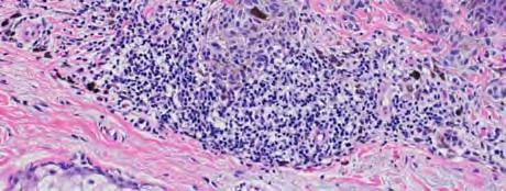
1 Las Vegas Fall Clinical 2016: The Assessment and Diagnosis of Melanoma Whitney A. High, MD, JD, MEng Associate Professor, Dermatology & Pathology Director of Dermatopathology (Dermatology) University of Colorado Health Sciences Center October 2016 Las Vegas, Nevada > 6000 Mutations in Melanoma Makes for Messy Model How good is grading? Duncan et al. (1993) 10 cases of nevi, dysplastic nevi (mild, mod, sev) and melanoma concordance 69-80% for nevus vs dysplastic nevus vs melanoma only 35-58% concordance for grading of dysplasia Piepkorn, et al. (1994) 149 atypical nevi graded by 6 expert dermatopathologists re-interpreted 6 mos later by same dermatopathologist correlation coefficients ( moderate to substantial ) best correlation by person using only two categories (mild/mod vs sev) Farmer, et al. (1996) 37 classic melanomas/nevi & 8 expert dermatopathologists unanimity for melanoma in only 11 cases (30%) What else can be done to discriminate atypical nevi from melanoma? Immunostains There is NO single Melanoma Stain Combination stains Kimart Mart1 highlights melanocytes Ki67 marks proliferative index 1


2 KiMart (MART-1/Ki67) P16 P16 is a tumor suppressor protein Often used for Spitz vs. melanoma ddx Dermal P16 staining was best discriminator: loss of nuclear staining (<25% of cells) 3 fold more likely to be melanoma loss of nuclear and cytoplasmic staining 8 fold more likely to be melanoma George E, et al: Am J Clin Pathol 130:370, 2010 P16 in Spitz Nevus HMB-45 Benign melanocytic lesions: junctional/superficial component stains normal zonation in the deeper component Confusing in situations of dusty cells: deeply pigmented nevi deep penetrating nevi clonal nevi Expression of p16 is favorable Compound nevus Melanoma Sensitivity vs Specificity S100 vs Mart1 ZONATION LACK OF ZONATION 2


3 Desmoplastic (Spindle Cell) Melanoma S100 Mart1 vs. S100 Melan A But atypical nevi, particularly mild and even moderate nevi are not that big a deal, right? But hold on Are we simply bad at distinguishing atypical nevi from melanoma? 3


4 Illustrative Example Re-examination of Original Called compound moderately atypical nevus by a well-known Colorado dermatopathologist. No stains or levels were performed. MART1/Ki67 4


5 HMB45 Spitz Nevus Classic Spitz Nevus ale of the Spitz tumor Histology 17 year-old girl seen in 2007 Eruptive lesion on leg only noticed for a few weeks No personal or FH of melanoma Otherwise healthy 5


6 No win situation. Overcall and a 17 year-old receives: huge disfiguring scar ruined insurance status chronic leg edema no real hope Undercall and there are: medicolegal issues is the patient harmed? Her Sentinel Lymph Node Patients may not always want (or need) what you think they want (or need) 6


 Patient")


7 Real case Ki67/Mart1 P16 Update Thoughts? Signed out as: Combined nevus (common nevus and Spitz nevus) Patient presented in 2016 with right sided cervical LAD and was told initially was mononucleosis. Ultimately, she had melanoma in a cervical lymph node, and tumor nodules in the right lung. 7

 Gene Expression Profiling (GEP) Comparative")


 11q13 (CCDN1) 6q23 (MYB) CEP6 86.")

8 On the verge of assessing the genetics of melanoma Tests That Cay Be Employed Comparative Genomic Hybridization (acgh) Fluorescent In Situ Hybridization (FISH) Gene Expression Profiling (GEP) Comparative Genomic Hybridization FISH for Additions/Deletions Examines entire genome Detects gains or losses in copy number Gains/losses exist in >95% of melanoma Performed on FFPE tx Bauer & Bastian,Derm Therapy, 2006; 19:40-9. (Am J Surg Pathol 2009;33: ) Best probe set 6p25 (RREB1) 11q13 (CCDN1) 6q23 (MYB) CEP6 86.7% sensitive 95.4% specific identified 6/6 metastasizing ambiguous lesions Only 60% sensitivity and only 60% specificity in ambiguous lesions. 8


9 23 Gene Expression Profiling qrt-pcr on FFPE that is microdissected Developed and validated on N=400+ nevi/mm Acknowledged that for difficult Spitzoid lesions the old probe set was probably only around 70% sensitive Proposes new probe set - 6p25, 9p21, 11q13, and 8q24 Particularly interesting is addition of loss of 9p21 (p16) Overall improvement in sensitivity reported to be 94% No lab test works all the time DermTech Another GEP based test Uses tape stripping Set for high SENSITIVITY and lesser SPECIFICITY Impact of a Threshold on Sensitivity and Specificity Conservative Dermpath Low FISH threshold Low proprietary threshold any test Cowboy Dermpath High FISH threshold High proprietary threshold any test 9
 Technology developed in uveal melanoma GEP accurately distinguishes low risk (95% 5-year metastasis-free survival) high risk (20% 5-year metastasis-free")

10 On the horizon Prognostic test for melanoma Clin Cancer Res. 2015; 21: Castle 31 GEP Test All stage disease (I-IV) Technology developed in uveal melanoma GEP accurately distinguishes low risk (95% 5-year metastasis-free survival) high risk (20% 5-year metastasis-free survival) Critics contend uveal MM is a genetically simpler problem than cutaneous MM almost all are GNAQ or GNA11 driven (80%) compared to 2% of GNAQ/GNA11 in all melanoma BAP-1 mutation confers increased metastatic risk Validation set data for the Castle Biosciences DecisionRx Kaplan-Meier Survival Curves for Low Risk vs High Risk GEP Measurements Only stage I & II Where this test might be used Patients that simply need to know everything that can be known Patients with lesions less than 1 mm but with high mitotic activity Patients s/p SLN to identify those at risk Class 2 67% and 83% of SLN negative cases with recurrence or death, respectively Zager et al. ASCO
> 6000 Mutations in Melanoma. Tests That Cay Be Employed. FISH for Additions/Deletions. Comparative Genomic Hybridization
 Winter Clinical 2017: The Assessment and Diagnosis of Melanoma Whitney A. High, MD, JD, MEng Associate Professor, Dermatology & Pathology Director of Dermatopathology (Dermatology) University of Colorado
Melanoma and the genes: Molecular alterations informing the diagnosis of melanocytic tumors
 Melanoma and the genes: Molecular alterations informing the diagnosis of melanocytic tumors Michael T. Tetzlaff MD, PhD Associate Professor Department of Pathology, Section of Dermatopathology Department
Michael T. Tetzlaff MD, PhD
 Molecular alterations informing the diagnosis of melanocytic tumors Michael T. Tetzlaff MD, PhD Associate Professor Department of Pathology, Section of Dermatopathology Department of Translational and
Melanocytic Lesions: Use of Immunohistochemistry and Special Studies Napa Valley 2018
 Melanocytic Lesions: Use of Immunohistochemistry and Special Studies Napa Valley 2018 Victor G. Prieto, MD, PhD Professor Depts. of Pathology and Dermatology University of Texas - MD Anderson Cancer Center
Update on Spitzoid and Blue nevus-like melanocytic lesions Emphasis on molecular studies informing diagnosis, prognosis and therapy
 Update on Spitzoid and Blue nevus-like melanocytic lesions Emphasis on molecular studies informing diagnosis, prognosis and therapy Michael T. Tetzlaff MD, PhD Associate Professor Department of Pathology,
Genetic Testing: When should it be ordered? Julie Schloemer, MD Dermatology
 Genetic Testing: When should it be ordered? Julie Schloemer, MD Dermatology Outline Germline testing CDKN2A BRCA2 BAP1 Somatic testing Gene expression profiling (GEP) BRAF Germline vs Somatic testing
A PRACTICAL APPROACH TO ATYPICAL MELANOCYTIC LESIONS BIJAN HAGHIGHI M.D, DIRECTOR OF DERMATOPATHOLOGY, ST. JOSEPH HOSPITAL
 A PRACTICAL APPROACH TO ATYPICAL MELANOCYTIC LESIONS BIJAN HAGHIGHI M.D, DIRECTOR OF DERMATOPATHOLOGY, ST. JOSEPH HOSPITAL OBJECTIVES Discuss current trends and changing concepts in our understanding of
Ways to get into trouble, ideas on avoiding trouble, and diagnostic approaches to keep trouble at bay
 Pitfalls in the diagnosis of melanocytic tumors Timothy McCalmont, MD University of California, San Francisco Ways to get into trouble, ideas on avoiding trouble, and diagnostic approaches to keep trouble
Vernon K. Sondak. Department of Cutaneous Oncology Moffitt Cancer Center Tampa, Florida
 Vernon K. Sondak Department of Cutaneous Oncology Moffitt Cancer Center Tampa, Florida Australasian Melanoma Conference 2016 Sydney, NSW, Australia October 29, 2016 Disclosures Dr. Sondak is a compensated
Molecular Aspects of Melanocytic Neoplasia. Iwei Yeh MD, PhD University of California, San Francisco
 Molecular Aspects of Melanocytic Neoplasia Iwei Yeh MD, PhD University of California, San Francisco Thanks to: Boris Bastian Timothy McCalmont Philip LeBoit Beth Ruben Jeff North Laura Pincus Thaddeus
MAPK Pathway. CGH Next Generation Sequencing. Molecular Tools in Care of Patients with Pigmented Lesions 7/20/2017
 Molecular Tools in Care of Patients with Pigmented Lesions Tammie Ferringer, MD Geisinger Medical Center, Danville, PA tferringer@geisinger.edu DISCLOSURE OF RELATIONSHIPS WITH INDUSTRY Tammie Ferringer,
The Enigmatic Spitz Lesion
 The Enigmatic Spitz Lesion The Dawn of Spitz S Spitz Sophie Spitz Melanomas of Childhood ; Am J Pathol 1948 1910-1956 13 children (18 mo - 12 yrs) 12/13 had a benign clinical course Sophie Spitz Born 1910
David B. Troxel, MD. Common Medicolegal Situations: Misdiagnosis of Melanoma
 Common Medicolegal Situations: Misdiagnosis of Melanoma David B. Troxel, MD Medical Director, The Doctors Company, Napa, California Clinical Professor Emeritus, University of California at Berkeley Past
Case 26 Male 37. Right jawline 5mm nodule?keloid. The best diagnosis is:
 Case 26 Male 37. Right jawline 5mm nodule?keloid. The best diagnosis is: A. Desmoplastic Spitz naevus B. Atypical Spitz Tumour C. Spitzoid melanoma D. Deep penetrating naevus E. Spitz naevus Case 26: M
Patricia Chevez-Barrrios AAOOP-USCAP /12/2016
 Biomarkers in Ocular Melanoma Patricia Chévez-Barrios, MD Pathology and Genomic Medicine, Houston Methodist Hospital Professor of Pathology and Laboratory Medicine and Ophthalmology, Weill Cornell Medical
Melanoma Update: 8th Edition of AJCC Staging System
 Melanoma Update: 8th Edition of AJCC Staging System Rosalie Elenitsas, M.D. Professor of Dermatology Director, Dermatopathology University of Pennsylvania DISCLOSURE OF RELATIONSHIPS WITH INDUSTRY None
6/22/2015. Original Paradigm. Correlating Histology and Molecular Findings in Melanocytic Neoplasms
 6 Correlating Histology and Molecular Findings in Melanocytic Neoplasms Pedram Gerami MD, Associate Professor of Dermatology and Pediatrics at Northwestern University Disclosures: I have been a consultant
Melanocytic proliferations in sundamaged
 Atypical Spitzoid Tumor: What Does It Mean And How Should It Be Managed? Melanocytic proliferations in sundamaged skin Jane L. Messina, Jane L. Messina MD International Melanoma Pathology Working Group
Molecular Methods in the Diagnosis and Prognostication of Melanoma: Pros & Cons
 Molecular Methods in the Diagnosis and Prognostication of Melanoma: Pros & Cons Ben J. Friedman, MD Senior Staff Physician Department of Dermatology Department of Pathology and Laboratory Medicine Henry
The Pathology of Neoplasia Part II
 The Pathology of Neoplasia Part II February 2018 PAUL BOGNER, MD A S S O C I A T E P R O F E S S O R O F O N C O L O G Y P A T H O L O G Y A N D D E R M A T O L O G Y Clinical goals of cancer pathology
Page 1 of 3. We suggest the following changes:
 Page 1 of 3 Loren E. Clarke, M.D. Myriad Genetic Laboratories, Inc. 320 Wakara Way, Salt Lake City, UT 84108 Phone: 801.883.3470 Email: lclarke@myriad.com Date of Request: June 2017 NCCN Guidelines Panel:
10/2/17. MELTUMP, SAMPUS, AST.An Algorithmic Approach to Challenging (Often Borderline) Melanocytic Tumors. An Introduction to SNP Arrays
 Melanocytic Tumors. An Introduction to SNP Arrays") MELTUMP, SAMPUS, AST.An Algorithmic Approach to Challenging (Often ) Melanocytic Tumors An Introduction to SNP Arrays Rajiv M. Patel, M.D. RCPA NZ ASM 2017 (11:45-12:30pm, Saturday, 23-09-17) Why do we
MELTUMP, SAMPUS, AST.An Algorithmic Approach to Challenging (Often ) Melanocytic Tumors An Introduction to SNP Arrays Rajiv M. Patel, M.D. RCPA NZ ASM 2017 (11:45-12:30pm, Saturday, 23-09-17) Why do we
Female 18. Deeply pigmented lesion on trunk.?warty naevus?seborrhoeic keratosis?malignant melanoma. The best diagnosis is:
 Female 18. Deeply pigmented lesion on trunk.?warty naevus?seborrhoeic keratosis?malignant melanoma. The best diagnosis is: A. deep penetrating naevus B. naevoid malignant melanoma C. pigment synthesising
Which melanoma patients benefit from genetic testing?
 Which melanoma patients benefit from genetic testing? Michael A. Marchetti, MD Assistant Attending, Dermatology Service Memorial Sloan Kettering Cancer Center American Academy of Dermatology Annual Meeting
Diagnoses of Cases 1. Lentigo, other melanosis and the acquired nevus 2. Variations on the acquired nevus 3. Dermal melanocytosis
 Diagnoses of Cases 1. Lentigo, other melanosis and the acquired nevus 1 1A. Lentigo simplex 4 1B. Psoralens and ultraviolet A (PUVA) lentigo 6 1C. Solar lentigo 8 1D. Café au lait macule 10 1E. Ink-spot
Desmoplastic Melanoma R/O BCC. Clinical Information. 74 y.o. man with lesion on left side of neck r/o BCC
 R/O BCC Sabine Kohler, M.D. Professor of Pathology and Dermatology Dermatopathology Service Stanford University School of Medicine Clinical Information 74 y.o. man with lesion on left side of neck r/o
Integrating Fluorescence in situ Hybridization and Genomic Array Results into the Diagnostic Workup of Melanoma
 Integrating Fluorescence in situ Hybridization and Genomic Array Results into the Diagnostic Workup of Melanoma Association for Molecular Pathology United States and Canadian Academy of Pathology Companion
Dermatopathology. Dr. Rafael Botella Estrada. Hospital La Fe de Valencia
 Dermatopathology Dr. Rafael Botella Estrada. Hospital La Fe de Valencia Melanoma and mimics Dr. Martin Mihm Malignant lesions result from the accumulation of mutations Class I lesions (benign) Class II
21/07/2017. The «gray zone» of diagnosis is visible. Nevus Atypical nevus Melanoma. Melanoma ex-blue nevus
 Update on the Clinico- Pathological and Molecular Diagnosis of Melanocytic Lesions None to declare Conflicts of interest Belfast pathology Arnaud de la Fouchardière MD, PhD Lyon, France What is new? Today
I have no relevant conflicts of interest to disclose. John T. Seykora MD PhD Departments of Dermatology & Pathology and Laboratory Medicine
 Molecular Characterization of Stage 1-3 Melanoma: Are we close to accurate prognostication and prediction? I have no relevant conflicts of interest to disclose. John T. Seykora MD PhD Departments of Dermatology
Michael T. Tetzlaff MD, PhD
 American Joint Cancer Committee (AJCC) staging system for primary cutaneous melanoma (8 th Edition) and principles of sentinel lymph node evaluation Emphasis on concise and accurate reporting of primary
Management of pediatric melanocytic lesions
 Open Journal of Clinical & Medical Case Reports Management of pediatric melanocytic lesions Volume 3 (2017) Issue 8 ISSN 2379-1039 Jin Kim, BS; Emmanuel Gabriel MD, PhD; Weiguo Liu MD, PhD; Lin Lin MD,
Conflict of Interest 9/2/2014. Pathogenesis and Comparison of Atypical Spitz Nevi vs Benign Spitz, and Childhood Melanoma
 Pathogenesis and Comparison of Atypical Spitz Nevi vs Benign Spitz, and Childhood Melanoma Martin C. Mihm Jr., M.D., F.A.C.P. Harvard Medical School Brigham and Women s Hospital Dana Farber Cancer Center
Associate Clinical Professor of Dermatology MUSC
 Re-excision of Moderately Dysplastic Nevi: Should we or shouldn t we? John C. Maize, Jr, M.D. Dermatologist and Dermatopathologist Trident Dermatology, Charleston SC Associate Clinical Professor of Dermatology
A diagnostic algorithm for atypical spitzoid tumors: guidelines for immunohistochemical and molecular assessment
 Modern Pathology (2016), 1 15 2016 USCAP, Inc All rights reserved 0893-3952/16 $32.00 1 A diagnostic algorithm for atypical spitzoid tumors: guidelines for immunohistochemical and molecular assessment
Whitney A. High, MD, JD, MEng
 ADS Dermatopathology Meeting 2014 Selected Adnexal Tumors Whitney A. High, MD, JD, MEng Associate Professor, Dermatology & Pathology Director of Dermatopathology (Dermatology) University of Colorado School
Approximately 2% of the United States population
 Differentiation of Malignant Melanoma From Benign Nevus Using a Novel Genomic Microarray With Low Specimen Requirements Wells M. Chandler, MD; Leslie R. Rowe, MS; Scott R. Florell, MD; Mona S. Jahromi,
Blue Melanocytic Proliferations
 Blue Melanocytic Proliferations Labib R. Zakka M.D., M.A. Research Fellow Melanoma Program Department of Dermatology Brigham and Women s Hospital Harvard Medical School Conflicts of Interest No conflicts
Impact of Prognostic Factors
 Melanoma Prognostic Factors: where we started, where are we going? Impact of Prognostic Factors Staging Management Surgical intervention Adjuvant treatment Suraj Venna, MD Assistant Clinical Professor,
Gene Expression Profiling for Cutaneous Melanoma
 Gene Expression Profiling for Cutaneous Melanoma Policy Number: 2.04.146 Last Review: 8/1/2018 Origination: 08/2018 Next Review: 8/1/2019 Policy Blue Cross and Blue Shield of Kansas City (Blue KC) will
2/6/2018. Original Paradigm. Clonal Chromosomal A berrations. Only 20% of Spitz Nevi 95% 6p, 7q, 17q, 20q, 4q,8q, 1q, 11q. Isolated Gain in 11p
 Molecular Diagnostics for Melanocytic Neoplasms: Moving towards a Revolution in the Management of Melanocytic Neoplasms Pedr am Gerami MD Associate Professor of Dermatology, Pathology and Pediatrics at
Financial disclosures
 Mesenchymal Neoplasms with Melanocytic Differentiation By Konstantinos Linos MD, FCAP, FASDP Bone, Soft Tissue and Dermatopathology Assistant Professor of Pathology Dartmouth-Hitchcock Medical Center Geisel
Simulators of melanoma
 Simulators of melanoma Philip E. LeBoit, M.D. Depts. of Pathology and Dermatology University of California, San Francisco Simulators of melanoma Simulators of melanoma in situ Melanocytic Non-melanocytic
Medical Policy An independent licensee of the Blue Cross Blue Shield Association
 Gene Expression Profiling for Cutaneous Melanoma Page 1 of 28 Medical Policy An independent licensee of the Blue Cross Blue Shield Association Title: Gene Expression Profiling for Cutaneous Melanoma Professional
Update on Genetic Testing for Melanoma
 Update on Genetic Testing for Melanoma Emily Y. Chu, M.D., Ph.D. Assistant Professor of Dermatology & Pathology and Laboratory Medicine Hospital of the University of Pennsylvania February 18, 2018 AAD
Update on Lymph Node Management in Melanoma
 Update on Lymph Node Management in Melanoma John T. Vetto MD, FACS Professor of Surgery Division of Surgical Oncology Oregon Health & Science University Portland, Oregon Lymph Nodes in Melanoma Outline
Melanoma-Back to Basics I Thought I Knew Ya! Paul K. Shitabata, M.D. Dermatopathologist APMG
 Melanoma-Back to Basics I Thought I Knew Ya! Paul K. Shitabata, M.D. Dermatopathologist APMG At tumor board, a surgeon insists that all level II melanomas are invasive since they have broken through the
Gene Expression Profiling for Cutaneous Melanoma
 Applies to all products administered or underwritten by Blue Cross and Blue Shield of Louisiana and its subsidiary, HMO Louisiana, Inc.(collectively referred to as the Company ), unless otherwise provided
Dermatologica Sinica
 DERMATOLOGICA SINICA 30 (2012) 57e61 Contents lists available at SciVerse ScienceDirect Dermatologica Sinica journal homepage: http://www.derm-sinica.com CASE REPORT Pigmented epithelioid melanocytoma:
Reviewers' comments: Reviewer #1 (Remarks to the Author):
:") Reviewers' comments: Reviewer #1 (Remarks to the Author): In this study the authors analysed 18 deep penetrating nevi for oncogenic genomic changes (single nucleotide variations, insertions/deletions,
Reviewers' comments: Reviewer #1 (Remarks to the Author): In this study the authors analysed 18 deep penetrating nevi for oncogenic genomic changes (single nucleotide variations, insertions/deletions,
Springer Healthcare. Staging and Diagnosing Cutaneous Melanoma. Concise Reference. Dirk Schadendorf, Corinna Kochs, Elisabeth Livingstone
 Concise Reference Staging and Diagnosing Cutaneous Melanoma Dirk Schadendorf, Corinna Kochs, Elisabeth Livingstone Extracted from Handbook of Cutaneous Melanoma: A Guide to Diagnosis and Treatment Published
Corporate Medical Policy
 Corporate Medical Policy Gene Expression Profiling for Cutaneous Melanoma File Name: Origination: Last CAP Review: Next CAP Review: Last Review: gene_expression_profiling_for_cutaneous_melanoma 5/2018
Malignant tumors of melanocytes: Part 1. Deba P Sarma, MD., Omaha
 Malignant tumors of melanocytes: Part 1 Deba P Sarma, MD., Omaha The melanocytic tumor is one of the most difficult and confusing areas in Dematopathology. It is true that most (95%) of such lesions are
Cutaneous Melanoma: Epidemiology (USA) The Sentinel Node in Head and Neck Melanoma. Cutaneous Melanoma: Epidemiology (USA)
 The Sentinel Node in Head and Neck Melanoma. Cutaneous Melanoma: Epidemiology (USA)") The Sentinel Node in Head and Neck Melanoma Cutaneous Melanoma: Epidemiology (USA) 6 th leading cause of cancer among men and women 68,720 new cases of invasive melanoma in 2009 8,650 deaths from melanoma
The Sentinel Node in Head and Neck Melanoma Cutaneous Melanoma: Epidemiology (USA) 6 th leading cause of cancer among men and women 68,720 new cases of invasive melanoma in 2009 8,650 deaths from melanoma
PHILIP E. LEBOIT. Histological Diagnosis of Nevi and Melanoma
 GUIDO MASSI PHILIP E. LEBOIT Histological Diagnosis of Nevi and Melanoma GUIDO MASSI PHILIP E. LEBOIT Histological Diagnosis of Nevi and Melanoma With Contributions by PAOLA PASQUINI and FRANCESCO FEDERICO,
Interesting Case Series. Desmoplastic Melanoma
 Interesting Case Series Desmoplastic Melanoma Anthony Maurice Kordahi, MD, Joshua B. Elston, MD, Ellen M. Robertson, MD, and C. Wayne Cruse, MD Division of Plastic Surgery, Department of Surgery, University
Melanoma and Mimickers
 Melanoma and Mimickers Kara Walton, MD Assistant Professor of Dermatology and Dermatopathology Medical College of Wisconsin Disclosures No relevant financial disclosures 1 Objectives Recognize common benign
Melanocytic lesions on Genital Skin Melanoma vs. Melanocytic Nevus, Revisited. Timothy H. McCalmont, MD University of California, San Francisco
 Melanocytic lesions on Genital Skin Melanoma vs. Melanocytic Nevus, Revisited Timothy H. McCalmont, MD, San Francisco I. IS IT BENIGN OR IS IT MALIGNANT? One of the commonest determinations we make as
Melanocytic Tumours. Molecular Biology 02/06/2015. Cutaneous Melanocytic Tumours Introduction. Thomas Brenn. Intermediate Malignancy
 Cutaneous Melanocytic Tumours Introduction Melanocytic Tumours: Update on Epidemiology and Molecular Biology Thomas Brenn Wide clinical and morphological spectrum Ranging from benign naevi to melanoma
Diagnosis of melanocytic proliferations remains one of
 Update on Fluorescence In Situ Hybridization in Melanoma State of the Art Pedram Gerami, MD; Artur Zembowicz, MD, PhD N Context. Recent advances in understanding the molecular basis of melanoma have resulted
STUDY. Sensitivity of Fluorescence In Situ Hybridization for Melanoma Diagnosis Using RREB1, MYB, Cep6, and 11q13 Probes in Melanoma Subtypes
 STUDY Sensitivity of Fluorescence In Situ Hybridization for Melanoma Diagnosis Using RREB1, MYB, Cep6, and 11q13 Probes in Melanoma Subtypes Pedram Gerami, MD; Mariam Mafee, BS; Teekay Lurtsbarapa, MD;
Financial disclosures
 Cutaneous Mesenchymal Neoplasms with EWSR1 Rearrangement By Konstantinos Linos MD, FCAP, FASDP Bone, Soft Tissue and Dermatopathology Assistant Professor of Pathology Dartmouth-Hitchc Geisel School of
Published Ahead of Print on December 14, 2009 as /JCO J Clin Oncol by American Society of Clinical Oncology
 Published Ahead of Print on December 14, 2009 as 10.1200/JCO.2009.24.7734 The latest version is at http://jco.ascopubs.org/cgi/doi/10.1200/jco.2009.24.7734 JOURNAL OF CLINICAL ONCOLOGY O R I G I N A L
None. Conflicts of Interest: Good News! NEJM 2011
 Napa Dermatoathology Meeting 2018: Medicolegal Issues in Dermpath Whitney A. High, MD, JD, MEng whitney.high@ucdenver.edu Professor of Dermatology & Pathology Vice-Chairman, Dermatology Director of Dermatopathology
Head & Neck Squamous Carcinoma: Artifacts, Challenges, and Controversies. Agenda
 Head & Neck Squamous Carcinoma: Artifacts, Challenges, and Controversies Jennifer L. Hunt, MD, MEd Aubrey J. Hough Jr, MD, Endowed Professor of Pathology Chair of Pathology and Laboratory Medicine University
Case RAC7783. M46. Ear. Mole. r/o MM.?Blue naevus RAC7783
 Case RAC7783. M46. Ear. Mole. r/o MM.?Blue naevus RAC7783 Pie Chart Participants N=74 Benign: 48 N=74 Blue naevus: 38 Intradermal: 12 DPN: 10 Compound 3 Clonal: 3; Spitz 2; Special Site: 1; Congenital:
1/10/2018. Soft Tissue Tumors Showing Melanocytic Differentiation. Overview. Desmoplastic/ Spindle Cell Melanoma
 2016 MFMER slide-1 2016 MFMER slide-2 2016 MFMER slide-3 Soft Tissue Tumors Showing Melanocytic Differentiation Andrew L. Folpe, M.D. Professor of Laboratory Medicine and Pathology Mayo Clinic, Rochester,
Protocol applies to melanoma of cutaneous surfaces only.
 Melanoma of the Skin Protocol applies to melanoma of cutaneous surfaces only. Procedures Biopsy (No Accompanying Checklist) Excision Re-excision Protocol revision date: January 2005 Based on AJCC/UICC
Conflicts of Interest
 Challenging Melanocytic Lesions Carlos N. Prieto-Granada M.D. Assistant Professor University of Alabama at Birmingham (UAB) Department of Pathology 2017 AAD Annual Meeting 3/2/17 - Orlando, FL None Conflicts
Society for Pediatric Pathology Spring Meeting Joint Symposium with American Society of Dermatopathology
 Society for Pediatric Pathology 2013 Spring Meeting Joint Symposium with American Society of Dermatopathology Update on Cutaneous Melanocytic, Mesenchymal and Lymphoproliferative Lesions in Children Melanocytic
Toby Maurer, MD University of California, San Francisco. Lifetime risk of an American developing melanoma
 Distinguishing Pigmented Skin Lesions and Melanoma Toby Maurer, MD University of California, San Francisco Epidemiology of Melanoma Lifetime risk of an American developing melanoma 1935: 1 in 1500 1980:
PODIATRIC PATHOLOGY REPORT IS;RL;MMR; 1 of 1. A Copy was sent to: DR. JANE DOE 456 SAMPLE BLVD NEW YORK, NY DIAGNOSIS
 Batch#: 10388 Tel#: 1-888-CUPTH ccession: Obtained: Received: 12:02 pm PTIENT: JCK JONES 12548 MIN ST FLUSHING, NY 11365 (718) 555-2541 DOB: 01/02/19XX Specimen Final Report Date cct#: 1018 ge: 57 Sex:
Time to reconsider Spitzoid neoplasms?
 DERMATOLOGY PRACTICAL & CONCEPTUAL www.derm101.com Time to reconsider Spitzoid neoplasms? Carmelo Urso 1 1 Department of Anatomic Pathology, Dermatopathology Section, SM Annunziata Hospital, AUSL Toscana
Original Articles Primary orbital melanoma in association with cellular blue nevus
 Original Articles Primary orbital melanoma in association with cellular blue nevus Tarek El-Sawy, MD, PhD, a Mathieu F. Bakhoum, PhD, a Michael Tetzlaff, MD, PhD, b Qasiem J. Nasser, MD, a Victor G. Prieto,
Melanoma. Kaushik Mukherjee MD A. Scott Pearson MD
 Melanoma Kaushik Mukherjee MD A. Scott Pearson MD Disclosures You still have to study Not all inclusive No Western blots Extensive use of Google Image Search and Sabiston Melanoma Basics 8 th most common
We are IntechOpen, the world s leading publisher of Open Access books Built by scientists, for scientists. International authors and editors
 We are IntechOpen, the world s leading publisher of Open Access books Built by scientists, for scientists 3,900 116,000 120M Open access books available International authors and editors Downloads Our
Diploma examination. Dermatopathology: First paper. Tuesday 21 March Candidates must answer FOUR questions ONLY. Time allowed: Three hours
 Dermatopathology: First paper Tuesday 21 March 2017 1. Discuss the role of fluorescent in-situ hybridization (FISH) and emerging molecular techniques in the diagnosis of cutaneous melanocytic lesions,
Guy Perrot (Ги Перро)
") НАУЧНО-ПРАКТИЧЕСКАЯ КОНФЕРЕНЦИЯ (МАСТЕР-КЛАСС) «ПРАКТИЧЕСКИЕ АСПЕКТЫ ДИАГНОСТИКИ И ЛЕЧЕНИЯ МЕЛАНОМЫ КОЖИ» DIAGNOSTIC AND PITFALLS IN MELANOMA Guy Perrot (Ги Перро) MD PHD pathologist, University Hospital
НАУЧНО-ПРАКТИЧЕСКАЯ КОНФЕРЕНЦИЯ (МАСТЕР-КЛАСС) «ПРАКТИЧЕСКИЕ АСПЕКТЫ ДИАГНОСТИКИ И ЛЕЧЕНИЯ МЕЛАНОМЫ КОЖИ» DIAGNOSTIC AND PITFALLS IN MELANOMA Guy Perrot (Ги Перро) MD PHD pathologist, University Hospital
K Blessing, J J H Grant, D S A Sanders, M M Kennedy, A Husain, P Coburn
 J Clin Pathol 2000;53:591 595 591 Papers Pathology, Aberdeen University, Foresterhill, Aberdeen AB25 2ZD, K Blessing Pathology, Birmingham University, Birmingham B15 2TT, D S A Sanders Pathology, Heartlands
Pathological diagnosis of melanocytic tumours: clues and pitfalls # Richard A. Scolyer 1,2,3* and Stanley W. McCarthy 1,2,3
 Pathological diagnosis of melanocytic tumours: clues and pitfalls # Richard A. Scolyer 1,2,3* and Stanley W. McCarthy 1,2,3 1 Tissue Pathology and Diagnostic Oncology, Royal Prince Alfred Hospital, Sydney,
An Overview of Melanoma. Harriet Kluger, M.D. Associate Professor Section of Medical Oncology Yale Cancer Center
 An Overview of Melanoma Harriet Kluger, M.D. Associate Professor Section of Medical Oncology Yale Cancer Center Melanoma Statistics Median age at presentation 45-55 55 years Incidence: 2003 54,200 cases
Desmoplastic Melanoma: Clinical Behavior and Management Implications
 Desmoplastic Melanoma: Clinical Behavior and Management Implications Collier S. Pace, MD, a Jyoti P. Kapil, MD, b Luke G. Wolfe, MS, c Brian J. Kaplan, MD, c and James P. Neifeld, MD c a Division of Plastic
Desmoplastic Melanoma: Surgical Management and Adjuvant Therapy
 Desmoplastic Melanoma: Surgical Management and Adjuvant Therapy Dale Han, MD Assistant Professor Department of Surgery Section of Surgical Oncology No disclosures Background Desmoplastic melanoma (DM)
IT S FUNDAMENTAL MY DEAR WATSON! A SHERLOCKIAN APPROACH TO DERMATOLOGY
 IT S FUNDAMENTAL MY DEAR WATSON! A SHERLOCKIAN APPROACH TO DERMATOLOGY Skin, Bones, and other Private Parts Symposium Dermatology Lectures by Debra Shelby, PhD, DNP, FNP-BC, FADNP, FAANP Debra Shelby,
We are IntechOpen, the world s leading publisher of Open Access books Built by scientists, for scientists. International authors and editors
 We are IntechOpen, the world s leading publisher of Open Access books Built by scientists, for scientists 3,500 108,000 1.7 M Open access books available International authors and editors Downloads Our
Artur Zembowicz, MD, PhD; Sung-Eun Yang, MD; Antonios Kafanas, MD; Stephen R. Lyle, MD, PhD
 Correlation Between Histologic Assessment and Fluorescence In Situ Hybridization Using MelanoSITE in Evaluation of Histologically Ambiguous Melanocytic Lesions Artur Zembowicz, MD, PhD; Sung-Eun Yang,
Update: Morphologic Considerations in Mesothelioma within the Pleural and Peritoneal Cavities. Douglas J. Hartman, MD June 7, 2018
 Update: Morphologic Considerations in Mesothelioma within the Pleural and Peritoneal Cavities Douglas J. Hartman, MD June 7, 2018 Objectives Review Historical Features associated with prognosis Present
Barriers to Understanding
 Behind the Scenes: The Critical Importance of Cancer Cell Pathology and the Pathologist Sherry T. Emery, M.D., Chief of Pathology Northeast Health System Barriers to Understanding Questions for 2010 What
Diploma Examination. Dermatopathology: First paper. Tuesday 20 March Candidates must answer FOUR questions. Time allowed: 3 hours
 Dermatopathology: First paper Tuesday 20 March 2018 Candidates must answer FOUR questions Time allowed: 3 hours 1. Give an account of the genetic aberrations encountered in Spitzoid neoplasms and how these
Case 231: F7. Exophytic naevus over left trapezious. Grown over a few weeks. Iniitally flat.?spitz naevus,?malignant
 Case 231: F7. Exophytic naevus over left trapezious. Grown over a few weeks. Iniitally flat.?spitz naevus,?malignant Dermoscopy: coarse vascular structures. c/o A, B, C RAC7750 Case 231: F7. Exophytic
Update on 8 th Edition Cutaneous AJCC Staging of Primary Cutaneous Melanoma. Michael T. Tetzlaff MD, PhD
 Update on 8 th Edition Cutaneous AJCC Staging of Primary Cutaneous Melanoma Michael T. Tetzlaff MD, PhD Associate Professor Departments of Pathology (Dermatopathology) and Translational and Molecular Pathology
By now you re probably sick of the relentless attack of the alien melanocytes DAILY in your practice. Melanocytic conundrums
 Melanocytic conundrums Ron Rapini MD, Chernosky Chair, Dept Derm Professor of Pathology Univ Texas Medical School at Houston MD Anderson Cancer Center Conflict of interest statement: None - I have no stock
EARLY ONLINE RELEASE
 EARLY ONLINE RELEASE Note: This article was posted on the Archives Web site as an Early Online Release. Early Online Release articles have been peer reviewed, copyedited, and reviewed by the authors. Additional
Quantification of Melanoma Micrometastases in Sentinel Lymph Nodes Using Real-Time RT-PCR
 See related Commentaries on pages vi and xiv Quantification of Melanoma Micrometastases in Sentinel Lymph Nodes Using Real-Time RT-PCR Thomas Giese, Monika Engstner,w Ulrich Mansmann,z Wolfgang Hartschuh,w
المركب النموذج--- سبيتز وحمة = Type Spitz's Nevus, Compound SPITZ NEVUS 1 / 7
 SPITZ NEVUS 1 / 7 Epidemiology An annual incidence rate of 1.4 cases of Spitz nevus per 100,000 individuals has been estimated in Australia, compared with 25.4 per 100,000 individuals for cutaneous melanoma
The Panel Approach to Diagnostics. Lauren Hopson International Product Specialist Cell Marque Corporation
 The Panel Approach to Diagnostics Lauren Hopson International Product Specialist Cell Marque Corporation Cell Marque Rocklin, California About Cell Marque: IVD primary antibody manufacturer Distributors
BAP-oma & BEYOND MICHAEL A NOWAK, MD
 BAP-oma & BEYOND MICHAEL A NOWAK, MD CONFLICTS No conflicts with the content of this lecture BAP-oma Wiesner 2011: Families with multiple tan dome-shaped papules of head, neck, trunk, and extremities.
Diagnostic utility of neural stem and progenitor cell markers nestin and SOX2 in distinguishing nodal melanocytic nevi from metastatic melanomas
 44 & 2013 USCAP, Inc All rights reserved 0893-3952/13 $32.00 Diagnostic utility of neural stem and progenitor cell markers nestin and SOX2 in distinguishing nodal melanocytic nevi from metastatic melanomas
Disclosure Information. Lecture Outline. Lecture Outline. Introduction. Molecular Pathology of Cutaneous Melanoma. Nothing to disclose
 Molecular Pathology of Cutaneous Melanoma Disclosure Information Nothing to disclose Jonathan L. Curry, MD Assistant Professor of Pathology and Dermatology University of Texas-MD Anderson Cancer Center